Article

The Modified eCura System for Identifying High-Risk Lymph Node Metastasis in Patients with Early Gastric Cancer Resected by Endoscopic Submucosal Dissection

1
Departments of Gastroenterology, School of Medicine, Aichi Medical University, Nagakute 480-1155, Aichi, Japan
2
Department of Gastroenterology and Metabolism, Nagoya City University Graduate School of Medical Sciences, Nagoya 467-8601, Aichi, Japan
3
Department of Gastroenterology, Japanese Red Cross Aichi Medical Center, Nagoya Daini Hospital, Nagoya 466-8650, Aichi, Japan
4
Department of Gastroenterology, Kasugai Municipal Hospital, Kasugai 486-8510, Aichi, Japan
5
Department of Gastroenterology, Toyokawa City Hospital, Toyokawa 442-8561, Aichi, Japan
6
Departments of Biostatics, Clinical Research Center, School of Medicine, Aichi Medical University, Nagakute 480-1155, Aichi, Japan
*
Author to whom correspondence should be addressed.
Gastroenterol. Insights 2022, 13(1), 60-67; https://0-doi-org.brum.beds.ac.uk/10.3390/gastroent13010007
Submission received: 29 December 2021 / Accepted: 12 February 2022 / Published: 14 February 2022
(This article belongs to the Special Issue Novel Therapeutics and Prognostications in Gastrointestinal Cancer)

Abstract

:
Background: Endoscopic submucosal dissection (ESD) is widely used for early gastric cancer (EGC) in patients without lymph node metastasis (LNM). Prediction of LNM after ESD is important to determine prognosis in patients with EGC. In this regard, the eCura system was applied to predict LNM after noncurative ESD for EGC. This study aimed to identify risk factors for LNM and improve the accuracy of the eCura system for predicting the risk of LNM after ESD. Methods: A total of 150 patients who underwent noncurative resection of EGC by ESD were retrospectively enrolled at five institutions in Japan. All patients underwent additional surgery with lymph node resection after ESD. The risk factors for LNM among clinicopathological parameters were examined and receiver operating characteristic curve (ROC) analysis was used to determine the optimal cutoff point for predicting high LNM risk using the modified eCura system. Results: Of 150 patients, 19 (13%) had LNM. In the multivariate analysis, lymphatic invasion, and tumor size >30 mm were independent risk factors for LNM. Using a cutoff score of ≥4 for predicting high risk based on the eCura system, the rate of LNM was significantly higher in the high-risk group (4–7 points) than in the low-risk group (0–3 points) (odds ratio 12.0, 95% confidence interval 3.7–54.2, p < 0.0001). Conclusions: An eCura score ≥4 may improve the prediction of LNM risk after ESD in patients with EGC in the intermediate-risk group (2–4 points) of the eCura system, suggesting better treatment strategies for patients. Further prospective and long-term follow-up studies are needed to validate the efficacy of the modified system.

1. Introduction

Early gastric cancer (EGC) without lymph node metastasis (LNM) can be resected by endoscopic resection [1,2,3]. Endoscopic submucosal dissection (ESD) for EGC is an effective treatment that was developed in Japan in the 1990s and is associated with a good long-term prognosis [4]. For lesions in which ESD is noncurative, radical gastrectomy with lymph node dissection is recommended [1,2,3]. However, LNM is seen in only 5–10% of such lesions [5,6,7,8]. Furthermore, preoperative staging of gastric cancer is difficult [9,10,11]. The accuracy rates of endoscopic ultrasonography in identifying T stage and N stage are 41% and 42.9%, respectively, and those of computed tomography (CT) are 4% and 56%, respectively [12].
In 2017, the eCura system to assess curability after ESD for EGC was reported to clarify the risk factors of LNM [12]. The rate of LNM after additional surgery in patients with noncurative resection is not very high [13]. Therefore, most patients do not actually require additional surgery. In the eCura system, lesions with noncurative resection are divided into three risk groups: high, intermediate, and low. The rate of LNM in the high-risk group is 22.7%, that in the intermediate-risk group is 6.7%, and that in the low-risk group is 2.5% [12]. In the high-risk group, cancer recurrence is significantly higher, and cancer-specific survival (CSS) tends to be lower in patients who did not receive additional treatment than in those who underwent radical surgery. In contrast, in the low-risk group, there was no significant difference in CSS between patients who did not receive additional treatment and those who underwent radical surgery after noncurative ESD of EGC (5-year CSS: 99.6% vs. 99.7%). A multivariate regression analysis also revealed that there was no significant difference in cancer recurrence and cancer-specific mortality between patients who did not receive additional treatment and those who underwent radical surgery [12].
Conversely, in the intermediate-risk group, patients who did not receive additional treatment have a high hazard ratio for cancer-specific mortality (1.66) and cancer recurrence (2.00); however, in multivariate regression analysis, there is no significant difference in cancer recurrence and cancer-specific mortality between patients who did not receive additional treatment after ESD and those who underwent radical surgery [14]. Therefore, in the intermediate-risk group, it is often difficult to decide the treatment policy. To address this, we examined the risk factors for LNM and modified the eCura system to stratify the risk of LNM by simplifying it into two groups, high-risk and low-risk, to avoid unnecessary radical surgery in patients with EGC who undergo ESD.

2. Materials and Methods

2.1. Study Design

The study was a retrospective, multicenter, hospital-based cohort study in Japan. We evaluated 2,434 patients with EGC who underwent ESD at five institutions in Japan between January 2004 and November 2018. Among these patients, 150 with noncurative resection who underwent additional surgery after ESD were included in this study. The study was approved by the Institutional Review Board of Aichi Medical University (No.2020-H149) and received ethical approval from each study center’s local ethics committee. Written informed consent was obtained from all study participants.

2.2. ESD procedure

All the institutions that participated in the study were staffed by clinical staff from Aichi Medical University that perform treatment of EGC with the same protocol and procedures for ESD. A conventional gastroscope (GIF-H260, GIF-Q260J, GIF-HQ290, Olympus Corp., Tokyo, Japan) was used for ESD. A mixture of hyaluronic acid (MucoUp; Johnson & Johnson K.K. Medical Co., Tokyo, Japan) and saline solution with indigo carmine dye was injected into the perilesional submucosa. Mucosal incision and submucosal dissection were performed using a Dual Knife (Endoscopy Medical System, Olympus Corp., Tokyo, Japan), Flush Knife (Fujifilm Medical Co., Ltd., Tokyo, Japan), Insulation-Tipped diathermic (IT) Knife (Olympus Corp. Tokyo, Japan), IT Knife2 (Olympus Corp.), Clutch Cutter (Fujifilm Medical Co., Ltd.), or SB Knife Jr. (Sumitomo Bakelite Co., Ltd., Tokyo, Japan), depending on the individual endoscopist’s preference. Details of the ESD procedure have been described by Gotoda et al. previously [15].

2.3. Outcome Measures

The resected specimens were microscopically evaluated for tumor depth of invasion, lateral and vertical margin involvement, and lymphatic and venous invasion. En bloc resection was defined as the endoscopic removal of the tumor in a single piece. Endoscopic complete resection was defined as en bloc resection without tumor at the lateral or vertical margins of the resected specimen. All lesions were scored based on both the eCura system and modified eCura system. The eCura system was developed to predict LNM in patients undergoing definitive surgery after noncurative ESD and consisted of a 7-point risk scoring system with three risk groups based on five clinicopathological parameters [12]. The eCura system estimated the risk of LNM after calculating the total score of the patient, in which 1 point is given for tumor size >30 mm, positive vertical margin, venous invasion, and deep submucosal invasion ≥500 μm (SM2) and 3 points are given for lymphatic invasion. In this scoring system, patients are categorized into three LNM risk groups based on the scores, namely, low-risk (0–1 points), intermediate-risk (2–4 points), and high-risk (5–7 points) [12,14]. The modified eCura system has been simplified from the three risk groups of the eCura system to two groups by determining thresholds for predicting LNM. Histopathologically, hematoxylin-eosin staining and, if histopathological evaluation was difficult, immunohistochemical staining were used to determine lymphatic and venous invasion. Histopathological evaluation was based on the Japanese Classification of Gastric Carcinoma. Esophagogastroduodenoscopy and CT were performed annually for follow-up of all patients according to the guideline of the Japanese Gastric Cancer Association. Cancer recurrence was defined as histopathologically or radiologically confirmed recurrence in the lymph nodes and/or other organs of the patients treated with EGC.

2.4. Statistical Analysis
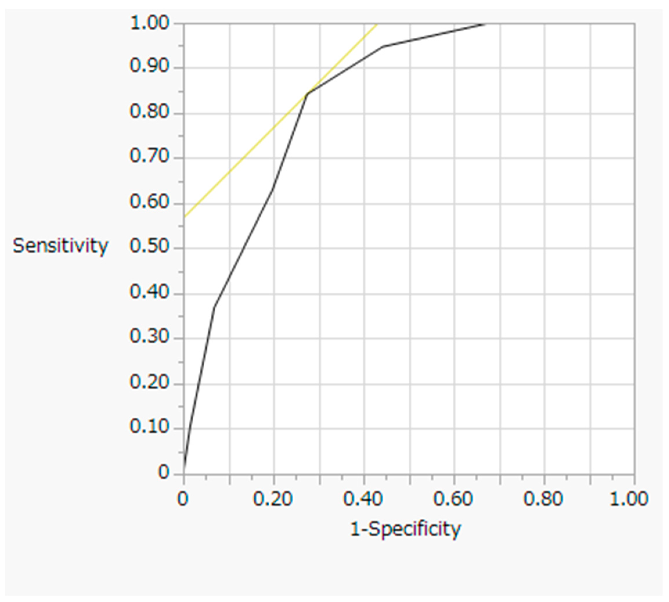
To identify risk factors for LNM, we collected data on sex, age, tumor location, tumor diameter, depth of invasion, degree of differentiation, lymphatic invasion, vascular invasion, ulceration, lateral margin positivity, vertical margin positivity, and LNM. The Mann–Whitney U-test and chi-square test with Bonferroni correction were used to compare continuous and categorical data among the low-risk, intermediate-risk, and high-risk groups. When the data followed a normal distribution based on the Shapiro–Wilks test, Student’s t-test was used. Odds ratios and 95% confidence intervals (CIs) were calculated using univariate and multivariate logistic regression analyses adjusted for age and sex. A receiver operating characteristic (ROC) curve was applied to estimate the clinical suitability of the model for predicting LNM. The sensitivity and specificity were examined using optimal cutoff points based on the ROC curve and categorized into two groups to explore potential improvements in stratification of the eCura system. Statistical analyses were performed using EZR version 1.54 (Saitama Medical Center, Jichi Medical University, Saitama, Japan) [16]. All statistical tests were two-sided, and a p-value of 0.05 was considered statistically significant.

3. Results

The enrolled patients were predominantly male (85%) and the median age (interquartile range, IQR) was 69 (65–76) (Table 1). One hundred and eleven patients (74%) had SM2 invasion. Eighteen lesions (12%) had undifferentiated histology, 70 (47%) had lymphatic invasion, and 32 (21%) had venous invasion. A positive horizontal margin was seen in seven lesions (5%), and a positive vertical margin was seen in 35 lesions (23%). There were 19 lesions (13%) with LNM. During a median follow-up of 5.0 years for all cases, two patients with no LNM experienced local recurrence and one patient with LNM experienced distant metastasis after the additional surgery.
Table 2 shows the results of univariate analysis of the risk factors for LNM. Patients with LNM were significantly older (median, 73 years, IQR, 69–78 years) than patients without LNM (median, 69 years, IQR, 63–75 years) (p = 0.044). Lymphatic invasion was significantly more common in patients with LNM (LNM vs. no LNM: 95% vs. 40%: p < 0.0001). Patients with LNM also had significantly larger tumors (median, 34 mm, IQR, 18–50 mm) than patients without LNM (median, 20 cm, IQR, 14–30 mm) (p = 0.0019). Venous invasion was also more common in patients with LNM (LNM vs. no LNM: 42% vs. 18%; p = 0.032). Lymphatic invasion and tumor size >30 mm were significant independent risk factors of LNM in multivariate analysis adjusted by age and sex (odds ratio, 22.9, 95% CI: 2.9–180.0, p = 0.003 and odds ratio, 2.9, 95% CI: 1.3–11.8, p = 0.017, respectively; Table 3).
The diagnostic efficacy of the modified eCura system for identifying the risk of LNM was assessed by ROC curve analysis (Figure 1). The cutoff for estimating the risk of LNM was 4 points based on the ROC curve analysis. The Youden index, sensitivity, and specificity were 0.563, 0.842, and 0.725, respectively, with a resulting area under the curve of 0.836 (95% CI, 0.702–0.970). Fifty-two patients with 4–7 points were defined as the high-risk group, and the rate of LNM in this group was 23.5%. Conversely, 98 patients with 0–3 points were defined as the low-risk group, and the rate of LNM in this group was 3.1% (4–7 points vs. 0–3 points: odds ratio 12.0, 95% CI: 3.7–54.2, p < 0.0001) (Table 4).

4. Discussion

Our results showed that lymphatic invasion and tumor size >30 mm were independent risk factors of LNM, as has been previously reported for the eCura system [9]. Hatta et al. established the eCura system to score the risk of LNM in EGC patients after ESD [12]. Based on this system, patients are divided into three risk groups, which may interfere with treatment policy decision-making [14]. Using this scoring system, the LNM risks are 2.5% in the low-risk group (0–1 points), 6.7% in the intermediate-risk group (2–4 points), and 22.7% in the high-risk group (5–7 points) [14]. In our study, ROC curve analysis indicated that the optimal cutoff point to determine LNM risk based on the eCura system was 4. Therefore, we divided the patients into two groups, namely, the low-risk group, with scores of 1–3 points, and the high-risk group, with scores of 4–7 points, which will contribute to better treatment policies for patients.
The risk of LNM is the most important factor when selecting a treatment strategy for EGC [17,18,19]. The curability of ESD and post-ESD treatment were factored into the gastric cancer treatment guidelines in 2018 [20]. According to the guidelines, EGCs resected by ESD are divided into three categories: absolute indication, expanded indication, and noncurative indication [5,21,22,23,24]. Absolute indication is defined as a risk of LNM <1% and good evidence of a long-term outcome. Expanded indication is defined as a risk of LNM <1% but little evidence of a long-term outcome. Jee et al. suggested that extending the indications for EMR and ESD according to the Japanese Gastric Cancer Association guidelines increased the risk of LNM because the long-term outcome of these lesions has not been discussed [25]. A systematic review indicated that expanding the indication for endoscopic resection of EGC to include lesions ≤3 cm suggests that T1b1 is associated with a 3% risk of LNM for EGC in Japan [2]. Additional surgery is recommended for lesions with noncurative resection; however, the rate of LNM among lesions with noncurative resection is only 2.6–10.6% [2]. Further, the risk of complications is statistically lower in ESD than in surgery [26,27,28]. With the aging of society, there is an increasing number of patients in whom surgery is difficult; therefore, less invasive treatment is needed.
With regard to tumor size, the result of our study indicated that tumor size >30 mm was a significant factor associated with LNM in EGC patients after noncurative ESD. Chu et al. reported that tumor size >30 mm was an independent risk factor for LNM (odds ratio, 1.900, p = 0.006) [17] and Feng et al. also reported an increase in the rate of LNM among patients with larger tumors [19]. In this study, we reaffirmed the usefulness of the eCura system in stratifying the risk of LNM. The eCura system is an excellent mechanism for stratifying risk; however, our results make it easier to decide whether to perform additional surgery by proposing more appropriate cutoff values.
There are some limitations in this study. First, although this study was a multicenter study, it was retrospective and had a small sample size. Therefore, further prospective randomized control studies will be needed. Second, we cannot show the long-term outcomes at this time. In this regard, we are considering tracking the long-term outcome of this cohort in the future to confirm the effectiveness of the modified eCura system.

5. Conclusions

Lymphatic invasion and tumor size >30 mm were independent factors associated with LNM in EGC patients after noncurative ESD, and patients with 0–3 points in the eCura system had a low risk of LNM, whereas those with 4–7 points had a high risk. The modified system should be applied in the clinical setting to the intermediate-risk group (2–4 points) of the eCura system to suggest better treatment strategies for patients. This stratification could improve the outcome of EGC treatment.

Author Contributions

Conceptualization, K.N. and M.E.; methodology, K.N. and M.E.; validation, K.N., M.E. and W.O.; formal analysis, T.S. (Takaya Shimura), T.Y., Y.H., T.I., T.O., T.S. (Tomoya Sugiyama), Y.Y., K.A. and S.I.; investigation, K.N. and M.E.; resources, T.S. (Takaya Shimura), T.Y., Y.H., T.I., T.O., T.S. (Tomoya Sugiyama), Y.Y., K.A. and S.I.; data curation, K.N. and M.E.; writing—original draft preparation, K.N. and M.E.; writing—review and editing, Y.F., N.O. and M.S.; visualization, K.N. and M.E.; supervision, H.K. and K.K.; project administration, K.K. All authors have read and agreed to the published version of the manuscript.

Funding

This research received no external funding.

Institutional Review Board Statement

The study was conducted according to the guidelines of the Declaration of Helsinki and approved by the Institutional Review Board of Aichi Medical University (No.2020-H149, date of approval November 2018) and received ethical approval from each study center’s local ethics committee.

Informed Consent Statement

Written informed consent was obtained from all study participants.

Data Availability Statement

The data are available upon request from the corresponding author.

Conflicts of Interest

The authors declare no conflict of interest.

References

  1. Sano, T.; Aiko, T. New Japanese classifications and treatment guidelines for gastric cancer: Revision concepts and major revised points. Gastric Cancer 2011, 14, 97–100. [Google Scholar] [CrossRef] [Green Version]
  2. Ono, H.; Yao, K.; Fujishiro, M.; Oda, I.; Nimura, S.; Yahagi, N.; Iishi, H.; Oka, M.; Ajioka, Y.; Ichinose, M.; et al. Guidelines for endoscopic submucosal dissection and endoscopic mucosal resection for early gastric cancer. Dig. Endosc. 2016, 28, 3–15. [Google Scholar] [CrossRef] [PubMed] [Green Version]
  3. Ono, H.; Yao, K.; Fujishiro, M.; Oda, I.; Uedo, N.; Nimura, S.; Yahagi, N.; Iishi, H.; Oka, M.; Ajioka, Y.; et al. Guidelines for endoscopic submucosal dissection and endoscopic mucosal resection for early gastric cancer (second edition). Dig. Endosc. 2021, 33, 4–20. [Google Scholar] [CrossRef] [PubMed]
  4. Nishizawa, T.; Yahagi, N. Long-Term Outcomes of Using Endoscopic Submucosal Dissection to Treat Early Gastric Cancer. Gut Liver 2018, 12, 119–124. [Google Scholar] [CrossRef] [PubMed] [Green Version]
  5. Kim, T.K.; Kim, G.H.; Park, D.Y.; Lee, B.E.; Jeon, T.Y.; Kim, D.H.; Jo, H.J.; Song, G.A. Risk factors for local recurrence in patients with positive lateral resection margins after endoscopic submucosal dissection for early gastric cancer. Surg. Endosc. 2015, 29, 2891–2898. [Google Scholar] [CrossRef]
  6. Oda, I.; Gotoda, T.; Sasako, M.; Sano, T.; Katai, H.; Fukagawa, T.; Shimoda, T.; Emura, F.; Saito, D. Treatment strategy after non-curative endoscopic resection of early gastric cancer. Br. J. Surg. 2008, 95, 1495–1500. [Google Scholar] [CrossRef]
  7. Son, S.Y.; Park, J.Y.; Ryu, K.W.; Eom, B.W.; Yoon, H.M.; Cho, S.J.; Lee, J.Y.; Kim, C.G.; Lee, J.H.; Kook, M.-C.; et al. The risk factors for lymph node metastasis in early gastric cancer patients who underwent endoscopic resection: Is the minimal lymph node dissection applicable? A retrospective study. Surg. Endosc. 2013, 27, 3247–3253. [Google Scholar] [CrossRef]
  8. Yang, T.C.; Hou, M.C.; Chen, P.H.; Hsin, I.F.; Chen, L.K.; Tsou, M.Y.; Lin, H.-C.; Lee, F.-Y. Clinical Outcomes and Complications of Endoscopic Submucosal Dissection for Superficial Gastric Neoplasms in the Elderly. Medicine 2015, 94, e1964. [Google Scholar] [CrossRef]
  9. Fairweather, M.; Jajoo, K.; Sainani, N.; Bertagnolli, M.M.; Wang, J. Accuracy of EUS and CT imaging in preoperative gastric cancer staging. J. Surg. Oncol. 2015, 111, 1016–1020. [Google Scholar] [CrossRef]
  10. Serrano, O.K.; Huang, K.; Ng, N.; Yang, J.; Friedmann, P.; Libutti, S.K.; Kennedy, T.J. Correlation between preoperative endoscopic ultrasound and surgical pathology staging of gastric adenocarcinoma: A single institution retrospective review. J. Surg. Oncol. 2016, 113, 42–45. [Google Scholar] [CrossRef]
  11. Nakagawa, M.; Choi, Y.Y.; An, J.Y.; Chung, H.; Seo, S.H.; Shin, H.B.; Bang, H.-J.; Li, S.; Kim, H.-I.; Cheong, J.-H.; et al. Difficulty of predicting the presence of lymph node metastases in patients with clinical early stage gastric cancer: A case control study. BMC Cancer 2015, 15, 943. [Google Scholar] [CrossRef] [PubMed] [Green Version]
  12. Hatta, W.; Gotoda, T.; Oyama, T.; Kawata, N.; Takahashi, A.; Yoshifuku, Y.; Hoteya, S.; Nakagawa, M.; Hirano, M.; Esaki, M.; et al. A Scoring System to Stratify Curability after Endoscopic Submucosal Dissection for Early Gastric Cancer: “eCura system”. Am. J. Gastroenterol. 2017, 112, 874–881. [Google Scholar] [CrossRef] [PubMed]
  13. Hatta, W.; Gotoda, T.; Koike, T.; Masamune, A. History and future perspectives in Japanese guidelines for endoscopic resection of early gastric cancer. Dig. Endosc. 2020, 32, 180–190. [Google Scholar] [CrossRef] [Green Version]
  14. Hatta, W.; Gotoda, T.; Oyama, T.; Kawata, N.; Takahashi, A.; Yoshifuku, Y.; Hoteya, S.; Nakagawa, M.; Hirano, M.; Esaki, M.; et al. Is the eCura system useful for selecting patients who require radical surgery after noncurative endoscopic submucosal dissection for early gastric cancer? A comparative study. Gastric Cancer 2018, 21, 481–489. [Google Scholar] [CrossRef] [PubMed]
  15. Gotoda, T.; Yamamoto, H.; Soetikno, R.M. Endoscopic submucosal dissection of early gastric cancer. J. Gastroenterol. 2006, 41, 929–942. [Google Scholar] [CrossRef]
  16. Kanda, Y. Investigation of the freely available easy-to-use software ‘EZR’ for medical statistics. Bone Marrow Transplant. 2013, 48, 452–458. [Google Scholar] [CrossRef] [Green Version]
  17. Chu, Y.N.; Yu, Y.N.; Jing, X.; Mao, T.; Chen, Y.Q.; Zhou, X.B.; Song, W.; Zhao, X.-Z.; Tian, Z.-B. Feasibility of endoscopic treatment and predictors of lymph node metastasis in early gastric cancer. World J. Gastroenterol. 2019, 25, 5344–5355. [Google Scholar] [CrossRef]
  18. Zhao, B.W.; Chen, Y.M.; Jiang, S.S.; Chen, Y.B.; Zhou, Z.W.; Li, Y.F. Lymph Node Metastasis, a Unique Independent Prognostic Factor in Early Gastric Cancer. PLoS ONE 2015, 10, e0129531. [Google Scholar] [CrossRef] [Green Version]
  19. Feng, H.; Wang, Y.; Cao, L.; Zhang, C.; Sun, B.; Zhao, Y.; Xu, J. Lymph node metastasis in differentiated-type early gastric cancer: A single-center retrospective analysis of surgically resected cases. Scand. J. Gastroenterol. 2016, 51, 48–54. [Google Scholar] [CrossRef]
  20. Japanese Gastric Cancer Association. Japanese gastric cancer treatment guidelines 2018 (5th edition). Gastric Cancer 2021, 24, 1–21. [Google Scholar] [CrossRef] [Green Version]
  21. Uedo, N.; Iishi, H.; Tatsuta, M.; Ishihara, R.; Higashino, K.; Takeuchi, Y.; Imanaka, K.; Yamada, T.; Yamamoto, S.; Yamamoto, S.; et al. Longterm outcomes after endoscopic mucosal resection for early gastric cancer. Gastric Cancer 2006, 9, 88–92. [Google Scholar] [CrossRef] [PubMed] [Green Version]
  22. Tanabe, S.; Ishido, K.; Matsumoto, T.; Kosaka, T.; Oda, I.; Suzuki, H.; Fujisaki, J.; Ono, H.; Kawata, N.; Oyama, T.; et al. Long-term outcomes of endoscopic submucosal dissection for early gastric cancer: A multicenter collaborative study. Gastric Cancer 2017, 20 (Suppl. S1), 45–52. [Google Scholar] [CrossRef] [Green Version]
  23. Shin, K.Y.; Jeon, S.W.; Cho, K.B.; Park, K.S.; Kim, E.S.; Park, C.K.; Chung, Y.J.; Kwon, J.G.; Jung, J.T.; Kim, K.O.; et al. Clinical outcomes of the endoscopic submucosal dissection of early gastric cancer are comparable between absolute and new expanded criteria. Gut Liver 2015, 9, 181–187. [Google Scholar] [CrossRef] [Green Version]
  24. Ahn, J.Y.; Jung, H.Y.; Choi, K.D.; Choi, J.Y.; Kim, M.Y.; Lee, J.H.; Choi, K.-S.; Kim, D.H.; Song, H.J.; Lee, G.H.; et al. Endoscopic and oncologic outcomes after endoscopic resection for early gastric cancer: 1370 cases of absolute and extended indications. Gastrointest. Endosc. 2011, 74, 485–493. [Google Scholar] [CrossRef] [PubMed]
  25. Jee, Y.S.; Hwang, S.H.; Rao, J.; Park, D.J.; Kim, H.H.; Lee, H.J.; Yang, H.; Lee, K.U. Safety of extended endoscopic mucosal resection and endoscopic submucosal dissection following the Japanese Gastric Cancer Association treatment guidelines. Br. J. Surg. 2009, 96, 1157–1161. [Google Scholar] [CrossRef] [PubMed]
  26. Li, H.; Feng, L.Q.; Bian, Y.Y.; Yang, L.L.; Liu, D.X.; Huo, Z.B.; Zeng, L. Comparison of endoscopic submucosal dissection with surgical gastrectomy for early gastric cancer: An updated meta-analysis. World J. Gastrointest. Oncol. 2019, 11, 161–171. [Google Scholar] [CrossRef] [PubMed]
  27. Liu, Q.; Ding, L.; Qiu, X.; Meng, F. Updated evaluation of endoscopic submucosal dissection versus surgery for early gastric cancer: A systematic review and meta-analysis. Int. J. Surg. 2020, 73, 28–41. [Google Scholar] [CrossRef]
  28. Hahn, K.Y.; Park, C.H.; Lee, Y.K.; Chung, H.; Park, J.C.; Shin, S.K.; Kim, H.; Cheong, J.-H.; Hyung, W.J.; Noh, S.H.; et al. Comparative study between endoscopic submucosal dissection and surgery in patients with early gastric cancer. Surg. Endosc. 2018, 32, 73–86. [Google Scholar] [CrossRef]
Figure 1. ROC curve analysis to determine the cutoff for predicting lymph node metastasis.
Gastroent 13 00007 g001
Table 1. Patient characteristics.
Variables N = 150
Sex, n (%)Male 128 (85)
Female 22 (15)
Age, years, median (IQR) 69 (65–76)
Invasion depth, n (%)M 6 (4)
SM1 33 (22)
SM2 111 (74)
Histological type, n (%)Differentiated 132 (88)
Undifferentiated 18 (12)
Lymphatic invasion, n (%)Positive 70 (47)
Negative 80 (53)
Venous invasion, n (%)Positive 32 (21)
Negative 118 (79)
Ulceration, n (%)Positive28 (19)
Negative 122 (81)
Horizontal margin, n (%)Positive 7 (5)
Negative140 (93)
Unclear3 (2)
Vertical margin, n (%)Positive 35 (23)
Negative 109 (73)
Unclear 6 (4)
Lymph node metastasis, n (%)Positive 19 (13)
Negative 131 (87)
Recurrence, n (%)None147 (98)
Local 2 (1)
Distant 1 (1)
IQR, interquartile range; M, mucosa; SM, submucosa.
Table 2. Univariate analysis of the risk for lymph node metastasis.
Variables Lymph Node Metastasisp-Value
NegativePositive
N = 131 (%)N = 19 (%)
Age (years), median (IQR) 69 (63–75)73 (69–78)0.044
Sex Male111 (85)17 (90)0.741
Female20 (15)2 (10)
LocationU32 (24)3 (16)0.773
M61 (47)10 (52)
L38 (29)6 (32)
Tumor size (mm), median (IQR) 20 (14–30)34 (18–50)0.0019
>3025100.002
≤301069
Invasion depthM6 (4)0 (0)0.717
SM130 (23)3 (16)
SM295 (73)16 (84)
Histological type Differentiated114 (87)18 (95)0.472
Undifferentiated17 (13)1 (5)
Lymphatic invasion Positive52 (40)18 (95)<0.0001
Negative79 (60)1 (5)
Venous invasion Positive24 (18)8 (42)0.032
Negative107 (82)11 (58)
Horizontal margin Positive7 (5)0 (0)0.597
Negative121 (92)19 (100)
Unclear3 (3)0 (0)
Vertical margin Positive29 (22)6 (32)0.389
Negative96 (73)13 (68)
Unclear6 (5)0 (0)
Ulceration Positive24 (18)4 (21)0.757
 Negative107 (82)15 (79)
RecurrenceLocal2 (2)0 (0)0.140
Distant0 (0)1 (5)
None129 (98)18 (95)
IQR, interquartile range; U, upper; M, middle; L, lower.
Table 3. Multivariate analysis of the risk factors of lymph node metastasis.
Risk FactorsOdds Ratio a95% CIp-Value
Lymphatic invasion22.92.9–180.00.003
Venous invasion2.60.8–8.10.112
Tumor size >30 mm2.91.3–11.80.017
CI, Confidence interval; a Adjusted by age and sex.
Table 4. Rate of lymph node metastasis based on the modified eCura system.
Risk Score Groups bnLymph Node Metastasis (%)Odd Ratio a (95% CI)p-Value
High risk
(4–7 points)
5216 (23.5)12.0 (3.7–54.2)<0.0001
Low risk
(0–3 points)
983 (3.1)1
CI, Confidence interval; a Adjusted by age and sex; b Based on the modified eCura system
Publisher’s Note: MDPI stays neutral with regard to jurisdictional claims in published maps and institutional affiliations.

