Article

Nasopharyngeal Carriage of Methicillin-Resistant Staphylococcus aureus (MRSA) among Sickle Cell Disease (SCD) Children in the Pneumococcal Conjugate Vaccine Era

by
Nicholas T. K. D. Dayie
1,*,
Deborah N. K. Sekoh
1,2,
Fleischer C. N. Kotey
1,2,
Beverly Egyir
3,
Patience B. Tetteh-Quarcoo
1,
Kevin Kofi Adutwum-Ofosu
4,
John Ahenkorah
4,
Mary-Magdalene Osei
1,2 and
Eric S. Donkor
1
1
Department of Medical Microbiology, University of Ghana Medical School, P.O. Box KB 4236, Korle Bu, Accra 00233, Ghana
2
FleRhoLife Research Consult, P.O. Box TS 853, Teshie, Accra 00233, Ghana
3
Department of Bacteriology, Noguchi Memorial Institute for Medical Research, University of Ghana, P.O. Box LG 581, Legon, Accra 00233, Ghana
4
Department of Anatomy, University of Ghana Medical School, P.O. Box KB 4236, Korle Bu, Accra 00233, Ghana
*
Author to whom correspondence should be addressed.
Infect. Dis. Rep. 2021, 13(1), 191-204; https://0-doi-org.brum.beds.ac.uk/10.3390/idr13010022
Submission received: 17 January 2021 / Revised: 11 February 2021 / Accepted: 23 February 2021 / Published: 1 March 2021

Abstract

:
The aim of this cross-sectional study was to investigate Staphylococcus aureus nasopharyngeal carriage epidemiology in relation to other nasopharyngeal bacterial colonizers among sickle cell disease (SCD) children about five years into pneumococcal conjugate vaccine 13 (PCV-13) introduction in Ghana. The study involved bacteriological culture of nasopharyngeal swabs obtained from 202 SCD children recruited from the Princess Marie Louise Children’s Hospital. S. aureus isolates were identified using standard methods and subjected to antimicrobial susceptibility testing using the Kirby-Bauer disc diffusion method. Cefoxitin-resistant S. aureus isolates were screened for carriage of the mecA, pvl, and tsst-1 genes using multiplex polymerase chain reaction. The carriage prevalence of S. aureus was 57.9% (n = 117), and that of methicillin-resistant S. aureus (MRSA) was 3.5% (n = 7). Carriage of the mecA, pvl, and tsst-1 genes were respectively demonstrated in 20.0% (n = 7), 85.7% (n = 30), and 11.4% (n = 4) of the cefoxitin-resistant S. aureus isolates. PCV-13 vaccination (OR = 0.356, p = 0.004) and colonization with coagulase-negative staphylococci (CoNS) (OR = 0.044, p < 0.0001) each protected against S. aureus carriage. However, none of these and other features of the participants emerged as a determinant of MRSA carriage. The following antimicrobial resistance rates were observed in MRSA compared to methicillin-sensitive S. aureus: clindamycin (28.6% vs. 4.3%), erythromycin (42.9% vs. 19.1%), tetracycline (100% vs. 42.6%), teicoplanin (14.3% vs. 2.6%), penicillin (100% vs. 99.1%), amoxiclav (28.6% vs. 3.5%), linezolid (14.3% vs. 0.0%), ciprofloxacin (42.9% vs. 13.9%), and gentamicin (42.9% vs. 13.0%). The proportion of S. aureus isolates that were multidrug resistant was 37.7% (n = 46). We conclude that S. aureus was the predominant colonizer of the nasopharynx of the SCD children, warranting the continuous monitoring of this risk group for invasive S. aureus infections.

1. Introduction

Staphylococcus aureus, though a commensal, has over the years evolved into a very important human pathogen, causing mild to severe infections, including folliculitis and furunculosis, meningitis, septicaemia, pneumonia, endocarditis, and osteomyelitis [1,2]. As a commensal, it is predominantly isolated from the anterior nares, but also co-colonizes the nasopharynx with other microbiota, such as Haemophilus influenzae, Moraxella catarrhalis, and the predominant nasopharyngeal colonizer—Streptococcus pneumoniae [3,4,5]. Of these colonizers, S. aureus and S. pneumoniae, which antagonize each other [6,7], appear to be the most clinically significant, owing to their greater invasive disease-causing capacity [8,9,10,11] and their propensity for multidrug resistance development [12,13,14,15,16,17,18]. Colonization with these organisms is a precursor of their infections [6,19]. As S. pneumoniae carriage is especially high in young children [20,21,22], who bear the brunt of its high disease burden and case fatality [23,24,25], pneumococcal conjugate vaccines (PCVs) were incorporated into paediatric vaccination programmes to protect the children against S. pneumoniae carriage and infections [23,25,26].
However, the nasopharyngeal microbiota of vaccinated individuals and their close contacts have been reportedly altered in response to PCV introduction [27,28]. Given the antagonism between S. aureus and S. pneumoniae, there are concerns that S. aureus could potentially replace S. pneumoniae vaccine serotypes and emerge as the dominant colonizer of the nasopharynx as coverage of PCVs increases [6,7,29,30,31]. This raises legitimate concerns, as S. aureus is frequently implicated in respiratory tract infections, and its presence in the nasopharynx significantly predicts subsequent invasive infections [31,32]. Moreover, some studies have demonstrated that among sickle cell disease (SCD) patients, S. aureus holds more potential in causing invasive diseases than S. pneumoniae [33,34,35,36,37]. Besides, owing to the fact that antibiotics are frequently used among SCD patients as prophylaxes, due to their immunocompromised nature and consequent high predisposition to infections, selective antibiotic pressure could result, generating multidrug resistant microbes, such as methicillin-resistant S. aureus (MRSA) [38]. Infections with MRSA are often in tandem with extended hospital stays, increased healthcare costs, and high mortality rates [39,40,41]. Moreover, a recent study has reported that MRSA is responsible for 74.1% of all healthcare associated S. aureus infections and 30.1% of all community-associated infections [42]. In Ghana, there have been several MRSA outbreaks since 2012 [43]; that was the same year in which one of the PCVs—PCV-13—was introduced in the country. It is unknown whether the introduction of PCV-13 played a role in the insurgence of the MRSA outbreaks.
Most of the studies carried out on S. aureus and MRSA carriage appear to have focused on the occurrence of the pathogen in its ecological niche, the anterior nares [17,18,44,45,46,47]. Little attention seems to have been given to the occurrence of the pathogen in the nasopharynx, and how vaccination with PCVs influences that. The few studies that have evaluated the dynamics of the pathogen in relation to the introduction of PCVs appear to have emanated from developed countries [27,28,29,30]. Moreover, it is unknown how the introduction of PCVs has influenced the genetic diversity and antimicrobial resistance patterns of S. aureus. In addition, no such study seems to have been conducted among sickle cell disease patients in the country. Preliminary data to the current study, involving samples collected from healthy school children [48], reported that the nasopharyngeal carriage prevalence of S. aureus was high (23.2%). Evidently, the implications of an increased risk of occurrence of S. aureus and MRSA, with increased levels of antimicrobial resistance, in PCV-vaccinated SCD patients cannot be overemphasized, and an evaluation of the role of PCVs in the dynamics of S. aureus within this risk group would certainly contribute to addressing the MRSA menace in the country. This study thus aimed to investigate S. aureus and MRSA nasopharyngeal carriage epidemiology in relation to other nasopharyngeal bacterial colonizers among SCD children about four years into PCV-13 introduction in Ghana, including S. aureus antimicrobial susceptibility patterns, occurrence of the LukF-PV (pvl) and toxic shock syndrome-1 (tsst-1) genes among cefoxitin-resistant S. aureus, as well as identification of determinants of S. aureus and MRSA carriage.

2. Methods

2.1. Study Site, Design, and Sampling

This study was conducted at the Princess Marie Louise Children’s Hospital (PML) in Accra, the capital city of Ghana. The city is inhabited by about two million people, and has 27 hospitals (http://www.statsghana.gov.gh/ accessed on 18 June 2019). PML is the only major public hospital in Accra that primarily provides paediatric care. Its SCD clinic operates on Thursdays, and attends to an average of 20 children per day [49]. The study was cross-sectional, entailing the use of archived nasopharyngeal samples previously collected from 202 SCD children recruited between September 2016 and June 2017, in the study of Dayie et al. [50], which focused on S. pneumoniae carriage.

2.2. Laboratory Analysis of Samples and Identification of Bacteria

The nasopharyngeal swab samples were processed, and the bacterial isolates therein identified following standard bacteriological methods [51]. To begin with, the archived samples, which had been stored in skim milk-tryptone-glucose-glycerin (STGG) medium and kept at −80 °C, were brought out of the freezer and allowed to thaw at room temperature, after which they were vortexed. The specimens were pre-enriched in 5 mL of brain heart infusion broth, and after 24 h of incubation at 37 °C, inoculated with a sterile loop on 5% sheep blood, chocolate, MacConkey, and mannitol salt agars (Oxoid, Basingstoke, Hants, UK). The blood and chocolate agar plates were incubated at 37 °C in 5% CO2, whereas the MacConkey and mannitol salt agar plates were incubated aerobically at 37 °C. Growth promotion test was also performed after media preparation as part of the quality assurance processes by inoculating a known S. aureus control strain (ATCC 25923) into brain heart infusion broth and incubated at 37 °C.
The plates were examined for growth after 18–24 h of incubation, followed by sub-culturing and identification of the isolates using a composite of colonial morphology, the Gram staining, and various biochemical tests—indole, citrate, triple sugar iron, oxidase, tube coagulase, and catalase. The tube coagulase test was done by inoculating isolated colonies into each tube containing one drop of diluted plasma and nine drops of peptone water and incubated for 4–24 h. This test was quality controlled using a known S. aureus (ATCC 25923) strain as positive control and a known Staphylococcus epidermidis as negative control. Catalase- and coagulase-positive staphylococcal isolates were identified as S. aureus, and those that were catalase-positive, but coagulase-negative, were identified as coagulase-negative staphylococci (CoNS).

2.3. Antimicrobial Susceptibility Testing

Using the Clinical and Laboratory Standards Institute (CLSI) [52] recommendations, antimicrobial susceptibility patterns of all the S. aureus isolates were determined by the Kirby-Bauer disc diffusion method using the following antibiotics: gentamicin (10 µg), linezolid (30 µg), teicoplanin (30 µg), cefoxitin (30 µg), clindamycin (2 µg), tetracycline (30 µg), erythromycin (15 µg), penicillin (1 unit), and ciprofloxacin (5 µg). S. aureus ATCC 25923 was used as the positive control strain. An equivalent of 0.5 McFarland suspension of isolates were prepared according to the CLSI guidelines by adjusting the turbidity of the inoculum using Becton Dickinson Pheonix Spec TM Nephelometer. Within 15 min after preparation of the suspension, a sterile cotton swab was dipped into the suspension and seeded on sterile, dried Muller Hinton plates in order to obtain semi-confluent growth. After 15 min, using sterile forceps, the various antimicrobial discs listed above were aseptically positioned on the seeded plates. The inoculated Muller Hinton agar plates were incubated aerobically for 18–24 h at 37 °C within 15 min after the application of the antimicrobial discs. Isolates that displayed resistance to cefoxitin, showing zones of inhibition less than 22 mm, were phenotypically identified as MRSA, whereas cefoxitin zones of inhibition greater than or equal to 22 mm were phenotypically identified as methicillin-sensitive S. aureus (MSSA). The presumptively identified MRSA isolates were subsequently confirmed as MRSA by polymerase chain reaction (PCR) targeting the mecA gene and screened for carriage of the pvl and tsst-1 genes.

2.4. Molecular Analyses

Crude DNA was extracted from overnight cultures of the isolates. For each isolate, a loopful of pure culture was harvested and emulsified in 200 µL nuclease-free water in a sterile 2 mL Eppendorf tube. The emulsion was vortexed and heated in a heat block compartment at 100 °C for 10 min, frozen at −20 °C for 10 min, and centrifuged at 3000× g for 10 min. The supernatant containing the bacterial DNA was aliquoted into a new, labelled Eppendorf tube, stored at −20 °C, and used as a DNA template for multiplex PCR [53].
The multiplex PCR involved the use of primer sets that targeted the mecA, pvl, and tsst-1 genes, as described by Larsen et al. [54]. The primers were reconstituted by adding specific volumes of nuclease-free water, and vortexed vigorously for even mixing, according to the manufacturer’s instructions, to make 100 mM of stock solution, which was then stored at −20 °C. A working solution of 200 µL volume of each primer was made by adding 180µL of nuclease-free water to 20 µL of stock primer solution. The mixture was vortexed, centrifuged, and stored at −20 ℃. The primer mix containing the forward and reverse primers of each of the genes (mecA, pvl, and tsst-1) was prepared according to the dilutions in Table 1.
Amplification was performed in a Veriti 96-well thermal cycler. The amplicons (PCR products) were visualized using 2% agarose gel electrophoresis, and the band sizes were compared to that of a DNA marker (100 bp ladder from New England BioLabs, Ipswich, MA, USA). In-house mecA-, pvl-, and tsst-1-harbouring S. aureus isolates were used as positive controls for the mecA, pvl, and tsst-1 genes, respectively, and nuclease-free water was used as negative control. Table 2 presents a list of primers and their sequences used for the polymerase chain reaction.

2.5. Data Analysis

The data were analyzed with the aid of Statistical Products and Services Solutions (SPSS), version 25. The sociodemographic and clinical data of the study participants were summarized using descriptive statistics. Independent samples Chi-square and point biserial correlation tests, followed by binary logistic regression, were used to determine predictors of S. aureus and MRSA colonization among the study participants. The significance of each of the predictors was determined by computing its p-value, odds ratio, and 95% confidence interval; predictors were deemed statistically significant if their p-values fell below 0.05.

2.6. Ethical Approval

The study was given approval by the Ethical and Protocol Review Committee of the College of Health Sciences, University of Ghana, with protocol identification number “CHS-Et/M.9-P 4.3/2015-2016”.

3. Results

3.1. Sociodemographic and Clinical Features of the Study Participants

The age of the participants ranged from 1 to 13 years (mean and standard deviation: 3.4 ± 1.9). Additionally, more than half of them were above five years of age (53.5%), were males (55.0%), and lived in compound houses (63.9%). Almost all (93.6%) of them were enrolled in school. The sociodemographic characteristics of the study participants are displayed in Table 3.
The clinical characteristics of the study participants displayed in Table 4 demonstrate that almost half (44.6%) of the children were vaccinated with PCV-13, and less than one-third had each had runny nose (30.7%), blocked nose (23.8%), productive cough (20.3%), ear infection (1.5%), and other conditions.

3.2. Bacterial Flora Colonizing the Nasopharynx of the SCD Children

Bacterial pathogens isolated from the nasopharynx of the participants are presented in Table 5. The carriage prevalence of MRSA and MSSA were 3.5% and 55.0%, respectively, comprising a composite S. aureus carriage of 57.9% (dual S. aureus carriage was observed among five participants, with one of these having dual MRSA-MSSA carriage and four having dual MSSA-MSSA carriage). S. aureus was thus the commonest colonizer, followed by Streptococcus pneumoniae (39.1%), coagulase negative Staphylococci (CoNS) (22.1%), and alpha haemolytic streptococci (19.8%). The rest of the isolates (Coliforms, Diphtheroids, Enterococcus spp., Klebsiella oxytoca, and Pseudomonas spp.) rarely colonized the study participants (1.5% and below). Moreover, the prevalence of S. aureus was significantly higher than that of S. pneumoniae (p < 0.0001).

3.3. Occurrence of Virulence Genes among the Cefoxitin-Resistant S. aureus Isolates

As observed in Table 6, of the 35 cefoxitin-resistant S. aureus isolates obtained in the current study, mecA gene carriage was confirmed in 20.0% (n = 7); the pvl and tsst-1 genes were demonstrated in 85.7% (n = 30) and 11.4% (n = 4), respectively. Figure 1 is a representation of the agarose gel electrophoresis of the multiplex PCR products of the three genes whose presence were screened for.

3.4. Determinants of S. aureus Nasopharyngeal Carriage

As observed in Table 7, colonization with CoNS (OR = 0.044, p < 0.0001) and PCV-13 vaccination (OR = 0.356, p = 0.004) each appeared to protect the study participants against S. aureus colonization. However, none of these and other features of the participants emerged as a determinant of MRSA colonization.

3.5. Antimicrobial Resistance Patterns of the MSSA and MRSA Isolates

Penicillin was the antimicrobial to which the S. aureus isolates in the current study displayed the highest resistance (100% for MRSA, and 99.1% for MSSA). On the other hand, linezolid recorded the lowest composite resistance (14.3% for MRSA, and 0.0% for MSSA). Moreover, the MRSA isolates recorded higher tetracycline (100% vs. 42.6%), erythromycin (42.9% vs. 19.1%), amoxiclav (28.6% vs. 3.5%), ciprofloxacin (42.9% vs. 13.9%), and gentamicin (42.9% vs. 13.0%) resistance in comparison to the MSSA isolates. The proportion of S. aureus isolates that were multidrug resistant was 37.7% (n = 46). Details of the antimicrobial resistance rates can be found in Figure 2.

4. Discussion

This study investigated S. aureus nasopharyngeal carriage epidemiology in relation to other nasopharyngeal bacterial colonizers among SCD children about five years into PCV-13 introduction in Ghana. It is the first to conduct such an evaluation among SCD children post-PCV-13 introduction in the country, thus providing insights into dynamics of PCV- and SCD-induced S. aureus nasopharyngeal carriage epidemiology.
The isolated bacterial flora colonizing the nasopharynx of the SCD children belonged to both the Gram-positive and Gram-negative categories, comprising S. aureus, CoNS, Streptococcus pneumoniae, alpha haemolytic streptococci, Diphtheroids, Enterococcus spp., Pseudomonas spp., and Klebsiella oxytoca. While the other bacteria isolated were present at low numbers, MRSA and MSSA prevalence were 3.5% and 55.0%, respectively, translating to S. aureus domination among the isolated nasopharyngeal colonizers at a prevalence of 57.9%. It is noted that Dayie et al. [50] (the parent study) reported a significantly lower carriage prevalence of S. pneumoniae (39.1%) compared to the carriage prevalence of S. aureus observed in the current study (57.9%). Interestingly, the nasopharynx is the ecological niche of S. pneumoniae, but not S. aureus. Hence, the significantly higher carriage prevalence of S. aureus relative to S. pneumoniae among the participants, as well as the presence of the other nasopharyngeal colonizers in low numbers, suggest that the modification of the nasopharyngeal microbiota resulting from PCV-13 vaccination led to nasopharyngeal proliferation of S. aureus as the dominant colonizer. The observations concurrently demonstrate that PCV-13 vaccination was effective in decolonizing the study participants of vaccine-captured serotypes of S. pneumoniae.
The S. aureus carriage prevalence recorded in the current study is higher than the post-PCV-13 vaccination anterior nasal carriage prevalence reported by Appiah et al. [18] among a cohort of SCD children (33%) and the 44.9% nasal carriage prevalence reported by Donkor et al. [19] among HIV-infected children, another immunologically challenged population, who were also studied during the post-PCV-13 vaccination era. The nasopharyngeal carriage prevalence observed in this study is higher than those reported in other countries, such as Ethiopia, The Gambia, Iran, and Portugal (10.3–30.6%) [5,56,57,58].
That S. aureus carriage was higher in this study than it was in the anterior nares of the SCD children in Appiah et al.’s [18] study and HIV-infected children in Donkor et al. [17] study seems to confirm an insurgence of S. aureus in the nasopharynx, as asserted above. This is because carriage is expected to be higher in the anterior nares (the anatomical site sampled by Appiah et al. [18] and Donkor et al.’s [17]), as it is the ecological niche of S. aureus, rather than the nasopharynx. It is possible that slight variations between the study population of the current study and each of the other two populations in question may account for this disparity. Indeed, should information on concurrent nasal carriage prevalence be available alongside the reported nasopharyngeal carriage prevalence, a more conclusive deduction could have been made. That notwithstanding, the higher S. aureus nasopharyngeal carriage prevalence of this study than the nasal carriage prevalence reported by Donkor et al. [17] also supports the deduction that S. aureus is becoming established in the nasopharynx. This claim is strengthened by the known proneness of HIV-infected persons to S. aureus nasal carriage [17,59].
Appiah et al.’s [18] study, although also conducted in the PCV-13 era, focused on nasal carriage of staphylococci, and hence it is not possible to glean from it the diversity of the colonizing bacteria for the purpose of comparing with the diversity observed in the current study. Another carriage study conducted in the study area [60], entailing surveillance of an additional organism to S. aureus among SCD patients, but done prior to the PCV-13 era, focused only on S. pneumoniae carriage, and its study design was atypical—it was an amalgam of two sub-studies, one involving a haemoglobin SS-haemoglobin AA case-control nasal carriage study focusing on S. aureus only, and the other involving a different cohort of SCD children who were studied for nasopharyngeal carriage of S. pneumoniae exclusively. Therefore, that study also did not provide data with which appropriate comparisons could be made in the context of colonizing nasopharyngeal bacterial flora.
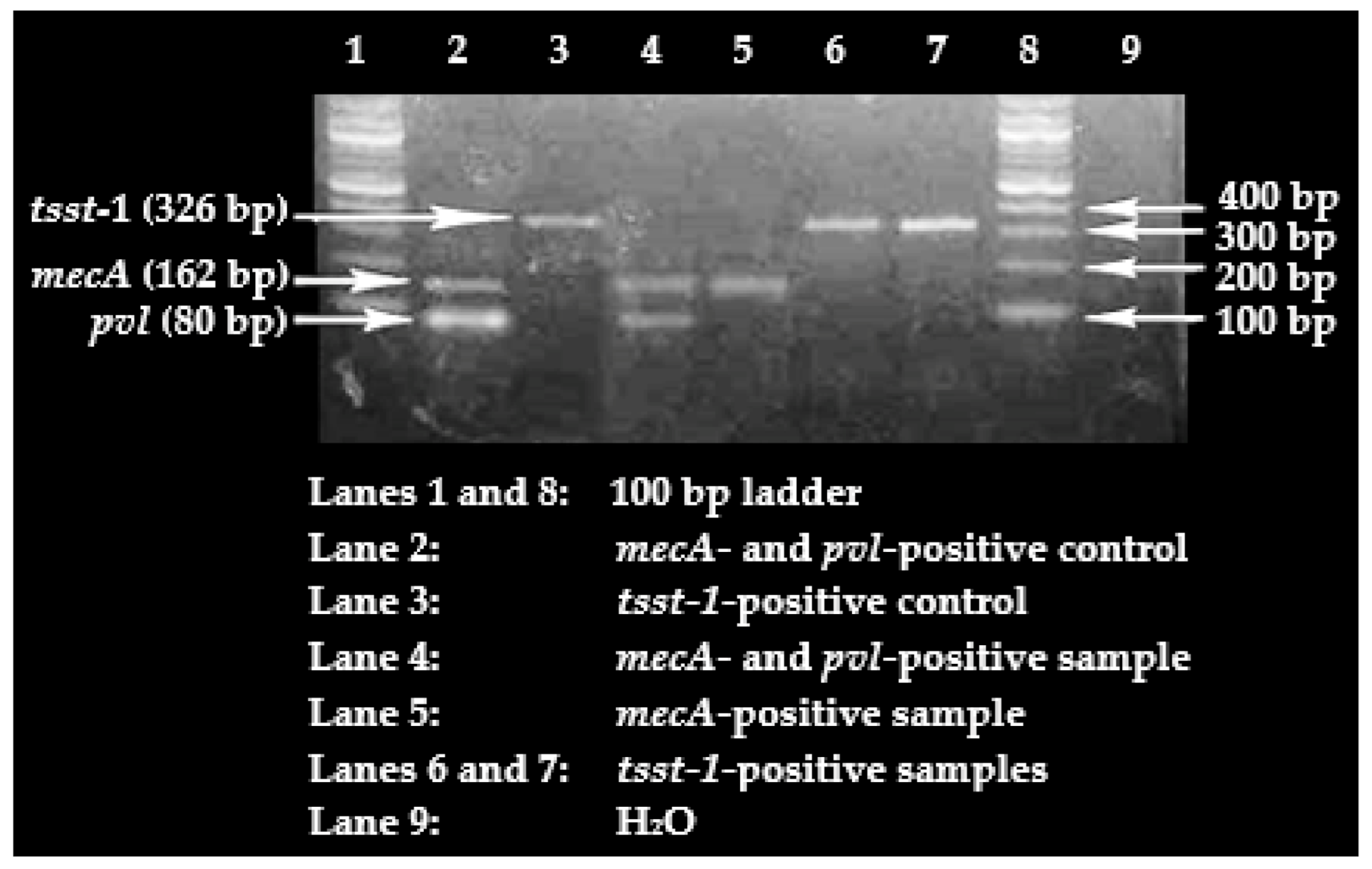
Only 20% of the cefoxitin-resistant S. aureus isolates were confirmed to carry the mecA gene, meaning that 80% of these isolates lacked the presence of the gene. This is of concern, given that in resource-poor settings, such as Ghana, clinical laboratories generally base the identification of MRSA on oxacillin or cefoxitin resistance. Even though it is possible that the cefoxitin-resistant, mecA-negative, S. aureus isolates may be harbouring other variants of the mec gene, such as mecC, a significant proportion (80%) lacked the gene. This could be an indictment on MRSA diagnosis that is exclusively based on phenotypic detection. Concerns similar to these have been raised elsewhere [61]. It may however be argued that this apparent flaw in MRSA diagnosis in resource-poor settings probably has an invariably little impact in clinical settings, particularly, when such laboratory investigations are focused on guiding the selection of an antimicrobial agent with a contemporarily demonstrated effectiveness against an infecting pathogen for the purpose of achieving success in treatment. Hence, in that case, phenotypic demonstration of methicillin resistance would suffice in ruling out the use of certain antimicrobial regimens. The flaw, nonetheless, becomes relevant when such clinical data are submitted directly to epidemiological studies involving MRSA surveillance, or published to reflect MRSA burden in hospitals, in which cases the data may be deemed misleading. Accordingly, it is important to prioritize the upgrade of clinical laboratories in resource-poor settings so as to ensure the use of present-day techniques, such as molecular detection methods, in diagnosis.
The high proportion of pvl carriage was not surprising, as immunosuppression is known to predispose individuals to a higher risk of pvl-positive S. aureus carriage [62]. Besides this fact, Ghana, like other countries in Africa, is a pvl-positive S. aureus-endemic region, with the high occurrence observed even among MSSA isolates. For instance, Eibach et al. [63] reported the pvl gene to be present in 58% of the MSSA isolates recovered in their study conducted in Kumasi, Ghana. Devine et al. [64] also reported a pvl carriage proportion of 72.1% in Nigeria. That said, the high proportion of pvl-positive nasopharyngeal S. aureus colonizers is of immense clinical significance, as it indicates that this cohort of SCD patients, a known immunocompromised population, would have a higher risk of developing invasive infections with S. aureus strains that could cause polymorphonuclear leucocyte lysis and tissue necrosis owing to their borne virulence determinant—pvl [65,66,67,68].
Based on available literature published in peer-reviewed journals, this study is the first to evaluate tsst-1 carriage among S. aureus strains recovered from SCD patients in Ghana. A similar proportion—9.8%—was reported in Nigeria [64]. The low carriage of the gene may mean that only a few of these SCD patients may be prone to developing the toxic shock syndrome.
It is quite interesting that having previously received PCV-13 vaccination appeared to protect the study participants against S. aureus colonization. This is because PCV-13, like other PCVs, was designed to specifically curtail S. pneumoniae carriage in the nasopharynx [69,70], and hence logically, it is feared that it would cause an insurgence of S. aureus nasopharyngeal carriage, given the S. aureusS. pneumoniae antagonism [6,7,71]. Thus, the apparent unusual protective effects observed with regard to PCV-13 vaccination in this cohort is most likely not directly linked to the PCV-13 itself, but must have been mediated by secondary factors, such as the presence of CoNS, which was reported at a high rate in the current study. CoNS are also known to be antagonistic to S. aureus, by virtue of the CoNS-produced autoinducing peptide [72]. In fact, in the current study, CoNS colonization was observed to protect against S. aureus colonization. Besides the potential contribution of CoNS, other members of the nasopharyngeal microbiome, which may have been missed by the culture methods relied on in the current study, may have a bearing on this seeming protectiveness of PCV-13 against S. aureus carriage. Future studies could employ high-throughput sequencing, such as 16S ribosomal ribonucleic acid (rRNA) sequencing, to further investigate the dynamics introduced by CoNS colonization as well as other members of the nasopharyngeal microbiome to this indirect and atypical PCV-13-S. aureus relationship.
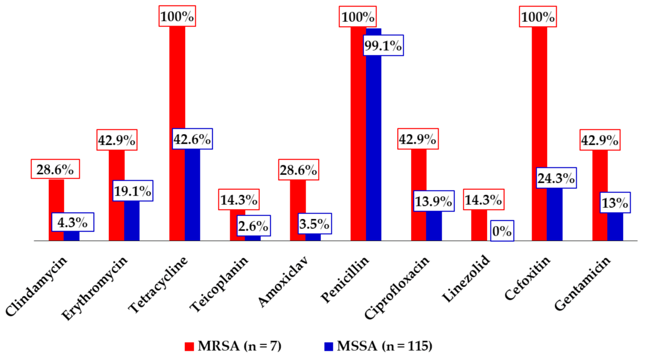
The highest antimicrobial resistance rate was recorded against penicillin, which was expected, as the antibiotic is routinely used among SCD individuals for prophylactic purposes against S. pneumoniae infections, has been in wide circulation for years, and is easily accessible in the country. Moreover, other S. aureus studies have reported similar resistance rates against it [17,18,47,73]. The low resistance rates recorded against linezolid was also expected, as the antibiotic does not have a wide coverage in the country, and previous studies conducted on S. aureus have reported comparable rates [17,74]. The observed rate is however at odds with the 30% resistance rate recorded among anterior nasal S. aureus isolates obtained in the above-cited recent study of Appiah et al. [18] among SCD children. That notwithstanding, the low rate signals that linezolid can still be administered in the treatment of MRSA infections. The high rate of tetracycline resistance, although alarming, lacks clinical significance, as the drug is rarely used in clinical practice. Moreover, the high resistance rates demonstrated by the MRSA isolates against erythromycin, amoxiclav, ciprofloxacin, and gentamicin relative to the MSSA isolates is consistent with a key attribute of MRSA—resistance to several antibiotic groups in routine use [12,75,76]. The 37.7% proportion of multidrug resistance observed in this study appears to be higher than those reported with regard to S. aureus in some studies in the country (3.2–16.7%) [16,48,73], whereas strikingly lower than others (62.3–100%) [17,18], and although it may reflect some beneficial effects of campaigns against irrational antimicrobial use, it synchronously underscores the need to intensify these campaigns, particularly among risk groups for S. aureus carriage and infections, such as SCD patients.
This study was limited by a few factors. First, the absence of information on concurrent S. aureus nasal carriage prevalence alongside the reported nasopharyngeal carriage prevalence makes the assertion of S. aureus insurgence of the nasopharynx a bit less conclusive. Furthermore, screening for virulent genes was restricted to the cefoxitin-resistant S. aureus isolates due to financial constraints, and hence, the study could not provide information on the proportion of S. aureus isolates that harboured the screened virulent genes.

5. Conclusions

We conclude that of the bacterial flora isolated from the nasopharynx of the sickle cell disease children, S. aureus was the predominant colonizer. Nasopharyngeal carriage prevalence of S. aureus and MRSA were 57.9% and 3.5%, respectively, suggesting an insurgence of S. aureus in the nasopharynx; the distribution of virulence genes among the cefoxitin-resistant S. aureus isolates was: 20% mecA, 85.7% pvl, and 11.4% tsst-1. Furthermore, no determinant of MRSA colonization was identified; colonization with coagulase-negative staphylococci and PCV-13 vaccination were each found to protect the SCD children against S. aureus colonization. The highest and lowest antimicrobial resistance rate were recorded against penicillin and linezolid, respectively, with 37.7% of the S. aureus isolates displaying multidrug resistance.
It is recommended that further studies be conducted on the S. aureus isolates obtained in this study to determine their sequence and spa types via whole-genome sequencing. In addition, this study needs to be extended to other risk groups, with concurrent sampling of the anterior nares and nasopharynx. Moreover, it is important to carry out continuous surveillance of the pathogen nationwide and intensify public health education against indiscriminate antibiotic use. There is also a need to monitor SCD patients for invasive bacterial diseases due to S. aureus.

Author Contributions

Conceptualization, N.T.K.D.D.; methodology, N.T.K.D.D., P.B.T.-Q., F.C.N.K., B.E., E.S.D., and D.N.K.S.; software, N.T.K.D.D., P.B.T.-Q., F.C.N.K., D.N.K.S., and E.S.D.; validation, N.T.K.D.D., F.C.N.K., B.E., P.B.T.-Q., J.A., K.K.A.-O., D.N.K.S., and E.S.D.; formal analysis, N.T.K.D.D., F.C.N.K., B.E., P.B.T.-Q., D.N.K.S., J.A., K.K.A.-O., M.-M.O., and E.S.D.; investigation, N.T.K.D.D., E.S.D., B.E., D.N.K.S., M.-M.O., P.B.T.-Q., J.A., and K.K.A.-O.; resources, N.T.K.D.D., P.B.T.-Q., F.C.N.K., D.N.K.S., M.-M.O., J.A., K.K.A.-O., B.E., and E.S.D.; data curation, F.C.N.K., N.T.K.D.D., D.N.K.S., P.B.T.-Q., J.A., K.K.A.-O., and E.S.D.; writing—original draft preparation, N.T.K.D.D., F.C.N.K., and D.N.K.S.; writing—review and editing, N.T.K.D.D., P.B.T.-Q., J.A., K.K.A.-O., D.N.K.S., M.-M.O., F.C.N.K., E.S.D., and B.E.; visualization, N.T.K.D.D., P.B.T.-Q., F.C.N.K., D.N.K.S., E.S.D., and B.E.; supervision, N.T.K.D.D., E.S.D., and B.E.; project administration, N.T.K.D.D. and F.C.N.K. All authors have read and agreed to the published version of the manuscript.

Funding

Funding for this work was obtained from Office of Research, Innovation and Development (ORID) (University of Ghana, Legon).

Institutional Review Board Statement

The study was conducted according to the guidelines of the Declaration of Helsinki, and approved by the Ethical and Protocol Review Committee of the College of Health Sciences, University of Ghana (Protocol Identification Number: CHS-Et/M.9-P4.3/2015-2016).

Informed Consent Statement

Informed consent was obtained from all subjects involved in the study.

Data Availability Statement

The data presented in this study are available upon reasonable request from the corresponding author via [email protected].

Acknowledgments

The authors acknowledge the contributions made by the study participants, the technical staff of the Department of Medical Microbiology and the assistance received from Georgina Dasi, Elizabeth Y. Tettey, and Prince J. Pappoe-Ashong.

Conflicts of Interest

The authors declare no conflicting interests.

References

  1. Lowy, F.D. Staphylococcus aureus infections. N. Engl. J. Med. 1998, 339, 520–532. [Google Scholar] [CrossRef] [PubMed]
  2. Todar, K. Todar’s Online Textbook of Bacteriology; Department of Bacteriology, University of Wisconsin-Madison: Madison, WI, USA, 2006. [Google Scholar]
  3. Williams, R.E.O. Healthy carriage of Staphylococcus aureus: Its prevalence and importance. Bacteriol. Rev. 1963, 27, 56–71. [Google Scholar] [CrossRef]
  4. Leiberman, A.; Dagan, R.; Leibovitz, E.; Yagupsky, P.; Fliss, D.M. The bacteriology of the nasopharynx in childhood. Int. J. Pediatric Otorhinolaryngol. 1999, 49, S151–S153. [Google Scholar] [CrossRef]
  5. Assefa, A.; Gelaw, B.; Shiferaw, Y.; Tigabu, Z. Nasopharyngeal carriage rate and antimicrobial susceptibility pattern of potential pathogenic bacteria among pediatric outpatients at Gondar University teaching hospital, Ethiopia. J. Infect. Dis. Ther. 2013, 1, 2. [Google Scholar] [CrossRef]
  6. Bogaert, D.; van Belkum, A.; Sluijter, M.; Luijendijk, A.; de Groot, R.; Rümpke, H.C.; Hermans, P.W.M. Colonisation by Streptococcus pneumoniae and Staphylococcus aureus in healthy children. Lancet 2004, 363, 1871–1872. [Google Scholar] [CrossRef]
  7. Regev-Yochay, G.; Dagan, R.; Raz, M.; Carmeli, Y.; Shainberg, B.; Derazne, E.; Rahav, G.; Rubinstein, E. Association between carriage of Streptococcus pneumoniae and Staphylococcus aureus in children. JAMA 2004, 292, 716–720. [Google Scholar] [CrossRef] [Green Version]
  8. Brogden, K.A.; Guthmiller, J.M.; Taylor, C.E. Human polymicrobial infections. Lancet 2005, 365, 253–255. [Google Scholar] [CrossRef]
  9. Pettigrew, M.M.; Gent, J.F.; Revai, K.; Patel, J.A.; Chonmaitree, T. Microbial interactions during upper respiratory tract infections. Emerg. Infect. Dis. 2008, 14, 1584. [Google Scholar] [CrossRef] [PubMed]
  10. Xu, Q.; Almudervar, A.; Casey, J.R.; Pichichero, M.E. Nasopharyngeal bacterial interactions in children. Emerg. Infect. Dis. 2012, 18, 1738. [Google Scholar] [CrossRef]
  11. Thevaranjan, N.; Whelan, F.J.; Puchta, A.; Ashu, E.; Rossi, L.; Surette, M.G.; Bowdish, D.M. Streptococcus pneumoniae colonization disrupts the microbial community within the upper respiratory tract of aging mice. Infect. Immun. 2016, 84, 906–916. [Google Scholar] [CrossRef] [Green Version]
  12. Han, L.L.; McDougal, L.K.; Gorwitz, R.J.; Mayer, K.H.; Patel, J.B.; Sennott, J.M.; Fontana, J.L. High frequencies of clindamycin and tetracycline resistance in methicillin-resistant Staphylococcus aureus pulsed-field type USA300 isolates collected at a Boston ambulatory health center. J. Clin. Microbiol. 2007, 45, 1350–1352. [Google Scholar] [CrossRef] [PubMed] [Green Version]
  13. Van Bambeke, F.; Reinert, R.R.; Appelbaum, P.C.; Tulkens, P.M.; Peetermans, W.E. Multidrug-resistant Streptococcus pneumoniae infections. Drugs 2007, 67, 2355–2382. [Google Scholar] [CrossRef]
  14. Chambers, H.F.; DeLeo, F.R. Waves of resistance: Staphylococcus aureus in the antibiotic era. Nat. Rev. Microbiol. 2009, 7, 629–641. [Google Scholar] [CrossRef]
  15. Dayie, N.T.K.D.; Arhin, R.E.; Newman, M.J.; Dalsgaard, A.; Bisgaard, M.; Frimodt-Møller, N.; Slotved, H.C. Multidrug-resistant Streptococcus pneumoniae isolates from healthy Ghanaian preschool children. Microb. Drug Resist. 2015, 21, 636–642. [Google Scholar] [CrossRef] [PubMed]
  16. Sampane-Donkor, E.; Badoe, E.V.; Annan, J.A.; Nii-Trebi, N. Colonisation of antibiotic resistant bacteria in a cohort of HIV infected children in Ghana. Pan Afr. Med. J. 2017, 26. [Google Scholar] [CrossRef]
  17. Donkor, E.S.; Kotey, F.C.N.; Dayie, N.T.K.D.; Duodu, S.; Tetteh-Quarcoo, P.B.; Osei, M.-M.; Tette, E. Colonization of HIV-Infected Children with Methicillin-Resistant Staphylococcus aureus. Pathogens 2019, 8, 35. [Google Scholar] [CrossRef] [Green Version]
  18. Appiah, V.A.; Pesewu, G.A.; Kotey, F.C.N.; Boakye, A.N.; Duodu, S.; Tette, E.; Nyarko, M.Y.; Donkor, E.S. Staphylococcus aureus nasal colonization among children with sickle cell disease at the Children’s Hospital, Accra: Prevalence, risk factors, and antibiotic resistance. Pathogens 2020, 9, 329. [Google Scholar] [CrossRef]
  19. Wertheim, H.F.L.; Melles, D.C.; Vos, M.C.; Leeuwen, W.; Van, B.A.; Verbrugh, H.A.; Nouwen, J.L. The role of nasal carriage in Staphylococcus aureus infections. Lancet Infect. Dis. 2005, 5, 751–762. [Google Scholar] [CrossRef]
  20. Donkor, E.S. Understanding the pneumococcus: Transmission and evolution. Front. Cell. Infect. Microbiol. 2013, 3, 7. [Google Scholar] [CrossRef] [Green Version]
  21. Navne, J.E.; Koch, A.; Slotved, H.-C.; Andersson, M.; Melbye, M.; Ladefoged, K.; Børresen, M. Effect of the 13-valent pneumococcal conjugate vaccine on nasopharyngeal carriage by respiratory pathogens among Greenlandic children. Int. J. Circumpolar Health 2017, 76, 1–8. [Google Scholar] [CrossRef] [Green Version]
  22. Fjeldhøj, S.; Laursen, R.P.; Larnkjær, A.; Mølgaard, C.; Fuursted, K.; Krogfelt, K.A.; Slotved, H.-C. Probiotics and carriage of Streptococcus pneumoniae serotypes in Danish children, a double-blind randomized controlled trial. Sci. Rep. 2018, 8, 15258. [Google Scholar] [CrossRef]
  23. Roca, A.; Bojang, A.; Bottomley, C.; Gladstone, R.A.; Adetifa, J.U.; Egere, U.; Greenwood, B. Effect on nasopharyngeal pneumococcal carriage of replacing PCV7 with PCV13 in the Expanded Programme of Immunization in the Gambia. Vaccine 2015, 33, 7144–7151. [Google Scholar] [CrossRef] [Green Version]
  24. Kobayashi, M.; Conklin, L.M.; Bigogo, G.; Jagero, G.; Hampton, L.; Fleming-Dutra, K.E.; Breiman, R.F. Pneumococcal carriage and antibiotic susceptibility patterns from two cross-sectional colonization surveys among children aged <5 years prior to the introduction of 10-valent pneumococcal conjugate vaccine—Kenya, 2009–2010. BMC Infect. Dis. 2017, 17, 25. [Google Scholar] [CrossRef]
  25. Wahl, B.; O’Brien, K.; Greenbaum, A.; Liu, L.; Chu, Y.; Majumder, A.; Knoll, M.D. Global, regional, and national burden of Streptococcus pneumoniae and Haemophilus influenzae type b disease in children in the era of conjugate vaccines: Updated estimates from 2000–2015. Lancet Glob. Health 2018, 6, e744–e757. [Google Scholar] [CrossRef] [Green Version]
  26. International Vaccine Access Center (IVAC), Johns Hopkins Bloomberg School of Public Health. Vaccine Information Management System (VIMS) Global Vaccine Introduction Report. Available online: http://www.jhsph.edu/research/centers-and-institutes/ivac/vims/ (accessed on 18 June 2019).
  27. Davis, S.M.; Deloria-Knoll, M.; Kassa, H.T.; O’Brien, K.L. Impact of pneumococcal conjugate vaccines on nasopharyngeal carriage and invasive disease among unvaccinated people: Review of evidence on indirect effects. Vaccine 2013, 32, 133–145. [Google Scholar] [CrossRef]
  28. Shak, J.R.; Vidal, J.E.; Klugman, K.P. Influence of bacterial interactions on pneumococcal colonization of the nasopharynx. Trends Microbiol. 2013, 21, 129–135. [Google Scholar] [CrossRef] [Green Version]
  29. Quintero, B.; Araque, M.; van der Gaast-de Jongh, C.; Escalona, F.; Correa, M.; Morillo-Puente, S.; Vielma, S.; Hermans, P.W.M. Epidemiology of Streptococcus pneumoniae and Staphylococcus aureus colonization in healthy Venezuelan children. Eur. J. Clin. Microbiol. Infect. Dis. 2011, 30, 1–13. [Google Scholar] [CrossRef] [Green Version]
  30. Huang, Y.-C.; Chen, C.-J. Nasal carriage of methicillin-resistant Staphylococcus aureus during the first 2 years of life in children in northern Taiwan. Pediatric Infect. Dis. J. 2015, 34, 131–135. [Google Scholar] [CrossRef]
  31. Nzenze, S.A. Effect of introduction of pneumococcal conjugate vaccine immunization on nasopharyngeal colonization of streptococcus pneumoniae in South Africa (Doctoral dissertation). Wits Inst. Environ. D-Space 2015, 212, 386. [Google Scholar]
  32. Pizzutto, S.J.; Hare, K.M.; Upham, J.W. Bronchiectasis in children: Current concepts in immunology and microbiology. Front. Pediatrics 2017, 29, 123. [Google Scholar] [CrossRef] [Green Version]
  33. Okuoghae, H.O.; Nwankwo, M.U.; Offor, E.C. Pattern of bacteraemia in febrile children with sickle cell anaemia. Ann. Trop. Paediatr. 1993, 13, 55–64. [Google Scholar] [CrossRef] [PubMed]
  34. Akuse, R.M. Variation in the pattern of bacterial infection in patients with sickle cell disease requiring admission. J. Trop. Pediatrics 1996, 42, 318–323. [Google Scholar] [CrossRef] [Green Version]
  35. Aken’ova, Y.A.; Bakare, R.A.; Okunade, M.A. Septicaemia in sickle cell anaemia patients: The Ibadan experience. Cent. Afr. J. Med. 1998, 44, 102. [Google Scholar]
  36. Thanni, L.O. Bacterial osteomyelitis in major sickling haemoglobinopathies: Geographic difference in pathogen prevalence. Afr. Health Sci. 2006, 6, 236–239. [Google Scholar]
  37. Kizito, E.; Mworozi, E.; Ndugwa, C.; Serjeant, G.R. Bacteraemia in homozygous sickle cell disease in Africa: Is pneumococcal prophylaxis justified? Arch. Dis. Child. 2007, 92, 21–23. [Google Scholar] [CrossRef] [PubMed] [Green Version]
  38. Steele, R.W.; Warrier, R.; Unkel, P.J.; Foch, B.J.; Howes, R.F.; Shah, S.; Williams, K.; Moore, S.; Jue, S.J. Colonization with antibiotic-resistant Streptococcus pneumoniae in children with sickle cell disease. J. Pediatrics 1996, 128, 531–535. [Google Scholar] [CrossRef]
  39. Elixhauser, A.; Steiner, C. Infections with methicillin-resistant Staphylococcus aureus (MRSA) in US Hospitals, 1993–2005. In Healthcare Cost and Utilization Project (HCUP) Statistical Briefs [Internet] 2007 July; Agency for Healthcare Research and Quality (US): Rockville, MD, USA, 2007. Available online: https://0-www-ncbi-nlm-nih-gov.brum.beds.ac.uk/books/NBK61977 (accessed on 13 August 2020).
  40. Klevens, R.M.; Edwards, J.R.; Richards, C.L., Jr.; Horan, T.C.; Gaynes, R.P.; Pollock, D.A.; Cardo, D.M. Estimating health care-associated infections and deaths in US hospitals, 2002. Public Health Rep. 2007, 122, 160–166. [Google Scholar] [CrossRef] [PubMed]
  41. De Kraker, M.E.; Wolkewitz, M.; Davey, P.G.; Koller, W.; Berger, J.; Nagler, J.; Icket, C.; Kalenic, S.; Horvatic, J.; Seifert, H.; et al. Burden Study Group 2011. Clinical impact of antimicrobial resistance in European hospitals: Excess mortality and length of hospital stay related to methicillin-resistant Staphylococcus aureus bloodstream infections. Antimicrob. Agents Chemother. 2011, 55, 1598–1605. [Google Scholar] [CrossRef] [PubMed] [Green Version]
  42. Song, Z.; Gu, F.F.; Guo, X.K.; Ni, Y.X.; He, P.; Han, L.Z. Antimicrobial resistance and molecular characterization of Staphylococcus aureus causing childhood pneumonia in Shanghai. Front. Microbiol. 2017, 8, 455. [Google Scholar] [CrossRef] [PubMed]
  43. Donkor, E.S.; Jamrozy, D.; Mills, R.O.; Dankwah, T.; Amoo, P.K.; Egyir, B.; Badoe, E.V.; Twasam, J.; Bentley, S.D. A genomic infection control study for Staphylococcus aureus in two Ghanaian hospitals. Infect. Drug Resist. 2018, 11, 1757–1765. [Google Scholar] [CrossRef] [PubMed] [Green Version]
  44. Donkor, E.S.; Nartey, E. Nasal colonisation of antibiotic resistant bacteria in Ghanaian children less than five years of age. Internet J. Microbiol. 2007, 5, 1–5. [Google Scholar]
  45. Cenizal, M.J.; Hardy, R.D.; Anderson, M.; Katz, K.; Skiest, D.J. Prevalence of and risk factors for methicillin-resistant Staphylococcus aureus (MRSA) nasal colonization in HIV-infected ambulatory Patients. J. Acquir. Immune Defic. Syndr. 2008, 48, 567–571. [Google Scholar] [CrossRef]
  46. Imaz, A.; Camoez, M.; Yacovo, S.; Di Gasch, O.; Dominguez, M.A.; Vila, A.; Maso-Serra, M.; Pujol, M.; Podzamczer, D. Prevalence of methicillin-resistant Staphylococcus aureus colonization in HIV-infected patients in Barcelona, Spain: A cross-sectional study. BMC Infect. Dis. 2015, 15, 243. [Google Scholar] [CrossRef] [PubMed] [Green Version]
  47. Egyir, B.; Oteng, A.A.; Owusu, E.; Newman, M.J.; Addo, K.K.; Rhod-Larsen, A. Characterization of Staphylococcus aureus from Human Immunodeficiency Virus (HIV) patients in Accra, Ghana. J. Infect. Dev. Ctries. 2016, 10, 453–456. [Google Scholar] [CrossRef] [Green Version]
  48. Dayie, N.T.K.D.; Osei, M.-M.; Opintan, J.A.; Tetteh-Quarcoo, P.B.; Kotey, F.C.N.; Ahenkorah, J.; Adutwum-Ofosu, K.; Egyir, B.; Donkor, E.S. Nasopharyngeal carriage and antimicrobial susceptibility profile of Staphylococcus aureus among children under five years in Accra. Pathogens 2021, 10, 136. [Google Scholar] [CrossRef]
  49. Boadu, I.; Ohemeng, A.; Renner, L.A. Dietary intakes and nutritional status of children with sickle cell dis- ease at the Princess Marie Louise Hospital, Accra—A survey. BMC Nutr. 2018, 4, 33. [Google Scholar] [CrossRef] [Green Version]
  50. Dayie, N.T.K.D.; Tetteh-Ocloo, G.; Labi, A.K.; Olayemi, E.; Slotved, H.C.; Lartey, M.; Donkor, E.S. Pneumococcal carriage among sickle cell disease patients in Accra, Ghana: Risk factors, serotypes and antibiotic resistance. PLoS ONE 2018, 13, e0206728. [Google Scholar] [CrossRef]
  51. Baron, S. (Ed.) Medical Microbiology, 4th ed.; University of Texas Medical Branch at Galveston: Galveston, TX, USA, 1996. Available online: https://0-www-ncbi-nlm-nih-gov.brum.beds.ac.uk/books/NBK7627/ (accessed on 28 September 2019).
  52. Humphries, R.M.; Ambler, J.; Mitchell, S.L.; Castanheira, M.; Dingle, T.; Hindler, J.A.; Koeth, L.; Sei, K. CLSI Methods Development and Standardization Working Group best practices for evaluation of antimicrobial susceptibility tests. J. Clin. Microbiol. 2018, 1, 56. [Google Scholar] [CrossRef] [Green Version]
  53. Rahman, M.M.; Amin, K.B.; Rahman, S.M.M.; Khair, A.; Rahman, M.; Hossain, A.; Alam, M.M. Investigation of methicillin-resistant Staphylococcus aureus among clinical isolates from humans and animals by culture methods and multiplex PCR. BMC Vet. Res. 2018, 14, 300. [Google Scholar] [CrossRef] [PubMed] [Green Version]
  54. Larsen, A.R.; Stegger, M.; Sørum, M. Spa typing directly from a mecA, spa and pvl multiplex PCR assay—A cost-effective improvement for methicillin-resistant Staphylococcus aureus surveillance. Clin. Microbiol. Infect. 2008, 14, 611–614. [Google Scholar] [CrossRef] [Green Version]
  55. Mehrotra, M.; Wang, G.; Johnson, W.M. Multiplex PCR for detection of genes for Staphylococcus aureus enterotoxins, exfoliative toxins, toxic shock syndrome toxin 1, and methicillin resistance. J. Clin. Microbiol. 2000, 38, 1032–1035. [Google Scholar] [CrossRef] [Green Version]
  56. Bottomley, C.; Bojang, A.; Smith, P.G.; Darboe, O.; Antonio, M.; Foster-Nyarko, E.; Kampmann, B.; Greenwood, B.; D’Alessandro, U.; Roca, A. The impact of childhood vaccines on bacterial carriage in the nasopharynx: A longitudinal study. Emerg. Themes Epidemiol. 2015, 12, 1. [Google Scholar] [CrossRef] [PubMed] [Green Version]
  57. Tavares, D.A.; Sá-Leão, R.; Miragaia, M.; de Lencastre, H. Large screening of CA-MRSA among Staphylococcus aureus colonizing healthy young children living in two areas (urban and rural) of Portugal. BMC Infect. Dis. 2010, 10, 110. [Google Scholar] [CrossRef] [Green Version]
  58. Sedighi, I.; Moez, H.; Alikhani, M. Nasal carriage of methicillin resistant Staphylococcus aureus and their antibiotic susceptibility patterns in children attending day-care centers. Acta Microbiol. Immunol. Hung. 2011, 58, 227–234. [Google Scholar] [CrossRef]
  59. Hidron, A.I.; Kempker, R.; Moanna, A.; Rimland, D. Methicillin-resistant Staphylococcus aureus in HIV-infected patients. Infect. Drug Resist. 2010, 3, 73–86. [Google Scholar] [CrossRef] [PubMed] [Green Version]
  60. Donkor, E.S.; Foster-Nyarko, E.; Enweronu-Laryea, C.C. Relationship between antibiotic resistance and sickle cell anemia: Preliminary evidence from a pediatric carriage study in Ghana. Infect. Drug Resist. 2013, 6, 71–77. [Google Scholar] [CrossRef] [Green Version]
  61. Velasco, D.; del Mar Tomas, M.; Cartelle, M.; Beceiro, A.; Perez, A.; Molina, F.; Moure, R.; Villanueva, R.; Bou, G. Evaluation of different methods for detecting methicillin (oxacillin) resistance in Staphylococcus aureus. J. Antimicrob. Chemother. 2005, 55, 379–382. [Google Scholar] [CrossRef] [PubMed]
  62. Schaumburg, F.; Alabi, A.S.; Peters, G.; Becker, K. New epidemiology of Staphylococcus aureus infection from Africa. Clin. Microbiol. Infect. 2014, 20, 589–596. [Google Scholar] [CrossRef] [PubMed] [Green Version]
  63. Eibach, D.; Nagel, M.; Hogan, B.; Azuure, C.; Krumkamp, R.; Dekker, D.; Gajdiss, M.; Brunke, M.; Sarpong, N.; Owusu-Dabo, E.; et al. Nasal carriage of Staphylococcus aureus among children in the Ashanti Region of Ghana. PLoS ONE 2017, 12, e0170320. [Google Scholar] [CrossRef]
  64. Devine, V.T.; Jefferies, J.M.; Clarke, S.C.; Faust, S.N. Nasopharyngeal bacterial carriage in the conjugate vaccine era with a focus on pneumococci. J. Immunol. Res. 2015, 1–9. [Google Scholar] [CrossRef] [Green Version]
  65. Tristan, A.; Ferry, T.; Durand, G.; Dauwalder, O.; Bes, M.; Lina, G.; Etienne, J. Virulence determinants in community and hospital methicillin-resistant Staphylococcus aureus. J. Hosp. Infect. 2007, 65, 105–109. [Google Scholar] [CrossRef]
  66. Boyle-Vavra, S.; Daum, R.S. Community-acquired methicillin-resistant Staphylococcus aureus: The role of Panton-Valentine leukocidin. Lab. Investig. 2007, 87, 3–9. [Google Scholar] [CrossRef] [PubMed] [Green Version]
  67. Brown, A.F.; Leech, J.M.; Rogers, T.R.; McLoughlin, R.M. Staphylococcus aureus colonization: Modulation of host immune response and impact on human vaccine design. Front. Immunol. 2014, 4. [Google Scholar] [CrossRef] [PubMed]
  68. Reddinger, R.M.; Luke-Marshall, N.R.; Sauberan, S.L.; Hakansson, A.P.; Campagnari, A.A. Streptococcus pneumoniae modulates Staphylococcus aureus biofilm dispersion and the transition from colonization to invasive disease. mBio 2018, 9, e02089-17. [Google Scholar] [CrossRef] [PubMed]
  69. Sucher, A.J.; Chahine, E.B.; Nelson, M.; Sucher, B.J. Prevnar 13, the new 13-valent pneumococcal conjugate vaccine. Ann. Pharmacother. 2011, 45, 1516–1524. [Google Scholar] [CrossRef]
  70. Donkor, E.S.; Dayie, N.T.; Badoe, E.V. Vaccination against pneumococcus in West Africa: Perspectives and prospects. Int. J. Gen. Med. 2013, 6, 757–764. [Google Scholar] [CrossRef] [PubMed]
  71. Reiss-Mandel, A.; Regev-Yochay, G. Staphylococcus aureus and Streptococcus pneumoniae interaction and response to pneumococcal vaccination: Myth or reality? Hum. Vaccines Immunother. 2016, 12, 351–357. [Google Scholar] [CrossRef] [Green Version]
  72. Paharik, A.E.; Parlet, C.P.; Chung, N.; Van Dyke, M.J.; Cech, N.B.; Horswill, A.R. Coagulase-negative staphylococcal strain prevents Staphylococcus aureus colonization and skin infection by blocking quorum sensing. Cell Host Microbe 2017, 22, 746–756. [Google Scholar] [CrossRef] [Green Version]
  73. Egyir, B.; Guardabassi, L.; Esson, J.; Nielsen, S.S.; Newman, M.J.; Addo, K.K.; Larsen, A.R. Insights into nasal carriage of Staphylococcus aureus in an urban and a rural community in Ghana. PLoS ONE 2014, 9, e96119. [Google Scholar] [CrossRef] [PubMed]
  74. Egyir, B.; Guardabassi, L.; Nielsen, S.S.; Larsen, J.; Addo, K.K.; Newman, M.J.; Larsena, A.R. Prevalence of nasal carriage and diversity of Staphylococcus aureus among inpatients and hospital staff at Korle Bu Teaching Hospital, Ghana. J. Glob. Antimicrob. Resist. 2013, 1, 189–193. [Google Scholar] [CrossRef] [PubMed]
  75. Tawara, Y.; Honma, K.; Naito, Y. Methicillin-resistant Staphylococcus aureus and Candida albicans on denture surfaces. Bull. Tokyo Dent. Coll. 1996, 37, 119–128. [Google Scholar] [PubMed]
  76. Chambers, H.F. Methicillin resistance in staphylococci: Molecular and biochemical basis and clinical implications. Clin. Microbiol. Rev. 1997, 10, 781–791. [Google Scholar] [CrossRef] [PubMed]
Figure 1. Agarose gel electrophoresis pattern for multiplex PCR amplification product of mecA, pvl, and tsst-1 genes.
Idr 13 00022 g001
Figure 2. Antimicrobial resistance patterns of the methicillin-resistant S. aureus (MRSA) and methicillin-sensitive S. aureus (MSSA) isolates.
Idr 13 00022 g002
Table 1. Primer mix preparations for mecA, pvl, and tsst-1 genes.
PrimerForward (µL)Reverse (µL)Concentration (µM)
mecA45450.45
pvl1001001
tsst-11001001
Volume of nuclease-free water = 510 µL; Total reaction volume = 1000 µL.
Table 2. Primers used for multiplex PCR screening of mecA, pvl, and tsst-1 genes.
GenePrimerSequenceSizeReference
mecAP4TCCAGATTACAACTTCACCAGG162 bpLarsen et al. [54]
P7CCACTTCATATCTTGTAACG
pvlForwardGCTGGACAAAACTTCTTGGAATAT80 bpLarsen et al. [54]
ReverseGATAGGACACCAATAAATTCTGGATTG
tsst-1ForwardACCCCTGTTCCCTTATCATC326 bpMehrota et al. [55]
ReverseTTTTCAGTATTTGTAACGCC
Table 3. Sociodemographic characteristics of the study participants (n = 202).
CharacteristicsFrequencyPercentage (%)
Age groups
<5 years9446.5
5–9 years7537.1
10–15 years3316.4
Gender
Male11155.0
Female9145.0
Type of residence
Compound house12963.9
Self-contained7336.1
Number of persons per household
<57135.1
5–1011858.4
11–20125.9
≥2110.5
Others
School attendance18993.6
Exposure to passive smoking00.0
Mean age = 3.4 ± 1.90 years; Mean number of persons per household = 6 ± 3.
Table 4. Clinical characteristics of study participants (n = 202).
CharacteristicsFrequencyPercentage (%)
PCV-13 vaccination9044.6
Asthma73.5
Pneumonia146.9
Ear infection31.5
Respiratory symptoms
Sore throat188.9
Chest pains167.9
Runny nose6230.7
Blocked nose4823.8
Productive cough4120.3
Difficulty in breathing105.0
PCV-13 refers to pneumococcal conjugate vaccine 13-valent.
Table 5. Prevalence of isolated nasopharyngeal colonizers.
Bacterial pathogen Number **Prevalence (%)
S. aureus##11757.9
MRSA73.5
MSSA11155.0
CoNS 4823.8
Streptococcus pneumoniae++7939.1
Alpha haemolytic streptococci4019.8
Coliforms31.5
Diphtheroids21.0
Enterococcus spp.21.0
Klebsiella oxytoca10.5
Pseudomonas spp.10.5
** Number refers to number of colonized participants; ## Five participants had dual S. aureus carriage (Dual MRSA-MSSA carriage [n = 1], Dual MSSA-MSSA carriage [n = 4]); ++ Extracted from Dayie et al. [50].
Table 6. Virulence genes among the cefoxitin-resistant S. aureus isolates.
GeneCarriage (N)Percentage (%)
mecA7/3520.0
pvl30/3585.7
tsst-14/3511.4
Table 7. Determinants of S. aureus colonization.
DeterminantOdds Ratio95% CIp Value
Colonization with CoNS0.044 *0.017–0.117<0.0001
PCV-13 vaccination0.356 *0.176–0.7180.004
* Significant at 0.05 alpha level; N/A = Not applicable.
Publisher’s Note: MDPI stays neutral with regard to jurisdictional claims in published maps and institutional affiliations.

