Article

Epstein–Barr Virus (EBV) Viral Load in Tumor Cells Did Not Predict Tumor Extensiveness in Nasopharyngeal Cancer

by
Soehartati A. Gondhowiardjo
1,
Handoko
1,*,
Marlinda Adham
2,
Lisnawati Rachmadi
3,
Henry Kodrat
1,
Demak Lumban Tobing
4,
I Made Haryoga
1,
Agustinus Gatot Dwiyono
1,
Yoseph Adi Kristian
1,
Tiara Bunga Mayang Permata
1 and
Takahiro Oike
5
1
Department of Radiation Oncology, Cipto Mangunkusumo National General Hospital, Faculty of Medicine Universitas Indonesia, Jl. Salemba Raya No. 6, Jakarta 10430, Indonesia
2
Department of ENT, Cipto Mangunkusumo National General Hospital, Faculty of Medicine Universitas Indonesia, Jl. Salemba Raya No. 6, Jakarta 10430, Indonesia
3
Department of Anatomical Pathology, Cipto Mangunkusumo National General Hospital, Faculty of Medicine Universitas Indonesia, Jl. Salemba Raya No. 6, Jakarta 10430, Indonesia
4
Department of Clinical Pathology, Dharmais National Cancer Hospital, Jl. Letjen S. Parman No. 84-86, Jakarta 11420, Indonesia
5
Department of Radiation Oncology, Gunma University Graduate School of Medicine, 3-39-22, Showa-Machi, Maebashi, Gunma 371-8511, Japan
*
Author to whom correspondence should be addressed.
Submission received: 4 April 2020 / Accepted: 17 August 2020 / Published: 5 March 2021

Abstract

:
Background: Nasopharyngeal cancer is commonly associated with Epstein–Barr virus (EBV) infection, especially undifferentiated non-keratinized histology. EBV DNA quantification through nasopharyngeal brushing was previously reported to be not related to disease stage. This study aimed to reinvestigate the relationship of EBV viral load in tumor tissue with tumor extensiveness by more accurate EBV DNA quantification through microscopically confirmed tumor cells from nasopharyngeal biopsy. Method: The specimens for EBV DNA quantification were derived from histopathology slides which were pre-treated following the QIAsymphony® SP protocol for tissue DNA extraction. Then, the extracted DNA underwent real-time polymerase chain reaction (RT-PCR) using the artus® EBV RG PCR Kit for EBV DNA quantification. The tumor volume was determined by delineating the gross tumor based on 3D imaging of the patient’s nasopharynx. Result: Twenty-four subjects were included in this study. All subjects were stage III and above, with more males (75%) than females. EBV viral load in tumor cells was found to have no correlation to tumor volume both in local and nodal regions. The median local tumor volume was 81.3 cm3 ± 80 cm3. The median EBV viral load in tumor cells was 95,644.8 ± 224,758.4 copies/100 ng of DNA. The median nodal or regional tumor volume was 35.7 ± 73.63 cm3. Conclusion: EBV viral load from tumor cells from nasopharyngeal biopsy has no relationship with tumor extensiveness in nasopharyngeal cancer. The presence and amount of EBV in tumor cells did not translate into larger or smaller tumors. The EBV viral proteins and RNAs were perhaps more likely to confer some prognostic information due to the fact that those molecules were related to carcinogenesis.

1. Introduction

Nasopharyngeal cancer is an endemic cancer in some part of the world such as Southern and South-East Asia [1]. The endemic type of nasopharyngeal cancer commonly had a histology of undifferentiated non-keratinized WHO type 3. However, in the Western world nasopharyngeal cancer is an uncommon cancer, usually with histology of differentiated keratinized WHO type 1 or differentiated non-keratinized WHO type 2 nasopharyngeal cancer [2]. Smoking and alcohol consumption have been shown to be associated with the differentiated type of nasopharyngeal cancer, but not the undifferentiated nasopharyngeal cancer [3].
The development of the endemic type of nasopharyngeal cancer is very much related to Epstein-Barr virus (EBV) infection [4]. EBV was found in approximately all nasopharyngeal cancers with undifferentiated non-keratinized histology WHO type 3 [5,6]. EBV infection could become dormant within host epithelial cells [7]. This EBV in its dormant or latent phase would express various viral molecules that have been shown to be responsible for inducing carcinogenesis in nasopharyngeal cancer [1,8,9,10].
Due to a tight association between EBV and undifferentiated nasopharyngeal cancer, EBV viral load has been proposed to be a biomarker that could possibly serve as a screening tool and guide treatment or inform prognosis. Detectable EBV viral loads in plasma have been shown to be able to inform asymptomatic patients regarding greater risk of developing nasopharyngeal cancer [11]. Furthermore, EBV viral load in plasma was found to be prognostic, especially in predicting risk of distant recurrence [12,13]. However, EBV viral loads from local nasopharyngeal specimens have not been shown to be prognostic [14].
The EBV genotype from circulating plasma was found to be similar to the EBV genotype from tumor cells [15]. That finding indicated that EBV detected from plasma was very likely to be derived from EBV in tumor cells from the nasopharynx. Therefore, with the knowledge suggesting a prognostic role of plasma EBV viral load, the EBV DNA from tumor cells in the nasopharynx warrants further investigation to seek a potential of an even stronger prognostic value.
The previous study that found a negative relationship between EBV from tumor cells and tumor burden or disease stage was thought to be due to the specimen having been collected from nasopharyngeal brushing, which might not represent the true EBV samples from tumor cells [14]. Therefore, a more accurate and representative design with EBV samples collected directly from microscopically confirmed tumor cells was used in this study to seek the role of EBV viral load in tumor cells.

2. Method

2.1. Study Design and Subject Recruitment

This study recruited subjects with microscopically confirmed nasopharyngeal carcinoma from nasopharynx biopsy. All subjects were 18 years and above. They all consented to be included in this study. All subjects underwent 3D imaging with either CT scan with contrast or MRI with contrast for the nasopharynx to assess the initial pre-treatment tumor extension. The scan was taken from the frontal sinus up to the clavicle with a slice thickness of 5 mm or below. The pre-treatment imaging utilized in this study was within 1 month of the nasopharynx biopsy to ensure the biopsy sample was approximately representative of the clinical tumor volume.

2.2. Sample Processing for EBV DNA Quantification

The specimens were derived from a paraffin embedded tissue of the confirmed nasopharyngeal cancer patients. A cut of a 4 µm thickness of the paraffin embedded tissues were put into a slide. The slide then underwent standard hematoxylin–eosin staining. The stained slides were reviewed, and the exact locations of tumor cells were marked initially with a permanent marker by a senior pathologist. The marked slides were then re-marked again by carving the glass slides with a diamond pen. The permanently carved slides underwent pre-treatment for deparaffinization using xylene. The pre-treatment process of the slides followed the QIAsymphony® SP protocol for tissue.
After pre-treatment, the DNA from the specimens was extracted and purified using the QIAsymphony DSP DNA Mini Kit. The DNA extracted was then quantified with a spectrophotometer following the manufacturers’ guidelines. The DNA specimens were then diluted until reaching 100 ng DNA per ml of solution. The homogenous DNA content from all the specimens was used for real-time polymerase chain reaction (RT-PCR) using the artus® EBV RG PCR Kit for EBV DNA quantification following the manufacturers’ guidelines.

2.3. Tumor Volume Determination

Tumor extensiveness in this study referd to tumor volume. The 3D imaging performed within 1 month of nasopharyngeal biopsy for all subjects was used for the basis of tumor volume determination. The tumor volume was determined by contouring the gross tumor locally in nasopharynx and regionally in cervical lymph nodes. The contouring was performed using Eclipse™ software. The contouring was structured separately for gross tumors in local and regional areas to obtain the local tumor volume and nodal tumor volume. The contours were performed by 2 radiation oncologists independently. The final contour structures were discussed together to reach a consensus to determine the final structures. The volume for each structure was then recorded.

2.4. Statistical Analysis

A correlation test using Pearson’s correlation test was performed between tumor volume and EBV viral load in tumor cells. The distribution for each numeric variable was determined using a histogram. For non-normal distribution, the median with interquartile range was reported. For a skewed distribution, if there was no correlation found, the data were re-arranged into a logarithmic scale to obtain normally distributed data. It was performed whenever it was sensible and the basis of the data permitted for conversion into a logarithmic scale. A re-analysis of the correlation using Pearson’s correlation test was performed after normalization of data distribution to ascertain the presence or absence of the correlation.

3. Results

3.1. Subject Characteristics

Twenty-four subjects were included in this study. There was an almost equal number of subjects aged 50 and below and aged over 50. There were more males (75%) compared to females. All subjects were in the locally advanced to advanced stage (stage III and above) based on the American Joint Committee on Cancer (AJCC) version 8 staging system. There was no subject in stage I and II because all patients came in later stages for treatment in the center where this study was performed. There were 33.3% of subjects in advanced metastatic stage IVB. (see Table 1) The histology was undifferentiated nasopharyngeal carcinoma WHO type 3 in all subjects. The median gross local tumor volumes segregated by cancer stage were 67.65 cm3 ± range 24.10 cm3, 77.55 cm3 ± range 344 cm3 and 107.05 cm3 ± range 254.30 cm3 for stage III, IVA and IVB, respectively. The median gross nodal tumor volumes segregated by cancer stage were 26.25 cm3 ± range 31.30 cm3, 34.85 cm3 ± range 534 cm3 and 71.35 cm3 ± range 182.10 cm3 for stage III, IVA and IVB, respectively.

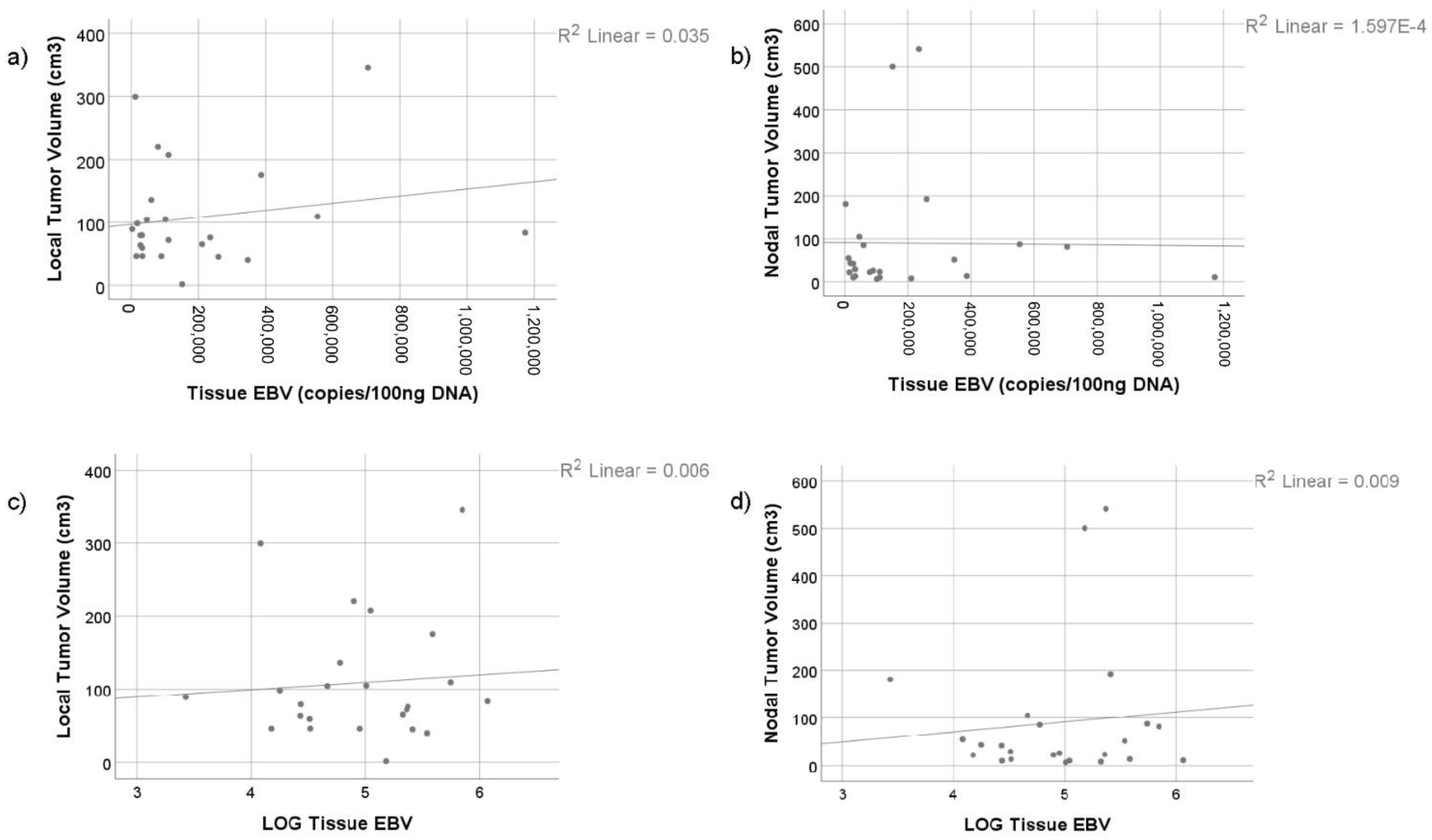
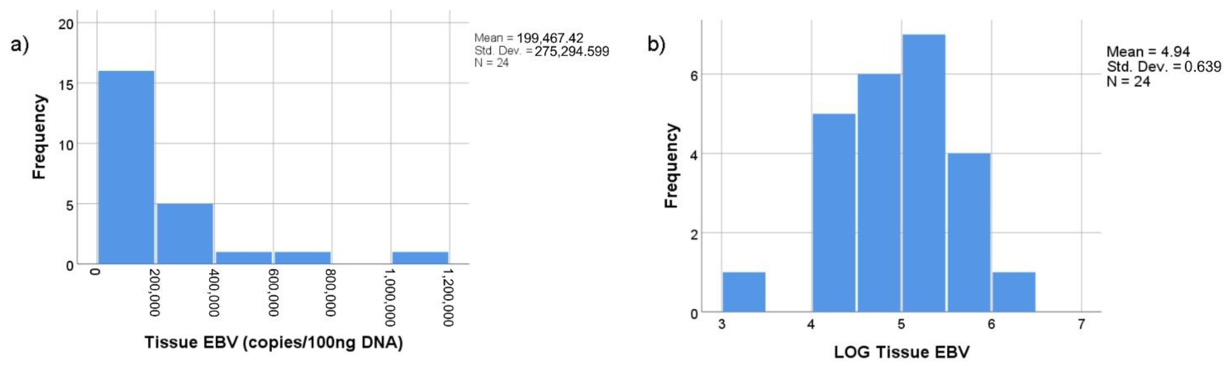
3.2. Relationship between Tumor Volume and EBV Viral Load in Tumor Cells

There was no correlation found between gross tumor volume at local nasopharynx with EBV viral load in tumor cells (see Figure 1a). The median local tumor volume was 81.3 cm3 ± range 80 cm3. The median EBV viral load in tumor cells was 95,644.8 copies/100 ng of DNA ± range 224,758.4 copies/100 ng of DNA. A higher EBV viral load in tumor cells in the local nasopharynx did not result in larger tumors in the local nasopharynx.
There was also no correlation found between gross tumor volume in the nodal or regional nasopharynx and EBV viral load in tumor cells (see Figure 1b). The median nodal or regional tumor volume was 35.7 cm3 ± range 73.63 cm3. A higher EBV viral load in tumor cells in the local nasopharynx did not result in larger or smaller tumors in the nodal or regional nasopharynx.
The replication of EBV followed a geometric order, therefore the data distribution of EBV viral load was skewed if a linear scale was used (see Figure 2a). A converted scale from linear to logarithmic for EBV viral load in tumor cells has resulted in those data being presented as a normal distribution (see Figure 2b). A re-analysis with EBV viral load in a logarithmic scale was performed.
There was also no correlation existing between both local tumor volume and nodal tumor volume with EBV viral load in tumor cells in logarithmic scale (Log of EBV viral load) from the local nasopharynx (See Figure 1c,d). The mean Log of EBV viral load in tumor cells from the local nasopharynx was 4.94 Log copies/100 ng of DNA ± range 0.64 Log copies/100 ng of DNA.

4. Discussion

EBV viral load in tumor tissue from nasopharyngeal biopsy has been shown in our study to not be related to baseline tumor extensiveness. A recent retrospective study also found no relationship between EBV viral load from nasopharyngeal lesions and cancer stages [16]. However, they found an association between a higher EBV viral load with more than 70 copies/cells and better outcomes, but the EBV viral load from that study varied very greatly [16]. In addition, patients’ performance status, patients’ comorbidities and the treatment were not described in that study. Those factors might also impact the overall survival. Hence, we could still argue that EBV viral load from nasopharyngeal tumor cells did not confer important prognostic information.
In our study, we confirmed that in all our samples with undifferentiated nasopharyngeal WHO type 3 histology, EBV is present in all samples. Although EBV was present, the amount of virus present within the cancer cells did not translate into greater or smaller tumor sizes. This finding indicates that EBV replication within the host cells does not affect cancer phenotype. There were other factors that might affect the aggressiveness of tumor growth, which ultimately affects patients’ survival. A study of immune checkpoint molecules in nasopharyngeal cancer with EBV DNA load in the endemic region found that EBV DNA load did not confer any prognostic information [17]. However, the immune checkpoint molecule PD-L1 did predict poorer prognosis [17,18]. This finding suggested that there was a complex interplay between host immune system, viral oncogenes and host genome that drive carcinogenesis and likely dictate the prognosis in nasopharyngeal cancer.
There is also ample evidence suggesting that the expression of several EBV viral oncogenic genes are related to cancer development [7,19]. These oncogenic proteins are probably the major driver of carcinogenesis, especially the proteins expressed by the latent EBV genes [7]. In acute EBV infection, the lytic phase will ensue with the expression of lytic genes. After lytic infection, the virus will reside and become dormant within the epithelial or lymphocytes by expression of various latent genes. These latent genes such as Latent Membrane Protein 1 (LMP1) and Epstein–Barr nuclear antigen 1 (EBNA-1) expressed in the latent phase of EBV infection were found to contribute to cancer development [7].
The viral protein LMP1 was associated with the inactivation of several important genes such as tumor suppressor genes within the host cells [7,20,21]. LMP1 inactivated tumor suppressor genes, for instance the Phosphatase and Tensin Homolog (PTEN) gene through the action of DNA methyltransferase 3b (DNMT3b) [21]. This DNMT3b resulted in PTEN CpG island methylation [21]. Furthermore, the LMP1 expressed by EBV has also been shown to be able to methylate the CDH-1 promoter region which eventually silences the E-cadherin gene. The cadherin is a cell adhesion molecule, in which a deficit of this protein will promote cancer cells to easily metastasize [22]. LMP1 has also been shown to be able to induce expression of PD-L1 in nasopharyngeal cancer [23]. The knockout of the LMP1 gene in the preclinical setting resulted in the suppression of PD-L1 [23]. The expression of PD-L1 has been correlated with poorer survival [23].
The EBNA-1 viral protein was associated with gene methylation process and genomic instability within the host genome [7,20]. The gene profiling study has shown that EBNA-1 could bind to the promoter sites of many host genes and result in upregulation and downregulation of specific genes [24]. The presence of EBNA-1 was also related to elevated production of reactive oxygen species (ROS) within the host cells [25]. This elevated ROS further increased the likelihood of genomic instability and DNA damage [25]. In the pre-clinical model, EBNA-1 expression was shown to be strongly associated with tumor growth [26]. All these findings indicate that EBNA-1 is required at least in part for tumorigenesis.
Another EBV molecule that is also highly transcribed in latent EBV infection was Epstein–Barr virus-encoded RNA (EBER). The EBER is a small segment of RNA molecule that is not expressed into protein, but it plays a major role in facilitating the process of immune escape [7]. The EBER can interfere with interferon-stimulating genes, thereby blunting the immune response [27]. In concert with other EBV viral proteins such as LMP1, the EBV could stimulate the recruitment and activation of Treg into a tumor microenvironment, therefore stimulating an even more permissive microenvironment for tumor growth [28].
Based on all the findings above and the results of our study, the expression of these oncogenic viral genes is probably more likely to correlate with tumor progression rather than the presence and the amount of EBV alone. EBV viral load quantification in tumor tissue was probably useful to assist doubtful diagnosis of nasopharyngeal cancer, especially the endemic type with WHO type 3 histopathology [29]. Quantification of EBV DNA in tumor cells might not be able to confer valuable prognostic information. However, quantification of viral proteins or RNAs might be able to provide some prognostic clues.

5. Conclusions

EBV viral load in tumor tissue from nasopharyngeal biopsy did not correlate with baseline pre-treatment tumor extensiveness. The presence and amount of virus in tumor cells was probably not very informative except for confirming diagnosis of nasopharyngeal carcinoma. EBV viral molecules such as LMP1, EBNA-1 and EBER were probably more likely to confer prognostic information.

Author Contributions

All authors have made equal contributions. All authors have read and agreed to the published version of the manuscript.

Funding

This study received a research grant from Universitas Indonesia. The funder has no role in specifying the design of the study and collection, analysis, and interpretation of data and in writing the manuscript.

Institutional Review Board Statement

The study was conducted according to the guidelines of the Declaration of Helsinki, and approved by the Institutional Review Board (or Ethics Committee) of Faculty of Medicine, Universitas Indonesia (protocol number 19-02-0133 and date of approval 11 March 2019).

Informed Consent Statement

Informed consent was obtained from all subjects involved in the study.

Acknowledgments

We acknowledge, express gratitude and appreciation to QIAGEN Indonesia for generously providing this study with their QIAGEN kits for free.

Conflicts of Interest

The authors declare no conflict of interest.

References

  1. Gondhowiardjo, S.A.; Adham, M.; Lisnawati, L.; Kodrat, H.; Tobing, D.L.; Handoko, H.; Haryoga, I.M.; Dwiyono, A.G.; Kristian, Y.A. Current Immune-Related Molecular Approach in Combating Nasopharyngeal Cancer. World J. Oncol. 2019, 10, 157–161. [Google Scholar] [CrossRef] [Green Version]
  2. Rumayor Pina, A.; Dos Santos, H.T.; Carlos, R.; Altemani, A.; de Almeida, O.P. Epstein-Barr Virus in Nasopharyngeal Carcinoma of Guatemalan and Brazilian Patients. Int. J. Surg. Pathol. 2017, 25, 304–309. [Google Scholar] [CrossRef]
  3. Vaughan, T.L.; A Shapiro, J.; Burt, R.D.; Swanson, G.M.; Berwick, M.; Lynch, C.F.; Lyon, J.L. Nasopharyngeal cancer in a low-risk population: Defining risk factors by histological type. Cancer Epidemiol. Biomarkers Prev. 1996, 5, 587–593. [Google Scholar] [PubMed]
  4. Cohen, J.I. Epstein-Barr virus infection. N. Engl. J. Med. 2000, 343, 481–492. [Google Scholar] [CrossRef] [PubMed]
  5. Klein, G.; Giovanella, B.C.; Lindahl, T.; Fialkow, P.J.; Singh, S.; Stehlin, J.S. Direct evidence for the presence of Epstein-Barr virus DNA and nuclear antigen in malignant epithelial cells from patients with poorly differentiated carcinoma of the nasopharynx. Proc. Natl. Acad. Sci. USA 1974, 71, 4737–4741. [Google Scholar] [CrossRef] [Green Version]
  6. Fåhraeus, R.; Fu, H.L.; Ernberg, I.; Finke, J.; Rowe, M.; Klein, G.; Falk, K.; Nilsson, E.; Yadav, M.; Busson, P.; et al. Expression of Epstein-Barr virus-encoded proteins in nasopharyngeal carcinoma. Int. J. Cancer 1988, 42, 329–338. [Google Scholar] [CrossRef] [PubMed]
  7. Tsao, S.W.; Tsang, C.M.; Lo, K.W. Epstein–Barr virus infection and nasopharyngeal carcinoma. Philos. Trans. R. Soc. B Biol. Sci. 2017, 372, 20160270. [Google Scholar] [CrossRef]
  8. Jain, A.; Chia, W.K.; Toh, H.C. Immunotherapy for nasopharyngeal cancer—A review. Chin. Clin. Oncol. 2016, 5, 22. [Google Scholar] [CrossRef]
  9. Gondhowiardjo, S. Epstein-Barr virus latent membrane protein 1 (EBV-LMP1) and tumor proliferation rate as predictive factors of nasopharyngeal cancer (NPC) radiation response. Gan kagaku ryoho. Cancer Chemother. 2000, 27, 323–331. [Google Scholar]
  10. Young, L.S.; Yap, L.F.; Murray, P.G. Epstein-Barr virus: More than 50 years old and still providing surprises. Nat. Rev. Cancer 2016, 16, 789–802. [Google Scholar] [CrossRef]
  11. Chan, K.A.; Woo, J.K.; King, A.; Zee, B.C.; Lam, W.J.; Chan, S.L.; Chu, S.W.; Mak, C.; Tse, I.O.; Leung, S.Y.; et al. Analysis of Plasma Epstein–Barr Virus DNA to Screen for Nasopharyngeal Cancer. N. Engl. J. Med. 2017, 377, 513–522. [Google Scholar] [CrossRef]
  12. Shen, T.; Tang, L.-Q.; Luo, D.-H.; Chen, Q.-Y.; Li, P.-J.; Mai, D.-M.; Guo, S.-S.; Liu, L.-T.; Qian, C.-N.; Guo, X.; et al. Different Prognostic Values of Plasma Epstein-Barr Virus DNA and Maximal Standardized Uptake Value of 18F-FDG PET/CT for Nasopharyngeal Carcinoma Patients with Recurrence. PLoS ONE 2015, 10, e0122756. [Google Scholar] [CrossRef] [Green Version]
  13. Chen, M.; Yin, L.; Wu, J.; Gu, J.-J.; Jiang, X.-S.; Wang, D.-J.; Zong, D.; Guo, C.; Zhu, H.-F.; Wu, J.-F.; et al. Impact of Plasma Epstein-Barr Virus-DNA and Tumor Volume on Prognosis of Locally Advanced Nasopharyngeal Carcinoma. BioMed Res. Int. 2015, 2015, 1–5. [Google Scholar] [CrossRef]
  14. Adham, M.; Greijer, A.E.; Verkuijlen, S.A.W.M.; Juwana, H.; Fleig, S.; Rachmadi, L. Epstein-Barr Virus DNA Load in Nasopharyngeal Brushings and Whole Blood in Nasopharyngeal Carcinoma Patients before and after Treatment. Clin. Cancer Res. 2013, 19, 2175–2186. [Google Scholar] [CrossRef] [Green Version]
  15. Lin, J.-C.; Wang, W.-Y.; Chen, K.Y.; Wei, Y.-H.; Liang, W.-M.; Jan, J.-S.; Jiang, R.-S. Quantification of Plasma Epstein–Barr Virus DNA in Patients with Advanced Nasopharyngeal Carcinoma. N. Engl. J. Med. 2004, 350, 2461–2470. [Google Scholar] [CrossRef] [Green Version]
  16. Nilsson, J.S.; Forslund, O.; Andersson, F.C.; Lindstedt, M.; Greiff, L. Intralesional EBV-DNA load as marker of prognosis for nasopharyngeal cancer. Sci. Rep. 2019, 9, 15432. [Google Scholar] [CrossRef] [Green Version]
  17. Zhou, Y.; Shi, D.; Miao, J.; Wu, H.; Chen, J.; Zhou, X. PD-L1 predicts poor prognosis for nasopharyngeal carcinoma irrespective of PD-1 and EBV-DNA load. Sci. Rep. 2017, 7, 43627. [Google Scholar] [CrossRef] [Green Version]
  18. Gondhowiardjo, S.A.; Handoko Adham, M.; Rachmadi, L.; Kodrat, H.; Tobing, D.L. Tumor microenvironment predicts local tumor extensiveness in PD-L1 positive nasopharyngeal cancer. PLoS ONE 2020, 15, e0230449. [Google Scholar]
  19. Elgui de Oliveira, D.; Muller-Coan, B.G.; Pagano, J.S. Viral Carcinogenesis Beyond Malignant Transformation: EBV in the Progression of Human Cancers. Trends Microbiol. 2016, 24, 649–664. [Google Scholar] [CrossRef] [PubMed] [Green Version]
  20. Li, L.; Zhang, Y.; Guo, B.B.; Chan, F.K.; Tao, Q. Oncogenic induction of cellular high CpG methylation by Epstein-Barr virus in malignant epithelial cells. Chin. J. Cancer 2014, 33, 604–608. [Google Scholar] [CrossRef] [PubMed]
  21. Peng, H.; Chen, Y.; Gong, P.; Cai, L.; Lyu, X.; Jiang, Q. Higher methylation intensity induced by EBV LMP1 via NF-κB/DNMT3b signaling contributes to silencing of PTEN gene. Oncotarget 2016, 7, 40025–40037. [Google Scholar] [CrossRef] [PubMed] [Green Version]
  22. Petrova, Y.I.; Schecterson, L.; Gumbiner, B.M. Roles for E-cadherin cell surface regulation in cancer. Mol. Biol. Cell 2016, 27, 3233–3244. [Google Scholar] [CrossRef]
  23. Fang, W.; Zhang, J.; Hong, S.; Zhan, J.; Chen, N.; Qin, T. EBV-driven LMP1 and IFN-γ up-regulate PD-L1 in nasopharyngeal carcinoma: Implications for oncotargeted therapy. Oncotarget 2014, 5, 12189–12202. [Google Scholar] [CrossRef] [PubMed]
  24. Canaan, A.; Haviv, I.; Urban, A.E.; Schulz, V.P.; Hartman, S.; Zhang, Z. EBNA1 regulates cellular gene expression by binding cellular promoters. Proc. Natl. Acad. Sci. USA 2009, 106, 22421–22426. [Google Scholar] [CrossRef] [PubMed] [Green Version]
  25. Gruhne, B.; Sompallae, R.; Marescotti, D.; Kamranvar, S.A.; Gastaldello, S.; Masucci, M.G. The Epstein-Barr virus nuclear antigen-1 promotes genomic instability via induction of reactive oxygen species. Proc. Natl. Acad. Sci. USA 2009, 106, 2313–2318. [Google Scholar] [CrossRef] [PubMed] [Green Version]
  26. Kaul, R.; Murakami, M.; Choudhuri, T.; Robertson, E.S. Epstein-Barr virus latent nuclear antigens can induce metastasis in a nude mouse model. J. Virol. 2007, 81, 10352–10361. [Google Scholar] [CrossRef] [Green Version]
  27. Takada, K. Role of EBER and BARF1 in nasopharyngeal carcinoma (NPC) tumorigenesis. Semin. Cancer Biol. 2012, 22, 162–165. [Google Scholar] [CrossRef] [PubMed]
  28. Baumforth, K.R.; Birgersdotter, A.; Reynolds, G.M.; Wei, W.; Kapatai, G.; Flavell, J.R.; Kalk, E.; Piper, K.; Lee, S.; Machado, L.; et al. Expression of the Epstein-Barr Virus-Encoded Epstein-Barr Virus Nuclear Antigen 1 in Hodgkin’s Lymphoma Cells Mediates Up-Regulation of CCL20 and the Migration of Regulatory T Cells. Am. J. Pathol. 2008, 173, 195–204. [Google Scholar] [CrossRef] [PubMed] [Green Version]
  29. Zheng, X.-H.; Lu, L.-X.; Li, X.-Z.; Jia, W.-H. Quantification of Epstein-Barr virus DNA load in nasopharyngeal brushing samples in the diagnosis of nasopharyngeal carcinoma in southern China. Cancer Sci. 2015, 106, 1196–1201. [Google Scholar] [CrossRef] [Green Version]
Figure 1. Scatterplot showing no correlation between (a,b) Epstein–Barr virus (EBV) viral load in nasopharyngeal tumor tissue with (a) local tumor volume, and (b) nodal tumor volume. (c,d) Re-analysis of correlation using normalized distribution in a logarithmic scale of EBV viral load in nasopharyngeal tumor tissue also showing no correlation with (c) local tumor volume, and (d) nodal tumor volume.
Microbiolres 12 00011 g001
Figure 2. Histogram showing distribution of data for EBV viral load in tumor tissue from nasopharyngeal biopsy: (a) non-normal distribution with linear scale, (b) normal distribution with logarithmic scale.
Microbiolres 12 00011 g002
Table 1. Subject Characteristics.
CharacteristicsNumber of Subjects (%)
Age
≤50 years old13 (54.2%)
>50 years old11 (45.8%)
Gender
Male18 (75%)
Female6 (25%)
Stage *
III4 (16.7%)
IVA12 (50%)
IVB8 (33.3%)
Total24 (100%)
* Based on AJCC Staging 8th Edition.
Publisher’s Note: MDPI stays neutral with regard to jurisdictional claims in published maps and institutional affiliations.

