Incessant Automatic Atrial Tachycardia in a Neonate Successfully Treated with Nadolol and Closely Spaced Doses of Flecainide: A Case Report

 and
and 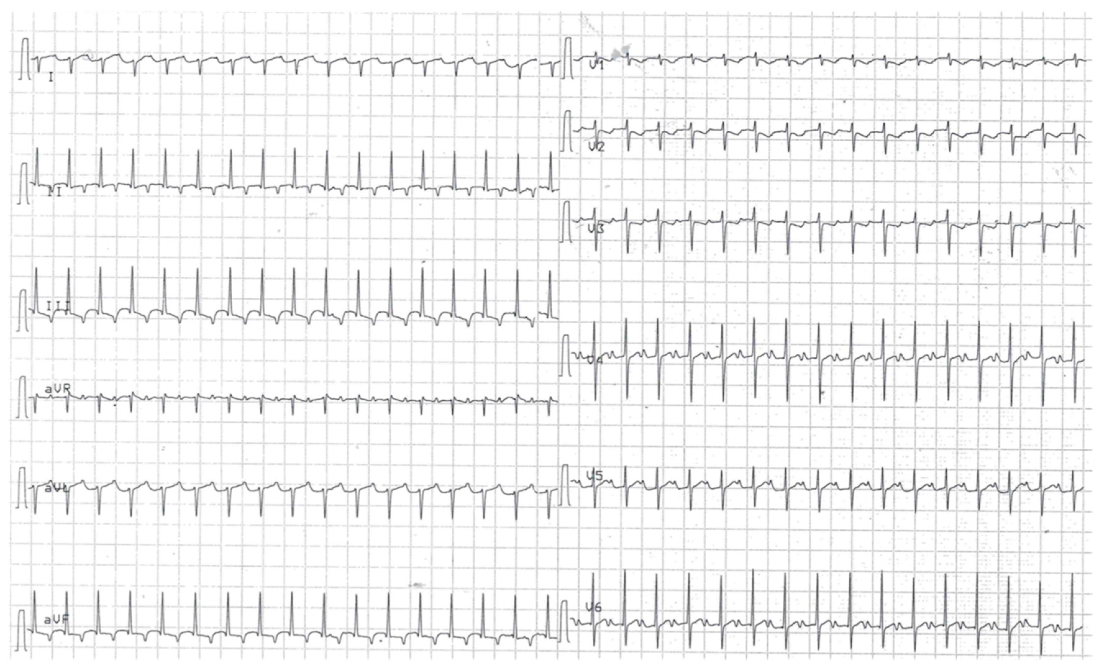
Abstract
:1. Introduction
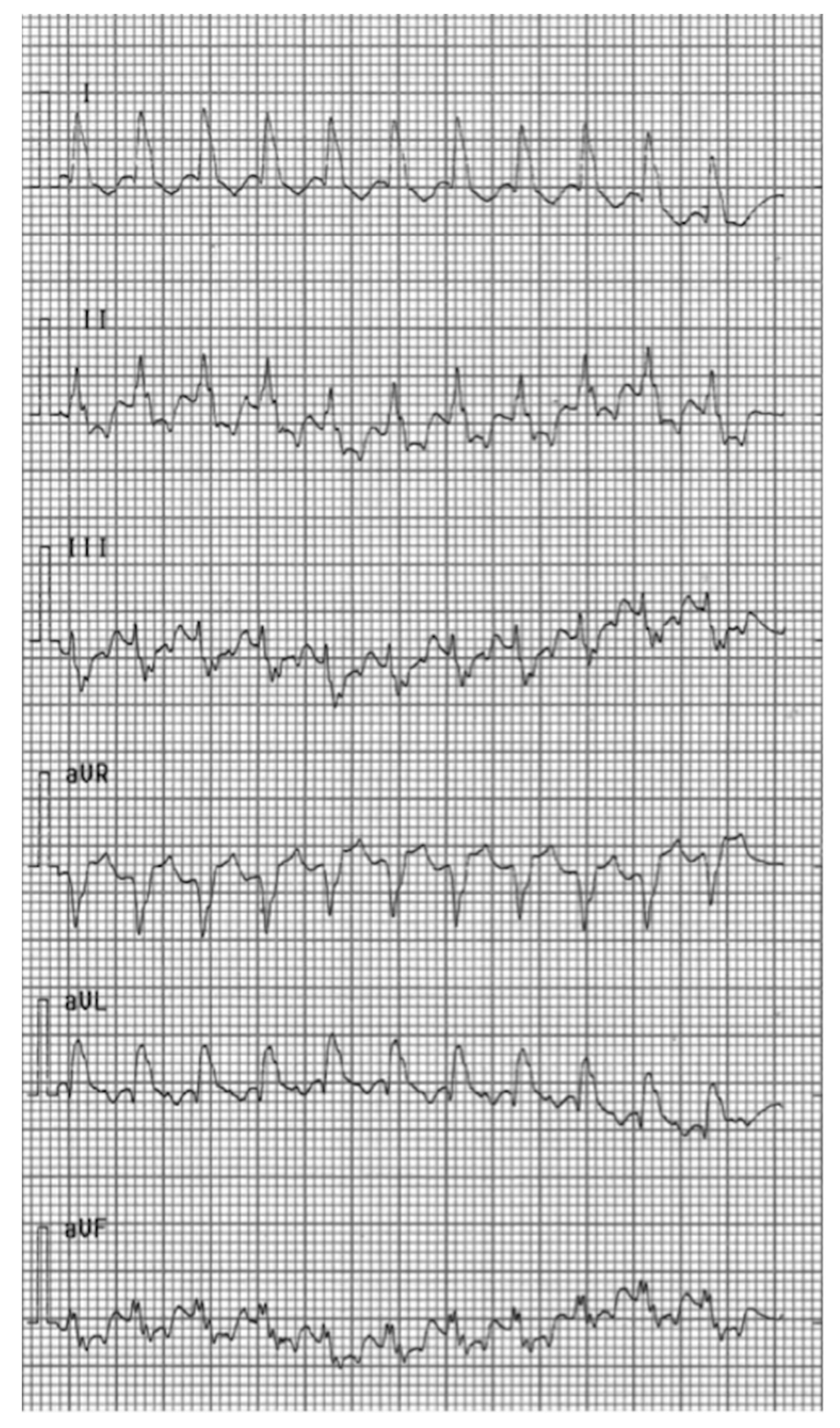
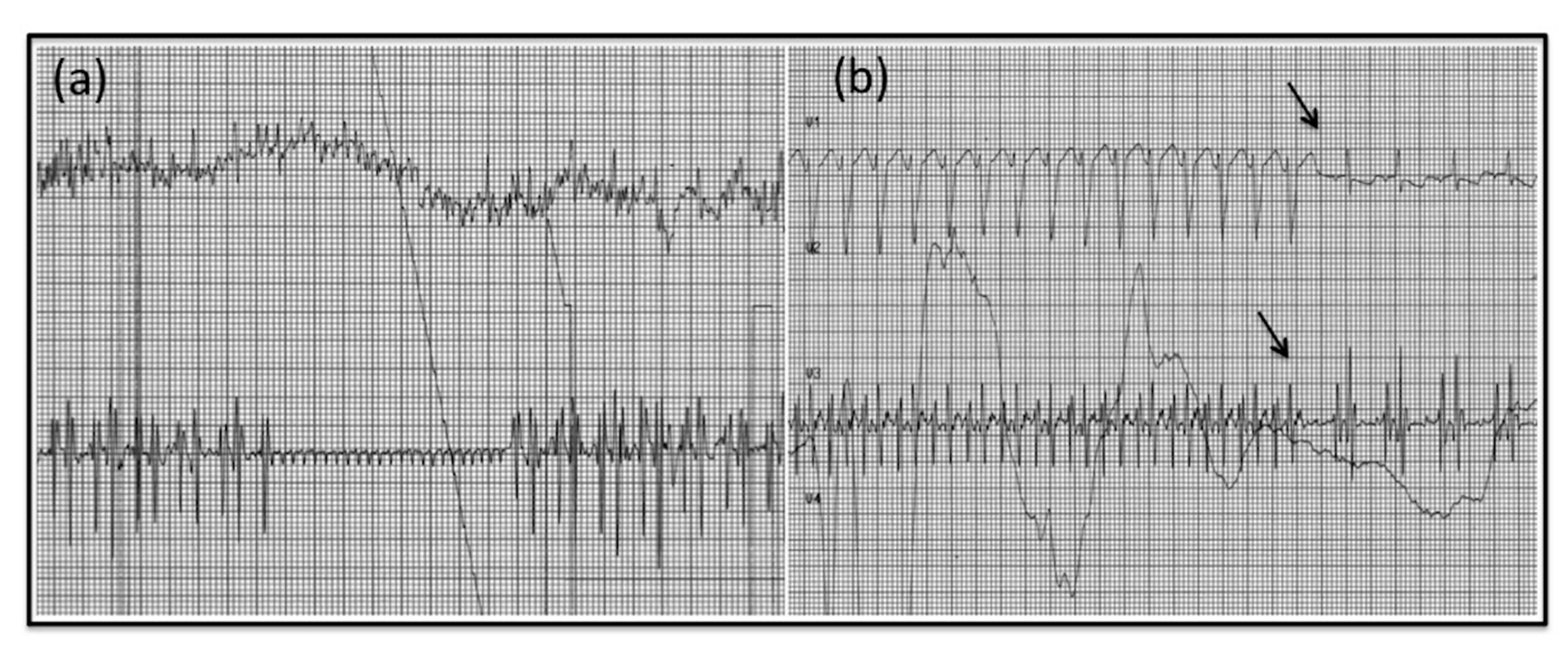
2. Case Report
3. Discussion
4. Conclusions
Author Contributions
Funding
Acknowledgments
Conflicts of Interest
References
- Vignati, G.; Annoni, G. Characterization of supraventricular tachycardia in infants: Clinical and instrumental diagnosis. Curr. Pharm. Des. 2008, 14, 729–735. [Google Scholar] [CrossRef] [PubMed]
- Luedtke, S.A.; Kuhn, R.J.; Mccaffrey, F.M. Pharmacologic management of supraventricular tachycardia in children part 2: Atrial Flutter, Atrial Fibrillation, and Junctional and Atrial Ectopic Tachycardia. Ann. Pharm. 1997, 31, 1347–1359. [Google Scholar] [CrossRef] [PubMed]
- Entenmann, A.; Michel, M.; Herberg, U.; Haas, N.; Kumpf, M.; Gass, M.; Egender, F.; Gebauer, R. Management of postoperative junctional ectopic tachycardia in pediatric patients: A survey of 30 centers in Germany, Austria, and Switzerland. Eur. J. Nucl. Med. Mol. Imaging 2017, 176, 1217–1226. [Google Scholar] [CrossRef] [PubMed]
- Calabrò, M.P.; Cerrito, M.; Luzza, F.; Oreto, G. Supraventricular tachycardia in infants: Epidemiology and clinical management. Curr. Pharm. Des. 2008, 14, 723–728. [Google Scholar] [CrossRef] [PubMed] [Green Version]
- Chu, P.Y.; Hill, K.D.; Clark, R.H.; Smith, P.B.; Hornik, C.P. Treatment of supraventricular tachycardia in infants: Analysis of a large multicenter database. Early Hum. Dev. 2015, 91, 345–350. [Google Scholar] [CrossRef] [PubMed] [Green Version]
- Scheinman, M.M.; Aliot, E.M. ACC/AHA/ESC guidelines for the management of patients with supraventricular arrhythmias. J. Am. Coll. Cardiol. 2003, 42, 1493–1531. [Google Scholar] [CrossRef] [Green Version]
- Brendorp, B.; Pedersen, O.; Torp-Pedersen, C.; Sahebzadah, N.; Køber, L. A Benefit-Risk Assessment of Class III Antiarrhythmic Agents. Drug Saf. 2002, 25, 847–865. [Google Scholar] [CrossRef] [PubMed]
- Naheed, Z.J.; Strasburger, J.F.; Woodrow, D.; Deal, B.J. Natural History and Management Automatic Atrial Tachycardia. Am. J. Cardiol. 1995, 75, 405–407. [Google Scholar] [CrossRef]
- Von Bernuth, G.; Engelhardt, W.; Kramer, H.H.; Singer, H.; Schneider, P.; Ulmer, H.; Brodherr-Heberlein, S.; Kienast, W.; Lang, D.; Lindinger, A.; et al. Atrial automatic tachycardia in infancy and childhood. Eur. Hear. J. 1992, 13, 1410–1415. [Google Scholar] [CrossRef] [PubMed]
- Ferlini, M.; Colli, A.M.; Bonanomi, C.; Salvini, L.; A Galli, M.; Salice, P.; Ravaglia, R.; Centola, M.; Danzi, G.B. Flecainide as first-line treatment for supraventricular tachycardia in newborns. J. Cardiovasc. Med. 2009, 10, 372–375. [Google Scholar] [CrossRef] [PubMed]
- Perry, J.C.; Garson, A. Flecainide acetate for treatment of tachyarrhythmias in children: Review of world literature on efficacy, safety, and dosing. Am. Hear. J. 1992, 124, 1614–1621. [Google Scholar] [CrossRef]
- Perry, J.C.; McQuinn, R.L.; Smith, R.T.; Gothing, C.; Fredell, P.; Garson, A. Flecainide acetate for resistant arrhythmias in the young: Efficacy and pharmacokinetics. J. Am. Coll. Cardiol. 1989, 14, 185–191. [Google Scholar] [CrossRef] [Green Version]
- Horie, A.; Ishida, K.; Shibata, K.; Taguchi, M.; Ozawa, A.; Hirono, K.; Ichida, F.; Hashimoto, Y. Pharmacokinetic variability of flecainide in younger Japanese patients and mechanisms for renal excretion and intestinal absorption. Biopharm. Drug Dispos. 2013, 35, 145–153. [Google Scholar] [CrossRef]
- A Till, J.; A Shinebourne, E.; Rowland, E.; E Ward, D.; Bhamra, R.; Haga, P.; Johnston, A.; Holt, D.W. Paediatric use of flecainide in supraventricular tachycardia: Clinical efficacy and pharmacokinetics. Heart 1989, 62, 133–139. [Google Scholar] [CrossRef] [PubMed]
- Tamargo, J.; Le Heuzey, J.-Y.; Mabo, P. Narrow therapeutic index drugs: A clinical pharmacological consideration to flecainide. Eur. J. Clin. Pharmacol. 2015, 71, 549–567. [Google Scholar] [CrossRef] [Green Version]


Publisher’s Note: MDPI stays neutral with regard to jurisdictional claims in published maps and institutional affiliations. |
© 2020 by the authors. Licensee MDPI, Basel, Switzerland. This article is an open access article distributed under the terms and conditions of the Creative Commons Attribution (CC BY) license (http://creativecommons.org/licenses/by/4.0/).
Share and Cite
Belli, G.; Giovannini, M.; Porcedda, G.; Moroni, M.; la Marca, G.; Capponi, G.; Favilli, S.; De Simone, L. Incessant Automatic Atrial Tachycardia in a Neonate Successfully Treated with Nadolol and Closely Spaced Doses of Flecainide: A Case Report. Pediatr. Rep. 2020, 12, 108-113. https://0-doi-org.brum.beds.ac.uk/10.3390/pediatric12030024
Belli G, Giovannini M, Porcedda G, Moroni M, la Marca G, Capponi G, Favilli S, De Simone L. Incessant Automatic Atrial Tachycardia in a Neonate Successfully Treated with Nadolol and Closely Spaced Doses of Flecainide: A Case Report. Pediatric Reports. 2020; 12(3):108-113. https://0-doi-org.brum.beds.ac.uk/10.3390/pediatric12030024
Chicago/Turabian StyleBelli, Gilda, Mattia Giovannini, Giulio Porcedda, Marco Moroni, Giancarlo la Marca, Guglielmo Capponi, Silvia Favilli, and Luciano De Simone. 2020. "Incessant Automatic Atrial Tachycardia in a Neonate Successfully Treated with Nadolol and Closely Spaced Doses of Flecainide: A Case Report" Pediatric Reports 12, no. 3: 108-113. https://0-doi-org.brum.beds.ac.uk/10.3390/pediatric12030024