Epiploic Appendagitis and Omental Infarction as Rare Causes of Acute Abdominal Pain in Children

 ,
,
Abstract
:1. Introduction
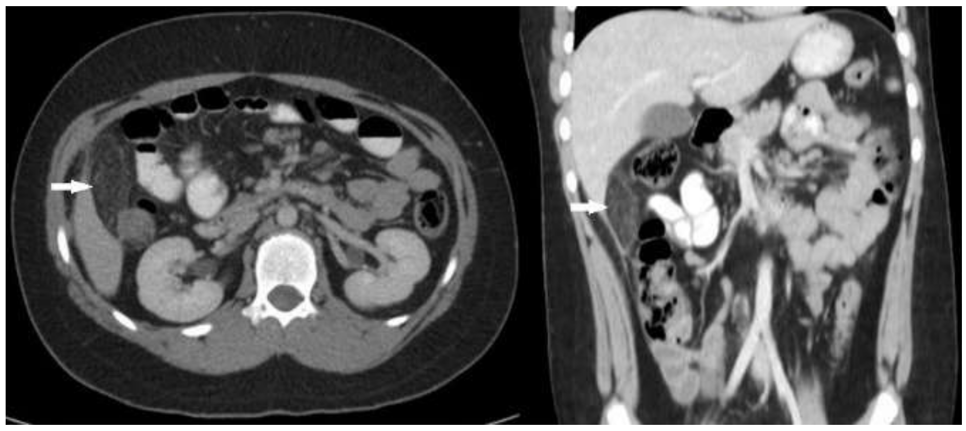
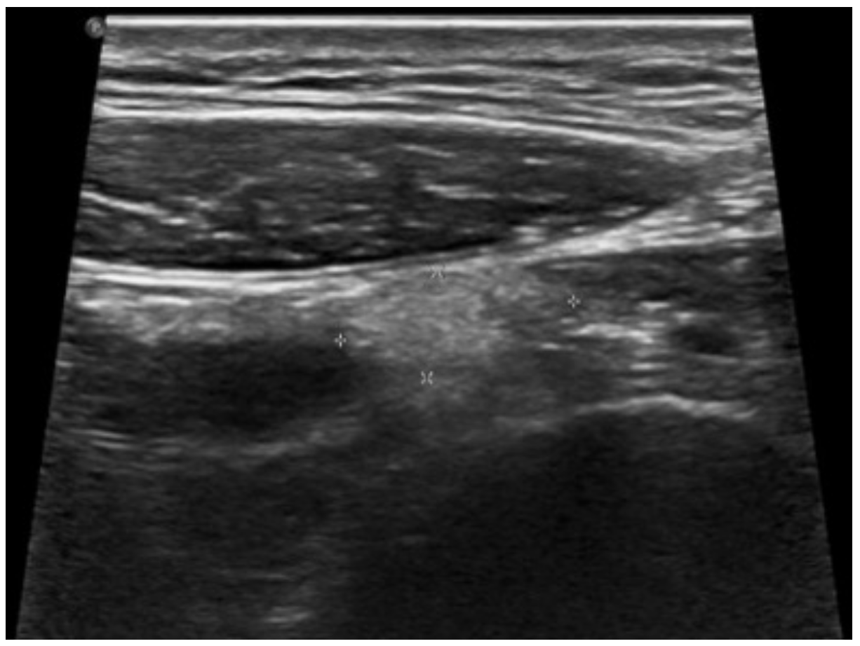
2. Case Report 1
3. Case Report 2
4. Discussion
5. Conclusions
Author Contributions
Funding
Institutional Review Board Statement
Informed Consent Statement
Data Availability Statement
Conflicts of Interest
Abbreviations
| OI | Omental infarction |
| EA | Epiploic appendagitis |
| AA | Acute appendicitis |
| US | Ultrasonography |
| CT | Computed Tomography |
| MRI | Magnetic resonance imaging |
References
- Helmrath, M.A.; Dorfman, S.R.; Minifee, P.K.; Bloss, R.S.; Brandt, M.L.; DeBakey, M.E. Right lower quadrant pain in children caused by omental infarction. Am. J. Surg. 2001, 182, 729–732. [Google Scholar] [CrossRef]
- Grattan-Smith, J.D.; Blews, D.E.; Brand, T. Omental Infarction in Pediatric Patients: Sonographic and CT Findings. Am. J. Roentgenol. 2002, 178, 1537–1539. [Google Scholar] [CrossRef]
- Houben, C.H.; Powis, M.; Wright, V.M. Segmental Infarction of the Omentum: A Difficult Diagnosis. Eur. J. Pediatr. Surg. 2003, 13, 57–59. [Google Scholar] [CrossRef]
- Nagar, H.; Kessler, A.; Ben-Sira, L.; Klepikov, I.; Wiess, J.; Graif, M. Omental infarction: An unusual cause of acute abdomen in children. Pediatr. Surg. Int. 2003, 19, 677–679. [Google Scholar] [CrossRef] [PubMed]
- Varjavandi, V.; Lessin, M.; Kooros, K.; Fusunyan, R.; McCauley, R.; Gilchrist, B. Omental infarction: Risk factors in children. J. Pediatr. Surg. 2003, 38, 233–235. [Google Scholar] [CrossRef]
- Sakellaris, G.; Stathopoulos, E.; Kafousi, M.; Arbiros, J.; Bitsori, M.; Charissis, G. Primary idiopathic segmental infarction of the greater omentum: Two cases of acute abdomen in childhood. J. Pediatr. Surg. 2004, 39, 1264–1266. [Google Scholar] [CrossRef]
- Loh, M.H.; Chui, H.C.; Yap, T.-L.; Sundfor, A.; Tan, C.E.L. Omental infarction—A mimicker of acute appendicitis in children. J. Pediatr. Surg. 2005, 40, 1224–1226. [Google Scholar] [CrossRef] [PubMed]
- Lee, W.; Ong, C.L.; Chong, C.C.L.; Hwang, W.S. Omental infarction in children: Imaging features with pathological correlation. Singap. Med. J. 2005, 46, 328–332. [Google Scholar]
- Coulier, B. Segmental omental infarction in childhood: A typical case diagnosed by CT allowing successful conservative treatment. Pediatr. Radiol. 2006, 36, 141–143. [Google Scholar] [CrossRef] [PubMed]
- Aoun, N.; Nader, L.; Haddad-Zebouni, S.; Ghossain, M.; Akatcherian, C. Left segmental omental infarction in a child: Conservative treatment. Archives de Pédiatrie 2006, 13, 1040–1042. [Google Scholar] [CrossRef]
- Van Kerkhove, F.; Coenegrachts, K.; Steyaert, L.; Ghekiere, J.; Gabriel, C.; Casselman, J.W. Omental infarction in childhood. JBR-BTR 2006, 89, 198–200. [Google Scholar] [PubMed]
- Fragoso, A.C.; Pereira, J.M.; Estevão-Costa, J. Nonoperative management of omental infarction: A case report in a child. J. Pediatr. Surg. 2006, 41, 1777–1779. [Google Scholar] [CrossRef] [PubMed]
- Zargar, N.U.; Kundal, A.K.; Krishna, A. Omental infarction—An unrecognized cause of acute abdomen. Indian J. Pediatr. 2007, 74, 87. [Google Scholar] [CrossRef] [PubMed]
- Foscolo, S.; Mandry, D.; Galloy, M.-A.; Champigneulles, J.; De Miscault, G.; Claudon, M. Segmental omental infarction in childhood: An unusual case of left-sided location with extension into the pelvis. Pediatr. Radiol. 2007, 37, 575–577. [Google Scholar] [CrossRef] [PubMed]
- Agresta, F.; Bedin, N. Primary Omental Infarction: Laparoscopic Approach in Two Pediatric Cases: A Case Review. J. Laparoendosc. Adv. Surg. Tech. 2007, 17, 831–832. [Google Scholar] [CrossRef] [PubMed]
- Nubi, A.; McBride, W.; Stringel, G. Primary omental infarct: Conservative vs. operative management in the era of ultrasound, computerized tomography, and laparoscopy. J. Pediatr. Surg. 2009, 44, 953–956. [Google Scholar] [CrossRef] [PubMed]
- Rimon, A.; Daneman, A.; Gerstle, J.T.; Ratnapalan, S. Omental Infarction in Children. J. Pediatr. 2009, 155, 427–431.e1. [Google Scholar] [CrossRef]
- Yang, Y.-L.; Huang, Y.-H.; Tiao, M.-M.; Tang, K.-S.; Huang, F.-C.; Lee, S.-Y. Comparison of Clinical Characteristics and Neutrophil Values in Omental Infarction and Acute Appendicitis in Children. Pediatr. Neonatol. 2010, 51, 155–159. [Google Scholar] [CrossRef] [Green Version]
- Ventham, N.T.; Velchuru, V.; Scout, E.; Studley, J. Unusual cause of acute abdomen—Omental infarction occurring in a child with cyclical neutropenia. Ann. R. Coll. Surg. Engl. 2010, 92, e32–e34. [Google Scholar] [CrossRef] [Green Version]
- Bradley, G.; Adger, H.; Splinter, A.; Leggett, B.D.; Madan, N.; Marks, M. Index of Suspicion. Pediatr. Rev. 2010, 31, 117–123. [Google Scholar] [CrossRef] [PubMed]
- Gosain, A.; Blakely, M.; Boulden, T.; Uffman, J.K.; Seetharamaiah, R.; Huang, E.; Langham, M.; EubanksIII, J.W. Omental Infarction: Preoperative Diagnosis and Laparoscopic Management in Children. J. Laparoendosc. Adv. Surg. Tech. 2010, 20, 777–780. [Google Scholar] [CrossRef]
- Kambouri, K.; Gardikis, S.; Giatromanolaki, A.; Tsalkidis, A.; Sivridis, E.; Vaos, G. Omental Infarction in an Obese 10-year-old Boy. Pediatr. Rep. 2011, 3, 91–92. [Google Scholar] [CrossRef] [PubMed]
- Tsunoda, T.; Sogo, T.; Komatsu, H.; Inui, A.; Fujisawa, T. A Case Report of Idiopathic Omental Infarction in an Obese Child. Case Rep. Pediatr. 2012, 2012, 513634. [Google Scholar] [CrossRef] [PubMed]
- Sandusky, M.F.; Herliczek, T.W. Pediatric omental infarction. Med. Health RI 2012, 95, 40–41. [Google Scholar]
- Wertheimer, J.; Galloy, M.-A.; Régent, D.; Champigneulle, J.; Lemelle, J.-L. Radiological, clinical and histological correlations in a right segmental omental infarction due to primary torsion in a child. Diagn. Interv. Imaging 2014, 95, 325–331. [Google Scholar] [CrossRef] [Green Version]
- Estevão-Costa, J.; Alvarenga, A.S.; Fragoso, A.C.; Garcia, M.; Campos, M. Omental Infarction: A Reappraisal of Conservative Management in Children. Acta Médica Port. 2014, 27, 433–436. [Google Scholar] [CrossRef] [PubMed] [Green Version]
- Álvarez, A.A.; Serna, J.P.; Chávez, L.R.; Perri, L.E.; Palacios, M.G.; Barca, P.R.; Gallart, R.M.; Martinez, E.E.; Casasnovas, A.B. Tratamiento conservador del infarto omental. Cir Pediátrica 2014, 27, 4. [Google Scholar]
- Hamchou, M.; Kothari, M.; Sahari, B.; Swid, A.; Al-Salem, A.H. Segmental omental infarction: A rare cause of acute abdominal pain in children. Surg. Laparosc. Endosc. Percutaneous Tech. 2014, 24, e38–e40. [Google Scholar] [CrossRef] [PubMed]
- Koay, H.T.; Mahmoud, H.E.S. Simultaneous omental infarction and acute appendicitis in a child. Med. J. Malays. 2015, 70, 2. [Google Scholar]
- Arigliani, M.; Dolcemascolo, V.; Nocerino, A.; Pasqual, E.; Avellini, C.; Cogo, P. A Rare Cause of Acute Abdomen: Omental Infarction. J. Pediatr. 2016, 176, 216–216.e1. [Google Scholar] [CrossRef] [PubMed] [Green Version]
- O’Rourke, R.W.; Saito, J.M.; Albanese, C.T. Laparoscopic Diagnosis and Resection of Pediatric Appendicitis Epiploicae: Case Report and Literature Review. Pediatr. Endosurgery Innov. Tech. 2001, 5, 319–322. [Google Scholar] [CrossRef]
- Hurreiz, H.; Madavo, C.M. Torsion of an epiploic appendix mimicking acute appendicitis. Saudi Med. J. 2005, 26, 2003–2004. [Google Scholar] [PubMed]
- Gupta, V.; Kumar, S. Appendicitis epiploicae: An unusual cause of acute abdomen in children. J. Indian Assoc. Pediatr. Surg. 2008, 13, 83–84. [Google Scholar] [CrossRef] [PubMed]
- Christianakis, E.; Paschalidis, N.; Filippou, G.K.; Smailis, D.; Chorti, M.; Rizos, S.; Filippou, D.K. Cecal epiploica appendix torsion in a female child mimicking acute appendicitis: A case report. Cases J. 2009, 2, 8023. [Google Scholar] [CrossRef] [PubMed] [Green Version]
- Fraser, J.D.; Aguayo, P.; Leys, C.M.; Peter, S.D.S.; Ostlie, D.J. Infarction of an epiploic appendage in a pediatric patient. J. Pediatr. Surg. 2009, 44, 1659–1661. [Google Scholar] [CrossRef] [PubMed]
- Matsunaga, H.; Fujii, Y.; Taniguchi, N. Ultrasonographic findings in epiploic appendagitis. J. Med Ultrason. 2009, 37, 31–32. [Google Scholar] [CrossRef] [PubMed]
- Goh, V.L.; Rudolph, C.D. Epiploic Appendagitis. J. Pediatr. Gastroenterol. Nutr. 2011, 53, 1. [Google Scholar] [CrossRef] [PubMed]
- Rashid, A.; Nazir, S.; Hakim, S.Y.; Chalkoo, M.A. Epiploic appendagitis of caecum: A diagnostic dilemma. Ger. Med. Sci. 2012, 10. [Google Scholar] [CrossRef] [PubMed]
- Cho, M.S.; Hwang-Bo, S.; Choi, U.Y.; Kim, H.S.; Hahn, S.H. A Case of Epiploic Appendagitis with Acute Gastroenteritis. Pediatr. Gastroenterol. Hepatol. Nutr. 2014, 17, 263–265. [Google Scholar] [CrossRef] [PubMed] [Green Version]
- Toprak, H.; Yildiz, S.; Kilicarslan, R.; Bilgin, M. Epiploic appendagitis:a rare cause of acute abdominal pain in children. Report of a case and review of the pediatric literature. J. Belg. Soc. Radiol. 2014, 97, 174–175. [Google Scholar] [CrossRef] [PubMed] [Green Version]
- Redmond, P.; Sawaya, D.E.; Miller, K.H.; Nowicki, M.J. Epiploic Appendagitis: A rare cause of acute abdominal pain in children. report of a case and review of the pediatric literature. Pediatr. Emerg. Care 2015, 31, 717–719. [Google Scholar] [CrossRef] [PubMed]
- Joshi, D.; Fleming, A.E.; Spottswood, S.E. It’s Not Appendicitis? Consideration of a Benign Mimicker. Hosp. Pediatr. 2015, 5, 101–105. [Google Scholar] [CrossRef] [PubMed] [Green Version]
- Ullah, I.; Mahajan, L.; Magnuson, D. Epiploic Appendagitis: A Rare Cause of Chronic Right Lower Quadrant Pain in a Child. J. Pediatr. 2017, 182, 400–400.e1. [Google Scholar] [CrossRef]
- Boscarelli, A.; Frediani, S.; Ceccanti, S.; Falconi, I.; Masselli, G.; Casciani, E.; Cozzi, D.A. Magnetic resonance imaging of epiploic appendagitis in children. J. Pediatr. Surg. 2016, 51, 2123–2125. [Google Scholar] [CrossRef] [PubMed]
- Ozturk, M.; Aslan, S.; Sağlam, D.; Bekci, T.; Bilgici, M.C. Epiploic Appendagitis as a Rare Cause of Acute Abdomen in the Pediatric Population: Report of Three Cases. Eurasian J. Med. 2018, 50, 56–58. [Google Scholar] [CrossRef] [PubMed] [Green Version]
- Nhamoucha, Y.; Bouabdellah, Y. Une cause rare d’abdomen aigu: l’appendagite épiploique. Pan Afr. Med. J. 2018, 30. [Google Scholar] [CrossRef] [PubMed]


| Authors (Year Reported) | Patients No. | Sex | Average Age (Range) in Years | Preoperative Diagnosis with Imaging Studies | Other Preoperative Diagnosis | Approach | Complications | Intraoperative Diagnosis |
| Helmrath et al. [1], 2001 | 18 | M (12) F (6) | 7.5 (2–13) | US (4) CT (2) | AA (6) | Surgery (18) | - | +(12) |
| Grattan-Simth et al. [2], 2002 | 9 | M (5) F (4) | 8.0 (3–11) | US + CT (5) | Undiagnosed (4) | Surgery (8) Conservative (1) | - | +(4) |
| Houben et al. [3], 2003 | 1 | M | 8 | - | AA (1) | Surgery (1) | - | +(1) |
| Nagar et al. [4], 2003 | 2 | M (2) | 9.0 (8–10) | US + CT (2) | - | Conservative (2) | - | |
| Varjavandi et al. [5], 2003 | 4 | M (3) F (1) | 12.5 (10–14) | CT (3) | Undiagnosed (1) | Surgery (4) | - | +(1) |
| Sakellaris et al. [6], 2004 | 2 | M (2) | 8.0 (7–9) | - | AA (2) | Surgery (2) | - | +(2) |
| Loh et al. [7], 2005 | 12 | M (10) F (2) | 9.0 (4–11) | CT (4) | AA (8) | Surgery (12) | - | +(8) |
| Lee et al. [8], 2005 | 6 | M (5) F (1) | 8.8 (5–11) | CT (2) | AA (3) Undiagnosed (1) | Surgery (6) | - | +(4) |
| Coulier [9], 2006 | 1 | M | 10 | US + CT (1) | - | Conservative (1) | - | |
| Aoun et al. [10], 2006 | 1 | M | 11 | US + CT (1) | - | Conservative (1) | - | |
| Van Kerkhove et al. [11], 2006 | 1 | F | 6 | - | Undiagnosed (1) | Surgery (1) | - | +(1) |
| Fragoso et al. [12], 2006 | 1 | M | 9 | US + CT (1) | - | Conservative (1) | - | |
| Zargar et al. [13], 2007 | 1 | M | 6 | - | AA (1) | Surgery (1) | - | +(1) |
| Foscolo et al. [14], 2007 | 1 | F | 6 | US + CT (1) | - | Surgery (1) | - | |
| Agresta and Bedin [15], 2007 | 2 | M (2) | 11.5 (9–14) | - | AA (2) | Surgery (2) | - | +(2) |
| Nubi et al. [16], 2009 | 10 | M (6) F (4) | 9.1 (5–14) | US + CT (2) CT (8) | - | Surgery (3) Conservative (7) | +(3) * | - |
| Rimon et al. [17], 2009 | 19 | M (10) F (9) | 9.3 (4–17) | US (5) US + CT (8) CT (1) | AA (5) | Surgery (5) Conservative (14) | - | +(5) |
| Yang et al. [18], 2010 | 7 | M (6) F (1) | 10.9 ± 0.6 | US (1) | AA (6) | Surgery (7) | - | +(6) |
| Ventham et al. [19], 2010 | 1 | M | 16 | - | AA (1) | Surgery (1) | - | +(1) |
| Bradley and Adger [20], 2010 | 1 | M | 8 | CT (1) | - | Conservative (1) | - | |
| Gosain et al. [21], 2010 | 10 | M (9) F (1) | 8.9 (7–11) | CT (9) | AA (1) | Surgery (10) | - | +(1) |
| Kambouri et al. [22], 2011 | 1 | M | 10 | - | AA (1) | Surgery (1) | - | +(1) |
| Tsunoda et al. [23], 2012 | 1 | F | 9 | CT (1) | - | Conservative (1) | - | |
| Sandusky and Herliczek [24], 2012 | 1 | M | 11 | 1 (MRI) | - | Conservative (1) | - | |
| Wertheimer et al. [25], 2014 | 1 | M | 12 | CT + MRI (1) | - | Conservative (1) | ||
| Estevão-Costa et al. [26], 2014 | 8 | M (4) F (4) | 8.5 (6–13) | US + CT (4) CT (2) | AA (2) | Surgery (2) Conservative (6) | - | +(2) |
| Armas Álvarez et al. [27], 2014 | 3 | M (2) F (1) | 7.3 (6–9) | US (2) CT (1) | - | Conservative (3) | - | |
| Hamchou et al. [28], 2014 | 2 | M (2) | 10.5 (10–11) | - | AA (2) | Surgery (2) | - | +(2) |
| Koay and Mahmoud [29], 2015 | 1 | F | 7 | US (1) | Simultaneous AA (1) | Surgery (1) | - | |
| Arigliani et al. [30], 2016 | 1 | M | 3 | - | AA (1) | Surgery (1) | - | +(1) |
| Authors (Year Reported) | Patients No. | Sex | Average Age (Range) in Years | Preoperative Diagnosis with Imaging Studies | Other Preoperative Diagnosis | Approach | Complications | Intraoperative Diagnosis |
| O’Rourke et al. [31], 2001 | 1 | M | 9 | CT (1) | - | Surgery (1) | - | |
| Hurreiz and Madavo [32], 2005 | 1 | F | 16 | - | AA (1) | Surgery (1) | - | +(1) |
| Gupta and Kumar [33], 2008 | 1 | M | 8 | - | AA (1) | Surgery (1) | - | +(1) |
| Christianaki et al. [34], 2009 | 1 | F | 8 | - | Intractable abdominal pain (1) | Surgery (1) | - | +(1) |
| Fraser et al. [35], 2009 | 1 | M | 8 | CT (1) | - | Surgery (1) | - | |
| Matsunaga et al. [36], 2010 | 1 | F | 5 | US + CT (1) | - | nr | nr | |
| Goh and Rudolph [37], 2011 | 1 | M | 17 | CT (1) | - | Conservative (1) | - | |
| Rashid et al. [38], 2012 | 1 | M | 7 | - | AA (1) | Surgery (1) | - | +(1) |
| Cho et al. [39], 2014 | 1 | F | 8 | CT (1) | - | Conservative (1) | - | |
| Toprak et al. [40], 2014 | 1 | F | 8 | CT (1) | - | Conservative (1) | +(1) * | |
| Redmond et al. [41], 2015 | 1 | M | 9 | CT (1) | - | Conservative (1) | - | |
| Joshi et al. [42], 2015 | 3 | M (2) F (1) | 15.3 (15–16) | US (1) CT (1) MRI (1) | - | Conservative (3) | - | |
| Ullah et al. [43], 2016 | 1 | M | 10 | CT (1) | - | Surgery (1) | - | |
| Boscarelli et al. [44], 2016 | 2 | M (2) | 6.0 (5–7) | MRI (2) | - | Surgery (1) Conservative (1) | - | |
| Ozturk et al. [45], 2018 | 3 | M (2) F (1) | 14.0 (10–17) | CT (3) | - | Conservative (3) | - | |
| Nhamoucha and Bouabdellah [46], 2018 | 1 | M | 14 | US + CT (1) | - | Conservative (1) | - |
Publisher’s Note: MDPI stays neutral with regard to jurisdictional claims in published maps and institutional affiliations. |
© 2021 by the authors. Licensee MDPI, Basel, Switzerland. This article is an open access article distributed under the terms and conditions of the Creative Commons Attribution (CC BY) license (http://creativecommons.org/licenses/by/4.0/).
Share and Cite
Bianchi, F.; Leganés Villanueva, C.; Brun Lozano, N.; Goruppi, I.; Boronat Guerrero, S. Epiploic Appendagitis and Omental Infarction as Rare Causes of Acute Abdominal Pain in Children. Pediatr. Rep. 2021, 13, 76-85. https://0-doi-org.brum.beds.ac.uk/10.3390/pediatric13010010
Bianchi F, Leganés Villanueva C, Brun Lozano N, Goruppi I, Boronat Guerrero S. Epiploic Appendagitis and Omental Infarction as Rare Causes of Acute Abdominal Pain in Children. Pediatric Reports. 2021; 13(1):76-85. https://0-doi-org.brum.beds.ac.uk/10.3390/pediatric13010010
Chicago/Turabian StyleBianchi, Federica, Carlos Leganés Villanueva, Núria Brun Lozano, Ilaria Goruppi, and Susana Boronat Guerrero. 2021. "Epiploic Appendagitis and Omental Infarction as Rare Causes of Acute Abdominal Pain in Children" Pediatric Reports 13, no. 1: 76-85. https://0-doi-org.brum.beds.ac.uk/10.3390/pediatric13010010