Article

Aspergillus foetidus Regulated the Biochemical Characteristics of Soybean and Sunflower under Heat Stress Condition: Role in Sustainability

1
Department of Botany, Abdul Wali Khan University Mardan, Mardan 23200, Pakistan
2
Department of Food Science & Technology, Abdul Wali Khan University Mardan, Mardan 23200, Pakistan
3
Centre of Biotechnology and Microbiology, University of Peshawar, Peshawar 25000, Pakistan
4
Department of Biotechnology, Abdul Wali Khan University Mardan, Mardan 23200, Pakistan
5
Department of Biological Sciences, Faculty of Basic and Applied Sciences, International Islamic University, Islamabad 1243, Pakistan
6
Smart Farm Research Center, Korea Institute of Science and Technology (KIST), Gangwon 25451, Korea
7
Department of Applied Biosciences, Kyungpook National University, Daegu 41566, Korea
*
Authors to whom correspondence should be addressed.
Sustainability 2021, 13(13), 7159; https://0-doi-org.brum.beds.ac.uk/10.3390/su13137159
Submission received: 10 April 2021 / Revised: 13 June 2021 / Accepted: 16 June 2021 / Published: 25 June 2021

Abstract

:
Plants are susceptible to various environmental constrains, including heat stress due to their sessile nature. Endophytic fungi can be used as a novel technique to protect crop plants against the injurious effects of thermal stress. Endophytic fungi were isolated from Adiantum capillus-veneris L. and tested against heat stress in Glycine max L. and Helianthus annuus L. The results exhibited increased levels of the plant’s chlorophyll, height and biomass in Aspergillus foetidus (AdR-13) inoculated host crop species. Conversely, a significant decrease in lipid peroxidation and reactive oxygen species (ROS) production was noted in A. foetidus-associated host crop species. Likewise, the amounts of ROS-degrading antioxidants (glutathione reductase (GR), peroxidase (POD), ascorbic acid oxidase (AAO), superoxide dismutase (SOD), catalase (CAT)) as well as phenolics were increased, while the amounts of proline and abscisic acid (ABA) were decreased in fungal-associated test crops. Total lipids, proteins and sugars were noted to be high in A. foetidus-associated test crops. From the results, we concluded that A. foetidus have a role in heat stress mitigation that might help to sustain the production of important crops in the future.

Graphical Abstract

1. Introduction

In recent times, crop production has been badly affected by various abiotic stresses, including heat stress [1,2,3]. Such elevation in worldwide temperature may lead to saline as well as drought conditions. The exposure of plants to one or a combination of these stresses can cause stunted growth and low yield [3,4]. In fact, plants can respond to such stresses in terms of high reactive oxygen species (ROS) production [5]. The negative effects of the ROS can be controlled by plant’s native antioxidant system [6]. To evade oxidative injuries, plants have well-defined antioxidant [7] in the form of self-activated-defense-mechanisms (SADMs). Generally, the antioxidant system of the living species can be divided in to enzymatic and non-enzymatic system. Both systems might work in harmony in order to reclaim ROS. Enzymatic antioxidant system encompasses glutathione reductase (GR), ascorbic acid oxidase (AAO), peroxidase (POD), superoxide dismutase (SOD) and catalase (CAT), whereas the non-enzymatic system comprises vitamin E, vitamin C, secondary metabolites, etc. [8]. Besides the enzymatic and non-enzymatic antioxidant systems, plant hormones might help the plant species to combat various biotic and abiotic stresses [9,10,11]. ABA, for example helps the plants to re-regulate the opening and closing of the stomata after sensing heat or drought stresses [12].
Higher plants can accumulate proline and phenolics under stressful conditions. The function of prolines and phenolics are to help in the stabilization of proteins and cellular membranes. They can also act as a ROS scavengers and buffer the cellular redox reactions under stress [13,14,15]. Additionally, proline necessitates the cellular metabolism, protein compatible hydrotrope and sustenance of cellular-acidosis by complementing the nicotineamide-adenine dinucleotide phosphate (NADP+)/Dihydronicotinamide-adenine dinucleotide phosphate (NADPH)ratio [16]. Once the plant get relieved from stressful condition, proline gets metabolised with the generation of massive amounts of reducing agents employed for adenosine triphosphate (ATP) production during oxidative phosphorylation in mitochondria to repair the damage [16].
Generally, plants are very susceptible to heat stress and, in crop plants, this is devastating to a country’s economy. Sunflower and soybean are important oil crops that are grown almost everywhere in the world for revenue. The optimal temperature for sunflower growth and development is around 25–28 °C [17,18], whereas the optimal temperature for soybean growth and development is 22–25 °C [19]. Both crops are heat sensitive and requires the development of resilient varieties to support the economy of an agriculture based country. However, the release of new resistant cultivars by means of classical breeding is laborious and time consuming, while the advanced molecular techniques are highly expensive. On the contrary, the use of endophytes in crop production under stress conditions is cheaper and less time consuming.
Endophytes can live inside the host tissues in a harmony and do not causes any harm to the host [20]. Being a symbiont the endophytes can restore the belted plant growth under stressful conditions by endowing resistance, minimizing the effect of diseases, hasten the assimilation of vital minerals and recuperate the biomass synthesis of the host plant [21]. Plants without accommodating the important endophytes in their tissues can be vulnerable to the environmental factors that hamper its growth [1,22]. Aspergillus sydowii was formerly known to build up the capacity of the host plant to resist noxious effect of the heavy metals by modulating the behaviour of lipid peroxidation, GR, POD, AAO, SOD, CAT and the contents of proline [2,23]. We investigated the characteristic of the A. foetidus (an endophytic fungus isolated from Adiantum capillus-veneris) in attributing resistance against high temperature stress in crop species. i.e., G. max and H. annuus. As mentioned earlier, both species are among the top edible oil producing crops around the world. Both crops are heat sensitive and facing high temperature stress now-a-day, therefore, we selected it as test crops in the present study.

2. Materials and Methods

The seeds of H. annuus (ICI Hyson 33) and G. Max (Swat 84) was sown in small plastic pots holding 100 g of sterilized sand. The pots were kept in two different growth chambers for two weeks. The temperature of the one growth chamber was maintained at 25 °C and the other at 40 °C. A light intensity of 472.5 µmol m−2 s−1, 12 h of day/night cycle, 70% relative humidity (RH) was maintained in growth chambers. All the experiments were performed in triplicates.

2.1. Isolation of the Fungal Endophyte AdR-13

A. capillus-veneris was used for the isolation of endophytes according to the standardized protocol of Benizri et al. [24]. A. capillus-veneris is a wild species and can be found in places that undergo hot summer. It is due to heat standing ability that we have selected A. capillus-veneris to isolate endophytes for the amelioration of heat stress. Initially, plant roots of A. capillus-veneris were collected in plastic zip bags and brought to the laboratory. The roots were carefully washed with ordinary tap water to eliminate dirt. Ethanol (70%) was used first for 2 min in order to sterilize the clean roots, then the roots were treated with sodium hypochlorite (5%) for 5 min. To remove traces of ethanol and NaOCl, the roots were washed with autoclaved distilled water. The sterilized roots were cut into pieces (1–2 cm) and 5 pieces of cut roots per plate were employed in a Hagem medium plate. The plates were shifted to the incubator, where they were incubated at 28 °C till the emergence of the fungal colonies. Individual colonies were obtained by repeated culturing on potato dextrose agar (PDA) plates.

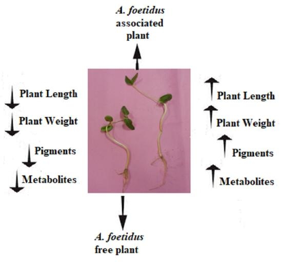
2.2. Early Assessment of the Potent Fungi Using Oryza sativa

The potency of fungal strain was initially checked in O. sativa seedlings. Fresh biomass of the isolated fungal endophyte (AdR13) was added to a flask holding 50 mL of the Czapek broth. The flask was placed in a shaking-incubator at 28 °C for a week, and 120 rpm was maintained till the end of the experiment. After one week of incubation, the Czapek medium was filtered and the culture filtrate was collected for further use. O. sativa variety Fakhr-e-Malakand was taken and the collected culture filtrate (100 µL) was applied to the apex of the seedlings already flourishing in water-agar medium (0.8%). After a week of incubation, the growth attributes of the O. sativa seedlings (i.e., total chlorophyll, fresh and dry weights of root and shoot, and root and shoot length) were calculated. O. sativa seedlings from control treatments were treated with distilled water or Czapek broth [25].

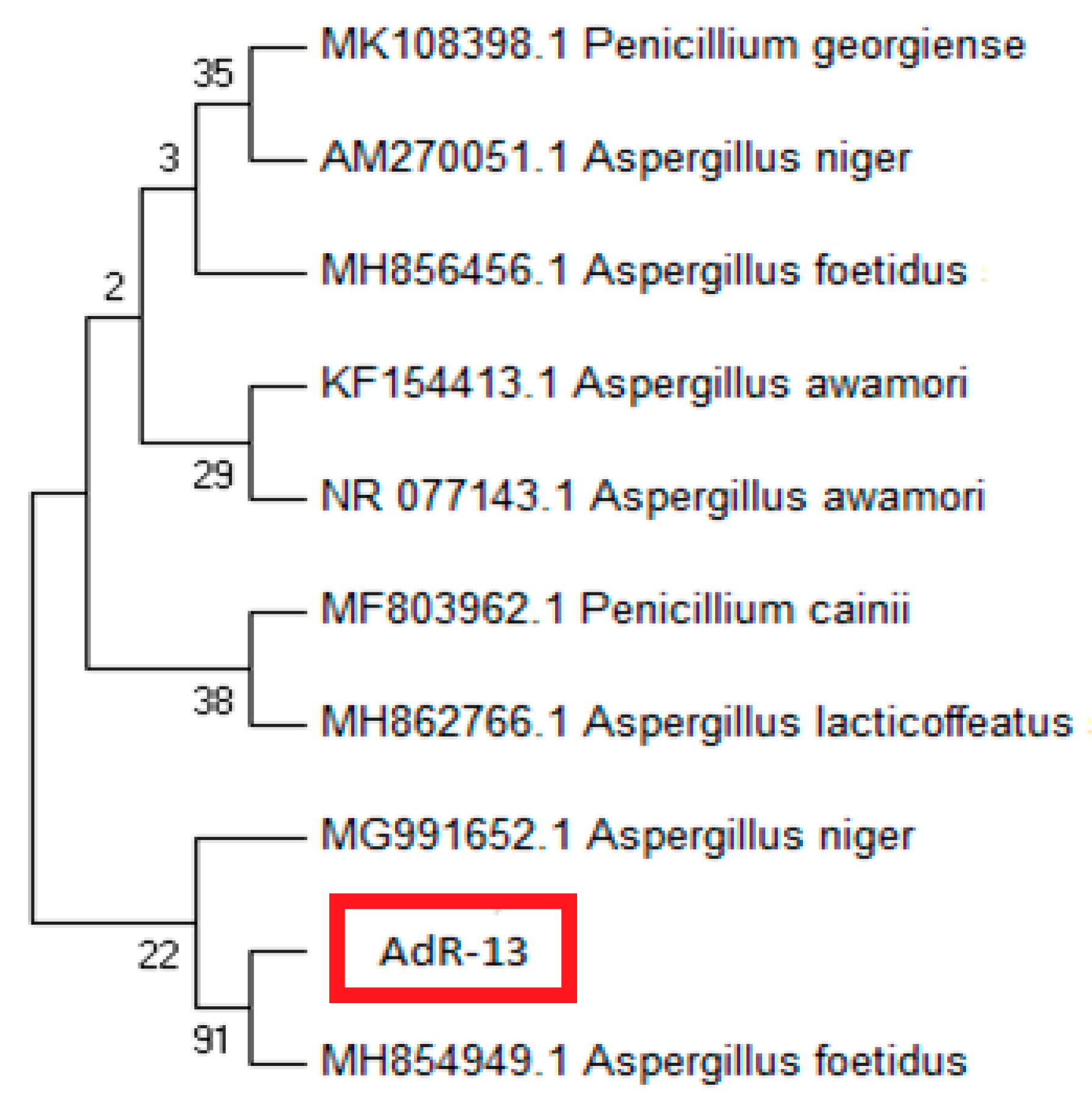
2.3. Identification of AdR13

The method of Khan et al. [26] with minor modification was adopted for the identification of AdR13 endophyte. The primers ITS1 (5′-TCC GTA GGT GAA CCT GCG G-3′) and ITS4 (5′-TCC TCC GCT TAT TGA TAT GC-3′) were utilised in the amplification of 18S rRNA. To find sequence homology approximation, the BLASTn1 program (National Institute of Health (NIH) Rockville Pike, Bethesda MD, USA) was used. The phylogenetic consensus tree was constructed through the neighbor joining (NJ) method using MEGA X software [27].
The method of Khan et al. [26] was adopted for the isolation of AdR13 DNA. Initially, a microfuge was taken and transferred 500 μL of 5% of sodium dodecyl sulfate in bead beating solution. The mycelium (200 mg) of AdR13 was then added to the microfuge tube and the contents of the tube were mixed by using vortex. suspended in the microfuge tube containing 500 μL of a bead beating solution. After mixing the contents of the microfuge tube, it was transferred to the centrifuge, where it was centrifuged at 4 °C and 11,000× g for 10 min. The supernatant (0.5 mL) was collected with great care and transferred to a fresh tube. To the supernatant, we added equal volumes of phenol: chloroform: isoamyl alcohol (25:24:1) and the contents were mixed with the help of the vortex. The vortexed sample was centrifuged again for 5 min at 10,000× g. Isopropanol (2.5 mL) was then added to the collected supernatant and the tube was transferred to the refrigerator, where it was kept for an hour. After incubation in the refrigerator the contents of the tube was centrifuged for 10 min at 14,000× g. The DNA was precipitated in the form of pellets. Cold ethanol (70%) was used to wash the pellet in order to wash out all the impurities. After air drying, 40 µL of Tris-ethylene diamine tetra acetic acid (TE) buffer was added to re-suspend the pellets and the DNA was quantified at 260 nm through Thermo Scientific Nano Drop spectrophotometer [27].

2.4. Inoculation of A. foetidus to G. max and H. annuus

Fresh fungal biomass (1 mg) was mixed with 100 g of autoclaved sand. After thorough mixing of fungal biomass with autoclaved sand, it was transferred to the pots. To each pot, 9 seeds of H. annuus or G. max were sown. The pots were moved to the growth chambers for 14 days. The temperature of the growth chambers was maintained at 25 °C or 40 °C. Hoagland solution (10 mL; Half strength) was employed to the plants at two day intervals until the end of the experiment. The growth attributes were finally measured after the termination of the experiment [28,29]. Lengths of plants were measured with a scale, whereas fresh weight of plants were measured with the help of an analytical balance. To measure the plant dry weight, the roots and shoot of the test species were initially dried in an oven operated at 105 °C ± 1 for 12 h. The dried weight was then measured using the analytical balance. A chlorophyll meter (Spad-502 plus, Konica Minolta, Tokyo, Japan) was used to compute the chlorophyll contents in the test crop species.

2.5. Estimation of Antioxidants

The protocol of Luck [30] was adopted to estimate the activity of catalase in test crop species, i.e., H. annuus and G. max. Leaves of the test crop species (2 g) were crushed in a 10 mL of phosphate buffer. The resultant homogenate was collected and centrifuged for 5 min at 10,000 rpm. The supernatant (40 µL) was carefully moved to a tube containing H2O2-phosphate buffer (3 mL) with the help of micropipette. After mixing the contents of the tube, absorbance (Abs. = 240 nm) was measured with a spectrophotometer. Estimation of the peroxidase activity (enzyme units/mg protein) was carried out by the method of Kar and Mishra [31]. Approximately, 20× diluted enzyme extract (1 mL) was mixed with 4 mL of 50 μmoles H2O2 + 50 μmoles pyrogallol + 125 μmoles of phosphate buffer (pH 6.8). Incubation of the mixture was done at 25 °C for 5 min. H2SO4 (5% v/v) was then added to stop the reaction and the concentration of purpurogallin was estimated with the help of spectrophotometer (Abs. = 420 nm). The activity of ascorbate oxidase was measured by a well established protocol [32]. Leaves of the test crop species (0.1 g) was crushed in 2 mL of phosphate buffer and the mixture was centrifuged at 3000× g for 5 min. The collected supernatant (100 µL) was mixed with a substrate solution (3 mL). The substrate solution comprised ascorbic acid (8.8. mg) and phosphate buffer of pH 5.6 (300 mL). The absorbance (Abs. = 265 nm) was observed for 5 min at intervals of 30 s with the help of the spectrophotometer. The superoxide dismutase activity in the test crops was calculated by the procedure of Beyer Jr and Fridovich [33]. Fresh leaves (0.5 g) of the test crop species were taken in a mortar and crushed with the help of pestle in a 1 mL of potassium phosphate (50 mM) + ethylenediaminetetraacetic acid (EDTA) (1 mM; pH 7.5). The resulting homogenate was transferred to centrifuge tubes for centrifugation. This step was done at 43,000× g and 4 °C for 15 min. The collected supernatant (50 µL) was then consumed to estimate the activity of SOD by mixing it with a reaction mixture (1 mM EDTA, 13 mM L-methionine, 75 µM nitro blue tetrazolium (NBT) in 50 mM potassium phosphate buffer (pH 7.8)). A 2 µM of riboflavin was then added to initiate the reaction. The SOD activity was measured as an increase in the optical density of nitroblue tetrazolium (NBT) after 1 min of the reaction under standard conditions at 560 nm. The amount of SOD was determined as enzyme units/mg protein. An enzyme unit is defined as the amount of protein consumed to cause 50% inhibition of NBT reduction. The method of Carlberg and Mannervik [34] was adopted to measure the activity of glutathione reductase. The GR activity was characterized by the reduction of glutathione (GSSG) by NADPH at an optical density of 340 nm. Initially, a reaction mixture was made, composed of NADPH (0.125 mmol/L), GSSG (1 mmol/L), EDTA (1 mmol/L) in potassium phosphate buffer (100 mmol/L; pH 7.0). Fresh leaves of the test crops were crushed in the mortar with help of pestle in the presence of liquid nitrogen, EDTA (1 mmol/L), Triton X-100 (0.1% v/v), dithiothreitol (2 mmol/L) in 100 mmol/L Tris-HCl (pH 8). The homogenate was centrifuged at 27,000× g to collect the supernatant. Added, 200 µL of the sample supernatant to 0.8 mL of the reaction mixture and the optical density was observed with the help of spectrophotometer (Abs. = 340 nm).

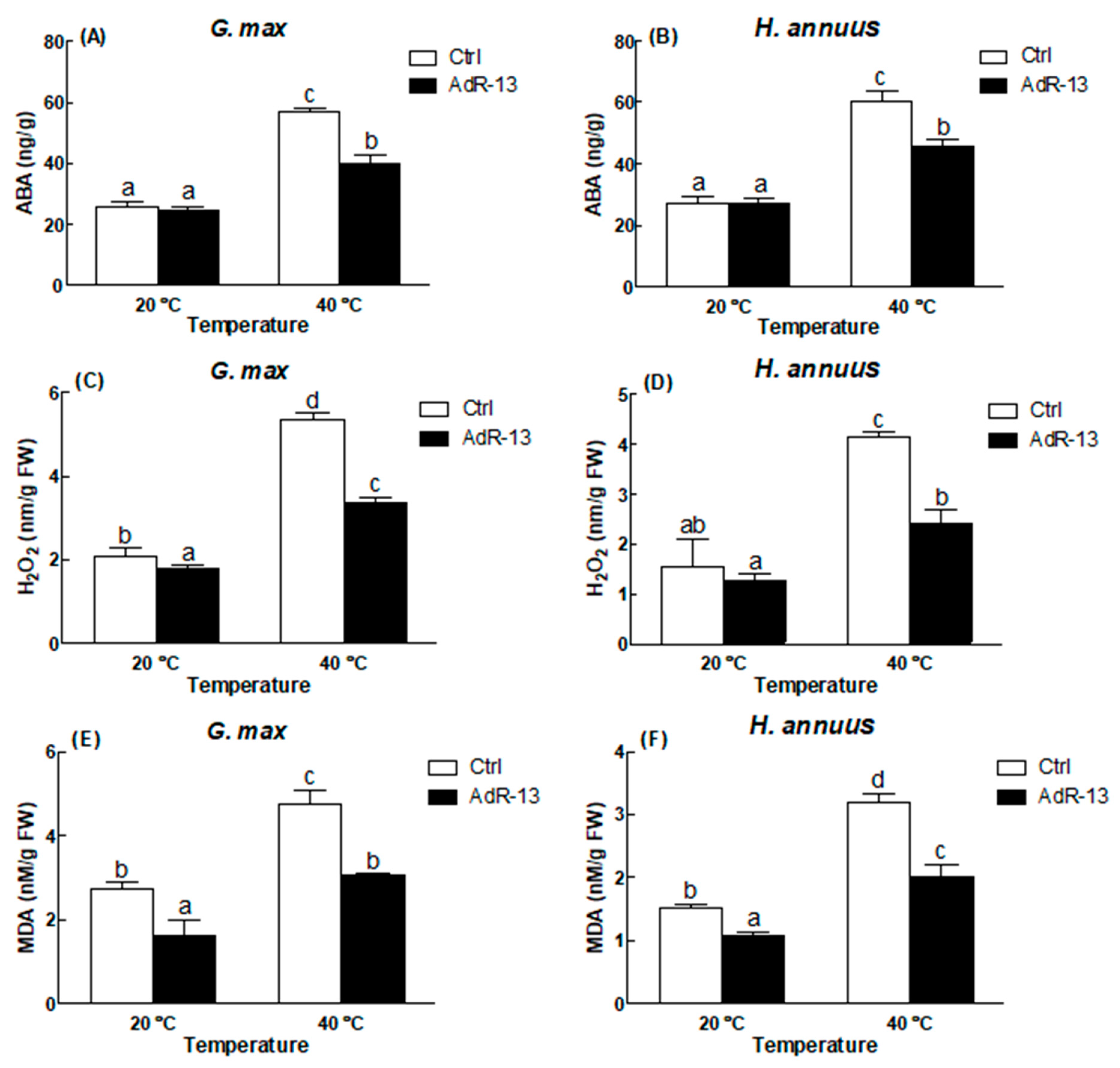
2.6. Estimation of ABA

Estimation of ABA in test crop species was undertaken according to the method of Yoon et al. [12]. Fresh leaves (0.5 g) of H. annuus and G. max were pulverized in liquid N2. The homogenate was then added to the 2 mL mixture, composed of isopropanol (1.5 mL) and glacial acetic acid (28.5 mL). The resultant mixture was concentrated in a rotary evaporator and then filtered. Diazomethane was added to the mixture and was examined via GC MS SIM (6890N setup GC Scheme furnished with 5973 System Mass Selective Detector; Agilent Technologies, Palo Alto, CA, USA). The Lab Base, Thermo Quset, Manchester, UK, Data System Software (DSS) was used to monitor retorts to ions with m/z standards of 190 and 162 for Me-ABA and 194 and 166 for Me-[2H6]-ABA. ABA ([2H6]-ABA) was applied as standard.

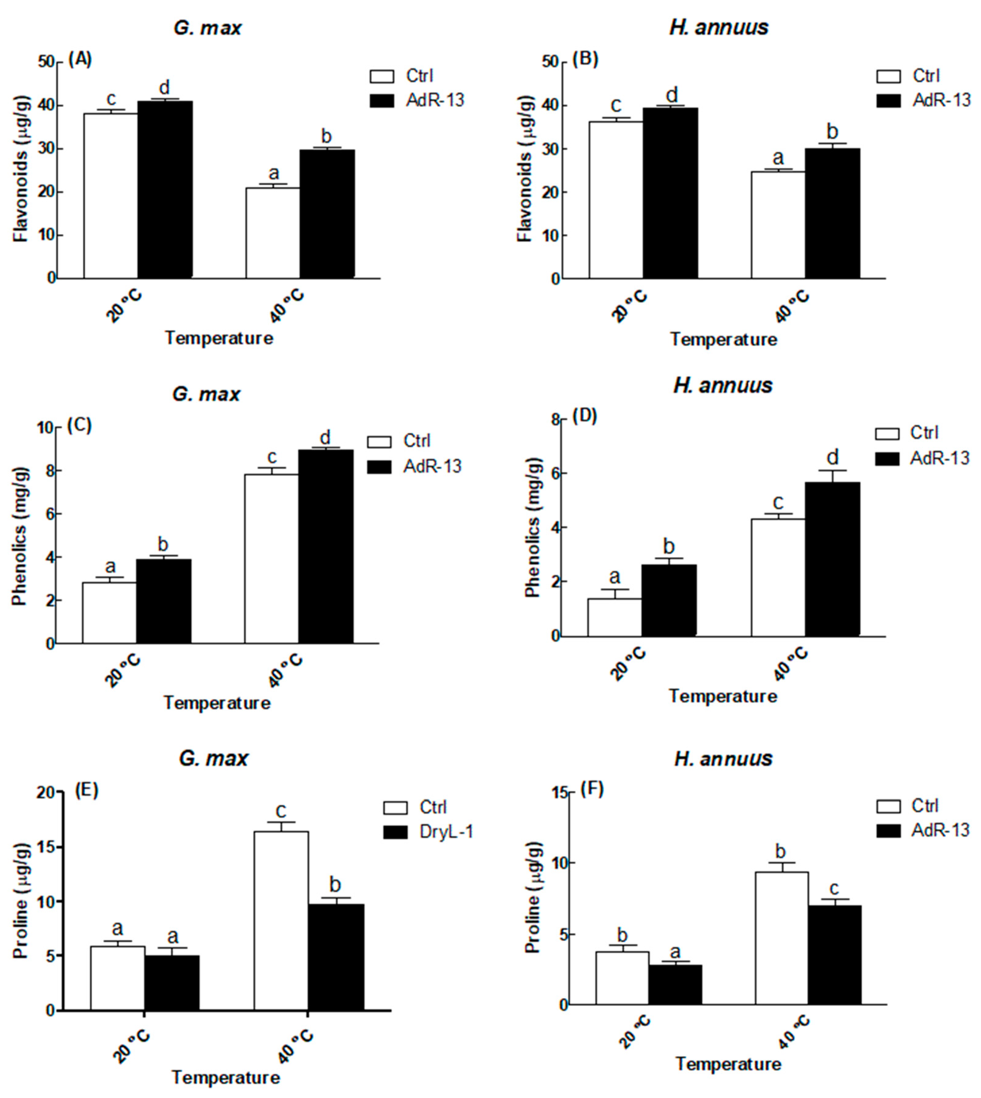
2.7. Estimation of Phenolics and Proline

Estimated total phenolics in test crop species were measured by the established protocol of Cai et al. [35]. To the sample (0.2 mL), 0.5 N of the Folin–Ciocalteu reagent was added and the mixture was kept for 4 min at 25 °C. Sodium carbonate (75 g/L) was then added and the contents were heated for 1 min at 100 °C. The heated mixture was transferred to the dark place and incubated for 2 h. After 2 h of incubation, the absorbance was measured at 650 nm. Gallic acid (Sigma Aldrich, Peshawar, Pakistan) was used as a standard. The concentration of proline in the test crops were estimated by an established method [36], but with minor modifications. Fresh leaves (0.1 g) of the test crops were pulverized in 3% of sulpho-salicylic acid (4 mL). The pulverized tissues in sulpho-salicylic acid was incubated for 24 h at 5 °C. After incubation, the homogenate was centrifuged at 3000 rpm for 5 min. A supernatant (2 mL) was mixed with acid ninhydrin (2 mL) and heated at 100 °C for 1 h. Toluene (4 mL) was finally added to the mixture and the absorbance was checked with the help of spectrophotometer (Abs. = 520 nm). Pure proline (Sigma Aldrich) was used as standard.

2.8. Estimation of Total Lipids, Proteins and Sugars

The protocol of Lowry et al. [37] was adopted to estimate the total proteins in the test crops. The filtered leaf homogenate of the test crops (0.1 mL) was added to 0.1 mL of NaOH (2 M) and then hydrolyzed at 100 °C. The hydrolysate was allowed to cool and then added to 1 mL of complex-forming solution (complex-forming solution = 1% (w/v) copper sulphate in distilled water, 2% (w/v) sodium carbonate in distilled water, 2% (w/v) sodium potassium tartrate in distilled water). The resultant mixture was allowed to stand for 10 min at room temperature. Folin reagent (1 mL) was added to the mixture and the solution was vortexed. The vortexed sample was again allowed to stand for 30 min at room temperature and the absorbance was observed with a spectrophotometer (Abs. = 650 nm). Bovine serum albumin (Sigma Aldrich) was used as a standard. The methodology of Van Handel [38] was adopted to estimate the total lipids in H. annuus and G. max seedlings. Leaves of the test crop species were crushed in 1 mL of 2% sodium sulfate. The homogenate was then centrifuged for 5 min at 11,000× g. About 1 mL of the supernatant was transferred to the tubes containing 1 mL chloroform/methanol (1:2 ratio v/v). The mixture was evaporated at 90 °C on a water bath until precipitates formed. The precipitates were then dissolved by using 2 mL of concentrated sulfuric acid. The mixture was heated for the second time at 90 °C for 20 min on a water bath. After cooling, 5 mL of vanillin-phosphoric acid was added and the mixture was left on a bench for half and hour for color development. The absorbance was ultimately recorded using spectrophotometer (Abs. = 525 nm). Pure canola oil was used as a standard. The well-established method of Mohammadkhani and Heidari [39] was adopted to estimate the soluble sugars in the leaves of test crops. Leaves (0.5 g) were crushed in a mortar and pestle in the presence of liquid nitrogen. The resultant homogenate was mixed with distilled water (5 mL) and the mixture was centrifuged for 5 min at 4000× g. Supernatant (0.1 mL) was collected in fresh tube and we added 1 mL of 80% phenol. The mixture was allowed to stand for 10 min at room temperature. After incubation, 5 mL of concentrated H2SO4 was added to the mixture and was then incubated at room temperature for an hour. The absorbance was finally observed at 485 nm. Glucose (Sigma Aldrich) was used as a standard.

2.9. Statistical Analysis

The data were analyzed by the analysis of variance. The significantly different means were separated by the Duncan multiple range test at p = 0.05, using SPSS-20 (SPSS Inc., Chicago, IL, USA). Moreover, the significant means were denoted by different letters as a superscript.

3. Results

3.1. Isolation and Plant Growth Promoting Activity of Endophytes

From the roots of A. capillus-veneris, potent endophytic fungi were isolated that were coded as AdR-13 before identification. The potency of the isolated stain was initially screened for growth promoting activity in O. sativa seedlings. The O. sativa seedlings inoculated with the endophytic strain (AdR-13) had longer roots and shoots as compared to the non-inoculated controls (Table 1). Similarly, the fresh and dry weight of the O. sativa seedlings inoculated with the endophytic strain (AdR-13) were higher when compared with the control seedlings. Higher chlorophyll contents were also noticed in the AdR-13 associated O. sativa seedlings (Table 1). After observing a positive impact of the isolated endophyte (AdR-13) on O. sativa, the strain was further utilized to check its growth-promoting activity in G. max and H. annuus exposed to 25 °C and 40 °C. The results revealed that the isolated AdR-13 strain has little effect on the chlorophyll contents, root and shoot lengths and weights of G. max exposed to 25 °C and 40 °C (Table 2). Similar observations were recorded in AdR-13 associated H. annuus exposed to 25 °C and 40 °C (Table 3).

3.2. Molecular Identification of Isolate AdR-13

The BLAST search program (Basic Local Alignment Search Tool, 2012) was used to align the nucleotides of AdR-13 isolate from the the Internal transcribed spacer (ITS) region for comparison. Maximum resemblance with A. foetidus was noticed by using 18 S rRNA sequence. From 21 taxa (20 reference and 1 clone), the phylogenetic consensus tree was constructed through the neighbor joining (NJ) method using MEGA X software (Figure 1). The isolated endophyte AdR-13 from A. capillus-veneris formed a clad with A. foetidus strengthened by 91% bootstrap value in the harmony tree. Sequence homology and phylogenetic analysis verified that A. foetidus was our isolate.

3.3. Arbitration of Enzymatic and Non-Enzymatic Antioxidants

The results regarding the enzymatic activity of various enzymes in G. max exposed to 25 °C and 40 °C are given in Table 4. The activity of AAO was noted higher in the A. foetidus associated G. max exposed to 40 °C temperature as compared to the other treatments. Similarly, the activity of other enzymes, i.e., CAT, POD, SOD and GR was recorded high in A. foetidus associated G. max exposed to 40 °C temperature (Table 4). By contrast, a non-significant difference (p = 0.05) in AAO activity was noticed in A. foetidus associated H. annuus compared to its respective control at 40 °C (Table 5). However, the activity of the CAT, POD, SOD and GR was significantly (p = 0.05) higher in A. foetidus associated H. annuus as compared to the A. foetidus free H. annuus exposed to 40 °C temperature (Table 5).
Figure 2 represents the results of flavonoids, phenolics and proline contents of the G. max and H. annuus (with or without A. foetidus association) exposed to 25 °C and 40 °C. A significant (p = 0.05) increase in the flavonoids contents was noticed in the A. foetidus associated G. max under 25 °C and 40 °C as compared to the controls (Figure 2A). The increase in flavonoid contents in A. foetidus associated G. max under 25 °C was 7.5%, whereas a 41% increase in flavonoid contents was noted in A. foetidus associated G. max under 40 °C as compared to their respective controls (Figure 2A). The value of flavonoids was increased by 8.9% and 22% in A. foetidus associated H. annuus at 25 °C and 40 °C, respectively (Figure 2B). In addition, higher amounts of phenolics (39%) were recorded in A. foetidus associated G. max under 25 °C, whereas 15% increase were recorded in A. foetidus associated G. max under 40 °C (Figure 2C). An almost doubled amount (91%) of phenolics was observed in A. foetidus associated H. annuus at 25 °C, while a 31% increase in phenolics was recorded in A. foetidus-associated H. annuus at 40 °C (Figure 2D). Conversely, a sizable decrease were noticed in the proline contents of A. foetidus associated G. max and H. annuus exposed to 25 °C and 40 °C. A 13% and 40% decrease in proline contents were recorded in A. foetidus-associated G. max at 25 °C and 40 °C, respectively, as compared to their respective controls (Figure 2E). Likewise, a decrease of 26% and 25% were noted in the A. foetidus-associated H. annuus as compared to their respective controls at 25 °C and 40 °C (Figure 2F).
Figure 3 presents the results of ABA, H2O2 and malondialdehyde (MDA) contents of the G. max and H. annuus (with or without A. foetidus association) exposed to 25 °C and 40 °C. A decrease in the amount of ABA (4% and 30%) was noticed in A. foetidus-associated G. max at 25 °C and 40 °C as compared to their respective controls; however, the decrease at 25 °C was non-significant (p = 0.05) (Figure 3A). Comparably, a 1% increase in ABA contents have been observed in A. foetidus-associated H. annuus at 25 °C, while a significant (p = 0.05) increase in ABA content (25%) was recorded in A. foetidus-associated H. annuus at 40 °C (Figure 3B). Additionally, lower amounts of H2O2 (14%) were recorded in A. foetidus-associated G. max under 25 °C, whereas a 37% decrease was recorded in A. foetidus-associated G. max under 40 °C (Figure 3C). A. foetidus-associated H. annuus at 25 °C showed 18% decrease in H2O2 contents, while 42% decrease were recorded in A. foetidus-associated H. annuus at 40 °C (Figure 3D). A 41% and 36% decrease in MDA contents were recorded in A. foetidus-associated G. max as compared to their respective controls at 25 °C and 40 °C, respectively (Figure 3E). Likewise, a decrease of 29% and 37% in MDA contents were noted in the A. foetidus-associated H. annuus as compared to their respective controls at 25 °C and 40 °C (Figure 3F).
Figure 4 represents the results of total proteins, sugars and lipids of the G. max and H. annuus (with or without A. foetidus association) exposed to 25 °C and 40 °C. An increase in the total protein (2% and 21%) was noticed in A. foetidus-associated G. max at 25 °C and 40 °C as compared to their respective controls; however, the increase at 25 °C was non-significant (p = 0.05) (Figure 4A). Similarly, a 6% increase in total proteins was observed in A. foetidus-associated H. annuus at 25 °C, while a significant (p = 0.05) increase in total proteins (33%) was recorded in A. foetidus-associated H. annuus at 40 °C (Figure 4B). Likewise, higher amounts of sugars (10%) were recorded in A. foetidus-associated G. max under 25 °C, whereas 25% increase were recorded in A. foetidus-associated G. max under 40 °C (Figure 4C). A. foetidus-associated H. annuus at 25 °C showed a 17% increase in total sugars, while the 26% increase in sugar content was recorded in A. foetidus-associated H. annuus at 40 °C (Figure 4D). An 11% and 21% increase in total lipids was recorded in A. foetidus-associated G. max as compared to their respective controls at 25 °C and 40 °C, respectively (Figure 4E). Likewise, an increase of 15% and 19% in total lipids was noted in the A. foetidus-associated H. annuus as compared to the respective controls at 25 °C and 40 °C (Figure 4F).

4. Discussion

Plant species when they undergo heat stress may defend themselves through biosynthetic responses, such as regulation of membrane lipids, re-adjustment of opening and closing of stomata, activation of antioxidant systems, and production of heat-shock-proteins [40]. Moreover, plants can establish a symbiotic relationship with endophytes, which can help the host plants under stress [21,41]. Endophytes can help the host plants under stress conditions by releasing higher amounts of alkaloids, flavonoids and phenolics [42,43]. By doing this, endophytes can improve the growth attributes and impart resistance to host plants against stress [13,44]. In this study, A. foetidus-associated test crops showed higher resistance against thermal stress than control seedlings. Culture filtrates of A. foetidus were initially assessed on O. sativa because of their prompt response to growth-promoting phytohormones, produced by endophytes [43]. Culture filtrates of A. foetidus profoundly encouraged the rice growth (length and weight of shoot and roots of host plant species) similar to the Gliocladium cibotii [11] and Penicillium glabrum [45]. Moreover, A. foetidus improved the plant growth parameters of G. max and H. annuus under thermal stress. A. foetidus inoculated G. max and H. annuus gained higher shoot and roots weight, longer roots and shoots, and higher chlorophyll contents as compared to the non-inoculated control plants. Comparable results have been recorded in previous reports, in which Gliocladium cibotii [11] and Penicillium glabrum [45] has supported the growth of G. max and H. annuus after exposure to 40 °C. A. foetidus association might accelerate the photosynthetic rate in G. max and H. annuus, which in turn helped in the promotion of growth attributes of the host plants [1,2]. Our results also confirmed the finding of of Sun et al. [46], who observed resistance in chlorophyll breakdown and sustenance in photosynthetic rate in Piriformospora indica-associated host plants under drought stress.
Plant can produce higher levels of phenolics against biotic and abiotic stresses to defend themselves [2]. The buildup of higher amounts of phenolics in A. foetidus-inoculated host plant species under 40 °C enabled the host to reduce the stocking of ROS, thus lowering the stress. A similar observation was made by Abd_Hallah et al. [6], who reported that endophytes can help the host plants to flourish under stress. Proline is an organic osmolyte that accumulates in plant species during stressful conditions, like heat or drought stress [47,48]. In our study, elevated amounts of proline were noticed in A. foetidus-inoculated G. max and H. annuus that might have a positive effect on buffering of cellular redox potential, enzymatic system, membrane stability and scavenging of free radicals during stress. In fact, proline can contribute towards the cytoplasmic acidosis and maintenance of NADP+/NADPH ratios [49]. Also, proline helps in delivering the stress-reducing agents in order to repair the stress-provoked damages and to produce ATP [50].
Our results demonstrated the role of endophytic fungi A. foetidus in associated G. max and H. annuus seedlings exposed to 40 °C. This role of A. foetidus in associated G. max and H. annuus seedlings might be linked with nutrients uptake and controlled antioxidant system and ROS declination [51]. During the stress, plant can protect themselves from the threatening effects of free radicals by regulating the activity of antioxidant enzymes (AAO, SOD, POD, GR, CAT) [6]. SOD is one of the vital antioxidant enzymes that can inhibit the Haber–Weiss reaction by producing hydroxyl radicals, thus scavenge superoxide radicals and H2O2. Extraordinary levels of POD might have a task of diminishing the lethal effect of stress through lignins biosynthesis [52]. CAT activity has a proven role in boosting the immunity of the plant species against the abiotic stress [53], which might be due to elimination of H2O2 [54]. AAO and GR have a vital role in the ascorbate–glutathione cycle, in which both of the enzymes can scavenge the ROS species [55]. In the current study, AAO and GR might have reduced the generation of superoxide radicals in order to protect the photosynthetic-electron transport chain under heat stress [55].
Abscisic acid is a well-known phytohormone that can be produced in plant species during stress. O. sativa was the first species in which higher productions of ABA was noticed under thermal stress [56]. The genes that are responsible for the production of ABA are upregulated, whereas the genes that are responsible for the breakdown of ABA are downregulated upon the exposure of plants to heat stress [57]. The results of the present study revealed lower amounts of ABA in A. foetidus-associated host plant species (G. max and H. annuus) exposed to 40 °C as compared to A. foetidus free plants. The presence of ABA in lower amounts in A. foetidus crop species (G. max and H. annuus) can be attributed to the ABA synthesis genes that were downregulated or the ABA digesting genes that were upregulated. It is also possible that lower amounts of ABA might result in A. foetidus-associated plants due to the involvement of GAs [2,23,29]. Furthermore, higher amounts of plants MDA under stressful conditions points towards the lipid peroxidation. In this study, the A. foetidus-associated G. max and H. annuus had lower levels of MDA as compared to the non-inoculated plants exposed to 40 °C temperature. The accumulation of abnormal amounts of MDA can trigger membrane desolation, whereas plant species associated with endophytes can keep the MDA levels under check [45]. Previously, low levels of MDA (27.5%) have been noticed in chickpea inoculated with Bacillus subtilis BERA 71 and exposed to salt stress [6]. We also found that A. foetidus can ease the effects of heat stress by dropping the concentration of H2O2 and MDA. Moreover, heat stress has lifted the hydrogen peroxide levels in plants that ultimately imitate membrane structural integrity and guided the fast lipids peroxidation in non-inoculated host plant species [1]. Higher amounts of total lipids, proteins and sugars were noticed in A. foetidus-associated G. max and H. annuus exposed to 40 °C temperature. These findings reflect the positive role of A. foetidus, reinforcing the case for its use as a bio-fertilizer. This type of stress is among the crucial constraints in the production of agricultural crops such as G. max and H. annuus.

5. Conclusions

A. foetidus enhanced the growth parameters of G. max and H. annuus exposed to thermal stress. Also, A. foetidus helped the host plants to scavenge ROS and elevation of AAO, POD, CAT, GR and SOD activity. From the results we concluded that A. foetidus can be one of the best choices for use as a bio-fertilizer or as a thermal stress-relieving bio-agent in future crop production.

Author Contributions

I., A.A., S.G., S.A.K. and H.-Y.K. designed and performed all the experiments. A.I. and A.H. analyzed the data and wrote the manuscript. M.H. and I.-J.L. supervised the research. A.I., M.H. and I.-J.L. edited the manuscript and arranged the resources for the work. All authors have read and agreed to the published version of the manuscript.

Funding

This research was supported by the Basic Science Research Program through the National Research Foundation of Korea (NRF) funded by the Ministry of Education (2017R1D1A1B04035601).

Institutional Review Board Statement

Not applicable.

Informed Consent Statement

Not applicable.

Data Availability Statement

All the data are included in the manuscript.

Acknowledgments

The authors are thankful to the laboratory team of Abdul Wali Khan Mardan and Kyungpook National University, Korea for providing the lab facilities.

Conflicts of Interest

The authors declare that there is no competing interest of any nature related to this manuscript.

References

  1. Ismail; Hamayun, M.; Hussain, A.; Afzal Khan, S.; Iqbal, A.; Lee, I.-J. Aspergillus flavus promoted the growth of soybean and sunflower seedlings at elevated temperature. Biomed Res. Int. 2019, 2019, 1295457. [Google Scholar] [CrossRef] [PubMed] [Green Version]
  2. Ismail; Hamayun, M.; Hussain, A.; Iqbal, A.; Khan, S.A.; Lee, I.-J. Endophytic fungus Aspergillus japonicus mediates host plant growth under normal and heat stress conditions. Biomed Res. Int. 2018, 2018, 7696831. [Google Scholar] [CrossRef] [PubMed]
  3. Ismail; Anwar, H.; Mehmood, A.; Qadir, M.; Husna; Iqbal, A.; Hamayun, M.; Khan, N. Thermal stress alleviating potential of endophytic fungus Rhizopus oryzae inoculated to sunflower (Helianthus annuus L.) And soybean (Glycine max L.). Pak. J. Bot. 2020, 52, 1–5. [Google Scholar] [CrossRef]
  4. Zahid, K.R.; Ali, F.; Shah, F.; Younas, M.; Shah, T.; Shahwar, D.; Hassan, W.; Ahmad, Z.; Qi, C.; Lu, Y.; et al. Response and tolerance mechanism of cotton Gossypium hirsutum L. to elevated temperature stress: A review. Front. Plant Sci. 2016, 7, 937. [Google Scholar] [CrossRef] [Green Version]
  5. Ashraf, M.; Harris, P. Potential biochemical indicators of salinity tolerance in plants. Plant. Sci. 2004, 166, 3–16. [Google Scholar] [CrossRef]
  6. Abd_Hallah, E.F.; Alqarawi, A.A.; Hashem, A.; Radhakrishnan, R.; Al-Huqail, A.A.; Al-Otibi, F.O.N.; Malik, J.A.; Alharbi, R.I.; Egamberdieva, D. Endophytic bacterium Bacillus subtilis (BERA 71) improves salt tolerance in chickpea plants by regulating the plant defense mechanisms. J. Plant Interact. 2018, 13, 37–44. [Google Scholar] [CrossRef] [Green Version]
  7. Mhamdi, A.; Queval, G.; Chaouch, S.; Vanderauwera, S.; Van Breusegem, F.; Noctor, G. Catalase function in plants: A focus on Arabidopsis mutants as stress-mimic models. J. Exp. Bot. 2010, 61, 4197–4220. [Google Scholar] [CrossRef] [Green Version]
  8. Abd_Hallah, E.F.; Hashem, A.; Alqarawi, A.A.; Bahkali, A.H.; Alwhibi, M.S. Enhancing growth performance and systemic acquired resistance of medicinal plant Sesbania sesban (L.) Merr using arbuscular mycorrhizal fungi under salt stress. Saudi J. Biol. Sci. 2015, 22, 274–283. [Google Scholar] [CrossRef] [PubMed]
  9. Gul Jan, F.; Hamayun, M.; Hussain, A.; Iqbal, A.; Jan, G.; Khan, S.A.; Khan, H.; Lee, I.-J. A promising growth promoting Meyerozyma caribbica from Solanum xanthocarpum alleviated stress in maize plants. Biosci. Rep. 2019, 39, 1–15. [Google Scholar]
  10. Raid, A.; Humaira, G.; Hamayun, M.; Mamoona, R.; Amjad, I.; Mohib, S.; Anwar, H.; Hamida, B.; In-Jung, L. Aspergillus awamori ameliorates the physicochemical characteristics and mineral profile of mung bean under salt stress. Chem. Biol. Technol. Agric. 2021, 8, 1–13. [Google Scholar]
  11. Ismail; Hamayun, M.; Hussain, A.; Iqbal, A.; Khan, S.A.; Khan, M.A.; Lee, I.-J. An Endophytic Fungus Gliocladium cibotii Regulates Metabolic and Antioxidant System of Glycine max and Helianthus annuus under Heat Stress. Pol. J. Environ. Stud. 2021, 30, 1631–1640. [Google Scholar] [CrossRef]
  12. Yoon, J.Y.; Hamayun, M.; Lee, S.-K.; Lee, I.-J. Methyl jasmonate alleviated salinity stress in soybean. J. Crop. Sci. Biotechnol. 2009, 12, 63–68. [Google Scholar] [CrossRef]
  13. Muhammad, I.; Niaz, A.; Gul, J.; Amjad, I.; Hamayun, M.; Farzana, G.J.; Anwar, H.; In-Jung, L. Trichoderma reesei improved the nutrition status of wheat crop under salt stress. J. Plant Interact. 2019, 14, 590–602. [Google Scholar]
  14. Khushdil, F.; Jan, F.G.; Jan, G.; Hamayun, M.; Iqbal, A.; Hussain, A.; Bibi, N. Salt stress alleviation in Pennisetum glaucum through secondary metabolites modulation by Aspergillus terreus. Plant Physiol. Biochem. 2019, 144, 127–134. [Google Scholar] [CrossRef] [PubMed]
  15. Nusrat, B.; Gul, J.; Farzana, G.J.; Hamayun, M.; Amjad, I.; Anwar, H.; Hazir, R.; Abdul, T.; Faiza, K. Cochliobolus sp. acts as a biochemical modulator to alleviate salinity stress in okra plants. Plant Physiol. Biochem. 2019, 139, 459–469. [Google Scholar]
  16. Ashraf, M.; Foolad, M. Roles of glycine betaine and proline in improving plant abiotic stress resistance. Environ. Exp. Bot. 2007, 59, 206–216. [Google Scholar] [CrossRef]
  17. Škorić, D. Sunflower breeding for resistance to abiotic stresses/mejoramiento de girasol por resistencia a estreses abióticos/sélection du tournesol pour la résistance aux stress abiotiques. Helia 2009, 32, 1–16. [Google Scholar]
  18. Van der Merwe, R.; Labuschagne, M.T.; Herselman, L.; Hugo, A. Effect of heat stress on seed yield components and oil composition in high-and mid-oleic sunflower hybrids. S. Afr. J. Plant. Soil 2015, 32, 121–128. [Google Scholar] [CrossRef]
  19. Alsajri, F.A.; Singh, B.; Wijewardana, C.; Irby, J.; Gao, W.; Reddy, K.R. Evaluating soybean cultivars for low-and high-temperature tolerance during the seedling growth stage. Agronomy 2019, 9, 13. [Google Scholar] [CrossRef] [Green Version]
  20. Mehmood, A.; Hussain, A.; Irshad, M.; Hamayun, M.; Iqbal, A.; Khan, N. In vitro production of IAA by endophytic fungus Aspergillus awamori and its growth promoting activities in Zea mays. Symbiosis 2019, 77, 225–235. [Google Scholar] [CrossRef]
  21. Mehmood, A.; Hussain, A.; Irshad, M.; Hamayun, M.; Iqbal, A.; Rahman, H.; Tawab, A.; Ahmad, A.; Ayaz, S. Cinnamic acid as an inhibitor of growth, flavonoids exudation and endophytic fungus colonization in maize root. Plant Physiol. Biochem. 2019, 135, 61–68. [Google Scholar] [CrossRef]
  22. Kang, S.-M.; Hamayun, M.; Khan, M.A.; Iqbal, A.; Lee, I.-J. Bacillus subtilis JW1 enhances plant growth and nutrient uptake of Chinese cabbage through gibberellins secretion. J. Appl. Bot. Food Qual. 2019, 92, 172–178. [Google Scholar]
  23. Ismail; Hamayun, M.; Anwar, H.; Sumera Afzal, K.; Amjad, I.; In-Jung, L. An endophytic fungus Aspergillus violaceofuscus can be used as heat stress adaptive tool for Glycine max L. and Helianthus annuus L. J. Appl. Bot. Food Qual. 2020, 93, 112–120. [Google Scholar]
  24. Benizri, E.; Courtade, A.; Picard, C.; Guckert, A. Role of maize root exudates in the production of auxins by Pseudomonas fluorescens M. 3.1. Soil Biol. Biochem. 1998, 30, 1481–1484. [Google Scholar] [CrossRef]
  25. Warrier, R.; Paul, M.; Vineetha, M. Estimation of salicylic acid in Eucalyptus leaves using spectrophotometric methods. Genet. Plant. Physiol. 2013, 3, 90–97. [Google Scholar]
  26. Khan, S.A.; Hamayun, M.; Yoon, H.; Kim, H.-Y.; Suh, S.-J.; Hwang, S.-K.; Kim, J.-M.; Lee, I.-J.; Choo, Y.-S.; Yoon, U.-H. Plant growth promotion and Penicillium citrinum. BMC Microbiol. 2008, 8, 231. [Google Scholar] [CrossRef] [PubMed] [Green Version]
  27. Chen, W.-p.; Kuo, T.-T. A simple and rapid method for the preparation of gram-negative bacterial genomic DNA. Nucleic Acids Res. 1993, 21, 2260. [Google Scholar] [CrossRef] [PubMed]
  28. Misra, N.; Dwivedi, U. Genotypic difference in salinity tolerance of green gram cultivars. Plant. Sci. 2004, 166, 1135–1142. [Google Scholar] [CrossRef]
  29. Ismail; Hamayun, M.; Hussain, A.; Afzal Khan, S.; Iqbal, A.; Lee, I.-J. Aspergillus niger boosted heat stress tolerance in sunflower and soybean via regulating their metabolic and antioxidant system. J. Plant Interact. 2020, 15, 223–232. [Google Scholar] [CrossRef]
  30. Luck, H. Methods in Enzymatic Analysis, 2nd ed.; Academic Press: New York, NY, USA, 1974. [Google Scholar]
  31. Kar, M.; Mishra, D. Catalase, peroxidase, and polyphenoloxidase activities during rice leaf senescence. Plant Physiol. 1976, 57, 315–319. [Google Scholar] [CrossRef] [Green Version]
  32. Oberbacher, M.; Vines, H. Spectrophotometric assay of ascorbic acid oxidase. Nature 1963, 197, 1203–1204. [Google Scholar] [CrossRef]
  33. Beyer, W.F., Jr.; Fridovich, I. Assaying for superoxide dismutase activity: Some large consequences of minor changes in conditions. Anal. Biochem. 1987, 161, 559–566. [Google Scholar] [CrossRef]
  34. Carlberg, I.; Mannervik, B. Glutathione reductase. In Methods Enzymol; Elsevier: Amsterdam, The Netherlands, 1985; Volume 113, pp. 484–490. [Google Scholar]
  35. Cai, Y.; Luo, Q.; Sun, M.; Corke, H. Antioxidant activity and phenolic compounds of 112 traditional Chinese medicinal plants associated with anticancer. Life Sci. 2004, 74, 2157–2184. [Google Scholar] [CrossRef]
  36. Bates, L.S.; Waldren, R.P.; Teare, I. Rapid determination of free proline for water-stress studies. Plant Soil 1973, 39, 205–207. [Google Scholar] [CrossRef]
  37. Lowry, O.H.; Rosebrough, N.J.; Farr, A.L.; Randall, R.J. Protein measurement with the Folin phenol reagent. J. Biol. Chem. 1951, 193, 265–275. [Google Scholar] [CrossRef]
  38. Van Handel, E. Rapid determination of total lipids in mosquitoes. J. Am. Mosq. Control Assoc. 1985, 1, 302–304. [Google Scholar] [PubMed]
  39. Mohammadkhani, N.; Heidari, R. Drought-induced accumulation of soluble sugars and proline in two maize varieties. World Appl. Sci. J. 2008, 3, 448–453. [Google Scholar]
  40. Rodriguez, R.J.; Redman, R.S.; Henson, J.M. The role of fungal symbioses in the adaptation of plants to high stress environments. Mitig. Adapt. Strateg. Glob. Chang. 2004, 9, 261–272. [Google Scholar] [CrossRef]
  41. Hamayun, M.; Hussain, A.; Khan, S.A.; Kim, H.-Y.; Khan, A.L.; Waqas, M.; Irshad, M.; Iqbal, A.; Rehman, G.; Jan, S. Gibberellins producing endophytic fungus Porostereum spadiceum AGH786 rescues growth of salt affected soybean. Front. Microbiol. 2017, 8, 1–13. [Google Scholar] [CrossRef] [PubMed] [Green Version]
  42. Bilal, L.; Asaf, S.; Hamayun, M.; Gul, H.; Iqbal, A.; Ullah, I.; Lee, I.-J.; Hussain, A. Plant growth promoting endophytic fungi Asprgillus fumigatus TS1 and Fusarium proliferatum BRL1 produce gibberellins and regulates plant endogenous hormones. Symbiosis 2018, 76, 117–127. [Google Scholar] [CrossRef]
  43. Hamayun, M.; Hussain, A.; Khan, S.A.; Irshad, M.; Khan, A.L.; Waqas, M.; Shahzad, R.; Iqbal, A.; Ullah, N.; Rehman, G. Kinetin modulates physio-hormonal attributes and isoflavone contents of Soybean grown under salinity stress. Front. Plant Sci. 2015, 6, 377. [Google Scholar] [CrossRef] [PubMed] [Green Version]
  44. Hussain, A.; Hamayun, M.; Rahman, H.; Iqbal, A.; Shah, M.; Irshad, M.; Qasim, M.; Islam, B. Bioremediation of hexavalent chromium by endophytic fungi; safe and improved production of Lactuca sativa L. Chemosphere 2018, 211, 653–663. [Google Scholar]
  45. Ismail; Hamayun, M.; Hussain, A.; Iqbal, A.; Khan, S.A.; Gul, S.; Khan, H.; Rehman, K.U.; Bibi, H.; Lee, I.-J. Penicillium Glabrum acted as a heat stress relieving endophyte in soybean and sunflower. Pol. J. Environ. Stud. 2021, 30, 3099–3110. [Google Scholar] [CrossRef]
  46. Sun, C.; Johnson, J.M.; Cai, D.; Sherameti, I.; Oelmüller, R.; Lou, B. Piriformospora indica confers drought tolerance in Chinese cabbage leaves by stimulating antioxidant enzymes, the expression of drought-related genes and the plastid-localized CAS protein. J. Plant Physiol. 2010, 167, 1009–1017. [Google Scholar] [CrossRef]
  47. Ilyas, N.; Mumtaz, K.; Akhtar, N.; Yasmin, H.; Sayyed, R.; Khan, W.; Enshasy, H.A.E.; Dailin, D.J.; Elsayed, E.A.; Ali, Z. Exopolysaccharides producing bacteria for the amelioration of drought stress in wheat. Sustainability 2020, 12, 8876. [Google Scholar] [CrossRef]
  48. Khan, I.; Awan, S.; Ikram, R.; Rizwan, M.; Akhtar, N.; Yasmin, H.; Sayyed, R.; Shafaqat, A.; Ilyas, N. Effect of 24-Epibrassinolide regulated antioxidants and osmolyte defense and endogenous hormones in two wheat varieties under drought stress. Physiol. Planta 2020, 172, 696–706. [Google Scholar] [CrossRef]
  49. Hare, P.; Cress, W. Metabolic implications of stress-induced proline accumulation in plants. Plant Growth Regul. 1997, 21, 79–102. [Google Scholar] [CrossRef]
  50. Hare, P.D.; Cress, W.A.; Van Staden, J. Dissecting the roles of osmolyte accumulation during stress. Plant Cell Environ. 1998, 21, 535–553. [Google Scholar] [CrossRef]
  51. Chang, J.; Wang, Y.; Shao, L.; Laberge, R.-M.; Demaria, M.; Campisi, J.; Janakiraman, K.; Sharpless, N.E.; Ding, S.; Feng, W. Clearance of senescent cells by ABT263 rejuvenates aged hematopoietic stem cells in mice. Nat. Med. 2016, 22, 78. [Google Scholar] [CrossRef] [Green Version]
  52. Boerjan, W.; Ralph, J.; Baucher, M. Lignin biosynthesis. Annu. Rev. Plant Biol. 2003, 54, 519–546. [Google Scholar] [CrossRef]
  53. Mittal, S.; Kumari, N.; Sharma, V. Differential response of salt stress on Brassica juncea: Photosynthetic performance, pigment, proline, D1 and antioxidant enzymes. Plant Physiol. Biochem. 2012, 54, 17–26. [Google Scholar] [CrossRef]
  54. Bienert, G.P.; Chaumont, F. Aquaporin-facilitated transmembrane diffusion of hydrogen peroxide. Biochim. Biophys. Acta Biochim. Biophys. Acta 2014, 1840, 1596–1604. [Google Scholar] [CrossRef] [PubMed]
  55. Noctor, G.; Foyer, C.H. Simultaneous measurement of foliar glutathione, γ-glutamylcysteine, and amino acids by high-performance liquid chromatography: Comparison with two other assay methods for glutathione. Anal. Biochem. 1998, 264, 98–110. [Google Scholar] [CrossRef] [PubMed]
  56. Raghavendra, A.S.; Gonugunta, V.K.; Christmann, A.; Grill, E. ABA perception and signalling. Trends Plant Sci. 2010, 15, 395–401. [Google Scholar] [CrossRef] [PubMed]
  57. Toh, S.; Imamura, A.; Watanabe, A.; Nakabayashi, K.; Okamoto, M.; Jikumaru, Y.; Hanada, A.; Aso, Y.; Ishiyama, K.; Tamura, N. High temperature-induced abscisic acid biosynthesis and its role in the inhibition of gibberellin action in Arabidopsis seeds. Plant Physiol. 2008, 146, 1368–1385. [Google Scholar] [CrossRef] [Green Version]
Figure 1. Identification of fungal isolate AdR-13 with 91% bootstrap value as Aspergillus foetidus using the neighbor joining (NJ) method.
Sustainability 13 07159 g001
Figure 2. (A) Flavonoid content in G. max, (B) flavonoid content in H. annuus, (C) phenolics content in G. max, (D) phenolics content in H. annuus, (E) proline content in G. max and (F) proline content in H. annuus inoculated with and without A. foetidus. Data are the mean of 3 replicates with standard error. Bars that are represented with different letters (i.e., a–d) are significantly different (p = 0.05) as estimated by Duncan’s multiple range test.
Sustainability 13 07159 g002
Figure 3. (A) ABA content in G. max, (B) ABA content in H. annuus, (C) H2O2 content in G. max, (D) H2O2 content in H. annuus, (E) MDA content in G. max and (F) MDA content in H. annuus inoculated with and without A. foetidus. Data are the mean of 3 replicates with standard error. Bars that are represented with different letters (i.e., a–d) are significantly different (p = 0.05) as estimated by Duncan’s multiple range test.
Sustainability 13 07159 g003
Figure 4. (A) Protein (ptn) content in G. max, (B) protein (ptn) content in H. annuus, (C) sugars content in G. max, (D) sugars content in H. annuus, (E) lipids content in G. max and (F) lipids content in H. annuus inoculated with A. foetidus. Data are the mean of 3 replicates with standard error. Bars that are represented with different letters (i.e., a–d) are significantly different (p = 0.05) as estimated by Duncan’s multiple range test.
Sustainability 13 07159 g004
Table 1. Effect of A. foetidus filtrate on the growth of O. sativa seedlings.
Growth AttributesCtrl (DW)Ctrl (Czk)A. foetidus
SL (cm)10.4 ± 0.8 a10.6 ± 1.5 a15 ± 0.6 b
RL (cm)4.9 ± 0.8 a6.3 ± 0.3 a8 ± 0.6 b
SFW (g)0.03 ± 0.0002 a0.0317 ± 0.0003 a0.0409 ± 0.0002 b
RFW (g)0.08 ± 0.007 a0.082 ± 0.006 a0.1031 ± 0.005 b
SDW (g)0.0047 ± 0.0003 a0.0043 ± 0.0003 a0.0063 ± 0.0009 b
RDW (g)0.0137 ± 0.0009 a0.015 ± 0.0001 a0.020 ± 0.0006 b
Chlorophyll content (SPAD)18.9 ± 1.3 a21.4 ± 0.3 a23.4 ± 0.7 b
SL = shoot length; RL = root length; SFW = fresh weight of shoots; RFW = fresh weight of roots; SDW = dry weight of shoots; RDW = dry weight of roots; Ctrl = control; Czk = Czapek medium, DW = distilled water. Data are mean of 3 replicates with standard error. Data that is followed by different letter (i.e., a,b) is significantly different (p = 0.05) as estimated by Duncan’s multiple range test.
Table 2. Effect of A. foetidus on the growth features of G. max.
Growth Attributes25 °C40 °C
CtrlA. foetidusControlA. foetidus
SL (cm)39 ± 1.7 b40 ± 0.6 b26 ± 0.9 a27 ± 2.0 a
RL (cm)13 ± 1.4 a,b14 ± 1.4 b10 ± 0.6 a11 ± 0.3 a,b
SFW (g)1.12 ± 0.02 a1.47 ± 0.20 a0.82 ± 0.04 a1.19 ± 0.06 a
RFW (g)0.18 ± 0.02 a0.25 ± 0.09 a0.13 ± 0.02 a0.16 ± 0.02 a
SDW(g)0.14 ± 0.012 b0.14 ± 0.0051 b0.08 ± 0.001 a0.11 ± 0.009 a
RDW (g)0.075 ± 0.002 b0.078 ± 0.003 b0.01 ± 0.001 a0.012 ± 0.001 a
Chlorophyll (SPAD)31 ± 0.2 a33 ± 1.6 a29 ± 2.3 a32 ± 3.1 a
Effect of A. foetidus on G. max seedlings, isolated from A. capillus-veneris L. SL = shoot length; RL = root length; SFW = fresh weight of shoots; RFW = fresh weight of roots; SDW = dry weight of shoots; RDW = dry weight of roots; Ctrl = control. Data are mean of 3 replicates with standard error. Data that is followed by different letter (i.e., a,b) is significantly different (p = 0.05) as estimated by Duncan’s multiple range test.
Table 3. Effect of A. foetidus on the growth features of H. annuus.
Growth Attributes/Temperature Stress25 °C40 °C
CtrlA. foetidusCtrlA. foetidus
SL (cm)23.7 ± 0.9 bc25.9 ± 0.9 c20.5 ± 0.4 a22.5 ± 0.5 ab
RL (cm)9.3 ± 0.6 b10.9 ± 0.5 b6.2 ± 0.6 a6.7 ± 0.9 a
SFW (g)1.22 ± 0.09 a1.32 ± 0.14 a0.82 ± 0.31 a0.93 ± 0.22 a
RFW (g)0.13± 0.021 b,c0.14 ± 0.084 c0.08 ± 0.074 a0.09 ± 0.006 a,b
SDW (g)0.08 ± 0.024 a,b0.09 ± 0.001 b0.04 ± 0.0001 a0.06 ± 0.009 a,b
RDW (g)0.024 ± 0.001 b0.032 ± 0.001 c0.014 ± 0.001 a0.016 ± 0.002 a
Chlorophyll (SPAD)40 ± 4.7 a45.6 ± 2.6 a38.4 ± 1.9 a39 ± 1.3 a
Effect of A. foetidus on H. annuus seedlings, isolated from A. capillus-veneris L. SL = shoot length; RL = root length; SFW = fresh weight of shoots; RFW = fresh weight of roots; SDW = dry weight of shoots; RDW = dry weight of roots; ctrl = control. Data are mean of 3 replicates with standard error. Data that is followed by different letter (i.e., a,b,c) is significantly different (p = 0.05) as estimated by Duncan’s multiple range test.
Table 4. Effect of A. foetidus on the activity of antioxidant enzymes in G. max.
Growth Attributes25 °C40 °C
CtrlA. foetidusCtrlA. foetidus
AAO (EU/mg ptn)1.51 ± 0.09 a1.61 ± 0.07 b1.85 ± 0.12 c2.04 + 0.14 d
CAT (EU/mg ptn)0.46 ± 0.02 a0.58 ± 0.04 b0.95 ± 0.41 c1.04 ± 0.17 d
POD (EU/mg ptn)1.42 ± 0.03 a1.64 + 0.11 a3.90 ± 0.22 b5.01 ± 0.64 c
SOD (EU/mg ptn)18 ± 0.64 a19 ± 0.55 a37 ± 0.73 b43 ± 1.34 c
GR (EU/mg ptn)1.54 ± 0.61 a0.16 ± 0.13 a1.86 ± 0.05 b2.09 ± 0.11 c
Effect of A. foetidus on G. max seedlings, isolated from A. capillus-veneris. AAO = ascorbic acid oxidase; CAT = catalase; POD = peroxidase, SOD = superoxide dismutase; GR = glutathione reductase; EU = enzyme unit; ptn = protein; ctrl = control. Data are mean of 3 replicates with standard error. Data that is followed by different letter (i.e., a,b,c,d) is significantly different (p = 0.05) as estimated by Duncan’s multiple range test.
Table 5. Effect of A. foetidus on the activity of antioxidant enzymes in H. annuus.
Growth Attributes25 °C40 °C
CtrlA. foetidusCtrlA. foetidus
AAO (EU/mg ptn)0.65 ± 0.06 a0.77 ± 0.05 b2.28 ± 0.36 c2.55 ± 0.28 c
CAT (EU/mg ptn)0.26 ± 0.03 a0.34 ± 0.03 b0.60 ± 0.43 c0.72 ± 0.04 d
POD (EU/mg ptn)1.11 ± 0.08 a1.37 ± 0.26 a2.73 ± 0.16 b3.78 ± 0.18 c
SOD (EU/mg ptn)12 ± 0.47 a13 ± 0.64 a22 ± 1.05 b26 ± 0.78 c
GR (EU/mg ptn)0.89 ± 0.05 a0.94 ± 0.04 a2.11 ± 0.19 b2.63 ± 0.13 c
Effect of A. foetidus on H. annuus seedlings, isolated from A. capillus-veneris. AAO = ascorbic acid oxidase; CAT = catalase; POD = peroxidase, SOD = superoxide dismutase; GR = glutathione reductase; EU = enzyme unit; ptn = protein; Ctrl = control. Data are mean of 3 replicates with standard error. Data that is followed by different letter (i.e., a,b,c,d) is significantly different (p = 0.05) as estimated by Duncan’s multiple range test.
Publisher’s Note: MDPI stays neutral with regard to jurisdictional claims in published maps and institutional affiliations.

