Article

Mass Spectrometry-Based Metabolomics of Agave Sap (Agave salmiana) after Its Inoculation with Microorganisms Isolated from Agave Sap Concentrate Selected to Enhance Anticancer Activity

by
Luis M. Figueroa
1,
Liliana Santos-Zea
1,
Adelfo Escalante
2 and
Janet A. Gutiérrez-Uribe
1,*
1
Tecnologico de Monterrey, Escuela de Ingeniería y Ciencias, Ave. Eugenio Garza Sada 2501, Col. Tecnológico, 64849 Monterrey, N.L., Mexico
2
Departamento de Ingeniería Celular y Biocatálisis, Instituto de Biotecnología, Universidad Nacional Autónoma de México (UNAM), Av. Universidad 2001, Col. Chamilpa, 62210 Cuernavaca, Mor., Mexico
*
Author to whom correspondence should be addressed.
Sustainability 2017, 9(11), 2095; https://0-doi-org.brum.beds.ac.uk/10.3390/su9112095
Submission received: 25 September 2017 / Revised: 4 November 2017 / Accepted: 4 November 2017 / Published: 16 November 2017

Abstract

:
Saponins have been correlated with the reduction of cancer cell growth and the apoptotic effect of agave sap concentrate. Empirical observations of this artisanal Mexican food have shown that fermentation occurs after agave sap is concentrated, but little is known about the microorganisms that survive after cooking, or their effects on saponins and other metabolites. The aim of this study was to evaluate the changes in metabolites found in agave (A. salmiana) sap after its fermentation with microorganisms isolated from agave sap concentrate, and demonstrate its potential use to enhance anticancer activity. Microorganisms were isolated by dilution plating and identified by 16S rRNA analysis. Isolates were used to ferment agave sap, and their corresponding butanolic extracts were compared with those that enhanced the cytotoxic activity on colon (Caco-2) and liver (Hep-G2) cancer cells. Metabolite changes were investigated by mass spectrometry-based metabolomics. Among 69 isolated microorganisms, the actinomycetes Arthrobacter globiformis and Gordonia sp. were used to analyze the metabolites, along with bioactivity changes. From the 939 ions that were mainly responsible for variation among fermented samples at 48 h, 96 h, and 192 h, four were correlated to anticancer activity. It was shown that magueyoside B, a kammogenin glycoside, was found at higher intensities in the samples fermented with Gordonia sp. that reduced Hep-G2 viability better than controls. These findings showed that microorganisms from agave sap concentrate change agave sap metabolites such as saponins. Butanolic extracts obtained after agave sap fermentation with Arthrobacter globiformis or Gordonia sp. increased the cancer cell growth inhibitory effect on colon or liver cancer cells, respectively.

1. Introduction

Agave sap concentrate is a food produced by thermal treatment of the fresh sap (“aguamiel”). Agave sap is composed of water, sugars (glucose, fructose, and sucrose), proteins, gums, and mineral salts [1]. It is obtained from the mature plant, which is between eight and 10 years old. Agave americana, A. salmiana, and A. atrovirens are agave species known as “maguey pulquero”, and used for production of “aguamiel” [2]. Regarding the concentrate, the high concentration of soluble solids (about 70%) confers stability to environmental conditions and resistance to microbial attack [3].
Saponins are the most commonly bioactive compounds found in Agave species, and they have shown biological activity against many types of cancer [4,5,6]. Saponins have been isolated mainly from the leaves of the plant [4], but recently they were reported in agave sap concentrate [5,6], and fresh agave sap [7]. These molecules were identified as glycosides of kammogenin, manogenin, gentrogenin, and hecogenin, and are the main source of variability among agave sap concentrates [6]. Particularly, a kammogenin glycoside identified as magueyoside B has been correlated with reduction in viability, as well as the induction of apoptosis in colon cancer cell lines [5,6].
Mass spectrometry (MS)-based metabolomics have been used as a platform for determining biochemical changes in food during processing, including fermentation [8,9,10]. In addition, the data obtained can be statistically processed using principal component analysis (PCA) to identify the most important biomarkers or informative metabolites in samples [9].
The high soluble solids content in the agave sap concentrate creates an environment that prevents the development of most microbial species. However, empirical observations of this artisanal Mexican food have shown that fermentation occurs after the agave sap is concentrated. The microbiota of agave sap and its fermented product, known as pulque, has been studied previously. Bacterial diversity in these products is dominated mainly by lactic acid bacteria and aerobic mesophiles belonging to α- and γ-proteobacteria [11]. However, little is known about the microorganisms that survive after cooking, or their effects on saponins and other metabolites. Additionally, environmental factors may affect the composition of the microbial community present in the soil of a particular site of cultivation, which could in turn affect the native microbial community found in the sap and its concentrate [12]. Therefore, the aim of this study was to evaluate the changes in metabolites found in agave sap (A. salmiana) after its fermentation with microorganisms isolated from agave sap concentrate. These microorganisms were selected based on a screening of viability reduction using agave sap butanolic extracts on colon (Caco-2) and liver cancer cells (Hep-G2).

2. Materials and Methods

2.1. Biological Material

Agave sap concentrate was provided by the local producer AGMEL S.A de C.V (Monterrey, Mexico), and stored at 4 °C until use for the isolation of microorganisms. Agave sap was obtained from A. salmiana plants grown in the state of Coahuila, Mexico. It was transported in dry ice and stored at −20 °C until use. Before use, the agave sap was autoclaved at 121 °C for 15 min.

2.2. Microorganism Isolation from Agave Sap Concentrate

Yeast Mold (YM, DifcoTM, Sparks, MD, USA), Potato Dextrose Agar (PDA, DifcoTM, Sparks, MD, USA), De Man, Rogosa and Sharpe (MRS, DifcoTM, Sparks, MD, USA), Reasoner’s 2A (R2A, Sigma-Aldrich, St. Louis, MO, USA) and NZ amine A media were used to isolate microorganisms from agave sap concentrate. NZ amine medium was prepared with NZ amine A (2 g/L) (Sigma-Aldrich, St. Louis, MO, USA) and Noble agar (15 g/L) (DifcoTM, Sparks, MD, USA). The rest of the media were prepared according to the instructions of the manufacturers. A culture broth was used for the preservation of isolates and to prepare the inoculum used in fermentations. This culture broth was composed of casein peptone (0.5 g/L), yeast extract (0.5 g/L), glucose (0.5 g/L), soluble starch (0.5 g/L), magnesium sulfate (0.024 g/L), and dibasic potassium phosphate (0.3 g/L).
For isolation, agave sap concentrate (10 g) was diluted with 90 mL of peptone (0.1%) and stirred for 30 min at 200 rpm. Dilutions from 10−1 to 10−6 were prepared, plated in Petri dishes, and incubated at 30 °C for seven days. MRS medium cultures were incubated at 37 °C. Culture media without inoculum were used as controls. For preservation, each colony was placed in culture broth supplemented with glycerol (20% v/v) and stored at −80 °C.

2.3. Experimental Strategy for the Selection of Microorganisms that Enhanced Agave Sap Anticancer Activity

For the first step, inoculums of each isolate were prepared and used to ferment sterile agave sap in a final volume of 3 mL. Each isolate was grown in 10 mL of culture broth, and incubated at 30 °C and 200 rpm for 96 h. These cultures were used to inoculate sterile agave sap, and were incubated individually at 30 °C and 200 rpm for 48 h, 96 h, or 192 h. Sterile agave sap without inoculum was incubated in the same conditions and used as control. Fermentations were performed by triplicate.
Butanolic extracts of fermentations were prepared and fractioned by solid phase extraction (SPE) to obtain saponin-rich extracts. These saponin-rich extracts were tested on liver (Hep-G2, ATCC® HB-8065™) and colon (Caco-2, ATCC® HTB-37™) cancer cell lines. According to the results of the biological assays, five isolates were selected and identified by 16S rRNA gene sequence analysis. Two of them were used to scale up to a final volume of 40 mL under the same fermentation conditions. Saponin-rich extracts were obtained and tested on liver and colon cancer cell lines. The effect on cancer cell lines was contrasted to that observed in murine fibroblasts (NIH-3T3, ATCC® CRL-1658™), which were used as the non-cancer control. Detailed procedures are described below.

2.4. Saponin-Rich Extracts Preparation

After fermentation, saponin-rich extracts were obtained using butanol (1:1), as described by Leal-Diaz and colleagues [7]. Butanol was removed under vacuum at 40 °C in a Rocket evaporator (Genevac, Ltd., Ipswich, UK). Then, dry samples (120 mg) were dissolved in 20 mL of high-performance liquid chromatography (HPLC) grade water (BDH, Poole, UK), filtered through a 0.45 μm polytetrafluoroethylene (PTFE) syringe filter (Agilent Technologies, Santa Clara, CA, USA) and fractionated by SPE to obtain a saponin-rich fraction. Strata C18 (8B-S001-LEG, Phenomenex, Torrance, CA, USA) cartridges were washed twice with 20 mL of HPLC grade water, and then twice with 20 mL of an HPLC grade methanol:water (60:40 v/v) solution. Saponins were eluted with two volumes of 20 mL HPLC grade methanol (BDH, Poole, UK) and dried under vacuum. Dry samples were dissolved in 50% methanol in water (HPLC grade) and stored at −20 °C until biological assays and metabolomics analysis.

2.5. Biological Assays

Saponin-rich extracts were tested on Hep-G2 and Caco-2 cancer cell lines, as well as in murine fibroblasts (NIH-3T3) non-cancer cell line. Bioassays were performed as described by Antunes-Ricardo et al. [13], using a final extract concentration of 50 µg/mL in all assays. Dulbecco’s Modified Eagle Medium (DMEM, Thermo Fisher Scientific, Waltham, MA, USA) supplemented with 5% of fetal bovine serum was used as negative control. Assays were performed by triplicate.

2.6. Microorganism Identification

The bacterial chromosomal DNA of selected isolates was extracted with the UltraClean® extraction kit (Mo Bio Laboratories, Inc., Carlsbad, CA, USA), and DNA amplification was conducted as described by Escalante et al. [11], with slight modifications. Briefly, each sample of DNA extracted was used as template for 16S rRNA gene fragment amplification with the primers targeted to Eubacteria, Eu530F, and Eu1449R [11,14]. The protocol used for most of the isolates consisted of an initial denaturation step (95 °C for 5 min), followed by 30 cycles of denaturation at 95 °C for 1 min, primer annealing at 42 °C for 30 s, and elongation at 72 °C for 1 min, plus an additional 5 min cycle. For isolate 31, the temperature of primer annealing was 55 °C. PCR reaction was performed in a Gradient 96 Robocycler (Stratagene, La Jolla, CA, USA). Samples of chromosomal DNA and PCR products were analyzed by 1% agarose gel electrophoresis in TBE 1× buffer. The gel was stained with ethidium bromide, and the bands were visualized under UV illumination.
PCR products were purified using the GeneJET Purification Kit (Thermo Fisher Scientific, Waltham, MA, USA), and quantified in a 2000c NanoDrop (Thermo Fisher Scientific, Waltham, MA, USA). After quantitation, PCR products were sequenced in an automated DNA sequencer of 16 capillaries (Applied Biosystems, model 3130xl, Foster City, CA, USA) at the Sequencing Unit from the Institute of Biotechnology-Universidad Nacional Autonoma de Mexico (UNAM). Forward and reverse sequences were analyzed with Chromatogram Explorer Lite and assembled using the DNA Baser program (http://www.dnabaser.com/). The obtained sequences were submitted to the non-redundant nucleotide database at GeneBank using the Basic Local Alignment Search Tool (BLAST) program (www.ncbi.nlm.nih.gov) to identify the isolates. A multiple alignment of 16S rRNA gene sequences from the microorganisms isolated with those retrieved from the GeneBank database was performed using the ClustalW function contained in the MEGA7 program [15]. A distance matrix calculation of nucleotide substitution rates and a phylogenetic tree were constructed with the Kimura (two parameters) algorithm and the neighbor-joining method, respectively, which are also included in the program MEGA7 [15]. The sequences of microorganisms Sulfolobus acidocaldarius strain ATCC 33909 (NR_043400) and Escherichia coli strain EcSD4 (KC504012) were included as outgroups.

2.7. Metabolomics and Saponins Analysis by Mass Spectrometry

Only samples from scale up fermentations, performed by triplicate, were used for metabolomics analysis. Methanol (100%) fractions obtained from SPE were analyzed according to the chromatographic conditions described by Leal-Diaz et al. [7]. Samples were analyzed by a high-performance liquid chromatographic system coupled to a time-of-flight (TOF) mass spectrometer equipped with an electrospray source (ESI) (HPLC/ESI-MS/TOF). Separation was performed at 25 °C in a Zorbax Eclipse XDB-C18 column 4.6 × 150 mm with a guard column of the same material 12.5 mm × 4.6 mm (Agilent Technologies, Santa Clara, CA, USA). Samples were injected randomly. Blanks were included between samples to reduce noise and eliminate potential contaminants from the column.
Saponin quantitation was performed using a 1200 Series HPLC coupled to an evaporative light scattering detector (HPLC/ELSD) (Agilent Technologies, Santa Clara, CA, USA), as previously reported [7]. Saponin concentrations were obtained as protodioscin equivalents (PE) using a standard curve from 10 to 500 ppm. Concentrations were reported as μg PE/mg extract. For saponin characterization, extracted ion chromatograms were analyzed considering the exact masses reported for saponins and sapogenins, as well as their fragmentation patterns (±0.05 units) using the Analyst QS 1.1 software (Applied Biosystems) [6,7,16].
For metabolomic analysis, raw mass spectrometry data were processed using Mass Hunter A.02.00 software (Agilent Technologies, Santa Clara, CA, USA). Information about retention time, exact mass, m/z, and the abundance of all ions from each sample were analyzed. Data acquired from Mass Hunter were processed using Excel. First, all of the ions from blanks were summed using an Excel table. Ions of all the samples were considered to exclude ions of contaminants. Protodioscin standard and the characteristic peak of kammogenin found in all the samples were used as references to determine the reliability of the mass, and the retention times of ions between different blanks or samples. A safety of 20 s for the retention time and 40 ppm for the exact mass was considered, and sample ions with a signal/noise ratio above five were picked. Ions with the same mass and retention time than those found in blanks were subtracted from the samples and ions were aligned manually. A single list with all of the ions detected in the samples with their corresponding exact masses, m/z, and abundances was obtained. Finally, a two-dimensional data matrix (mass versus abundance) was generated for all of the samples. Data analysis was conducted using PCA to select the ions that generated more variability among samples. Metabolites that were related to the decrease in viability were characterized by their retention times and mass spectra and compared with previous literature reports [6,7,16], and the Metlin database [17].

2.8. Data Analysis

Results of bioassays were analyzed using a three-factor ANOVA (fermentation time, strain, and cell line). Saponin quantitation was analyzed using a two-factor ANOVA (fermentation time and strain). Comparisons were made using Tukey’s honest significant difference (HSD) tests, values of p < 0.05 were accepted as statistically significant. All statistical analyses were conducted using the JMP Version 12 software (SAS Institute Inc., Cary, NC, USA).

3. Results and Discussion

3.1. Microorganisms Isolation, Screening, and Identification

Agave sap microorganisms were mainly grown in R2A and NZ amine A media with 38 and 29 isolates, respectively. No growth was detected in the MRS medium. Only one isolate was detected in YM and PDA media, which was indicative that the presence of yeasts in the concentrate is limited. Fermentations conducted in a final volume of 3 mL showed that 15 of 69 isolates produced changes in agave sap that reduced cancer cell viability when their corresponding extracts were tested in vitro. Particularly, extracts obtained from 48 h agave sap fermentation using isolates 16, 28, 31, and 44 had a significant reduction in the viability of Caco-2 cells (Supplementary Materials Figure S1). In the case of Hep-G2 cells, isolates that produced extracts with the highest bioactivity were 4, 16, 28, and 38 (Supplementary Materials Figure S2).
The phylogenetic analysis of the 16S rRNA gene sequences revealed that isolates 16 and 31 formed a branch from the genus Pseudomonas and Roseomonas, respectively (Figure 1). Isolate 16 showed a closer identity with Roseomonas aerilata and Roseomonas pecuniae, whereas isolate 31 was more similar to Pseudomonas stutzeri and Pseudomonas xanthomarina. R. aerilata had been isolated from the air [18], whereas R. pecuniae had been isolated from the surface of a copper-alloy coin [19]. P. stutzeri had been isolated from diverse environments [20,21]. P. xanthomarina had been isolated from samples of sea squirts, and it was able to grow in 8% NaCl [22]. Isolate 4 was within the same branch as Arthrobacter globiformis (Figure 1). The genus Arthrobacter includes coryneform non-pathogenic bacteria isolated from environmental sources such as soil, water, and plant material. A. globiformis possesses the enzyme choline oxidase, which catalyzes the oxidation of choline to glycine betaine, a compound that prevents dehydration and plasmolysis in hyperosmotic environments [23]. A. globiformis has been included in the European Food and Feed Cultures Association (EFFCA) and the International Dairy Federation (IDF) inventory of microorganisms with documented history of use in human food [24]. Isolates 28 and 38 were placed in the same branch as Gordonia terrae and G. lacunae (Figure 1). The first strains of the genus Gordonia were detected as opportunistic pathogens from human patients, but other strains play a role in the bioremediation and biodegradation of xenobiotics [25]. Isolate 4 (A. globiformis), along with isolates 28 and 38 (Gordonia sp.), belong to the group of actinomycetes. Strains of A. globiformis have shown the ability to produce a compound with high antioxidant capacity [26]. The actinomycete G. lacunae exhibited antibiotic activity [25], whereas G. terrae was reported as a carotenoids producer [27]. Interestingly, none of the genera identified in this study had previously been reported in agave sap or pulque (fermented agave sap).

3.2. Metabolomics

Based on the screening results, the actinomycetes A. globiformis and Gordonia sp. (isolates 4 and 28, respectively) were selected for scaled up fermentations. Mass spectrometry data analysis showed the presence of 2913 ions in all of the samples. After analyzing the abundance of all of the ions, 939 ions were mainly responsible for the variability observed among the saponin-rich extracts obtained from 48 h, 96 h, or 192 h fermentations. According to the principal component 1, extracts from agave sap fermented with Gordonia sp. (isolate 28) were separated from those of control agave sap and fermented with A. globiformis (isolate 4) (Figure 2).
The 939 ions responsible for the variability between samples were numbered based on the ion abundance detected by MS. Ions 1–4 were correlated with the inhibitory activity on the cancer cell lines, and were further investigated for identification based on their fragmentation patterns and previous reports in the literature (Table 1). Ion 1 was tentatively identified as dehydro-phytosphingosine, a sphingolipid found as a structural component of the membrane in plants and yeasts [28]. Ions 2 and 4, along with other ions detected at the same retention time, were tentatively identified as kammogenin glycosides [6,7]. On the other hand, ion 3 corresponded to the kammogenin aglycone, which was previously found in agave sap [7].
Mass spectra analysis of compound 2 showed that two kammogenin glycosides were co-eluting. These saponins exhibited characteristic ions at m/z 1217.51 ([M + Na]+) and 1085.48 ([M + Na]+), and both were isolated previously from flowers of A. offoyana and identified as magueyoside A and magueyoside B, respectively [16]. Similarly, at the retention time of compound 4, two kammogenin glycosides were found. The saponin that showed the ion at m/z 1247.51 ([M + Na]+) was also previously detected in agave sap [7], and the ion at m/z 1115.51 ([M + Na]+) was similar but without the pentose residue. According to mass spectra and the Metlin database [17], compound 1 tentatively corresponded to dehydro-phytosphingosine. Dehydro-phytosphingosine belongs to the sphingolipids, a class of lipids found in all living organisms as essential structural membrane constituents. Some sphingolipids from plants have shown anti-inflammatory properties and reduced the activity of several signaling pathways [28]. Particularly, dehydro-phytosphingosine from maize seeds has showed to induce apoptosis in Caco-2 cells [29].

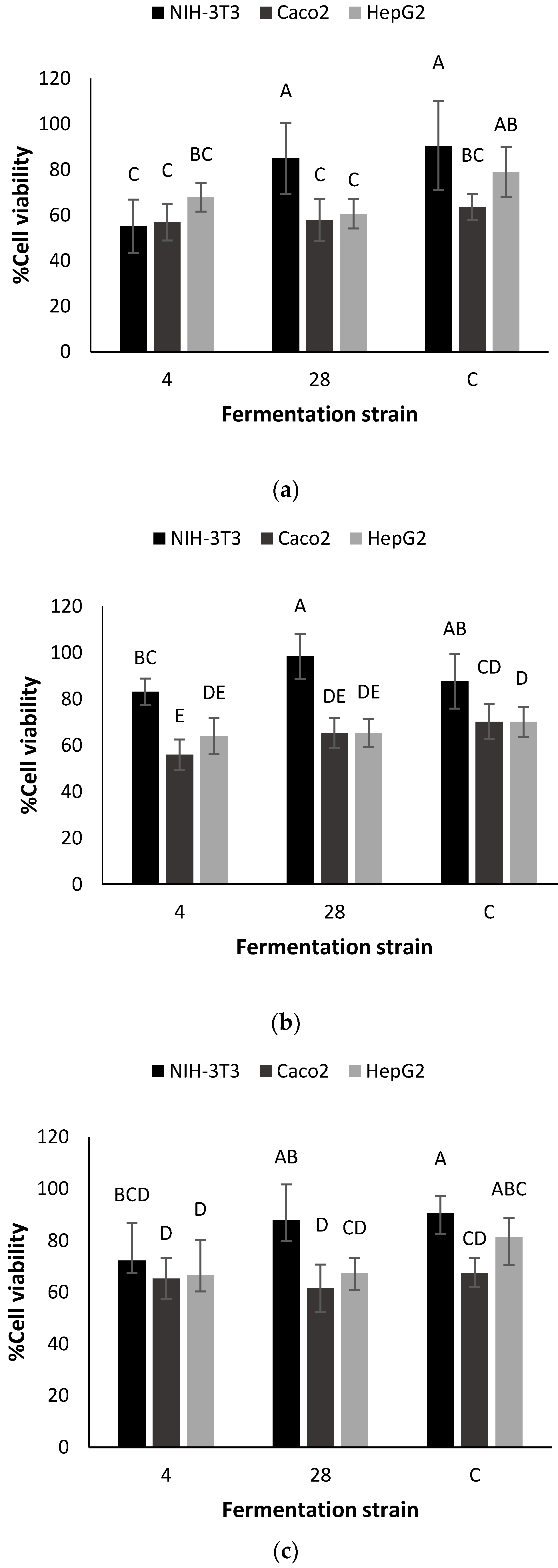
3.3. Saponins Analysis and Effects on Cancer Cell Viability

The extract from agave sap fermented for 48 h with A. globiformis (4) reduced the viability of mouse fibroblasts (NIH-3T3) in comparison with control, but the change on the viability of cancer cells was not significant (Figure 3a). The effect on Caco-2 cells viability for the sample obtained from the 48 h fermentation with Gordonia sp. (28) was similar to that obtained for control, but it reduced the viability of Hep-G2 cells from 78.9 % to 60.6%. At 48 h fermentation, A. globiformis (4) did not enhance the anticancer activity of agave sap butanolic extracts.
Extracts obtained after 96 h fermentation with A. globiformis (4) reduced the viability of Caco-2 cells from 70.2% to 56.0% (Figure 3b). NIH-3T3 cell viability was not negatively affected after the treatment with the butanolic extracts obtained after 96 h fermentation; only the sample obtained after 192 h fermentation with A. globiformis (4) reduced the viability of non-cancer cells (Figure 3c). This extract also reduced the hepatic cancer cells (Hep-G2) viability from 81.4% to 66.6%, and did not decrease the viability of Caco-2 cells in comparison with control (Figure 3c).
Therefore, considering cytotoxicity on NIH-3T3, Gordonia sp. (28) enhanced the anticancer activity of agave sap in Hep-G2 cells after 48 h fermentation. At 96 h, A. globiformis (4) enhanced the anticancer effect on Caco-2 cells; but 192 h fermentation with A. globiformis (4) would not be recommended to enhance the bioactivity of butanolic extracts, since it similarly affected Hep-G2 cells and NIH-3T3 cells.
Saponins derived from kammogenin, manogenin, gentrogenin, and hecogenin were detected in all of the extracts. Despite the correlation of ion abundance with inhibitory activity, no differences were found in the concentration of the most abundant saponins found in butanolic extracts (Table 2). Magueyosides A and B co-eluted, and were the most abundant saponins in all of the samples, which agrees with previous reports [5,6,7]. Based on the relative intensity of the extracted ion chromatograms for these compounds, there was an increase of magueyoside B concentration and a slight reduction of magueyoside A in the extracts obtained from Gordonia sp. fermentation (Figure 4A,B). These findings, in conjunction with the results of the bioassays, indicate that the inhibitory activity of the extracts depends not only on the total concentration of saponins, but also on the saponins’ profile; a method with higher resolution would be useful to quantitate them separately. Additionally, Magueyoside B has been correlated with a significant reduction in the viability of colon cancer cells (Caco-2) [6], and it has been shown to induce apoptosis in colon cancer cell line HT-29 [5].

4. Conclusions

Microorganisms that survive the thermal treatment of agave sap have the potential to modify agave sap metabolites such as saponins. Then, their anticancer enhancement effect depended on the fermentation time and the cancer cell line tested. Gordonia sp. enhanced the effect on Hep-G2 cells after 48 h fermentation. Agave sap fermentation after 96 h fermentation with A. globiformis increased the Caco-2 cancer cell growth inhibitory effect of the corresponding saponin-rich extract. It was shown that magueyoside B, a kammogenin glycoside, was found at higher intensities in the samples fermented with Gordonia sp. that reduced Hep-G2 viability better than controls. However, the fact that the concentration of these saponins was not affected after fermentation indicates that the anti-cancer activity depends not only on the concentration of total saponins, but also on the profile of these compounds within each sample. The changes in the saponins profile observed in this experiment demonstrate that it is important to characterize the microorganisms that survive thermal treatment of foods and affect the phytochemicals composition of traditional Mexican foods.

Supplementary Materials

The following are available online at www.mdpi.com/2071-1050/9/11/2095/s1, Figure S1: Viability of colon cancer cells (Caco-2) after treatment with extracts obtained after 48, 96 or 192 h of fermentation with isolates (50 µg/mL), Figure S2: Viability of liver cancer cells (HepG2) after treatment with extracts obtained after 48, 96 or 192 h of fermentation with isolates (50 µg/mL).

Acknowledgments

We appreciate financial support from CONACYT CVU 490590, NutriOmics Chair from Tecnológico de Monterrey, Nutrigenomics Research Chair from Fundación FEMSA, PAPIIT DGAPA project IN 207914 from IBT UNAM, as well as Carlos Rodríguez-López for his knowledge and assistance in metabolomics analysis and Albino Vargas from AGMEL S.A de C.V for providing agave sap and agave sap concentrate.

Author Contributions

Luis M. Figueroa carried out all the experiments related to microorganism isolation and identification, compiled and analyzed all the data, and drafted the manuscript. Liliana Santos-Zea obtained the data for saponin profile, collaborated in saponin identification and participated in the writing process. Janet A. Gutiérrez Uribe and Adelfo Escalante obtained the funding for the project and advised throughout the design and experimental phases of the study, as well as through the writing process.

Conflicts of Interest

The authors declare no conflict of interest.

References

  1. Lappe-Oliveras, P.; Moreno-Terrazas, R.; Arrizón-Gaviño, J.; Herrera-Suárez, T.; García-Mendoza, A.; Gschaedler-Mathis, A. Yeasts associated with the production of Mexican alcoholic nondistilled and distilled Agave beverages. FEMS Yeast Res. 2008, 8, 1037–1052. [Google Scholar] [CrossRef] [PubMed]
  2. Escalante, A.; López Soto, D.R.; Velázquez Gutiérrez, J.E.; Giles-Gómez, M.; Bolívar, F.; López-Munguía, A. Pulque, a traditional Mexican alcoholic fermented beverage: Historical, microbiological, and technical aspects. Front. Microbiol. 2016, 7, 1026. [Google Scholar] [CrossRef] [PubMed]
  3. Gutiérrez-Uribe, J.A.; Serna-Saldívar, S. Agave Syrup Extract Having Anticancer Activity. U.S. Patent 8,470,858, 25 June 2013. [Google Scholar]
  4. Chen, P.-Y.; Chen, C.-H.; Kuo, C.-C.; Lee, T.-H.; Kuo, Y.-H.; Lee, C.-K. Cytotoxic steroidal saponins from Agave Sisalana. Plant. Med. 2011, 77, 929–933. [Google Scholar] [CrossRef] [PubMed]
  5. Santos-Zea, L.; Fajardo-Ramírez, O.R.; Romo-López, I.; Gutiérrez-Uribe, J.A. Fast centrifugal partition chromatography fractionation of concentrated agave (Agave salmiana) sap to obtain saponins with apoptotic effect on colon cancer cells. Plant Foods Hum. Nutr. 2016, 71, 57–63. [Google Scholar] [CrossRef] [PubMed]
  6. Santos-Zea, L.; Rosas-Pérez, A.M.; Leal-Díaz, A.M.; Gutiérrez-Uribe, J.A. Variability in saponin content, cancer antiproliferative activity and physicochemical properties of concentrated agave sap. J. Food Sci. 2016, 81, H2069–H2075. [Google Scholar] [CrossRef] [PubMed]
  7. Leal-Díaz, A.M.; Santos-Zea, L.; Martínez-Escobedo, H.C.; Guajardo-Flores, D.; Gutiérrez-Uribe, J.A.; Serna-Saldivar, S.O. Effect of Agave americana and Agave salmiana ripeness on saponin content from aguamiel (agave sap). J. Agric. Food Chem. 2015, 63, 3924–3930. [Google Scholar] [CrossRef] [PubMed]
  8. Gao, P.; Xu, G. Mass-spectrometry-based microbial metabolomics: Recent developments and applications. Anal. Bioanal. Chem. 2015, 407, 669–680. [Google Scholar] [CrossRef] [PubMed]
  9. Wishart, D.S. Metabolomics: Applications to food science and nutrition research. Trends Food Sci. Technol. 2008, 19, 482–493. [Google Scholar] [CrossRef]
  10. Settachaimongkon, S.; Nout, M.J.R.; Antunes Fernandes, E.C.; Hettinga, K.A.; Vervoort, J.M.; van Hooijdonk, T.C.M.; Zwietering, M.H.; Smid, E.J.; van Valenberg, H.J.F. Influence of different proteolytic strains of Streptococcus thermophilus in co-culture with Lactobacillus delbrueckii subsp. bulgaricus on the metabolite profile of set-yoghurt. Int. J. Food Microbiol. 2014, 177, 29–36. [Google Scholar] [CrossRef] [PubMed]
  11. Escalante, A.; Giles-Gómez, M.; Hernández, G.; Córdova-Aguilar, M.S.; López-Munguía, A.; Gosset, G.; Bolívar, F. Analysis of bacterial community during the fermentation of pulque, a traditional Mexican alcoholic beverage, using a polyphasic approach. Int. J. Food Microbiol. 2008, 124, 126–134. [Google Scholar] [CrossRef] [PubMed]
  12. He, Y.; Xu, M.; Qi, Y.; Dong, Y.; He, X.; Li, J.; Liu, X.; Sun, L. Differential responses of soil microbial community to four-decade long grazing and cultivation in a semi-arid grassland. Sustainability 2017, 9, 128. [Google Scholar] [CrossRef]
  13. Antunes-Ricardo, M.; Moreno-García, B.E.; Gutiérrez-Uribe, J.A.; Aráiz-Hernández, D.; Alvarez, M.M.; Serna-Saldivar, S.O. Induction of apoptosis in colon cancer cells treated with isorhamnetin glycosides from Opuntia ficus-indica pads. Plant Foods Hum. Nutr. 2014, 69, 331–336. [Google Scholar] [CrossRef] [PubMed]
  14. Escalante, A.; Rodríguez, M.E.; Martínez, A.; López-Munguía, A.; Bolívar, F.; Gosset, G. Characterization of bacterial diversity in pulque, a traditional Mexican alcoholic fermented beverage, as determined by 16S rDNA analysis. FEMS Microbiol. Lett. 2004, 235, 273–279. [Google Scholar] [CrossRef] [PubMed]
  15. Kumar, S.; Stecher, G.; Tamura, K. MEGA7: Molecular volutionary Genetics Analysis Version 7.0 for bigger datasets. Mol. Biol. Evol. 2016, 33, 1870–1874. [Google Scholar] [CrossRef] [PubMed]
  16. Pérez, A.J.; Calle, J.M.; Simonet, A.M.; Guerra, J.O.; Stochmal, A.; Macías, F.A. Bioactive steroidal saponins from Agave offoyana flowers. Phytochemistry 2013, 95, 298–307. [Google Scholar] [CrossRef] [PubMed]
  17. METLIN: A metabolite Mass Spectral Database. Available online: http://metlin.scripps.edu (accesed on 1 May 2017).
  18. Yoo, S.-H.; Weon, H.-Y.; Noh, H.-J.; Hong, S.-B.; Lee, C.-M.; Kim, B.-Y.; Kwon, S.-W.; Go, S.-J. Roseomonas aerilata sp. nov., isolated from an air sample. Int. J. Syst. Evol. Microbiol. 2008, 58, 1482–1485. [Google Scholar] [CrossRef] [PubMed]
  19. Lopes, A.; Santo, C.E.; Grass, G.; Chung, A.P.; Morais, P.V. Roseomonas pecuniae sp. nov., isolated from the surface of a copper-alloy coin. Int. J. Syst. Evol. Microbiol. 2011, 61, 610–615. [Google Scholar] [CrossRef] [PubMed]
  20. Kuroda, M.; Notaguchi, E.; Sato, A.; Yoshioka, M.; Hasegawa, A.; Kagami, T.; Narita, T.; Yamashita, M.; Sei, K.; Soda, S.; et al. Characterization of Pseudomonas stutzeri NT-I capable of removing soluble selenium from the/aqueous phase under aerobic conditions. J. Biosci. Bioeng. 2011, 112, 259–264. [Google Scholar] [CrossRef] [PubMed]
  21. Zhang, J.; Cao, X.; Xin, Y.; Xue, S.; Zhang, W. Purification and characterization of a dehalogenase from Pseudomonas stutzeri DEH130 isolated from the marine sponge Hymeniacidon perlevis. World J. Microbiol. Biotechnol. 2013, 29, 1791–1799. [Google Scholar] [CrossRef] [PubMed]
  22. Romanenko, L.A.; Uchino, M.; Falsen, E.; Lysenko, A.M.; Zhukova, N.V.; Mikhailov, V.V. Pseudomonas xanthomarina sp. nov., a novel bacterium isolated from marine ascidian. J. Gen. Appl. Microbiol. 2005, 51, 65–71. [Google Scholar] [CrossRef] [PubMed]
  23. Fan, F.; Ghanem, M.; Gadda, G. Cloning, sequence analysis, and purification of choline oxidase from Arthrobacter globiformis: A bacterial enzyme involved in osmotic stress tolerance. Arch. Biochem. Biophys. 2004, 421, 149–158. [Google Scholar] [CrossRef] [PubMed]
  24. Mogensen, G.; Salminen, S.; O’Brien, J.; Ouwenhand, A.; Holzapfel, W.; Shortt, C.; Fonden, R.; Miller, G.D.; Donohue, D.; Playne, M.; et al. Inventory of microorganisms with a documented history of use in food. Bull. Int. Dairy Fed. 2002, 377, 10–19. [Google Scholar]
  25. Roes, M.; Goodwin, C.M.; Meyers, P.R. Gordonia lacunae sp. nov., isolated from an estuary. Syst. Appl. Microbiol. 2008, 31, 17–23. [Google Scholar] [CrossRef] [PubMed]
  26. Teng, W.H.; Sun, W.J.; Yu, B.; Cui, F.J.; Qian, J.Y.; Liu, J.Z.; Wang, L.; Qi, X.H.; Wei, H. Continuous conversion of rice starch hydrolysate to 2-keto-d-gluconic acid by Arthrobacter globiformis C224. Biotechnol. Bioprocess Eng. 2013, 18, 709–714. [Google Scholar] [CrossRef]
  27. Takaichi, S.; Maoka, T.; Akimoto, N.; Carmona, M.L.; Yamaoka, Y. Carotenoids in a Corynebacterineae, Gordonia terrae AIST-1: Carotenoid glucosyl mycoloyl esters. Biosci. Biotechnol. Biochem. 2008, 72, 2615–2622. [Google Scholar] [CrossRef] [PubMed]
  28. Canela, N.; Herrero, P.; Mariné, S.; Nadal, P.; Ras, M.R.; Rodríguez, M.Á.; Arola, L. Analytical methods in sphingolipidomics: Quantitative and profiling approaches in food analysis. J. Chromatogr. A 2016, 1428, 16–38. [Google Scholar] [CrossRef] [PubMed]
  29. Aida, K.; Kinoshita, M.; Susgawara, T.; Ono, J.; Miyazawa, T.; Ohnishi, M. Apoptosis Inducement by Plant and Fungus Sphingoid Bases in Human Colon Cancer Cells. J. Oleo Sci. 2004, 53, 503–510. [Google Scholar] [CrossRef]
Figure 1. Phylogenetic tree of 16S rDNA sequences from isolates and their closest neighbors from bacteria or environmental clones available in the National Center for Biotechnology Information (NCBI) database. The 16S rRNA gene sequence of Sulfolobus acidocaldarius served as outgroup. Identity percentage with closest reference to 16S rRNA gene clones in the database is indicated in parenthesis.
Sustainability 09 02095 g001
Figure 2. Principal Component Analysis (PCA) score plot of the 939 ions that were among those mainly responsible for the variability observed in the saponin-rich extracts obtained from 48 h, 96 h, or 192 h fermentations with A. globiformis (isolate 4), Gordonia sp. (isolate 28), and unfermented agave sap (C).
Sustainability 09 02095 g002
Figure 3. Effect of extract from agave sap fermented with A. globiformis (4) and Gordonia sp. (28) on three different cell lines at (a) 48 h; (b) 96 h; and (c) 192 h fermentation. Non-fermented controls (C) were included. All assays were performed at a final concentration of 50 µg/mL. Bars not connected with the same letter were significantly different.
Sustainability 09 02095 g003
Figure 4. (A) Comparison of extracted ion chromatograms for magueyoside A after 96 h of agave sap fermentation with A. globiformis or Gordonia sp; (B) Comparison of extracted ion chromatograms for magueyoside B after 96 h of agave sap fermentation with A. globiformis or Gordonia sp.
Sustainability 09 02095 g004
Table 1. Suggested identification of compounds found in agave sap fermented with Gordonia sp.
CompoundSuggested CompoundsAccurate Massm/z
1Dehydro-phytosphingosine315.27316.28 (M + H)+,
338.25 (M + Na)+
2Magueyoside A1194.48445.29 (M-3Hex-2Pen + H)+,
607.34 (M-2Hex-2Pen + H)+,
769.35 (M-1Hex-2Pen + H)+,
1217.51 (M + Na)+
Magueyoside B1062.44445.29 (M-3Hex-1Pen + H)+,
607.34 (M-2Hex-1Pen + H)+,
769.36 (M-1Hex-1Pen + H)+,
1085.48 (M + Na)+
3Kammogenin444.28445.29 (M + H)+,
467.25 (M + Na)+,
483.23 (M + K)+,
911.52 (2M + Na)+
4Kammogenin glycoside 1 (KG1)1092.44445.29 (M-4Hex + H)+,
607.34 (M-3Hex + H)+,
769.36 (M-2Hex + H)+,
931.40 (M-1Hex + H)+,
1115.51 (M + Na)+
Kammogenin glycoside 2 (KG2)1224.48445.29 (M-4Hex-1Pen + H)+,
607.34 (M-3Hex-1Pen + H)+,
769.36 (M-2Hex-1Pen + H)+,
1247.47 (M + Na)+
Table 2. Concentration of saponins in fermented and unfermented agave sap *.
StrainFermentation Time (h)MagA + MagB §KG1 + KG2
(µg PE/mg Extract)
A. globiformis482.99 ± 0.68 a2.05 ± 0.30 a,b
962.73 ± 0.58 a1.81 ± 0.32 a,b
1922.33 ± 0.71 a1.71 ± 0.57 a,b
Gordonia sp.482.71 ± 0.29 a2.04 ± 0.21 a,b
963.09 ± 0.65 a2.43 ± 0.67 a,b
1922.70 ± 1.01 a2.80 ± 0.71 a
Control483.56 ± 0.05 a2.51 ± 0.11 a,b
962.57 ± 0.39 a1.84 ± 0.20 a,b
1921.70 ± 0.59 a1.27 ± 0.41 b
§ Magueyoside A and magueyoside B; Kammogenin glycosides with m/z of 1115.51 (M + Na)+ and 1247.51 (M + Na)+; PE: protodioscin equivalents. * Mean values ± SD of replicate samples analyzed in triplicate. Mean values within the same column followed by different letters a to b are significantly different (p < 0.05).

