Review

Food Components and Dietary Habits: Keys for a Healthy Gut Microbiota Composition

by
Emanuele Rinninella
1,2,*,
Marco Cintoni
3,
Pauline Raoul
2,
Loris Riccardo Lopetuso
2,4,
Franco Scaldaferri
2,4,
Gabriele Pulcini
3,
Giacinto Abele Donato Miggiano
1,2,
Antonio Gasbarrini
2,4 and
Maria Cristina Mele
1,2
1
UOC di Nutrizione Clinica, Dipartimento di Scienze Gastroenterologiche, Endocrino‑Metaboliche e Nefro‑Urologiche, Fondazione Policlinico Universitario A. Gemelli IRCCS, Largo A. Gemelli 8, 00168 Rome, Italy
2
Istituto di Patologia Speciale Medica, Università Cattolica del Sacro Cuore, Largo F. Vito 1, 00168 Rome, Italy
3
Scuola di Specializzazione in Scienza dell’Alimentazione, Università di Roma Tor Vergata, Via Montpellier 1, 00133 Rome, Italy
4
UOC di Medicina Interna e Gastroenterologia, Dipartimento di Scienze Gastroenterologiche, Endocrino‑Metaboliche e Nefro‑Urologiche, Fondazione Policlinico Universitario A. Gemelli IRCCS, Largo A. Gemelli 8, 00168 Rome, Italy
*
Author to whom correspondence should be addressed.
Submission received: 21 June 2019 / Revised: 25 September 2019 / Accepted: 27 September 2019 / Published: 7 October 2019
(This article belongs to the Special Issue Nutrients 2009–2019: The Present and the Future of Nutrition)

Abstract

:
The gut microbiota is a changing ecosystem, containing trillions of bacteria, continuously shaped by many factors, such as dietary habits, seasonality, lifestyle, stress, antibiotics use, or diseases. A healthy host–microorganisms balance must be respected in order to optimally maintain the intestinal barrier and immune system functions and, consequently, prevent disease development. In the past several decades, the adoption of modern dietary habits has become a growing health concern, as it is strongly associated with obesity and related metabolic diseases, promoting inflammation and both structural and behavioral changes in gut microbiota. In this context, novel dietary strategies are emerging to prevent diseases and maintain health. However, the consequences of these different diets on gut microbiota modulation are still largely unknown, and could potentially lead to alterations of gut microbiota, intestinal barrier, and the immune system. The present review aimed to focus on the impact of single food components (macronutrients and micronutrients), salt, food additives, and different dietary habits (i.e., vegan and vegetarian, gluten-free, ketogenic, high sugar, low FODMAP, Western-type, and Mediterranean diets) on gut microbiota composition in order to define the optimal diet for a healthy modulation of gut microbiota.

1. Introduction

The human gastrointestinal (GI) tract harbors more than 100,000 billion microorganisms, representing 10–100 times the number of human cells [1]. Bacteria are classified according to phyla, classes, orders, families, genera, and species. Only a few phyla are represented in the gut, accounting for more than 160 species [2]. The dominant gut microbial phyla are Firmicutes, Bacteroidetes, Actinobacteria, Proteobacteria, Fusobacteria, and Verrucomicrobia, with the two phyla Firmicutes and Bacteroidetes [3] representing 90% of gut microbiota. The Firmicutes phylum is composed of more than 200 different genera, such as Lactobacillus, Bacillus, Clostridium, Enterococcus, and Ruminococcus. Clostridium genera represent 95% of the Firmicutes phyla. Bacteroidetes consists of predominant genera such as Bacteroides and Prevotella. The Actinobacteria phylum is proportionally less abundant and mainly represented by the Bifidobacterium genus. There is no singular optimal gut microbiota composition since it is different for each individual [4]. Indeed, the human gut microbiota is characterized by an inter-individual variability due to infant transitions, antibiotics use, as well as lifestyle, dietary, and cultural habits. A rich and diverse microbial community leads to a well-balanced and healthy gut microbiota composition.
In recent years, many studies have shown the close association between gut microbiota dysbiosis and numerous non-communicable diseases, such as cardiovascular diseases [5], obesity [6], diabetes [7], cancer [8], gastrointestinal diseases such as irritable bowel syndrome (IBS) [9], and neurological disorders [10,11,12]. Furthermore, the reciprocal interactions between gut microbiota and the brain, well-known as the “gut–brain axis”, have been demonstrated through bidirectional neuroendocrine signaling and immune activations [13], playing a key role in several neurological disorders such as Parkinson’s disease [14] and autism spectrum disorders [15]. Although it is not entirely known if changes in gut microbiota composition are a cause or consequence of a given disease, the association between the richness and diversity of gut microbiota and health has been demonstrated [4].
Diet is one of the key modulators of gut microbiota composition which directly influences host homeostasis and biological processes but also via metabolites derived from the microbial fermentation of nutrients—particularly short-chain fatty acids (SCFAs) [16]. This crucial mutualism between the human host and its bacterial symbionts can be altered through novel dietary habits, potentially impacting intestinal barrier functions and the immune system. The present review aimed to explain how food components and dietary choices can modulate gut microbiota composition and, consequently, intestinal barrier functions. First, we describe the interplay between gut microbiota, intestinal barrier, and the immune system. Secondly, after defining the interactions between food components and gut microbiota, we focus on the relationship between dietary habits and gut microbiota to define the optimal dietary habits to shape a healthy microbiota composition and to maintain host gut barrier and immune functions.

2. Gut Microbiota, Intestinal Barrier, and Immune System

A dynamic network orchestrates the interplay among the several components of the gastrointestinal tract, which is organized as a semipermeable multi-layer ecosystem [17,18,19,20]. A complex and mutualistic symbiosis regulates the relationship between the host and the gut microbiota [21,22,23]. This interplay is constantly challenged with numerous factors, and can lead to the collapse of the microbial community structure [17]. The intestinal barrier represents a functional unit responsible for two main tasks that are crucial for the survival of the individual: allowing nutrient absorption and defending the body from the penetration of unwanted, often dangerous, macromolecules. The gut mucosa is in fact a multi-layered system consisting of an external “anatomical” barrier and an inner “functional” immunological barrier [19]. Commensal gut microbiota, the mucous layer, and the intestinal epithelial monolayer constitute the anatomical barrier [21,22]. The deeper, inner layer consists of a complex network of immune cells organized in a specialized and compartmentalized system known as gut-associated lymphoid tissue (GALT). GALT represents both isolated and aggregated lymphoid follicles, and is one of the largest lymphoid organs, containing up to 70% of the body’s total number of immunocytes. Moreover, it is involved in the response to pathogenic microorganisms and provides immune tolerance to commensal bacteria. The ability of GALT to interact with luminal antigens rests on specific mucosal immune cells (i.e., dendritic cells and M-cells), primarily localized to Peyer’s patches within the ileum that are intimately positioned at the mucosa–environment interface and internalize microorganisms and macromolecules [17]. These specialized immune cells have the ability to present antigens to naïve T-lymphocytes, which subsequently produce cytokines and activate mucosal immune responses [23], when needed. Many factors such as alterations in the gut microflora, modifications of the mucus layer, and epithelial damage induced by the diet can alter this balance, leading to increased intestinal permeability and the translocation of luminal contents to the underlying mucosa [24]. The integrity of these structures is necessary for the maintenance of normal intestinal barrier function. The dysregulation of any of the aforementioned components has been implicated not only in the pathogenesis of IBD, but many other GI disorders, including infectious enterocolitis, irritable bowel syndrome, small intestinal bowel overgrowth, and allergic food intolerance [25,26,27]. In particular, several lines of evidence have shown that the microbial flora is critical for the development of a normal gut immune system, but can also play a central role in the development of IBD [28,29,30,31]. In support of this concept, the majority of genetically susceptible murine models of colitis do not develop significant inflammation when raised in a germ-free environment [32,33,34], while in others, diseases can be attenuated or completely abolished with antibiotic treatment [35,36]. In this context, innate immune responses that recognize conserved microbial products such as lipopolysaccharide (LPS) and peptidoglycan [37] are likely to be important in microbe–host interactions and intestinal homeostasis [38]. Critical to the host’s sensing of microbes are members of the toll-like receptor (TLR) family that, alone or in combination, recognize a wide array of microbe-associated molecular patterns (MAMPs) on either pathogenic or commensal microorganisms [39,40]. Furthermore, emerging lines of evidence indicate that intestinal homeostasis and inflammation are driven by cellular elements and soluble mediators that mediate both processes, with several cytokines exhibiting opposing roles depending upon the specific setting. In this context, all dietary components we introduce (or not introduce) may play a crucial role in shaping gut microbiota composition.

3. Interplays Between Food Components and Gut Microbiota

3.1. Carbohydrates and Gut Microbiota

3.1.1. Carbohydrates

Carbohydrates can be categorized into digestible and indigestible substrates. Digestible carbohydrates such as glucose, fructose, and galactose are enzymatically degraded in the small intestine and rapidly released as glucose in the bloodstream. Conversely, indigestible carbohydrates, also called “dietary fiber” are resistant to digestion in the small intestine, and reach the large intestine. Dietary fibers include non-starch polysaccharides, lignin, resistant starches, and non-digestible oligosaccharides [41]. Non-starch polysaccharides include cellulose and hemicellulose (glucans, gums, and pectin). Resistant starch is commonly found within whole or partly milled grains or seeds. Finally, non-digestible oligosaccharides consist of raffinose, stachyose, oligofructose, and inulin. Dietary fibers may be categorized according to their fermentability (fermentable or non-fermentable) in the colon or to their solubility (soluble or insoluble) in water. Fermentable dietary fibers such as inulin, pectin, beta-glucan, fructo-oligosaccharides (FOSs), and galactooligosaccharides (GOSs) are considered water-soluble in nature, while non-fermentable dietary fibers such as cellulose, hemicellulose, lignin, and resistant starch are considered insoluble [42]. Fermentable fibers are easily fermented by bacteria in the colon, while non-fermentable fibers are not [42].

3.1.2. SCFAs

Fermentable dietary fibers undergo saccharolytic fermentation, essentially under the action of gut bacteria, which in turn yield monosaccharides, SCFAs (i.e., butyrate (15%), acetate (60%), and propionate (25%)) and gases (i.e., methane and carbon dioxide) [43]. On the one hand, acetate and propionate are taken up by the liver through the portal vein where they are used as substrates for lipid, glucose, and cholesterol metabolism. Indeed, acetate is a precursor for cholesterol synthesis and lipogenesis while propionate is a gluconeogenic substrate [44]. On the other hand, butyrate plays a crucial role in maintaining tissue barrier function [45] and regulating gene expression and immunoregulation [46]. SCFAs are also involved in the colonic homeostasis, stimulating the proliferation and differentiation of epithelial cells, the absorption of salts and water, the maintenance of mucosal integrity, and decreasing inflammation [47,48]. Additionally, SCFAs can exert other beneficial effects, acting as histone deacetylase inhibitors, playing a crucial role in epigenetic regulation and acting as anti-cancer agents [16,49]; and increasing transit time and satiety by activating hormones such as glucagon-like peptide 1, peptide YY, and leptin via their endogenous receptors Free Fatty Acid Receptors 2 (FFAR2) and 3 (FFAR3) [50]. The types and amounts of SCFAs are mainly determined by the composition of intestinal microbiota and by how much carbohydrates are consumed [51]. Consequently, changes in the type and quantity of non-digestible carbohydrates in the human diet influence the bacterial populations detected in feces [52].

3.1.3. Prebiotics

Prebiotics are non-digestible (by the host) food ingredients that have a beneficial effect through their selective metabolism in the intestinal tract [53]. Microbiota-accessible carbohydrates (MACs) are carbohydrates that are resistant to digestion by a host’s metabolism, and are made available for gut microbiota as prebiotics to metabolize into SCFAs [54]. Sonnenburg et al. [54] demonstrated that a low-MAC diet leads to an increase of Bacteroides thetaiotaomicrondegrading intestinal mucus glycans. Indeed, these bacteria have the capacity to use host mucus glycans when dietary MACs are scarce, [24] leading to intestinal barrier thinning. Singh et al. [55] demonstrated that, in high-fat-fed male rats, inulin dose-dependently altered fecal microbiota by suppressing the number of Firmicutes (Roseburia, Clostridium clusters I, IV, and XIV) and promoting the number of Bifidobacterium spp. and Bacteroidetes. Vandeputte et al. [56] assessed the effects of inulin consumption on stool frequency in healthy adults with mild constipation, and detected specific inulin-induced changes in relative abundances of Anaerostipes, Bilophila, and Bifidobacterium. The observed decrease in Bilophila abundance following inulin consumption was associated with both softer stools and a favorable change in constipation-specific quality-of-life measures [56]. Therefore, all these dietary prebiotics interventions’ effects on the colon microbiota represent a promising novel target for mechanistic research.

3.2. Proteins and Gut Microbiota

The fermentation of amino acids occurs in the distal colon by major microbial phyla including Firmicutes, Bacteroidetes, and Proteobacteria. The proteolytic fermentation produces less SCFAs than saccharolytic fermentation, but also branched-chain fatty acids BCFAs (e.g., isobutyrate, 2-methyl butyrate, isovalerate) and potentially toxic substrates such as ammonia, the amines of which include nitrosamines and trimethylamine N-oxide [57]. Scott et al. demonstrated that aerobic genera such as Escherichia, Pseudomonas, Proteus, and Klebsiella were able to produce nitrosamine [57].
The effects of proteins on gut microbiota composition vary according to the protein type. The consumption of animal-based proteins—particularly from red meat and dairy products—may lead to an increase in abundance of bile-tolerant anaerobic bacteria such as Bacteroides, Alistipes, and Bilophila [58,59]. These gut microbiota alterations induce an increase of trimethylamine N-oxide (TMAO), a compound known for its proatherogenic potential [60], playing a role in cardiovascular diseases. Another study [61] showed that high consumption of animal-based proteins might increase the risk of inflammatory bowel diseases (IBDs) through an accrued production of hydrogen sulfide (H2S) by sulfate-reducing bacteria (SRB; e.g., Desulfovibrio spp.) from dietary inorganic sulfur and sulfated amino acids (i.e., methionine, cysteine, cysteine, and taurine). Moreover, animal-based protein fermentation decreased Bifidobacterium abundance and SCFA production, potentially increasing the risk of IBD [62].
On the other hand, one study [63] demonstrated that the consumption of vegetal proteins such as pea proteins increased gut-commensal Bifidobacterium and Lactobacillus and decreased pathogenic Bacteroides fragilis and Clostridium perfringens. Moreover, the supplementation of soy protein concentrates, after a Western-style diet for 3 weeks, led to significant increases in Bifidobacteriaceae, Clostridiales, and Deferribacteraceae abundances and decreases in Bacteroidetes levels in a Golden Syrian hamster model [64]. A beneficial impact of soy consumption on gut microbiota could be enhanced by soy isoflavones [65], while this benefit could be counterbalanced by a detrimental effect of soybean saponins on gut barrier [66]. Finally, the fermentation of plant-based protein may be associated with an increase of Bifidobacterium and Lactobacillus abundance, stimulating SCFA production [62].

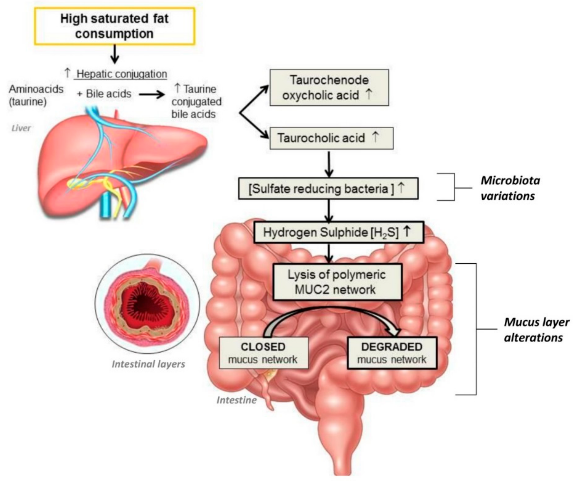
3.3. Fats and Gut Microbiota

Dietary fat quantity and quality influence gut microbiota composition [67]. Dietary fatty acids can be divided into saturated (SFAs), monounsaturated (MUFAs) and polyunsaturated fatty acids (PUFAs) according to the presence of double bonds between carbon molecules.
Mammalian animal products are the main source of SFAs. Several animal studies [68,69] described a decrease in Bacteroidetes and an increase in Firmicutes and Proteobacteria in mice fed with a high-fat diet (HFD)—specifically SFAs. Interestingly, these changes may be gradually reverted by a control diet [68,69]. Another study [70] also showed a decrease of Bacillus bifidus in mice fed an HFD. Therefore, high intake of dietary fats and particularly SFA could lead to intestinal dysbiosis.
Furthermore, HFD-induced dysbiosis could lead to intestinal barrier alterations. Indeed, sulfate-reducing bacteria (SRB) are more abundant in hosts consuming high-fat diets such as milk fat [71,72]. The high concentration of sulfide produced by particular SRB such as Bilophila wadsworthia may reduce disulfide bonds in the mucus, lysing the network of polymeric proteins MUC2 (oligomeric mucus gel-forming) secreted by goblet cells and having a key role in mucus layer stability and mucosal repair [73]. Recent lines of evidence have indicated that consumption of a high-SFA diet can stimulate the production of SRB, causing a defective mucus layer and increasing gut inflammation [73], colitis scores [73], and IBD [74,75]. The effects of HFD on gut microbiota are illustrated in Figure 1.
MUFAs such as the oleic acid present in extra virgin olive oil (EVOO) are among the main components of the “Mediterranean diet”. Extra virgin olive oil consumption has been demonstrated to hold most of the cardioprotective properties of the Mediterranean diet, and it is particularly advisable to reduce the risk of coronary heart disease [76]. However, its antioxidant and anti-inflammatory properties do not seem to be as related to the MUFA oleic acid as to its phenolic compounds [77]. A recent systematic review [78] showed that high-MUFA diets have no effect on richness/diversity indexes, phylum distribution, or Bacteroidetes-to-Firmicutes ratio. At family and genus level, MUFA-rich diets could be positively correlated with Parabacteroides, Prevotella, and Turicibacter genera and Enterobacteriaceae family, and with a lower number of Bifidobacterium genus. Moreover, the abundance of Blautia detected in high body mass index (BMI) individuals could be positively associated with MUFA serum metabolites, whereas the abundance of the phylum Tenericutes, correlating with lower triglyceride levels, was negatively associated with MUFA metabolites [78].
PUFAs are largely found in sunflower, soybean, and corn oil, as well as nuts and seeds. They are subdivided into omega-3 PUFAs (including linolenic acid) and omega-6 PUFAs (including linoleic acid). PUFAs are also called “essential fatty acids” since they cannot be synthesized by the human body and need to be obtained from the diet. Omega-3 PUFAs, especially found in fatty fish, can exert a positive action by restoring a healthy microbiota composition and increasing the production of anti-inflammatory compounds. Several studies have demonstrated that omega-3 PUFAs are able to restore the ratio of Firmicutes/Bacteroidetes and increase Lachnospiraceae taxa [79,80,81]—both of which are associated with increased production of the anti-inflammatory SCFA butyrate [79,80,81].
Unfortunately, the increased dietary intake of omega-6 PUFAs and the decreased intake of omega-3 PUFAs have dramatically shifted the human evolutionary ratio of ~1:1 to the modern ratio ranging from 10:1 to 50:1 [82]. This effect has been correlated to the epidemic spread of cardiovascular and chronic diseases [83,84]. A high omega-6/omega-3 PUFA ratio, predominant in the Western diet, has been related to an enhanced gut barrier permeability and metabolic endotoxemia through a gut-microbiota-driven mechanism [82]. Restoring this ratio towards a major uptake of omega-3 PUFAs could ameliorate gut microbiota composition and consequently reduce metabolic endotoxemia.
A distinct family of PUFAs are the conjugated linoleic acids (CLAs), of which the most naturally abundant are 18:2cis-9, trans-11 (9,11 CLA, or rumenic acid), and 18:2trans-10, cis-12 (10,12 CLA). These types of PUFA derive from the biohydrogenation of linoleic acid by ruminant bacteria expressing linoleic acid isomerase. For this reason, CLAs are found in ruminant animal food products such as beef, lamb, butter, and dairy products. Interestingly, several lines of evidence have shown anti-atherosclerotic, anti-obesogenic, and anti-cancer properties of CLAs, which are considered “Generally Regarded as Safe (GRAS)” by the Food and Drug Administration (FDA) [85]. It has been demonstrated in murine models that diet supplementation with 10,12 CLA can promote notable changes in gut microbiota composition at the phylum level such as a decrease of Firmicutes and an increase of Bacteroidetes [86] and at the species level with an enrichment of Butyrivibrio, Roseburia, and Lactobacillus, resulting in significant elevations of the SCFAs butyrate in the feces and acetate in plasma [87]. Such effects on gut microbiota composition could partially explain the beneficial properties attributed to CLAs.

3.4. Salt and Gut Microbiota

The World Health Organization recommends a maximum salt intake of 5 g/day [88]. A high-salt diet (HSD) is one of the major risk factors in the development of hypertension [89], kidney injury, and cardiovascular diseases (CVDs). HSD is also associated with an increased risk of gastric cancer by directly damaging gastric mucosa, leading to hyperplasia of the epithelium [90]. Moreover, considering H. pylori CagA-positive strains as markers of gastric cancer risk, Loth et al. showed that, when exposed to HSD, gastric H. pylori changed its virulence by inducing the expression of CagA [91].
Regarding gut microbiota, several studies in mice models demonstrated that HSD is associated with a decreased abundance of Lactobacillus spp. [92,93,94], Oscillibacter [91], Pseudoflavonifractor [91], Clostridium XIVa [91], Johnsonella [94], and Rothia [94], and an increased abundance of Parasutterella spp. [56,91], Erwinia genus [95], Christensenellaceae [95] and Corynebacteriaceae [95], Lachnospiraceae [93], and Ruminococcus [93]. In particular, Lactobacillus spp. reduction associated with HSD increased Th17 cells, pro-inflammatory hallmarks of many inflammatory diseases [92]. Miranda et al. [92] went further, showing that the exacerbation of colitis in mice induced by HSD was associated with a reduction in Lactobacillus spp., leading to the alteration of protective SCFA production, and hypothesizing that these changes alter gut immune homeostasis and lead to increased vulnerability to inflammatory insults [92]. HSD may result in alterations of gut microbiota composition, with a possible increase of Firmicutes/Bacteroidetes ratio, which may alter SCFAs production associated with modifications of gut permeability and immune homeostasis [92].

3.5. Food Additives and Gut Microbiota

With the development of the ultra-processed foods mainly characterizing the Western-type diet, the number of food additives such as non-nutritive sweeteners and emulsifiers approved for alimentary use by the industry has been soaring over the past few decades [96]. Artificial sweeteners are incorporated into almost all processed foods, often to aid stability and shelf life, and to improve taste and texture. Many reports [97,98] have shown that artificial sweetener consumption could alter gut microbiota and induce microbiota-mediated adverse effects in the host (e.g., glucose intolerance). Indeed, Suez et al. [98] demonstrated that non-caloric artificial sweeteners (NASs) altered microbial metabolic pathways and linked these changes to host susceptibility to metabolic disease. In this study, a proportion of healthy volunteers who did not normally consume NAS and who were given saccharin for one week at a dose of 5 mg/kg developed poorer glucose tolerance. The analysis of “NAS” responders’ stools showed an increase of Bacteroides spp. and Lactobacillus spp. and a decrease of Clostridiales spp. Several studies [97,99] confirmed that NAS intake increased the abundance of Bacteroides and some Clostridiales spp., and decreased the abundance of some Clostridiales spp., Bifidobacterium, and Lactobacillus. Another study by Palmnäs et al. reported significant gut microbiota changes in rats drinking water with low doses (5–7 mg/day) of aspartame, such as an increased abundance of Enterobacteriaceae and Clostridium leptum along with elevated fasting glucose levels and impaired insulin responses [100]. As these microbiota variations could cause glucose intolerance, these studies raise interesting questions on the harmlessness of artificial sweeteners and the functional meaning of gut microbiota.
Unlike other low-calorie sweeteners, steviol glycosides (extracted from stevia leaf) have no reported consistent microbial changes on anaerobic fecal cultures taken from healthy human subjects [101]. Specifically, no changes among Bacteroidaceae and Clostridia species were reported [102]. To date, there is no evidence that steviol glycosides adversely impact colonic bacteria [103].
In addition to artificial sweeteners, dietary emulsifiers such as lecithins, mono- and diglycerides of fatty acids, could increase bacterial translocation across epithelia in vitro, promoting systemic inflammation, altering microbiota localization and composition [104]. Emulsifier intake decreased gut microbial diversity, decreasing Bacteroides abundance and increasing Verrumicrobia (Akkermansia muciniphila), Proteobacteria abundances, and mucolytic operational taxonomic units (OTUs) including Ruminicoccus gnavus. These microbiota alterations led to dysbiosis and chronic gut inflammation, promoting colitis and metabolic syndrome [104].

3.6. Micronutrients and Gut Microbiota

Micronutrients play an important role in shaping the gut microbiota, which in turn is an efficient player in mediating their protective health effects [105].
Vitamins and minerals play a key role in regulating energy metabolism, cellular growth and differentiation, and immune functions. Various vitamins, such as thiamine, riboflavin, niacin, biotin, pantothenic acid, and folate (B vitamins), as well as vitamin K, can be synthesized by the fecal microbiota [106]. Indeed, Magnúsdóttir [107] assessed that B-vitamins are biosynthesized by the cooperation of various gut bacteria. For example, all bacteria from the phyla Bacteroidetes, Fusobacteria, and Proteobacteria possessed the necessary pathways for riboflavin and biotin biosynthesis. Moreover, all the Fusobacteria were predicted to be producers of vitamin B12 while Bacteroidetes appeared to be the phylum with the greatest number of predicted B vitamin producers. On the other hand, several studies demonstrated that vitamins such as vitamin D could impact gut microbiota composition. For example, it has been demonstrated that vitamin D had a positive effect on IBD patients by modulating the gut microbiome and increasing the abundance of potentially beneficial bacterial strains [108]. Furthermore, in a recent study of infant gut microbiome at age 3–6 months, researchers showed that vitamin D was associated with increased Lachnobacterium but decreased Lactococcus [109], and suggested that these correlations could have possible long-term implications for immune system modulation and asthma/allergic disease incidence.
Other antioxidant vitamins such as carotenoids, responsible for the yellow, orange, and red colors of many fruits and vegetables, could influence gut microbiota composition. A recent phytotherapy study on humans [110] identified lutein as a component of blackcurrant extract powder and demonstrated that lutein significantly promoted the growth of bifidobacteria and lactobacilli and reduced other bacteria populations, such as Bacteroides spp. and Clostridium spp. On the other hand, serum carotenoid levels could be impacted by gut microbiota composition. Indeed, Karlsson et al. [111] conducted a study to identify the association between alterations of the gut metagenome with atherosclerosis and suggested that high levels of beta-carotene in the serum of healthy controls could be due to the potential production of this anti-oxidant by the gut microbiota [111]. This study suggested that the anti-inflammatory effects of beta-carotene are mediated by the gut microbiota.
Like vitamins, metals are involved in numerous bacterial physiological processes impacting the gut microbiota [16]. Indeed, in mice colonized with the well-known nosocomial pathogen Clostridium difficile, excess dietary zinc severely exacerbated C. difficile-associated disease by increasing toxin activity and altering the host immune response [112]. Iron is another key element involved as a cofactor in iron-containing proteins for redox reactions, metabolic pathways, and electron transport chain mechanisms [113]. Iron availability influences the composition of the microbiota. Indeed, Constante et al. [114] demonstrated in mice that a heme-rich diet decreased microbial diversity, increased the abundance of Proteobacteria, and reduced the abundance of Firmicutes. Furthermore, the heme-rich diet may stimulate the growth of bacteria-coding genes related to heme uptake and release from red blood cells [114].

3.7. Polyphenols and Gut Microbiota

More than 10,000 polyphenol compounds have been identified in various plants and foods, such as fruits, vegetables, tea, medical plants, microalgae, herbs, seeds, and cereals, and in beverages such as coffee, tea, cocoa, and wine [115]. Some edible and wild fruits such as grape, olive, blueberry, sweetsop, mango, and citrus fruits contain high contents of polyphenols [116]. As these compounds are known to be implicated in preventing CVD [117], diabetes, obesity, and many other diseases [118], they currently represent a topic of growing interest for the scientific community. However, the absorption and bioavailability of these compounds in humans remain unclear and controversial. Nevertheless, there is strong agreement among researchers that reciprocal interactions of gut microbiota and phenolic compounds have an important impact on the bioavailability of phenolic compounds [119].
Over the past decade, several studies [120,121,122,123] demonstrated that phenolic compounds can alter the gut microbiota, resulting in a greater abundance of beneficial microbes. For example, quercetin supplementation resulted in an altered composition of gut microbiota at different taxonomic levels, including the relative Firmicutes:Bacteroidetes ratio and inhibiting the growth of bacterial species associated with diet-induced obesity such as Erysipelotrichaceae, Bacillus spp., and Eubacterium cylindroides [120]. The anthocyanins significantly stimulate the growth of Bifidobacterium spp., Lactobacillus and Enterococcus spp. [121], suggesting that anthocyanins may positively select for beneficial members of the gut microbial community [121]. As phenolic compounds could exert prebiotic activity, it is crucial to understand their inhibitory or stimulatory effects on beneficial or pathogenic bacteria [119].
At the same time, gut microbiota can modulate the transformation of phenolic compounds into smaller metabolites, influencing their bioavailability and modifying their properties [119]. The gut microbiota plays a key role in modulating the bioavailability of pro-anthocyanidins [124]. Pro-anthocyanidins could exert local beneficial biological actions on colonic epithelial cells, resulting in protective effects against inflammation-mediated diseases including colorectal cancer [124].
Phenolic compounds could affect gut microbiota composition [120,121,122,123]. Moreover, the gut microbiota has an impact on the biotransformation of phenolic compounds [119]. Although reciprocal interactions have been demonstrated [119,120,121,122,123], they are not yet understood.

4. Effects of Dietary Habits on Gut Microbiota

Since our dietary habits are the result of a specific mixture of micro and macronutrient amounts, continuously and indefinitely delivered to our gut ecosystem, we considered it useful to evaluate the impact of innovative and current dietary habits on gut microbiota associated with host mucosal barrier and immune functions. Figure 2 illustrates the effects of different types of diet on commensal bacterial species, mucus layer, and lamina propria hosting immune cells.

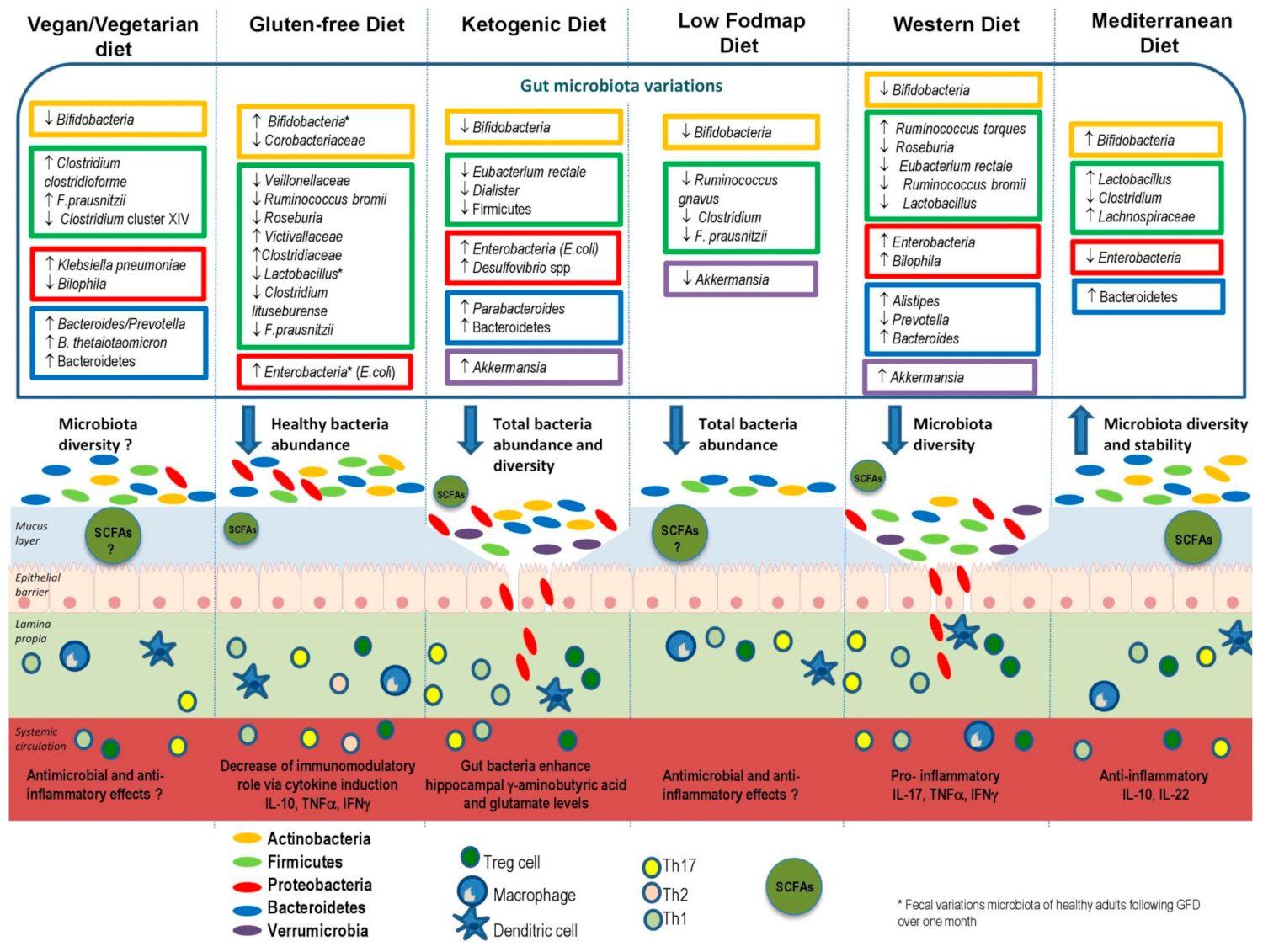
4.1. Vegan and Vegetarian Diets

Unlike omnivores, vegetarians refrain from consuming all types of meat and seafood. Vegans represent a subgroup of vegetarians, excluding also animal products such as eggs, milk and dairy products, and honey from their diet. Several studies have compared the gut microbiota of omnivores, vegetarians, and vegan individuals. Some of them [125,126] showed higher ratios of Bacteroides/Prevotella [125], Bacteroides thetaiotaomicron [125], Clostridium clostridioforme [125], Klebsiella pneumoniae [126], and Faecalibacterium prausnitzii [125] and lower ratios of Clostridium cluster XIVa [125] and Bilophila wadsworthia [126] in vegetarians and vegans compared to omnivores. Another study [127] demonstrated both vegans and vegetarians had lower counts of Bifidobacterium and Bacteroides species, but the quantification of fecal SCFA levels and methane production by breath revealed no difference between vegans and omnivores, demonstrating that vegan and vegetarian diets could decrease gut microbiota diversity but not decrease SCFAs and methane levels. A recent cross-sectional study [128] suggested that vegan and vegetarian diets influence the microbiota but do not allow conclusions to be drawn about gut microbial composition. Indeed, all these results should be interpreted cautiously due to different methodologies for microbiota identification, varied sample sizes, and the influences of geographical origin, age, gender, and body mass [129]. Additionally, the effects of polyphenols, which are abundant in plant foods and thus in vegan and vegetarian diets, on gut microbiota modulation should be considered. Indeed, these components increase the abundance of beneficial bacteria such as Bifidobacterium and Lactobacillus. However, further studies are warranted to clarify the complex mechanisms and interrelationships between vegan/vegetarian diets and gut microbiota.

4.2. Gluten-Free Diet (GFD)

Celiac disease (CD) is an autoimmune-mediated disease in which gluten sets off intestinal inflammation. In the 1960s, the gluten-free diet (GFD) was recognized as a potential cure, restoring the normal intestinal mucosa in coeliac patients [130]. At present, GFD is still known as the unique therapy in CD and the most commonly adopted special diet worldwide [131].
De Palma et al. [132] studied the gut microbiota of healthy subjects following a GFD over 1 month, and demonstrated a decrease of Bifidobacterium, Clostridium lituseburense, and Faecalibacterium prausnitzii proportions and an increase of Enterobacteriaceae and Escherichia coli counts after the GFD. Moreover, Bonder et al. [133] showed that the strongest gut microbiota variations in GFD occur in the family Veillonellaceae, whereby the abundance in the gut drops significantly in GFD. They also demonstrated a decrease of Ruminicoccus bromii and Roseburia feces in GFD while families Victivallaceae, Clostridiaceae, and Coriobacteriaceae increased in abundance in GFD. Sanz et al. [134] demonstrated that the fecal microbiota of healthy adults following a GFD over one month was eroded in healthy bacteria such as Bifidobacterium, B. longum, and Lactobacillus, and unhealthy bacteria increased (e.g., Enterobacteriaceae, in particular, E. coli).
Although most of these studies have important limitations including small sample sizes and the use of low-throughput techniques (e.g., culture techniques and non-sequencing-based molecular techniques [135]), a decrease of healthy bacteria such as Bifidobacterium and Lactobacillus has been demonstrated, leading to a diminution of SCFAs production and their beneficial metabolic and host immunity effects. Long-term GFD could constitute an environmental variable to be considered in treated CD patients for its possible effects on gut health, improving coeliac disease symptoms [132]. However, the increase of detrimental species such as Enterococcus, Staphylococcus, Salmonella, Shigella, and Klebsiella demonstrated by several studies [132,133,134,136] could influence the microbial profiles and impact the long-term homeostasis of the intestinal mucosa of healthy subjects [137].

4.3. Ketogenic Diet

The ketogenic diet (KD) is a high-fat, very-low-carbohydrate normocaloric diet used for drug-resistant epilepsy and GLUT1 Deficiency Syndrome [138]. In obese patients, KD seems to act as an efficient diet therapy for weight reduction [139]. However, maintaining body weight after weight loss is usually a major problem. Moreover, regarding the long-term impact of KD on cardiovascular risk factors, study results are controversial [140,141,142,143]. Some studies [141,143,144] demonstrated adverse events such as the development of non-alcoholic fatty liver disease (NAFLD) or insulin resistance.
The study of Tagliabue et al. [138] on patients treated with KD comparing their fecal microbiota composition before and after three months on the diet demonstrated that fecal microbiota increased in Desulfovibrio spp. involved in the exacerbation of gut inflammation. Another study [145] analyzed taxonomic changes in the children’s microbiota before and 3 months after starting KD, and demonstrated that alpha diversity did not change significantly during the diet. However, differences in both taxonomic and functional composition were detected, with a decrease in the abundance of bifidobacteria, E. rectale, and Dialister and an increase of E. coli during the intervention. Lindefeldt et al. [145] also showed, in children with refractory epilepsy following a week of KD, a reduction in the richness of gut microbiota with an increase of Bacteroidetes and reduction of Proteobacteria after KD. At the genus level, Bacteroides, Bifidobacterium, and Prevotella were increased after KD, while Cronobacter diminished [146]. Olson et al. [147] went further, showing an anti-seizure effect of KD in mice, mediated by Akkermansia and Parabacteroides involving changes in systemic gammaglutamylated amino acids and elevated hippocampal GABA/glutamate levels.
KD results in a reduction of carbohydrate intake, and leads to a decrease of polysaccharide content associated with a decrease in beneficial gut microbiota bacteria such as Bifidobacteria. Moreover, all these studies showed microbial increases such as Akkermansia or E. coli during KD. This confirms a potential detrimental effect of an HFD on the gut mucus barrier, as described above. Therefore, although KD has a positive impact on a wide range of diseases [145], we raise a concern about the long-term effects of KD on the gut microbiota composition and consequently mucus layer homeostasis and immunity functions—more specifically in healthy subjects adopting KD for weight loss. Further studies are warranted to understand the role of gut microbiota variations during KD and its therapeutic effects.

4.4. High-Glucose or -Fructose Diets

The excess of sugar in modern dietary habits has been linked to obesity and several metabolic diseases, including diabetes mellitus type II, NAFLD, and cardiovascular diseases [148]. The impact of high-sugar diet on the gut microbiota has recently been elucidated by Do et al. [149]: the authors assigned six-week-old C57BL/6J mice to receive distinct dietary regimes such as normal diet (ND), high-glucose diet (HGD), and high-fructose diet (HFrD). After 12 weeks, HGD- and HFrD-fed mice showed lower microbial diversity (i.e., fewer operational taxonomic units and lower Shannon indices) than ND-fed mice, with a lower abundance of Bacteroidetes and an increased abundance of Proteobacteria at the phylum level, as well as an increase of the Desulfovibrio vulgaris species. These metabolic and microbial differences were accompanied by a significant (2.5-fold greater) increase in gut permeability. Consequently, the expression of inflammatory cytokines (TNF-alpha and IL-1beta) in the colon was higher in HGD- and HFrD-fed mice than in ND-fed mice. This evidence suggests that an HGD and/or HFrD can shape the gut microbiota, increasing the Firmicutes-to-Bacteroidetes ratio and the proportion of Proteobacteria—one of the best sources of LPS [150]. Moreover, these dietary regimens significantly alter gut permeability, boosting metabolic endotoxemia and systemic inflammation through modulation of the gut microbiota [105].

4.5. Low-FODMAP Diet

In 2004, the Monash University created the term “FODMAP” (fermentable oligo-, di-, mono-saccharides, and polyols) to describe a group of highly fermentable but poorly absorbed carbohydrates and polyols [151]. In recent years, this alternative diet gained visibility with physicians especially for its use as a treatment option in IBD and IBS [152,153]. Halmos et al. [154] demonstrated in IBS patients on a low-FODMAP diet, similar SCFA concentrations with a reduction of total bacterial abundance to 47% compared with a habitual diet. Several studies showed a decrease of Clostridium cluster IV [154], Propionibacteriaceae [155], mucus-associated Akkermansia muciniphila [154], Ruminococcus gnavus [115], and Bifidobacteria [154,156,157] in low-FODMAP diet compared to control diet. Indeed, a low-FODMAP diet could lead to a reduction of potential prebiotics (FOSs and GOSs), thus leading to a reduction in beneficial bacteria and fermentative effects. The integration of a low-FODMAP diet with probiotics seems to counteract gut microbiota imbalances and, in particular, restore Bifidobacterium levels [131]. Probiotics are live microorganisms that, when administered in adequate amounts, confer a health benefit on the host [158]. Each probiotic may have different characteristics, including variable effects on cytokines, host microbiota, and other potential targets [159]. Larger studies are needed to further understand the potential contributions of probiotics supplementation and long-term effects of low-FODMAP diets on intestinal microbiota composition.

4.6. Western Diet

The Western diet (WD) is a dietary habit, as part of the Western lifestyle chosen by many people in developed countries, and increasingly in developing countries, associated with economic growth. The WD is enriched in total fat, animal proteins, and refined sugars. Martinez Medina et al. [160] demonstrated that a combination of high-fat/high-sugar diet led to dysbiosis with increased Bacteroides spp. and Ruminococcus torques in mice. A study [161] of European children fed with the WD and Burkina Faso (BF) children assuming a diet rich in millet/sorghum + local vegetables containing very few lipids and animal proteins revealed that Proteobacteria were more abundant in EU than in BF children. Moreover, the BF children’s microbiota was enriched with Prevotella and Xylanibacter compared to EU children’s microbiota. Another study [162] determined the microbiota composition of volunteers from Venezuela, Malawi, and the United States, and reported that irrespective of age, Malawian and Venezuelan microbiota compositions were similar, compared with the US American microbiota characterized by the least microbial diversity. These two studies demonstrated that the genus Prevotella was underrepresented in WD microbiota and could be a taxonomic discriminator. Furthermore, the WD based on animal protein consumption increases the abundance of bile-tolerant microorganisms such as Alistipes, Bilophila, and Bacteroides and decreases the levels of Firmicutes that metabolize dietary plant polysaccharides such as Roseburia, Eubacterium rectale, and RRuminococcus bromii [58]. Moreover, several studies suggest that the detrimental effects of WD on gut microbiota may also be driven by food additives, inducing dysbiosis and consequently adverse intestinal mucosal effects and inflammation [104]. Moreover, a large body of research supports the hypothesis that the WD, by causing dysbiosis in gut microbiota composition, is associated with obesity and metabolic diseases [163].

4.7. Mediterranean Diet

The concept of the Mediterranean diet (MD) was developed to reflect the typical dietary habits followed during the early 1960s by inhabitants of the Mediterranean basin, mainly in Crete, much of the rest of Greece, and Southern Italy [164]. The MD, centered around fruits, vegetables, olive oil, nuts, legumes, and whole grains, has been linked to a large number of health benefits, including reduced mortality risk and the prevention of many diseases such as CVD [165], diabetes [166], metabolic syndrome [167], cognitive impairment [168,169], and depression [169]. The MD is based on the regular consumption of MUFAs and PUFAs, polyphenols and other antioxidants, a high intake of prebiotic fiber and low-glycemic carbohydrates, and greater consumption of plant proteins than animal proteins. The recent study of Garcia-Mantrana et al. [170] indicated that a higher ratio of Firmicutes–Bacteroidetes was related to lower adherence to the MD, whereas the greater presence of Bacteroidetes was associated with lower animal protein intake. Furthermore, higher bifidobacterial counts and higher total SCFAs were related to greater consumption of plant-based nutrients, such as vegetable proteins and polysaccharides. Finally, they demonstrated that better adherence to the MD was associated with significantly higher levels of total SCFAs. De Filippis and colleagues [171] also reported that high-level consumption of plant-based foods and high-level adherence to an MD beneficially impact the gut microbiota and associated metabolomic profile [171]. Mitsou et al [172] confirmed these findings, with positive correlations between adherence to MD and increase of total bacteria, Bifidobacteria/E. coli ratio, the relative share of Bacteroides, C. albicans, and total SCFAs, as well as decrease of E. coli levels. All these findings demonstrated a link between adherence to the MD and improvements to the diversity and richness of gut microbiota.

5. Conclusions

Food components have a key impact on the gut microbiota, influencing its composition in terms of richness and diversity. On the one hand, high intake of animal proteins, saturated fat, sugar, and salt could stimulate the growth of pathogenic bacteria to the detriment of beneficial bacteria, leading to potential alterations of the intestinal barrier. On the other hand, the consumption of complex polysaccharides and plant protein could be associated with an increase of beneficial bacteria quantity, stimulating SCFA production. Moreover, omega-3, polyphenols, and micronutrients appear to have the potential to confer health benefits via modulation of the gut microbiota.
Dietary habits can strongly influence gut microbiota composition. The Westernization of the diet, including additives, may reduce gut microbial diversity in terms of phyla and genus leading to dysbiosis, alteration of barrier function and permeability, and abnormal activation of immune cells, leading to high incidences of chronic diseases. Although elimination diets such as low-FODMAP and GFD can improve the symptoms of some diseases like IBS and CD in selected patients, the long-term effects on gut microbiota require elucidations. To date, the Mediterranean diet remains the evergreen solution to optimally modulating microbiota diversity and stability as well as the regular permeability and activity of immune functions of the human host. Modifying dietary habits and adopting a MD may be the solution to prevent microbiota dysbiosis, and consequently, to prevent many GI and neurological disorders [13].
Future advances on the knowledge of the interactions between food compounds and specific intestinal bacteria would lead to a better understanding of both positive and negative interactions with dietary habits. A novel nutritional approach may be adopted by building a personalized diet subsequent to microbiota analyses, in order to modulate and restore a healthy gut microbiota.

Author Contributions

E.R. conceived the topic and revised the manuscript; M.C. and G.P. selected bibliographic sources; P.R. prepared the manuscript and generated the schematic diagrams; L.R.L. and F.S. contributed to the immunological section; A.G., G.A.D.M., and M.C.M. revised the final version; M.C.M. coordinated the working group. All authors made significant contributions to this review article.

Funding

This research received no external funding.

Conflicts of Interest

The authors declare no conflicts of interest.

References

  1. Thursby, E.; Juge, N. Introduction to the human gut microbiota. Biochem. J. 2017, 474, 1823–1836. [Google Scholar] [CrossRef] [PubMed]
  2. Laterza, L.; Rizzatti, G.; Gaetani, E.; Chiusolo, P.; Gasbarrini, A. The gut microbiota and immune system relationship in human graft-versus-host disease. Mediterr. J. Hematol. Infect. Dis. 2016, 8, e2016025. [Google Scholar] [CrossRef] [PubMed]
  3. Arumugam, M.; Raes, J.; Pelletier, E.; Le Paslier, D.; Yamada, T.; Mende, D.R.; Fernandes, G.R.; Tap, J.; Bruls, T.; Batto, J.M.; et al. Enterotypes of the human gut microbiome. Nature 2011, 473, 174–180. [Google Scholar] [CrossRef] [PubMed]
  4. Rinninella, E.; Raoul, P.; Cintoni, M.; Franceschi, F.; Miggiano, G.A.D.; Gasbarrini, A.; Mele, M.C. What is the healthy gut microbiota composition? a changing ecosystem across age, environment, diet, and diseases. Microorganisms 2019, 7, 14. [Google Scholar] [CrossRef] [PubMed]
  5. Tang, W.H.W.; Bäckhed, F.; Landmesser, U.; Hazen, S.L. Intestinal microbiota in cardiovascular health and disease. J. Am. Coll. Cardiol. 2019, 73, 2089–2105. [Google Scholar] [CrossRef] [PubMed]
  6. Ley, R.E.; Bäckhed, F.; Turnbaugh, P.; Lozupone, C.A.; Knight, R.D.; Gordon, J.I. Obesity alters gut microbial ecology. Proc. Natl. Acad. Sci. USA 2005, 102, 11070–11075. [Google Scholar] [CrossRef] [PubMed] [Green Version]
  7. Pascale, A.; Marchesi, N.; Govoni, S.; Coppola, A.; Gazzaruso, C. The role of gut microbiota in obesity, diabetes mellitus, and effect of metformin: new insights into old diseases. Curr. Opin. Pharmacol. 2019, 49, 1–5. [Google Scholar] [CrossRef] [PubMed]
  8. Raza, M.H.; Gul, K.; Arshad, A.; Riaz, N.; Waheed, U.; Rauf, A.; Aldakheel, F.; Alduraywish, S.; Rehman, M.U.; Abdullah, M.; et al. Microbiota in cancer development and treatment. J. Cancer Res. Clin. Oncol. 2019, 145, 49–63. [Google Scholar] [CrossRef] [PubMed]
  9. Bhattarai, Y.; Muniz Pedrogo, D.A.; Kashyap, P.C. Irritable bowel syndrome: a gut microbiota-related disorder? Am. J. Physiol. Gastrointest. Liver Physiol. 2017, 312, G52–G62. [Google Scholar] [CrossRef]
  10. Collins, S.M.; Surette, M.; Bercik, P. The interplay between the intestinal microbiota and the brain. Nat. Rev. Microbiol. 2012, 10, 735–742. [Google Scholar] [CrossRef]
  11. Finegold, S.M.; Molitoris, D.; Song, Y.; Liu, C.; Vaisanen, M.L.; Bolte, E.; McTeague, M.; Sandler, R.; Wexler, H.; Marlowe, E.M.; et al. Gastrointestinal microflora studies in late-onset autism. Clin. Infect Dis. 2002, 35, S6–S16. [Google Scholar] [CrossRef] [PubMed]
  12. Finegold, S.M. Desulfovibrio species are potentially important in regressive autism. Med. Hypotheses 2011, 77, 270–274. [Google Scholar] [CrossRef] [PubMed]
  13. Mayer, E.A.; Tillisch, K.; Gupta, A. Gut/brain axis and the microbiota. J. Clin. Investig. 2015, 125, 926–938. [Google Scholar] [CrossRef] [PubMed]
  14. Hill-Burns, E.M.; Debelius, J.W.; Morton, J.T.; Wissemann, W.T.; Lewis, M.R.; Wallen, Z.D.; Peddada, S.D.; Factor, S.A.; Molho, E.; Zabetian, C.P.; et al. Parkinson’s disease and Parkinson’s disease medications have distinct signatures of the gut microbiome. Mov. Disord. 2017, 32, 739–749. [Google Scholar] [CrossRef] [PubMed]
  15. Li, Q.; Han, Y.; Dy, A.B.C.; Hagerman, R.J. The gut microbiota and autism spectrum disorders. Front. Cell Neurosci. 2017, 11, 120. [Google Scholar] [CrossRef] [PubMed]
  16. Gentile, C.L.; Weir, T.L. The gut microbiota at the intersection of diet and human health. Science 2018, 362, 776–780. [Google Scholar] [CrossRef] [PubMed] [Green Version]
  17. Scaldaferri, F.; Pizzoferrato, M.; Gerardi, V.; Lopetuso, L. The gut barrier: New acquisitions and therapeutic approaches. J. Clin. Gastroenterol. 2012, 46, S12–S17. [Google Scholar] [CrossRef]
  18. Leser, T.D.; Molbak, L. Better living through microbial action: the benefits of the mammalian gastrointestinal microbiota on the host. Environ. Microbiol. 2009, 11, 2194–2206. [Google Scholar] [CrossRef]
  19. Neish, A.S. Microbes in gastrointestinal health and disease. Gastroenterology 2009, 136, 65–80. [Google Scholar] [CrossRef]
  20. Scanlan, P.D.; Marchesi, J.R. Micro-eukaryotic diversity of the human distal gut microbiota: Qualitative assessment using culture-dependent and -independent analysis of faeces. ISME J. 2008, 2, 1183–1193. [Google Scholar] [CrossRef]
  21. Backhed, F.; Ley, R.E.; Sonnenburg, J.L.; Peterson, D.A.; Gordon, J.I. Host-bacterial mutualism in the human intestine. Science 2005, 307, 1915–1920. [Google Scholar] [CrossRef] [PubMed]
  22. McCracken, V.J.; Lorenz, R.G. The gastrointestinal ecosystem: A precarious alliance among epithelium, immunity and microbiota. Cell Microbiol. 2001, 3, 1–11. [Google Scholar] [CrossRef] [PubMed]
  23. Lievin-Le Moal, V.; Servin, A.L. The front line of enteric host defense against unwelcome intrusion of harmful microorganisms: Mucins, antimicrobial peptides, and microbiota. Clin. Microbiol. Rev. 2006, 19, 315–337. [Google Scholar] [CrossRef] [PubMed]
  24. Mu, Q.; Kirby, J.; Reilly, C.M.; Luo, X.M. Leaky gut as a danger signal of autoimmune diseases. Front. Immunol. 2017, 8, 598. [Google Scholar] [CrossRef] [PubMed]
  25. Camilleri, M.; Madsen, K.; Spiller, R.; Greenwood-Van Meerveld, B.; Verne, G.N. Intestinal barrier function in health and gastrointestinal disease. Neurogastroenterol. Motil. 2012, 24, 503–512. [Google Scholar] [CrossRef] [PubMed]
  26. Fasano, A. Leaky gut and autoimmune diseases. Clin. Rev. Allergy Immunol. 2012, 42, 71–78. [Google Scholar] [CrossRef] [PubMed]
  27. Fasano, A. Zonulin and its regulation of intestinal barrier function: The biological door to inflammation, autoimmunity, and cancer. Physiol. Rev. 2011, 91, 151–175. [Google Scholar] [CrossRef]
  28. Peloquin, J.M.; Nguyen, D.D. The microbiota and inflammatory bowel disease: Insights from animal models. Anaerobe 2013, 24, 102–106. [Google Scholar] [CrossRef] [PubMed] [Green Version]
  29. Scaldaferri, F.; Petito, V.; Lopetuso, L.; Bruno, G.; Gerardi, V.; Ianiro, G.; Sgambato, A.; Gasbarrini, A.; Cammarota, G. Pre- and posttherapy assessment of intestinal soluble mediators in IBD: Where we stand and future perspectives. Mediat. Inflamm. 2013, 2013, 391473. [Google Scholar] [CrossRef] [PubMed]
  30. Scaldaferri, F.; Gerardi, V.; Lopetuso, L.R.; Del Zompo, F.; Mangiola, F.; Boškoski, I.; Bruno, G.; Petito, V.; Laterza, L.; Cammarota, G.; et al. Gut microbial flora, prebiotics, and probiotics in IBD: Their current usage and utility. BioMed Res. Int. 2013, 2013, 435268. [Google Scholar] [CrossRef] [PubMed]
  31. Purchiaroni, F.; Tortora, A.; Gabrielli, M.; Bertucci, F.; Gigante, G.; Ianiro, G.; Ojetti, V.; Scarpellini, E.; Gasbarrini, A. The role of intestinal microbiota and the immune system. Eur. Rev. Med. Pharmacol. Sci. 2013, 17, 323–333. [Google Scholar] [PubMed]
  32. Mombaerts, P.; Mizoguchi, E.; Grusby, M.J.; Glimcher, L.H.; Bhan, A.K.; Tonegawa, S. Spontaneous development of inflammatory bowel disease in T cell receptor mutant mice. Cell 1993, 75, 274–282. [Google Scholar] [CrossRef]
  33. Sadlack, B.; Merz, H.; Schorle, H.; Schimpl, A.; Feller, A.C.; Horak, I. Ulcerative colitis-like disease in mice with a disrupted interleukin-2 gene. Cell 1993, 75, 253–261. [Google Scholar] [CrossRef]
  34. Kuhn, R.; Lohler, J.; Rennick, D.; Rajewsky, K.; Müller, W. Interleukin-10-deficient mice develop chronic enterocolitis. Cell 1993, 75, 263–274. [Google Scholar] [CrossRef]
  35. Madsen, K.L.; Doyle, J.S.; Tavernini, M.M.; Jewell, L.D.; Rennie, R.P.; Fedorak, R.N. Antibiotic therapy attenuates colitis in interleukin 10 gene-deficient mice. Gastroenterology 2000, 118, 1094–1105. [Google Scholar] [CrossRef]
  36. Kang, S.S.; Bloom, S.M.; Norian, L.A.; Geske, M.J.; Flavell, R.A.; Stappenbeck, T.S.; Allen, P.M. An antibiotic-responsive mouse model of fulminant ulcerative colitis. PLoS Med. 2008, 5, e41. [Google Scholar] [CrossRef] [PubMed]
  37. Guarino, A.; Albano, F.; Ashkenazi, S.; Gendrel, D.; Hoekstra, J.H.; Shamir, R.; Szajewska, H. ESPGHAN/ESPID Evidence-Based Guidelines for the Management of Acute Gastroenteritis in Children in Europe Expert Working Group. European Society for Paediatric Gastroenterology, Hepatology, and Nutrition/European Society for Paediatric Infectious Diseases evidence-based guidelines for the management of acute gastroenteritis in children in Europe: Executive summary. J. Pediatr. Gastroenterol. Nutr. 2008, 46, 619–621. [Google Scholar] [PubMed]
  38. Akira, S.; Uematsu, S.; Takeuchi, O. Pathogen recognition and innate immunity. Cell 2006, 124, 783–801. [Google Scholar] [CrossRef]
  39. Bamias, G.; Corridoni, D.; Pizarro, T.T.; Cominelli, F. New insights into the dichotomous role of innate cytokines in gut homeostasis and inflammation. Cytokine 2012, 59, 451–459. [Google Scholar] [CrossRef] [Green Version]
  40. Lopetuso, L.R.; Chowdhry, S.; Pizarro, T.T. Opposing functions of classic and novel IL-1 family members in gut health and disease. Front. Immunol. 2013, 4, 181. [Google Scholar] [CrossRef]
  41. Mudgil, D.; Barak, S. Composition, properties and health benefits of indigestible carbohydrate polymers as dietary fiber: A review. Int. J. Biol. Macromol. 2013, 61, 1–6. [Google Scholar] [CrossRef] [PubMed]
  42. Galanakis, C. (Ed.) Dietary Fiber: Properties, Recovery, and Applications, 1st ed.; Academic Press: Cambridge, MA, USA, 2019; ISBN 9780128164952. [Google Scholar]
  43. Cummings, J.H.; Pomare, E.W.; Branch, W.J.; Naylor, C.P.; Macfarlane, G.T. Short chain fatty acids in human large intestine, portal, hepatic and venous blood. Gut 1987, 28, 1221–1227. [Google Scholar] [CrossRef] [PubMed]
  44. Levy, M.; Thaiss, C.A.; Elinav, E. Metabolites: Messengers between the microbiota and the immune system. Genes Dev. 2016, 30, 1589–1597. [Google Scholar] [CrossRef] [PubMed]
  45. Kelly, C.J.; Zheng, L.; Campbell, E.L.; Saeedi, B.; Scholz, C.C.; Bayless, A.J.; Wilson, K.E.; Glover, L.E.; Kominsky, D.J.; Magnuson, A.; et al. Crosstalk between microbiota-derived short-chain fatty acids and intestinal epithelial hif augments tissue barrier function. Cell Host Microbe 2015, 17, 662–671. [Google Scholar] [CrossRef] [PubMed]
  46. Roediger, W.E. Role of anaerobic bacteria in the metabolic welfare of the colonic mucosa in man. Gut 1980, 21, 793–798. [Google Scholar] [CrossRef] [PubMed]
  47. Tsukahara, T.; Iwasaki, Y.; Nakayama, K.; Ushida, K. Stimulation of butyrate production in the large intestine of weaning piglets by dietary fructooligosaccharides and its influence on the histological variables of the large intestinal mucosa. J. Nutr. Sci. Vitaminol. 2003, 49, 414–421. [Google Scholar] [CrossRef]
  48. Klampfer, L.; Huang, J.; Sasazuki, T.; Shirasawa, S.; Augenlicht, L. Inhibition of interferon gamma signaling by the short chain fatty acid butyrate. Mol. Cancer Res. 2003, 1, 855–862. [Google Scholar]
  49. Waldecker, M.; Kautenburger, T.; Daumann, H.; Busch, C.; Schrenk, D. Inhibition of histone-deacetylase activity by short-chain fatty acids and some polyphenol metabolites formed in the colon. J. Nutr. Biochem. 2008, 19, 587–593. [Google Scholar] [CrossRef]
  50. Blaut, M. Gut microbiota and energy balance: Role in obesity. Proc. Nutr. Soc. 2015, 74, 227–234. [Google Scholar] [CrossRef]
  51. Nylund, L.; Kaukinen, K.; Lindfors, K. The microbiota as a component of the celiac disease and non-celiac gluten sensitivity. Clin. Nutr. Exp. 2016, 6, 17–24. [Google Scholar] [CrossRef] [Green Version]
  52. Maukonen, J.; Saarela, M. Human gut microbiota: Does diet matter? Proc. Nutr. Soc. 2015, 74, 23–36. [Google Scholar] [CrossRef] [PubMed]
  53. Gibson, G.R.; Probert, H.M.; Loo, J.V.; Rastall, R.A.; Roberfroid, M.B. Dietary modulation of the human colonic microbiota: Updating the concept of prebiotics. Nutr. Res. Rev. 2004, 17, 259–275. [Google Scholar] [CrossRef] [PubMed]
  54. Sonnenburg, E.D.; Smits, S.A.; Tikhonov, M.; Higginbottom, S.K.; Wingreen, N.S.; Sonnenburg, J.L. Diet-induced extinction in the gut microbiota compounds over generations. Nature 2016, 529, 212–215. [Google Scholar] [CrossRef] [PubMed]
  55. Singh, A.; Zapata, R.C.; Pezeshki, A.; Reidelberger, R.D.; Chelikani, P.K. Inulin fiber dose-dependently modulates energy balance, glucose tolerance, gut microbiota, hormones and diet preference in high-fat-fed male rats. J. Nutr. Biochem. 2018, 59, 142–152. [Google Scholar] [CrossRef] [PubMed]
  56. Vandeputte, D.; Falony, G.; Vieira-Silva, S.; Wang, J.; Sailer, M.; Theis, S.; Verbeke, K.; Raes, J. Prebiotic inulin-type fructans induce specific changes in the human gut microbiota. Gut 2017, 66, 1968–1974. [Google Scholar] [CrossRef] [PubMed]
  57. Scott, K.P.; Gratz, S.W.; Sheridan, P.O.; Flint, H.J.; Duncan, S.H. The influence of diet on the gut microbiota. Pharmacol. Res. 2013, 69, 52–60. [Google Scholar] [CrossRef]
  58. David, L.A.; Maurice, C.F.; Carmody, R.N.; Gootenberg, D.B.; Button, J.E.; Wolfe, B.E.; Ling, A.V.; Devlin, A.S.; Varma, Y.; Fischbach, M.A.; et al. Diet rapidly and reproducibly alters the human gut microbiome. Nature 2014, 505, 559–563. [Google Scholar] [CrossRef]
  59. Reddy, B.S.; Weisburger, J.H.; Wynder, E.L. Effects of high risk and low risk diets for colon carcinogenesis on fecal microflora and steroids in man. J. Nutr. 1975, 105, 878–884. [Google Scholar] [CrossRef]
  60. Barrea, L.; Annunziata, G.; Muscogiuri, G.; Laudisio, D.; Somma, C.D.; Maisto, M.; Tenore, G.C.; Colao, A.; Savastano, S. Trimethylamine n-oxide (tmao), mediterranean diet and nutrition in healthy, normal-weight subjects: Is it also a matter of gender? Nutrition 2018, 62, 7–17. [Google Scholar] [CrossRef]
  61. Jantchou, P.; Morois, S.; Clavel-Chapelon, F.; Boutron-Ruault, M.C.; Carbonnel, F. Animal protein intake and risk of inflammatory bowel disease: the E3N prospective study. Am. J. Gastroenterol. 2010, 105, 2195–2201. [Google Scholar] [CrossRef]
  62. Singh, R.K.; Chang, H.W.; Yan, D.; Lee, K.; Ucmak, D.; Wong, K.; Abrouk, M.; Farahnik, B.; Nakamura, M.; Zhu, T.H.; et al. Influence of diet on the gut microbiome and implications for human health. J. Transl. Med. 2017, 15, 73. [Google Scholar] [CrossRef]
  63. Świątecka, D.; Dominika, Ś.; Narbad, A.; Ridgway, K.P.; Kostyra, H. The study on the impact of glycated pea proteins on human intestinal bacteria. Int. J. Food. Microbiol. 2011, 145, 267–272. [Google Scholar] [PubMed]
  64. Butteiger, D.N.; Hibberd, A.A.; McGraw, N.J.; Napawan, N.; Hall-Porter, J.M.; Krul, E.S. Soy protein compared with milk protein in a western diet increases gut microbial diversity and reduces serum lipids in golden syrian hamsters. J. Nutr. 2016, 146, 697–705. [Google Scholar] [CrossRef] [PubMed]
  65. Vázquez, L.; Flórez, A.B.; Guadamuro, L.; Mayo, B. Effect of Soy Isoflavones on growth of representative bacterial species from the human gut. Nutrients 2017, 9, 727. [Google Scholar] [CrossRef]
  66. Miao, S.; Zhao, C.; Zhu, J.; Hu, J.; Dong, X.; Sun, L. Dietary soybean meal affects intestinal homoeostasis by altering the microbiota, morphology and inflammatory cytokine gene expression in northern snakehead. Sci. Rep. 2018, 8, 113. [Google Scholar] [CrossRef]
  67. Cândido, F.G.; Valente, F.X.; Grześkowiak, Ł.M.; Moreira, A.P.B.; Rocha, D.M.U.P.; Alfenas, R.C.G. Impact of dietary fat on gut microbiota and low-grade systemic inflammation: mechanisms and clinical implications on obesity. Int J Food Sci Nutr. 2018, 69, 125–143. [Google Scholar] [CrossRef] [PubMed]
  68. Hildebrandt, M.A.; Hoffmann, C.; Sherrill-Mix, S.A.; Keilbaugh, S.A.; Hamady, M.; Chen, Y.Y.; Knight, R.; Ahima, R.S.; Bushman, F.; Wu, G.D. High-fat diet determines the composition of the murine gut microbiome independently of obesity. Gastroenterology 2009, 137, 1716–1724. [Google Scholar] [CrossRef] [PubMed]
  69. Wu, G.D.; Chen, J.; Hoffmann, C.; Bittinger, K.; Chen, Y.Y.; Keilbaugh, S.A.; Bewtra, M.; Knights, D.; Walters, W.A.; Knight, R.; et al. Linking longterm dietary patterns with gut microbial enterotypes. Science 2011, 334, 105–108. [Google Scholar] [CrossRef]
  70. Zhang, C.; Zhang, M.; Pang, X.; Zhao, Y.; Wang, L.; Zhao, L. Structural resilience of the gut microbiota in adult mice under high-fat dietary perturbations. ISME J. 2012, 6, 1848–1857. [Google Scholar] [CrossRef]
  71. Ijssennagger, N.; Van der Meer, R.; Van Mil, S.W.C. Sulfide as a mucus barrier-breaker in inflammatory bowel disease? Trends Mol. Med. 2016, 22, 190–199. [Google Scholar] [CrossRef]
  72. Johansson, M.E.; Phillipson, M.; Petersson, J.; Velcich, A.; Holm, L.; Hansson, G.C. The inner of the two Muc2 mucin-dependent mucus layers in colon is devoid of bacteria. Proc. Natl. Acad. Sci. USA 2008, 105, 15064–15069. [Google Scholar] [CrossRef] [PubMed] [Green Version]
  73. Devkota, S.; Wang, Y.; Musch, M.W.; Leone, V.; Fehlner-Peach, H.; Nadimpalli, A.; Antonopoulos, D.A.; Jabri, B.; Chang, E.B. Dietary-fat-induced taurocholic acid promotes pathobiont expansion and colitis in Il10-/- mice. Nature 2012, 487, 104–108. [Google Scholar] [CrossRef] [PubMed]
  74. Gruber, L.; Kisling, S.; Lichti, P.; Martin, F.P.; May, S.; Klingenspor, M.; Lichtenegger, M.; Rychlik, M.; Haller, D. High fat diet accelerates pathogenesis of murine Crohn’s disease-like ileitis independently of obesity. PLoS ONE 2013, 8, e71661. [Google Scholar] [CrossRef] [PubMed]
  75. Devkota, S.; Chang, E.B. Devkota interactions between diet, bile acid metabolism, gut microbiota, and inflammatory bowel diseases. Dig. Dis. 2015, 33, 351–356. [Google Scholar] [CrossRef] [PubMed]
  76. Colica, C.; Di Renzo, L.; Trombetta, D.; Smeriglio, A.; Bernardini, S.; Cioccoloni, G.; de Miranda, R.C.; Gualtieri, P.; Salimei, P.S.; De Lorenzo, A. Effects of a hydroxytyrosol-based pharmaceutical formulation on body composition, metabolic state, and gene expression: A randomized double-blinded, placebo-controlled crossover trial. Oxid. Med. Cell Longev. 2017, 2017, 2473495. [Google Scholar] [CrossRef]
  77. Bulotta, S.; Celano, M.; Lepore, S.M.; Montalcini, T.; Pujia, A.; Russo, D. Beneficial effects of the olive oil phenolic components oleuropein and hydroxytyrosol: Focus on protection against cardiovascular and metabolic diseases. J. Transl. Med. 2014, 12, 219. [Google Scholar] [CrossRef] [PubMed]
  78. Wolters, M.; Ahrens, J.; Romaní-Pérez, M.; Watkins, C.; Sanz, Y.; Benítez-Páez, A.; Stanton, C.; Günther, K. Dietary fat, the gut microbiota, and metabolic health—A systematic review conducted within the MyNewGut project. Clin. Nutr. 2018. [Google Scholar] [CrossRef] [PubMed]
  79. Watson, H.; Mitra, S.; Croden, F.C.; Taylor, M.; Wood, H.M.; Perry, S.L.; Spencer, J.A.; Quirke, P.; Toogood, G.J.; Lawton, C.L. A randomised trial of the effect of omega-3 polyunsaturated fatty acid supplements on the human intestinal microbiota. Gut 2018, 67, 1974–1983. [Google Scholar] [CrossRef]
  80. Noriega, B.S.; Sanchez-Gonzalez, M.A.; Salyakina, D.; Coffman, J. Understanding the impact of omega-3 rich diet on the gut microbiota. Case Rep. Med. 2016, 2016, 3089303. [Google Scholar] [CrossRef]
  81. Menni, C.; Zierer, J.; Pallister, T.; Jackson, M.A.; Long, T.; Mohney, R.P.; Steves, C.J.; Spector, T.D.; Valdes, A.M. Omega-3 fatty acids correlate with gut microbiome diversity and production of N-carbamylglutamate in middle aged and elderly women. Sci. Rep. 2017, 7, 11079. [Google Scholar] [CrossRef]
  82. Kaliannan, K.; Wang, B.; Li, X.Y.; Kim, K.J.; Kang, J.X. A host-microbiome interaction mediates the opposing effects of omega-6 and omega-3 fatty acids on metabolic endotoxemia. Sci. Rep. 2015, 5, 11276. [Google Scholar] [CrossRef] [PubMed]
  83. Simopoulos, A.P. The importance of the omega-6/omega-3 fatty acid ratio in cardiovascular disease and other chronic diseases. Exp. Biol. Med. 2008, 233, 674–688. [Google Scholar] [CrossRef] [PubMed]
  84. Kan, J.X. The omega-6/omega-3 fatty acid ratio in chronic diseases: Animal models and molecular aspects. World Rev. Nutr. Diet. 2011, 102, 22–29. [Google Scholar] [CrossRef]
  85. Den Hartigh, L.J. Conjugated linoleic acid effects on cancer, obesity, and atherosclerosis: A review of pre-clinical and human trials with current perspectives. Nutrients 2019, 11, 370. [Google Scholar] [CrossRef] [PubMed]
  86. Marques, T.M.; Wall, R.; O’Sullivan, O.; Fitzgerald, G.F.; Shanahan, F.; Quigley, E.M.; Cotter, P.D.; Cryan, J.F.; Dinan, T.G.; Ross, R.P.; et al. Dietary trans-10, cis-12-conjugated linoleic acid alters fatty acid metabolism and microbiota composition in mice. Br. J. Nutr. 2015, 113, 728–738. [Google Scholar] [CrossRef] [PubMed]
  87. Den Hartigh, L.J.; Gao, Z.; Goodspeed, L.; Wang, S.; Das, A.K.; Burant, C.F.; Chait, A.; Blaser, M.J. Obese mice losing weight due to trans-10, cis-12 conjugated linoleic acid supplementation or food restriction harbor distinct gut microbiota. J. Nutr. 2018, 148, 562–572. [Google Scholar] [CrossRef] [PubMed]
  88. World Health Organization. WHO Guideline: Sodium Intake for Adults and Children; Report; WHO Press: Geneva, Switzerland, 2012. [Google Scholar]
  89. Vega-Vega, O.; Fonseca-Correa, J.I.; Mendoza-De la Garza, A.; Rincón-Pedrero, R.; Espinosa-Cuevas, A.; Baeza-Arias, Y.; Dary, O.; Herrero-Bervera, B.; Nieves-Anaya, I.; Correa-Rotter, R. Contemporary dietary intake: Too much sodium, not enough potassium, yet sufficient iodine: The salmex cohort results. Nutrients 2018, 10, 816. [Google Scholar] [CrossRef]
  90. Peleteiro, B.; Lopes, C.; Figueiredo, C.; Lunet, N. Salt intake and gastric cancer risk according to Helicobacter pylori infection, smoking, tumour site and histological type. Br. J. Cancer 2011, 104, 198–207. [Google Scholar] [CrossRef]
  91. Loh, J.T.; Friedman, D.B.; Piazuelo, M.B.; Bravo, L.E.; Wilson, K.T.; Peek, R.M., Jr.; Correa, P.; Cover, T.L. Analysis of helicobacter pylori cagA promoter elements required for salt induced upregulation of cagA expression. Infect. Immun. 2012, 80, 3094–3106. [Google Scholar] [CrossRef]
  92. Miranda, P.M.; De Palma, G.; Serkis, V.; Lu, J.; Louis-Auguste, M.P.; McCarville, J.L.; Verdu, E.F.; Collins, S.M.; Bercik, P. High salt diet exacerbates colitis in mice by decreasing Lactobacillus levels and butyrate production. Microbiome 2018, 6, 57. [Google Scholar] [CrossRef]
  93. Wilck, N.; Matus, M.G.; Kearney, S.M.; Olesen, S.W.; Forslund, K.; Bartolomaeus, H.; Haase, S.; Mähler, A.; Balogh, A.; Markó, L.; et al. Salt-responsive gut commensal modulates TH17 axis and disease. Nature 2017, 551, 585–589. [Google Scholar] [CrossRef] [PubMed]
  94. Li, J.; Sun, F.; Guo, Y.; Fan, H. High-salt diet gets involved in gastrointestinal diseases through the reshaping of gastroenterological milieu. Digestion 2019, 99, 267–274. [Google Scholar] [CrossRef] [PubMed]
  95. Bier, A.; Braun, T.; Khasbab, R.; Di Segn, A.; Grossman, E.; Haberman, Y.; Leibowitz, A. A high salt diet modulates the gut microbiota and short chain fatty acids production in a salt sensitive hypertension rat model. Nutrients 2018, 10, 1154. [Google Scholar] [CrossRef] [PubMed]
  96. Carocho, M.; Barreiro, M.F.; Morales, P.; Ferreira, I.C.F.R. Adding molecules to food, pros and cons: A review on synthetic and natural food additives. Compr. Rev. Food Sci. Food Saf. 2014, 13, 377–399. [Google Scholar] [CrossRef]
  97. Spencer, M.; Gupta, A.; Dam, L.V.; Shannon, C.; Menees, S.; Chey, W.D. Artificial sweeteners: A systematic review and primer for gastroenterologists. J. Neurogastroenterol. Motil. 2016, 22, 168–180. [Google Scholar] [CrossRef] [PubMed]
  98. Suez, J.; Korem, T.; Zeevi, D.; Zilberman-Schapira, G.; Thaiss, C.A.; Maza, O.; Israeli, D.; Zmora, N.; Gilad, S.; Weinberger, A.; et al. Artificial sweeteners induce glucose intolerance by altering the gut microbiota. Nature 2014, 514, 181–186. [Google Scholar] [CrossRef] [PubMed]
  99. Roca-Saavedra, P.; Mendez-Vilabrille, V.; Miranda, J.M.; Nebot, C.; Cardelle-Cobas, A.; Franco, C.M.; Cepeda, A. Food additives, contaminants and other minor components: Effects on human gut microbiota—A review. J. Physiol. Biochem. 2018, 74, 69–83. [Google Scholar] [CrossRef]
  100. Palmnäs, M.S.; Cowan, T.E.; Bomhof, M.R.; Su, J.; Reimer, R.A.; Vogel, H.J.; Hittel, D.S.; Shearer, J. Low-dose aspartame consumption differentially affects gut microbiota-host metabolic interactions in the diet-induced obese rat. PLoS ONE 2014, 9, e109841. [Google Scholar] [CrossRef]
  101. Magnuson, B.A.; Carakostas, M.C.; Moore, N.H.; Poulos, S.P.; Renwick, A.G. Biological fate of low-calorie sweeteners. Nutr. Rev. 2016, 74, 670–689. [Google Scholar] [CrossRef] [Green Version]
  102. Gardana, C.; Simonetti, P.; Canzi, E.; Zanchi, R.; Pietta, P. Metabolism of stevioside and rebaudioside A from Stevia rebaudiana extracts by human microflora. J. Agric. Food Chem. 2003, 51, 6618–6622. [Google Scholar] [CrossRef]
  103. Renwick, A.G.; Tarka, S.M. Microbial hydrolysis of steviol glycosides. Food Chem. Toxicol. 2008, 46, S70–S74. [Google Scholar] [CrossRef] [PubMed]
  104. Chassaing, B.; Koren, O.; Goodrich, J.K.; Poole, A.C.; Srinivasan, S.; Ley, R.E.; Gewirtz, A.T. Dietary emulsifiers impact the mouse gut microbiota promoting colitis and metabolic syndrome. Nature 2015, 519, 92–96. [Google Scholar] [CrossRef] [PubMed] [Green Version]
  105. Rinninella, E.; Mele, M.C.; Merendino, N.; Cintoni, M.; Anselmi, G.; Caporossi, A.; Gasbarrini, A.; Minnella, A.M. The role of diet, micronutrients and the gut microbiota in age-related macular degeneration: New perspectives from the gut-retina axis. Nutrients 2018, 10, 1677. [Google Scholar] [CrossRef] [PubMed]
  106. Biesalski, H.K. Nutrition meets the microbiome: Micronutrients and the microbiota. Ann. N. Y. Acad. Sci. 2016, 1372, 53–64. [Google Scholar] [CrossRef] [PubMed]
  107. Magnúsdóttir, S.; Ravcheev, D.; de Crécy-Lagard, V.; Thiele, I. Systematic genome assessment of B-vitamin biosynthesis suggests co-operation among gut microbes. Front. Genet. 2015, 6, 148. [Google Scholar] [CrossRef] [PubMed]
  108. Sun, J. Dietary vitamin D, vitamin D receptor, and microbiome. Curr. Opin. Clin. Nutr. Metab. Care 2018, 21, 471–474. [Google Scholar] [CrossRef] [PubMed]
  109. Sordillo, J.E.; Zhou, Y.; McGeachie, M.J.; Ziniti, J.; Lange, N.; Laranjo, N.; Savage, J.R.; Carey, V.; O’Connor, G.; Sandel, M.; et al. Factors influencing the infant gut microbiome at age 3–6 months: Findings from the ethnically diverse vitamin D antenatal asthma reduction trial (vdaart). J. Allergy Clin. Immunol. 2017, 139, 482–491. [Google Scholar] [CrossRef]
  110. Molan, A.L.; Liu, Z.; Plimmer, G. Evaluation of the effect of blackcurrant products on gut microbiota and on markers of risk for colon cancer in humans. Phytother. Res. 2014, 28, 416–422. [Google Scholar] [CrossRef]
  111. Karlsson, F.H.; Fåk, F.; Nookaew, I.; Tremaroli, V.; Fagerberg, B.; Petranovic, D.; Bäckhed, F.; Nielsen, J. Symptomatic atherosclerosis is associated with an altered gut metagenome. Nat. Commun. 2012, 3, 1245. [Google Scholar] [CrossRef] [Green Version]
  112. Zackular, J.P.; Moore, J.L.; Jordan, A.T.; Juttukonda, L.J.; Noto, M.J.; Nicholson, M.R.; Crews, J.D.; Semler, M.W.; Zhang, Y.; Ware, L.B.; et al. Dietary zinc alters the microbiota and decreases resistance to Clostridium difficile infection. Nat. Med. 2016, 22, 1330–1334. [Google Scholar] [CrossRef]
  113. Frawley, E.R.; Fang, F.C. The ins and outs of bacterial iron metabolism. Mol. Microbiol. 2014, 93, 609–616. [Google Scholar] [CrossRef] [PubMed]
  114. Constante, M.; Fragoso, G.; Lupien-Meilleur, J.; Calvé, A.; Santos, M.M. Iron supplements modulate colon microbiota composition and potentiate the protective effects of probiotics in dextran sodium sulfate-induced colitis. Inflamm. Bowel Dis. 2017, 23, 753–766. [Google Scholar] [CrossRef] [PubMed]
  115. Vinson, J.A.; Su, X.; Zubik, L.; Bose, P. Phenol antioxidant quantity and quality in foods: Fruits. J. Agric. Food Chem. 2001, 49, 5315–5321. [Google Scholar] [CrossRef] [PubMed]
  116. Li, A.N.; Li, S.; Zhang, Y.J.; Xu, X.R.; Chen, Y.M.; Li, H.B. Resources and biological activities of natural polyphenols. Nutrients 2014, 6, 6020–6047. [Google Scholar] [CrossRef] [PubMed]
  117. Khurana, S.; Venkataraman, K.; Hollingsworth, A.; Piche, M.; Tai, T.C. Polyphenols: Benefits to the cardiovascular system in health and in ageing. Nutrients 2013, 5, 3779–3827. [Google Scholar] [CrossRef] [PubMed]
  118. Scalbert, A.; Manach, C.; Morand, C.; Remesy, C.; Jimenez, L. Dietary polyphenols and the prevention ofdiseases. Crit. Rev. Food Sci. Nutr. 2005, 45, 287–306. [Google Scholar] [CrossRef] [PubMed]
  119. Ozdal, T.; Sela, D.; Xiao, J.; Boyacioglu, D.; Chen, F.; Capanoglu, E. The reciprocal interactions between polyphenols and gut microbiota and effects on bioaccessibility. Nutrients 2016, 8, 78. [Google Scholar] [CrossRef]
  120. Etxeberria, U.; Arias, N.; Boqué, N.; Macarulla, M.T.; Portillo, M.P.; Martínez, J.A.; Milagro, F.I. Reshaping faecal gut microbiota composition by the intake of trans-resveratrol and quercetin in high-fat sucrose diet-fed rats. J. Nutr. Biochem. 2015, 26, 651–660. [Google Scholar] [CrossRef]
  121. Hidalgo, M.; Oruna-Concha, M.J.; Kolida, S.; Walton, G.E.; Kallithraka, S.; Spencer, J.P.; de Pascual-Teresa, S. Metabolism of anthocyanins by human gut microflora and their influence on gut bacterial growth. J. Agric. Food Chem. 2012, 60, 3882–3890. [Google Scholar] [CrossRef]
  122. Kawabata, K.; Sugiyama, Y.; Sakano, T.; Ohigashi, H. Flavonols enhanced production of anti-inflammatory substance(s) by bifidobacterium adolescentis: Prebiotic actions of galangin, quercetin, and fisetin. Biofactors 2013, 39, 422–429. [Google Scholar] [CrossRef]
  123. Parkar, S.G.; Stevenson, D.E.; Skinner, M.A. The potential influence of fruit polyphenols on colonic microflora and human gut health. Int. J. Food Microbiol. 2008, 124, 295–298. [Google Scholar] [CrossRef] [PubMed]
  124. He, J.; Magnuson, B.A.; Giusti, M.M. Analysis of anthocyanins in rat intestinal contentsimpact of anthocyanin chemical structure on fecal excretion. J. Agric. Food Chem. 2005, 53, 2859–2866. [Google Scholar] [CrossRef] [PubMed]
  125. Matijašić, B.B.; Obermajer, T.; Lipoglavšek, L.; Grabnar, I.; Avguštin, G.; Rogelj, I. Association of dietary type with fecal microbiota in vegetarians and omnivores in Slovenia. Eur. J. Nutr. 2014, 53, 1051–1064. [Google Scholar] [CrossRef]
  126. Ruengsomwong, S.; La-Ongkham, O.; Jiang, J.; Wannissorn, B.; Nakayama, J.; Nitisinprasert, S. Microbial community of healthy thai vegetarians and non-vegetarians, their core gut microbiota, and pathogen risk. J. Microbiol. Biotechnol. 2016, 26, 1723–1735. [Google Scholar] [CrossRef] [PubMed]
  127. Zimmer, J.; Lange, B.; Frick, J.S.; Sauer, H.; Zimmermann, K.; Schwiertz, A.; Rusch, K.; Klosterhalfen, S.; Enck, P. A vegan or vegetarian diet substantially alters the human colonic faecal microbiota. Eur. J. Clin. Nutr. 2012, 66, 53–60. [Google Scholar] [CrossRef]
  128. Losasso, C.; Eckert, E.M.; Mastrorilli, E.; Villiger, J.; Mancin, M.; Patuzzi, I.; Di Cesare, A.; Cibin, V.; Barrucci, F.; Pernthaler, J.; et al. Assessing the influence of vegan, vegetarian and omnivore oriented westernized dietary styles on human gut microbiota: A cross sectional study. Front. Microbiol. 2018, 9, 317. [Google Scholar] [CrossRef]
  129. Wong, M.W.; Yi, C.H.; Liu, T.T.; Lei, W.Y.; Hung, J.S.; Lin, C.L.; Lin, S.Z.; Chen, C.L. Impact of vegan diets on gut microbiota: An update on the clinical implications. Ci Ji Yi Xue Za Zhi 2018, 30, 200–203. [Google Scholar] [CrossRef]
  130. Newnham, E.D. Coeliac disease in the 21st century: Paradigm shifts in the modern age. J. Gastroenterol. Hepatol. 2017, 32, 82–85. [Google Scholar] [CrossRef]
  131. McAllister, B.P.; Williams, E.; Clarke, K. A comprehensive review of celiac disease/gluten-sensitive enteropathies. Clin. Rev. Allergy Immunol. 2018. [Google Scholar] [CrossRef]
  132. De Palma, G.; Nadal, I.; Collado, M.C.; Sanz, Y. Effects of a gluten-free diet on gut microbiota and immune function in healthy adult human subjects. Br. J. Nutr. 2009, 102, 1154–1160. [Google Scholar] [CrossRef] [Green Version]
  133. Bonder, M.J.; Tigchelaar, E.F.; Cai, X.; Trynka, G.; Cenit, M.C.; Hrdlickova, B.; Zhong, H.; Vatanen, T.; Gevers, D.; Wijmenga, C.; et al. The influence of a short-term gluten-free diet on the human gut microbiome. Genome Med. 2016, 8, 45. [Google Scholar] [CrossRef] [PubMed]
  134. Sanz, Y. Effects of a gluten-free diet on gut microbiota and immune function in healthy adult humans. Gut Microbes 2010, 1, 135–137. [Google Scholar] [CrossRef] [PubMed] [Green Version]
  135. Garcia-Mazcorro, J.F.; Noratto, G.; Remes-Troche, J.M. The effect of gluten-free diet on health and the gut microbiota cannot be extrapolated from one population to others. Nutrients 2018, 10, 1421. [Google Scholar] [CrossRef] [PubMed]
  136. Di Cagno, R.; De Angelis, M.; De Pasquale, I.; Ndagijimana, M.; Vernocchi, P.; Ricciuti, P.; Gagliardi, F.; Laghi, L.; Crecchio, C.; Guerzoni, M.E.; et al. Duodenal and faecal microbiota of celiac children: Molecular, phenotype and metabolome characterization. BMC Microbiol. 2011, 11, 219. [Google Scholar] [CrossRef] [PubMed]
  137. Reddel, S.; Putignani, L.; Del Chierico, F. The impact of low-fodmaps, gluten-free, and ketogenic diets on gut microbiota modulation in pathological conditions. Nutrients 2019, 11, 373. [Google Scholar] [CrossRef]
  138. Tagliabue, A.; Ferraris, C.; Uggeri, F.; Trentani, C.; Bertoli, S.; de Giorgis, V.; Veggiotti, P.; Elli, M. Short-term impact of a classical ketogenic diet on gut microbiota in GLUT1 deficiency syndrome: A 3-month prospective observational study. Clin. Nutr. ESPEN 2017, 17, 33–37. [Google Scholar] [CrossRef] [PubMed]
  139. Kosinski, C.; Jornayvaz, F. Effects of ketogenic diets on cardiovascular risk factors: Evidence from animal and human studies. Nutrients 2017, 9, 517. [Google Scholar] [CrossRef]
  140. Yancy, W.S., Jr.; Olsen, M.K.; Guyton, J.R.; Bakst, R.P.; Westman, E.C. A low-carbohydrate, ketogenic diet versus a low-fat diet to treat obesity and hyperlipidemia: A randomized, controlled trial. Ann. Intern. Med. 2004, 140, 769–777. [Google Scholar] [CrossRef]
  141. Jornayvaz, F.R.; Jurczak, M.J.; Lee, H.-Y.; Birkenfeld, A.L.; Frederick, D.W.; Zhang, D.; Zhang, X.M.; Samuel, V.T.; Shulman, G.I. A high-fat, ketogenic diet causes hepatic insulin resistance in mice, despite increasing energy expenditure and preventing weight gain. Am. J. Physiol. Endocrinol. Metab. 2010, 299, E808–E815. [Google Scholar] [CrossRef]
  142. Tay, J.; Luscombe-Marsh, N.D.; Thompson, C.H.; Noakes, M.; Buckley, J.D.; Wittert, G.A.; Yancy, W.S.; Brinkworth, G.D. A very low-carbohydrate, low–saturated fat diet for type 2 diabetes management: A randomized trial. Diabetes Care 2014, 37, 2909–2918. [Google Scholar] [CrossRef]
  143. Ellenbroek, J.H.; van Dijck, L.; Tons, H.A.; Rabelink, T.J.; Carlotti, F.; Ballieux, B.E.; de Koning, E.J.P. Long-term ketogenic diet causes glucose intolerance and reduced B- and a-cell mass but no weight loss in mice. Am. J. Physiol. Endocrinol. Metab. 2014, 306, E552–E558. [Google Scholar] [CrossRef] [PubMed]
  144. Dashti, H.M.; Bo-Abbas, Y.Y.; Asfar, S.K.; Mathew, T.C.; Hussein, T.; Behbahani, A.; Khoursheed, M.A.; Al-Sayer, H.M.; Al-Zaid, N.S. Ketogenic diet modifies the risk factors of heart disease in obese patients. Nutrition 2003, 19, 901–902. [Google Scholar] [CrossRef]
  145. Lindefeldt, M.; Eng, A.; Darban, H.; Bjerkner, A.; Zetterström, C.K.; Allander, T.; Andersson, B.; Borenstein, E.; Dahlin, M.; Prast-Nielsen, S. The ketogenic diet influences taxonomic and functional composition of the gut microbiota in children with severe epilepsy. NPJ Biofilms Microbiomes 2019, 5, 5. [Google Scholar] [CrossRef] [PubMed]
  146. Xie, G.; Zhou, Q.; Qiu, C.Z.; Dai, W.K.; Wang, H.P.; Li, Y.H.; Liao, J.X.; Lu, X.G.; Lin, S.F.; Ye, J.H.; et al. Ketogenic diet poses a significant effect on imbalanced gut microbiota in infants with refractory epilepsy. World J. Gastroenterol. 2017, 23, 6164–6171. [Google Scholar] [CrossRef] [PubMed]
  147. Olson, C.A.; Vuong, H.E.; Yano, J.M.; Liang, Q.Y.; Nusbaum, D.J.; Hsiao, E.Y. The gut microbiota mediates the anti-seizure effects of the ketogenic diet. Cell 2018, 173, 1728–1741. [Google Scholar] [CrossRef]
  148. Stanhope, K.L. Sugar consumption, metabolic disease and obesity: The state of the controversy. Crit. Rev. Clin. Lab. Sci. 2016, 53, 52–67. [Google Scholar] [CrossRef] [PubMed]
  149. Do, M.H.; Lee, E.; Oh, M.J.; Kim, Y.; Park, H.Y. High-glucose or -fructose diet cause changes of the gut microbiota and metabolic disorders in mice without body weight change. Nutrients 2018, 10, 761. [Google Scholar] [CrossRef]
  150. Rizzatti, G.; Lopetuso, L.R.; Gibino, G.; Binda, C.; Gasbarrini, A. Proteobacteria: A common factor in human diseases. Biomed. Res. Int. 2017, 2017, 9351507. [Google Scholar] [CrossRef]
  151. Gibson, P.R. History of the low FODMAP diet. J. Gastroenterol. Hepatol. 2017, 3, 5–7. [Google Scholar] [CrossRef]
  152. Catassi, G.; Lionetti, E.; Gatti, S.; Catassi, C. The low FODMAP Diet: Many question marks for a catchy acronym. Nutrients 2017, 9, 292. [Google Scholar] [CrossRef]
  153. Marsh, A.; Eslick, E.M.; Eslick, G.D. Does a diet low in FODMAPs reduce symptoms associated with functional gastrointestinal disorders? A comprehensive systematic review and meta-analysis. Eur. J. Nutr. 2016, 55, 897–906. [Google Scholar] [CrossRef] [PubMed]
  154. Halmos, E.P.; Power, V.A.; Shepherd, S.J.; Gibson, P.R.; Muir, J.G. A diet low in FODMAPs reduces symptoms of irritable bowel syndrome. Gastroenterology 2014, 146, 67–75. [Google Scholar] [CrossRef] [PubMed]
  155. McIntosh, K.; Reed, D.E.; Schneider, T.; Dang, F.; Keshteli, A.H.; De Palma, G.; Madsen, K.; Bercik, P.; Vanner, S. FODMAPs alter symptoms and the metabolome of patients with IBS: A randomised controlled trial. Gut 2017, 66, 1241–1251. [Google Scholar] [CrossRef] [PubMed]
  156. Staudacher, H.M.; Lomer, M.C.; Anderson, J.L.; Barrett, J.S.; Muir, J.G.; Irving, P.M.; Whelan, K. Fermentable carbohydrate restriction reduces luminal bifidobacteria and gastrointestinal symptoms in patients with irritable bowel syndrome. J. Nutr. 2012, 142, 1510–1518. [Google Scholar] [CrossRef] [PubMed]
  157. Staudacher, H.M.; Lomer, M.C.E.; Farquharson, F.M.; Louis, P.; Fava, F.; Franciosi, E.; Scholz, M.; Tuohy, K.M.; Lindsay, J.O.; Irving, P.M.; et al. A diet low in FODMAPs reduces symptoms in patients with irritable bowel syndrome and a probiotic restores bifidobacterium species: A randomized controlled trial. Gastroenterology 2017, 153, 936–947. [Google Scholar] [CrossRef]
  158. Hill, C.; Guarner, F.; Reid, G.; Gibson, G.R.; Merenstein, D.J.; Pot, B.; Morelli, L.; Canani, R.B.; Flint, H.J.; Salminen, S.; et al. The International Scientific Association for Probiotics and Prebiotics consensus statement on the scope and appropriate use of the term probiotic. Nat. Rev. Gastroenterol. Hepatol. 2014, 11, 506–514. [Google Scholar] [CrossRef] [Green Version]
  159. Nanayakkara, W.S.; Skidmore, P.M.; O’Brien, L.; Wilkinson, T.J.; Gearry, R.B. Efficacy of the low FODMAP diet for treating irritable bowel syndrome: the evidence to date. Clin. Exp. Gastroenterol. 2016, 9, 131–142. [Google Scholar] [CrossRef]
  160. Martinez-Medina, M.; Denizot, J.; Dreux, N.; Robin, F.; Billard, E.; Bonnet, R.; Darfeuille-Michaud, A.; Barnich, N. Western diet induces dysbiosis with increased E. coli in CEABAC10 mice, alters host barrier function favouring AIEC colonisation. Gut 2014, 63, 116–124. [Google Scholar] [CrossRef]
  161. De Filippo, C.; Cavalieri, D.; Di Paola, M.; Ramazzotti, M.; Poullet, J.B.; Massart, S.; Collini, S.; Pieraccini, G.; Lionetti, P. Impact of diet in shaping gut microbiota revealed by a comparative study in children from Europe and rural Africa. Proc. Natl. Acad. Sci. USA 2010, 107, 14691–14696. [Google Scholar] [CrossRef] [Green Version]
  162. Yatsunenko, T.; Rey, F.E.; Manary, M.J.; Trehan, I.; Dominguez Bello, M.G.; Contreras, M.; Magris, M.; Hidalgo, G.; Baldassano, R.N.; Anokhin, A.P.; et al. Human gut microbiome viewed across age and geography. Nature 2012, 486, 2227. [Google Scholar] [CrossRef]
  163. Zinöcker, M.K.; Lindseth, I.A. The western diet-microbiome-host interaction and its role in metabolic disease. Nutrients 2018, 10, 365. [Google Scholar] [CrossRef] [PubMed]
  164. Willett, W.C.; Sacks, F.; Trichopoulou, A.; Drescher, G.; Ferro-Luzzi, A.; Helsing, E.; Trichopoulos, D. Mediterranean diet pyramid: A cultural model for healthy eating. Am. J. Clin. Nutr. 1995, 61, 1402S–1406S. [Google Scholar] [CrossRef] [PubMed]
  165. De Lorgeril, M.; Salen, P.; Martin, J.L.; Monjaud, I.; Delaye, J.; Mamelle, N. Mediterranean diet, traditional risk factors, and the rate of cardiovascular complications after myocardial infarction: Final report of the Lyon Diet Heart Study. Circulation 1999, 99, 779–785. [Google Scholar] [CrossRef] [PubMed]
  166. Salas-Salvado, J.; Bullo, M.; Estruch, R.; Ros, E.; Covas, M.I.; Ibarrola-Jurado, N.; Corella, D.; Aros, F.; Gomez-Gracia, E.; Ruiz-Gutierrez, V.; et al. Prevention of diabetes with Mediterranean diets: A subgroup analysis of a randomized trial. Ann. Intern. Med. 2014, 160, 1–10. [Google Scholar] [CrossRef] [PubMed]
  167. Kastorini, C.M.; Milionis, H.J.; Esposito, K.; Giugliano, D.; Goudevenos, J.A.; Panagiotakos, D.B. The effect of Mediterranean diet on metabolic syndrome and its components: A meta-analysis of 50 studies and 534,906 individuals. J. Am. Coll. Cardiol. 2011, 57, 1299–1313. [Google Scholar] [CrossRef] [PubMed]
  168. Lourida, I.; Soni, M.; Thompson-Coon, J.; Purandare, N.; Lang, I.A.; Ukoumunne, O.C.; Llewellyn, D.J. Mediterranean diet, cognitive function, and dementia: A systematic review. Epidemiology 2013, 24, 479–489. [Google Scholar] [CrossRef] [PubMed]
  169. Psaltopoulou, T.; Sergentanis, T.N.; Panagiotakos, D.B.; Sergentanis, I.N.; Kosti, R.; Scarmeas, N. Mediterranean diet, stroke, cognitive impairment, and depression: A meta-analysis. Ann. Neurol. 2013, 74, 580–591. [Google Scholar] [CrossRef]
  170. Garcia-Mantrana, I.; Selma-Royo, M.; Alcantara, C.; Collado, M.C. Shifts on gut microbiota associated to mediterranean diet adherence and specific dietary intakes on general adult population. Front. Microbiol. 2018, 9, 890. [Google Scholar] [CrossRef]
  171. De Filippis, F.; Pellegrini, N.; Vannini, L.; Jeffery, I.B.; La Storia, A.; Laghi, L.; Serrazanetti, D.I.; Di Cagno, R.; Ferrocino, I.; Lazzi, C.; et al. High-level adherence to a Mediterranean diet beneficially impacts the gut microbiota and associated metabolome. Gut 2016, 65, 1812–1821. [Google Scholar] [CrossRef]
  172. Mitsou, E.K.; Kakali, A.; Antonopoulou, S.; Mountzouris, K.C.; Yannakoulia, M.; Panagiotakos, D.B.; Kyriacou, A. Adherence to the Mediterranean diet is associated with the gut microbiota pattern and gastrointestinal characteristics in an adult population. Br. J. Nutr. 2017, 117, 1645–1655. [Google Scholar] [CrossRef] [Green Version]
Figure 1. Impact of a high-fat diet on gut microbiota and mucus barrier. [ ], concentrations; ↑, increase; MUC2, Mucin 2.
Nutrients 11 02393 g001
Figure 2. Effects of different types of diet on gut microbiota, mucus layer, and immune cells. Bacteria species variations are indicated in rectangular frames. The arrows pointing up or down respectively indicate an increase or decrease of bacteria abundance. Each color of the rectangular frames represents one phylum: yellow for Actinobacteria, green for Firmicutes, red for Proteobacteria, blue for Bacteroides, and purple for Verrumicrobia. In the illustration of the intestinal epithelium, oval shapes represent microbiota. Each color represents one phylum. Abbreviations: FODMAP: fermentable oligo-, di-, mono-saccharides, and polyols; GFD: gluten-free diet; SCFAs: short-chain fatty acids.
Nutrients 11 02393 g002

