Decrease in Serum Vitamin D Level of Older Patients with Fatigue

 ,
,  , ,
, ,  ,
,
Abstract
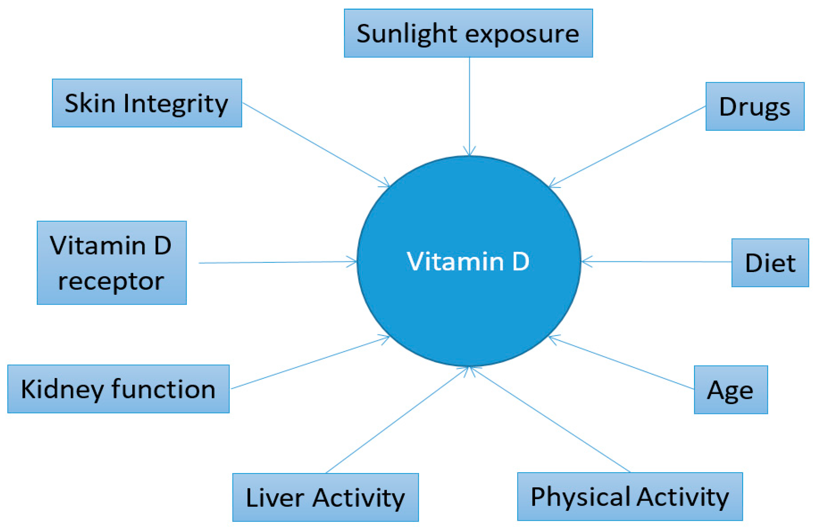
:1. Introduction
2. Materials and Methods
2.1. Subjects
2.2. Questionnaire Evaluating Fatigue
2.3. Laboratory Measurements
2.4. Statistics Analysis
3. Results
4. Discussion
4.1. Main Findings
4.2. Limitations
5. Conclusions
Author Contributions
Funding
Conflicts of Interest
References
- Chaudhuri, A.; Behan, P.O. Fatigue in neurological disorders. Lancet 2004, 363, 978–988. [Google Scholar] [CrossRef]
- Meng, H.; Hale, L.; Friedberg, F. Prevalence and Predictors of Fatigue among Middle-Aged and Older Adults: Evidence from the Health and Retirement Study. J. Am. Geriatr. Soc. 2010, 58, 2033–2034. [Google Scholar] [CrossRef] [PubMed]
- Arlington, V.A. Diagnostic and Statistical Manual of Mental Disorders: DSM-5TM, 5th ed.; American Psychiatric Publishing, Inc.: Washington, DC, USA, 2013. [Google Scholar]
- Bower, J.E. Fatigue, brain, behavior, and immunity: Summary of the 2012 Named Series on fatigue. Brain Behav. Immun. 2012, 26, 1220–1223. [Google Scholar] [CrossRef] [PubMed]
- Kroenke, K.; Arrington, M.E.; Mangelsdorff, A.D. The prevalence of symptoms in medical outpatients and the adequacy of therapy. Arch. Intern. Med. 1990, 150, 1685–1689. [Google Scholar] [CrossRef] [PubMed]
- Swain, M.G. Fatigue in chronic disease. Clin. Sci. 2000, 99, 1–8. [Google Scholar] [CrossRef] [PubMed] [Green Version]
- Huppertz-Hauss, G.; Høivik, M.L.; Jelsness-Jørgensen, L.-P.; Opheim, R.; Henriksen, M.; Høie, O. Fatigue in a population-based cohort of patients with inflammatory bowel disease 20 years after diagnosis: The IBSEN study. Scand. J. Gastroenterol. 2017, 52, 351–358. [Google Scholar] [CrossRef] [PubMed]
- Roy, S.; Sherman, A.; Monari-Sparks, M.J.; Schweiker, O.; Hunter, K. Correction of Low Vitamin D Improves Fatigue: Effect of Correction of Low Vitamin D in Fatigue Study (EViDiF Study). N. Am. J. Med. Sci. 2014, 6, 396–402. [Google Scholar] [CrossRef]
- Nowak, A.; Boesch, L.; Andres, E.; Battegay, E.; Hornemann, T.; Schmid, C.; Bischoff-Ferrari, H.A.; Suter, P.M.; Krayenbuehl, P.A. Effect of vitamin D3 on self-perceived fatigue: A double-blind randomized placebo-controlled trial. Medicine 2016, 95, e5353. [Google Scholar] [CrossRef]
- Holick, M.F. Vitamin D deficiency. N. Engl. J. Med. 2007, 357, 266–281. [Google Scholar] [CrossRef]
- Boccardi, V.; Lapenna, M.; Gaggi, L.; Garaffa, F.M.; Croce, M.F.; Baroni, M.; Ercolani, S.; Mecocci, P.; Ruggiero, C. Hypovitaminosis D: A Disease Marker in Hospitalized Very Old Persons at Risk of Malnutrition. Nutrients 2019, 11, 128. [Google Scholar] [CrossRef]
- US Centers for Disease Control and Prevention. Second National Report on Biochemical Indicators of Diet and Nutrition in the U.S. Population 2012; National Center for Environmental Health: Atlanta, GA, USA, April 2012. [Google Scholar]
- Holick, M.F.; Binkley, N.C.; Bischoff-Ferrari, H.A.; Gordon, C.M.; Hanley, D.A.; Heaney, R.P.; Murad, M.H.; Weaver, C.M. Evaluation, treatment, and prevention of vitamin D deficiency: An Endocrine Society clinical practice guideline. J. Clin. Endocrinol. Metab. 2011, 96, 1911–1930. [Google Scholar] [CrossRef] [PubMed]
- Stewart, J.W.; Alekel, D.L.; Ritland, L.M.; Van Loan, M.; Gertz, E.; Genschel, U. Serum 25-hydroxyvitamin D is related to indicators of overall physical fitness in healthy postmenopausal women. Menopause 2009, 16, 1093–1101. [Google Scholar] [CrossRef] [PubMed] [Green Version]
- Plotnikoff, G.A.; Quigley, J.M. Prevalence of Severe Hypovitaminosis D in Patients with Persistent, Nonspecific Musculoskeletal Pain. Mayo Clin. Proc. 2003, 78, 1463–1470. [Google Scholar] [CrossRef] [PubMed]
- Turner, M.K.; Hooten, W.M.; Schmidt, J.E.; Kerkvliet, J.L.; Townsend, C.O.; Bruce, B.K. Prevalence and clinical correlates of vitamin D inadequacy among patients with chronic pain. Pain Med. 2008, 9, 979–984. [Google Scholar] [CrossRef] [PubMed]
- Lotfi, A.; Abdel-Nasser, A.M.; Hamdy, A.; Omran, A.A.; El-Rehany, M.A. Hypovitaminosis D in female patients with chronic low back pain. Clin. Rheumatol. 2007, 26, 1895–1901. [Google Scholar] [CrossRef] [PubMed]
- Goldstein, M.R. Myopathy, statins, and vitamin D deficiency. Am. J. Cardiol. 2007, 100, 1328. [Google Scholar] [CrossRef]
- Bolton, C.F. Neuromuscular manifestations of critical illness. Muscle Nerv. 2005, 32, 140–163. [Google Scholar] [CrossRef]
- Hoeck, A.D.; Pall, M.L. Will vitamin D supplementation ameliorate diseases characterized by chronic inflammation and fatigue? Med. Hypotheses 2011, 76, 208–213. [Google Scholar] [CrossRef]
- Tague, S.E.; Clarke, G.L.; Winter, M.K.; McCarson, K.E.; Wright, D.E.; Smith, P.G. Vitamin D Deficiency Promotes Skeletal Muscle Hypersensitivity and Sensory Hyperinnervation. J. Neurosci. 2011, 31, 13728–13738. [Google Scholar] [CrossRef]
- Choi, M.; Park, H.; Cho, S.; Lee, M. Vitamin D3 supplementation modulates inflammatory responses from the muscle damage induced by high-intensity exercise in SD rats. Cytokine 2013, 63, 27–35. [Google Scholar] [CrossRef]
- Jamka, M.; Woźniewicz, M.; Jeszka, J.; Mardas, M.; Bogdański, P.; Stelmach-Mardas, M. The effect of vitamin D supplementation on insulin and glucose metabolism in overweight and obese individuals: Systematic review with meta-analysis. Sci. Rep. 2015, 5, 16142. [Google Scholar] [CrossRef] [PubMed]
- Peterson, C.A.; Heffernan, M.E. Serum tumor necrosis factor-alpha concentrations are negatively correlated with serum 25(OH)D concentrations in healthy women. J. Inflamm. 2008, 5, 10. [Google Scholar] [CrossRef] [PubMed]
- Krupp, L.B.; LaRocca, N.G.; Muir-Nash, J.; Steinberg, A.D. The fatigue severity scale. Application to patients with multiple sclerosis and systemic lupus erythematosus. Arch. Neurol. 1989, 46, 1121–1123. [Google Scholar] [CrossRef] [PubMed]
- Wessely, S.; Powell, R. Fatigue syndromes: Acomparison of chronic postviral fatigue with neuromuscular affective disorders. J. Neurol. Neurosurg. Psychiatry 1989, 52, 940–948. [Google Scholar] [CrossRef]
- Chalder, T.; Berelowitz, G.; Pawlikowska, T.; Watts, L.; Wessely, S.; Wright, D.; Wallace, E.P. Development of a fatigue scale. J. Psychosom. Med. 1993, 37, 147–153. [Google Scholar] [CrossRef] [Green Version]
- Pistone, G.; Marino, A.; Leotta, C.; Dell’Arte, S.; Finocchiaro, G.; Malaguarnera, M. Levocarnitine administration in elderly subjects with rapid muscle fatigue: Effect on body composition, lipid profile and fatigue. Drugs Aging 2003, 20, 761–767. [Google Scholar] [CrossRef]
- Malaguarnera, M.; Gargante, M.P.; Cristaldi, E.; Colonna, V.; Messano, M.; Koverech, A.; Neri, S.; Vacante, M.; Cammalleri, L.; Motta, M. Acetyl L-carnitine (ALC) treatment in elderly patients with fatigue. Arch. Gerontol. Geriatr. 2008, 46, 181–190. [Google Scholar] [CrossRef]
- Pennisi, M.; Di Bartolo, G.; Malaguarnera, G.; Bella, R.; Lanza, G.; Malaguarnera, M. Vitamin D Serum Levels in Patients with Statin-Induced Musculoskeletal Pain. Dis. Markers 2019, 2019, 3549402. [Google Scholar] [CrossRef]
- de Carvalho, J.F.; da Rocha Araújo, F.A.G.; da Mota, L.M.A.; Aires, R.B.; de Araujo, R.P. Vitamin D Supplementation Seems to Improve Fibromyalgia Symptoms: Preliminary Results. Isr. Med. Assoc. J. 2018, 20, 379–381. [Google Scholar]
- Earl, K.E.; Sakellariou, G.K.; Sinclair, M.; Fenech, M.; Croden, F.; Owens, D.J.; Tang, J.; Miller, A.; Lawton, C.; Dye, L.; et al. Vitamin D status in chronic fatigue syndrome/myalgic encephalomyelitis: A cohort study from the North-West of England. BMJ Open 2017, 7, e015296. [Google Scholar] [CrossRef]
- Latham, N.K.; Anderson, C.S.; Lee, A.; Bennett, D.A.; Moseley, A.; Cameron, I.D.; Fitness Collaborative Group. A randomized, controlled trial of quadriceps resistance exercise and vitamin D in frail older people: The Frailty Interventions Trial in Elderly Subjects (FITNESS). J. Am. Geriatr. Soc. 2003, 51, 291–299. [Google Scholar] [CrossRef] [PubMed]
- Vayá, A.; Bonet, E.; Romagnoli, M.; Nuñez, C.; Todoli, J. Erythrocyte deformability in macrocytosis determined by means of ektacytometry techniques. Clin. Hemorheol. Microcirc. 2010, 45, 27–33. [Google Scholar] [PubMed]
- Cita, K.C.; Brureau, L.; Lemonne, N.; Billaud, M.; Connes, P.; Ferdinand, S.; Tressières, B.; Tarer, V.; Etienne-Julan, M.; Blanchet, P.; et al. Men with Sickle Cell Anemia and Priapism Exhibit Increased Hemolytic Rate, Decreased Red Blood Cell Deformability and Increased Red Blood Cell Aggregate Strength. PLoS ONE 2016, 11, e0154866. [Google Scholar] [CrossRef] [PubMed]
- Tsuda, K. Associations between High-Sensitivity C-Reactive Protein and Membrane Fluidity of Red Blood Cells in Hypertensive Elderly Men: An Electron Spin Resonance Study. Int. J. Hypertens. 2012, 2012, 292803. [Google Scholar] [CrossRef]
- Doudin, A.; Becker, A.; Rothenberger, A.; Meyer, T. Relationship between serum 25-hydroxyvitamin D and red blood cell indices in German adolescents. Eur. J. Pediatr. 2018, 177, 583–591. [Google Scholar] [CrossRef]
- Saha, A.K.; Schmidt, B.R.; Wilhelmy, J.; Nguyen, V.; Abugherir, A.; Do, J.K.; Nemat-Gorgani, M.; Davis, R.W.; Ramasubramanian, A.K. Red blood cell deformability is diminished in patients with Chronic Fatigue Syndrome. Clin. Hemorheol. Microcirc. 2019, 71, 113–116. [Google Scholar] [CrossRef]
- MacLaughlin, J.; Holick, M.F. Aging decreases the capacity of human skin to produce vitamin D3. J. Clin. Investig. 1985, 76, 1536–1538. [Google Scholar] [CrossRef]
- Van der Wielen, R.P.; De Groot, L.C.P.G.M.; Van Staveren, W.A.; Löwik, M.R.H.; Van den Berg, H.; Haller, J.; Moreiras, O. Serum vitamin D concentrations among elderly people in Europe. Lancet 1995, 346, 207–210. [Google Scholar] [CrossRef]
- Chapuy, M.C.; Schott, A.M.; Garnero, P.; Hans, D.; Delmas, P.D.; Meunier, P.J. Healthy elderly French women living at home have secondary hyperparathyroidism and high bone turnover in winter. EPIDOS Study Group. J. Clin. Endocrinol. Metab. 1996, 81, 1129–1133. [Google Scholar]
- Aguado, P.; del Campo, M.T.; Garcés, M.V.; González-Casaús, M.L.; Bernad, M.; Gijón-Baños, J.; Martín Mola, E.; Torrijos, A.; Martínez, M.E. Low vitamin D levels in outpatient postmenopausal women from a rheumatology clinic in Madrid, Spain: Their relationship with bone mineral density. Osteoporos. Int. 2000, 11, 739–744. [Google Scholar] [CrossRef]
- Romagnoli, E.; Caravella, P.; Scarnecchia, L.; Martinez, P.; Minisola, S. Hypovitaminosis D in an Italian population of healthy subjects and hospitalized patients. Br. J. Nutr. 1999, 81, 133–137. [Google Scholar] [CrossRef] [PubMed] [Green Version]
- Reginster, J.Y.; Halkin, V.; Henrotin, Y.; Gosset, C. Treatment of osteoporosis: Role of bone-forming agents. Osteoporos. Int. 1999, 9 (Suppl. 2), S91–S96. [Google Scholar] [CrossRef] [PubMed]
- Eriksen, E.F.; Glerup, H. Vitamin D deficiency and aging: Implications for general health and osteoporosis. Biogerontology 2002, 3, 73–77. [Google Scholar] [CrossRef] [PubMed]
- Chan, R.; Woo, J. The value of vitamin D supplementation in older people. Nutr. Ther. Metab. 2011, 29, 8–21. [Google Scholar]
- Holick, M.F. Vitamin D: A d-lightful solution for health. J. Investig. Med. 2011, 59, 872–880. [Google Scholar] [CrossRef]
- Cesari, M.; Incalzi, R.A.; Zamboni, V.; Pahor, M. Vitamin D hormone: A multitude of actions potentially influencing the physical function decline in older persons. Geriatr. Gerontol. Int. 2011, 11, 133–142. [Google Scholar] [CrossRef]
- Havdahl, A.; Mitchell, R.; Paternoster, L.; Davey Smith, G. Investigating causality in the association between vitamin D status and self-reported tiredness. Sci. Rep. 2019, 9, 2880. Available online: https://0-www-ncbi-nlm-nih-gov.brum.beds.ac.uk/pmc/articles/PMC6393455/ (accessed on 27 February 2019). [CrossRef]
- Engberg, I.; Segerstedt, J.; Waller, G.; Wennberg, P.; Eliasson, M. Fatigue in the general population-associations to age, sex, socioeconomic status, physical activity, sitting time and self-rated health: The northern Sweden MONICA study 2014. BMC Public Health 2017, 17, 654. [Google Scholar] [CrossRef]
- Ginde, A.A.; Liu, M.C.; Camargo, C.A. Demographic Differences and Trends of Vitamin D Insufficiency in the US Population, 1988–2004. Arch. Intern. Med. 2009, 169, 626–632. [Google Scholar] [CrossRef]
- Goedendorp, M.M.; Knoop, H.; Schippers, G.M.; Bleijenberg, G. The lifestyle of patients with chronic fatigue syndrome and the effect on fatigue and functional impairments. J. Hum. Nutr. Diet. 2009, 22, 226–231. [Google Scholar] [CrossRef]
- Taylor, B.A.; Lorson, L.; White, C.M.; Thompson, P.D. Low vitamin D does not predict statin associated muscle symptoms but is associated with transient increases in muscle damage and pain. Atherosclerosis 2017, 256, 100–104. [Google Scholar] [CrossRef] [PubMed]
- Bhat, M.; Ismail, A. Vitamin D treatment protects against and reverses oxidative stress induced muscle proteolysis. J. Steroid Biochem. Mol. Biol. 2015, 152, 171–179. [Google Scholar] [CrossRef] [PubMed]
- Oliveri, B.; Plantalech, L.; Bagur, A.; Wittich, A.C.; Rovai, G.; Pusiol, E.; López Giovanelli, J.; Ponce, G.; Nieva, A.; Chaperón, A.; et al. High prevalence of vitamin D insufficiency in healthy elderly people living at home in Argentina. Eur. J. Clin. Nutr. 2004, 58, 337–342. [Google Scholar] [CrossRef] [PubMed] [Green Version]
- Bettica, P.; Bevilacqua, M.; Vago, T.; Norbiato, G. High prevalence of hypovitaminosis D among free-living postmenopausal women referred to an osteoporosis outpatient clinic in northern Italy for initial screening. Osteoporos. Int. 1999, 9, 226–229. [Google Scholar] [CrossRef] [PubMed]


| Variable | Patients | Controls | p |
|---|---|---|---|
| Age range, years (mean ± SD) | 69.10 ± 5.80 | 69.20 ± 5.10 | / |
| Heart rate, bpm (mean ± SD) | 82.80 ± 8.20 | 81.80 ± 8.60 | NS |
| Systolic blood pressure, mmHg (mean ± SD) | 140.00 ± 9.10 | 138.20 ± 9.60 | <0.001 |
| Diastolic blood pressure, mmHg (mean ± SD) | 79.00 ± 7.50 | 79.20 ± 7.20 | NS |
| Body Mass Index, Kg/m2 (mean ± SD) | 24.80 ± 3.40 | 24.40 ± 3.20 | NS |
| Current/Former smoker (%) | 38.70 | 37.91 | / |
| Diabetes Mellitus (%) | 9.16 | 10.00 | / |
| Hypertension (%) | 17.08 | 17.50 | / |
| Heart insufficiency (%) | 4.58 | 4.16 | / |
| Education, no Diploma (%) | 48.33 | 42.91 | / |
| Education. High School Diploma (%) | 30.41 | 30.41 | / |
| Education, University Degree (%) | 20.83 | 14.16 | / |
| Variable | Normal Range | Patients | Controls | p |
|---|---|---|---|---|
| CK, IU/L | 10.00–171.00 | 36.70 ± 11.80 | 37.00 ± 11.40 | NS |
| Bilirubin, mg/dl | 0.30–1.20 | 0.80 ± 0.10 | 0.81 ± 0.10 | NS |
| Albumin, g/dl | 3.50–4.80 | 4.20 ± 0.35 | 4.20 ± 0.31 | NS |
| AST, IU/L | 5.00–45.00 | 36.00 ± 7.20 | 35.00 ± 6.80 | NS |
| ALT, IU/L | 5.00–45.00 | 37.10 ± 9.00 | 36.50 ± 9.20 | NS |
| γGT, IU/L | 5.00–55.00 | 33.20 ± 8.60 | 32.10 ± 9.70 | NS |
| Creatinine, mg/dL | 0.50–1.10 | 0.93 ± 0.20 | 0.91 ± 0.23 | NS |
| Vitamin D, nmol/L (median; range) | 75.00–200.00 | 39.50 ± 11.80 (41.60; 25.00–51.00) | 48.10 ± 13.80 (60.20; 36.00–68.00) | <0.001 |
| CRP, mg/L (median; range) | 0.00–4.80 | 4.31 ± 1.70 (3.96; 1.60–5.20) | 1.44 ± 1.10 (1.30; 0.50–2.00) | <0.001 |
| Calcium, mg/dl | 8.10–10.10 | 8.50 ± 1.90 | 8.40 ± 2.00 | NS |
| Phosphorus, mg/dl | 2.50–4.50 | 2.40 ± 0.70 | 2.90 ± 0.70 | <0.001 |
| WBC, 103 U/L | 4.00–9.50 | 6.70 ± 1.20 | 6.50 ± 1.30 | NS |
| PLT, 103 U/L | 150.00–410.00 | 304.00 ± 26.00 | 298.00 ± 32.00 | <0.05 |
| RBC, 106 U/L | 4.50–6.10 | 4.64 ± 0.82 | 4.52 ± 0.91 | NS |
| HGB, g/dl | 13.00–18.00 | 14.20 ± 0.70 | 14.00 ± 0.87 | <0.05 |
| HCT, % | 38.00–50.00 | 44.10 ± 0.60 | 43.90 ± 0.80 | <0.05 |
| MCV, fL | 80.00–102.00 | 94.10 ± 0.80 | 93.80 ± 0.90 | <0.001 |
| MCH, pg | 26.00–33.00 | 30.20 ± 0.80 | 30.60 ± 0.70 | <0.001 |
| RDW, % | 9.80–16.00 | 14.10 ± 1.80 | 13.80 ± 2.00 | NS |
| Iron, µg/dL | 65.00–170.00 | 87.40 ± 11.80 | 82.60 ± 13.60 | <0.001 |
| Vitamin B12, pg/dL | 180.00–914.00 | 424.20 ± 26.80 | 412.80 ± 31.90 | <0.001 |
| Folic acid, ng/dL | 3.10–19.90 | 8.78 ± 0.34 | 8.61 ± 0.42 | <0.001 |
| Scale (mean ± SD) | Patients | Controls | p |
|---|---|---|---|
| Physical fatigue scale score | 11.5 ± 1.2 | 6.9 ± 1.3 | <0.0001 |
| Mental fatigue scale score | 6.7 ± 1.8 | 5.3 ± 1.2 | <0.0001 |
| Fatigue severity score | 46.1 ± 3.2 | 29.5 ± 3.2 | <0.0001 |
| Variable | Patients | Controls | Intergroup Comparison | |||||
|---|---|---|---|---|---|---|---|---|
| F | M | P (M vs. F) | F | M | P (M vs. F) | p (Control F vs. Patient F) | P (Control M vs. Patient M) | |
| Physical fatigue scale | 11.0 ± 1.2 | 12.1 ± 1.3 | <0.0001 | 6.7 ± 1.2 | 7.1 ± 1.4 | 0.0731 | <0.0001 | <0.0001 |
| Mental fatigue scale | 6.4 ± 1.8 | 7.1 ± 1.7 | 0.0017 | 5.1 ± 1.1 | 5.6 ± 1.2 | 0.0468 | <0.0001 | <0.0001 |
| Fatigue severity scale | 44.2 ± 3.2 | 48.2 ± 3.2 | <0.0001 | 28.2 ± 3.1 | 31.8 ± 3.6 | <0.0001 | <0.0001 | <0.0001 |
| Vitamin D, nmol/L | 39.2 ± 14.9 | 40.2 ± 12.8 | NS | 46.9 ± 21.4 | 48.2 ± 22.7 | NS | 0.0045 | 0.0075 |
© 2019 by the authors. Licensee MDPI, Basel, Switzerland. This article is an open access article distributed under the terms and conditions of the Creative Commons Attribution (CC BY) license (http://creativecommons.org/licenses/by/4.0/).
Share and Cite
Pennisi, M.; Malaguarnera, G.; Di Bartolo, G.; Lanza, G.; Bella, R.; Chisari, E.M.; Cauli, O.; Vicari, E.; Malaguarnera, M. Decrease in Serum Vitamin D Level of Older Patients with Fatigue. Nutrients 2019, 11, 2531. https://0-doi-org.brum.beds.ac.uk/10.3390/nu11102531
Pennisi M, Malaguarnera G, Di Bartolo G, Lanza G, Bella R, Chisari EM, Cauli O, Vicari E, Malaguarnera M. Decrease in Serum Vitamin D Level of Older Patients with Fatigue. Nutrients. 2019; 11(10):2531. https://0-doi-org.brum.beds.ac.uk/10.3390/nu11102531
Chicago/Turabian StylePennisi, Manuela, Giulia Malaguarnera, Giuseppe Di Bartolo, Giuseppe Lanza, Rita Bella, Eleonora Margherita Chisari, Omar Cauli, Enzo Vicari, and Michele Malaguarnera. 2019. "Decrease in Serum Vitamin D Level of Older Patients with Fatigue" Nutrients 11, no. 10: 2531. https://0-doi-org.brum.beds.ac.uk/10.3390/nu11102531