Article

Hesperetin, a Citrus Flavonoid, Attenuates LPS-Induced Neuroinflammation, Apoptosis and Memory Impairments by Modulating TLR4/NF-κB Signaling

Division of Applied Life Science (BK 21), College of Natural Sciences, Gyeongsang National University, Jinju 52828, Korea
*
Author to whom correspondence should be addressed.
These authors contributed equally to this paper.
Submission received: 1 February 2019 / Revised: 9 March 2019 / Accepted: 13 March 2019 / Published: 17 March 2019

Abstract

:
Glial activation and neuroinflammation play significant roles in apoptosis as well as in the development of cognitive and memory deficits. Neuroinflammation is also a critical feature in the pathogenesis of neurodegenerative disorders such as Alzheimer and Parkinson’s diseases. Previously, hesperetin has been shown to be an effective antioxidant and anti-inflammatory agent. In the present study, in vivo and in vitro analyses were performed to evaluate the neuroprotective effects of hesperetin in lipopolysaccharide (LPS)-induced neuroinflammation, oxidative stress, neuronal apoptosis and memory impairments. Based on our findings, LPS treatment resulted in microglial activation and astrocytosis and elevated the expression of inflammatory mediators such as phosphorylated-Nuclear factor-κB (p-NF-κB), tumor necrosis factor-α (TNF-α), and interleukin-1β (IL-1β) in the cortical and hippocampal regions and in BV2 cells. However, hesperetin cotreatment markedly reduced the expression of inflammatory cytokines by ameliorating Toll-like receptor-4 (TLR4)-mediated ionized calcium-binding adapter molecule 1/glial fibrillary acidic protein (Iba-1/GFAP) expression. Similarly, hesperetin attenuated LPS-induced generation of reactive oxygen species/lipid per oxidation (ROS/LPO) and improved the antioxidant protein level such as nuclear factor erythroid 2-related factor 2 (Nrf2) and Haem-oxygenase (HO-1) in the mouse brain. Additionally, hesperetin ameliorated cytotoxicity and ROS/LPO induced by LPS in HT-22 cells. Moreover, hesperetin rescued LPS-induced neuronal apoptosis by reducing the expression of phosphorylated-c-Jun N-terminal kinases (p-JNK), B-cell lymphoma 2 (Bcl-2)-associated X protein (Bax), and Caspase-3 protein and promoting the Bcl-2 protein level. Furthermore, hesperetin enhanced synaptic integrity, cognition, and memory processes by enhancing the phosphorylated-cAMP response element binding protein (p-CREB), postsynaptic density protein-95 (PSD-95), and Syntaxin. Overall, our preclinical study suggests that hesperetin conferred neuroprotection by regulating the TLR4/NF-κB signaling pathway against the detrimental effects of LPS.

Graphical Abstract

1. Introduction

Neuroinflammation is a critical feature in the pathophysiology of neurodegenerative diseases such as Alzheimer’s disease (AD), Parkinson’s disease (PD), amyotrophic lateral sclerosis, and frontotemporal dementia [1]. Accumulating evidence suggests that reactive microglia and astrocytosis (gliosis) play significant roles in neuroinflammation [2], and the activation of these glial cells releases several inflammatory cytokines, which eventually leads to neuroinflammation-mediated neurodegeneration [3]. Lipopolysaccharide (LPS) is a membrane component of Gram-negative bacteria [4], which activates the immune system and leads to behavioral and memory impairments, oxygen species (ROS) generation, lipid per oxidation (LPO), and consequently oxidative brain damage [5,6]. Previous studies suggest that dormant, non-growing bacteria are a crucial contributing factor in AD, and these bacteria release several inflammatory components such as LPS [3,7]. The LPS level in the plasma is raised in AD and is closely associated with aggregated protein in the brain [7].
The Toll-like receptor (TLR) is a pattern-recognition receptor that detects microbial components, and plays an important role in the initiation of the immune response [8]. The TLR-mediated signaling pathway is involved in removing bacteria but can also have detrimental effects on brain cells. TLR4, which is expressed in microglia, astrocytes, and macrophages in the brain, recognizes LPS and is involved in the release of inflammatory mediators by activating the NF-κB signaling pathway. Studies have demonstrated that TLR4-deficient/knockout mice models were protected and from neuroinflammation and other cognitive impairments [9,10,11]. A recent study indicates that amyloid-β oligomer (AβO) activates glial cells and elevates the expression of inflammatory cytokine suggesting its role in TLR4-induced memory impairments [12]. Additionally, LPS induces oxidative stress, an imbalance between the levels of ROS/LPO and the biological system’s ability to detoxify the reactive byproducts or its failure to repair the subsequent damage [13]. Oxidative brain damage is implicated in the pathogenesis of numerous neurological disorders [14], and the main targets of the elevated level of ROS are the endogenous antioxidant mechanisms such as Nrf-2 and HO-1 [15]. Additionally, LPS-induced neuroinflammation and oxidative stress-mediated neuronal apoptosis by promoting ROS generation and caspase activation as well as behavioral and memory impairments [9,16,17].
Flavonoid intake is associated with a lower risk of several disease conditions, such as neurodegenerative and cardiovascular disease [18]. Hesperetin, a flavanone class of flavonoid, is a derivative of hesperidin found in citrus fruits such as oranges and grapefruit [19]. Hesperetin has also been shown to protect primary mouse neurons and protects the mouse brain against STZ-induced memory deficits and neuronal apoptosis [20,21,22]. Moreover, another study has demonstrated that hesperetin inhibits inflammation in animal models and protects prostatic endothelial cells [23,24]. In the current preclinical study, we explored the neuroprotective effect of hesperetin against LPS-induced glial activation, neuroinflammation, neurodegeneration, synaptic dysfunction, and memory impairments in adult mice. Moreover, we further confirmed the anti-inflammatory, cytoprotective and anti-oxidant effects of hesperetin in BV2 and HT-22 cells respectively.

2. Materials and Methods

2.1. Chemicals

LPS, hesperetin, 2′7′-dichlorodihydrofluorescein diacetate (DCFHDA), TAK242, and BAY 11-7082 were obtained from Sigma-Aldrich Chemicals Company (St. Louis, MO, USA).

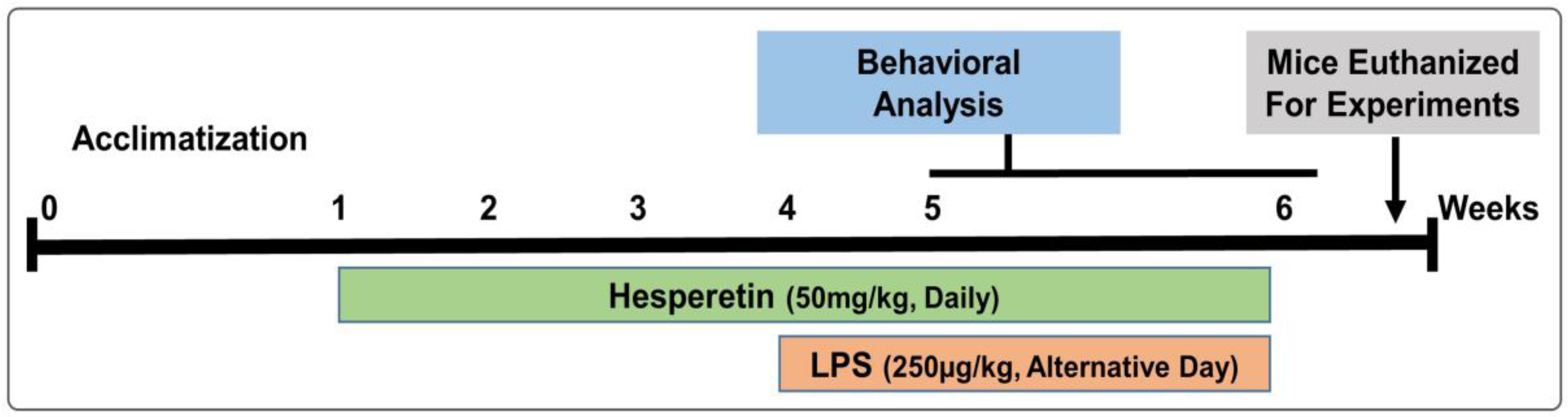
2.2. Animals and Drug Treatment

Male 7–8-week-old C57BL/6 N mice (n = 60, 23 ± 4 gm) were purchased from Samtako Bio (Osan, Republic of Korea). The study was approved and conducted in accordance with the guidelines of the Institutional Animal Care and Use Committee (IACUC) of the Division of Applied Life Science, Gyeongsang National University, South Korea (Approval ID: 125). Mice were acclimated for 1 week in the animal care facility under normal laboratory conditions with a 12 h light/dark cycle, at 40% humidity and room temperature, and given ad libitum access to food and water. The mice were divided into four groups randomly; (1) the control vehicle (normal saline) administered group; (2) the LPS-treated (250 µg/kg/day) group; (3) the LPS (250 µg/kg of body weight, alternative day) + hesperetin-treated (50 mg/kg of body weight, daily) group; and (4) the hesperetin (50 mg/kg of body weight) [25] alone group. LPS dissolved in saline was administered intraperitoneally (i.p) for 2 weeks (a total of 7 doses administered on an alternate day). Hesperetin suspended in water containing 0.1% carboxymethyl cellulose, was administered daily via oral gavage which was started three weeks before LPS treatment and continued for another 2 weeks together with LPS that is a total of five weeks (Figure 1).
Extraction of the proteins from the brain of mice (8–10 mice per group) was performed as previously described [26,27]. Briefly, mice were euthanized after giving the proper anesthesia (combination of 10 µL tiletamine/zolazepam and 10 µL xylazine), and the brain parts (specifically cortical and hippocampal regions) were collected [28] and deep frozen in liquid nitrogen followed by transfer to −80 °C.

2.3. Behavioral Analysis

Animal behavior analysis was performed by using Morris Water Maze (MWM) and Y-maze tests by using a video tracking software (SMART Panlab, Harvard Apparatus, Holliston, MA, USA). Behavioral analysis was conducted daily (after 1 h of drug/chemical administration) according to the plan of experiment. To evaluate the memory and learning performance, MWM was conducted as described previously [29,30] with some small modifications. After receiving 2 consecutive days of training, latency (sec) was measured to assess the time taken to reach the hidden platform for 6 consecutive days. On the following day, we performed the probe test for the evaluation of memory consolidation by removing the platform and allowing the animal to swim freely for 1 min. The crossing numbers over the previously hidden platform and the time spent in the specific target quadrant were measured.
Next, for Y-maze analysis, we placed each mouse in the middle of the device to freely move for 3–8 min in the maze. Entries into the arms were recorded digitally. Spontaneous alternation behavior is defined as the [successive triplet sets (consecutive entries into three different arms)/total number of arm entries-2] × 100. Improved memory and cognitive function were reflected by a higher % of spontaneous alternation behavior.

2.4. Western Blot Analyses

The brain tissues were homogenized in PRO-PREP extraction solution. We quantified the proteins via BIO-RAD assay and run through SDS-PAGE gel followed by transferring the proteins from the gels to PVDF membrane and blocking with 5% skim milk. The primary antibodies were attached and incubated overnight at 4 °C, washed with 1× TBST, and reacted with secondary antibodies for 1–2 h. The luminescence was observed by spraying the antibodies with a fluorescent solution, and the bands were obtained using X-ray films. The densities of the bands were quantified by using ImageJ, while graphs were generated by GraphPad Prism 6 software.

2.5. Antibodies

A list of antibodies (primary and secondary) used in this study is provided in the table below (Table 1).

2.6. Tissue Sample Preparation for Morphological Analysis

Mice were perfused transcardially with ice-cold PBS (0.01 M) followed by 4% neutrally buffered paraformaldehyde (NBP), and the brains were then post-fixed in 4% NBP for about 48–72 h. Next, the NBP was removed and washed with 1% PBS (0.01 M), and a 20% sucrose solution was then added for another 48 h period. After that, the brain sections were frozen in O.C.T and followed by obtaining 14 μM coronal cortical and hippocampal sections on a microscopic slide via CM-3050C-Cryostat (Leica, Germany).

2.7. Immunofluorescence Analysis

The immunofluorescence analysis was performed as described previously [31,32]. Briefly, mice 7–8/group) were euthanized and the brain were carefully collected and deep frozen in liquid nitrogen and then shifted to −80 °C. The brain blocks were made using OCT and the sections were taken via microtome (Leica) brain slicer. The slides with brain sections were washed with 1% PBS (0.01 M), incubated with primary antibodies (1:100 ratio in 1% PBS i.e., 0.01 M), and then washed with 1% PBS (0.01 M) and then secondary (FITC/TRITC, 1:50 ratio in 1% PBS i.e., 0.01 M) antibody attachments. Finally, brain sections were washed with 1% PBS (0.01 M). For nuclear staining, the sections were treated with 4′,6-diamidino-2-phenylindole dihydrochloride (DAPI) for 7–10 min and covered with glass coverslips using fluorescent mounting media, and the fluorescence was visualized under a laser-scanning confocal FluoView FV 1000 MPE (Olympus, Tokyo, Japan). Integrated density was used for the quantification of staining intensity and amount in the immunofluorescent microscopic image. ImageJ software ([email protected]., https://imagej.nih.gov/ij/) was used to quantify the integrated density which represents the sum of pixel values in an image.

2.8. In Vivo ROS and LPO Assays

The ROS level was evaluated as previously described with minor modifications [2], which is based on the oxidation of 7-dichlorodihydrofluorescein diacetate (DCFH-DA) to 2,7-dichlorodihydrofluorescein. Briefly, the cortical and hippocampal protein homogenates were diluted with Lock’s buffer (1:20 ratio) to a final 5 mg tissue/mL concentration, and 1 mL of Lock’s buffer, 0.2 mL of homogenate, and 10 mL of DCFH-DA (5 mM) followed by incubation at room temperature for 15 min to attain a fluorescent DCF (Dichlorofluorescin) product. The resulting fluorescent DCF level was measured via microplate reader (excitation at 484/530 nm). We used blanks (parallel) for any background fluorescence in the absence of a DCF product. The data are expressed as pmol DCF formed per min per mg of protein.
To measure the LPO for the evaluation of oxidative stress, free malondialdehyde (MDA) indicator of LPO was measured in the cortex and hippocampus protein homogenate using the MDA colorimetric/fluorometric kit (Cat #K739-100) in accordance with the instructions of the manufacturer.

2.9. Cresyl Violet (Nissl’s) Staining

For morphological examinations, we conducted Nissl’s staining, which indicates the number of neuronal cells loss. Briefly, as previously described [33,34], the brain slides were washed with 1% PBS (0.01 M) followed by staining with a solution of Cresyl violet (0.5%) comprising a few drops of glacial acetic acids for 8–10 min. After that, sections were washed with distilled water followed by dehydration in graded ethanol of 70%, 95%, and 100%. Finally, slides were immersed in xylene and covered with cover slips by using mounting medium (non-florescent). The effects of LPS with or without hesperetin were analyzed under a light microscope. The analysis was performed using ImageJ software, and the histograms were generated via GraphPad Prism 6 software.

2.10. In Vitro Cell Culture and Drug Treatment

Mouse hippocampal (HT-22) and murine microglia (BV2) cell lines were cultured in Dulbecco’s modified Eagle’s medium (DMEM) which was supplemented with FBS (10%) and antibiotic/antimycotic (1%) in an incubator supplied with 5% CO2 at 37 °C. After gaining 70% confluency, cells were treated with LPS (1 µg/mL), hesperetin (50 µM, the dose of hesperetin that significantly attenuated the LPS toxicity was selected on the basis of MTT assay and TAK242 (2 µM), or BAY 11-7082 (15 μM) for specific inhibition of the TLR4 and NF-κB. All the chemicals/drugs were treated and incubated for 24 h at 5% CO2 and 37 °C temperature.

2.11. In Vitro Western Blot Analysis

Similarly, BV2 cells were collected in 1% PBS (0.01 M) solution and centrifuged followed by the removal of supernatant. The remaining pellets were dissolved in PRO-PREP solution by vertexing and ultrasonication. Finally, the Western blot analysis was performed as mentioned for in vivo experiments.

2.12. In Vitro ROS and LPO Assays

For in vitro ROS analysis, we cultured HT-22 cells in DMEM (200 µL/well) in 96-well plates followed by incubation for 24 h in a humidified incubator (provided with 5% CO2 at 37 °C of temperature). After 24 h, LPS, hesperetin, or their combination was diluted in fresh media and used to replace the previous one. Again, after 24 h of the treatment, cells were exposed to DCFH-DA (50 μM) and incubated at 37 °C for 30 min followed by measurement (at 484/530 nm) of the relative absorbance of the ROS-positive cells treated with DCFHD.
Similarly, for in vitro LPO, we cultured HT-22 cells (2 × 10⁴/mL) in a DMEM medium in 96-well plates followed by incubation for 24 h in the humidified incubator (provided with 5% CO2 at 37 °C of temperature). After 24 h, LPS, hesperetin, or their combination was diluted in a fresh DMEM medium and used to replace the previous one, and LPO analysis was performed as mentioned for in vivo experiments.

2.13. MTT Assay

As previously described [35], an MTT assay was performed to evaluate the effect of LPS and/or hesperetin on cell viability. HT-22 cells were cultured at a density of 2 × 10⁴ cells/mL. After the cell attachment, media were replaced with fresh media containing 1, 10, and 50 µg/mL concentration of hesperetin or 1 µg/mL of LPS and incubated for further for 24 h at 5% CO2 at 37 °C temperature. After 24 h, 20 µL of MTT solution (5 mg/mL) was added for 4 h. Next, we removed 50 µL of media (containing MTT solution) from each well and added DMSO (100 µL/well i.e., 1:1.7 ratio). Finally, the plate was smoothly agitated for 10–20 min on a shaker, and the absorbance was then measured at 570 nm through a microplate reader. The experiments were repeated three times.

2.14. Statistical Analyses

In brief, for Western blot and morphological analysis, ImageJ software is used to measure the density to arbitrary units (A.U) and integrated density to A.U respectively, and data are presented as the mean ± SEM of triplicate wells for in vitro experiments and of 7–10 mice per group and are representative of three independent experiments. By using GraphPad Prism 6 software (San Diego, CA, USA), we performed one-way ANOVA with Tukey’s post hoc test. p < 0.05 is considered significant (significance: * p ≤ 0.05; ** p ≤ 0.01; *** p ≤ 0.001; **** p ≤ 0.0001).

3. Results

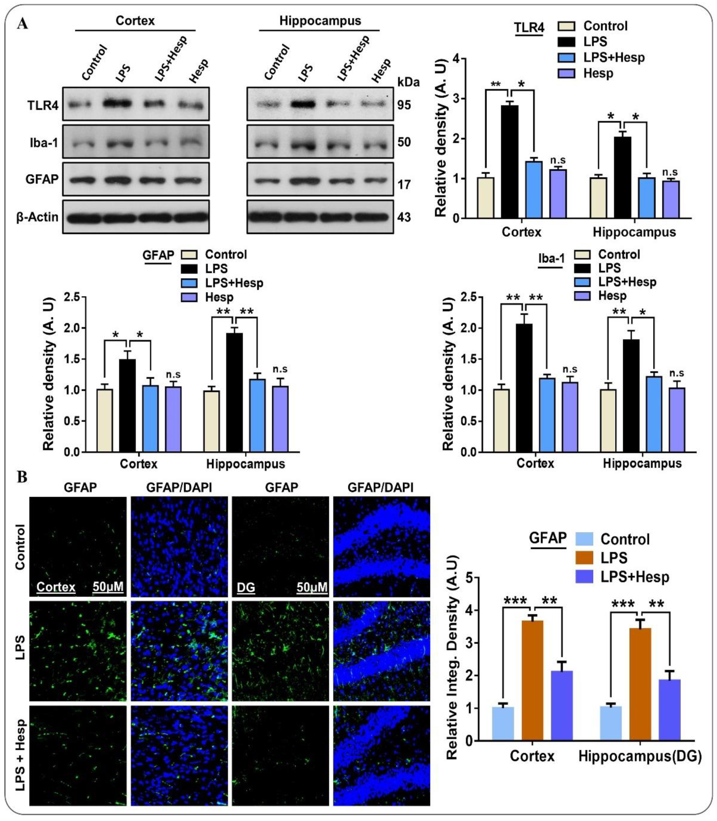
3.1. Effect of Hesperetin on LPS-Induced TLR4-Mediated Reactive Gliosis in the Mouse Brain

Microglia are the resident brain cells that maintain the microenvironment under the normal physiological conditions and play a critical role in the immune response [36]. Mounting literature indicates that glial cells such as microglia and astrocytes are the source of various mediators that significantly contribute to neuroinflammation, oxidative brain damage, and neuronal apoptosis [37,38]. For this, we assessed the potential beneficial effect of hesperetin against Iba-1 and GFAP which indicates activated microglia and astrocytosis respectively. Our immunoblot and immunofluorescence results indicated that LPS remarkably induced gliosis by significantly elevating the TLR4, GFAP, and Iba-1 protein expression in mice hippocampal and cortical regions compared to control saline-treated and hesperetin alone-treated groups. On the contrary, the LPS+hesperetin group presented significantly reduced levels (Figure 2A). Further, the immunofluorescence also confirmed the GFAP immunoblot results in the cortex and hippocampus. Hesperetin cotreatment substantially reduced the GFAP-reactive astrocyte cells compared to the LPS alone-treated group (Figure 2B).

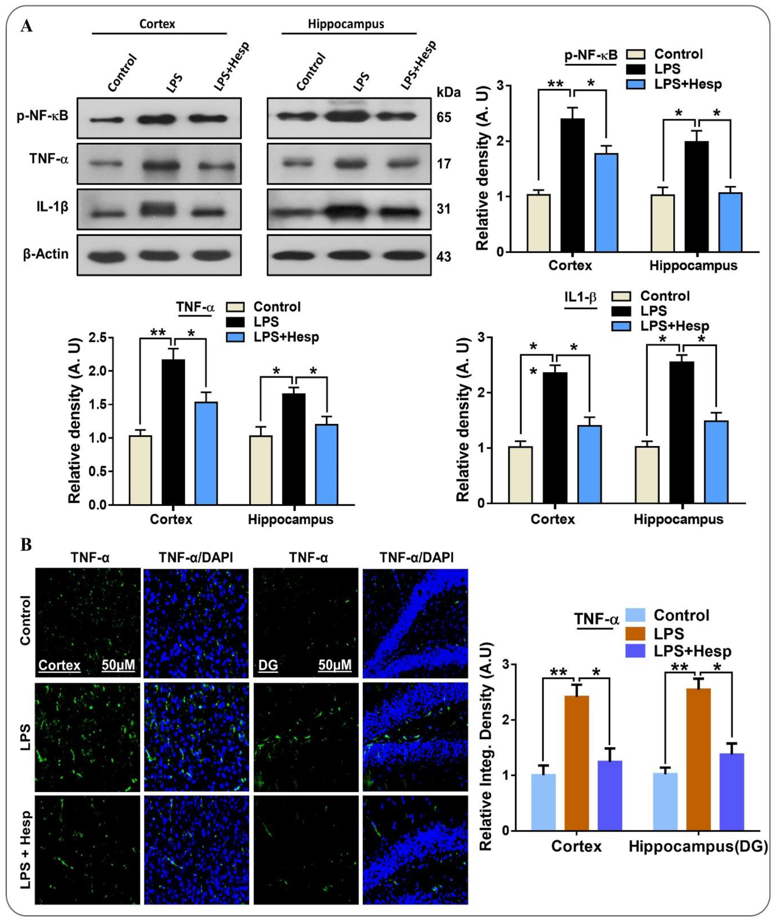
3.2. Hesperetin Mitigated Elevated p-NF-κB/TNF-α/IL-1β Expression in the LPS-Treated Mouse

Neuroinflammatory changes such as gliosis play an important role in the release of pro-inflammatory cytokines, and experimental and clinical studies have established that the expression of TNF-α and IL-1β and their receptors have been elevated in AD [36]. Similarly, the elevated p-NF-κB protein expression is the key transcription factor in neuroinflammation and apoptosis [37]. Additionally, the elevated NF-κB level has been found in the brains of AD patients, predominantly in the neuronal and glial cells near the neurofibrillary tangles and Aβ plaques [38]. LPS administration significantly increased p-NF-κB expression, but hesperetin cotreatment with LPS significantly reduced the p-NF-κB levels. The expression of inflammatory cytokines in LPS-induced activated glial cells is regulated by NF-κB signaling pathways [39]. Next, we examined the levels of TNF-α and IL-1β in cortex and hippocampus as well as in the BV2 cell lines. In the LPS-treated mice group, the level of these inflammatory cytokines was significantly increased in the cortex and hippocampus region. Conversely, hesperetin administration markedly reduced the expression of TNF-α and IL-1β (Figure 3A). Similarly, we confirmed TNF-α through confocal microscopy. The immunoreactivity TNF-α result showed that its protein expression was significantly increased by LPS treatment in the cortex and hippocampus and that hesperetin considerably reduced immunoreactivity (Figure 3B).

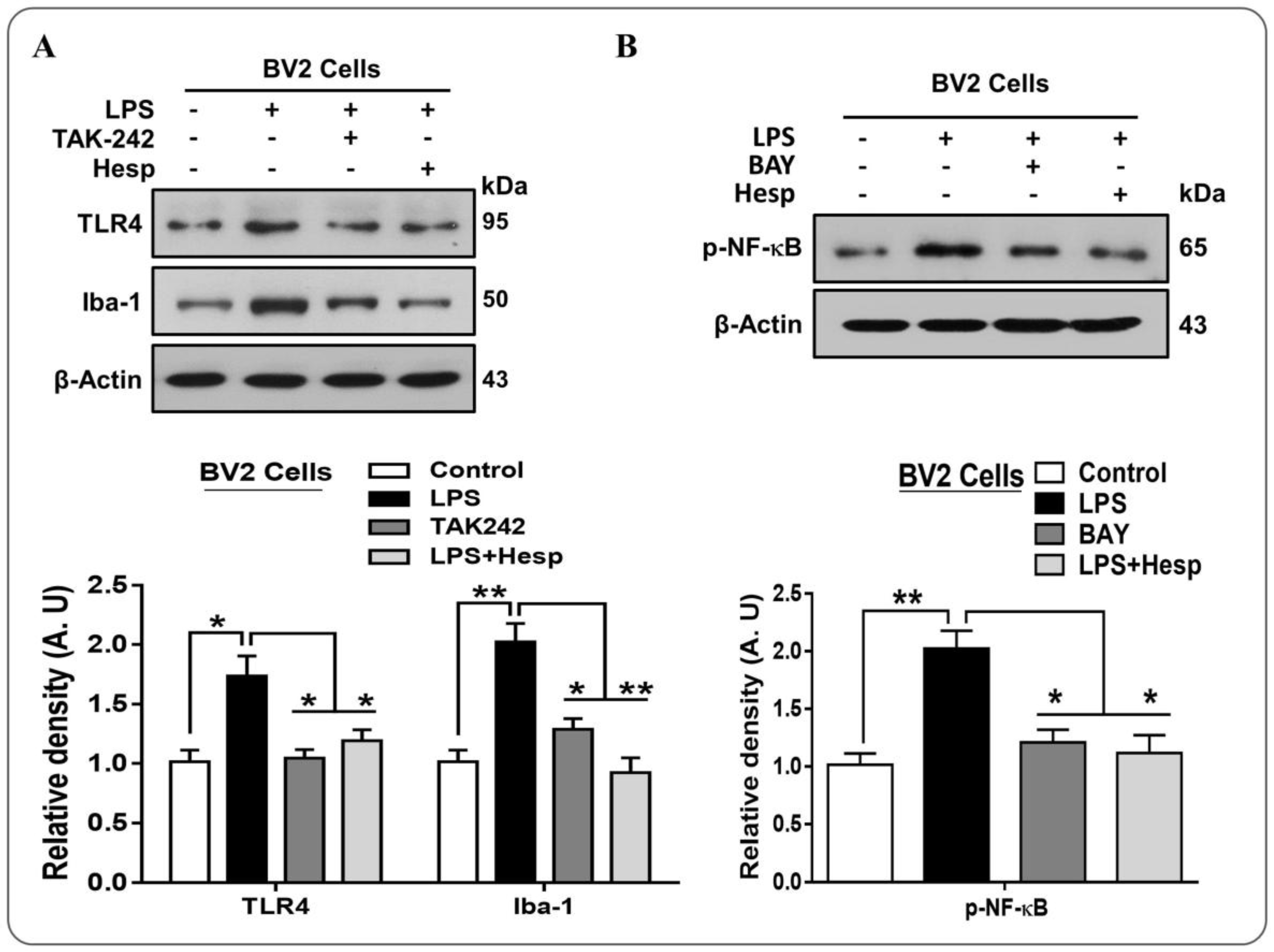
3.3. Hesperetin Alleviated the p-NF-κB/Iba-1/TNF-α Expression in LPS-Treated BV2 cells

Next, we confirmed the TLR4 and Iba-1 results in vitro in BV2 cells line, which supported the in vivo immunoblot results that LPS significantly exalted the TLR4 and Iba-1 protein level. However, hesperetin and TAK-242 (a TLR4 inhibitor) prevented LPS-induced overexpression of these proteins (Figure 4A). Furthermore, we confirmed these findings in vitro in BV2 cells, which demonstrated that LPS increased the p-NF-κB protein level, while hesperetin and BAY (BAY 11-7082, an NF-κB inhibitor) reduced this level significantly (Figure 4B).

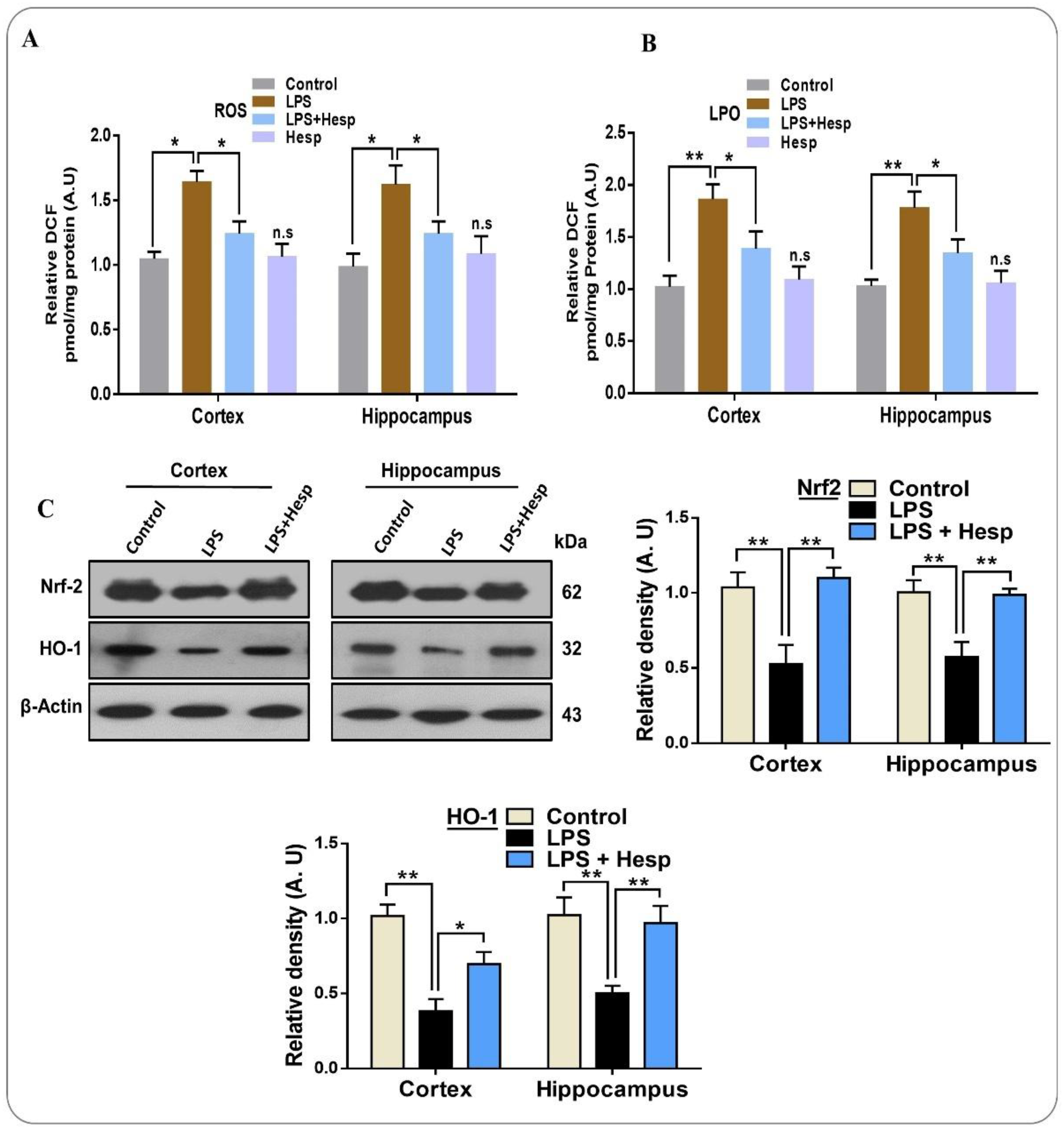
3.4. Hesperetin Reduced the ROS/LPO and Enhanced Nrf-2/HO-1 Level in LPS-Treated Mouse Brains

LPS-induced neuroinflammation results in the activation and recruitment of an increasing amount of neutrophils and macrophages, which propagates the generation of inflammatory cytokines and ROS, a key factor in the oxidative stress-mediated detrimental effects [40]. A higher ROS level further triggers a number of oxidative stress-related factors and reduces antioxidant proteins such as Nrf2 and HO1 [41]. Our results demonstrate that LPS-induced neuroinflammation considerably elevated the generation of ROS compared to the control group. Conversely, hesperetin significantly reduced the level of ROS in the cortex and hippocampus regions of the mouse brain. Likewise, hesperetin ameliorated the higher LPO level induced by LPS in the mouse brain (Figure 5A,B). Moreover, we checked the Nrf-2 and HO-1 expression level through immunoblot to further confirm the anti-oxidant effect of hesperetin. Our results revealed that the expression of antioxidant protein Nrf-2 and HO-1 in the cortex and hippocampus was substantially reduced by LPS administration. On the other hand, hesperetin recovered the expression of these proteins, suggestive of the antioxidant effects of hesperetin (Figure 5C).

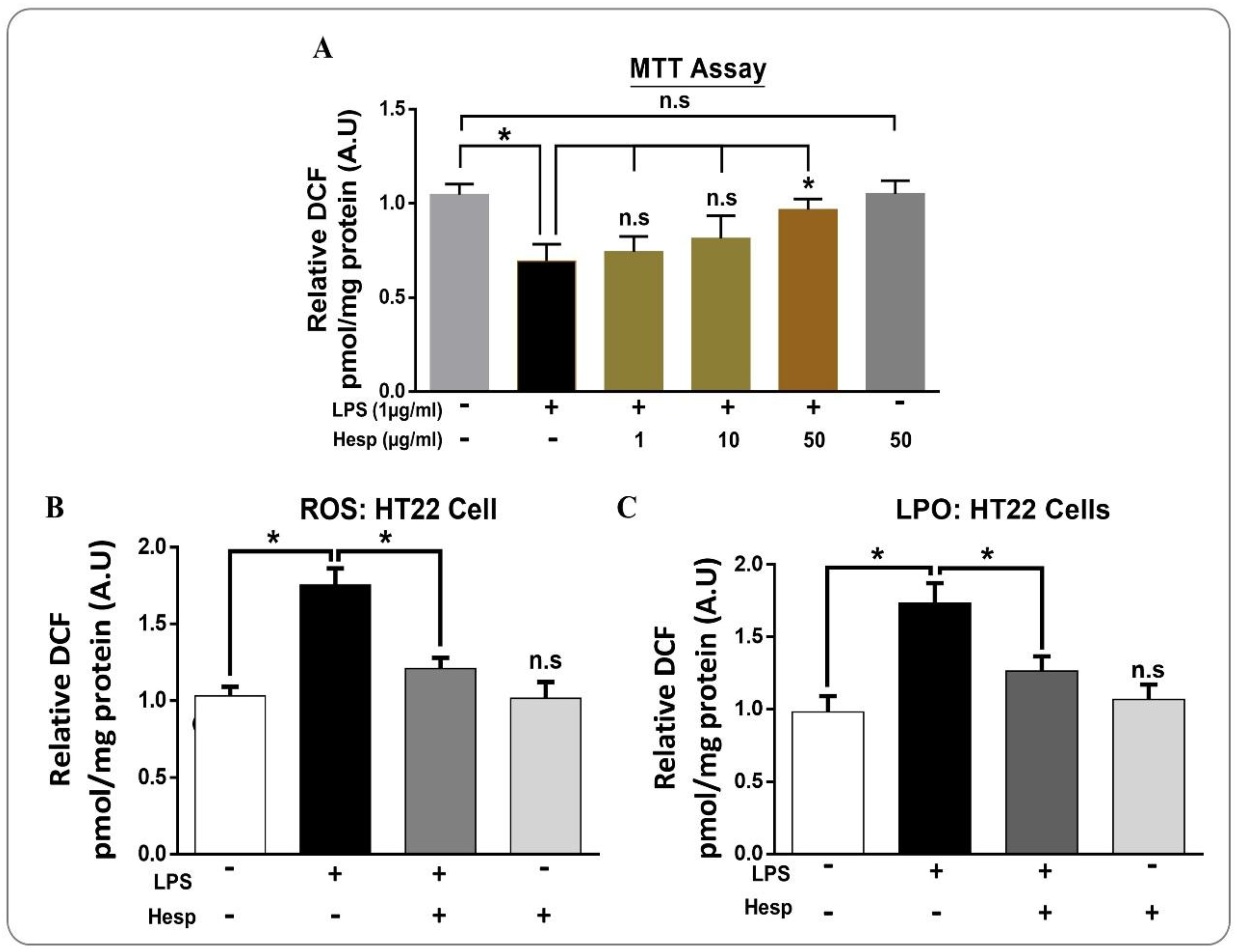
3.5. Hesperetin Attenuated LPS-Induced Cytotoxicity and ROS/LPO in HT-22 Cells

Next, we evaluated the effect of different concentrations of hesperetin against LPS on cell viability via the MTT assay. The result demonstrated that LPS significantly reduced cell viability significantly. On the other hand, hesperetin (50 µM) markedly reduced the LPS-induced cell toxicity in HT-22 cells (Figure 6A). Next, we performed the ROS and LPO analysis in vitro in HT-22 cells. Our in vitro findings demonstrated that the ROS and LPO levels in the LPS-treated HT-22 cells were significantly in LPS treated group as compare to the control group. On the other hand, hesperetin treatment significantly mitigated the elevated ROS and LPO levels (Figure 6B,C).

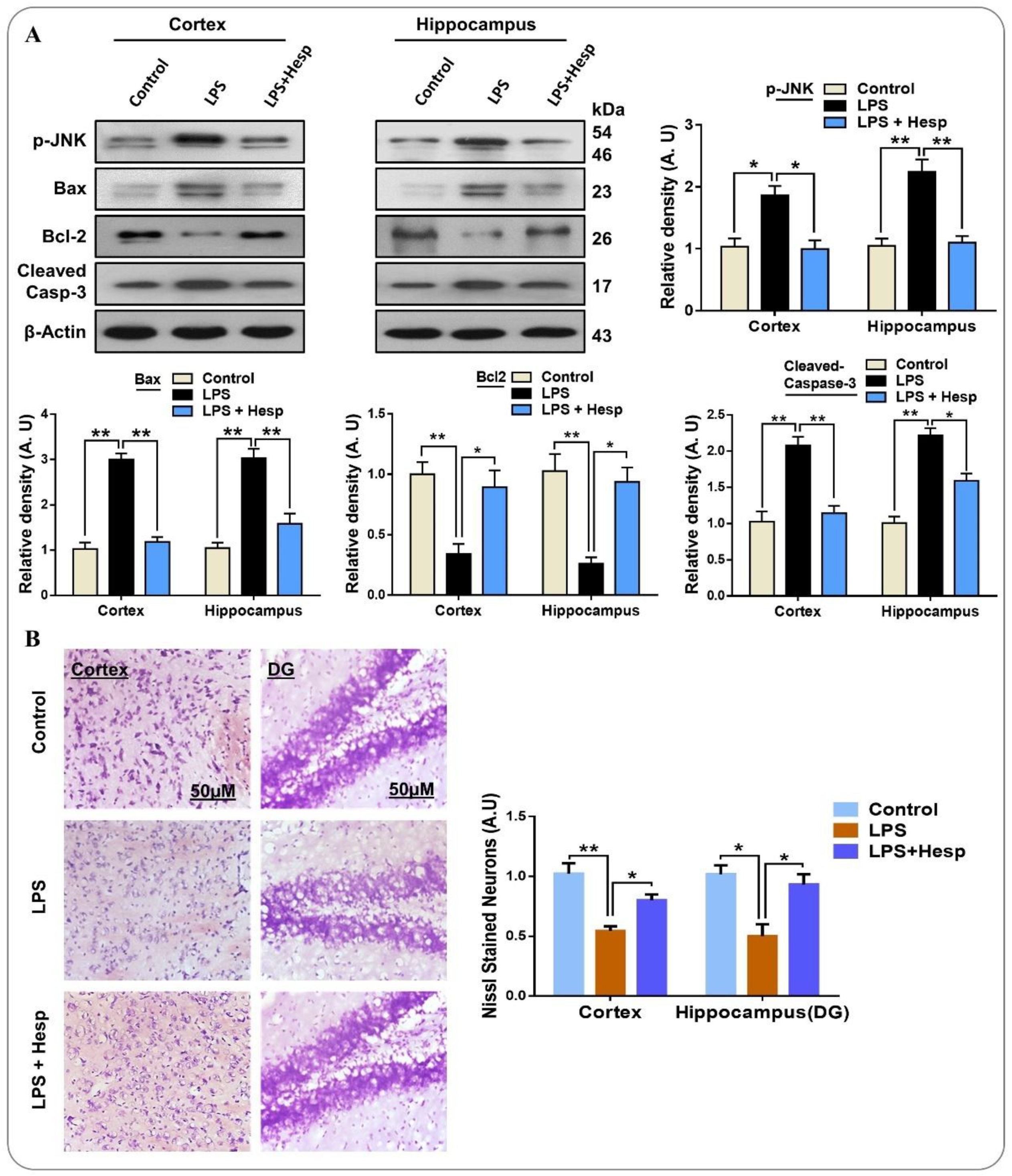
3.6. Hesperetin Reversed LPS-Mediated Neuronal Apoptosis and Neurodegeneration in Mouse Brain

Similarly, we found that LPS elevated the JNK level, a mitogen-activated protein kinase (MAPK) family level, enhanced the protein expression of pro-apoptotic Bax, and reduced anti-apoptotic Bcl2 expression; consequently, the ratio of Bax/Bcl2 was increased in the cortex and hippocampus of LPS-treated mice compared to the control vehicle-treated mice group. On the contrary, co-treatment of hesperetin attenuated the detrimental effects of LPS by significantly reducing the p-JNK expression, and decreased the ratio of Bax/Bcl2 compared to the LPS-administered mice group (Figure 7A). Additionally, we tested for the protective effect of hesperetin against the cleaved form of Caspase-3, a critical apoptotic caspase. Our results indicated that LPS-treatment increased the expression of Caspase-3, but hesperetin co-treatment substantially reduced its protein expression level compared to the LPS-treated group. Next, to confirm the anti-apoptotic effects of hesperetin, we performed Nissl’s (Cresyl violet) staining, which stains the Nissl bodies. These bodies or substances are lost in neurodegeneration/neuronal loss. As anticipated, the neuron loss in LPS-treated cortical and hippocampal regions (DG) was markedly increased as compared to the control vehicle-treated group. Hesperetin, however, reduced the neuronal loss and increased its survival in both the cortex and hippocampus region of the brain (Figure 7B).

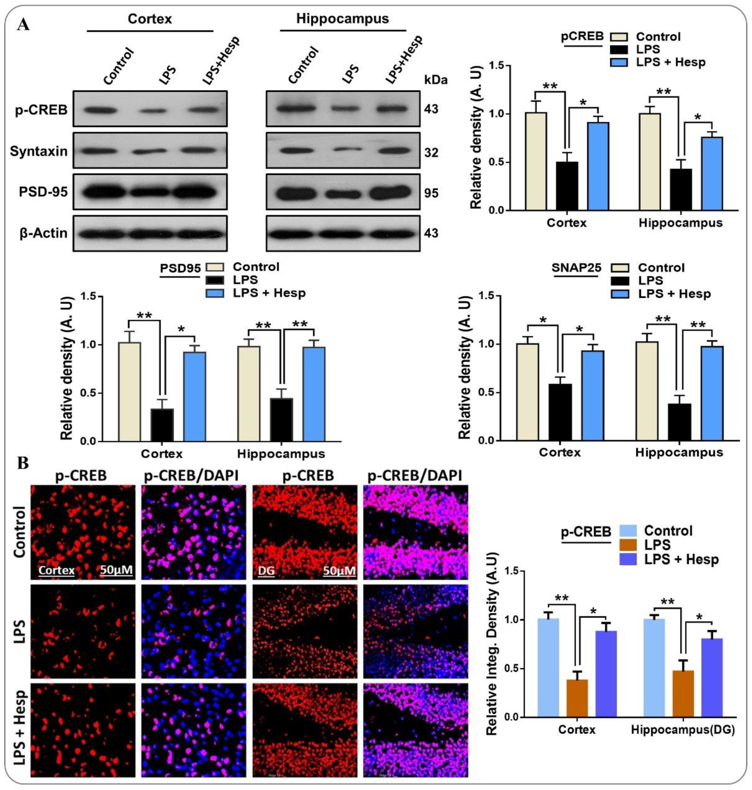
3.7. Hesperetin Enhanced the Expression of p-CREB and Improved Synaptic Integrity

We examined the effects of hesperetin on the memory and synaptic integrity related protein expression in the LPS mice model. For this, we checked the phosphorylation of CREB, a memory-associated transcription factor. As expected, the LPS-treated group showed a remarkable decrease in the expression of p-CREB compared to the control vehicle-treated group. However, hesperetin significantly enhanced the p-CREB expression in both hippocampal and cortical regions. Our findings also showed that LPS markedly induced synaptic dysfunction by reducing the expression level of presynaptic Syntaxin and PSD-95 in the cortex and hippocampus of mouse brains. On the other hand, hesperetin significantly recovered the level of Syntaxin and PSD-95 by improving their protein expression as compared to the LPS alone-treated group (Figure 8A). Additionally, we confirmed the p-CREB immunoblot results in the immunofluorescent confocal microscopy. The result indicated that LPS markedly reduced the immunoreactivity of p-CREB in the cortex and DG region of the hippocampus, while hesperetin significantly improved its immunoreactivity. These results indicated that hesperetin treatment reverses the LPS-induced synaptic dysfunction and upregulates the level of memory-associated p-CREB (Figure 8B).

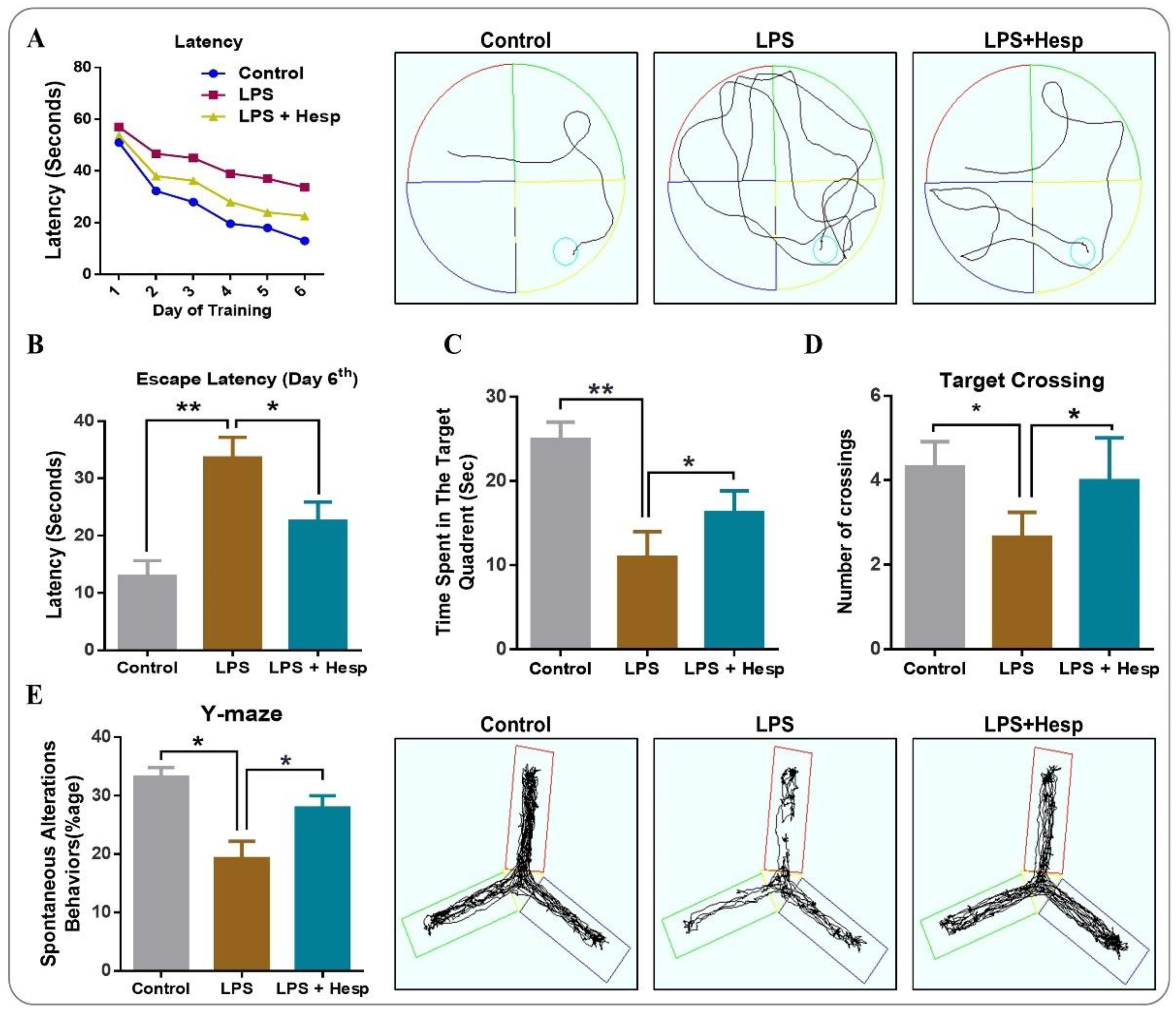
3.8. Hesperetin Ameliorated the Spatial and Learning Behavior Impaired By LPS Administration

We further investigated the effect of hesperetin on the spatial and learning behavior by MWM and Y-Maze in LPS-treated mice. Our results show that the mean escape latency was markedly higher in the LPS-treated group. Hesperetin co-treatment enhanced the learning and memory processes significantly and decreased the mean escape latency to the platform (Figure 9A). We further assessed the escape latency at the final day (Day 6) to reach the hidden platform. The control and hesperetin-treated mice groups took less time than did the LPS-treated mice group (Figure 9B). Next, we performed the probe test by removing the hidden platform on Day 7. LPS-treated mice showed fewer crossings over the previous site of the hidden platform and less time spent in the target quadrant (where a hidden platform was available previously) as compared to the saline-treated control group. The hesperetin+LPS group showed a markedly increased number of crossings as well as more time spent in the target quadrant, indicating the potential benefits of hesperetin in improving learning and memory formation (Figure 9C,D). After that, we conducted the Y-Maze test to assess spatial learning using the percentage of spontaneous alternation behavior. The Y-Maze data revealed that the LPS-treated mice had a reduced percentage of spontaneous alteration behavior than did the vehicle-treated control group, demonstrating impaired working memory. Hesperetin, on the other hand, significantly enhanced the percentage of spontaneous alteration compared to the LPS alone-treated group (Figure 9E).

4. Discussion

Phytochemicals have been shown to have potential neuroprotective effects in various neurological disorders. Recently, the use of phytotherapeutics has greatly increased; however, there is still much research that needs to be done before extensive use and acceptance as neuropsychotropic agents [42]. Mounting literature has demonstrated that hesperetin, a flavonoid mainly found in citrus fruits, exhibits numerous biological properties including anti-inflammatory, free radical scavenging, neuroprotective, anxiolytic, antidepressant, antinociceptive, and anticonvulsant effects [43,44,45]. In this study, we explored the anti-inflammatory, neuroprotective, and nootropic properties of hesperetin against LPS-induced neuroinflammation, neuronal apoptosis, and memory impairments.
Neuroinflammation plays a critical role in the pathogenesis of numerous neurological disorders, and a complex glial interplay is involved in the initiation of neuroinflammatory response. Glial cells play a major role in the defense and repair of the central nerve system of mammals [46,47]. Previous studies have demonstrated that, due to its poor passage through the blood-brain barrier (BBB), only a minute amount of peripheral LPS can enter the brain, suggesting that neuroinflammation is likely an indirect consequence of the peripheral inflammation [48]. On the other hand, several studies have shown that hesperetin crosses BBB and exerts anti-inflammatory and neuroprotective effects which further suggest the hesperetin is able to directly modulate neuroinflammation by directly crossing the BBB as well as indirectly by reducing the peripheral inflammatory cascades [49,50,51]. LPS leads to glial activation, that is astrocytosis and reactive microglia, which releases inflammatory cytokines such as TNF-α and IL-1β. These inflammatory mediators are involved in various neurodegenerative disorders such as Alzheimer’s disease, Parkinson’s disease, multiple sclerosis, and cerebral ischemia [52,53,54]. Herein, we observed that LPS markedly elevated microglial activation and astrocytosis by enhancing the TLR4 receptor expression, but hesperetin ameliorated the reactive gliosis and astrocytosis by downregulating the TLR4-mediated GFAP and Iba-1 expression. Next, to determine whether the beneficial effects of hesperetin against LPS are TLR4-dependent, we used the pharmacological inhibitor of TLR4 (TAK-242) in HT-22 cells. Interestingly, our in vitro results also supported the in vivo findings where LPS substantially induced TLR4/Iba-1 expression while hesperetin, consistent with TAK-242, markedly reduced its expression (Figure 2).
Furthermore, NF-κB, a pleiotropic transcription factor, regulates several other genes that are involved in inflammatory responses and stimulates numerous cellular signaling pathways, which leads to the increased production of inflammatory cytokines. The LPS-induced NF-κB activation in glia has been reported to upregulate the expression of inflammatory mediators [55,56]. Moreover, TNF-α signaling plays an important role in immunity by preventing infection and tumor growth. However, studies have found that TNF-α triggers cytotoxic cascades and apoptotic cell death, and Aβ in AD requires TNFR1 (TNF-α receptor1)-mediated signaling for neuronal death. Moreover, TNF-α effects learning and memory processes by disrupting synaptic plasticity and has detrimental effects on synaptic transmission and plasticity [57,58]. Thus, inhibiting the NF-κB-mediated cellular and molecular processes may provide a potential target for neuroinflammation-related neurological disorders. Our results demonstrated that hesperetin treatment markedly reduced the p-NF-κB expression level in the mouse brain stimulated by LPS. Additionally, hesperetin significantly reduced the protein expression level of TNF-α and IL-1β cytokines. Once more, we evaluated whether this protective effect is mediated by NF-κB signaling. For this, we treated hesperetin and BAY (NF-κB inhibitor) together with LPS in BV2 cells. As expected, hesperetin and BAY reduced the p-NF-κB/TNF-α expression significantly after LPS treatment. These findings suggest that the effect of hesperetin on neuroinflammation is significantly mediated through the NF-κB signaling pathway, which is consistent with the specific NF-κB inhibitor BAY (Figure 3).
Several lines of studies have shown that ROS are implicated in pathogenesis neurodegenerative disorders. Similarly, transcription factor Nrf-2 and HO-1 are the main cellular mechanisms that regulate antioxidant and cytoprotective genes, and are downregulated in neurodegenerative disorders. LPS induces ROS generation, which leads to an oxidant/antioxidant imbalance and oxidative stress [59,60,61,62]. The activation of Nrf-2 is reflected as a critical therapeutic target for cytoprotective strategies. Herein, our results have shown that LPS resulted in oxidative stress by inducing LPO and ROS generation in mouse brains and in HT-22 cells. As expected, Western blot results indicated that LPS also downregulated the expression of Nrf-2 and HO-1 in the mouse brain. Interestingly, elevated LPO and ROS as well as the reduced expression of Nrf-2 and HO-1 were significantly regulated in the hesperetin-treated group (Figure 4), supporting the hypothesis that hesperetin has potential antioxidant effects.
JNK, a stress kinase, has been found to be associated with apoptotic processes. Inflammatory mediators, UV radiation, and oxidative stress trigger the JNK cascades [63]. Similarly, the anti- and pro-apoptotic protein markers Bcl-2 and Bax play an important role in the regulation of apoptotic cell death pathways at the mitochondrial level [64,65]. Another factor responsible for apoptotic neurodegeneration is the caspase cascade, especially Caspase-3, a principal effector in apoptotic cascades resulting in neurodegeneration [66]. According to our findings, the expressions of JNK/Bax and cleaved caspase-3 were substantially elevated, and the antiapoptotic Bcl-2 level was significantly reduced in the LPS-treated mice group. Hesperetin markedly downregulated the LPS-induced elevation in pro-apoptotic markers and enhanced the expression of the antiapoptotic protein marker. Furthermore, our Cresyl Violet staining also demonstrated that LPS administration significantly reduced histochemical reactivity for Nissl bodies in the neurons, suggesting degenerated, shrunk, and fragmented neurons. The co-treatment of hesperetin reversed the detrimental effects of LPS and improved the histochemical reactivity for healthy normal neurons (Figure 5).
A growing body of evidence has also shown that LPS-induced neuroinflammation, oxidative stress, and neuronal apoptosis mediate synaptic dysfunction and memory impairment by damaging the hippocampus [67,68,69]. We, therefore, evaluated the memory and synaptic protein markers. Our results indicated that LPS decreased the expression of p-CREB, Syntaxin, and PSD-95 in the cortex and hippocampus regions. The hesperetin cotreatment significantly restored the expression of these markers to basal levels (Figure 6). Additionally, we analyzed the cognitive and learning dysfunction using MWM and Y-Maze tests. LPS significantly impaired the cognitive and learning behavior, but hesperetin cotreatment substantially reversed the effects and ameliorated cognition, spatial learning, and memory processing (Figure 7). These results demonstrated that hesperetin recovers LPS-induced neurodegeneration in mouse brains.

5. Conclusions

The findings of our study have revealed that hesperetin ameliorated the LPS-induced pathological hallmarks in vivo (in adult mouse brains) and in vitro (in BV2 and HT-22 cells). Our data suggest that hesperetin attenuates the neuroinflammation-mediated neurodegeneration, cognitive and learning decline, and memory impairments. However, more detailed studies are needed to assess the mechanistic role of hesperetin in neuroinflammatory and neurodegenerative disorders.

Author Contributions

T.M. designed and conducted experiments, wrote manuscript and performed statistical analysis; M.I., R.U. conducted experiments, provided the experimental reagents; S.U.R. reviewed and edit the manuscript; M.O.K. supervised and organized the final version of the manuscript. All authors reviewed and approved the paper.

Funding

This research received no external funding.

Acknowledgments

This research was supported by the Brain Research Program through the National Research Foundation of Korea (NRF) funded by the Ministry of Science, ICT (2016M3C7A1904391).

Conflicts of Interest

The authors declared no competing financial interests.

References

  1. Catorce, M.N.; Gevorkian, G. LPS-induced Murine Neuroinflammation Model: Main Features and Suitability for Pre-clinical Assessment of Nutraceuticals. Curr. Neuropharmacol. 2016, 14, 155–164. [Google Scholar] [CrossRef] [PubMed]
  2. Muhammad, T.; Ali, T.; Ikram, M.; Khan, A.; Alam, S.I.; Kim, M.O. Melatonin Rescue Oxidative Stress-Mediated Neuroinflammation/Neurodegeneration and Memory Impairment in Scopolamine-Induced Amnesia Mice Model. J. Neuroimmune Pharmacol. 2018, 1–17. [Google Scholar] [CrossRef] [PubMed]
  3. Khan, A.; Ali, T.; Rehman, S.U.; Khan, M.S.; Alam, S.I.; Ikram, M.; Muhammad, T.; Saeed, K.; Badshah, H.; Kim, M.O. Neuroprotective effect of quercetin against the detrimental effects of LPS in the adult mouse brain. Front. Pharmacol. 2018, 9, 1383. [Google Scholar] [CrossRef]
  4. Pulido-Salgado, M.; Vidal-Taboada, J.M.; Barriga, G.G.; Sola, C.; Saura, J. RNA-Seq transcriptomic profiling of primary murine microglia treated with LPS or LPS + IFNgamma. Sci. Rep. 2018, 8, 16096. [Google Scholar] [CrossRef] [PubMed]
  5. Barron, H.; Hafizi, S.; Andreazza, A.C.; Mizrahi, R. Neuroinflammation and Oxidative Stress in Psychosis and Psychosis Risk. Int. J. Mol. Sci. 2017, 18, 651. [Google Scholar] [CrossRef]
  6. Pepe, G.; Sommella, E.; Cianciarulo, D.; Ostacolo, C.; Manfra, M.; Di Sarno, V.; Musella, S.; Russo, M.; Messore, A.; Parrino, B.; et al. Polyphenolic Extract from Tarocco (Citrus sinensis L. Osbeck) Clone “Lempso” Exerts Anti-Inflammatory and Antioxidant Effects via NF-kB and Nrf-2 Activation in Murine Macrophages. Nutrients 2018, 10, 1961. [Google Scholar] [CrossRef] [PubMed]
  7. Pretorius, E.; Bester, J.; Page, M.J.; Kell, D.B. The Potential of LPS-Binding Protein to Reverse Amyloid Formation in Plasma Fibrin of Individuals With Alzheimer-Type Dementia. Front. Aging Neurosci. 2018, 10, 257. [Google Scholar] [CrossRef]
  8. Rau, C.-S.; Wu, S.-C.; Lu, T.-H.; Wu, Y.-C.; Wu, C.-J.; Chien, P.-C.; Kuo, P.-J.; Lin, C.-W.; Tsai, C.-W.; Hsieh, C.-H. Effect of Low-Fat Diet in Obese Mice Lacking Toll-like Receptors. Nutrients 2018, 10, 1464. [Google Scholar] [CrossRef]
  9. Hsieh, Y.-H.; Deng, J.-S.; Chang, Y.-S.; Huang, G.-J. Ginsenoside Rh2 Ameliorates Lipopolysaccharide-Induced Acute Lung Injury by Regulating the TLR4/PI3K/Akt/mTOR, Raf-1/MEK/ERK, and Keap1/Nrf2/HO-1 Signaling Pathways in Mice. Nutrients 2018, 10, 1208. [Google Scholar]
  10. Badshah, H.; Ali, T.; Kim, M.O. Osmotin attenuates LPS-induced neuroinflammation and memory impairments via the TLR4/NFκB signaling pathway. Sci. Rep. 2016, 6, 24493. [Google Scholar] [CrossRef]
  11. Vasconcelos, A.R.; Yshii, L.M.; Viel, T.A.; Buck, H.S.; Mattson, M.P.; Scavone, C.; Kawamoto, E.M. Intermittent fasting attenuates lipopolysaccharide-induced neuroinflammation and memory impairment. J. Neuroinflamm. 2014, 11, 85. [Google Scholar] [CrossRef] [PubMed]
  12. Balducci, C.; Frasca, A.; Zotti, M.; La Vitola, P.; Mhillaj, E.; Grigoli, E.; Iacobellis, M.; Grandi, F.; Messa, M.; Colombo, L. Toll-like receptor 4-dependent glial cell activation mediates the impairment in memory establishment induced by β-amyloid oligomers in an acute mouse model of Alzheimer’s disease. Brain. Behav. Immun. 2017, 60, 188–197. [Google Scholar] [CrossRef]
  13. El Kamouni, S.; El Kebbaj, R.; Andreoletti, P.; El Ktaibi, A.; Rharrassi, I.; Essamadi, A.; El Kebbaj, M.S.; Mandard, S.; Latruffe, N.; Vamecq, J.; et al. Protective Effect of Argan and Olive Oils against LPS-Induced Oxidative Stress and Inflammation in Mice Livers. Int. J. Mol. Sci. 2017, 18, 2181. [Google Scholar] [CrossRef]
  14. Lushchak, V.I. Free radicals, reactive oxygen species, oxidative stress and its classification. Chem. Biol. Interact. 2014, 224, 164–175. [Google Scholar] [CrossRef] [PubMed]
  15. Loboda, A.; Damulewicz, M.; Pyza, E.; Jozkowicz, A.; Dulak, J. Role of Nrf2/HO-1 system in development, oxidative stress response and diseases: An evolutionarily conserved mechanism. Cell. Mol. Life Sci. 2016, 73, 3221–3247. [Google Scholar] [CrossRef]
  16. You, L.H.; Yan, C.Z.; Zheng, B.J.; Ci, Y.Z.; Chang, S.Y.; Yu, P.; Gao, G.F.; Li, H.Y.; Dong, T.Y.; Chang, Y.Z. Astrocyte hepcidin is a key factor in LPS-induced neuronal apoptosis. Cell Death Dis. 2017, 8, e2676. [Google Scholar] [CrossRef] [Green Version]
  17. Wu, X.; Lv, Y.G.; Du, Y.F.; Hu, M.; Reed, M.N.; Long, Y.; Suppiramaniam, V.; Hong, H.; Tang, S.S. Inhibitory effect of INT-777 on lipopolysaccharide-induced cognitive impairment, neuroinflammation, apoptosis, and synaptic dysfunction in mice. Prog. Neuropsychopharmacol. Biol. Psychiatry 2019, 88, 360–374. [Google Scholar] [CrossRef] [PubMed]
  18. Knekt, P.; Kumpulainen, J.; Jarvinen, R.; Rissanen, H.; Heliovaara, M.; Reunanen, A.; Hakulinen, T.; Aromaa, A. Flavonoid intake and risk of chronic diseases. Am. J. Clin. Nutr. 2002, 76, 560–568. [Google Scholar] [CrossRef] [Green Version]
  19. Kim, H.K.; Jeong, T.S.; Lee, M.K.; Park, Y.B.; Choi, M.S. Lipid-lowering efficacy of hesperetin metabolites in high-cholesterol fed rats. Clin. Chim. Acta 2003, 327, 129–137. [Google Scholar] [CrossRef]
  20. Yang, H.-L.; Chen, S.-C.; Senthil Kumar, K.; Yu, K.-N.; Lee Chao, P.-D.; Tsai, S.-Y.; Hou, Y.-C.; Hseu, Y.-C. Antioxidant and anti-inflammatory potential of hesperetin metabolites obtained from hesperetin-administered rat serum: An ex vivo approach. J. Agric. Food Chem. 2011, 60, 522–532. [Google Scholar] [CrossRef]
  21. Rainey-Smith, S.; Schroetke, L.-W.; Bahia, P.; Fahmi, A.; Skilton, R.; Spencer, J.P.; Rice-Evans, C.; Rattray, M.; Williams, R.J. Neuroprotective effects of hesperetin in mouse primary neurones are independent of CREB activation. Neurosci. Lett. 2008, 438, 29–33. [Google Scholar] [CrossRef] [PubMed] [Green Version]
  22. Kheradmand, E.; Hajizadeh Moghaddam, A.; Zare, M. Neuroprotective effect of hesperetin and nano-hesperetin on recognition memory impairment and the elevated oxygen stress in rat model of Alzheimer’s disease. Biomed. Pharmacother. 2018, 97, 1096–1101. [Google Scholar] [CrossRef]
  23. Homma, M.; Oka, K.; Taniguchi, C.; Niitsuma, T.; Hayashi, T. Systematic analysis of post-administrative saiboku-to urine by liquid chromatography to determine pharmacokinetics of traditional Chinese medicine. Biomed. Chromatogr. 1997, 11, 125–131. [Google Scholar] [CrossRef]
  24. Vaya, J.; Belinky, P.A.; Aviram, M. Antioxidant constituents from licorice roots: Isolation, structure elucidation and antioxidative capacity toward LDL oxidation. Free Radic. Biol. Med. 1997, 23, 302–313. [Google Scholar] [CrossRef]
  25. Choi, E.J.; Ahn, W.S. Neuroprotective effects of chronic hesperetin administration in mice. Arch. Pharm. Res. 2008, 31, 1457. [Google Scholar] [CrossRef]
  26. Jo, M.G.; Ikram, M.; Jo, M.H.; Yoo, L.; Chung, K.C.; Nah, S.Y.; Hwang, H.; Rhim, H.; Kim, M.O. Gintonin Mitigates MPTP-Induced Loss of Nigrostriatal Dopaminergic Neurons and Accumulation of alpha-Synuclein via the Nrf2/HO-1 Pathway. Mol. Neurobiol. 2019, 56, 39–55. [Google Scholar] [CrossRef]
  27. Paterniti, I.; Impellizzeri, D.; Cordaro, M.; Siracusa, R.; Bisignano, C.; Gugliandolo, E.; Carughi, A.; Esposito, E.; Mandalari, G.; Cuzzocrea, S. The Anti-Inflammatory and Antioxidant Potential of Pistachios (Pistacia vera L.) In Vitro and In Vivo. Nutrients 2017, 9, 915. [Google Scholar] [CrossRef] [PubMed]
  28. Spijker, S. Dissection of rodent brain regions. In Neuroproteomics; Humana Press: Totowa, NJ, USA, 2011; pp. 13–26. [Google Scholar]
  29. Ravichandran, V.A.; Kim, M.; Han, S.K.; Cha, Y.S. Stachys sieboldii Extract Supplementation Attenuates Memory Deficits by Modulating BDNF-CREB and Its Downstream Molecules, in Animal Models of Memory Impairment. Nutrients 2018, 10, 917. [Google Scholar] [CrossRef] [PubMed]
  30. Rehman, S.U.; Ali, T.; Alam, S.I.; Ullah, R.; Zeb, A.; Lee, K.W.; Rutten, B.P.; Kim, M.O. Ferulic acid rescues LPS-induced neurotoxicity via modulation of the TLR4 receptor in the mouse hippocampus. Mol. Neurobiol. 2018, 1–17. [Google Scholar] [CrossRef]
  31. Amato, R.; Rossino, M.G.; Cammalleri, M.; Locri, F.; Pucci, L.; Dal Monte, M.; Casini, G. Lisosan G Protects the Retina from Neurovascular Damage in Experimental Diabetic Retinopathy. Nutrients 2018, 10, 1932. [Google Scholar] [CrossRef]
  32. Kim, M.J.; Rehman, S.U.; Amin, F.U.; Kim, M.O. Enhanced neuroprotection of anthocyanin-loaded PEG-gold nanoparticles against Aβ1-42-induced neuroinflammation and neurodegeneration via the NF-KB/JNK/GSK3β signaling pathway. Nanomed. Nanotechnol. Biol. Med. 2017, 13, 2533–2544. [Google Scholar] [CrossRef] [PubMed]
  33. Yon, J.-M.; Kim, Y.-B.; Park, D. The Ethanol Fraction of White Rose Petal Extract Abrogates Excitotoxicity-Induced Neuronal Damage In Vivo and In Vitro through Inhibition of Oxidative Stress and Proinflammation. Nutrients 2018, 10, 1375. [Google Scholar] [CrossRef]
  34. Alam, S.I.; Ur Rehman, S.; Ok Kim, M. Nicotinamide Improves Functional Recovery via Regulation of the RAGE/JNK/NF-κB Signaling Pathway after Brain Injury. J. Clin. Med. 2019, 8, 271. [Google Scholar] [CrossRef]
  35. Ikram, M.; Muhammad, T.; Rehman, S.U.; Khan, A.; Jo, M.G.; Ali, T.; Kim, M.O. Hesperetin Confers Neuroprotection by Regulating Nrf2/TLR4/NF-κB Signaling in an Aβ Mouse Model. Mol. Neurobiol. 2019, 1–17. [Google Scholar] [CrossRef] [PubMed]
  36. Wang, W.Y.; Tan, M.S.; Yu, J.T.; Tan, L. Role of pro-inflammatory cytokines released from microglia in Alzheimer’s disease. Ann. Transl. Med. 2015, 3, 136. [Google Scholar] [CrossRef] [PubMed]
  37. Ali, T.; Rehman, S.U.; Shah, F.A.; Kim, M.O. Acute dose of melatonin via Nrf2 dependently prevents acute ethanol-induced neurotoxicity in the developing rodent brain. J. Neuroinflamm. 2018, 15, 119. [Google Scholar] [CrossRef]
  38. Yun, J.; Yeo, I.J.; Hwang, C.J.; Choi, D.Y.; Im, H.S.; Kim, J.Y.; Choi, W.R.; Jung, M.H.; Han, S.B.; Hong, J.T. Estrogen deficiency exacerbates Abeta-induced memory impairment through enhancement of neuroinflammation, amyloidogenesis and NF-kB activation in ovariectomized mice. Brain. Behav. Immun. 2018, 73, 282–293. [Google Scholar] [CrossRef]
  39. Chen, G.; Liu, J.; Jiang, L.; Ran, X.; He, D.; Li, Y.; Huang, B.; Wang, W.; Liu, D.; Fu, S. Peiminine Protects Dopaminergic Neurons from Inflammation-Induced Cell Death by Inhibiting the ERK1/2 and NF-kappaB Signalling Pathways. Int. J. Mol. Sci. 2018, 19, 821. [Google Scholar] [CrossRef]
  40. Li, L.; Shoji, W.; Takano, H.; Nishimura, N.; Aoki, Y.; Takahashi, R.; Goto, S.; Kaifu, T.; Takai, T.; Obinata, M. Increased susceptibility of MER5 (peroxiredoxin III) knockout mice to LPS-induced oxidative stress. Biochem. Biophys. Res. Commun. 2007, 355, 715–721. [Google Scholar] [CrossRef] [PubMed]
  41. Itsumi, M.; Inoue, S.; Elia, A.J.; Murakami, K.; Sasaki, M.; Lind, E.F.; Brenner, D.; Harris, I.S.; Chio, C., II; Afzal, S.; et al. Idh1 protects murine hepatocytes from endotoxin-induced oxidative stress by regulating the intracellular NADP(+)/NADPH ratio. Cell Death Differ. 2015, 22, 1837–1845. [Google Scholar] [CrossRef]
  42. Kumar, G.P.; Khanum, F. Neuroprotective potential of phytochemicals. Pharmacogn. Rev. 2012, 6, 81–90. [Google Scholar] [CrossRef] [PubMed] [Green Version]
  43. Cho, J. Antioxidant and neuroprotective effects of hesperidin and its aglycone hesperetin. Arch. Pharm. Res. 2006, 29, 699–706. [Google Scholar] [CrossRef] [PubMed]
  44. Roohbakhsh, A.; Parhiz, H.; Soltani, F.; Rezaee, R.; Iranshahi, M. Neuropharmacological properties and pharmacokinetics of the citrus flavonoids hesperidin and hesperetin—A mini-review. Life Sci. 2014, 113, 1–6. [Google Scholar] [CrossRef]
  45. Shimoda, K.; Hamada, H. Production of Hesperetin Glycosides by Xanthomonas campestris and Cyclodextrin Glucanotransferase and Their Anti-allergic Activities. Nutrients 2010, 2, 171. [Google Scholar] [CrossRef] [PubMed]
  46. Liu, C.Y.; Yang, Y.; Ju, W.N.; Wang, X.; Zhang, H.L. Emerging Roles of Astrocytes in Neuro-Vascular Unit and the Tripartite Synapse with Emphasis on Reactive Gliosis in the Context of Alzheimer’s Disease. Front. Cell. Neurosci. 2018, 12, 193. [Google Scholar] [CrossRef] [PubMed]
  47. Zhou, J.; Mao, L.; Xu, P.; Wang, Y. Effects of (−)-Epigallocatechin Gallate (EGCG) on Energy Expenditure and Microglia-Mediated Hypothalamic Inflammation in Mice Fed a High-Fat Diet. Nutrients 2018, 10, 1681. [Google Scholar] [CrossRef] [PubMed]
  48. Qin, L.; Wu, X.; Block, M.L.; Liu, Y.; Breese, G.R.; Hong, J.S.; Knapp, D.J.; Crews, F.T. Systemic LPS causes chronic neuroinflammation and progressive neurodegeneration. Glia 2007, 55, 453–462. [Google Scholar] [CrossRef]
  49. Bai, X.; Yang, P.; Zhou, Q.; Cai, B.; Buist-Homan, M.; Cheng, H.; Jiang, J.; Shen, D.; Li, L.; Luo, X. The protective effect of the natural compound hesperetin against fulminant hepatitis in vivo and in vitro. Br. J. Pharmacol. 2017, 174, 41–56. [Google Scholar] [CrossRef]
  50. Vauzour, D.; Vafeiadou, K.; Rodriguez-Mateos, A.; Rendeiro, C.; Spencer, J.P. The neuroprotective potential of flavonoids: A multiplicity of effects. Genes Nutr. 2008, 3, 115. [Google Scholar] [CrossRef]
  51. Youdim, K.A.; Dobbie, M.S.; Kuhnle, G.; Proteggente, A.R.; Abbott, N.J.; Rice-Evans, C. Interaction between flavonoids and the blood–brain barrier: In vitro studies. J. Neurochem. 2003, 85, 180–192. [Google Scholar] [CrossRef]
  52. Taki-Nakano, N.; Kotera, J.; Ohta, H. 12-oxo-phytodienoic acid, a plant-derived oxylipin, attenuates lipopolysaccharide-induced inflammation in microglia. Biochem. Biophys. Res. Commun. 2016, 473, 1288–1294. [Google Scholar] [CrossRef] [PubMed] [Green Version]
  53. Huang, Y.N.; Ho, Y.J.; Lai, C.C.; Chiu, C.T.; Wang, J.Y. 1,25-Dihydroxyvitamin D3 attenuates endotoxin-induced production of inflammatory mediators by inhibiting MAPK activation in primary cortical neuron-glia cultures. J. Neuroinflamm. 2015, 12, 147. [Google Scholar] [CrossRef]
  54. Subedi, L.; Ji, E.; Shin, D.; Jin, J.; Yeo, J.H.; Kim, S.Y. Equol, a Dietary Daidzein Gut Metabolite Attenuates Microglial Activation and Potentiates Neuroprotection In Vitro. Nutrients 2017, 9, 207. [Google Scholar] [CrossRef]
  55. Kim, B.W.; Koppula, S.; Hong, S.S.; Jeon, S.B.; Kwon, J.H.; Hwang, B.Y.; Park, E.J.; Choi, D.K. Regulation of microglia activity by glaucocalyxin-A: Attenuation of lipopolysaccharide-stimulated neuroinflammation through NF-kappaB and p38 MAPK signaling pathways. PLoS ONE 2013, 8, e55792. [Google Scholar] [CrossRef] [PubMed]
  56. Negri, A.; Naponelli, V.; Rizzi, F.; Bettuzzi, S. Molecular Targets of Epigallocatechin—Gallate (EGCG): A Special Focus on Signal Transduction and Cancer. Nutrients 2018, 10, 1936. [Google Scholar] [CrossRef] [PubMed]
  57. Cheng, X.; Shen, Y.; Li, R. Targeting TNF: A therapeutic strategy for Alzheimer’s disease. Drug Discov. Today 2014, 19, 1822–1827. [Google Scholar] [CrossRef] [PubMed]
  58. Li, D.H.; Havell, E.A.; Brown, C.L.; Cullen, J.M. Woodchuck lymphotoxin-alpha, -beta and tumor necrosis factor genes: Structure, characterization and biological activity. Gene 2000, 242, 295–305. [Google Scholar] [CrossRef]
  59. Liu, Z.; Zhou, T.; Ziegler, A.C.; Dimitrion, P.; Zuo, L. Oxidative Stress in Neurodegenerative Diseases: From Molecular Mechanisms to Clinical Applications. Oxid. Med. Cell. Longev. 2017, 2017, 2525967. [Google Scholar] [CrossRef]
  60. He, M.; Zhao, L.; Wei, M.J.; Yao, W.F.; Zhao, H.S.; Chen, F.J. Neuroprotective effects of (-)-epigallocatechin-3-gallate on aging mice induced by d-galactose. Biol. Pharm. Bull. 2009, 32, 55–60. [Google Scholar] [CrossRef]
  61. Ishii, T.; Itoh, K.; Takahashi, S.; Sato, H.; Yanagawa, T.; Katoh, Y.; Bannai, S.; Yamamoto, M. Transcription factor Nrf2 coordinately regulates a group of oxidative stress-inducible genes in macrophages. J. Biol. Chem. 2000, 275, 16023–16029. [Google Scholar] [CrossRef]
  62. Nuzzo, D.; Amato, A.; Picone, P.; Terzo, S.; Galizzi, G.; Bonina, F.P.; Mulè, F.; Di Carlo, M. A Natural Dietary Supplement with a Combination of Nutrients Prevents Neurodegeneration Induced by a High Fat Diet in Mice. Nutrients 2018, 10, 1130. [Google Scholar] [CrossRef] [PubMed]
  63. Li, J.; Xu, B.; Chen, Z.; Zhou, C.; Liao, L.; Qin, Y.; Yang, C.; Zhang, X.; Hu, Z.; Sun, L.; et al. PI3K/AKT/JNK/p38 signalling pathway-mediated neural apoptosis in the prefrontal cortex of mice is involved in the antidepressant-like effect of pioglitazone. Clin. Exp. Pharmacol. Physiol. 2018, 45, 525–535. [Google Scholar] [CrossRef] [PubMed]
  64. Chao, D.T.; Korsmeyer, S.J. BCL-2 family: Regulators of cell death. Annu. Rev. Immunol. 1998, 16, 395–419. [Google Scholar] [CrossRef] [PubMed]
  65. Sang Eun, H.; Seong Min, K.; Ho Jeong, L.; Vetrivel, P.; Venkatarame Gowda Saralamma, V.; Jeong Doo, H.; Eun Hee, K.; Sang Joon, L.; Gon Sup, K. Scutellarein Induces Fas-Mediated Extrinsic Apoptosis and G2/M Cell Cycle Arrest in Hep3B Hepatocellular Carcinoma Cells. Nutrients 2019, 11, 263. [Google Scholar] [CrossRef]
  66. Louneva, N.; Cohen, J.W.; Han, L.Y.; Talbot, K.; Wilson, R.S.; Bennett, D.A.; Trojanowski, J.Q.; Arnold, S.E. Caspase-3 is enriched in postsynaptic densities and increased in Alzheimer’s disease. Am. J. Pathol. 2008, 173, 1488–1495. [Google Scholar] [CrossRef] [PubMed]
  67. Choi, D.-Y.; Lee, J.W.; Lin, G.; Lee, Y.K.; Lee, Y.H.; Choi, I.S.; Han, S.B.; Jung, J.K.; Kim, Y.H.; Kim, K.H. Obovatol attenuates LPS-induced memory impairments in mice via inhibition of NF-κB signaling pathway. Neurochem. Int. 2012, 60, 68–77. [Google Scholar] [CrossRef]
  68. Bilbo, S.D.; Biedenkapp, J.C.; Der-Avakian, A.; Watkins, L.R.; Rudy, J.W.; Maier, S.F. Neonatal infection-induced memory impairment after lipopolysaccharide in adulthood is prevented via caspase-1 inhibition. J. Neurosci. 2005, 25, 8000–8009. [Google Scholar] [CrossRef]
  69. Zarifkar, A.; Choopani, S.; Ghasemi, R.; Naghdi, N.; Maghsoudi, A.H.; Maghsoudi, N.; Rastegar, K.; Moosavi, M. Agmatine prevents LPS-induced spatial memory impairment and hippocampal apoptosis. Eur. J. Pharmacol. 2010, 634, 84–88. [Google Scholar] [CrossRef]
Figure 1. Schematic diagram of the experimental design showing the duration of the lipopolysaccharide (LPS) and/or hesperetin administration in adult mice and behavioral analysis.
Nutrients 11 00648 g001
Figure 2. Hesperetin ameliorates TLR4/gliosis-mediated neuroinflammation in the LPS-treated mouse brain: (A) Western blot analysis showing the expression of TLR4, GFAP, and Iba-1 in the experimental mice (cortex and hippocampus regions); (B) confocal photomicrographs showing the immunoreactivity of GFAP in the cortex and DG region of hippocampus in different experimental mice groups. The data are presented as the mean ± SEM of 7–10 mice per group and are representative of three independent experiments, * p ≤ 0.05, ** p ≤ 0.01, *** p ≤ 0.001.
Nutrients 11 00648 g002
Figure 3. Hesperetin mitigates the expression of p-NF-κB and inflammatory cytokines in LPS-treated mouse brains and BV2 cells: (A) Western blot analysis of p-NF-κB, TNF-α, and IL-1β expression in the cortex and hippocampus of mice; (B) immunofluorescence analysis of TNF-α immunoreactivity in the cortex and DG region of the hippocampus. The data are presented as the mean ± SEM of 7–10 mice per group and are representative of three independent experiments, * p ≤ 0.05, ** p ≤ 0.01.
Nutrients 11 00648 g003
Figure 4. (A) Western blot analysis of the TLR4 and Iba-1 expression in BV2 cells treated with hesperetin and TAK-242 (TLR4 inhibitor) and their respective histograms; (B) Western blot analysis of p-NF-κB and TNF-α protein expression level in BV2 cell line treated with LPS and/or hesperetin/BAY (NF-κB inhibitor). The data are shown as the mean ± SEM of triplicate wells for in vitro experiments are representative of three independent experiments, * p ≤ 0.05, ** p ≤ 0.01.
Nutrients 11 00648 g004
Figure 5. Effects of hesperetin against LPS-induced oxidative stress in the cortical and hippocampal region of mouse brains and in HT-22 cells: (A,B) the analysis of the generation of ROS and LPO production in vivo in the mouse brain (cortex and hippocampus regions); (C) Western blot analysis of Nrf2 and HO-1 in the cortex and hippocampus of adult mouse brains. The data are presented as the mean ± SEM of 8–10 mice per group and are representative of three independent experiments, * p ≤ 0.05, ** p ≤ 0.01.
Nutrients 11 00648 g005
Figure 6. The protective effects of hesperetin in LPS-treated in HT-22 cells: (A) the MTT assay conducted in HT-22 cells representing the cell viability in different treated groups (number of experiments = 3); (B,C) Analysis of the ROS generation and LPO production in HT-22 Cells. The data are shown as the mean ± SEM of triplicate wells for in vitro experiments are representative of three independent experiments, * p ≤ 0.05.
Nutrients 11 00648 g006
Figure 7. Hesperetin reversed the LPS-induced apoptotic cell death in the mouse brain and enhanced cell viability in vitro: (A) Western blot analysis of stress kinase p-JNK and pro-apoptotic protein Bax and cleaved-Caspase-3 and anti-apoptotic protein expression differential experiential groups and their histograms respectively; (B) photomicrograph of Nissl’s staining in the cortex and DG regions of the hippocampus in the mouse brain. The data are presented as the mean ± SEM of 7–10 mice per group and are representative of three independent experiments, * p ≤ 0.05, ** p ≤ 0.01.
Nutrients 11 00648 g007
Figure 8. Hesperetin attenuated the detrimental effects of LPS on the synaptic and memory-related proteins in the mouse brain: (A) Western blot analysis representing the expression level of p-CREB, Syntaxin, and PSD-95 in the cortex and hippocampus of the experimental groups; (B) confocal immunofluorescent photomicrographs of p-CREB reactivity in the cortex and DG region of hippocampus of the experimental groups. The data are presented as the mean ± SEM of 7–10 mice per group and are representative of three independent experiments, * p ≤ 0.05, ** p ≤ 0.01.
Nutrients 11 00648 g008
Figure 9. Hesperetin improved memory, learning, and cognitive behavior in LPS-treated adult mice: (A) mean escape latency to reach the hidden platform during training (5 days) with its representative trajectories at day-6 and (B) at the 6th day after training; (C,D) the time spent in the target quadrant where the hidden platform was previously present and the number of crossings over that location in the absence of a platform; (E) the Y-Maze analysis representing the spontaneous alteration behaviors of mice and its representative trajectories. The data are presented as the mean ± SEM of 8–10 mice per group and are representative of three independent experiments, * p ≤ 0.05, ** p ≤ 0.01.
Nutrients 11 00648 g009
Table 1. List of primary antibodies and their information used in this study.
AntibodyCatalogApplication (Conc.)HostManufacturer
anti-β-actinsc-47,778WB (1:1000)MouseSanta Cruz Biotech. (Dallas, TX, USA)
anti-TLR4sc-16240WB (1:1000)MouseSanta Cruz Biotech. (Dallas, TX, USA)
anti-Iba-1sc-32725WB (1:1000)MouseSanta Cruz Biotech. (Dallas, TX, USA)
anti-GFAPsc-33673WB/IF (1:1000/1:100)MouseSanta Cruz Biotech. (Dallas, TX, USA)
anti-p-NF-κBsc-136548WB (1:1000)MouseSanta Cruz Biotech. (Dallas, TX, USA)
anti-TNF-αsc-52746WB/IF (1:1000/1:100)MouseSanta Cruz Biotech. (Dallas, TX, USA)
anti-HO1sc-136961WB (1:1000)MouseSanta Cruz Biotech. (Dallas, TX, USA)
anti-IL-1βsc-32294WB (1:1000)MouseSanta Cruz Biotech. (Dallas, TX, USA)
anti-Nrf2sc-722WB (1:1000)MouseSanta Cruz Biotech. (Dallas, TX, USA)
anti-p-JNKsc-6254WB (1:1000)MouseSanta Cruz Biotech. (Dallas, TX, USA)
anti-Baxsc-7480WB (1:1000)MouseSanta Cruz Biotech. (Dallas, TX, USA)
anti-PSD-95sc-71,933WB (1:1000)MouseSanta Cruz Biotech. (Dallas, TX, USA)
anti-Bcl2sc-7382WB (1:1000)MouseSanta Cruz Biotech. (Dallas, TX, USA)
anti-Syntaxinsc-12,736WB (1:1000)MouseSanta Cruz Biotech. (Dallas, TX, USA)
anti-p-CREB#87G3WB/IF (1:1000/1:100)RabbitCell Signaling Tech. (Danvers, MA, USA)
anti-Cl-Caspase-3#9664WB (1:1000)RabbitCell Signaling Tech. (Danvers, MA, USA)
(WB: Western blotting, and IF: immunofluorescence).

