Association of Basal Metabolic Rate and Nutrients Oxidation with Cardiometabolic Risk Factors and Insulin Sensitivity in Sedentary Middle-Aged Adults

 , , and
Abstract
:1. Introduction
2. Materials and Methods
2.1. Design and Participants
2.2. Procedures
2.2.1. Anthropometry and Body Composition
2.2.2. Basal Metabolic Rate and Basal Nutrients Oxidation
2.2.3. Blood Pressure
2.2.4. Blood Samples
2.2.5. Metabolic Syndrome
2.2.6. Dietary Intake
2.2.7. Sedentary Behavior and Physical Activity Levels
2.3. Statistical Analysis
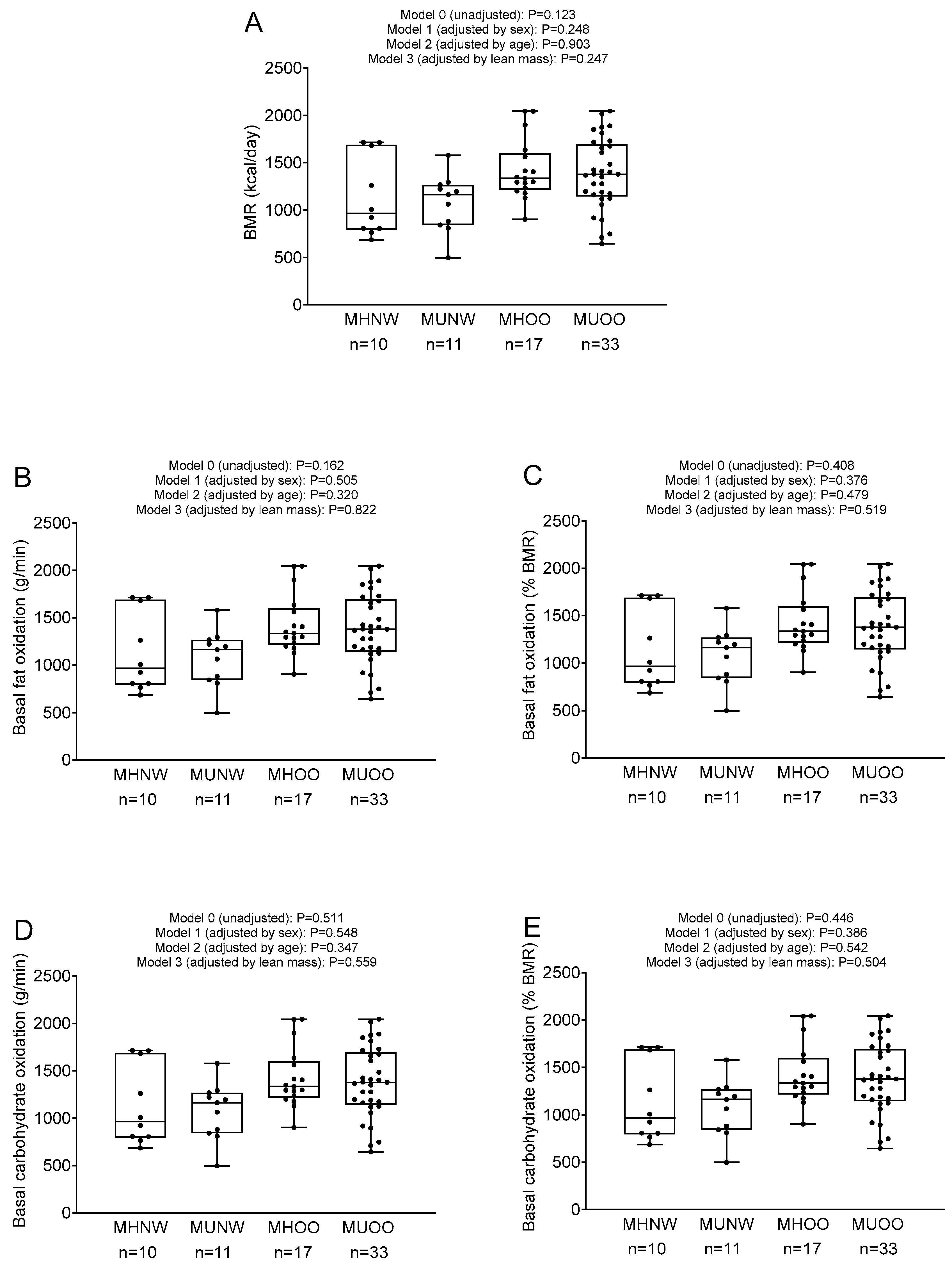
3. Results
4. Discussion
Author Contributions
Funding
Acknowledgments
Conflicts of Interest
References
- North, B.J.; Sinclair, D.A. The intersection between aging and cardiovascular disease. Circ. Res. 2012, 110, 1097–1108. [Google Scholar] [CrossRef] [PubMed]
- WHO. Noncommunicable Diseases Country Profiles, 2018; WHO: Geneva, Switzerland, 2018; Volume 369, ISBN 9789241514620. [Google Scholar]
- Goodpaster, B.H.; Sparks, L.M. Metabolic Flexibility in Health and Disease. Cell Metab. 2017, 25, 1027–1036. [Google Scholar] [CrossRef] [PubMed] [Green Version]
- Amaro-Gahete, F.; Jurado-Fasoli, L.; De-la-O, A.; Gutierrez, Á.; Castillo, M.; Ruiz, J. Accuracy and Validity of Resting Energy Expenditure Predictive Equations in Middle-Aged Adults. Nutrients 2018, 10, 1635. [Google Scholar] [CrossRef] [PubMed] [Green Version]
- Amaro-Gahete, F.J.; Sanchez-Delgado, G.; Alcantara, J.M.A.; Martinez-Tellez, B.; Muñoz-Hernandez, V.; Merchan-Ramirez, E.; Löf, M.; Labayen, I.; Ruiz, J.R. Congruent Validity of Resting Energy Expenditure Predictive Equations in Young Adults. Nutrients 2019, 11, 223. [Google Scholar] [CrossRef] [PubMed] [Green Version]
- Kitazoe, Y.; Kishino, H.; Tanisawa, K.; Udaka, K.; Tanaka, M. Renormalized basal metabolic rate describes the human aging process and longevity. Aging Cell 2019, 18, e12968. [Google Scholar] [CrossRef] [PubMed]
- Hand, G.A.; Blair, S.N. Energy flux and its role in obesity and metabolic disease. Eur. Endocrinol. 2014, 10, 131–135. [Google Scholar] [CrossRef]
- Amaro-Gahete, F.J.; De-la-O, A.; Jurado-Fasoli, L.; Sanchez-Delgado, G.; Ruiz, J.R.; Castillo, M.J. Metabolic rate in sedentary adults, following different exercise training interventions: The FIT-AGEING randomized controlled trial. Clin. Nutr. 2020. [Google Scholar] [CrossRef]
- Longo, V.D.; Mattson, M.P. Fasting: Molecular Mechanisms and Clinical Applications. Cell Metab. 2014, 19, 181–192. [Google Scholar] [CrossRef] [Green Version]
- Smith, R.L.; Soeters, M.R.; Wüst, R.C.I.; Houtkooper, R.H. Metabolic flexibility as an adaptation to energy resources and requirements in health and disease. Endocr. Rev. 2018, 39, 489–517. [Google Scholar] [CrossRef] [Green Version]
- Ali, N.; Mahmood, S.; Manirujjaman, M.; Perveen, R.; Al Nahid, A.; Ahmed, S.; Khanum, F.A.; Rahman, M. Hypertension prevalence and influence of basal metabolic rate on blood pressure among adult students in Bangladesh. BMC Public Health 2017, 18, 1–9. [Google Scholar] [CrossRef] [Green Version]
- Sriram, N.; Hunter, G.R.; Fisher, G.; Brock, D.W. Resting Energy Expenditure and Systolic Blood Pressure Relationships in Women Across 4.5 Years. J. Clin. Hypertens. 2014, 16, 172–176. [Google Scholar] [CrossRef] [PubMed] [Green Version]
- Brock, D.W.; Tompkins, C.L.; Fisher, G.; Hunter, G.R. Influence of resting energy expenditure on blood pressure is independent of body mass and a marker of sympathetic tone. Metabolism 2012, 61, 237–241. [Google Scholar] [CrossRef] [Green Version]
- Hopkins, J.L.; Hopkins, P.N.; Brinton, E.A.; Adams, T.D.; Davidson, L.E.; Nanjee, M.N.; Hunt, S.C. Expression of Metabolic Syndrome in Women with Severe Obesity. Metab. Syndr. Relat. Disord. 2017, 15, 283–290. [Google Scholar] [CrossRef] [PubMed]
- Piaggi, P.; Thearle, M.S.; Bogardus, C.; Krakoff, J. Fasting hyperglycemia predicts lower rates of weight gain by increased energy expenditure and fat oxidation rate. J. Clin. Endocrinol. Metab. 2015, 100, 1078–1087. [Google Scholar] [CrossRef] [PubMed] [Green Version]
- Alawad, A.O.; Merghani, T.H.; Ballal, M.A. Resting metabolic rate in obese diabetic and obese non-diabetic subjects and its relation to glycaemic control. BMC Res. Notes 2013, 6, 1. [Google Scholar] [CrossRef] [PubMed] [Green Version]
- Sampath Kumar, A.; Arun Maiya, G.; Shastry, B.A.; Vaishali, K.; Maiya, S.; Umakanth, S. Correlation between basal metabolic rate, visceral fat and insulin resistance among type 2 diabetes mellitus with peripheral neuropathy. Diabetes Metab. Syndr. Clin. Res. Rev. 2019, 13, 344–348. [Google Scholar] [CrossRef] [PubMed]
- Ravussin, E.; Swinburn, B.A. Metabolic predictors of obesity: Cross-sectional versus longitudinal data. Int. J. Obes. Relat. Metab. Disord. J. Int. Assoc. Study Obes. 1993, 17, S28–S31. [Google Scholar]
- Buscemi, S.; Verga, S.; Caimi, G.; Cerasola, G. A low resting metabolic rate is associated with metabolic syndrome. Clin. Nutr. 2007, 26, 806–809. [Google Scholar] [CrossRef]
- Lavie, C.J.; Milani, R.V.; Ventura, H.O. Obesity and Cardiovascular Disease. Risk Factor, Paradox, and Impact of Weight Loss. J. Am. Coll. Cardiol. 2009, 53, 1925–1932. [Google Scholar] [CrossRef] [Green Version]
- Galgani, J.E.; Santos, J.L. Insights about weight loss-induced metabolic adaptation. Obesity 2016, 24, 277–278. [Google Scholar] [CrossRef] [Green Version]
- Melzer, K. Carbohydrate and fat utilization during rest and physical activity. E. Spen. Eur. E. J. Clin. Nutr. Metab. 2011, 6, e45–e52. [Google Scholar] [CrossRef] [Green Version]
- El Bacha, T.; Luz, M.; Da Poian, A. Dynamic adaptation of nutrient utilization in humans. Nat. Educ. 2010, 3, 8. [Google Scholar]
- Amaro-Gahete, F.J.; De-la-O, A.; Jurado-Fasoli, L.; Espuch-Oliver, A.; Robles-Gonzalez, L.; Navarro-Lomas, G.; de Haro, T.; Femia, P.; Castillo, M.J.; Gutierrez, A. Exercise training as S-Klotho protein stimulator in sedentary healthy adults: Rationale, design, and methodology. Contemp. Clin. Trials Commun. 2018, 11, 10–19. [Google Scholar] [CrossRef] [PubMed]
- Arvidsson, D.; Fridolfsson, J.; Börjesson, M.; Andersen, L.B.; Ekblom, Ö.; Dencker, M.; Brønd, J.C. Re-examination of accelerometer data processing and calibration for the assessment of physical activity intensity. Scand. J. Med. Sci. Sports 2019, 29, 1442–1452. [Google Scholar] [CrossRef] [PubMed]
- Fullmer, S.; Benson-Davies, S.; Earthman, C.P.; Frankenfield, D.C.; Gradwell, E.; Lee, P.; Trabulsi, J.; Fullmer, S.; Benson-Davies, S.; Earthman, C.P.; et al. Evidence analysis library review of best practices for performing indirect calorimetry in healthy and non-critically ill individuals. J. Acad. Nutr. Diet. 2015, 115, 1417–1446.e2. [Google Scholar] [CrossRef] [PubMed]
- Alcantara, J.M.A.; Sanchez-Delgado, G.; Martinez-Tellez, B.; Merchan-Ramirez, E.; Labayen, I.; Ruiz, J.R. Congruent validity and inter-day reliability of two breath by breath metabolic carts to measure resting metabolic rate in young adults. Nutr. Metab. Cardiovasc. Dis. 2018, 28, 929–936. [Google Scholar] [CrossRef] [PubMed]
- Sanchez-Delgado, G.; Alcantara, J.M.A.; Ortiz-Alvarez, L.; Xu, H.; Martinez-Tellez, B.; Labayen, I.; Ruiz, J.R. Reliability of resting metabolic rate measurements in young adults: Impact of methods for data analysis. Clin. Nutr. 2018, 37, 1618–1624. [Google Scholar] [CrossRef] [PubMed] [Green Version]
- Weir, J. New methods for calculating metabolic rate with special reference to protein metabolism. J. Physiol. 1949, 109, 1–9. [Google Scholar] [CrossRef]
- Frayn, K.N. Calculation of substrate oxidation rates in vivo from gaseous exchange. J. Appl. Physiol. 1983, 55, 628–634. [Google Scholar] [CrossRef] [Green Version]
- Whelton, P.K.; Williams, B. The 2018 European Society of Cardiology/European Society of Hypertension and 2017 American College of Cardiology/American Heart Association Blood Pressure Guidelines. JAMA 2018, 320, 1749. [Google Scholar] [CrossRef]
- Katz, A.; Nambi, S.S.; Mather, K.; Baron, A.D.; Follmann, D.A.; Sullivan, G.; Quon, M.J. Quantitative insulin sensitivity check index: A simple, accurate method for assessing insulin sensitivity in humans. J. Clin. Endocrinol. Metab. 2000, 85, 2402–2410. [Google Scholar] [CrossRef] [PubMed]
- Ascaso, J.F.; Romero, P.; Real, J.T.; Priego, A.; Valdecabres, C.; Carmena, R. Insulin resistance quantification by fasting insulin plasma values and HOMA index in a non-diabetic population. Med. Clin. (Barc). 2001, 117, 530–533. [Google Scholar] [CrossRef]
- Ortega, F.B.; Ruiz, J.R.; Labayen, I.; Lavie, C.J.; Blair, S.N. The Fat but Fit paradox: What we know and don’t know about it. Br. J. Sports Med. 2018, 52, 151–153. [Google Scholar] [CrossRef] [PubMed]
- Carracher, A.M.; Marathe, P.H.; Close, K.L. International Diabetes Federation 2017. J. Diabetes 2018, 10, 353–356. [Google Scholar] [CrossRef] [PubMed] [Green Version]
- Hildebrand, M.; Hansen, B.H.; van Hees, V.T.; Ekelund, U. Evaluation of raw acceleration sedentary thresholds in children and adults. Scand. J. Med. Sci. Sport. 2017, 27, 1814–1823. [Google Scholar] [CrossRef]
- Hildebrand, M.; Van Hees, V.T.; Hansen, B.H.; Ekelund, U. Age group comparability of raw accelerometer output from wrist-and hip-worn monitors. Med. Sci. Sports Exerc. 2014, 46, 1816–1824. [Google Scholar] [CrossRef]
- Migueles, J.H.; Cadenas-Sanchez, C.; Ekelund, U.; Delisle Nyström, C.; Mora-Gonzalez, J.; Löf, M.; Labayen, I.; Ruiz, J.R.; Ortega, F.B. Accelerometer Data Collection and Processing Criteria to Assess Physical Activity and Other Outcomes: A Systematic Review and Practical Considerations. Sport. Med. 2017, 47, 1821–1845. [Google Scholar] [CrossRef]
- Drabsch, T.; Holzapfel, C.; Stecher, L.; Petzold, J.; Skurk, T.; Hauner, H. Associations between C-reactive protein, insulin sensitivity, and resting metabolic rate in adults: A mediator analysis. Front. Endocrinol. (Lausanne) 2018, 9, 1–9. [Google Scholar] [CrossRef] [Green Version]
- Buscemi, S.; Donatelli, M.; Grosso, G.; Vasto, S.; Galvano, F.; Costa, F.; Rosafio, G.; Verga, S. Resting energy expenditure in type 2 diabetic patients and the effect of insulin bolus. Diabetes Res. Clin. Pract. 2014, 106, 605–610. [Google Scholar] [CrossRef]
- Karelis, A.D. Metabolically healthy but obese individuals. Lancet (London, England) 2008, 372, 1281–1283. [Google Scholar] [CrossRef]
- Primeau, V.; Coderre, L.; Karelis, A.D.; Brochu, M.; Lavoie, M.-E.; Messier, V.; Sladek, R.; Rabasa-Lhoret, R. Characterizing the profile of obese patients who are metabolically healthy. Int. J. Obes. (Lond.) 2011, 35, 971–981. [Google Scholar] [CrossRef] [PubMed] [Green Version]
- Lee, C.C.; Lorenzo, C.; Haffner, S.M.; Wagenknecht, L.E.; Goodarzi, M.O.; Stefanovski, D.; Norris, J.M.; Rewers, M.J.; Hanley, A.J. Components of metabolic syndrome and 5-year change in insulin clearance—The Insulin Resistance Atherosclerosis Study. Diabetes, Obes. Metab. 2013, 15, 441–447. [Google Scholar] [CrossRef] [PubMed] [Green Version]
- Karwi, Q.G.; Uddin, G.M.; Ho, K.L.; Lopaschuk, G.D. Loss of Metabolic Flexibility in the Failing Heart. Front. Cardiovasc. Med. 2018, 5, 1–19. [Google Scholar] [CrossRef] [PubMed] [Green Version]


| All (n = 71) | Men (n = 34) | Women (n = 37) | ||||
| Age (years) | 53.4 | (4.9) | 54.2 | (5.2) | 52.7 | (4.6) |
| Anthropometry and body composition | (n = 71) | (n = 34) | (n = 37) | |||
| Body mass index (kg/m2) | 26.82 | (3.79) | 28.50 | (3.51) | 25.29 | (3.40) * |
| Waist circumference (cm) | 95.29 | (11.89) | 103.13 | (8.45) | 88.09 | (9.91) * |
| Lean mass index (kg/m2) | 15.29 | (2.86) | 17.49 | (2.06) | 13.26 | (1.78) * |
| Fat mass index (kg/m2) | 10.74 | (3.10) | 10.15 | (3.20) | 11.28 | (2.95) * |
| Visceral adipose tissue mass (g) | 788.9 | (391.8) | 986.4 | (388.8) | 607.4 | (298.7) * |
| Basal metabolic rate | (n = 68) | (n = 34) | (n = 34) | |||
| BMR (kcal/d) | 1323 | (376) | 1548 | (305) | 1098 | (301) * |
| BMR (kcal/kgleanmass/d) | ||||||
| Nutrients oxidation | (n = 57) | (n = 28) | (n = 29) | |||
| BFox (g/min) | 0.116 | (0.098) | 0.135 | (0.117) | 0.098 | (0.074) * |
| BFox (% BMR) | 48.32 | (35.72) | 47.90 | (38.13) | 48.73 | (33.91) |
| BCHox (g/min) | 0.112 | (0.096) | 0.137 | (0.115) | 0.088 | (0.069) * |
| BCHox (% BMR) | 48.06 | (34.34) | 50.00 | (37.33) | 46.28 | (31.77) |
| Blood pressure | (n = 67) | (n = 31) | (n = 36) | |||
| Systolic (mm Hg) | 127.09 | (15.78) | 134.26 | (13.84) | 120.92 | (14.85) * |
| Diastolic (mm Hg) | 81.12 | (11.72) | 85.16 | (10.87) | 77.64 | (11.44) * |
| Mean (mm Hg) | 104.10 | (13.15) | 109.71 | (11.70) | 99.28 | (12.52) * |
| Glycemic profile | (n = 70) | (n = 33) | (n = 37) | |||
| Plasma glucose (mg/dL) | 93.56 | (11.36) | 95.00 | (13.60) | 92.27 | (8.90) |
| Plasma insulin (UI/mL) | 8.08 | (5.68) | 8.94 | (6.75) | 7.32 | (4.48) |
| QUICKI | 0.362 | (0.036) | 0.357 | (0.039) | 0.365 | (0.033) |
| HOMA | 1.933 | (1.668) | 2.209 | (2.107) | 1.686 | (1.120) * |
| Lipid profile | (n = 70) | (n = 33) | (n = 37) | |||
| Total cholesterol (mg/dL) | 206.14 | (32.17) | 200.67 | (32.29) | 211.03 | (31.70) |
| HDL-C (mg/dL) | 58.71 | (12.28) | 55.33 | (12.86) | 61.73 | (11.06) * |
| LDL-C (mg/dL) | 131.39 | (64.57) | 138.79 | (78.00) | 124.78 | (49.88) |
| Triglycerides (mg/dL) | 126.23 | (27.07) | 125.06 | (27.93) | 127.27 | (26.63) |
| LDL-C/HDL-C | 2.234 | (0.632) | 2.358 | (0.667) | 2.123 | (0.585) * |
| Dietary intake | (n = 70) | (n = 34) | (n = 36) | |||
| Energy (kcal/d) | 2140 | (699) | 2396 | (841) | 1901 | (417) * |
| Fat (g/d) | 87.8 | (24.9) | 98.2 | (23.8) | 78.0 | (22.0) * |
| Carbohydrate (g/d) | 227.9 | (113.5) | 255.9 | (145.6) | 201.4 | (62.6) * |
| Protein (g/d) | 88.8 | (38.2) | 94.2 | (39.1) | 83.6 | (37.2) * |
| Sedentary behaviour and physical activity | (n = 70) | (n = 34) | (n = 36) | |||
| Sedentary time (min/d) | 746.9 | (84.3) | 770.0 | (80.3) | 725.1 | (83.3) |
| Time in MVPA (min/d) | 95.9 | (35.6) | 96.6 | (35.5) | 95.2 | (36.2) |
| Model 0 | Model 1 | Model 2 | Model 3 | |||||||||||||
|---|---|---|---|---|---|---|---|---|---|---|---|---|---|---|---|---|
| F1,68 | p value | R2 | β | F1,67 | p value | R2 | β | F1,67 | p value | R2 | β | F1,67 | p value | R2 | β | |
| Basal Metabolic Rate (kcal/d) | ||||||||||||||||
| Systolic blood pressure (mm Hg) | 1.243 | 0.269 | 0.004 | 0.137 | 8.377 | 0.145 | 0.183 | −0.210 | 0.753 | 0.231 | −0.008 | 0.155 | 6.062 | 0.523 | 0.133 | −0.086 |
| Diastolic blood pressure (mm Hg) | 1.359 | 0.248 | 0.005 | 0.143 | 3.933 | 0.530 | 0.082 | −0.095 | 0.710 | 0.238 | −0.009 | 0.153 | 3.731 | 0.834 | 0.076 | −0.029 |
| Mean blood pressure (mm Hg) | 1.415 | 0.239 | 0.006 | 0.146 | 6.844 | 0.250 | 0.150 | −0.169 | 0.797 | 0.213 | −0.006 | 0.161 | 5.498 | 0.634 | 0.120 | −0.064 |
| Plasma glucose (mg/dL) | 1.017 | 0.317 | 0.001 | 0.121 | 0.618 | 0.626 | 0.018 | 0.076 | 0.134 | 0.291 | −0.013 | 0.134 | 0.504 | 0.362 | −0.015 | 0.126 |
| Plasma insulin (UI/mL) | 4.101 | 0.047 | 0.043 | 0.238 | 2.022 | 0.112 | 0.029 | 0.243 | 5.472 | 0.181 | 0.115 | 0.159 | 3.390 | 0.013 | 0.065 | 0.339 |
| QUICKI | 3.652 | 0.060 | 0.037 | −2.226 | 1.854 | 0.096 | 0.024 | −0.256 | 4.612 | 0.203 | 0.095 | −0.153 | 2.822 | 0.021 | 0.078 | −0.314 |
| HOMA | 5.528 | 0.022 | 0.062 | 0.274 | 3.091 | 0.021 | 0.084 | 0.350 | 4.836 | 0.078 | 0.100 | 0.212 | 4.048 | 0.006 | 0.081 | 0.371 |
| Total cholesterol (mg/dL) | 0.086 | 0.771 | −0.013 | −0.035 | 1.145 | 0.494 | 0.004 | 0.105 | 2.533 | 0.380 | 0.043 | −0.108 | 3.705 | 0.315 | 0.073 | 0.133 |
| HDL-C (mg/dL) | 3.361 | 0.071 | 0.033 | −0.217 | 2.655 | 0.555 | 0.046 | −0.089 | 3.003 | 0.030 | 0.055 | −0.269 | 4.290 | 0.551 | 0.087 | −0.078 |
| LDL-C (mg/dL) | 1.143 | 0.289 | 0.002 | 0.129 | 1.402 | 0.106 | 0.012 | 0.250 | 2.175 | 0.569 | 0.061 | 0.070 | 2.844 | 0.063 | 0.051 | 0.262 |
| Triglycerides (mg/dL) | 0.063 | 0.802 | −0.014 | −0.030 | 0.947 | 0.304 | −0.002 | −0.159 | 0.379 | 0.984 | −0.018 | −0.002 | 0.361 | 0.547 | −0.019 | −0.084 |
| LDL-C/HDL-C | 4.378 | 0.040 | 0.047 | 0.246 | 2.229 | 0.165 | 0.034 | 0.212 | 2.207 | 0.059 | 0.034 | 0.236 | 2.254 | 0.108 | 0.035 | 0.219 |
| F1,56 | p value | R2 | β | F1,55 | p value | R2 | β | F1,55 | p value | R2 | β | F1,55 | p value | R2 | β | |
| Basal Fat Oxidation (g/min) | ||||||||||||||||
| Systolic blood pressure (mm Hg) | 2.781 | 0.100 | 0.026 | 0.203 | 7.484 | 0.401 | 0.164 | 0.099 | 1.467 | 0.094 | 0.014 | 0.224 | 5.937 | 0.657 | 0.156 | 0.056 |
| Diastolic blood pressure (mm Hg) | 2.594 | 0.112 | 0.024 | 0.196 | 4.254 | 0.327 | 0.090 | 0.120 | 1.519 | 0.086 | 0.015 | 0.229 | 3.927 | 0.531 | 0.081 | 0.081 |
| Mean blood pressure (mm Hg) | 2.964 | 0.090 | 0.029 | 0.209 | 6.585 | 0.343 | 0.145 | 0.113 | 1.626 | 0.077 | 0.019 | 0.236 | 5.544 | 0.582 | 0.148 | 0.070 |
| Plasma glucose (mg/dL) | 1.102 | 0.297 | 0.001 | −0.126 | 1.427 | 0.180 | 0.012 | −0.167 | 0.669 | 0.253 | −0.010 | −0.149 | 1.102 | 0.297 | 0.001 | −0.126 |
| Plasma insulin (UI/mL) | 6.559 | 0.013 | 0.075 | −0.297 | 5.392 | 0.003 | 0.113 | −0.355 | 5.992 | 0.102 | 0.126 | −0.200 | 3.420 | 0.012 | 0.066 | −0.326 |
| QUICKI | 6.379 | 0.014 | 0.072 | 0.293 | 4.645 | 0.005 | 0.096 | 0.342 | 5.294 | 0.094 | 0.111 | 0.206 | 3.419 | 0.012 | 0.066 | 0.328 |
| HOMA | 8.211 | 0.006 | 0.108 | −0.328 | 5.507 | 0.002 | 0.116 | −0.376 | 5.546 | 0.038 | 0.116 | −0257 | 4.525 | 0.004 | 0.093 | −0.373 |
| Total cholesterol (mg/dL) | 2.321 | 0.132 | 0.019 | −0.182 | 1.667 | 0.226 | 0.019 | −0.150 | 2.509 | 0.395 | 0.042 | −0.108 | 3.366 | 0.526 | 0.064 | −0.080 |
| HDL-C (mg/dL) | 0.965 | 0.329 | 0.014 | −0.118 | 2.579 | 0.648 | 0.044 | −0.056 | 0.734 | 0.513 | −0.008 | −0.085 | 4.092 | 0.931 | 0.082 | 0.011 |
| LDL-C (mg/dL) | 1.611 | 0.209 | 0.009 | −0.152 | 0.794 | 0.229 | −0.006 | −0.151 | 2.196 | 0.546 | 0.034 | −0.077 | 1.203 | 0.414 | 0.006 | −0.107 |
| Triglycerides (mg/dL) | 0.827 | 0.366 | −0.003 | 0.110 | 0.652 | 0.486 | −0.010 | 0.088 | 0.582 | 0.528 | −0.012 | 0.082 | 0.444 | 0.469 | −0.016 | 0.096 |
| LDL-C/HDL-C | 0.033 | 0.856 | −0.014 | −0.022 | 1.394 | 0.553 | 0.011 | −0.074 | 0.348 | 0.903 | −0.019 | 0.016 | 1.191 | 0.310 | 0.006 | −0.099 |
| Basal Fat Oxidation (% Basal Metabolic Rate) | ||||||||||||||||
| Systolic blood pressure (mm Hg) | 0.853 | 0.359 | −0.002 | 0.114 | 7.848 | 0.257 | 0.172 | 0.128 | 0.444 | 0.362 | −0.017 | 0.127 | 6.046 | 0.538 | 0.133 | 0.071 |
| Diastolic blood pressure (mm Hg) | 0.450 | 0.505 | −0.008 | 0.083 | 4.066 | 0.428 | 0.085 | 0.094 | 0.290 | 0.450 | −0.022 | 0.105 | 3.797 | 0.687 | 0.078 | 0.048 |
| Mean blood pressure (mm Hg) | 0.728 | 0.397 | −0.004 | 0.105 | 6.692 | 0.300 | 0.147 | 0.119 | 0.402 | 0.377 | −0.018 | 0.123 | 5.544 | 0.582 | 0.121 | 0.064 |
| Plasma glucose (mg/dL) | 1.426 | 0.237 | 0.006 | −0.143 | 1.178 | 0.251 | 0.005 | −0.139 | 0.937 | 0.177 | −0.002 | −0.183 | 0.862 | 0.216 | −0.004 | −0.152 |
| Plasma insulin (UI/mL) | 8.336 | 0.005 | 0.096 | −0.330 | 4.853 | 0.006 | 0.100 | −0.326 | 6.263 | 0.077 | 0.132 | −0.224 | 4.112 | 0.006 | 0.083 | −0.329 |
| QUICKI | 6.853 | 0.011 | 0.078 | 0.303 | 3.781 | 0.012 | 0.075 | 0.299 | 5.137 | 0.112 | 0.107 | 0.204 | 3.376 | 0.012 | 0.064 | 0.303 |
| HOMA | 10.262 | 0.002 | 0.118 | −0.362 | 5.354 | 0.002 | 0.112 | −0.359 | 6.041 | 0.023 | 0.127 | −0.291 | 5.066 | 0.002 | 0.105 | −0.364 |
| Total cholesterol (mg/dL) | 1.320 | 0.255 | 0.005 | −0.138 | 1.647 | 0.232 | 0.018 | −0.144 | 2.165 | 0.768 | 0.033 | −0.039 | 3.573 | 0.379 | 0.069 | −0.104 |
| HDL-C (mg/dL) | 0.033 | 0.855 | −0.014 | −0.022 | 2.504 | 0.791 | 0.042 | −0.031 | 0.557 | 0.773 | −0.013 | 0.039 | 4.103 | 0.873 | 0.083 | 0.019 |
| LDL-C (mg/dL) | 1.905 | 0.172 | 0.013 | −0.165 | 1.106 | 0.171 | 0.001 | −0.167 | 2.177 | 0.566 | 0.033 | −0.076 | 1.633 | 0.223 | 0.018 | −0.148 |
| Triglycerides (mg/dL) | 0.413 | 0.523 | −0.009 | 0.078 | 0.633 | 0.503 | −0.011 | 0.082 | 0.421 | 0.774 | −0.017 | 0.039 | 0.340 | 0.570 | −0.019 | 0.070 |
| LDL-C/HDL-C | 0.531 | 0.469 | −0.007 | −0.088 | 1.450 | 0.498 | 0.013 | −0.082 | 0.423 | 0.687 | −0.017 | −0.055 | 1.315 | 0.368 | 0.009 | −0.109 |
| Model 0 | Model 1 | Model 2 | Model 3 | |||||||||||||
|---|---|---|---|---|---|---|---|---|---|---|---|---|---|---|---|---|
| F1,56 | p value | R2 | β | F1,55 | p value | R2 | β | F1,55 | p value | R2 | β | F1,55 | p value | R2 | β | |
| Basal Carbohydrate Oxidation (g/min) | ||||||||||||||||
| Systolic blood pressure (mm Hg) | 0.046 | 0.832 | −0.015 | −0.026 | 8.109 | 0.192 | 0.177 | −0.153 | 0.031 | 0.889 | −0.030 | −0.019 | 6.030 | 0.552 | 0.132 | −0.069 |
| Diastolic blood pressure (mm Hg) | 0.136 | 0.713 | −0.013 | −0.046 | 4.490 | 0.242 | 0.096 | −0.144 | 0.087 | 0.679 | −0.028 | −0.057 | 3.966 | 0.497 | 0.110 | −0.081 |
| Mean blood pressure (mm Hg) | 0.086 | 0.771 | −0.014 | −0.036 | 7.088 | 0.190 | 0.156 | −0.156 | 0.042 | 0.788 | −0.030 | −0.037 | 5.626 | 0.506 | 0.123 | −0.078 |
| Plasma glucose (mg/dL) | 1.159 | 0.285 | 0.002 | 0.129 | 0.844 | 0.435 | 0.025 | 0.104 | 0.721 | 0.235 | −0.008 | 0.157 | 0.618 | 0.305 | −0.011 | 0.126 |
| Plasma insulin (UI/mL) | 7.196 | 0.009 | 0.082 | 0.309 | 3.690 | 0.018 | 0.072 | 0.292 | 6.105 | 0.091 | 0.129 | 0.209 | 3.828 | 0.008 | 0.076 | 0.318 |
| QUICKI | 6.991 | 0.010 | 0.080 | −0.305 | 3.472 | 0.016 | 0.067 | −0.298 | 5.416 | 0.083 | 0.113 | −0.216 | 3.631 | 0.009 | 0.071 | −0.312 |
| HOMA | 11.156 | 0.001 | 0.128 | 0.375 | 5.501 | 0.002 | 0.115 | 0.378 | 6.670 | 0.013 | 0.141 | 0.309 | 5.698 | 0.001 | 0.120 | 0.382 |
| Total cholesterol (mg/dL) | 0.639 | 0.427 | −0.005 | 0.096 | 1.686 | 0.221 | 0.019 | 0.153 | 2.118 | 0.980 | 0.031 | 0.003 | 3.793 | 0.280 | 0.075 | 0.127 |
| HDL-C (mg/dL) | 0.049 | 0.826 | −0.014 | −0.027 | 2.555 | 0.685 | 0.043 | 0.050 | 0.738 | 0.510 | −0.008 | −0.087 | 4.090 | 0.959 | 0.082 | 0.006 |
| LDL-C (mg/dL) | 1.767 | 0.188 | 0.011 | 0.011 | 1.151 | 0.144 | 0.004 | 0.185 | 2.206 | 0.536 | 0.034 | 0.080 | 1.979 | 0.144 | 0.028 | 0.176 |
| Triglycerides (mg/dL) | 0.516 | 0.475 | −0.007 | −0.087 | 0.921 | 0.316 | −0.002 | −0.127 | 0.465 | 0.682 | −0.016 | −0.054 | 0.481 | 0.439 | −0.015 | −0.095 |
| LDL-C/HDL-C | 1.214 | 0.274 | 0.003 | 0.132 | 1.464 | 0.486 | 0.013 | 0.087 | 1.384 | 0.332 | 0.011 | 0.118 | 1.384 | 0.332 | 0.011 | 0.118 |
| Basal Carbohydrate Oxidation (% Basal Metabolic Rate) | ||||||||||||||||
| Systolic blood pressure (mm Hg) | 0.046 | 0.832 | −0.015 | −0.026 | 8.109 | 0.192 | 0.177 | −0.153 | 0.031 | 0.889 | −0.030 | −0.019 | 6.030 | 0.552 | 0.132 | −0.069 |
| Diastolic blood pressure (mm Hg) | 0.136 | 0.713 | −0.013 | −0.046 | 4.490 | 0.242 | 0.096 | −0.144 | 0.087 | 0.679 | −0.028 | −0.057 | 3.966 | 0.497 | 0.082 | −0.081 |
| Mean blood pressure (mm Hg) | 0.086 | 0.771 | −0.014 | −0.036 | 7.088 | 0.190 | 0.156 | −0.156 | 0.042 | 0.788 | −0.030 | −0.037 | 5.626 | 0.506 | 0.123 | −0.078 |
| Plasma glucose (mg/dL) | 1.159 | 0.285 | 0.002 | 0.129 | 0.844 | 0.411 | −0.005 | 0.104 | 0.721 | 0.235 | −0.008 | 0.157 | 0.618 | 0.305 | −0.011 | 0.126 |
| Plasma insulin (UI/mL) | 7.196 | 0.009 | 0.082 | 0.309 | 3.690 | 0.018 | 0.072 | 0.292 | 6.105 | 0.091 | 0.129 | 0.209 | 3.828 | 0.008 | 0.076 | 0.318 |
| QUICKI | 6.991 | 0.010 | 0.080 | −0.305 | 4.472 | 0.016 | 0.067 | −0.298 | 5.416 | 0.083 | 0.113 | −0.216 | 3.631 | 0.009 | 0.071 | −0.312 |
| HOMA | 11.156 | 0.001 | 0.128 | 0.375 | 5.501 | 0.002 | 0.115 | 0.378 | 6.670 | 0.013 | 0.141 | 0.309 | 5.698 | 0.001 | 0.120 | 0.382 |
| Total cholesterol (mg/dL) | 0.639 | 0.427 | −0.005 | 0.096 | 1.686 | 0.221 | 0.019 | 0.153 | 2.118 | 0.980 | 0.031 | 0.003 | 3.793 | 0.280 | 0.075 | 0.127 |
| HDL-C (mg/dL) | 0.049 | 0.826 | −0.014 | −0.027 | 2.555 | 0.685 | 0.043 | 0.050 | 0.738 | 0.510 | −0.008 | −0.087 | 4.090 | 0.959 | 0.082 | 0.006 |
| LDL-C (mg/dL) | 1.767 | 0.188 | 0.011 | 0.159 | 1.151 | 0.144 | 0.004 | 0.185 | 2.206 | 0.536 | 0.034 | 0.080 | 1.979 | 0.144 | 0.028 | 0.176 |
| Triglycerides (mg/dL) | 0.516 | 0.475 | −0.007 | −0.087 | 0.921 | 0.316 | −0.002 | −0.127 | 0.465 | 0.682 | −0.016 | −0.054 | 0.481 | 0.439 | −0.015 | −0.095 |
| LDL-C/HDL-C | 1.214 | 0.274 | 0.003 | 0.132 | 1.464 | 0.486 | 0.013 | 0.087 | 0.698 | 0.403 | −0.009 | 0.113 | 1.384 | 0.332 | 0.011 | 0.118 |
© 2020 by the authors. Licensee MDPI, Basel, Switzerland. This article is an open access article distributed under the terms and conditions of the Creative Commons Attribution (CC BY) license (http://creativecommons.org/licenses/by/4.0/).
Share and Cite
Amaro-Gahete, F.J.; Jurado-Fasoli, L.; Ruiz, J.R.; Castillo, M.J. Association of Basal Metabolic Rate and Nutrients Oxidation with Cardiometabolic Risk Factors and Insulin Sensitivity in Sedentary Middle-Aged Adults. Nutrients 2020, 12, 1186. https://0-doi-org.brum.beds.ac.uk/10.3390/nu12041186
Amaro-Gahete FJ, Jurado-Fasoli L, Ruiz JR, Castillo MJ. Association of Basal Metabolic Rate and Nutrients Oxidation with Cardiometabolic Risk Factors and Insulin Sensitivity in Sedentary Middle-Aged Adults. Nutrients. 2020; 12(4):1186. https://0-doi-org.brum.beds.ac.uk/10.3390/nu12041186
Chicago/Turabian StyleAmaro-Gahete, Francisco J., Lucas Jurado-Fasoli, Jonatan R. Ruiz, and Manuel J. Castillo. 2020. "Association of Basal Metabolic Rate and Nutrients Oxidation with Cardiometabolic Risk Factors and Insulin Sensitivity in Sedentary Middle-Aged Adults" Nutrients 12, no. 4: 1186. https://0-doi-org.brum.beds.ac.uk/10.3390/nu12041186