
Visceral Adiposity Index (VAI) in Children and Adolescents with Obesity: No Association with Daily Energy Intake but Promising Tool to Identify Metabolic Syndrome (MetS)

 , , , , , and
Abstract
:1. Introduction
2. Materials and Methods
2.1. Cohort
2.2. Measurements
2.2.1. Anthropometry
2.2.2. Adiposity Indices
2.2.3. Biochemistry
2.2.4. Dietary Habits
2.2.5. Metabolic Syndrome
2.2.6. Cardiometabolic Risk Assessment
2.3. Statistical Analysis
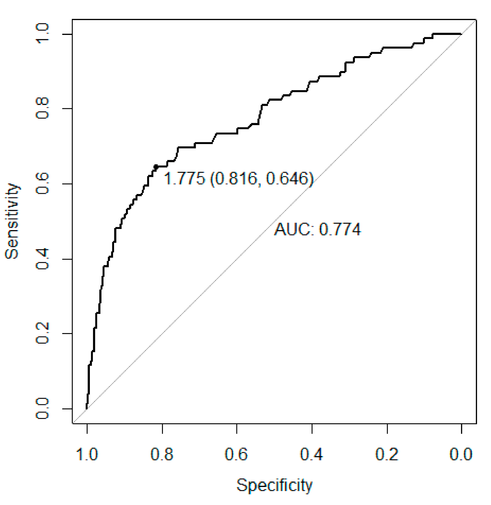
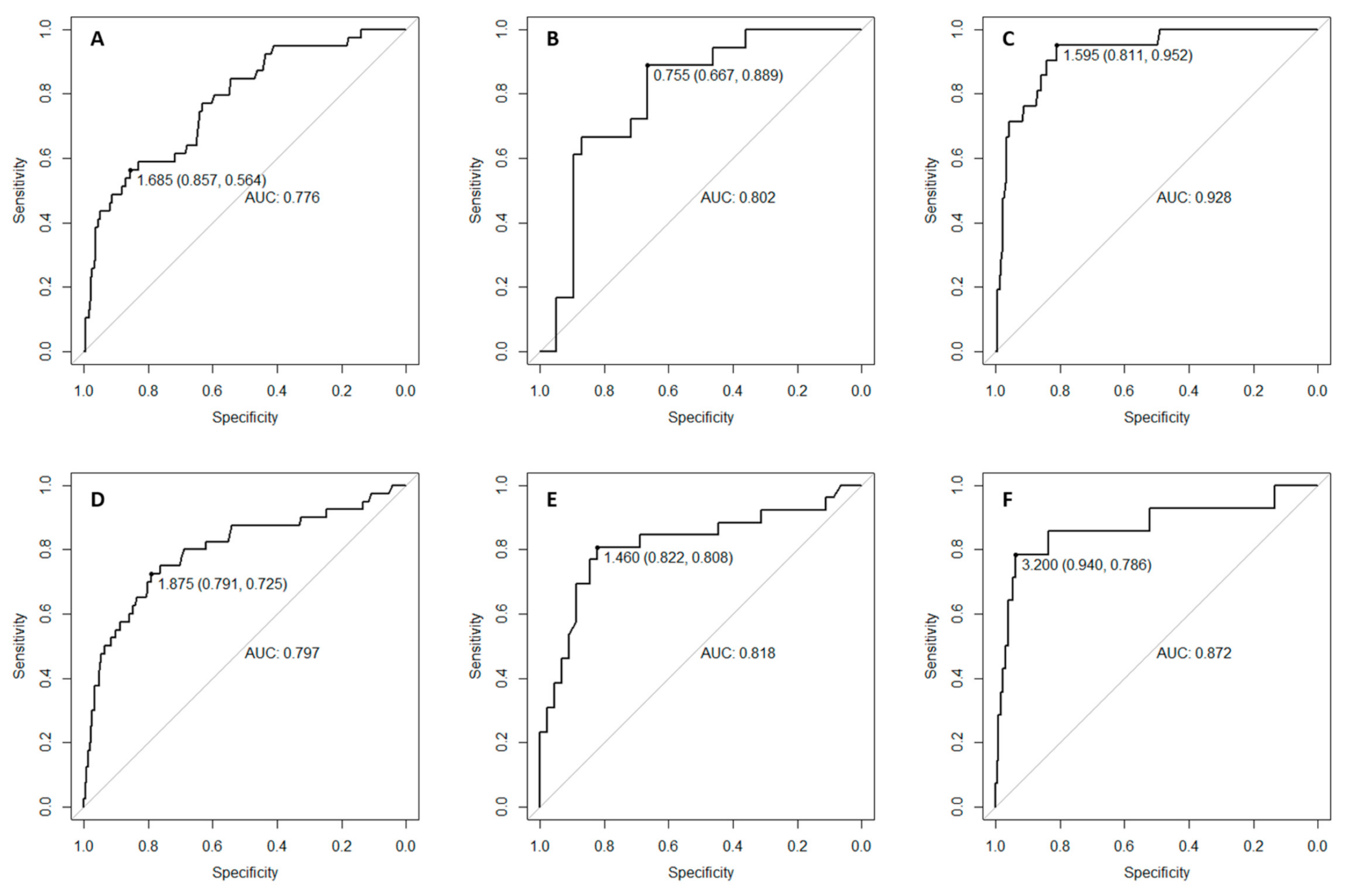
3. Results
4. Discussion
5. Conclusions
Supplementary Materials
Author Contributions
Funding
Institutional Review Board Statement
Informed Consent Statement
Data Availability Statement
Conflicts of Interest
References
- Arroyo-Johnson, C.; Mincey, K.D. Obesity Epidemiology Worldwide. Gastroenterol. Clin. N. Am. 2016, 45, 571–579. [Google Scholar] [CrossRef] [PubMed] [Green Version]
- Tremmel, M.; Gerdtham, U.-G.; Nilsson, P.; Saha, S. Economic Burden of Obesity: A Systematic Literature Review. Int. J. Environ. Res. Public Health 2017, 14, 435. [Google Scholar] [CrossRef] [PubMed]
- World Health Organization Childhood Overweight and Obesity. Available online: http://www.who.int/dietphysicalactivity/childhood/en/ (accessed on 30 December 2020).
- Zimmet, P.; Alberti, G.K.M.M.; Kaufman, F.; Tajima, N.; Silink, M.; Arslanian, S.; Wong, G.; Bennett, P.; Shaw, J.; Caprio, S. The metabolic syndrome in children and adolescents—An IDF consensus report. Pediatr. Diabetes 2007, 8, 299–306. [Google Scholar] [CrossRef] [PubMed]
- Engin, A. The Definition and Prevalence of Obesity and Metabolic Syndrome; Springer: Cham, Switzerland, 2017; pp. 1–17. [Google Scholar]
- Kelishadi, R.; Mirmoghtadaee, P.; Najafi, H.; Keikha, M. Systematic review on the association of abdominal obesity in children and adolescents with cardio-metabolic risk factors. J. Res. Med. Sci. 2015, 20, 294–307. [Google Scholar]
- Styne, D.M.; Arslanian, S.A.; Connor, E.L.; Farooqi, I.S.; Murad, M.H.; Silverstein, J.H.; Yanovski, J.A. Pediatric obesity-assessment, treatment, and prevention: An endocrine society clinical practice guideline. J. Clin. Endocrinol. Metab. 2017, 102, 709–757. [Google Scholar] [CrossRef]
- Valerio, G.; Maffeis, C.; Saggese, G.; Ambruzzi, M.A.; Balsamo, A.; Bellone, S.; Bergamini, M.; Bernasconi, S.; Bona, G.; Calcaterra, V.; et al. Diagnosis, treatment and prevention of pediatric obesity: Consensus position statement of the Italian Society for Pediatric Endocrinology and Diabetology and the Italian Society of Pediatrics. Ital. J. Pediatr. 2018, 44, 88. [Google Scholar] [CrossRef] [Green Version]
- Katzmarzyk, P.T.; Barlow, S.; Bouchard, C.; Catalano, P.M.; Hsia, D.S.; Inge, T.H.; Lovelady, C.; Raynor, H.; Redman, L.M.; Staiano, A.E.; et al. An evolving scientific basis for the prevention and treatment of pediatric obesity. Int. J. Obes. 2014, 38, 887–905. [Google Scholar] [CrossRef] [Green Version]
- Al-Khudairy, L.; Loveman, E.; Colquitt, J.L.; Mead, E.; Johnson, R.E.; Fraser, H.; Olajide, J.; Murphy, M.; Velho, R.M.; O’Malley, C.; et al. Diet, physical activity and behavioural interventions for the treatment of overweight or obese adolescents aged 12 to 17 years. Cochrane Database Syst. Rev. 2017, 6, CD012691. [Google Scholar] [CrossRef] [Green Version]
- August, G.P.; Caprio, S.; Fennoy, I.; Freemark, M.; Kaufman, F.R.; Lustig, R.H.; Silverstein, J.H.; Speiser, P.W.; Styne, D.M.; Montori, V.M.; et al. Prevention and Treatment of Pediatric Obesity: An Endocrine Society Clinical Practice Guideline Based on Expert Opinion. J. Clin. Endocrinol. Metab. 2008, 93, 4576–4599. [Google Scholar] [CrossRef] [Green Version]
- Romieu, I.; Dossus, L.; Barquera, S.; Blottière, H.M.; Franks, P.W.; Gunter, M.; Hwalla, N.; Hursting, S.D.; Leitzmann, M.; Margetts, B.; et al. Energy balance and obesity: what are the main drivers? Cancer Causes Control 2017, 28, 247–258. [Google Scholar] [CrossRef] [Green Version]
- Flegal, K.M.; Ogden, C.L. Childhood Obesity: Are We All Speaking the Same Language? Adv. Nutr. 2011, 2, 159S–166S. [Google Scholar] [CrossRef] [PubMed]
- World Health Organization. Waist Circumference and Waist-Hip Ratio: Report of a WHO Expert Consultation, Geneva, Switzerland, 8–11 December 2008; World Health Organization: Geneva, Switzerland, 2011; ISBN 9789241501491. [Google Scholar]
- Lentferink, Y.E.; Elst, M.A.J.; Knibbe, C.A.J.; van der Vorst, M.M.J. Predictors of Insulin Resistance in Children versus Adolescents with Obesity. J. Obes. 2017, 2017, 3793868. [Google Scholar] [CrossRef] [PubMed] [Green Version]
- Rodea-Montero, E.R.; Evia-Viscarra, M.L.; Apolinar-Jiménez, E. Waist-to-Height Ratio Is a Better Anthropometric Index than Waist Circumference and BMI in Predicting Metabolic Syndrome among Obese Mexican Adolescents. Int. J. Endocrinol. 2014, 2014, 195407. [Google Scholar] [CrossRef] [PubMed]
- Ashwell, M.; Gibson, S. Waist-to-height ratio as an indicator of ‘early health risk’: simpler and more predictive than using a ‘matrix’ based on BMI and waist circumference. BMJ Open 2016, 6, e010159. [Google Scholar] [CrossRef] [PubMed] [Green Version]
- Ochoa Sangrador, C.; Ochoa-Brezmes, J. Waist-to-height ratio as a risk marker for metabolic syndrome in childhood. A meta-analysis. Pediatr. Obes. 2018, 13, 421–432. [Google Scholar] [CrossRef] [PubMed]
- Tee, J.Y.H.; Gan, W.Y.; Lim, P.Y. Comparisons of body mass index, waist circumference, waist-to-height ratio and a body shape index (ABSI) in predicting high blood pressure among Malaysian adolescents: A cross-sectional study. BMJ Open 2020, 10, e032874. [Google Scholar] [CrossRef] [Green Version]
- Freedman, D.S.; Kahn, H.S.; Mei, Z.; Grummer-Strawn, L.M.; Dietz, W.H.; Srinivasan, S.R.; Berenson, G.S. Relation of body mass index and waist-to-height ratio to cardiovascular disease risk factors in children and adolescents: the Bogalusa Heart Study. Am. J. Clin. Nutr. 2007, 86, 33–40. [Google Scholar] [CrossRef] [Green Version]
- Wormser, D.; Kaptoge, S.; Di Angelantonio, E.; Wood, A.M.; Pennells, L.; Thompson, A.; Sarwar, N.; Kizer, J.R.; Lawlor, D.A.; Nordestgaard, B.G.; et al. Separate and combined associations of body-mass index and abdominal adiposity with cardiovascular disease: Collaborative analysis of 58 prospective studies. Lancet 2011, 377, 1085–1095. [Google Scholar] [CrossRef] [Green Version]
- Brambilla, P.; Bedogni, G.; Heo, M.; Pietrobelli, A. Waist circumference-to-height ratio predicts adiposity better than body mass index in children and adolescents. Int. J. Obes. 2013, 37, 943–946. [Google Scholar] [CrossRef] [Green Version]
- Bertoli, S.; Leone, A.; Krakauer, N.Y.; Bedogni, G.; Vanzulli, A.; Redaelli, V.I.; De Amicis, R.; Vignati, L.; Krakauer, J.C.; Battezzati, A. Association of Body Shape Index (ABSI) with cardio-metabolic risk factors: A cross-sectional study of 6081 Caucasian adults. PLoS ONE 2017, 12, e0185013. [Google Scholar] [CrossRef] [Green Version]
- Wang, F.; Chen, Y.; Chang, Y.; Sun, G.; Sun, Y. New anthropometric indices or old ones: which perform better in estimating cardiovascular risks in Chinese adults. BMC Cardiovasc. Disord. 2018, 18, 14. [Google Scholar] [CrossRef] [PubMed] [Green Version]
- Bozorgmanesh, M.; Sardarinia, M.; Hajsheikholeslami, F.; Azizi, F.; Hadaegh, F. CVD-predictive performances of “a body shape index” versus simple anthropometric measures: Tehran lipid and glucose study. Eur. J. Nutr. 2016, 55, 147–157. [Google Scholar] [CrossRef] [PubMed]
- Mameli, C.; Krakauer, N.Y.; Krakauer, J.C.; Bosetti, A.; Ferrari, C.M.; Moiana, N.; Schneider, L.; Borsani, B.; Genoni, T.; Zuccotti, G. The association between a body shape index and cardiovascular risk in overweight and obese children and adolescents. PLoS ONE 2018, 13, e0190426. [Google Scholar] [CrossRef] [Green Version]
- Leone, A.; Vizzuso, S.; Brambilla, P.; Mameli, C.; Ravella, S.; De Amicis, R.; Battezzati, A.; Zuccotti, G.; Bertoli, S.; Verduci, E. Evaluation of Different Adiposity Indices and Association with Metabolic Syndrome Risk in Obese Children: Is there a Winner? Int. J. Mol. Sci. 2020, 21, 4083. [Google Scholar] [CrossRef]
- Amato, M.C.; Giordano, C.; Pitrone, M.; Galluzzo, A. Cut-off points of the visceral adiposity index (VAI) identifying a visceral adipose dysfunction associated with cardiometabolic risk in a Caucasian Sicilian population. Lipids Health Dis. 2011, 10, 183. [Google Scholar] [CrossRef] [Green Version]
- Mohammadreza, B.; Farzad, H.; Davoud, K.; Prof, A.F. Prognostic significance of the Complex “Visceral Adiposity Index” vs. simple anthropometric measures: Tehran lipid and glucose study. Cardiovasc. Diabetol. 2012, 11, 20. [Google Scholar] [CrossRef] [Green Version]
- Al-Daghri, N.M.; Al-Attas, O.S.; Alokail, M.S.; Alkharfy, K.M.; Charalampidis, P.; Livadas, S.; Kollias, A.; Sabico, S.L.; Chrousos, G.P. Visceral adiposity index is highly associated with adiponectin values and glycaemic disturbances. Eur. J. Clin. Investig. 2013, 43, 183–189. [Google Scholar] [CrossRef]
- Petta, S.; Amato, M.; Cabibi, D.; Cammà, C.; Di Marco, V.; Giordano, C.; Galluzzo, A.; Craxì, A. Visceral adiposity index is associated with histological findings and high viral load in patients with chronic hepatitis C due to genotype 1. Hepatology 2010, 52, 1543–1552. [Google Scholar] [CrossRef]
- Ejtahed, H.-S.; Kelishadi, R.; Hasani-Ranjbar, S.; Angoorani, P.; Motlagh, M.E.; Shafiee, G.; Ziaodini, H.; Taheri, M.; Qorbani, M.; Heshmat, R. Discriminatory Ability of Visceral Adiposity Index as an Indicator for Modeling Cardio-Metabolic Risk Factors in Pediatric Population: The CASPIAN-V Study. J. Cardiovasc. Thorac. Res. 2019, 11, 280–286. [Google Scholar] [CrossRef] [Green Version]
- Moslehi, N.; Ehsani, B.; Mirmiran, P.; Hojjat, P.; Azizi, F. Association of dietary proportions of macronutrients with visceral adiposity index: Non-substitution and iso-energetic substitution models in a prospective study. Nutrients 2015, 7, 8859–8870. [Google Scholar] [CrossRef] [Green Version]
- WHO. BMI-for-Age; WHO: Geneva, Switzerland, 2014. [Google Scholar]
- Lohman, T.G.; Roche, A.F.; Martorell, R. Anthropometric Standardization Reference Manual; Human Kinetics Books: Champaign, IL, USA, 1988; ISBN 9780873221214. [Google Scholar]
- NHLBI Obesity Education Initiative Expert Panel on the Identification Evaluation and Treatment Obesity in Adults. Clinical Guidelines on the Identification, Evaluation, and Treatment of Overweight and Obesity in Adults; National Heart, Lung and Blood Institute, Ed.; NHLBI Obesity Education Initiative Expert Panel on the Identification, Evaluation, and Treatment of Obesity in Adults (US): Bethesda, MD, USA, 1998. [Google Scholar]
- Völgyi, E.; Tylavsky, F.A.; Lyytikäinen, A.; Suominen, H.; Alén, M.; Cheng, S. Assessing body composition with DXA and bioimpedance: effects of obesity, physical activity, and age. Obesity (Silver Spring Md.) 2008, 16, 700–705. [Google Scholar] [CrossRef] [PubMed]
- Flynn, J.T.; Kaelber, D.C.; Baker-Smith, C.M.; Blowey, D.; Carroll, A.E.; Daniels, S.R.; de Ferranti, S.D.; Dionne, J.M.; Falkner, B.; Flinn, S.K.; et al. Clinical Practice Guideline for Screening and Management of High Blood Pressure in Children and Adolescents. Pediatrics 2017, 140, e20171904. [Google Scholar] [CrossRef] [PubMed] [Green Version]
- Krakauer, N.Y.; Krakauer, J.C. A new body shape index predicts mortality hazard independently of body mass index. PLoS ONE 2012, 7, e39504. [Google Scholar] [CrossRef] [PubMed]
- Yoo, E.-G. Waist-to-height ratio as a screening tool for obesity and cardiometabolic risk. Korean J. Pediatr. 2016, 59, 425–431. [Google Scholar] [CrossRef]
- Santoro, N.; Amato, A.; Grandone, A.; Brienza, C.; Savarese, P.; Tartaglione, N.; Marzuillo, P.; Perrone, L.; Miraglia Del Giudice, E. Predicting metabolic syndrome in obese children and adolescents: look, measure and ask. Obes. Facts 2013, 6, 48–56. [Google Scholar] [CrossRef]
- 42. Expert Panel on Integrated Guidelines for Cardiovascular Health and Risk Reduction in Children and Adolescents. National Heart, Lung, and Blood Institute Expert panel on integrated guidelines for cardiovascular health and risk reduction in children and adolescents: summary report. Pediatrics 2011, 128 (Suppl. 5), S213–S256. [CrossRef] [Green Version]
- Block, G.; Hartman, A.M.; Dresser, C.M.; Carroll, M.D.; Gannon, J.; Gardner, L. A data-based approach to diet questionnaire design and testing. Am. J. Epidemiol. 1986, 124, 453–469. [Google Scholar] [CrossRef]
- Bellù, R.; Ortisi, M.T.; Riva, E.; Banderali, G.; Cucco, I.; Giovannini, M. Validity assessment of a food frequency questionnaire for school-age children in Northern Italy. Nutr. Res. 1995, 15, 1121–1128. [Google Scholar] [CrossRef]
- Istituto Nazionale di Ricerca Per gli Alimenti e la Nutrizione. Tabelle di Composizione Degli Alimenti. Available online: http://nut.entecra.it/646/tabelle_di_composizione_degli_alimenti.html (accessed on 20 June 2020).
- Kolodziejczyk, J.K.; Merchant, G.; Norman, G.J. Reliability and validity of child/adolescent food frequency questionnaires that assess foods and/or food groups. J. Pediatr. Gastroenterol. Nutr. 2012, 55, 4–13. [Google Scholar] [CrossRef]
- Ahrens, W.; Moreno, L.A.; Mårild, S.; Molnár, D.; Siani, A.; De Henauw, S.; Böhmann, J.; Günther, K.; Hadjigeorgiou, C.; Iacoviello, L.; et al. Metabolic syndrome in young children: definitions and results of the IDEFICS study. Int. J. Obes. 2014, 38, S4–S14. [Google Scholar] [CrossRef] [Green Version]
- Nagy, P.; Kovacs, E.; Moreno, L.A.; Veidebaum, T.; Tornaritis, M.; Kourides, Y.; Siani, A.; Lauria, F.; Sioen, I.; Claessens, M.; et al. Percentile reference values for anthropometric body composition indices in European children from the IDEFICS study. Int. J. Obes. 2014, 38, S15–S25. [Google Scholar] [CrossRef] [Green Version]
- Barba, G.; Buck, C.; Bammann, K.; Hadjigeorgiou, C.; Hebestreit, A.; Mårild, S.; Molnár, D.; Russo, P.; Veidebaum, T.; Vyncke, K.; et al. Blood pressure reference values for European non-overweight school children: The IDEFICS study. Int. J. Obes. 2014, 38, S48–S56. [Google Scholar] [CrossRef] [PubMed] [Green Version]
- De Henauw, S.; Michels, N.; Vyncke, K.; Hebestreit, A.; Russo, P.; Intemann, T.; Peplies, J.; Fraterman, A.; Eiben, G.; de Lorgeril, M.; et al. Blood lipids among young children in Europe: results from the European IDEFICS study. Int. J. Obes. 2014, 38, S67–S75. [Google Scholar] [CrossRef] [Green Version]
- Peplies, J.; Jiménez-Pavón, D.; Savva, S.C.; Buck, C.; Günther, K.; Fraterman, A.; Russo, P.; Iacoviello, L.; Veidebaum, T.; Tornaritis, M.; et al. Percentiles of fasting serum insulin, glucose, HbA1c and HOMA-IR in pre-pubertal normal weight European children from the IDEFICS cohort. Int. J. Obes. 2014, 38, S39–S47. [Google Scholar] [CrossRef] [PubMed] [Green Version]
- Li, C.; Ford, E.S.; Mokdad, A.H.; Cook, S. Recent Trends in Waist Circumference and Waist-Height Ratio Among US Children and Adolescents. Pediatrics 2006, 118, e1390–e1398. [Google Scholar] [CrossRef] [PubMed]
- D’Annunzio, G.; Vanelli, M.; Pistorio, A.; Minuto, N.; Bergamino, L.; Iafusco, D.; Lorini, R.; Diabetes Study Group of the Italian Society for Pediatric Endocrinology; Diabetes, T.; Banin, P.; et al. Insulin resistance and secretion indexes in healthy Italian children and adolescents: A multicentre study. Acta Bio-Med. Atenei Parm. 2009, 80, 21–28. [Google Scholar]
- Matthews, D.R.; Hosker, J.P.; Rudenski, A.S.; Naylor, B.A.; Treacher, D.F.; Turner, R.C. Homeostasis model assessment: insulin resistance and beta-cell function from fasting plasma glucose and insulin concentrations in man. Diabetologia 1985, 28, 412–419. [Google Scholar] [CrossRef] [PubMed] [Green Version]
- Shashaj, B.; Luciano, R.; Contoli, B.; Morino, G.S.; Spreghini, M.R.; Rustico, C.; Sforza, R.W.; Dallapiccola, B.; Manco, M. Reference ranges of HOMA-IR in normal-weight and obese young Caucasians. Acta Diabetol. 2016, 53, 251–260. [Google Scholar] [CrossRef] [PubMed]
- Brown, R.J.; Yanovski, J.A. Estimation of insulin sensitivity in children: methods, measures and controversies. Pediatr. Diabetes 2014, 15, 61–151. [Google Scholar] [CrossRef] [PubMed]
- Katz, A.; Nambi, S.S.; Mather, K.J.; Baron, A.D.; Follmann, D.A.; Sullivan, G.; Quon, M.J. Quantitative Insulin Sensitivity Check Index: A Simple, Accurate Method for Assessing Insulin Sensitivity in Humans. J. Clin. Endocrinol. Metab. 2000, 85, 2402–2410. [Google Scholar] [CrossRef] [PubMed]
- Placzkowska, S.; Pawlik-Sobecka, L.; Kokot, I.; Piwowar, A. Indirect Insulin Resistance Detection: Current Clinical Trends and Laboratory Limitations. Biomed. Pap. Med. Fac. Univ. Palacky Olomouc Czechoslov. 2019, 163, 187–199. [Google Scholar] [CrossRef] [PubMed] [Green Version]
- Calcaterra, V.; Montalbano, C.; de Silvestri, A.; Pelizzo, G.; Regalbuto, C.; Paganelli, V.; Albertini, R.; Cave, F.D.; Larizza, D.; Cena, H. Triglyceride Glucose Index as a Surrogate Measure of Insulin Sensitivity in a Caucasian Pediatric Population. J. Clin. Res. Pediatr. Endocrinol. 2019, 1, 1–11. [Google Scholar] [CrossRef] [PubMed]
- Manco, M.; Grugni, G.; Di Pietro, M.; Balsamo, A.; Di Candia, S.; Morino, G.S.; Franzese, A.; Di Bonito, P.; Maffeis, C.; Valerio, G. Triglycerides-to-HDL cholesterol ratio as screening tool for impaired glucose tolerance in obese children and adolescents. Acta Diabetol. 2016, 53, 493–498. [Google Scholar] [CrossRef]
- Di Bonito, P.; Moio, N.; Scilla, C.; Cavuto, L.; Sibilio, G.; Sanguigno, E.; Forziato, C.; Saitta, F.; Iardino, M.R.; Di Carluccio, C.; et al. Usefulness of the High triglyceride-to-HDL Cholesterol Ratio to Identify Cardiometabolic Risk Factors and Preclinical Signs of Organ Damage in Outpatient Children. Diabetes Care 2012, 35, 158–162. [Google Scholar] [CrossRef] [PubMed] [Green Version]
- Zaki, M.E.; El-Bassyouni, H.T.; El-Gammal, M.; Kamal, S. Indicators of the metabolic syndrome in obese adolescents. Arch. Med. Sci. AMS 2015, 11, 92–98. [Google Scholar] [CrossRef] [Green Version]
- Jaber, L.A.; Brown, M.B.; Hammad, A.; Zhu, Q.; Herman, W.H. The prevalence of the metabolic syndrome among arab americans. Diabetes Care 2004, 27, 234–238. [Google Scholar] [CrossRef] [Green Version]
- Bustos, P.; Saez, K.; Gleisner, A.; Ulloa, N.; Calvo, C.; Asenjo, S. Metabolic syndrome in obese adolescents. Pediatr. Diabetes 2010, 11, 55–60. [Google Scholar] [CrossRef]
- Bussler, S.; Penke, M.; Flemming, G.; Elhassan, Y.S.; Kratzsch, J.; Sergeyev, E.; Lipek, T.; Vogel, M.; Spielau, U.; Körner, A.; et al. Novel Insights in the Metabolic Syndrome in Childhood and Adolescence. Horm. Res. Paediatr. 2017, 88, 181–193. [Google Scholar] [CrossRef]
- Magge, S.N.; Goodman, E.; Armstrong, S.C. The Metabolic Syndrome in Children and Adolescents: Shifting the Focus to Cardiometabolic Risk Factor Clustering. Pediatrics 2017, 140, e20171603. [Google Scholar] [CrossRef] [Green Version]
- Ritchie, S.A.; Connell, J.M.C. The link between abdominal obesity, metabolic syndrome and cardiovascular disease. Nutr. Metab. Cardiovasc. Dis. NMCD 2007, 17, 26–319. [Google Scholar] [CrossRef]
- Vizzuso, S.; Amatruda, M.; Del Torto, A.; D’auria, E.; Ippolito, G.; Zuccotti, G.V.; Verduci, E. Is macronutrients intake a challenge for cardiometabolic risk in obese adolescents? Nutrients 2020, 12, 1785. [Google Scholar] [CrossRef] [PubMed]


| Cohort | Boys | Girls | <10 Years | ≥10 Years | ||||||||
|---|---|---|---|---|---|---|---|---|---|---|---|---|
| (n = 637) | (n = 313) | (n = 324) | p | (n = 129) | (n = 508) | p | ||||||
| Age | 11 | (10–13) | 11 | (10–13) | 12 | (10–13) | 0.757 | 9 | (8–9) | 12 | (11–13) | <0.001 |
| Girls | 324 | (50.9%) | - | - | - | 72 | (55.8%) | 252 | (49.6%) | 0.208 | ||
| Height | 1.54 | (1.45–1.62) | 1.55 | (1.45–1.65) | 1.53 | (1.45–1.60) | 0.026 | 1.4 | (1.35–1.44) | 1.57 | (1.50–1.64) | <0.001 |
| Weight | 70 | (56.3–83.4) | 69.5 | (56–85.3) | 72.4 | (56.4–82.7) | 0.968 | 49.3 | (44.6–53.9) | 75.8 | (63.6–88) | <0.001 |
| BMI | 29.3 | (26.4–31.9) | 28.8 | (26.2–31.1) | 30 | (26.6–32.7) | 0.003 | 25.2 | (24–27) | 30.2 | (27.6–32.7) | <0.001 |
| BMI z-score | 2.9 | (2.6–3.1) | 2.9 | (2.6–3.2) | 2.8 | (2.6–3) | <0.001 | 3 | (2.7–3.3) | 2.8 | (2.6–3) | 0.003 |
| WC | 94 | (86–102) | 94 | (87–103) | 93 | (84–101) | 0.052 | 82 | (78–87) | 96 | (90–104) | <0.001 |
| WHR | 0.61 | (0.58–0.65) | 0.61 | (0.58–0.65) | 0.62 | (0.57–0.65) | 0.612 | 0.59 | (0.56–0.64) | 0.62 | (0.58–0.65) | 0.001 |
| ABSI | 0.0800 | (0.0766–0.0833) | 0.0814 | (0.0783–0.0837) | 0.0785 | (0.0747–0.0824) | <0.001 | 0.0805 | (0.0781–0.0839) | 0.0798 | (0.0759–0.0831) | 0.007 |
| Glucose | 85 | (80–90) | 86 | (81–90) | 85 | (80–90) | 0.144 | 83 | (79–88) | 86 | (81–90) | 0.002 |
| HOMA-IR | 3.22 | (2.18–4.72) | 3.12 | (2.05–4.57) | 3.32 | (2.39–4.87) | 0.050 | 2.34 | (1.61–3.75) | 3.47 | (2.46–4.9) | <0.001 |
| HOMA β | 256.9 | (180.0–373.8) | 237.9 | (163.3–340.6) | 271.2 | (194.3–420.0) | 0.001 | 222.1 | (144.2–315.9) | 269.0 | (187.6–386.3) | 0.001 |
| QUICKI | 0.321 | (0.305–0.340) | 0.322 | (0.306–0.342) | 0.320 | (0.303–0.335) | 0.055 | 0.336 | (0.315–0.355) | 0.318 | (0.303–0.334) | <0.001 |
| TyG index | 4.42 | (4.25–4.58) | 4.43 | (4.24–4.58) | 4.41 | (4.25–4.57) | 0.979 | 4.38 | (4.24–4.57) | 4.42 | (4.27–4.58) | 0.155 |
| TG | 80 | (58–108) | 80 | (57–109) | 79 | (61–108) | 0.893 | 76 | (57–106) | 81 | (59–111) | 0.306 |
| TC | 156 | (140–177) | 157 | (142–180) | 155 | (138–175) | 0.101 | 155 | (142–177) | 156 | (139–177) | 0.996 |
| HDL-C | 47 | (40–54) | 47 | (39–54) | 47 | (40–54) | 0.892 | 48 | (42–55) | 46 | (39–54) | 0.038 |
| LDL-C | 93 | (78–111) | 96 | (78–113) | 92 | (78–110) | 0.307 | 94 | (80–110) | 93 | (78–111) | 0.939 |
| TG/HDL ratio | 1.7 | (1.2–2.5) | 1.7 | (1.1–2.5) | 1.7 | (1.2–2.5) | 0.893 | 1.6 | (1–3.1) | 1.8 | (1.2–2.6) | 0.094 |
| VAI | 1.13 | (0.75–1.76) | 0.95 | (0.62–1.47) | 1.37 | (0.95–1.97) | <0.001 | 1.02 | (0.66–1.62) | 1.17 | (0.77–1.77) | 0.026 |
| SBP | 110 | (105–120) | 111 | (105–120) | 110 | (105–120) | 0.455 | 105 | (100–111) | 115 | (110–120) | <0.001 |
| DBP | 70 | (60–71) | 70 | (60–74) | 70 | (60–71) | 0.711 | 60 | (58–70) | 70 | (62–75) | <0.001 |
| MetS | 79 | (12.4%) | 39 | (12.5%) | 40 | (12.3%) | 0.965 | 44 | (34.1%) | 35 | (6.9%) | <0.001 |
| BMI z-Score < 3 | BMI z-Score ≥3 | Presence of MetS | Absence of MetS | |||||||
|---|---|---|---|---|---|---|---|---|---|---|
| (n = 422) | (n = 215) | p | (n = 79) | (n = 558) | p | |||||
| Age | 12 | (10–13) | 11 | (9–13) | 0.002 | 9 | (8–12) | 12 | (10–13) | <0.001 |
| Girls | 231 | (54.7%) | 93 | (43.3%) | 0.006 | 40 | (50.6%) | 284 | (50.9%) | 0.965 |
| Height | 1.55 | (1.46–1.63) | 1.51 | (1.44–1.6) | 0.026 | 1.47 | (1.39–1.57) | 1.54 | (1.46–1.62) | <0.001 |
| Weight | 68.4 | (55.2–81.8) | 73.5 | (59–91) | <0.001 | 58 | (47.3–81) | 71.8 | (57.5–84.1) | 0.001 |
| BMI | 28.4 | (25.7–30.9) | 31.4 | (28.1–35.3) | <0.001 | 27.8 | (24.3–33.2) | 29.4 | (26.7–31.9) | 0.025 |
| BMI z-score | 2.7 | (2.5–2.9) | 3.3 | (3.1–3.7) | <0.001 | 2.9 | (2.6–3.4) | 2.8 | (2.6–3.1) | 0.095 |
| WC | 92 | (84–100) | 98 | (90–108) | <0.001 | 88 | (82–104) | 94 | (86–102) | 0.019 |
| WHR | 0.6 | (0.56–0.63) | 0.65 | (0.61–0.68) | <0.001 | 0.62 | (0.58–0.68) | 0.61 | (0.58–0.65) | 0.153 |
| ABSI | 0.0798 | (0.0762–0.0833) | 0.0805 | (0.0771–0.0834) | 0.337 | 0.081 | (0.0782–0.0838) | 0.0798 | (0.0762–0.0833) | 0.064 |
| Glucose | 85 | (80–90) | 86 | (81–91) | 0.041 | 85 | (82–91) | 85 | (80–90) | 0.313 |
| HOMA-IR | 2.98 | (2.09–4.45) | 3.67 | (2.51–5.02) | <0.001 | 4.15 | (3.05–5.74) | 3.06 | (2.11–4.53) | <0.001 |
| HOMA β | 259.2 | (170.3–359.5) | 267.4 | (196.2–424.6) | 0.037 | 316.8 | (241.1–459.5) | 245.2 | (171.2–356.6) | <0.001 |
| QUICKI | 0.324 | (0.307–0.341) | 0.315 | (0.303–0.332) | <0.001 | 0.310 | (0.297–0.323) | 0.323 | (0.307–0.341) | <0.001 |
| TyG index | 4.39 | (4.24–4.57) | 4.45 | (4.29–4.59) | 0.062 | 4.67 | (4.51–4.78) | 4.39 | (4.24–4.53) | <0.001 |
| TG | 77 | (57–107) | 85 | (61–113) | 0.105 | 134 | (99–172) | 76 | (56–100) | <0.001 |
| TC | 155 | (140–174) | 156 | (141–181) | 0.094 | 160 | (145–182) | 155 | (139–176) | 0.037 |
| HDL-C | 46 | (40–54) | 48 | (39–54) | 0.622 | 38 | (34–47) | 48 | (41–54) | <0.001 |
| LDL-C | 93 | (78–109) | 91 | (79–106) | 0.155 | 98 | (85–119) | 92 | (77–110) | 0.008 |
| TG/HDL ratio | 1.7 | (1.2–2.4) | 1.8 | (1.2–2.7) | 0.241 | 3.3 | (2–5.2) | 1,6 | (1.2–2.3) | <0.001 |
| VAI | 1.12 | (0.75–1.75) | 1.19 | (0.72–1.79) | 0.745 | 2.36 | (1.17–3.52) | 1.09 | (0.72–1.58) | <0.001 |
| SBP | 110 | (105–120) | 114 | (105–120) | 0.103 | 115 | (108–123) | 110 | (105–120) | 0.014 * |
| DBP | 70 | (60–70) | 70 | (61–80) | 0.002 | 65 | (58–73) | 70 | (60–71) | 0.115 |
| MetS | 43 | (10.2%) | 36 | (16.7%) | 0.018 | - | - | - | ||
| BMI | BMI z-Score | WC | WHR | ABSI | Glucose | HOMA—IR | HOMA -β | QUICKI | TyG | TG | TC | HDL-C | LDL-C | TG/HDL Ratio | VAI | SBP | DBP | |||
|---|---|---|---|---|---|---|---|---|---|---|---|---|---|---|---|---|---|---|---|---|
| index | ||||||||||||||||||||
| BMI | ρ | 1.00 | ||||||||||||||||||
| p | ||||||||||||||||||||
| BMI z-score | ρ | 0.45 | 1.00 | |||||||||||||||||
| p | <0.001 | |||||||||||||||||||
| WC | ρ | 0.82 | 0.37 | 1.00 | ρ | |||||||||||||||
| p | <0.001 | <0.001 | −1.00 | |||||||||||||||||
| WHR | ρ | 0.50 | 0.53 | 0.66 | 1.00 | −0.75 | ||||||||||||||
| p | <0.001 | <0.001 | <0.001 | −0.50 | ||||||||||||||||
| ABSI | ρ | −0.19 | 0.11 | 0.30 | 0.52 | 1.00 | −0.25 | |||||||||||||
| p | <0.001 | 0.008 | <0.001 | <0.001 | 0.00 | |||||||||||||||
| Glucose | ρ | 0.19 | 0.11 | 0.14 | 0.07 | −0.09 | 1.00 | 0.25 | ||||||||||||
| p | <0.001 | 0.008 | 0.001 | 0.101 | 0.032 | 0.50 | ||||||||||||||
| HOMA—IR | ρ | 0.40 | 0.16 | 0.36 | 0.22 | −0.07 | 0.43 | 1.00 | 0.75 | |||||||||||
| p | <0.001 | <0.001 | <0.001 | <0.001 | 0.115 | <0.001 | 1.00 | |||||||||||||
| HOMA -β | ρ | 0.29 | 0.09 | 0.27 | 0.17 | −0.03 | −0.29 | 0.70 | 1.00 | |||||||||||
| p | <0.001 | 0.028 | <0.001 | <0.001 | 0.502 | <0.001 | <0.001 | |||||||||||||
| QUICKI | ρ | −0.40 | −0.16 | −0.35 | −0.22 | 0.07 | −0.43 | −1.00 | −0.70 | 1.00 | ||||||||||
| p | <0.001 | <0.001 | <0.001 | <0.001 | 0.109 | <0.001 | <0.001 | <0.001 | ||||||||||||
| TyG index | ρ | 0.15 | 0.06 | 0.19 | 0.18 | 0.08 | 0.14 | 0.44 | 0.35 | −0.44 | 1.00 | |||||||||
| p | <0.001 | 0.149 | <0.001 | <0.001 | 0.051 | <0.001 | <0.001 | <0.001 | <0.001 | |||||||||||
| TG | ρ | 0.12 | 0.04 | 0.17 | 0.17 | 0.09 | −0.03 | 0.36 | 0.40 | −0.36 | 0.98 | 1.00 | ||||||||
| p | 0.002 | 0.265 | <0.001 | <0.001 | 0.025 | 0.538 | <0.001 | <0.001 | <0.001 | <0.001 | ||||||||||
| TC | ρ | 0.07 | 0.10 | 0.07 | 0.14 | 0.09 | 0.10 | 0.13 | 0.04 | −0.14 | 0.40 | 0.39 | 1.00 | |||||||
| p | 0.091 | 0.01 | 0.065 | <0.001 | 0.024 | 0.011 | 0.001 | 0.303 | 0.001 | <0.001 | <0.001 | |||||||||
| HDL-C | ρ | −0.15 | 0.03 | −0.20 | −0.13 | −0.06 | 0.08 | −0.21 | −0.30 | 0.21 | −0.34 | −0.35 | 0.19 | 1.00 | ||||||
| p | <0.001 | 0.425 | <0.001 | 0.001 | 0.111 | 0.049 | <0.001 | <0.001 | <0.001 | <0.001 | <0.001 | <0.001 | ||||||||
| LDL-C | ρ | 0.08 | 0.09 | 0.09 | 0.13 | 0.07 | 0.09 | 0.17 | 0.10 | −0.18 | 0.36 | 0.35 | 0.88 | −0.11 | 1.00 | |||||
| p | 0.042 | 0.019 | 0.032 | 0.001 | 0.066 | 0.019 | <0.001 | 0.011 | <0.001 | <0.001 | <0.001 | <0.001 | 0.007 | |||||||
| TG/HDL ratio | ρ | 0.16 | 0.03 | 0.21 | 0.19 | 0.10 | −0.05 | 0.37 | 0.43 | −0.37 | 0.91 | 0.93 | 0.25 | −0.65 | 0.32 | 1.00 | ||||
| p | <0.001 | 0.475 | <0.001 | <0.001 | 0.017 | 0.228 | <0.001 | <0.001 | <0.001 | <0.001 | <0.001 | <0.001 | <0.001 | <0.001 | ||||||
| VAI | ρ | 0.21 | −0.02 | 0.26 | 0.23 | 0.10 | −0.06 | 0.38 | 0.46 | −0.38 | 0.86 | 0.88 | 0.21 | −0.63 | 0.28 | 0.95 | 1.00 | |||
| p | <0.001 | 0.716 | <0.001 | <0.001 | 0.015 | 0.116 | <0.001 | <0.001 | <0.001 | <0.001 | <0.001 | <0.001 | <0.001 | <0.001 | <0.001 | |||||
| SBP | ρ | 0.39 | 0.05 | 0.31 | 0.04 | −0.21 | 0.12 | 0.24 | 0.19 | −0.24 | 0.12 | 0.11 | −0.02 | −0.08 | 0.02 | 0.11 | 0.11 | 1.00 | ||
| p | <0.001 | 0.254 | <0.001 | 0.288 | <0.001 | 0.004 | <0.001 | <0.001 | <0.001 | 0.004 | 0.009 | 0.71 | 0.042 | 0.646 | 0.007 | 0.009 | ||||
| DBP | ρ | 0.41 | 0.22 | 0.43 | 0.23 | 0.06 | 0.13 | 0.13 | 0.06 | −0.13 | −0.02 | −0.04 | 0.03 | −0.04 | 0.04 | −0.01 | 0.03 | 0.41 | 1.00 | |
| p | <0.001 | <0.001 | <0.001 | <0.001 | 0.166 | 0.002 | 0.002 | 0.13 | 0.002 | 0.579 | 0.376 | 0.519 | 0.314 | 0.307 | 0.881 | 0.456 | <0.001 | |||
| BMI z-Score | WHR z-Score | ABSI z-Score | VAI z-Score | |
|---|---|---|---|---|
| Male sex | 0.076 (0.255) | 0.022 (0.250) | 0.025 (0.252) | −0.684 * (0.298) |
| Age | −0.299 * (0.065) | −0.319 * (0.066) | −0.307 * (0.066) | −0.413 * (0.076) |
| BMI z-score | 0.416 (0.213) | |||
| WHR z-score | 0.319 * (0.098) | |||
| ABSI z-score | 0.135 (0.140) | |||
| VAI z-score | 1.203 * (0.164) | |||
| Costant | 0.055 (1.043) | 1.531 * (0.721) | 1.415 (0.730) | 2.698 (0.836) |
| Cases | 637 | 631 | 631 | 628 |
| Pseudo R2 | 0.062 | 0.076 | 0.057 | 0.229 |
| AIC | 430 | 442 | 455 | 374 |
Publisher’s Note: MDPI stays neutral with regard to jurisdictional claims in published maps and institutional affiliations. |
© 2021 by the authors. Licensee MDPI, Basel, Switzerland. This article is an open access article distributed under the terms and conditions of the Creative Commons Attribution (CC BY) license (http://creativecommons.org/licenses/by/4.0/).
Share and Cite
Vizzuso, S.; Del Torto, A.; Dilillo, D.; Calcaterra, V.; Di Profio, E.; Leone, A.; Gilardini, L.; Bertoli, S.; Battezzati, A.; Zuccotti, G.V.; et al. Visceral Adiposity Index (VAI) in Children and Adolescents with Obesity: No Association with Daily Energy Intake but Promising Tool to Identify Metabolic Syndrome (MetS). Nutrients 2021, 13, 413. https://0-doi-org.brum.beds.ac.uk/10.3390/nu13020413
Vizzuso S, Del Torto A, Dilillo D, Calcaterra V, Di Profio E, Leone A, Gilardini L, Bertoli S, Battezzati A, Zuccotti GV, et al. Visceral Adiposity Index (VAI) in Children and Adolescents with Obesity: No Association with Daily Energy Intake but Promising Tool to Identify Metabolic Syndrome (MetS). Nutrients. 2021; 13(2):413. https://0-doi-org.brum.beds.ac.uk/10.3390/nu13020413
Chicago/Turabian StyleVizzuso, Sara, Alberico Del Torto, Dario Dilillo, Valeria Calcaterra, Elisabetta Di Profio, Alessandro Leone, Luisa Gilardini, Simona Bertoli, Alberto Battezzati, Gian Vincenzo Zuccotti, and et al. 2021. "Visceral Adiposity Index (VAI) in Children and Adolescents with Obesity: No Association with Daily Energy Intake but Promising Tool to Identify Metabolic Syndrome (MetS)" Nutrients 13, no. 2: 413. https://0-doi-org.brum.beds.ac.uk/10.3390/nu13020413