
Early Enteral Feeding in Preterm Infants: A Narrative Review of the Nutritional, Metabolic, and Developmental Benefits


Department of Pediatrics, College of Medicine, University of Nebraska Medical Center, Omaha, NE 68198-1205, USA
*
Author to whom correspondence should be addressed.
Nutrients 2021, 13(7), 2289; https://0-doi-org.brum.beds.ac.uk/10.3390/nu13072289
Submission received: 19 April 2021
/
Revised: 25 May 2021
/
Accepted: 3 June 2021
/
Published: 1 July 2021
(This article belongs to the Special Issue Early-Life Nutrition and Metabolic Disorders in Later Life)
Abstract
:Enteral feeding is the preferred method of nutrient provision for preterm infants. Though parenteral nutrition remains an alternative to provide critical nutrition after preterm delivery, the literature suggests that enteral feeding still confers significant nutritional and non-nutritional benefits. Therefore, the purpose of this narrative review is to summarize health and clinical benefits of early enteral feeding within the first month of life in preterm infants. Likewise, this review also proposes methods to improve enteral delivery in clinical care, including a proposal for decision-making of initiation and advancement of enteral feeding. An extensive literature review assessed enteral studies in preterm infants with subsequent outcomes. The findings support the early initiation and advancement of enteral feeding impact preterm infant health by enhancing micronutrient delivery, promoting intestinal development and maturation, stimulating microbiome development, reducing inflammation, and enhancing brain growth and neurodevelopment. Clinicians must consider these short- and long-term implications when caring for preterm infants.
1. Introduction
Appropriate nutrition and growth are critical therapies for the preterm infant population. Multiple studies have concluded that early and higher nutrient provision influences growth and clinical outcomes in preterm infants, such as neurodevelopment, prevention of bronchopulmonary dysplasia (BPD), or decreased risk of retinopathy of prematurity (ROP) [1,2,3,4,5]. A growth-related study by Ehrenkranz et al. explicitly quantified that increasing quartiles for weight gain for extremely low birth weight infants was associated with improved neurodevelopmental outcomes at 18 months of age [1]. Early postnatal nutrition delivery must be prioritized, as increased energy and protein intakes solely during the first week of life have been associated with long-term neurodevelopment [2]. However, when considering most optimal sources of nutrition for preterm infants, the provision of mother’s own milk is the recommended choice [6]. Multiple benefits have been described, with examples including enhanced immunity and a reduced risk of necrotizing enterocolitis (NEC), BPD, late onset sepsis, and ROP in preterm infants [7,8,9]. While the literature strongly supports provision of adequate nutrition, there is evidence that additional benefit is conferred when enterally provided compared to parenterally. Therefore, the purpose of this narrative review is to summarize benefits of enteral feeding in preterm infants (<37 weeks gestation) during the first one month of life (Table 1).
2. Review of the Literature
As this was not a systematic review or meta-analysis, literature was reviewed as available and applicable to early life enteral feeding in preterm infants. Though recently published articles were preferable, less recent publications were included, if relevant. Literature results are summarized as a narrative review.
2.1. Enteral Nutrient Provision
Early enteral feeding provides vital nutrients and promotes necessary growth in preterm infants. Though priority may often be on adequacy of total energy and protein provision for these infants, adequate delivery of micronutrients (including 13 vitamins and multiple minerals) also remains essential to promote optimal physical growth and neurodevelopment [36]. Early delivery of enteral feeding may markedly increase overall provision of essential micronutrients via fortification or nutrient supplementation compared to parenteral nutrition alone, especially if parenteral additive shortages exist [37]. In addition, parenteral micronutrient formulations may not provide sufficient amounts for preterm infants receiving total parenteral nutrition. In the example of vitamin D, joint guidelines for parenteral provision are 200–1000 International Units (IU) per day (or 80–400 IU/kilogram(kg)/day) [38]. However, recommendations may not be met by extremely low birth weight infants who only receive 120 IU/day based on dosing instructions for standard parenteral multivitamin formulations [37]. Consequently, enteral provision becomes the most feasible alternative to enhance delivery, with human milk fortification able to be initiated at enteral volumes as low as 20 milliliters/kg/day (mL/kg/day) or at first feeding [32,33], though the addition of such will increase enteral feeding osmolality [35]. Higher collective provisions of multiple micronutrients (e.g., calcium, phosphorus, vitamin D) via early initiation and attainment of full enteral feeding theoretically supports appropriate postnatal growth [28], such as bone mineralization. Given that 80% of bone mineralization occurs in the third trimester of fetal development [39], optimizing the provision of these nutrients in the first one month of life is crucial, as this accounts for one third of this vital period.
Benefits conferred to preterm infants by providing mother’s own milk compared to preterm infant formula or heat-processed donor human milk has been analyzed [7,40,41,42]. However, beyond these considerations, early enteral feeding also allows for the supplementation of essential nutrients not standardly added to parenteral nutrition formulations. One example is iron, which is associated with improved hematological status and may have implications related to neurodevelopment in preterm infants [43,44]. Though the age of starting supplementation may vary, initiation at 2 weeks of life may reduce the need for transfusion and later iron-deficiency anemia [45,46]. Thus, neonates receiving minimal enteral feeding in the first month of life will not receive early benefit from this critical nutrient.
Further benefit of early enteral feeding is provision of nutrients not deemed essential for life but having potential to impact health. Carotenoids are one example of non-essential micronutrients found in human milk or infant formula, including lutein which concentrates in brain and eye tissue at levels dependent on dietary intake [47,48]. An enteral supplementation trial of lutein in infants born <33 weeks gestation (n = 203) reported that among infants with retinopathy, fewer supplemented infants progressed to severe stages (8 vs. 28%) [49]. As this nutrient is not included or available as a parenteral nutrition additive, enteral feeding becomes the only method of delivery, with provision increasing parallel to higher volume of enteral intake. Furthermore, these nutrients may have cumulative effects on retinal development [50], so the most benefit will be attained by early initiation and progressive advancement of enteral feeding after preterm birth.
2.2. Gastrointestinal Development
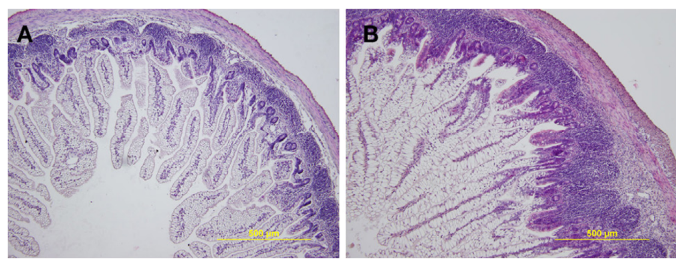
Preterm infants are born with incomplete organ development, including the intestinal tract, as evidenced in Figure 1.
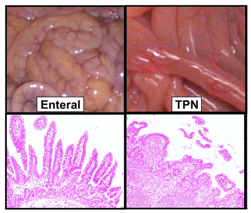
However, in utero, the fetal gastrointestinal tract remains in use, with the fetus estimated to swallow amniotic fluid at 200–250 mL/kg/day of weight [12]. In addition to enzymes and electrolytes, amniotic fluid also contains growth hormones, carbohydrates, proteins, and lipids [12]. Therefore, nutrients from the swallowed amniotic fluid support fetal growth, as well as the proliferation of the intestinal epithelial cells. However, preterm birth disrupts this normal physiology, with intestinal development further hindered by late provision of enteral feeding [12]. In animal and human studies, exclusive parenteral nutrition provision increases intestinal villous atrophy—as demonstrated in Figure 2—which is reversed with enteral feeding provision [13,14].
Findings thus support early enteral feeding, with a Cochrane review further concluding no increased risk of NEC in infants born <28 weeks gestation or <1500 g by initiating enteral feeding within the first four days of life [10]. With further consideration to NEC, the American Society for Parenteral and Enteral Nutrition (ASPEN) recommends starting minimal enteral feeding within the first two days of life for infants ≥1000 g [11]. For infants receiving enteral feeding but in need of blood transfusion, a 2019 Cochrane review concluded there is insufficient evidence to indicate if holding enteral feeding has any effect on developing NEC [53]. Similarly, monitoring gastric residuals is not necessary as an assessment of feeding tolerance to prevent NEC, as a 2019 Cochrane review found no benefit to this practice [54]. Continuing enteral feeding also remains feasible and safe for infants receiving Indomethacin as a treatment for patent ductus arteriosus [55,56]. Delayed introduction of feeding, discontinuation of enteral feeding, prolonged trophic feeding, and slow enteral advancement only results in longer duration for achieving full-volume enteral feeding [10,23]. Enteral feeding was previously advanced by 15–20 mL/kg/day, but a 2017 Cochrane review indicates that more rapid advancement by 30–40 mL/kg/day does not increase risk of adverse outcomes in very or extremely low birth weight infants [23].
In combination with preventing intestinal villous atrophy, early enteral feedings in preterm infants also promotes intestinal development. In examples from a piglet model, Hansen et al. compared intestinal outcomes for preterm-born piglets between two groups: group 1 received parenteral nutrition only for the first 5 days of life and group 2 received minimal volume enteral trophic feedings for the first 5 days in addition to parenteral nutrition. Significant results showed that relative to body weight, intestinal weight was higher in the enterally fed group (p < 0.001) [57], theorized to be associated with enhanced intestinal development. Similarly, lack of enteral feeding may promote increased permeability of the intestine, which could include pathogenic organisms. In the example from mice, lack of enteral feeding and exclusive parenteral nutrition provision increases risk of bacterial translocation [13]. Clinical evidence from a 2017 Cochrane meta-analysis of very low birth weight infants (n = 3753) reported slow enteral advancement to result in “borderline increased risk of invasive infection”, Relative Risk (RR) 1.15 (though 95%Confidence Interval (CI) 1.00–1.32) [23]. Comparatively, Shulman et al. compared intestinal permeability in infants born 26–30 weeks gestation (n = 132), in which differences were assessed in infants started on minimal enteral feeding at 4 vs. 15 days of life [15]. Results identified that intestinal permeability was lower at 10 days of life in early-fed infants (p < 0.01) [15], supporting the theory that early feeding promotes enhanced intestinal development.
Enteral nutrient provision also promotes the maturation of intestinal function and hormones in infants following preterm birth. For example, research by Berseth and Nordyke assessed intestinal manometry in infants born 26–33 weeks gestation (n = 32) receiving enteral feeding during the first week of life of either formula or sterile water (at 24 mL/kg/day) in addition to parenteral nutrition [20]. While both groups showed an initial intestinal motor response to enteral provision, after 10 days infants receiving formula had less clustered phasic activity (p < 0.05) and more migrating activity (p < 0.01) during fasting states [20]. Furthermore, Berseth compared gastrointestinal hormones and peptides in a small subset of infants (n = 27) born at 28–32 weeks gestation with enteral feeding initiated early (3–5 days of life) vs. late (10–14 days of life) [21]. While no differences initially existed, after 10 days, fasting plasma gastrin (p = 0.03) and gastric inhibitory peptide (p = 0.001) were significantly higher in early fed infants [21]. After another 10–14 days, gastrin levels also remained significantly higher in early fed infants (p < 0.02) [21]. More recently, Shanahan et al. analyzed serum intestinal-related hormones in infants born <30 weeks gestation (n = 64) [22]. The results showed that serum gastric inhibitory peptide (p = 0.001) and peptide tyrosine tyrosine (peptide YY) (p = 0.0002) on day of life 7 correlated with percent of nutrition provided enterally, and furthermore with singular enteral fat, carbohydrate, and protein provision (all p < 0.001) [22]. Additionally, a multivariate analysis found that peptide YY, glucose-dependent insulinotropic peptide, and leptin levels on day of life 7 were all associated with reduced time to achievement of full enteral feeding (all p ≤ 0.05) [22].
2.3. Development of the Intestinal Microbiome
The microbiome continually develops throughout the neonatal and pediatric period and many modifiable and non-modifiable factors contribute to its evolution and diversity. One modifiable factor is enteral feeding, which promotes the growth of bacteria in the developing gastrointestinal tract of preterm neonates. In example, Dahlgren et al. compared two groups of infants (total n = 47) born >32 weeks gestation during the first month of life: one group received enteral feeding and one group exclusively received total parenteral nutrition. At four weeks of life, the fecal bacterial diversity was significantly reduced in the parenterally fed group (p < 0.05) [16]. The impact of enteral nutrition on microbiome development in the preterm infant is complex and not completely understood. Development of the intestinal microbiome in terms of bacterial type, amount, and diversity varies in preterm infants according to type of substrate provided, such as mother’s own milk, heat-processed donor human milk, or formula. However, it is less understood how compositional differences within these substrates impact microbiome development, such as structure of the protein molecules or the type or quantity of fatty acids or prebiotic carbohydrates. Yet, additional and less easily modifiable factors must be considered regarding their impact on microbiome evolution, such as presence of intestinal inflammation or feeding intolerance. Likewise, the microbiome may also vary according to gestational age and postnatal age [58,59,60,61,62,63]. Multiple additional factors may impact the intestinal microbiome, such as mode of delivery or antibiotic use [64,65,66,67].
Consideration of the how the microbiome develops should be of interest to clinicians caring for preterm infants. Research has attempted to quantify if the microbiome of preterm infants who develop NEC is altered compared to healthy controls. A recent meta-analysis reported infants who developed NEC had increased fecal levels of Proteobacteria and decreased quantities of Firmicutes and Bacteroidetes [68]. More prospectively, a review by Stiemsma and Michels summarized the interconnection between early microbiome development and developmental origins of health and disease [69]. Early results from both animal and human studies have linked intestinal dysbiosis with adverse outcomes in later life include asthma, allergic disease, obesity, and neurological or behavioral variations [69]. Ultimately, findings suggest that strategies to optimize the intestinal and microbiota health of these fragile infants may be beneficial. Therefore, in addition to enteral feeding promoting a healthy microbiome, initiation of enteral feeding also poses opportunity to introduce probiotic supplementation. However, as no unanimous recommendations exist for optimal probiotic strain, dose, timing of introduction, or length of duration; supplementation must be evaluated on an individual patient, clinician, or unit level. Risks of supplementation include bacterial translocation or unidentified adverse implications for future health. Alternatively, a 2014 Cochrane review reports a lower risk of severe NEC (>Stage II), RR 0.43, 95% CI 0.33–0.56 (n = 5529 infants) and mortality, RR 0.65, 95% CI 0.52–0.81 (n = 5112) in preterm or low birth weight infants given probiotics as compared to those who were unsupplemented [70]. For those favoring probiotic supplementation, recommendations have been set forth in a position paper by the European Society for Paediatric Gastroenterology, Hepatology, and Nutrition (ESPGHAN) to guide clinical strategies for probiotic administration and strain selection in preterm infants [71].
2.4. Inflammatory Response and Clinical Comorbidities
Fetuses swallow amniotic fluid in utero [12], so preterm birth interrupts this normal physiology. If enteral feeding is not initiated after birth, atrophy of the intestinal villous lining will occur. Further findings identify additional detrimental impacts associated with delayed initiation of enteral feeding after birth. For instance, Konnikova et al. analyzed outcomes for infants born <33 weeks gestation, further comparing early vs. late (< vs. >72 h of life) introduction of enteral feeding after birth [17]. While the late feeding group was born smaller and more critically ill at baseline, they demonstrated higher blood C-Reactive Protein levels (p = 0.02) and increased fecal levels of pro-inflammatory interleukin-8 (p < 0.05) at two weeks of age. After multivariate analysis (adjusting for baseline characteristics, nutrition, and illness severity), these findings still remained significant. Notably after multivariate analysis, late enteral feeding was associated with a 4.5-fold increase in chronic lung disease with oxygen use at 36 weeks gestation (95% CI 1.8–11.5; p = 0.002) and a 2.9-fold increase in any stage of ROP (95% CI 1.1–7.8; p = 0.03) [17]. Within a cohort consisting only of extremely low birth weight non-growth-restricted infants, late enteral feeding was associated with a 5.97-fold increase in the odds of developing chronic lung disease (p = 0.02) [17]. Overall, infants receiving late enteral feeding were more likely to have two or more comorbidities compared to early fed infants (25 vs. 8%) [17]. Similarly, Wemhöner et al. analyzed infants born <31 weeks gestation and ≤1500 g (n = 95) to identify how nutrition differs between infants who developed BPD vs. non-diseased controls [31]. Results found that, while calorie, protein, and carbohydrate intakes were statistically similar during the first two weeks of life, infants who developed BPD received a significantly lower volume of enteral feeding (p < 0.04) [31].
2.5. Neurodevelopment
Increased nutrition provision in the early weeks of life enhance neurodevelopmental outcomes in preterm infants [2]. However, there may be an advantage to delivering nutrition enterally compared to parenterally. In example, Tottman et al. assessed enteral feeding volume, total fluid, and macronutrient intake during the first one week and one month of life for infants born <30 weeks gestation or <1500 g. Though growth, total macronutrient, and enteral intakes were statistically similar between boys vs. girls (n = 478), nutrition-related sex-specific differences were identified [29]. After adjustment for birth gestational age, NEC, and sepsis, increased quartiles of enteral feeding volume in mL/kg/day during the first week (p = 0.001) and one month of life (p = 0.01) was associated with a higher odds of survival without neuroimpairment in girls (but not boys) [29]. Coviello et al. also compared nutritional intakes in the first four weeks of life for infants born <31 weeks gestation (n = 131) with brain volumes assessed by magnetic resonance imaging at term equivalent age [30]. After adjusting for cumulative macronutrient intakes, parenteral nutrition duration, and postnatal growth; increased enteral fat, enteral protein, and enteral calorie provision was each associated with increased brain volumes in the cerebellum and basal ganglia (all p < 0.05) [30]. Comparatively, the duration of parenteral nutrition days was inversely associated with volume of the cerebellum, cortical grey matter, basal ganglia, and total brain (all p < 0.05), though parenteral macronutrient provision was not assessed [30]. Cognitive outcomes at two years of age were not associated with enteral intake during the first four weeks of life in this study, and there was no comparison between brain volume and developmental outcomes at this timepoint [30].
2.6. Barriers to Early Life Enteral Feedings
Table 2 lists barriers to early life enteral feeding practices in preterm infants, with suggested strategies to address these barriers.
One of the most significant barriers to initiating and advancing enteral feeding in preterm infants is clinician fear of NEC. As reviewed previously, multiple systematic reviews and meta-analyses conclude that early enteral feeding initiation and more rapid progression does not increase risk of NEC and, in contrast, instead promotes better immediate and lasting outcomes for the preterm infant population. Therefore, the best strategy to address this barrier is implementation of an evidenced-based enteral feeding protocol within each neonatal care unit [28]. Review of additional clinical practices and protocols should also be assessed by individual neonatal care units that may have secondary effects on enteral feeding, such as use of sedation or paralysis that alter gastrointestinal function and ability to tolerate enteral feeding [74]. These strategies aim to alleviate clinician fear by implementing proven successful feeding strategies, create consistency of enteral feeding practices across different providers, and promote a positive “culture change” within neonatal care [79]. In example from one level III neonatal care unit, initial implementation of an enteral feeding protocol for very low birth weight infants yielded significant improvement in clinical outcomes [72]. This protocol was later modified to parallel newfound evidenced-based practices including enteral feeding initiation within the first day of life, reduction in the trophic feeding period to 48 h in infants born <28 weeks gestation with elimination of the trophic feeding period for infants born >28 weeks gestation, earlier milk fortification (at enteral volumes of 50–60 mL/kg/day) and higher enteral volume initiation and advancement (30–35 mL/kg/day) [24,80]. Clinical implementation found this protocol to be feasible and to result in the faster achievement of full enteral feedings, a decreased need for indwelling central line to deliver parenteral nutrition, and fewer infants with weights plotting <10th% on the Fenton preterm infant growth curve at time of discharge [24].
A second barrier to initiating and achieving early enteral feeding is determining appropriate methods to use among infants born <750 g, given less available evidence within this population. However, similar strategies and protocols may be implemented as in older or larger preterm populations, but with some modifications, as suggested in Figure 3. For example, enteral volume initiation and advancement may be slower in infants born <750 g, such as with volumes at 15–25 mL/kg/day [81], though some practices may be even slower [26]. Nonetheless, individualized care must be tailored to meet the medical needs of each patient while prioritizing nutrition, especially in those born at the limits of viability.
Concern may exist that the early achievement of full enteral feedings contributes to accelerated growth in this patient population, which contributes to high adiposity. However, risk of excessive growth may be less concerning during this period than growth failure and diagnosable malnutrition [82], as evidenced from 2013 data by the Vermont Oxford Network summarizing that half of very low birth weight infants in North America were discharged with weights plotting <10th% for age on their respective growth chart [83]. Consequently, experienced clinicians may advocate that establishing full volume enteral feeding with appropriate fortification in early life actually supports appropriate body composition by preventing nutrition deficits that cause neonatal growth failure and linear stunting. In example, Miller et al. reported increased risk of growth failure during the transition from parenteral to enteral nutrition [34]. Nutrition management during this transitional period must be carefully evaluated and modified to meet infant needs, especially given the length of this period may vary significantly based on unit nutrition protocols, birth weight and/or gestational age, and infant illness severity. Liotto et al. demonstrated the importance of close observation and monitoring during the transitional phase, as low protein provision during this period was associated with decreased growth velocity and fat-free mass composition at term-corrected age in very low birth weight infants [84]. Likewise, Brennan et al. compared hourly intakes of macronutrients during the transitional phase, exemplifying the need for higher parenteral volume when enteral feeding was <40 mL/kg/day as well as early fortification of human milk to achieve targeted protein provision when infants began receiving more enteral compared to parenteral nutrition [85]. Thus, initiating and achieving targeted fortification levels (that will achieve desired micronutrient, energy, and protein goals at targeted enteral volumes) prior to discontinuation of parenteral nutrition will promote consistent adequate provision of daily nutrition. This may be most beneficial to the smallest preterm infants who consequently have the highest nutritional needs [86], yet may require longer time to transition from parenteral to full enteral nutrition. Enteral feeding fortification is ideally achieved through provision of human milk with designated human milk fortifiers, provision of preterm infant formulas (e.g., 24 calorie/ounce), and additional supplementation (e.g., protein modular or micronutrient supplement) as available and indicated to achieve targeted goals.
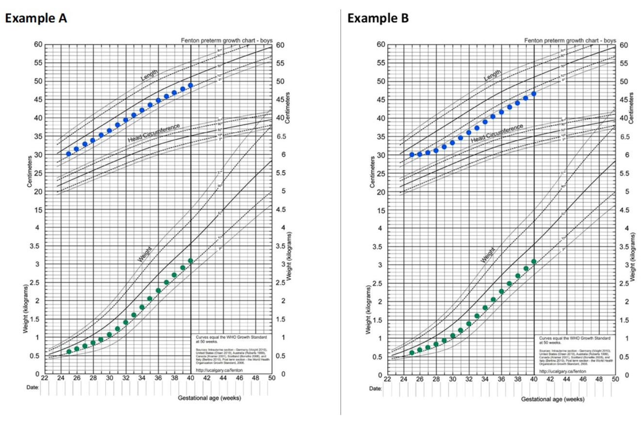
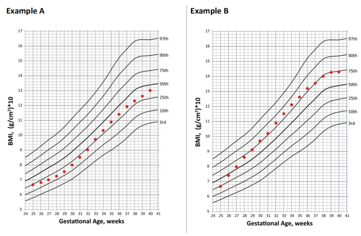
Clinical experience suggests catch-up growth is more easily attained for weight than for linear growth. Thus, stunted lean body mass accretion in early life may not be regained by term corrected age like weight, and therefore may contribute to a higher proportion of weight as adipose tissue [87,88]. This concept is exemplified in Figure 4 and Figure 5 which were constructed to demonstrate growth from 25–40 weeks gestation for an example infant (birth weight 600 g) on the 2013 Fenton growth curve, with estimated body mass index (BMI) size plotted on the Olsen BMI Curves for Preterm Infants [89,90,91,92]. Figure 4, Example A demonstrates growth appropriately maintained for weight and length, with corresponding BMI plotted in Figure 5, Example A. In contrast, Figure 4, Example B demonstrates the same maintained weight as in Example A, but with linear growth failure in the first one month of life before establishing maintenance. In Figure 5, Example B shows correlating BMI as result of early linear growth failure, which is estimated to plot approximately 0.9 standard deviations higher at 40 weeks gestation compared to if linear growth had been adequately maintained since birth (Example A). While evaluating differences in BMI serves as an indirect estimate of body composition, alternative measures are more accurate, such as air displacement plethysmography [93].
Research additionally indicates that delayed enteral feeding after birth promotes inflammation [17], and inflammation increases risk of both inadequate linear growth and development of comorbidities (e.g., BPD) [94,95]. These may perpetuate the risk of altered body composition and therapies to manage comorbidities (e.g., steroid administration) can further impair linear growth [96]. Conclusively, an early transition to full volume enteral feedings should be viewed as an ideal therapy to promote appropriate growth and body composition in preterm infants. Ultimately, clinicians must consider the comprehensive multi-system short and long-term implications for the management of enteral feeding in preterm infants, especially within the first one month of life.
3. Conclusions
Literature supports that enteral feeding, especially early initiation and more rapid enteral advancement, impact preterm infant health during the first one month of life by enhancing micronutrient delivery, promoting intestinal development and maturation, stimulating microbiome development, reducing inflammation, and enhancing brain growth and neurodevelopment. Clinicians must seriously consider the multi-system short and long-term implications that result from the management of enteral feeding in preterm infants and should revise clinical feeding protocols accordingly.
Author Contributions
Conceptualization, M.T. and A.A.-B.; methodology, M.T.; data curation, M.T.; writing—original draft preparation, M.T.; writing—review and editing, M.T. and A.A.-B. All authors have read and agreed to the published version of the manuscript.
Funding
This research received no external funding.
Institutional Review Board Statement
Not applicable.
Informed Consent Statement
Not applicable.
Conflicts of Interest
The authors declare no conflict of interest.
References
- Ehrenkranz, R.; Dusick, A.; Vohr, B.; Wright, L.; Wrage, L.; Poole, W. Growth in the Neonatal Intensive Care Unit Influences Neurodevelopmental and Growth Outcomes of Extremely Low Birth Weight Infants. J. Pediatr. 2006, 117, 1253–1261. [Google Scholar] [CrossRef] [PubMed] [Green Version]
- Stephens, B.E.; Walden, R.V.; Gargus, R.A.; Tucker, R.; McKinley, L.; Mance, M.; Nye, J.; Vohr, B.R. First-Week Protein and Energy Intakes are Associated with 18-Month Developmental Outcomes in Extremely Low Birth Weight Infants. Pediatrics 2009, 123, 1337–1343. [Google Scholar] [CrossRef] [PubMed]
- Fang, J.L.; Sorita, A.; Carey, W.A.; Colby, C.E.; Murad, M.H.; Alahdab, F. Interventions to Prevent Retinopathy of Prematurity: A Meta-Analysis. Pediatrics 2016, 137, e20153387. [Google Scholar] [CrossRef] [PubMed] [Green Version]
- Stoltz, S.; Lundgren, P.; Öhlund, I.; Holmström, G.; Hellström, A.; Domellöf, M. Low Energy Intake during the First 4 weeks of Life Increases the Risk for Severe Retinopathy of Prematurity in Extremely Preterm Infants. Archives of Disease in Childhood. Fetal Neonatal Ed. 2015, 102, F108–F113. [Google Scholar] [CrossRef] [Green Version]
- Klevebro, S.; Westin, V.; Stoltz Sjostrom, E.; Norman, M.; Domellof, M.; Edstedt Bonamy, A.K.; Hallberg, B. Early Energy and Protein Intakes and Associations with Growth, BPD, and ROP in Extremely Preterm Infants. Clin. Nutr. 2019, 38, 1289–1295. [Google Scholar] [CrossRef] [Green Version]
- Eidelman, A.I.; Schanler, R.J. Section on Breastfeeding. Breastfeeding and the use of Human Milk. Pediatrics 2012, 129, e827–e841. [Google Scholar]
- Miller, J.; Tonkin, E.; Damarell, R.A.; McPhee, A.J.; Suganuma, M.; Suganuma, H.; Middleton, P.F.; Makrides, M.; Collins, C.T. A Systematic Review and Meta-Analysis of Human Milk Feeding and Morbidity in very Low Birth Weight Infants. Nutrients 2018, 10, 707. [Google Scholar] [CrossRef] [Green Version]
- Villamor-Martinez, E.; Pierro, M.; Cavallaro, G.; Mosca, F.; Kramer, B.W.; Villamor, E. Donor Human Milk Protects Against Bronchopulmonary Dysplasia: A Systematic Review and Meta-Analysis. Nutrients 2018, 10, 238. [Google Scholar] [CrossRef] [Green Version]
- Villamor-Martinez, E.; Pierro, M.; Cavallaro, G.; Mosca, F.; Villamor, E. Mother’s Own Milk and Bronchopulmonary Dysplasia: A Systematic Review and Meta-Analysis. Front. Pediatr. 2019, 7, 224. [Google Scholar] [CrossRef]
- Morgan, J.; Young, L.; McGuire, W. Delayed Introduction of Progressive Enteral Feeds to Prevent Necrotising Enterocolitis in very Low Birth Weight Infants. Cochrane Database Syst. Rev. 2014, 12, CD001970. [Google Scholar] [CrossRef]
- Fallon, E.M.; Nehra, D.; Potemkin, A.K.; Gura, K.M.; Simpser, E.; Compher, C. American Society for Parenteral and Enteral Nutrition (A.S.P.E.N.) Board of Directors; Puder, M. A.S.P.E.N. Clinical Guidelines: Nutrition Support of Neonatal Patients at Risk for Necrotizing Enterocolitis. JPEN J. Parenter. Enteral Nutr. 2012, 36, 506–523. [Google Scholar] [CrossRef]
- Underwood, M.A.; Gilbert, W.M.; Sherman, M.P. Amniotic Fluid: Not just Fetal Urine Anymore. J. Perinatol. 2005, 25, 341–348. [Google Scholar] [CrossRef] [Green Version]
- Wildhaber, B.E.; Yang, H.; Spencer, A.U.; Drongowski, R.A.; Teitelbaum, D.H. Lack of Enteral Nutrition—Effects on the Intestinal Immune System. J. Surg. Res. 2005, 123, 8–16. [Google Scholar] [CrossRef]
- Buchman, A.L.; Moukarzel, A.A.; Bhuta, S.; Belle, M.; Ament, M.E.; Eckhert, C.D.; Hollander, D.; Gornbein, J.; Kopple, J.D.; Vijayaroghavan, S.R. Parenteral Nutrition is Associated with Intestinal Morphologic and Functional Changes in Humans. JPEN J. Parenter. Enteral Nutr. 1995, 19, 453–460. [Google Scholar] [CrossRef]
- Shulman, R.J.; Schanler, R.J.; Lau, C.; Heitkemper, M.; Ou, C.N.; Smith, E.O. Early Feeding, Antenatal Glucocorticoids, and Human Milk Decrease Intestinal Permeability in Preterm Infants. Pediatr. Res. 1998, 44, 519–523. [Google Scholar] [CrossRef]
- Dahlgren, A.F.; Pan, A.; Lam, V.; Gouthro, K.C.; Simpson, P.M.; Salzman, N.H.; Nghiem-Rao, T.H. Longitudinal Changes in the Gut Microbiome of Infants on Total Parenteral Nutrition. Pediatr. Res. 2019, 86, 107–114. [Google Scholar] [CrossRef]
- Konnikova, Y.; Zaman, M.M.; Makda, M.; D’Onofrio, D.; Freedman, S.D.; Martin, C.R. Late Enteral Feedings are Associated with Intestinal Inflammation and Adverse Neonatal Outcomes. PLoS ONE 2015, 10, e0132924. [Google Scholar] [CrossRef] [Green Version]
- Dutta, S.; Singh, B.; Chessell, L.; Wilson, J.; Janes, M.; McDonald, K.; Shahid, S.; Gardner, V.A.; Hjartarson, A.; Purcha, M.; et al. Guidelines for Feeding very Low Birth Weight Infants. Nutrients 2015, 7, 423–442. [Google Scholar] [CrossRef] [Green Version]
- McClave, S.A.; Martindale, R.G.; Vanek, V.W.; McCarthy, M.; Roberts, P.; Taylor, B.; Ochoa, J.B.; Napolitano, L.; Cresci, G.; Gervasio, J.M.; et al. Guidelines for the Provision and Assessment of Nutrition Support Therapy in the Adult Critically Ill Patient: Society of Critical Care Medicine (SCCM) and American Society for Parenteral and Enteral Nutrition (A.S.P.E.N.). JPEN J. Parenter. Enteral Nutr. 2009, 33, 277–316. [Google Scholar] [CrossRef]
- Berseth, C.L.; Nordyke, C. Enteral Nutrients Promote Postnatal Maturation of Intestinal Motor Activity in Preterm Infants. Am. J. Physiol. 1993, 264, G1046–G1051. [Google Scholar] [CrossRef]
- Berseth, C.L. Effect of Early Feeding on Maturation of the Preterm Infant’s Small Intestine. J. Pediatr. 1992, 120, 947–953. [Google Scholar] [CrossRef]
- Shanahan, K.H.; Yu, X.; Miller, L.G.; Freedman, S.D.; Martin, C.R. Early Serum Gut Hormone Concentrations Associated with Time to Full Enteral Feedings in Preterm Infants. J. Pediatr. Gastroenterol. Nutr. 2018, 67, 97–102. [Google Scholar] [CrossRef]
- Oddie, S.J.; Young, L.; McGuire, W. Slow Advancement of Enteral Feed Volumes to Prevent Necrotising Enterocolitis in very Low Birth Weight Infants. Cochrane Database Syst. Rev. 2017, 8, CD001241. [Google Scholar] [CrossRef]
- Thoene, M.K.; Lyden, E.; Anderson-Berry, A. Improving Nutrition Outcomes for Infants < 1500 Grams with a Progressive, Evidenced-Based Enteral Feeding Protocol. Nutr. Clin. Pract. 2018, 5, 647–655. [Google Scholar]
- Viswanathan, S.; McNelis, K.; Super, D.; Einstadter, D.; Groh-Wargo, S.; Collin, M. Standardized Slow Enteral Feeding Protocol and the Incidence of Necrotizing Enterocolitis in Extremely Low Birth Weight Infants. JPEN J. Parenter. Enteral Nutr. 2015, 39, 644–654. [Google Scholar] [CrossRef] [Green Version]
- Viswanathan, S.; Merheb, R.; Wen, X.; Collin, M.; Groh-Wargo, S. Standardized Slow Enteral Feeding Protocol Reduces Necrotizing Enterocolitis in Micropremies. J. Neonatal Perinatal Med. 2017, 10, 171–180. [Google Scholar] [CrossRef]
- Salas, A.A.; Kabani, N.; Travers, C.P.; Phillips, V.; Ambalavanan, N.; Carlo, W.A. Short Versus Extended Duration of Trophic Feeding to Reduce Time to Achieve Full Enteral Feeding in Extremely Preterm Infants: An Observational Study. Neonatology 2017, 112, 211–216. [Google Scholar] [CrossRef]
- Koletzko, B.; Poindexter, B.; Uauy, R. Nutritional Care of Preterm Infants: Scientific Basis and Practical Guidelines; Karger: Basel, Switzerland, 2014. [Google Scholar]
- Tottman, A.C.; Bloomfield, F.H.; Cormack, B.E.; Harding, J.E.; Taylor, J.; Alsweiler, J.M. Sex-Specific Relationships between Early Nutrition and Neurodevelopment in Preterm Infants. Pediatr. Res. 2019, 5, 872–878. [Google Scholar] [CrossRef]
- Coviello, C.; Keunen, K.; Kersbergen, K.J.; Groenendaal, F.; Leemans, A.; Peels, B.; Isgum, I.; Viergever, M.A.; de Vries, L.S.; Buonocore, G.; et al. Effects of Early Nutrition and Growth on Brain Volumes, White Matter Microstructure, and Neurodevelopmental Outcome in Preterm Newborns. Pediatr. Res. 2018, 83, 102–110. [Google Scholar] [CrossRef] [Green Version]
- Wemhoner, A.; Ortner, D.; Tschirch, E.; Strasak, A.; Rudiger, M. Nutrition of Preterm Infants in Relation to Bronchopulmonary Dysplasia. BMC Pulm. Med. 2011, 11, 7. [Google Scholar] [CrossRef] [Green Version]
- Shah, S.D.; Dereddy, N.; Jones, T.L.; Dhanireddy, R.; Talati, A.J. Early Versus Delayed Human Milk Fortification in very Low Birth Weight Infants-A Randomized Controlled Trial. J. Pediatr. 2016, 174, 126–131.e1. [Google Scholar] [CrossRef] [PubMed]
- Tillman, S.; Brandon, D.H.; Silva, S.G. Evaluation of Human Milk Fortification from the Time of the First Feeding: Effects on Infants of Less than 31 Weeks Gestational Age. J. Perinatol. 2012, 32, 525–531. [Google Scholar] [CrossRef] [Green Version]
- Miller, M.; Vaidya, R.; Rastogi, D.; Bhutada, A.; Rastogi, S. From Parenteral to Enteral Nutrition: A Nutrition-Based Approach for Evaluating Postnatal Growth Failure in Preterm Infants. JPEN J. Parenter. Enteral Nutr. 2014, 38, 489–497. [Google Scholar] [CrossRef] [PubMed]
- Donovan, R.; Kelly, S.G.; Prazad, P.; Talaty, P.N.; Lefaiver, C.; Hastings, M.L.; Everly, D.N. The Effects of Human Milk Fortification on Nutrients and Milk Properties. J. Perinatol. 2017, 37, 42–48. [Google Scholar] [CrossRef] [PubMed]
- Institute of Medicine of the National Academies. Dietary Reference Intakes: The Essential Guide to Nutrient Requirements; National Academies Press: Washington, DC, USA, 2006; p. 541. [Google Scholar]
- Hanson, C.; Thoene, M.; Wagner, J.; Collier, D.; Lecci, K.; Anderson-Berry, A. Parenteral Nutrition Additive Shortages: The Short-Term, Long-Term and Potential Epigenetic Implications in Premature and Hospitalized Infants. Nutrients 2012, 4, 1977–1988. [Google Scholar] [CrossRef] [PubMed] [Green Version]
- Bronsky, J.; Campoy, C.; Braegger, C. ESPGHAN/ESPEN/ESPR/CSPEN working group on pediatric parenteral nutrition. ESPGHAN/ESPEN/ESPR/CSPEN Guidelines on Pediatric Parenteral Nutrition: Vitamins. Clin. Nutr. 2018, 37, 2366–2378. [Google Scholar] [CrossRef] [PubMed] [Green Version]
- Pohlandt, F.; Abrams, S.; Cooke, R. Calcium, Magnesium, Phosphorus, and Vitamin D. In Nutrition of the Preterm Infant: Scientific Basis and Practical Guidelines, 2nd ed.; Tsang, R.C., Uauy, R., Koletzko, B., Zlotkin, S.H., Eds.; Digital Educational Publishing, Inc.: Cincinnati, OH, USA, 2005; p. 245. [Google Scholar]
- Quigley, M.; Embleton, N.D.; McGuire, W. Formula Versus Donor Breast Milk for Feeding Preterm or Low Birth Weight Infants. Cochrane Database Syst. Rev. 2018, 6, CD002971. [Google Scholar] [CrossRef] [PubMed]
- Henderson, G.; Anthony, M.Y.; McGuire, W. Formula Milk Versus Maternal Breast Milk for Feeding Preterm or Low Birth Weight Infants. Cochrane Database Syst. Rev. 2007, 4, CD002972. [Google Scholar] [CrossRef]
- McGuire, W.; Anthony, M.Y. Formula Milk Versus Preterm Human Milk for Feeding Preterm Or Low Birth Weight Infants. Cochrane Database Syst. Rev. 2001, 3, CD002972. [Google Scholar]
- Mills, R.J.; Davies, M.W. Enteral Iron Supplementation in Preterm and Low Birth Weight Infants. Cochrane Database Syst. Rev. 2012, 3, CD005095. [Google Scholar] [CrossRef] [Green Version]
- Georgieff, M.K. The Role of Iron in Neurodevelopment: Fetal Iron Deficiency and the Developing Hippocampus. Biochem. Soc. Trans. 2008, 36, 1267–1271. [Google Scholar] [CrossRef] [Green Version]
- Arnon, S.; Shiff, Y.; Litmanovitz, I.; Regev, R.H.; Bauer, S.; Shainkin-Kestenbaum, R.; Bental, Y.; Dolfin, T. The Efficacy and Safety of Early Supplementation of Iron Polymaltose Complex in Preterm Infants. Am. J. Perinatol. 2007, 24, 95–100. [Google Scholar] [CrossRef]
- Lundstrom, U.; Siimes, M.A.; Dallman, P.R. At what Age does Iron Supplementation Become Necessary in Low-Birth-Weight Infants? J. Pediatr. 1977, 91, 878–883. [Google Scholar] [CrossRef]
- Loskutova, E.; Nolan, J.; Howard, A.; Beatty, S. Macular Pigment and its Contribution to Vision. Nutrients 2013, 5, 1962–1969. [Google Scholar] [CrossRef]
- Vishwanathan, R.; Kuchan, M.J.; Sen, S.; Johnson, E.J. Lutein and Preterm Infants with Decreased Concentrations of Brain Carotenoids. J. Pediatr. Gastroenterol. Nutr. 2014, 59, 659–665. [Google Scholar] [CrossRef]
- Rubin, L.P.; Chan, G.M.; Barrett-Reis, B.M.; Fulton, A.B.; Hansen, R.M.; Ashmeade, T.L.; Oliver, J.S.; Mackey, A.D.; Dimmit, R.A.; Hartmann, E.E.; et al. Effect of Carotenoid Supplementation on Plasma Carotenoids, Inflammation and Visual Development in Preterm Infants. J. Perinatol. 2012, 32, 418–424. [Google Scholar] [CrossRef] [Green Version]
- Stringham, J.M.; Johnson, E.J.; Hammond, B.R. Lutein Across the Lifespan: From Childhood Cognitive Performance to the Aging Eye and Brain. Curr. Dev. Nutr. 2019, 3, nzz066. [Google Scholar] [CrossRef] [Green Version]
- Splichalova, A.; Slavikova, V.; Splichalova, Z.; Splichal, I. Preterm Life in Sterile Conditions: A Study on Preterm, Germ-Free Piglets. Front. Immunol. 2018, 9, 220. [Google Scholar] [CrossRef] [Green Version]
- Jain, A.K.; Stoll, B.; Burrin, D.G.; Holst, J.J.; Moore, D.D. Enteral Bile Acid Treatment Improves Parenteral Nutrition-Related Liver Disease and Intestinal Mucosal Atrophy in Neonatal Pigs. Am. J. Physiol. Gastrointest. Liver Physiol. 2012, 302, G218–G224. [Google Scholar] [CrossRef] [Green Version]
- Yeo, K.T.; Kong, J.Y.; Sasi, A.; Tan, K.; Lai, N.M.; Schindler, T. Stopping Enteral Feeds for Prevention of Transfusion-Associated Necrotising Enterocolitis in Preterm Infants. Cochrane Database Syst. Rev. 2019, 2019, CD012888. [Google Scholar] [CrossRef]
- Abiramalatha, T.; Thanigainathan, S.; Ninan, B. Routine Monitoring of Gastric Residual for Prevention of Necrotising Enterocolitis in Preterm Infants. Cochrane Database Syst. Rev. 2019, 7, CD012937. [Google Scholar] [CrossRef]
- Clyman, R.; Wickremasinghe, A.; Jhaveri, N.; Hassinger, D.C.; Attridge, J.T.; Sanocka, U.; Polin, R.; Gillam-Krakauer, M.; Reese, J.; Mammel, M.; et al. Enteral Feeding during Indomethacin and Ibuprofen Treatment of a Patent Ductus Arteriosus. J. Pediatr. 2013, 163, 406–411. [Google Scholar] [CrossRef] [Green Version]
- Louis, D.; Torgalkar, R.; Shah, J.; Shah, P.S.; Jain, A. Enteral Feeding during Indomethacin Treatment for Patent Ductus Arteriosus: Association with Gastrointestinal Outcomes. J. Perinatol. 2016, 36, 544–548. [Google Scholar] [CrossRef]
- Hansen, C.F.; Thymann, T.; Andersen, A.D.; Holst, J.J.; Hartmann, B.; Hilsted, L.; Langhorn, L.; Jelsing, J.; Sangild, P.T. Rapid Gut Growth but Persistent Delay in Digestive Function in the Postnatal Period of Preterm Pigs. Am. J. Physiol. Gastrointest. Liver Physiol. 2016, 310, G550–G560. [Google Scholar] [CrossRef] [Green Version]
- Zanella, A.; Silveira, R.C.; Roesch, L.F.W.; Corso, A.L.; Dobbler, P.T.; Mai, V.; Procianoy, R.S. Influence of Own Mother’s Milk and Different Proportions of Formula on Intestinal Microbiota of very Preterm Newborns. PLoS ONE 2019, 14, e0217296. [Google Scholar] [CrossRef]
- Parra-Llorca, A.; Gormaz, M.; Alcantara, C.; Cernada, M.; Nunez-Ramiro, A.; Vento, M.; Collado, M.C. Preterm Gut Microbiome Depending on Feeding Type: Significance of Donor Human Milk. Front. Microbiol. 2018, 9, 1376. [Google Scholar] [CrossRef]
- Ford, S.L.; Lohmann, P.; Preidis, G.A.; Gordon, P.S.; O’Donnell, A.; Hagan, J.; Venkatachalam, A.; Balderas, M.; Luna, R.A.; Hair, A.B. Improved Feeding Tolerance and Growth are Linked to Increased Gut Microbial Community Diversity in very-Low-Birth-Weight Infants Fed Mother’s Own Milk Compared with Donor Breast Milk. Am. J. Clin. Nutr. 2019, 109, 1088–1097. [Google Scholar] [CrossRef]
- Gregory, K.E.; Samuel, B.S.; Houghteling, P.; Shan, G.; Ausubel, F.M.; Sadreyev, R.I.; Walker, W.A. Influence of Maternal Breast Milk Ingestion on Acquisition of the Intestinal Microbiome in Preterm Infants. Microbiome 2016, 4, 68. [Google Scholar] [CrossRef] [Green Version]
- Butcher, J.; Unger, S.; Li, J.; Bando, N.; Romain, G.; Francis, J.; Mottawea, W.; Mack, D.; Stintzi, A.; O’Connor, D.L. Independent of Birth Mode or Gestational Age, very-Low-Birth-Weight Infants Fed their Mothers’ Milk Rapidly Develop Personalized Microbiotas Low in Bifidobacterium. J. Nutr. 2018, 148, 326–335. [Google Scholar] [CrossRef]
- Cong, X.; Judge, M.; Xu, W.; Diallo, A.; Janton, S.; Brownell, E.A.; Maas, K.; Graf, J. Influence of Feeding Type on Gut Microbiome Development in Hospitalized Preterm Infants. Nurs. Res. 2017, 66, 123–133. [Google Scholar] [CrossRef]
- Rutayisire, E.; Huang, K.; Liu, Y.; Tao, F. The Mode of Delivery Affects the Diversity and Colonization Pattern of the Gut Microbiota during the First Year of Infants’ Life: A Systematic Review. BMC Gastroenterol. 2016, 16, 86. [Google Scholar] [CrossRef] [PubMed] [Green Version]
- Montoya-Williams, D.; Lemas, D.J.; Spiryda, L.; Patel, K.; Carney, O.O.; Neu, J.; Carson, T.L. The Neonatal Microbiome and its Partial Role in Mediating the Association between Birth by Cesarean Section and Adverse Pediatric Outcomes. Neonatology 2018, 114, 103–111. [Google Scholar] [CrossRef] [PubMed]
- Fjalstad, J.W.; Esaiassen, E.; Juvet, L.K.; van den Anker, J.N.; Klingenberg, C. Antibiotic Therapy in Neonates and Impact on Gut Microbiota and Antibiotic Resistance Development: A Systematic Review. J. Antimicrob. Chemother. 2018, 73, 569–580. [Google Scholar] [CrossRef] [PubMed] [Green Version]
- Zou, Z.H.; Liu, D.; Li, H.D.; Zhu, D.P.; He, Y.; Hou, T.; Yu, J.L. Prenatal and Postnatal Antibiotic Exposure Influences the Gut Microbiota of Preterm Infants in Neonatal Intensive Care Units. Ann. Clin. Microbiol. Antimicrob. 2018, 17, 9. [Google Scholar] [CrossRef] [Green Version]
- Pammi, M.; Cope, J.; Tarr, P.I.; Warner, B.B.; Morrow, A.L.; Mai, V.; Gregory, K.E.; Kroll, J.S.; McMurtry, V.; Ferris, M.J.; et al. Intestinal Dysbiosis in Preterm Infants Preceding Necrotizing Enterocolitis: A Systematic Review and Meta-Analysis. Microbiome 2017, 5, 31. [Google Scholar] [CrossRef] [Green Version]
- Stiemsma, L.T.; Michels, K.B. The Role of the Microbiome in the Developmental Origins of Health and Disease. Pediatrics 2018, 141, e20172437. [Google Scholar] [CrossRef] [Green Version]
- AlFaleh, K.; Anabrees, J. Probiotics for Prevention of Necrotizing Enterocolitis in Preterm Infants. Cochrane Database Syst. Rev. 2014, 4, CD005496. [Google Scholar]
- van den Akker, C.H.P.; van Goudoever, J.B.; Shamir, R.; Domellöf, M.; Embleton, N.D.; Hojsak, I.; Lapillonne, A.; Mihatsch, W.A.; Berni Canani, R.; Bronsky, J.; et al. Probiotics and Preterm Infants: A Position Paper by the European Society for Paediatric Gastroenterology Hepatology and Nutrition Committee on Nutrition and the European Society for Paediatric Gastroenterology Hepatology and Nutrition Working Group for Probiotics and Prebiotics. J. Pediatr. Gastroenterol. Nutr. 2020, 70, 664–680. [Google Scholar]
- Hanson, C.; Sundermeier, J.; Dugick, L.; Lyden, E.; Anderson-Berry, A.L. Implementation, Process, and Outcomes of Nutrition Best Practices for Infants < 1500 g. Nutr. Clin. Pract. 2011, 26, 614–624. [Google Scholar]
- Ibrahim, T.; Li Wei, C.; Bautista, D.; Sriram, B.; Xiangzhen Fay, L.; Rajadurai, V.S. Saline Enemas Versus Glycerin Suppositories to Promote Enteral Feeding in Premature Infants: A Pilot Randomized Controlled Trial. Neonatology 2017, 112, 347–353. [Google Scholar] [CrossRef]
- McPherson, C. Sedation and Analgesia in Mechanically Ventilated Preterm Neonates: Continue Standard of Care or Experiment? J. Pediatr. Pharmacol. Ther. 2012, 17, 351–364. [Google Scholar] [CrossRef] [Green Version]
- Priyadarshia, A.; Hindera, M.; Badawib, N.; Luiga, M.; Tracy, M. Continuous Positive Airway Pressure Belly Syndrome: Challenges of a Changing Paradigm. Int. J. Clin. Pediatr. 2020, 9, 9–15. [Google Scholar] [CrossRef]
- Jaile, J.C.; Levin, T.; Wung, J.T.; Abramson, S.J.; Ruzal-Shapiro, C.; Berdon, W.E. Benign Gaseous Distension of the Bowel in Premature Infants Treated with Nasal Continuous Airway Pressure: A Study of Contributing Factors. AJR Am. J. Roentgenol. 1992, 158, 125–127. [Google Scholar] [CrossRef] [Green Version]
- Eichenwald, E.C.; Committee on Fetus and Newborn. Diagnosis and Management of Gastroesophageal Reflux in Preterm Infants. Pediatrics 2018, 142, e20181061. [Google Scholar] [CrossRef] [Green Version]
- Watson, J.; McGuire, W. Transpyloric Versus Gastric Tube Feeding for Preterm Infants. Cochrane Database Syst. Rev. 2013, 2013, CD003487. [Google Scholar] [CrossRef] [Green Version]
- Rodrigues, C.; Severo, M.; Zeitlin, J.; Barros, H.; Portuguese EPICE (Effective Perinatal Intensive Care in Europe) Network. The Type of Feeding at Discharge of very Preterm Infants: Neonatal Intensive Care Units Policies and Practices make a Difference. Breastfeed Med. 2018, 13, 50–59. [Google Scholar] [CrossRef]
- Thoene, M.K.; Anderson-Berry, A.L. A Review of Best Evidenced-Based Enteral and Parenteral Nutrition Support Practices for Preterm Infants Born <1500 Grams. Pediatr. Med. 2018, 1. [Google Scholar] [CrossRef]
- Senterre, T. Practice of Enteral Nutrition in very Low Birth Weight and Extremely Low Birth Weight Infants. World Rev. Nutr. Diet. 2014, 110, 201–214. [Google Scholar]
- Goldberg, D.L.; Becker, P.J.; Brigham, K.; Carlson, S.; Fleck, L.; Gollins, L.; Sandrock, M.; Fullmer, M.; Van Poots, H.A. Identifying Malnutrition in Preterm and Neonatal Populations: Recommended Indicators. J. Acad. Nutr. Diet. 2018, 118, 1571–1582. [Google Scholar] [CrossRef]
- Horbar, J.D.; Ehrenkranz, R.A.; Badger, G.J.; Edwards, E.M.; Morrow, K.A.; Soll, R.F.; Buzas, J.S.; Bertino, E.; Gagliardi, L.; Bellu, R. Weight Growth Velocity and Postnatal Growth Failure in Infants 501 to 1500 Grams: 2000–2013. Pediatrics 2015, 136, e84–e92. [Google Scholar] [CrossRef] [Green Version]
- Liotto, N.; Amato, O.; Piemontese, P.; Menis, C.; Orsi, A.; Corti, M.G.; Colnaghi, M.; Cecchetti, V.; Pugni, L.; Mosca, F.; et al. Protein Intakes during Weaning from Parenteral Nutrition Drive Growth Gain and Body Composition in very Low Birth Weight Preterm Infants. Nutrients 2020, 12, 1298. [Google Scholar] [CrossRef]
- Brennan, A.M.; Kiely, M.E.; Fenton, S.; Murphy, B.P. Standardized Parenteral Nutrition for the Transition Phase in Preterm Infants: A Bag that Fits. Nutrients 2018, 10, 170. [Google Scholar] [CrossRef] [Green Version]
- American Academy of Nutrition and Dietetics. Pediatric Nutrition Care Manual: Preterm and High-Risk Infants Nutrition Support. Available online: https://www.nutritioncaremanual.org (accessed on 20 May 2021).
- Bruckner, M.; Khan, Z.; Binder, C.; Morris, N.; Windisch, B.; Holasek, S.; Urlesberger, B. Extremely Preterm Infants have a Higher Fat Mass Percentage in Comparison to very Preterm Infants at Term-Equivalent Age. Front. Pediatr. 2020, 8, 61. [Google Scholar] [CrossRef]
- Johnson, M.J.; Wootton, S.A.; Leaf, A.A.; Jackson, A.A. Preterm Birth and Body Composition at Term Equivalent Age: A Systematic Review and Meta-Analysis. Pediatrics 2012, 130, e640–e649. [Google Scholar] [CrossRef] [Green Version]
- Fenton, T.R.; Kim, J.H. A Systematic Review and Meta-Analysis to Revise the Fenton Growth Chart for Preterm Infants. BMC Pediatr. 2013, 13, 59. [Google Scholar] [CrossRef] [Green Version]
- Olsen, I.E.; Lawson, M.L.; Ferguson, A.N.; Cantrell, R.; Grabich, S.C.; Zemel, B.S.; Clark, R.H. BMI Curves for Preterm Infants. Pediatrics 2015, 135, 572–581. [Google Scholar] [CrossRef] [Green Version]
- Chou, J. PediTools: Clinical Tools for Pediatric Providers. 2020. Available online: https://peditools.org (accessed on 22 January 2020).
- Chou, J.H.; Roumiantsev, S.; Singh, R. PediTools Electronic Growth Chart Calculators: Applications in Clinical Care, Research, and Quality Improvement. J. Med. Internet Res. 2020, 22, e16204. [Google Scholar] [CrossRef] [PubMed]
- Andrews, E.T.; Beattie, R.M.; Johnson, M.J. Measuring Body Composition in the Preterm Infant: Evidence Base and Practicalities. Clin. Nutr. 2019, 38, 2521–2530. [Google Scholar] [CrossRef]
- Cuestas, E.; Aguilera, B.; Cerutti, M.; Rizzotti, A. Sustained Neonatal Inflammation is Associated with Poor Growth in Infants Born very Preterm during the First Year of Life. J. Pediatr. 2019, 205, 91–97. [Google Scholar] [CrossRef]
- Twilhaar, E.S.; Wade, R.M.; de Kieviet, J.F.; van Goudoever, J.B.; van Elburg, R.M.; Oosterlaan, J. Cognitive Outcomes of Children Born Extremely or very Preterm since the 1990s and Associated Risk Factors: A Meta-Analysis and Meta-Regression. JAMA Pediatr. 2018, 172, 361–367. [Google Scholar] [CrossRef]
- Linafelter, A.; Cuna, A.; Liu, C.; Quigley, A.; Truog, W.E.; Sampath, V.; Oschman, A. Extended Course of Prednisolone in Infants with Severe Bronchopulmonary Dysplasia. Early Hum. Dev. 2019, 136, 1–6. [Google Scholar] [CrossRef] [PubMed]
Figure 1.
Cross Section Stain of Villi in the Ileum of Preterm (A) vs. Term (B) Piglets. Figure reproduced from Frontiers in Immunology, Vol. 9, Page 5 as available via Open Access and under terms of the Creative Commons Attribution License. Original article published as “Preterm Life in Sterile Conditions: A Study on Preterm, Germ-Free Piglets” by Splichalova et al., 2018 [51].

Figure 2.
A Macroscopic Appearance and Stained Cross Section of Distal Small Intestine in Preterm Piglets Receiving Enteral Feeding vs. Total Parenteral Nutrition (TPN). Figure modified and reproduced with copyright permission from American Journal of Physiology-Gastrointestinal and Liver Physiology, Vol. 302, page 221, Copyright © 2012 by The American Physiological Society. Original article published as “Enteral Bile Acid Treatment Improves Parenteral Nutrition-Related Liver Disease and Intestinal Mucosal Atrophy in Neonatal Pigs” by Jain et al. [52].

Figure 3.
Proposed decision making for initiating and advancing enteral feeding in preterm infants.

Figure 4.
Comparison of adequate (Example A) vs. early growth failure (Example B) for linear growth in an extremely low birth weight infant from 25–40 weeks gestation with adequate weight gain. Examples A and B 2013 Fenton growth charts constructed and reprinted from the PediTools.org website [89,91,92] as available via open access and under terms of the Creative Commons Attribution License.

Figure 5.
Comparison of body mass index trend from 25–40 weeks gestation in an extremely low birth weight infant with adequate (Example A) vs. early growth failure (Example B) for linear growth when weight gain is adequate. Examples A and B modified and reproduced with permission from first author Irene Olsen, published in Pediatrics, Vol. 135, page 575, Copyright © 2015 by the American Academy of Pediatrics. Original article published as “BMI Curves for Preterm Infants” by Olsen et al. [90].


Table 1.
Benefits and risks of early life enteral feeding practices in preterm infants.
| Enteral Feeding Practice | Benefits | Risks |
|---|---|---|
| ||
|
| |
|
| |
|
|
Table 2.
Barriers and strategies to initiating and advancing early life enteral feeding in preterm infants.
Table 2.
Barriers and strategies to initiating and advancing early life enteral feeding in preterm infants.
| Barriers | Strategies to Address Barriers |
|---|---|
|
|
| |
| |
|
|
|
|
Publisher’s Note: MDPI stays neutral with regard to jurisdictional claims in published maps and institutional affiliations. |
© 2021 by the authors. Licensee MDPI, Basel, Switzerland. This article is an open access article distributed under the terms and conditions of the Creative Commons Attribution (CC BY) license (https://creativecommons.org/licenses/by/4.0/).
Share and Cite
MDPI and ACS Style
Thoene, M.; Anderson-Berry, A. Early Enteral Feeding in Preterm Infants: A Narrative Review of the Nutritional, Metabolic, and Developmental Benefits. Nutrients 2021, 13, 2289. https://0-doi-org.brum.beds.ac.uk/10.3390/nu13072289
AMA Style
Thoene M, Anderson-Berry A. Early Enteral Feeding in Preterm Infants: A Narrative Review of the Nutritional, Metabolic, and Developmental Benefits. Nutrients. 2021; 13(7):2289. https://0-doi-org.brum.beds.ac.uk/10.3390/nu13072289
Chicago/Turabian StyleThoene, Melissa, and Ann Anderson-Berry. 2021. "Early Enteral Feeding in Preterm Infants: A Narrative Review of the Nutritional, Metabolic, and Developmental Benefits" Nutrients 13, no. 7: 2289. https://0-doi-org.brum.beds.ac.uk/10.3390/nu13072289
Note that from the first issue of 2016, this journal uses article numbers instead of page numbers. See further details here.