Review

Immune-Relevant and Antioxidant Activities of Vitellogenin and Yolk Proteins in Fish

Laboratory for Evolution & Development, Institute of Evolution & Marine Biodiversity and Department of Marine Biology, Ocean University of China, Qingdao 266003, China
*
Author to whom correspondence should be addressed.
Nutrients 2015, 7(10), 8818-8829; https://0-doi-org.brum.beds.ac.uk/10.3390/nu7105432
Submission received: 15 July 2015 / Revised: 24 August 2015 / Accepted: 25 August 2015 / Published: 22 October 2015
(This article belongs to the Special Issue Egg Consumption and Human Health)

Abstract

:
Vitellogenin (Vtg), the major egg yolk precursor protein, is traditionally thought to provide protein- and lipid-rich nutrients for developing embryos and larvae. However, the roles of Vtg as well as its derived yolk proteins lipovitellin (Lv) and phosvitin (Pv) extend beyond nutritional functions. Accumulating data have demonstrated that Vtg, Lv and Pv participate in host innate immune defense with multifaceted functions. They can all act as multivalent pattern recognition receptors capable of identifying invading microbes. Vtg and Pv can also act as immune effectors capable of killing bacteria and virus. Moreover, Vtg and Lv are shown to possess phagocytosis-promoting activity as opsonins. In addition to these immune-relevant functions, Vtg and Pv are found to have antioxidant activity, which is able to protect the host from oxidant stress. These non-nutritional functions clearly deepen our understanding of the physiological roles of the molecules, and at the same time, provide a sound basis for potential application of the molecules in human health.

1. Introduction

Most fishes are oviparous, with their eggs being fertilized externally [1]. Eggs or haploid reproductive cells, which develop into viable embryos after fertilization, are the final product of oocyte growth and differentiation [2]. Generally, several steps are involved in oocyte development: formation of primordial germ-cells (PGCs), and transformation of PGCs into oogonia and then to oocytes. Subsequently, massive maternal information and molecules needed for early embryo development are deposited in growing oocytes during vitellogenesis, including RNAs, proteins, lipids, vitamins, and hormones [2,3]. One of the most important proteins deposited in oocytes is vitellogenin (Vtg), a member of the large lipid transfer protein (LLTP) superfamily [3,4,5]. Vtg is a high molecular mass glycolipophosphoprotein, usually circulating in the blood (vertebrates)/hemolymph (invertebrates) as a homodimer [4,6,7,8]. There are usually several isoforms of Vtg in a given species, which are encoded by a multigene family [9,10]. For instance, three vtg genes have been identified in chicken Gallus gallus [11,12], four in Africa frog Xenopus laevis [13,14], and six in nematode Caenorhabditis elegans [15]. Multiple vtg genes are also common in teleosts. It has been documented that there are seven vtg genes in zebrafish Danio rerio [16,17], two vtg genes in carp Cyprinus carpio [18], four vtg genes in medaka Oryzias latipes [10], three vtg genes in striped bass Morone saxatilis [19], and three vtg genes in white perch Morone americana [20]. All vitellogenins (Vtgs) encoded by multiple genes display a similar structure in vertebrates, such as fish, and invertebrates, particularly insects [21,22]. In most cases, Vtg contains three conserved domains, the LPD_N (also known as vitellogenin_N or LLT domain), which is identified at the N-terminus, the domain of unknown function (DUF) 1943, and the von Willebrand factor type D domain (vWD), which is located at the C-terminus and distributed over a wide range of proteins [21]. Occasionally, a domain of unknown function called DUF1944 is found to be present in between DUF1943 and vWD in some Vtg proteins from vertebrates such as chicken and fish [22]. Beginning at the N-terminus, a complete fish Vtg consists of a signal peptide, a lipovitellin heavy chain (LvH), a phosphorylated serine-rich phosvitin (Pv), a lipovitellin light chain (LvL), and a β-component (β-C) plus a C-terminal coding region (CT) comprising the vWD (Figure 1) [4,19,20,23]. Notably, some teleostean Vtgs lack Pv and much of the carboxyl-terminus (β-component and C-terminal peptide), consisting of only LvH and LvL [23]. Pv are also absent in most invertebrate Vtg [8,16].
Figure 1. Schematic summary of structure of a complete teleost vitellogenin (Vtg). A short signal peptide (SP) is shown at the N-terminus, flowing by a lipovitellin heave chain (LvH), a phosphorylated serine-rich phosvitin (Pv), a lipovitellin light chain (LvL), and a β-component (β-C) plus a C-terminal coding region (CT).
Nutrients 07 05432 g001
Vtgs, the precursors of egg yolk proteins, are present in the females of nearly all oviparous species including fish, amphibians, reptiles, birds, most invertebrates and the platypus. Vtgs are usually synthesized in an extra-ovarian tissue (in the liver of vertebrates, the hepatopancreas of crustaceans and the fat body of insects) and transported by the circulation system to the ovary, where it is internalized into growing oocytes via receptor-mediated endocytosis during vitellogenesis with diverse proportional composition [2,7,19,24,25,26,27,28,29,30,31]. Interestingly, the rates of different Vtgs internalized by growing oocytes are not always equal to the rates of circulating Vtgs in the blood, which may be due to the regulation of the system of multiple ovarian receptors engaged in endocytosis of different Vtgs [32,33,34,35]. Once internalized into the oocytes, Vtgs are proteolytically cleaved by the aspartic protease cathepsin D to generate yolk proteins, such as Lv subunits, Pv and β-C [36,37,38,39,40,41,42,43]. Lv subunits and Pv are stored in yolk globules or platelets, while β-C remains in cytoplasm as a soluble fraction [44,45,46]. Lv, the largest yolk protein derived from the proteolytic processing of Vtgs, is an apoprotein delivering mainly phospholipids into developing oocytes [36,47]. Pv, the smallest yolk protein, largely consists of phosphorylated serine residues thought to stabilize nascent Vtg structure during lipid loading and to enhance solubility of Vtg in the blood [4,47]. β-C and CT, the small cleavage products of vWD that contains a highly conserved motif of repeated cysteine residues, are postulated to stabilize the Vtg dimer for cellular recognition and receptor binding, and to protect Vtg or its product yolk proteins from premature or inappropriate proteolysis [4,19,20]. All these yolk proteins are later used as the nutrients by developing embryos to nourish their cells [48,49].
Vtgs were once regarded as a female-specific protein [50,51]; however, synthesis, albeit in smaller quantities, has been shown to occur in male and even sexually immature animals [52,53,54], suggesting that Vtgs presumably fulfill a more general role independent of gender. Recently, both Vtgs and yolk proteins have been shown to be connected with the immune defense and antioxidant activity in fish, challenging the traditional view that Vtgs and yolk proteins were simple source of nutrients for the developing embryos. Below we will discuss the immune-relevant and antioxidant activities of Vtgs and yolk proteins in fish.

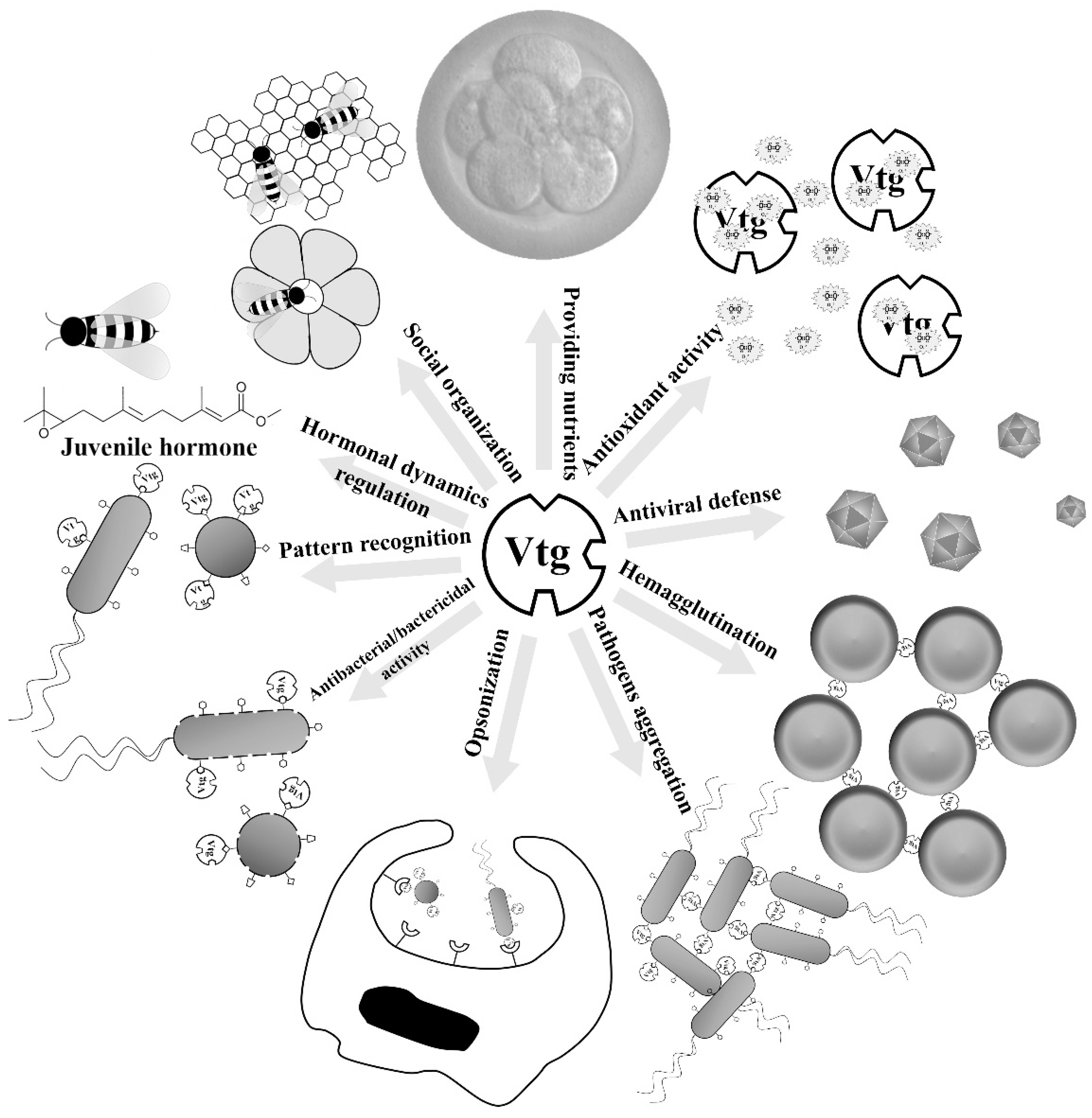
2. Immune-Relevant Activities of Vtgs and Yolk Proteins

2.1. Immune Roles of Vtgs

Accumulating data demonstrated several non-nutritional roles for Vtg. For instance, Vtgs were shown to be associated with the social organization, temporal division of labor and foraging specialization, regulation of hormonal dynamics and change in gustatory responsiveness in the honeybee Apis mellifera, an advanced eusocial insect (Figure 2) [55,56,57,58]. Recent studies show that Vtgs also play immune-relevant roles (Figure 2). The first solid evidence showing that Vtg preforms an immune-relevant role was the observation by Zhang et al., that Vtg purified from the ovaries of the protochordate amphioxus (Branchiostoma japonicum) exhibited hemagglutinating activity against chick, toad and grass carp erythrocytes as well as antibacterial activity against the Gram-negative bacterium E. coli [59]. Soon after that, Vtg purified from the rosy barb Puntius conchonius was found to be capable of inhibiting the growth of the Gram-negative bacteria E. coli, E. aerogenes and Pseudomonas putida as well as the Gram-positive bacteria Staphylococcus aureus, Bacillus subtilis and Streptococcus pyogenes [60], and Vtg from the carp capable of suppressing the growth of E. coli and S. aureus in a dose dependent-manner [61]. Interestingly, Vtgs from protostomes also appear to have antibacterial activity. Vtg from the scallop (Patinopecten yessoensis) was recently shown to have antibacterial activity against Gram-positive and Gram-negative bacteria [62]. In addition, Vtg in the nematode C. elegans seems also involved in its antibacterial defense. A reduced survival was observed in the vtg-knockdown C. elegans after pathogen infection [63]. Another evidence for a role of invertebrate Vtg associated with resistance against bacteria was provided by the enhancement of resistance of nematode against the pathogen Photorhabdus luminescens, when the production of Vtg was stimulated by estrogen 17β-estradiol and phytoestrogen daidzein. However, reduction of Vtg caused by soy isoflavone genistein diminished the host resistance to P. luminescens [64]. Taken together, it appears that the antibacterial activity is a universal property of Vtgs from both vertebrates and invertebrates.
Figure 2. Multiple roles of vitellogenin (Vtg). Vtg is traditionally thought to provide protein- and lipid-rich nutrients for developing embryos and larvae. However, accumulating data demonstrate that its roles extend beyond the nutritional function. In the advanced eusocial insect honeybee, Vtgs were shown to be associated with the social organization, temporal division of labor and foraging specialization, regulation of hormonal dynamics and change in gustatory responsiveness. Recent studies show that Vtgs also play immune-relevant roles. Vtg is able to recognize the invading microbes as a multivalent pattern recognition receptor, kill bacteria or neutralize virus as an effector molecule as well as enhance phagocytosis as an opsonin. Besides, Vtg also exhibits activities to hemagglutinate erythrocytes and aggregate pathogens. In addition to immune roles, Vtg plays another novel role as an antioxidant.
Nutrients 07 05432 g002
Shi et al. showed that intraperitoneal injection of E. coli was able to enhance the level of serum Vtg in male P. conchonius [60]. This has recently been confirmed by Lu et al., who showed that expression of vtg genes in the skin of zebrafish was induced following the challenge with Gram-negative bacterium Citrobacter freundii [65,66]. Moreover, an increased expression of vtg was also detectable in the insect Bactericera cockerelli after infected by “Candidatus Liberibacter solanacearum” via transcriptome analyses [67]. These data together suggest that Vtg may play an active role in the anti-infection of the host in vivo. Actually, Tong et al. showed that Vtg produced in male zebrafish as a consequence of induction by lipopolysaccharide (LPS) and lipoteichoic acid (LTA) is an acute phase reactant, with antibacterial activity against E. coli and S. aureus [68].
Vtg appears to play a multifaceted immune-relevant functions. Li et al. demonstrated that Vtg of the marine fish Hexagrammos otakii was able to bind to Gram-negative bacterium E. coli and Gram-positive bacterium S. aureus as well as fungus Pichia pastoris [69]. The binding to E. coli and S. aureus was also detected for carp and zebrafish Vtgs [61,68]. The binding of Vtgs to bacteria provides them ability to aggregate pathogens as well as to recognize the invading microbes [61]. Further examination via ELISA assay showed that Vtgs exhibited specific affinities to the components conserved within a class of microbes, called pathogen-associated molecular patterns (PAMPs), including LPS of Gram-negative bacteria, LTA of Gram-positive bacteria, peptidoglycan (PGN) of Gram-negative and positive bacteria, and glucan of fungi [61,69]. These observations indicate that Vtg first functions as a multivalent pattern recognition receptor capable of identifying invading Gram-negative and Gram-positive bacteria as well as fungi, and is involved in host immune defense as a detector. In a recent study attempting to search PGN recognition proteins in giant tiger shrimp (Penaeus monodon), an 83 kDa protein was isolated by in vitro PGN pull-down binding assay and identified as a Vtg-like protein via mass spectrometry as well as Western blots with monoclonal antibodies specific of Vtgs reported from P. monodon [70], implicating that invertebrate Vtg may also play a pattern recognition receptor role. Scanning electron microscopy as well as bacterial cell and protoplast lysis assays showed that H. otakii Vtg was able to kill pathogenic bacteria by lysing the whole cells (with cell walls) instead of protoplast (without cell walls) via interaction with LPS and LTA [71]. These show that Vtg functions as an effector molecule, capable of directly killing bacteria. Interestingly, Vtg was also shown to be able to enhance the phagocytosis of microbes by macrophages. Li et al. first reported that H. otakii Vtg could facilitate engulfing of the microbes E. coli, S. aureus and P. pastoris by head-kidney-derived macrophages in vitro [69]. Later, Vtg of carp was shown to possess similar phagocytosis-promoting activity [61]. In an in-situ study of impacts of urban wastewater on freshwater mussel Elliptio complanata, it was observed that the production of Vtg-like proteins was strongly associated with phagocytosis [72], suggesting a relation between Vtg and phagocytosis in invertebrates. Besides, H. otakii Vtg was found to be capable of binding to the cell surface of macrophages but not that of red blood cells [61,69]. Collectively, these observations indicate that Vtg is an opsonin functioning as a bridging molecule between host macrophages and invading pathogens, thereby leading to enhanced phagocytosis. Notably, Liu et al., established that the H. otakii Vtg was able to opsonize the fungus P. pastoris for phagocytosis by macrophages isolated from sea bass Lateolabrax japonicas, implying that the opsonization of Vtg was not species-specific [73]. Further study revealed that Vtg-opsonized phagocytosis showed properties typical of type I phagocytosis, including pseudopod extension, tyrosine kinase dependence, and up-regulation of pro-inflammatory cytokine genes tnf-α and il-1β [73]. Therefore, Vtg is a pattern recognition receptor capable of identifying pathogens, a bactericidal molecule capable of damaging bacterial cell walls, and an opsonin capable of enhancing phagocytosis of pathogens by macrophages. The multifaceted immune-relevant activities of Vtg are in part endowed with its different domains. It was reported by Sun et al. that both DUF1943 and DUF1944 as well as vWD contribute to the function of Vtg as a pattern recognition receptor, and DUF1943 and DUF1944 (but not vWD) also contribute to the function of Vtg as an opsonin [21].
Recently, Garcia et al. showed that Atlantic salmon Vtg possessed neutralizing ability for infectious pancreatic necrosis virus [74], suggesting that Vtg is also involved in host antiviral immunity. This seems further supported by the observation that the mosquito (Anopheles gambiae) Vtg was able to interfere with anti-plasmodium response [75]. These denote that in addition to antibacterial activity, Vtg also has antiviral activity, which demands detailed study in the future.

2.2. Immune Roles of Yolk Proteins

Lv and Pv are the principal yolk proteins generated by the proteolytic cleavage of Vtg. As Vtg is an immune-competent molecule, it is thus reasonable to hypothesize that Lv and Pv also have similar immune activities. This hypothesis was first tested by Zhang and Zhang [76]. They demonstrated that the native Lv purified from ovulated eggs of the rosy barb P. conchonius was able to interact with LPS, LTA and PGN, as well as E. coli and S. aureus, but not with self-molecules such as the egg extracts prepared, indicating that Lv is a molecule capable of recognizing non-self components. Moreover, the bacterial binding activity of Lv enabled it to enhance the phagocytosis of bacteria by macrophages, suggesting that Lv is also an opsonin functional in developing embryos/larvae [76]. Similarly, Pv was also shown to play a critical role in the immunity of zebrafish embryos via acting as a pattern recognition receptor and an antimicrobial effector molecule [77]. In line with this, hen egg yolk Pv was also shown to be able to inhibit the growth of the Gram-negative bacterium E. coli and the Gram-positive bacterium S. aureus under thermal stress [78,79]. Of note, the affinity of Pv to LPS enabled the protein a capacity to neutralize endotoxin, promoting the survival rate of endotoxemia mice [79]. It was recently shown that a truncated Pv (Pt5) consisting of the C-terminal 55 residues of zebrafish Pv also displayed similar immune activities with Pv, including antimicrobial activity against E. coli, Aeromonas hydrophila and S. aureus, and specific affinity to LPS, LTA, and PGN [77]. Intraperitoneal injection of this Pv-derived peptide was able to increase the survival rate of zebrafish challenged with pathogenic A. hydrophila and to markedly decrease the number of the pathogen in multiple tissues, suggesting that Pt5 could inhibit multiplication/dissemination of pathogen in host as an antimicrobial agent. In addition to direct antimicrobial activity, Pt5 was also shown to be able to regulate the host immune responses via suppressing the expression of pro-inflammatory cytokine genes (il-1β, il-6, tnf-α and ifn-γ) and simultaneously enhancing the expression of anti-inflammatory cytokine genes (il-10 and il-4), suggesting a dual role of Pt5 as both immune effector and modulator [80]. Recently, a mutant peptide of Pt5 (designated as Pt5e), generated by site-directed mutagenesis, was shown to have stronger bactericidal activity and LPS-neutralizing activity [81]. Besides, Sun et al. demonstrated that recombinant zebrafish Pv was capable of inhibiting the formation of the cytopathic effect in lymphocystis disease virus (LCDV)-infected cells and reducing the virus quantities in the infected cells as well as in the infected zebrafish, suggesting that Pv possesses an antiviral activity and participates in immune defense of host against the infection by viruses like LCDV [82]. Taken together, these data show that like Vtg, Lv and Pv are both immune-competent molecules involved in immune response of the host against invading pathogenic microbes.

3. Antioxidant Activities of Vtgs and Yolk Proteins

In addition to immune roles, another novel role of Vtg is antioxidant activity (Figure 2). It was first shown by Ando and Yanagida that Vtg from the eel Anguilla japonica was able to resist the copper-induced oxidation, and could protect the very low density lipoprotein (VLDL) against copper-induced oxidation [83]. This was the first observation reporting that Vtg has antioxidant activity, and serves to suppress the free-radical reactions in fish oocytes. Similar antioxidant activity was also suggested for the nematode (C. elegans) Vtg [84]. In the honeybee, Vtg was demonstrated to be able to reduce oxidative stress by scavenging free radicals, thereby increasing the lifespan in the facultatively sterile worker castes and reproductive queen castes [85,86]. The honeybee Vtg was also demonstrated in a recent study to be capable of recognizing cell damage through its binding to membrane and shielding living cells from damage by reactive oxygen species (ROS) [87]. It is clear that Vtg protects cells from ROS damage in both invertebrates and vertebrates.
It is well known that hen egg yolk Pv, as Vtg-derived major protein, show strong antioxidant activity owing to its high serine and phosphorus content, which makes this protein one of the strongest iron-chelating agents [88,89,90]. Very recently, we showed that zebrafish recombinant phosvitin (rPv) was an antioxidant agent capable of inhibiting the oxidation of the linoleic acid, and scavenging the 2,2-diphenyl-1-picrylhydrazyl (DPPH) radical. We also showed that zebrafish rPv is a cellular antioxidant capable of protecting radical-mediated oxidation of cellular biomolecules. Importantly, zebrafish rPv is non-cytotoxic to murine macrophages RAW264.7 [91]. These results show that Pv in fish is also a strong antioxidant agent. If Lv, another major Vtg-derived protein, has any antioxidant activity remains open, which is worthwhile exploring.

4. Potential Applications in Human Health

Antibiotics are globally utilized to control microbial infections in clinical practice up to date, however cases of resistance to the majority of antibiotic classes have been reported, which has become a serious threat to human health in many parts of the world [92,93]. It is thus essential to develop new antibiotic agents to combat these resistant pathogens. Antimicrobial proteins/peptides (AMPs) are potential candidates to solve this problem. As a protein/peptide with antimicrobial activity widely present in plants, animals and microbes, AMP commonly is a cationic and amphipathic molecule with a net positive charge and a high percentage of hydrophobic residues [94]. These structure characteristics provide AMP the ability to interact with the anionic cell wall and phospholipid membranes of microorganisms, which makes it more difficult for pathogens to evolve resistance [95]. Vtg and its derived protein Pv from oviparous species, especially teleost fishes, both display antibacterial activities with a broad antibacterial spectrum [59,60,61,62,68,71,77,78,79], and hence can be used as pro-drug to develop novel antibiotic agents. For example, based on the residual sequence of Pt5, the C-terminal peptide of zebrafish Pv, a total of six mutant peptides were generated by a single or double mutagenesis; among them, a mutant called Pt5e showed stronger antibacterial activities against E. coli and S. aureus [81], and was able to kill five strains of multiple drug resistance bacteria isolated from clinical cases via disturbing their cell membrane integrity (Data not shown).
Sepsis is a serious disease characterized by a systemic inflammatory response syndrome caused by infection. Severe sepsis is complicated by tissue damage and organ dysfunction, which can lead to sequential multi-organ failure followed by death [96]. The primary trigger of sepsis is thought to be LPS, or endotoxin, a major component of the cell wall of Gram-negative bacteria, which is released when bacteria grow or are abolished by antibiotics or host immunity. LPS, as a conserved molecular signature of Gram-negative bacteria, can specifically interact with Vtg, Lv, Pv and their derived peptides [21,61,62,69,71,76,77,79,81], making it possible to utilize these proteins/peptides to develop LPS-neutralizing agents for sepsis treatment. In line with this, both Pv and Pt5e exhibiting LPS-neutralizing ability had been shown to be able to promote the survival rate of endotoxemia mice. Moreover, both Pv and Pt5e displayed neither cytotoxicity to murine RAW264.7 macrophages nor hemolytic activity towards human red blood cells [79,81], suggesting that they can be a safe potential candidate for therapeutics of sepsis.
Antioxidant agents have attracted a great deal of attention in recent years because of their roles in prevention of chronic diseases and utilization as preservatives in food and cosmetics [97,98,99]. Vtg and its derived Pv both have antioxidant activities against ROS. As these proteins are components of our food source, they are thus natural antioxidant agents. These suggest that they can be can be an important antioxidant with a potential in preservation of food and cosmetics as well as in mediation of chronic disease states.

5. Conclusions

Vtg, the precursor of major egg yolk proteins, is traditionally thought to provide protein- and lipid-rich nutrients for developing embryos and larvae. However, accumulating data indicate that Vtg as well as its derived yolk proteins Lv and Pv also play non-nutritional functions: they are not only involved in immune defense but also antioxidant reaction. These non-nutritional functions clearly better and deepen our understanding of the physiological roles of the molecules, and at the same time, provide a sound basis for potential application of the molecules in human health.

Acknowledgments

This work was supported by Natural Science Foundation of China (No. 31402030; 31372505), Fundamental Research Funds for the Central Universities (201462004), China Postdoctoral Science Foundation (2015T80745; 2014M560580) and Qingdao Municipal Science and Technology Commission (No. 13-1-4-206-jch).

Author Contributions

C. Sun wrote the manuscript. S. C. Zhang read and revised the manuscript.

Conflicts of Interest

The authors declare no conflict of interest.

References

  1. Jalabert, B. Particularities of reproduction and oogenesis in teleost fish compared to mammals. Reprod. Nutr. Dev. 2005, 45, 261–279. [Google Scholar] [CrossRef] [PubMed]
  2. Lubzens, E.; Young, G.; Bobe, J.; Cerda, J. Oogenesis in teleosts: How eggs are formed. Gen. Comp. Endocrinol. 2010, 165, 367–389. [Google Scholar] [CrossRef]
  3. Patiño, R.; Sullivan, C. Ovarian follicle growth, maturation, and ovulation in teleost fish. Fish Physiol. Biochem. 2002, 26, 57–70. [Google Scholar] [CrossRef]
  4. Finn, R.N. Vertebrate yolk complexes and the functional implications of phosvitins and other subdomains in vitellogenins. Biol. Reprod. 2007, 76, 926–935. [Google Scholar] [CrossRef] [PubMed]
  5. Smolenaars, M.M.; Madsen, O.; Rodenburg, K.W.; van der Horst, D.J. Molecular diversity and evolution of the large lipid transfer protein superfamily. J. Lipid Res. 2007, 48, 489–502. [Google Scholar] [CrossRef] [PubMed]
  6. Avarre, J.C.; Lubzens, E.; Babin, P.J. Apolipocrustacein, formerly vitellogenin, is the major egg yolk precursor protein in decapod crustaceans and is homologous to insect apolipophorin II/I and vertebrate apolipoprotein B. BMC Evol. Biol. 2007, 7. [Google Scholar] [CrossRef] [PubMed]
  7. Tufail, M.; Takeda, M. Molecular characteristics of insect vitellogenins. J. Insect Physiol. 2008, 54, 1447–1458. [Google Scholar] [PubMed]
  8. Matozzo, V.; Gagne, F.; Marin, M.G.; Ricciardi, F.; Blaise, C. Vitellogenin as a biomarker of exposure to estrogenic compounds in aquatic invertebrates: A review. Environ. Int. 2008, 34, 531–545. [Google Scholar] [PubMed]
  9. Wu, L.T.; Hui, J.H.; Chu, K.H. Origin and evolution of yolk proteins: Expansion and functional diversification of large lipid transfer protein superfamily. Biol. Reprod. 2013, 88. [Google Scholar] [CrossRef] [PubMed]
  10. Finn, R.N.; Kolarevic, J.; Kongshaug, H.; Nilsen, F. Evolution and differential expression of a vertebrate vitellogenin gene cluster. BMC Evol. Biol. 2009, 9. [Google Scholar] [CrossRef] [PubMed]
  11. Silva, R.; Fischer, A.H.; Burch, J.B. The major and minor chicken vitellogenin genes are each adjacent to partially deleted pseudogene copies of the other. Mol. Cell. Biol. 1989, 9, 3557–3562. [Google Scholar] [CrossRef] [PubMed]
  12. Van het Schip, F.D.; Samallo, J.; Broos, J.; Ophuis, J.; Mojet, M.; Gruber, M.; Ab, G. Nucleotide sequence of a chicken vitellogenin gene and derived amino acid sequence of the encoded yolk precursor protein. J. Mol. Biol. 1987, 196, 245–260. [Google Scholar] [CrossRef]
  13. Germond, J.E.; Walker, P.; ten Heggeler, B.; Brown-Luedi, M.; de Bony, E.; Wahli, W. Evolution of vitellogenin genes: Comparative analysis of the nucleotide sequences downstream of the transcription initiation site of four Xenopus laevis and one chicken gene. Nucleic Acids Res. 1984, 12, 8595–8609. [Google Scholar] [CrossRef] [PubMed]
  14. Wahli, W.; Dawid, I.B.; Wyler, T.; Jaggi, R.B.; Weber, R.; Ryffel, G.U. Vitellogenin in Xenopus laevis is encoded in a small family of genes. Cell 1979, 16, 535–549. [Google Scholar] [CrossRef]
  15. Blumenthal, T.; Squire, M.; Kirtland, S.; Cane, J.; Donegan, M.; Spieth, J.; Sharrock, W. Cloning of a yolk protein gene family from Caenorhabditis elegans. J. Mol. Biol. 1984, 174, 1–18. [Google Scholar] [CrossRef]
  16. Wang, H.; Yan, T.; Tan, J.T.; Gong, Z. A zebrafish vitellogenin gene (vg3) encodes a novel vitellogenin without a phosvitin domain and may represent a primitive vertebrate vitellogenin gene. Gene 2000, 256, 303–310. [Google Scholar] [CrossRef]
  17. Wang, H.; Tan, J.T.; Emelyanov, A.; Korzh, V.; Gong, Z. Hepatic and extrahepatic expression of vitellogenin genes in the zebrafish, Danio rerio. Gene 2005, 356, 91–100. [Google Scholar] [PubMed]
  18. Kang, B.J.; Jung, J.H.; Lee, J.M.; Lim, S.G.; Saito, H.; Kim, M.H.; Kim, Y.J.; Saigusa, M.; Han, C.H. Structural and expression analyses of two vitellogenin genes in the carp, Cyprinus carpio. Comp. Biochem. Physiol. Part B Biochem. Mol. Biol. 2007, 148, 445–453. [Google Scholar] [CrossRef] [PubMed]
  19. Williams, V.N.; Reading, B.J.; Hiramatsu, N.; Amano, H.; Glassbrook, N.; Hara, A.; Sullivan, C.V. Multiple vitellogenins and product yolk proteins in striped bass, Morone saxatilis: Molecular characterization and processing during oocyte growth and maturation. Fish Physiol. Biochem. 2014, 40, 395–415. [Google Scholar] [CrossRef] [PubMed]
  20. Reading, B.J.; Hiramatsu, N.; Sawaguchi, S.; Matsubara, T.; Hara, A.; Lively, M.O.; Sullivan, C.V. Conserved and variant molecular and functional features of multiple egg yolk precursor proteins (vitellogenins) in white perch (Morone americana) and other teleosts. Mar. Biotechnol. 2009, 11, 169–187. [Google Scholar] [CrossRef] [PubMed]
  21. Sun, C.; Hu, L.; Liu, S.; Gao, Z.; Zhang, S. Functional analysis of domain of unknown function (DUF) 1943, DUF1944 and von Willebrand factor type D domain (VWD) in vitellogenin 2 in zebrafish. Dev. Comp. Immunol. 2013, 41, 469–476. [Google Scholar] [CrossRef] [PubMed]
  22. Hayward, A.; Takahashi, T.; Bendena, W.G.; Tobe, S.S.; Hui, J.H. Comparative genomic and phylogenetic analysis of vitellogenin and other large lipid transfer proteins in metazoans. FEBS Lett. 2010, 584, 1273–1278. [Google Scholar] [CrossRef] [PubMed]
  23. Finn, R.N.; Kristoffersen, B.A. Vertebrate vitellogenin gene duplication in relation to the “3R hypothesis”: Correlation to the pelagic egg and the oceanic radiation of teleosts. PLoS ONE 2007, 2, e169. [Google Scholar] [CrossRef] [PubMed]
  24. Meusy, J.J. Vitellogenin, the extraovarian precursor of the protein yolk in Crustacea: A review. Reprod. Nutr. Dev. 1980, 20, 1–21. [Google Scholar] [CrossRef] [PubMed]
  25. Girish, B.P.; Swetha, C.; Reddy, P.S. Hepatopancreas but not ovary is the site of vitellogenin synthesis in female fresh water crab, Oziothelphusa senex senex. Biochem. Biophys. Res. Commun. 2014, 447, 323–327. [Google Scholar] [CrossRef] [PubMed]
  26. Mak, A.S.; Choi, C.L.; Tiu, S.H.; Hui, J.H.; He, J.G.; Tobe, S.S.; Chan, S.M. Vitellogenesis in the red crab Charybdis feriatus: Hepatopancreas-specific expression and farnesoic acid stimulation of vitellogenin gene expression. Mol. Reprod. Dev. 2005, 70, 288–300. [Google Scholar] [CrossRef] [PubMed]
  27. Kolarevic, J.; Nerland, A.; Nilsen, F.; Finn, R.N. Goldsinny wrasse (Ctenolabrus rupestris) is an extreme vtgAa-type pelagophil teleost. Mol. Reprod. Dev. 2008, 75, 1011–1020. [Google Scholar] [CrossRef] [PubMed]
  28. Sawaguchi, S.; Ohkubo, N.; Koya, Y.; Matsubara, T. Incorporation and utilization of multiple forms of vitellogenin and their derivative yolk proteins during vitellogenesis and embryonic development in the mosquitofish, Gambusia affinis. Zool. Sci. 2005, 22, 701–710. [Google Scholar] [CrossRef] [PubMed]
  29. Amano, H.; Fujita, T.; Hiramatsu, N.; Kagawa, H.; Matsubara, T.; Sullivan, C.V.; Hara, A. Multiple vitellogenin-derived yolk proteins in gray mullet (Mugil cephalus): Disparate proteolytic patterns associated with ovarian follicle maturation. Mol. Reprod. Deve. 2008, 75, 1307–1317. [Google Scholar] [CrossRef] [PubMed]
  30. Wallace, R.A.; Selman, K. Ultrastructural aspects of oogenesis and oocyte growth in fish and amphibians. J. Electron Microsc. Tech. 1990, 16, 175–201. [Google Scholar] [PubMed]
  31. Conner, S.D.; Schmid, S.L. Regulated portals of entry into the cell. Nature 2003, 422, 37–44. [Google Scholar] [CrossRef] [PubMed]
  32. Hiramatsu, N.; Todo, T.; Sullivan, C.V.; Schilling, J.; Reading, B.J.; Matsubara, T.; Ryu, Y.W.; Mizuta, H.; Luo, W.; Nishimiya, O.; et al. Ovarian yolk formation in fishes: Molecular mechanisms underlying formation of lipid droplets and vitellogenin-derived yolk proteins. Gen. Comp. Endocrinol. 2015. [Google Scholar] [CrossRef] [PubMed]
  33. Reading, B.J.; Hiramatsu, N.; Schilling, J.; Molloy, K.T.; Glassbrook, N.; Mizuta, H.; Luo, W.; Baltzegar, D.A.; Williams, V.N.; Todo, T.; et al. Lrp13 is a novel vertebrate lipoprotein receptor that binds vitellogenins in teleost fishes. J. Lipid Res. 2014, 55, 2287–2295. [Google Scholar] [CrossRef] [PubMed]
  34. Reading, B.J.; Hiramatsu, N.; Sullivan, C.V. Disparate binding of three types of vitellogenin to multiple forms of vitellogenin receptor in white perch. Biol. Reprod. 2011, 84, 392–399. [Google Scholar] [CrossRef] [PubMed]
  35. Williams, V.N.; Reading, B.J.; Amano, H.; Hiramatsu, N.; Schilling, J.; Salger, S.A.; Williams, T.I.; Gross, K.; Sullivan, C.V. Proportional accumulation of yolk proteins derived from multiple vitellogenins is precisely regulated during vitellogenesis in striped bass (Morone saxatilis). J. Exp. Zool. A Ecol. Genet. Physiol. 2014, 321, 301–315. [Google Scholar] [CrossRef] [PubMed]
  36. Romano, M.; Rosanova, P.; Anteo, C.; Limatola, E. Vertebrate yolk proteins: A review. Mol. Reprod. Dev. 2004, 69, 109–116. [Google Scholar] [CrossRef] [PubMed]
  37. Carnevali, O.; Carletta, R.; Cambi, A.; Vita, A.; Bromage, N. Yolk formation and degradation during oocyte maturation in seabream Sparus aurata: Involvement of two lysosomal proteinases. Biol. Reprod. 1999, 60, 140–146. [Google Scholar] [CrossRef] [PubMed]
  38. Carnevali, O.; Cionna, C.; Tosti, L.; Lubzens, E.; Maradonna, F. Role of cathepsins in ovarian follicle growth and maturation. Gen. Comp. Endocrinol. 2006, 146, 195–203. [Google Scholar] [CrossRef] [PubMed]
  39. Hiramatsu, N.; Ichikawa, N.; Fukada, H.; Fujita, T.; Sullivan, C.V.; Hara, A. Identification and characterization of proteases involved in specific proteolysis of vitellogenin and yolk proteins in salmonids. J. Exp. Zool. 2002, 292, 11–25. [Google Scholar] [CrossRef] [PubMed]
  40. Fabra, M.; Cerda, J. Ovarian cysteine proteinases in the teleost Fundulus heteroclitus: Molecular cloning and gene expression during vitellogenesis and oocyte maturation. Mol. Reprod. Dev. 2004, 67, 282–294. [Google Scholar] [CrossRef] [PubMed]
  41. Opresko, L.K.; Karpf, R.A. Specific proteolysis regulates fusion between endocytic compartments in Xenopus oocytes. Cell 1987, 51, 557–568. [Google Scholar] [CrossRef]
  42. Sire, M.F.; Babin, P.J.; Vernier, J.M. Involvement of the lysosomal system in yolk protein deposit and degradation during vitellogenesis and embryonic development in trout. J. Exp. Zool. 1994, 269, 69–83. [Google Scholar] [CrossRef]
  43. Retzek, H.; Steyrer, E.; Sanders, E.J.; Nimpf, J.; Schneider, W.J. Molecular cloning and functional characterization of chicken cathepsin D, a key enzyme for yolk formation. DNA Cell Biol. 1992, 11, 661–672. [Google Scholar] [CrossRef] [PubMed]
  44. Babin, P.J. Apolipoproteins and the association of egg yolk proteins with plasma high density lipoproteins after ovulation and follicular atresia in the rainbow trout (Salmo gairdneri). J. Biol. Chem. 1987, 262, 4290–4296. [Google Scholar] [PubMed]
  45. Matsubara, T.; Ohkubo, N.; Andoh, T.; Sullivan, C.V.; Hara, A. Two forms of vitellogenin, yielding two distinct lipovitellins, play different roles during oocyte maturation and early development of barfin flounder, Verasper moseri, a marine teleost that spawns pelagic eggs. Dev. Biol. 1999, 213, 18–32. [Google Scholar] [CrossRef] [PubMed]
  46. Tyler, C.R.; Sumpter, J.P.; Bromage, N.R. In vivo ovarian uptake and processing of vitellogenin in the rainbow trout, Salmo gairdneri. J. Exp. Zool. 1988, 246, 171–179. [Google Scholar] [CrossRef]
  47. Yilmaz, O.; Prat, F.; Ibanez, A.J.; Amano, H.; Koksoy, S.; Sullivan, C.V. Estrogen-induced yolk precursors in European sea bass, Dicentrarchus labrax: Status and perspectives on multiplicity and functioning of vitellogenins. Gen. Comp. Endocrinol. 2015. [Google Scholar] [CrossRef] [PubMed]
  48. Finn, R.N.; Fyhn, H.J. Requirement for amino acids in ontogeny of fish. Aquac. Res. 2010, 41, 684–716. [Google Scholar] [CrossRef]
  49. Arukwe, A.; Goksoyr, A. Eggshell and egg yolk proteins in fish: Hepatic proteins for the next generation: Oogenetic, population, and evolutionary implications of endocrine disruption. Comp. Hepatol. 2003, 2. [Google Scholar] [CrossRef] [Green Version]
  50. Pan, M.L.; Bell, W.J.; Telfer, W.H. Vitellogenic blood protein synthesis by insect fat body. Science 1969, 165, 393–394. [Google Scholar] [CrossRef] [PubMed]
  51. Nath, P.; Sundararaj, B.I. Isolation and identification of female-specific serum lipophosphoprotein (vitellogenin) in the catfish, Heteropneustes fossilis (Bloch). Gen. Comp. Endocrinol. 1981, 43, 184–190. [Google Scholar] [CrossRef]
  52. Shyu, A.B.; Raff, R.A.; Blumenthal, T. Expression of the vitellogenin gene in female and male sea urchin. Proc. Natl. Acad. Sci. USA 1986, 83, 3865–3869. [Google Scholar] [CrossRef] [PubMed]
  53. Piulachs, M.D.; Guidugli, K.R.; Barchuk, A.R.; Cruz, J.; Simoes, Z.L.; Belles, X. The vitellogenin of the honey bee, Apis mellifera: Structural analysis of the cdna and expression studies. Insect Biochem. Mol. Biol. 2003, 33, 459–465. [Google Scholar] [CrossRef]
  54. Scharf, M.E.; Wu-Scharf, D.; Zhou, X.; Pittendrigh, B.R.; Bennett, G.W. Gene expression profiles among immature and adult reproductive castes of the termite Reticulitermes flavipes. Insect Mol. Biol. 2005, 14, 31–44. [Google Scholar] [CrossRef] [PubMed]
  55. Amdam, G.V.; Norberg, K.; Hagen, A.; Omholt, S.W. Social exploitation of vitellogenin. Proc. Natl. Acad. Sci. USA 2003, 100, 1799–1802. [Google Scholar] [CrossRef] [PubMed]
  56. Amdam, G.V.; Norberg, K.; Page, R.E., Jr.; Erber, J.; Scheiner, R. Downregulation of vitellogenin gene activity increases the gustatory responsiveness of honey bee workers (Apis mellifera). Behav. Brain Res. 2006, 169, 201–205. [Google Scholar] [CrossRef] [PubMed]
  57. Guidugli, K.R.; Nascimento, A.M.; Amdam, G.V.; Barchuk, A.R.; Omholt, S.; Simoes, Z.L.; Hartfelder, K. Vitellogenin regulates hormonal dynamics in the worker caste of a eusocial insect. FEBS Lett. 2005, 579, 4961–4965. [Google Scholar] [CrossRef] [PubMed]
  58. Nelson, C.M.; Ihle, K.E.; Fondrk, M.K.; Page, R.E.; Amdam, G.V. The gene vitellogenin has multiple coordinating effects on social organization. PLoS Biol. 2007, 5, e62. [Google Scholar] [CrossRef] [PubMed]
  59. Zhang, S.; Sun, Y.; Pang, Q.; Shi, X. Hemagglutinating and antibacterial activities of vitellogenin. Fish Shellfish Immunol. 2005, 19, 93–95. [Google Scholar] [CrossRef] [PubMed]
  60. Shi, X.; Zhang, S.; Pang, Q. Vitellogenin is a novel player in defense reactions. Fish Shellfish Immunol. 2006, 20, 769–772. [Google Scholar] [CrossRef] [PubMed]
  61. Liu, Q.H.; Zhang, S.C.; Li, Z.J.; Gao, C.R. Characterization of a pattern recognition molecule vitellogenin from carp (Cyprinus carpio). Immunobiology 2009, 214, 257–267. [Google Scholar] [CrossRef] [PubMed]
  62. Wu, B.; Liu, Z.; Zhou, L.; Ji, G.; Yang, A. Molecular cloning, expression, purification and characterization of vitellogenin in scallop Patinopecten yessoensis with special emphasis on its antibacterial activity. Deve. Comp. Immunol. 2015, 49, 249–258. [Google Scholar] [CrossRef] [PubMed]
  63. Fischer, M.; Regitz, C.; Kull, R.; Boll, M.; Wenzel, U. Vitellogenins increase stress resistance of Caenorhabditis elegans after Photorhabdus luminescens infection depending on the steroid-signaling pathway. Microbes Infect. 2013, 15, 569–578. [Google Scholar] [CrossRef] [PubMed]
  64. Fischer, M.; Regitz, C.; Kahl, M.; Werthebach, M.; Boll, M.; Wenzel, U. Phytoestrogens genistein and daidzein affect immunity in the nematode Caenorhabditis elegans via alterations of vitellogenin expression. Mol. Nutr. Food Res. 2012, 56, 957–965. [Google Scholar] [CrossRef] [PubMed]
  65. Lu, A.; Hu, X.; Wang, Y.; Shen, X.; Zhu, A.; Shen, L.; Ming, Q.; Feng, Z. Comparative analysis of the acute response of zebrafish Danio rerio skin to two different bacterial infections. J. Aquat. Anim. Health 2013, 25, 243–251. [Google Scholar] [CrossRef] [PubMed]
  66. Lu, A.; Hu, X.; Xue, J.; Zhu, J.; Wang, Y.; Zhou, G. Gene expression profiling in the skin of zebrafish infected with Citrobacter freundii. Fish Shellfish Immunol. 2012, 32, 273–283. [Google Scholar] [CrossRef] [PubMed]
  67. Nachappa, P.; Levy, J.; Tamborindeguy, C. Transcriptome analyses of Bactericera cockerelli adults in response to “Candidatus Liberibacter solanacearum” infection. Mol. Genet. Genom. 2012, 287, 803–817. [Google Scholar] [CrossRef] [PubMed]
  68. Tong, Z.; Li, L.; Pawar, R.; Zhang, S. Vitellogenin is an acute phase protein with bacterial-binding and inhibiting activities. Immunobiology 2010, 215, 898–902. [Google Scholar] [CrossRef] [PubMed]
  69. Li, Z.; Zhang, S.; Liu, Q. Vitellogenin functions as a multivalent pattern recognition receptor with an opsonic activity. PLoS ONE 2008, 3, e1940. [Google Scholar] [CrossRef] [PubMed]
  70. Udompetcharaporn, A.; Junkunlo, K.; Senapin, S.; Roytrakul, S.; Flegel, T.W.; Sritunyalucksana, K. Identification and characterization of a QM protein as a possible peptidoglycan recognition protein (PGRP) from the giant tiger shrimp Penaeus monodon. Dev. Comp. Immunol. 2014, 46, 146–154. [Google Scholar] [CrossRef] [PubMed]
  71. Li, Z.; Zhang, S.; Zhang, J.; Liu, M.; Liu, Z. Vitellogenin is a cidal factor capable of killing bacteria via interaction with lipopolysaccharide and lipoteichoic acid. Mol. Immunol. 2009, 46, 3232–3239. [Google Scholar] [CrossRef] [PubMed]
  72. Bouchard, B.; Gagne, F.; Fortier, M.; Fournier, M. An in-situ study of the impacts of urban wastewater on the immune and reproductive systems of the freshwater mussel Elliptio complanata. Comp. Biochem. Physiol. C Toxicol. Pharm. 2009, 150, 132–140. [Google Scholar] [CrossRef] [PubMed]
  73. Liu, M.; Pan, J.; Ji, H.; Zhao, B.; Zhang, S. Vitellogenin mediates phagocytosis through interaction with FcγR. Mol. Immunol. 2011, 49, 211–218. [Google Scholar] [CrossRef] [PubMed]
  74. Garcia, J.; Munro, E.S.; Monte, M.M.; Fourrier, M.C.; Whitelaw, J.; Smail, D.A.; Ellis, A.E. Atlantic salmon (Salmo salar L.) serum vitellogenin neutralises infectivity of infectious pancreatic necrosis virus (IPNV). Fish Shellfish Immunol. 2010, 29, 293–297. [Google Scholar] [CrossRef] [PubMed]
  75. Rono, M.K.; Whitten, M.M.; Oulad-Abdelghani, M.; Levashina, E.A.; Marois, E. The major yolk protein vitellogenin interferes with the anti-plasmodium response in the malaria mosquito Anopheles gambiae. PLoS Biol. 2010, 8, e1000434. [Google Scholar] [CrossRef] [PubMed]
  76. Zhang, J.; Zhang, S. Lipovitellin is a non-self recognition receptor with opsonic activity. Mar. Biotechnol. 2011, 13, 441–450. [Google Scholar] [CrossRef] [PubMed]
  77. Wang, S.; Wang, Y.; Ma, J.; Ding, Y.; Zhang, S. Phosvitin plays a critical role in the immunity of zebrafish embryos via acting as a pattern recognition receptor and an antimicrobial effector. J. Biol. Chem. 2011, 286, 22653–22664. [Google Scholar] [CrossRef] [PubMed]
  78. Sattar Khan, M.A.; Nakamura, S.; Ogawa, M.; Akita, E.; Azakami, H.; Kato, A. Bactericidal action of egg yolk phosvitin against Escherichia coli under thermal stress. J. Agric. Food Chem. 2000, 48, 1503–1506. [Google Scholar] [CrossRef] [PubMed]
  79. Ma, J.; Wang, H.; Wang, Y.; Zhang, S. Endotoxin-neutralizing activity of hen egg phosvitin. Mol. Immunol. 2013, 53, 355–362. [Google Scholar] [CrossRef] [PubMed]
  80. Ding, Y.; Liu, X.; Bu, L.; Li, H.; Zhang, S. Antimicrobial-immunomodulatory activities of zebrafish phosvitin-derived peptide Pt5. Peptides 2012, 37, 309–313. [Google Scholar] [CrossRef] [PubMed]
  81. Hu, L.; Sun, C.; Wang, S.; Su, F.; Zhang, S. Lipopolysaccharide neutralization by a novel peptide derived from phosvitin. Int. J. Biochem. Cell Biol. 2013, 45, 2622–2631. [Google Scholar] [CrossRef] [PubMed]
  82. Sun, C.; Hu, L.; Liu, S.; Hu, G.; Zhang, S. Antiviral activity of phosvitin from zebrafish Danio rerio. Deve. Comp. Immunol. 2013, 40, 28–34. [Google Scholar] [CrossRef] [PubMed]
  83. Ando, S.; Yanagida, K. Susceptibility to oxidation of copper-induced plasma lipoproteins from Japanese eel: Protective effect of vitellogenin on the oxidation of very low density lipoprotein. Comp. Biochem. Physiol. Part C Pharmacol. Toxicol. Endocrinol. 1999, 123, 1–7. [Google Scholar] [CrossRef]
  84. Nakamura, A.; Yasuda, K.; Adachi, H.; Sakurai, Y.; Ishii, N.; Goto, S. Vitellogenin-6 is a major carbonylated protein in aged nematode, Caenorhabditis elegans. Biochem. Biophys. Res. Commun. 1999, 264, 580–583. [Google Scholar] [CrossRef] [PubMed]
  85. Seehuus, S.C.; Norberg, K.; Gimsa, U.; Krekling, T.; Amdam, G.V. Reproductive protein protects functionally sterile honey bee workers from oxidative stress. Proc. Natl. Acad. Sci. USA 2006, 103, 962–967. [Google Scholar] [CrossRef] [PubMed]
  86. Corona, M.; Velarde, R.A.; Remolina, S.; Moran-Lauter, A.; Wang, Y.; Hughes, K.A.; Robinson, G.E. Vitellogenin, juvenile hormone, insulin signaling, and queen honey bee longevity. Proc. Natl. Acad. Sci. USA 2007, 104, 7128–7133. [Google Scholar] [CrossRef] [PubMed]
  87. Havukainen, H.; Munch, D.; Baumann, A.; Zhong, S.; Halskau, O.; Krogsgaard, M.; Amdam, G.V. Vitellogenin recognizes cell damage through membrane binding and shields living cells from reactive oxygen species. J. Biol. Chem. 2013, 288, 28369–28381. [Google Scholar] [CrossRef] [PubMed]
  88. Lu, C.L.; Baker, R.C. Characteristics of egg yolk phosvitin as an antioxidant for inhibiting metal-catalyzed phospholipid oxidations. Poult. Sci. 1986, 65, 2065–2070. [Google Scholar] [CrossRef] [PubMed]
  89. Ishikawa, S.; Yano, Y.; Arihara, K.; Itoh, M. Egg yolk phosvitin inhibits hydroxyl radical formation from the fenton reaction. Biosci. Biotechnol. Biochem. 2004, 68, 1324–1331. [Google Scholar] [CrossRef] [PubMed]
  90. Guérin-Dubiard, C.; Anton, M.; Dhene-Garcia, A.; Martinet, V.; Brulé, G. Hen egg and fish egg phosvitins: Composition and iron binding properties. Eur. Food Res. Technol. 2002, 214, 460–464. [Google Scholar] [CrossRef]
  91. Hu, L.; Sun, C.; Luan, J.; Lu, L.; Zhang, S. Zebrafish phosvitin is an antioxidant with non-cytotoxic activity. Acta Biochim. Biophys. Sin. 2015, 47, 349–354. [Google Scholar] [CrossRef] [PubMed]
  92. Maria-Neto, S.; de Almeida, K.C.; Macedo, M.L.; Franco, O.L. Understanding bacterial resistance to antimicrobial peptides: From the surface to deep inside. Biochim. Biophys. Acta 2015. [Google Scholar] [CrossRef] [PubMed]
  93. Grundmann, H.; Klugman, K.P.; Walsh, T.; Ramon-Pardo, P.; Sigauque, B.; Khan, W.; Laxminarayan, R.; Heddini, A.; Stelling, J. A framework for global surveillance of antibiotic resistance. Drug Resist. Updates 2011, 14, 79–87. [Google Scholar] [CrossRef] [PubMed]
  94. Wiesner, J.; Vilcinskas, A. Antimicrobial peptides: The ancient arm of the human immune system. Virulence 2010, 1, 440–464. [Google Scholar] [CrossRef] [PubMed]
  95. Brown, K.L.; Hancock, R.E. Cationic host defense (antimicrobial) peptides. Curr. Opin. Immunol. 2006, 18, 24–30. [Google Scholar] [CrossRef] [PubMed]
  96. Uppu, D.S.; Ghosh, C.; Haldar, J. Surviving sepsis in the era of antibiotic resistance: Are there any alternative approaches to antibiotic therapy? Microb. Pathog. 2015, 80, 7–13. [Google Scholar] [CrossRef] [PubMed]
  97. Knight, J.A. The biochemistry of aging. Adv. Clin. Chem. 2000, 35, 1–62. [Google Scholar] [PubMed]
  98. Lupo, M.P. Antioxidants and vitamins in cosmetics. Clin. Dermatol. 2001, 19, 467–473. [Google Scholar] [CrossRef]
  99. Bonilla, J.; Atares, L.; Chiralt, A.; Vargas, M. Recent patents on the use of antioxidant agents in food. Recent Pat. Food Nutr. Agric. 2011, 3, 123–132. [Google Scholar] [CrossRef] [PubMed]

