The Incidence of Marine Toxins and the Associated Seafood Poisoning Episodes in the African Countries of the Indian Ocean and the Red Sea


Abstract
:1. Introduction
2. Marine Toxins and Their Producers
2.1. Lipophilic Toxins
2.1.1. Okadaic Acid and Analogs
2.1.2. Ciguatoxins
2.1.3. Cyclic Imines
2.1.4. Brevetoxins
2.1.5. Pectenotoxin Group
2.1.6. Yessotoxins
2.1.7. Azaspiracids
2.2. Hydrophilic Toxins
2.2.1. Domoic Acid and Analogs
2.2.2. Paralytic Shellfish Toxins.
2.2.3. Tetrodotoxins
2.2.4. Palytoxin
2.3. Marine Cyanotoxins
3. Incidence of Harmful Algal Blooms MarineToxins and Consequent Poisoning Incidents along African Indian and the Red Sea Coasts
3.1. South Africa
3.2. Mozambique
3.3. Tanzania
3.4. Kenya
3.5. Madagascar
3.6. Indian Ocean French Islands
3.7. Mauritius
3.8. The Archipelago of Comoros
3.9. Somalia and Seychelles
3.10. Mediterranean and Red Sea (Djibouti, Eritrea, Sudan, Egypt)
4. Final Considerations and Recomendations
Author Contributions
Funding
Acknowledgments
Conflicts of Interest
References
- Cembella, A.D.; Lewis, N.I.; Quilliam, M.A. The marine dinoflagellate Alexandrium ostenfeldii [Dinophyceae] as the causative organism of spirolide shellfish toxins. Phycologia 2000, 39, 67–74. [Google Scholar] [CrossRef]
- MacKenzie, L.; de Salas, M.; Adamson, J.; Beuzenberg, V. The dinoflagellate genus Alexandrium [Halim] in New Zealand coastal waters: Comparative morphology, toxicity and molecular genetics. Harmful Algae 2004, 3, 71–92. [Google Scholar] [CrossRef]
- Beppu, R.; Nojima, K.; Tsuruda, S.; Gomez-Delan, G.; Barte-Quilantang, M.; Taniyama, S.; Sagara, T.; Nishio, S.; Takayama, H.; Miyazawa, K.; et al. Occurrence of PSP-producing dinoflagellate Alexandrium tamiyavanichii in Bingo-Nada, the central coastal water of the Seto Inland Sea, Hiroshima Prefecture, Japan. Mar. Pollut. Bull. 2008, 56, 758–763. [Google Scholar] [CrossRef] [PubMed]
- Wang, D.-Z. Neurotoxins from marine dinoflagellates: A brief review. Mar. Drugs 2008, 6, 349–371. [Google Scholar] [CrossRef] [PubMed]
- Seki, T.; Satake, M.; Mackenzie, L.; Kaspar, H.F.; Yasumoto, T. Gymnodimine, a new marine toxin of unprecedented structure isolated from New Zealand oysters and the dinoflagellate, Gymnodinium sp. Tetrahedron Lett. 1995, 36, 7093–7096. [Google Scholar] [CrossRef]
- Draisci, R.; Lucentini, L.; Giannetti, L.; Boria, P.; Poletti, R. First report of pectenotoxin-2 [PTX-2] in algae [Dinophysis fortii] related to seafood poisoning in Europe. Toxicon 1996, 34, 923–935. [Google Scholar] [CrossRef]
- Martin, J.L.; Hanke, A.R.; LeGresley, M.M. Long term phytoplankton monitoring, including harmful algal blooms, in the Bay of Fundy, eastern Canada. J. Sea Res. 2009, 61, 76–83. [Google Scholar] [CrossRef]
- Jeffrey, L.C. Identification of DTX-4, a new water-soluble phosphatase inhibitor from the toxic dinoflagellate Prorocentrum lima. J. Chem. Soc. Chem. Commun. 1995, 597–599. [Google Scholar] [CrossRef]
- MacKenzie, L.; Beuzenberg, V.; Holland, P.; McNabb, P.; Suzuki, T.; Selwood, A. Pectenotoxin and okadaic acid-based toxin profiles in Dinophysis acuta and Dinophysis acuminata from New Zealand. Harmful Algae 2005, 4, 75–85. [Google Scholar] [CrossRef]
- Ten-Hage, L.; Turquet, J.; Quod, J.P.; Couté, A. Coolia areolata sp. nov. [Dinophyceae], a new sand-dwelling dinoflagellate from the southwestern Indian Ocean. Phycologia 2000, 39, 377–383. [Google Scholar] [CrossRef]
- Ten-Hage, L.; Delaunay, N.; Pichon, V.; Couté, A.; Puiseux-Dao, S.; Turquet, J. Okadaic acid production from the marine benthic dinoflagellate Prorocentrum arenarium Faust [Dinophyceae] isolated from Europa Island coral reef ecosystem [SW Indian Ocean]. Toxicon 2000, 38, 1043–1054. [Google Scholar] [CrossRef]
- Pitcher, G.C.; Cembella, A.D.; Krock, B.; Macey, B.M.; Mansfield, L.; Probyn, T.A. Identification of the marine diatom Pseudo-nitzschia multiseries [Bacillariophyceae] as a source of the toxin domoic acid in Algoa Bay, South Africa. Afr. J. Mar. Sci. 2014, 36, 523–528. [Google Scholar] [CrossRef]
- Mohamed, Z.A.; Al-Shehri, A.M. Biodiversity and toxin production of cyanobacteria in mangrove swamps in the Red Sea off the southern coast of Saudi Arabia. Bot. Mar. 2015, 58, 23–34. [Google Scholar] [CrossRef]
- Lenoir, S.; Ten-Hage, L.; Turquet, J.; Quod, J.; Bernard, C.; Hennion, M. First evidence of palytoxin analogues from an Ostreopsis mascarenensis (Dinophyceae) benthic bloom in Southwestern Indian Ocean. J. Phycol. 2004, 40, 1042–1051. [Google Scholar] [CrossRef]
- Jørgensen, K.; Andersen, P. Relation between the concentration of Dinophysis acuminata and diarrheic shellfish poisoning toxins in blue mussels [Mytilus edulis] during a toxic episode in the Limfjord [Denmark], 2006. J. Shellfish Res. 2007, 26, 1081–1087. [Google Scholar] [CrossRef]
- Landsberg, J.H.; Flewelling, L.J.; Naar, J. Karenia brevis red tides, brevetoxins in the food web, and impacts on natural resources: Decadal advancements. Harmful Algae 2009, 8, 598–607. [Google Scholar] [CrossRef]
- El-Sayed, M.; Yacout, G.A.; El-Samra, M.; Ali, A.; Kotb, S.M. Toxicity of the Red Sea pufferfish Pleuranacanthus sceleratus “El-Karad. ” Ecotoxicol. Environ. Saf. 2003, 56, 367–372. [Google Scholar] [CrossRef]
- Onuma, Y.; Satake, M.; Ukena, T.; Roux, J.; Chanteau, S.; Rasolofonirina, N.; Ratsimaloto, M.; Naoki, H.; Yasumoto, T. Identification of putative palytoxin as the cause of clupeotoxism. Toxicon 1999, 37, 55–65. [Google Scholar] [CrossRef]
- Pitcher, G.C.; Krock, B.; Cembella, A.D. Accumulation of diarrhetic shellfish poisoning toxins in the oyster Crassostrea gigas and the mussel Choromytilus meridionalis in the southern Benguela ecosystem. Afr. J. Mar. Sci. 2011, 33, 273–281. [Google Scholar] [CrossRef]
- Diogène, J.; Reverté, L.; Rambla-Alegre, M.; Río, V.; Iglesia, P.; Campàs, M.; Palacios, O.; Flores, C.; Caixach, J.; Ralijaona, C.; et al. Identification of ciguatoxins in a shark involved in a fatal food poisoning in the Indian Ocean. Sci. Rep. 2017, 7, 8240. [Google Scholar] [CrossRef]
- Habermehl, G.G.; Krebs, H.C.; Rasoanaivo, P.; Ramialiharisoa, A. Severe ciguatera poisoning in Madagascar: A case report. Toxicon 1994, 32, 1539–1542. [Google Scholar] [CrossRef]
- Pitcher, G.C.; Franco, J.M.; Doucette, G.J.; Powell, C.L.; Mouton, A. Paralytic Shellfish Poisoning in the abalone Haliotis midae on the West Coast of South Africa. J. Shellfish Res. 2001, 20, 895–904. [Google Scholar]
- Silva, M.; Rodriguez, I.; Barreiro, A.; Kaufmann, M.; Neto, A.I.; Hassouani, M.; Sabour, B.; Alfonso, A.; Botana, L.M.; Vasconcelos, V. First report of ciguatoxins in two starfish species: Ophidiaster ophidianus and Marthasterias glacialis. Toxins 2015, 7, 3740–3757. [Google Scholar] [CrossRef] [PubMed]
- Vale, P.; de M Sampayo, M.A. First confirmation of human diarrhoeic poisonings by okadaic acid esters after ingestion of razor clams [Solen marginatus] and green crabs [Carcinus maenas] in Aveiro lagoon, Portugal and detection of okadaic acid esters in phytoplankton. Toxicon 2002, 40, 989–996. [Google Scholar] [CrossRef]
- Ahmed, S. Puffer fish tragedy in Bangladesh: An incident of Takifugu oblongus poisoning in Degholia, Khulna. Afr. J. Mar. Sci. 2006, 28, 457–458. [Google Scholar] [CrossRef]
- Mbaé, S.B.A.; Mlindassé, M.; Mihidjaé, S.; Seyler, T. Food-poisoning outbreak and fatality following ingestion of sea turtle meat in the rural community of Ndrondroni, Mohéli Island, Comoros, December 2012. Toxicon 2016, 120, 38–41. [Google Scholar] [CrossRef] [PubMed]
- Yong, Y.S.; Quek, L.S.; Lim, E.K.; Ngo, A. A case report of puffer fish poisoning in Singapore. Case Rep. Med. 2013. [Google Scholar] [CrossRef]
- Hwang, P.-A.; Tsai, Y.-H.; Lu, Y.-H.; Hwang, D.-F. Paralytic toxins in three new gastropod [Olividae] species implicated in food poisoning in southern Taiwan. Toxicon 2003, 41, 529–533. [Google Scholar] [CrossRef]
- Rafiqui Islam, M.; Chowdhury, F.R.; Das, S.K.; Rahman, S.; Mahmudur, M.D.; Amin, M.D.R. Outbreak of Puffer Fish Poisoning in Dhaka City. J. Med. 2018, 19, 30–34. [Google Scholar] [CrossRef]
- Field, J. Puffer fish poisoning. Emerg. Med. J. 1998, 15, 334–336. [Google Scholar] [CrossRef]
- Chopra, S.A. A case of fatal puffer-fish poisoning in a Zanzibari fisherman. East Afr. Med. J. 1967, 44, 493–496. [Google Scholar]
- Ellis, R.; Jelinek, G.A. Never eat an ugly fish: Three cases of tetrodotoxin poisoning from Western Australia. Emerg. Med. 1997, 9, 136–142. [Google Scholar] [CrossRef]
- Ghose, A.; Ahmed, H.; Basher, A.; Amin, M.R.; Sayeed, A.A.; Faiz, M.A. Tetrodotoxin poisoning in Blangadesh: A case study. J. Med. Toxicol. 2008, 4, 216. [Google Scholar]
- Halstead, B.W.; Cox, K.W. An investigation on fish poisoning in Mauritius. Proc. R. Soc. Arts Sci. Maruritius 1973, 4, 1–26. [Google Scholar]
- Puech, B.; Batsalle, B.; Roget, P.; Turquet, J.; Quod, J.-P.; Allyn, J.; Idoumbin, J.P.; Chane-Ming, J.; Villefranque, J.; Mougin-Damour, K.; et al. Family tetrodotoxin poisoning in Reunion Island [Southwest Indian Ocean] following the consumption of Lagocephalus sceleratus [Pufferfish]. Bull. Soc. Pathol. Exot. 2014, 107, 79–84. [Google Scholar] [CrossRef] [PubMed]
- Ribes, G.C.; Ramarokoto, S.; Rabearintsoa, S.; Robinson, R.; Ranaivoson, G.; Rakotonjanabelo, L.A.; Rabeson, D. Seafood poisoning in Madagascar: Current state of knowledge and results of a retrospective study of the inhabitants of coastal villages [Internet]. Sante 1999, 9, 235–241. Available online: http://0-search-ebscohost-com.brum.beds.ac.uk/login.aspx?direct=true&db=mnh&AN=10623871&site=eds-live (accessed on 20 June 2018). [PubMed]
- Grindley, J.R.; Sapeika, N. The cause of mussel poisoning in South Africa. S. Afr. Med. J. 1969, 43, 275–279. [Google Scholar]
- Linlawan, S.; Suteparuk, S. Puffer fish poisoning from illicit fish trading in Bangkok, Thailand. J. Med. Toxicol. 2008, 4, 215. [Google Scholar]
- Popkiss, M.E.; Horstman, D.A.; Harpur, D. Paralytic shellfish poisoning. A report of 17 cases in Cape Town. S. Afr. Med. J. Suid-Afrikaanse Tydskr vir Geneeskd 1979, 55, 1017–1023. [Google Scholar]
- Mann, N.M.; Winship, W.S. Paralytic mussel poisoning in Natal. S. Afr. Med. J. 1958, 32, 548–549. [Google Scholar]
- Ravaonindrina, N.; Andriamaso, T.H.; Rasolofonirina, N. Puffer fish poisoning in Madagascar: Four case reports. Arch. Inst. Pasteur Madag. 2001, 67, 61–64. [Google Scholar]
- Jong, E.C. Fish and shellfish poisoning: Toxic syndromes. In The Travel and Tropical Medicine Manual; Jong, E.C., Sanford, C., Eds.; W.B. Saunders: Edinburgh, 2008; pp. 474–480. [Google Scholar]
- Laurent, D.; Kerbrat, A.-S.; Darius, H.T.; Girard, E.; Golubic, S.; Benoit, E.; Sauviat, M.-P.; Chinain, M.; Molgo, J.; Pauillac, S.; et al. Are cyanobacteria involved in Ciguatera Fish Poisoning-like outbreaks in New Caledonia? Harmful Algae 2008, 7, 827–838. [Google Scholar] [CrossRef]
- Ishida, H.; Muramatsu, N.; Nukaya, H.; Kosuge, T.; Tsuji, K. Study on neurotoxic shellfish poisoning involving the oyster, Crassostrea gigas, in New Zealand. Toxicon 1996, 34, 1050–1053. [Google Scholar] [CrossRef]
- Boisier, P.; Ranaivoson, G.; Rasolofonirina, N.; Roux, J.; Chanteau, S.; Takeshi, Y. Fatal mass poisoning in Madagascar following ingestion of a shark [Carcharhinus leucas]: Clinical and epidemiological aspects and isolation of toxins. Toxicon 1995, 33, 1359–1364. [Google Scholar] [CrossRef]
- F.E.R. Paralytic shellfish poisoning in eastern canada: Prackash, A., Medcof, J. C. And Tennant, A.D. Fisheries research board of canada, bull. 71, ottawa, 1971, 88 p. Toxicon 1973, 11, 209–210. [Google Scholar] [CrossRef]
- Ranaivoson, G.; de Ribes Champetier, G.; Mamy, E.R.; Jeannerod, G.; Razafinjato, P.; Chanteau, S. Mass food poisoning after eating sea turtle in the Antalaha district. Arch. Inst. Pasteur Madag. 1994, 61, 84–86. [Google Scholar]
- Islam, Q.T.; Razzak, M.A.; Islam, M.A.; Bari, M.I.; Basher, A.; Chowdhury, F.R.; Sayeduzzaman, A.B.; Ahasan, H.A.; Faiz, M.A.; Arakawa, O.; et al. Puffer fish poisoning in Bangladesh: Clinical and toxicological results from large outbreaks in 2008. Trans. R. Soc. Trop. Med. Hyg. 2011, 105, 74–80. [Google Scholar] [CrossRef]
- Champetier, D.R.G.; Rasolofonirina, R.N.; Ranaivoson, G.; Razafimahefa, N.; Rakotoson, J.D.; Rabeson, D. Intoxication by marine animal venoms in Madagascar [ichthyosarcotoxism and chelonitoxism]: Recent epidemiological data. Bull. Soc. Pathol. Exot. 1997, 90, 286–290. [Google Scholar]
- Hallegraeff, G.M. A review of harmful algal blooms and their apparent global increase. Phycologia 1993, 32, 79–99. [Google Scholar] [CrossRef]
- Council of the European Union; Council Directive 86/609/EEC of 24 November 1986 on the approximation of laws, regulations and administrative provisions of the Member States regarding the protection of animals used for experimental and other scientific purposes. Off. J. Eur. Commun. 1986, 29, L358.
- Regulation, C. COMMISSION REGULATION [EU] No 15/2011 of 10 January 2011 amending Regulation [EC] No 2074/2005 as regards recognised testing methods for detecting marine biotoxins in live bivalve molluscs. Off. J. Eur. Commun. 2011, 50, 3–4. [Google Scholar]
- Spatharis, S.; Dolapsakis, N.P.; Economou-Amilli, A.; Tsirtsis, G.; Danielidis, D.B. Dynamics of potentially harmful microalgae in a confined Mediterranean Gulf—Assessing the risk of bloom formation. Harmful Algae 2009, 8, 736–743. [Google Scholar] [CrossRef]
- Raho, N.; Pizarro, G.; Escalera, L.; Reguera, B.; Marín, I. Morphology, toxin composition and molecular analysis of Dinophysis ovum Schütt, a dinoflagellate of the “Dinophysis acuminata complex”. Harmful Algae 2008, 7, 839–848. [Google Scholar] [CrossRef]
- Caroppo, C.; Congestri, R.; Bruno, M. On the presence of Phalacroma rotundatum in the southern Adriatic Sea [Italy]. Aquat. Microb. Ecol. 1999, 17, 301–310. [Google Scholar] [CrossRef]
- McCarron, P.; Kilcoyne, J.; Hess, P. Effects of cooking and heat treatment on concentration and tissue distribution of okadaic acid and dinophysistoxin-2 in mussels [Mytilus edulis]. Toxicon 2008, 51, 1081–1089. [Google Scholar] [CrossRef] [PubMed]
- Tanti, J.-F.; Gremeaux, T.; Van Obberghen, E.; Le Marchand-Brustel, Y. Effects of okadaic acid, an inhibitor of protein phosphatases-1 and-2A, on glucose transport and metabolism in skeletal muscle. J. Biol. Chem. 1991, 266, 2099–2103. [Google Scholar] [PubMed]
- Louzao, M.C.; Vieytes, M.R.; Botana, L.M. Effect of okadaic acid on glucose regulation. Mini Rev. Med. Chem. 2005, 5, 207–215. [Google Scholar] [CrossRef]
- Yasumoto, T.; Seino, N.; Murakami, Y.; Murata, M. Toxins produced by benthic dinoflagellates. Biol. Bull. 1987, 172, 128–131. [Google Scholar] [CrossRef]
- Naoki, H.; Fujita, T.; Cruchet, P.; Legrand, A.M.; Igarashi, T.; Yasumoto, T. Structural determination of new ciguatoxin congeners by tandem mass spectrometry. In International IUPAC Symposium on Mycotoxins and Phycotoxins Ponsen & Looyen; Ponsen and Looijen: Wageningen, The Netherlands, 2001; pp. 475–482. [Google Scholar]
- Lewis, R.J.; Sellin, M.; Poli, M.A.; Norton, R.S.; MacLeod, J.K.; Sheil, M.M. Purification and characterization of ciguatoxins from moray eel [Lycodontis javanicus, Muraenidae]. Toxicon 1991, 29, 1115–1127. [Google Scholar] [CrossRef]
- Lewis, R.J. The changing face of ciguatera. Toxicon 2001, 39, 97–106. [Google Scholar] [CrossRef]
- Lehane, L.; Lewis, R.J. Ciguatera: Recent advances but the risk remains. Int. J. Food Microbiol. 2000, 61, 91–125. [Google Scholar] [CrossRef]
- Satake, M.; Murata, M.; Yasumoto, T. The structure of CTX3C, a ciguatoxin congener isolated from cultured Gambierdiscus toxicus. Tetrahedron Lett. 1993, 34, 1975–1978. [Google Scholar] [CrossRef]
- Satake, M.; Murata, M.; Yasumoto, T. Gambierol: A new toxic polyether compound isolated from the marine dinoflagellate Gambierdiscus toxicus. J. Am. Chem. Soc. 1993, 115, 361–362. [Google Scholar] [CrossRef]
- Satake, M.; Fukui, M.; Legrand, A.-M.; Cruchet, P.; Yasumoto, T. Isolation and structures of new ciguatoxin analogs, 2, 3-dihydroxyCTX3C and 51-hydroxyCTX3C, accumulated in tropical reef fish. Tetrahedron Lett. 1998, 39, 1197–1198. [Google Scholar] [CrossRef]
- Pottier, I.; Vernoux, J.-P.; Jones, A.; Lewis, R.J. Characterisation of multiple Caribbean ciguatoxins and congeners in individual specimens of horse-eye jack [Caranx latus] by high-performance liquid chromatography/mass spectrometry. Toxicon 2002, 40, 929–939. [Google Scholar] [CrossRef]
- Bagnis, R.; Kuberski, T.; Laugier, S. Clinical observations on 3,009 cases of ciguatera [fish poisoning] in the South Pacific. Am. J. Trop. Med. Hyg. 1979, 28, 1067–1073. [Google Scholar] [CrossRef] [PubMed]
- Lewis, R.J.; Vernoux, J.-P.; Brereton, I.M. Structure of Caribbean ciguatoxin isolated from Caranx latus. J. Am. Chem. Soc. 1998, 120, 5914–5920. [Google Scholar] [CrossRef]
- Hamilton, B.; Hurbungs, M.; Jones, A.; Lewis, R.J. Multiple ciguatoxins present in Indian Ocean reef fish. Toxicon 2002, 40, 1347–1353. [Google Scholar] [CrossRef]
- Hamilton, B.; Hurbungs, M.; Vernoux, J.-P.; Jones, A.; Lewis, R.J. Isolation and characterisation of Indian Ocean ciguatoxin. Toxicon 2002, 40, 685–693. [Google Scholar] [CrossRef]
- Hokama, Y.; Abad, M.A.; Kimura, L.H. A rapid enzyme-immunoassay for the detection of ciguatoxin in contaminated fish tissues. Toxicon 1983, 21, 817–824. [Google Scholar] [CrossRef]
- Panel, E.C. Scienti fi c opinion on marine biotoxins in shell fi sh-emerging toxins: Ciguatoxin-group toxins. EFSA Panel Contam. Food Chain EFSA J. 2010, 8, 1627–1638. [Google Scholar]
- Pottier, I.; Vernoux, J.P.; Jones, A.; Lewis, R.J. Analysis of toxin profiles in three different fish species causing ciguatera fish poisoning in Guadeloupe, French West Indies. Food Addit. Contam. 2002, 19, 1034–1042. [Google Scholar] [CrossRef]
- Mello, F.D.; Braidy, N.; Marcal, H.; Guillemin, G.; Nabavi, S.M.; Neilan, B.A. Mechanisms and effects posed by neurotoxic products of cyanobacteria/microbial eukaryotes/dinoflagellates in algae blooms: A review. Neurotox. Res. 2018, 33, 153–167. [Google Scholar] [CrossRef] [PubMed]
- Touzet, N.; Franco, J.M.; Raine, R. Morphogenetic diversity and biotoxin composition of Alexandrium [Dinophyceae] in Irish coastal waters. Harmful Algae 2008, 7, 782–797. [Google Scholar] [CrossRef]
- Miles, C.O.; Wilkins, A.L.; Stirling, D.J.; MacKenzie, A.L. Gymnodimine C, an isomer of gymnodimine B, from Karenia selliformis. J. Agric. Food Chem. 2003, 51, 4838–4840. [Google Scholar] [CrossRef] [PubMed]
- Nézan, E.; Chomérat, N. Vulcanodinium rugosum gen. et sp. nov. [Dinophyceae], un nouveau dinoflagellé marin de la côte méditerranéenne française. Cryptogam. Algol. 2011, 32, 3–18. [Google Scholar] [CrossRef]
- Selwood, A.I.; Miles, C.O.; Wilkins, A.L.; van Ginkel, R.; Munday, R.; Rise, F.; McNabb, P. Isolation, structural determination and acute toxicity of pinnatoxins E, F and G. J. Agric. Food Chem. 2010, 58, 6532–6542. [Google Scholar] [CrossRef] [PubMed]
- Krock, B.; Tillmann, U.; John, U.; Cembella, A. LC-MS-MS aboard ship: Tandem mass spectrometry in the search for phycotoxins and novel toxigenic plankton from the North Sea. Anal. Bioanal. Chem. 2008, 392, 797–803. [Google Scholar] [CrossRef]
- Gill, S.; Murphy, M.; Clausen, J.; Richard, D.; Quilliam, M.; MacKinnon, S.; LaBlanc, P.; Mueller, R.; Pulido, O. Neural injury biomarkers of novel shellfish toxins, spirolides: A pilot study using immunochemical and transcriptional analysis. Neurotoxicology 2003, 24, 593–604. [Google Scholar] [CrossRef]
- Lawrence, J.; Loreal, H.; Toyofuku, H.; Hess, P.; Iddya, K. Assessment and Management of Biotoxin Risks in Bivalve Molluscs. 2011. Available online: https://archimer.ifremer.fr/doc/00085/19588/ (accessed on 10 November 2018).
- Silva, M.; Pratheepa, V.K.; Botana, L.M.; Vasconcelos, V. Emergent toxins in North Atlantic temperate waters: A challenge for monitoring programs and legislation. Toxins 2015, 7, 859–885. [Google Scholar] [CrossRef]
- Cembella, A.; Krock, B. Cyclic Imine Toxins: Chemistry, Biogeography, Biosynthesis and Pharmacology. In Seaf Freshw toxins Pharmacol Physiol Detect; Botana, L.M., Ed.; CRC Press: Boca Raton, FL, USA, 2007; pp. 561–580. [Google Scholar]
- Rundberget, T.; Aasen, J.A.B.; Selwood, A.I.; Miles, C.O. Pinnatoxins and spirolides in Norwegian blue mussels and seawater. Toxicon 2011, 58, 700–711. [Google Scholar] [CrossRef] [PubMed]
- Otero, P.; Alfonso, A.; Alfonso, C.; Vieytes, M.R.; Louzao, M.C.; Botana, A.M.; Botana, L.M. New protocol to obtain spirolides from Alexandrium ostenfeldii cultures with high recovery and purity. Biomed. Chromatogr. 2010, 24, 878–886. [Google Scholar] [PubMed]
- Watkins, S.M.; Reich, A.; Fleming, L.E.; Hammond, R. Neurotoxic shellfish poisoning. Mar. Drugs 2008, 6, 431–455. [Google Scholar] [CrossRef] [PubMed]
- Abraham, A.; Plakas, S.M.; Wang, Z.; Jester, E.L.E.; El Said, K.R.; Granade, H.R.; Henry, M.S.; Blum, P.C.; Pierce, R.H.; Dickey, R.W. Characterization of polar brevetoxin derivatives isolated from Karenia brevis cultures and natural blooms. Toxicon 2006, 48, 104–115. [Google Scholar] [CrossRef] [PubMed]
- Dickey, R.; Jester, E.; Granade, R.; Mowdy, D.; Moncreiff, C.; Rebarchik, D.; Robl, M.; Musser, S.; Poli, M. Monitoring brevetoxins during a Gymnodinium breve red tide: Comparison of sodium channel specific cytotoxicity assay and mouse bioassay for determination of neurotoxic shellfish toxins in shellfish extracts. Nat. Toxins 1999, 7, 157–165. [Google Scholar] [CrossRef]
- Ishida, H.; Nozawa, A.; Hamano, H.; Naoki, H.; Fujita, T.; Kaspar, H.F.; Tsuji, K. Brevetoxin B5, a new brevetoxin analog isolated from cockle Austrovenus stutchburyi in New Zealand, the marker for monitoring shellfish neurotoxicity. Tetrahedron Lett. 2004, 45, 29–33. [Google Scholar] [CrossRef]
- Murata, M.; Legrand, A.M.; Ishibashi, Y.; Fukui, M.; Yasumoto, T. Structures and configurations of ciguatoxin from the moray eel Gymnothorax javanicus and its likely precursor from the dinoflagellate Gambierdiscus toxicus. J. Am. Chem. Soc. 1990, 112, 4380–4386. [Google Scholar] [CrossRef]
- Morohashi, A.; Satake, M.; Murata, K.; Naoki, H.; Kaspar, H.F.; Yasumoto, T. Brevetoxin B3, a new brevetoxin analog isolated from the greenshell mussel Perna canaliculus involved in neurotoxic shellfish poisoning in New Zealand. Tetrahedron Lett. 1995, 36, 8995–8998. [Google Scholar] [CrossRef]
- Plakas, S.M.; El Said, K.R.; Jester, E.L.E.; Granade, H.R.; Musser, S.M.; Dickey, R.W. Confirmation of brevetoxin metabolism in the Eastern oyster [Crassostrea virginica] by controlled exposures to pure toxins and to Karenia brevis cultures. Toxicon 2002, 40, 721–729. [Google Scholar] [CrossRef]
- Wang, Z.; Plakas, S.M.; El Said, K.R.; Jester, E.L.E.; Granade, H.R.; Dickey, R.W. LC/MS analysis of brevetoxin metabolites in the Eastern oyster [Crassostrea virginica]. Toxicon 2004, 43, 455–465. [Google Scholar] [CrossRef]
- Baden, D.G. Brevetoxins: Unique polyether dinoflagellate toxins. FASEB J. 1989, 3, 1807–1817. [Google Scholar] [CrossRef] [PubMed]
- Poli, M.A. Laboratory procedures for detoxification of equipment and waste contaminated with brevetoxins PbTx-2 and PbTx-3. J. Assoc. Off. Anal. Chem. 1988, 71, 1000–1002. [Google Scholar] [PubMed]
- Baden, D.G.; Bourdelais, A.J.; Jacocks, H.; Michelliza, S.; Naar, J. Natural and derivative brevetoxins: Historical background, multiplicity, and effects. Environ. Health Perspect. 2005, 113, 621–625. [Google Scholar] [CrossRef] [PubMed]
- U.S. FDA [United States Food and Drug Administration]. Fish and Fisheries Products Hazards and Controls Guidance, 3rd ed.; Appendix 5—FDA & EPA Safety Levels in Regulations and Guidance; June 2001. Available online: http://www.fda.gov/Food/GuidanceComplianceRegulatoryInformation/GuidanceDocuments/Seafood/ucm 091782.htm (accessed on 24 July 2018 ).
- FSANZ [Food Standards Australia New Zealand]. Food Standard Code, Incorporating Amendments up to and Including Amendment 116, Standard 4.1.1, Primary Production and Processing Standards, Preliminary provisisons, Standard 1.4.1, Contaminants and Natural toxicants, [Internet]. 2010. Available online: http://www.foodstandards.gov.au/_srcfiles/Standard_1_4_1_Contaminants_v113.pdf (accessed on 24 July 2018).
- NZFSA (New Zealand Food). Animal products [specification for Bivalve Molluscan Shellfish]. 2006. Available online: http://www.nzfsa.govt.nz/animalproducts/legislation/notices/animal-material-product/shellfish/bmsrcsspecv-16_2_signed.pdf (accessed on 24 July 2018).
- Miles, C.O.; Wilkins, A.L.; Munday, R.; Dines, M.H.; Hawkes, A.D.; Briggs, L.R.; Sandvik, M.; Jensen, D.J.; Cooney, J.M.; Holland, P.T.; et al. Isolation of pectenotoxin-2 from Dinophysis acuta and its conversion to pectenotoxin-2 seco acid, and preliminary assessment of their acute toxicities. Toxicon 2004, 43, 1–9. [Google Scholar] [CrossRef] [PubMed]
- Miles, C.O. Pectenotoxins. In Phycotoxins Chemistry Biochemistry; Botana, L.B., Alfonso, A., Eds.; Wiley-Blackwell: Hoboken, NJ, USA, 2007; pp. 159–186. [Google Scholar]
- Allingham, J.S.; Miles, C.O.; Rayment, I. A structural basis for regulation of actin polymerization by pectenotoxins. J. Mol. Biol. 2007, 371, 959–970. [Google Scholar] [CrossRef] [PubMed]
- Sasaki, K.; Wright, J.L.C.; Yasumoto, T. Identification and characterization of pectenotoxin [PTX] 4 and PTX7 as spiroketal stereoisomers of two previously reported pectenotoxins. J. Org. Chem. 1998, 63, 2475–2480. [Google Scholar] [CrossRef]
- Suzuki, T.; Walter, J.A.; LeBlanc, P.; MacKinnon, S.; Miles, C.O.; Wilkins, A.L.; Munday, R.; Beuzenberg, V.; MacKenzie, A.L.; Jensen, D.J.; et al. Identification of pectenotoxin-11 as 34 S-hydroxypectenotoxin-2, a new pectenotoxin analogue in the toxic dinoflagellate Dinophysis acuta from New Zealand. Chem. Res. Toxicol. 2006, 19, 310–318. [Google Scholar] [CrossRef]
- Zhou, Z.; Komiyama, M.; Terao, K.; Shimada, Y. Effects of pectenotoxin-1 on liver cells in vitro. Nat. Toxins 1994, 2, 132–135. [Google Scholar] [CrossRef]
- Cañete, E.; Diogène, J. Comparative study of the use of neuroblastoma cells [Neuro-2a] and neuroblastoma× glioma hybrid cells [NG108-15] for the toxic effect quantification of marine toxins. Toxicon 2008, 52, 541–550. [Google Scholar] [CrossRef]
- Toyofuku, H. Joint FAO/WHO/IOC activities to provide scientific advice on marine biotoxins. Mar. Pollut. Bull. 2006, 52, 1735–1745. [Google Scholar] [CrossRef]
- Loader, J.I.; Hawkes, A.D.; Beuzenberg, V.; Jensen, D.J.; Cooney, J.M.; Wilkins, A.L.; Fitzgerald, J.M.; Briggs, L.R.; Miles, C.O. Convenient large-scale purification of yessotoxin from Protoceratium reticulatum culture and isolation of a novel furanoyessotoxin. J. Agric. Food Chem. 2007, 55, 11093–11100. [Google Scholar] [CrossRef] [PubMed]
- Samdal, I.A. Yessotoxins in algae and mussels: Studies on its sources, disposition, and levels. uitgever niet vastgesteld. 2005. Available online: https://scholar.google.com.br/scholar?hl=pt-BR&as_sdt=0%2C5&q=Yessotoxins+in+algae+and+mussels%3A+Studies+on+its+sources%2C+disposition%2C+and+levels.+uitgever+niet+vastgesteld&btnG= (accessed on 10 June 2018).
- EFSA. Opinion of the Scientific Panel on Contaminants in the Food chain on a request from the European Commission on marine biotoxins in shellfish—Yessotoxin group. EFSA J. 2008, 907, 1–62. [Google Scholar]
- Alfonso, A.; de la Rosa, L.; Vieytes, M.R.; Yasumoto, T.; Botana, L.M. Yessotoxin, a novel phycotoxin, activates phosphodiesterase activity: Effect of yessotoxin on cAMP levels in human lymphocytes. Biochem. Pharmacol. 2003, 65, 193–208. [Google Scholar] [CrossRef]
- Malagoli, D.; Casarini, L.; Ottaviani, E. Algal toxin yessotoxin signalling pathways involve immunocyte mussel calcium channels. Cell Biol. Int. 2006, 30, 721–726. [Google Scholar] [CrossRef] [PubMed]
- Pierotti, S.; Malaguti, C.; Milandri, A.; Poletti, R.; Rossini, G.P. Functional assay to measure yessotoxins in contaminated mussel samples. Anal. Biochem. 2003, 312, 208–216. [Google Scholar] [CrossRef]
- Malagoli, D.; Ottaviani, E. Yessotoxin affects fMLP-induced cell shape changes in Mytilus galloprovincialis immunocytes. Cell Biol. Int. 2004, 28, 57–61. [Google Scholar] [CrossRef] [PubMed]
- Dell’Ovo, V.; Bandi, E.; Coslovich, T.; Florio, C.; Sciancalepore, M.; Decorti, G.; Sosa, S.; Lorenzon, P.; Yasumoto, T.; Tubaro, A. In vitro effects of yessotoxin on a primary culture of rat cardiomyocytes. Toxicol. Sci. 2008, 106, 392–399. [Google Scholar] [CrossRef]
- Tillmann, U.; Elbrächter, M.; Krock, B.; John, U.; Cembella, A. Azadinium spinosum gen. et sp. nov. [Dinophyceae] identified as a primary producer of azaspiracid toxins. Eur.J. Phycol. 2009, 44, 63–79. [Google Scholar] [CrossRef]
- James, K.J.; Moroney, C.; Roden, C.; Satake, M.; Yasumoto, T.; Lehane, M.; Furey, A. Ubiquitous ‘benign’alga emerges as the cause of shellfish contamination responsible for the human toxic syndrome, azaspiracid poisoning. Toxicon 2003, 41, 145–151. [Google Scholar] [CrossRef]
- Satake, M.; Ofuji, K.; Naoki, H.; James, K.J.; Furey, A.; McMahon, T.; Silke, J.; Yasumoto, T. Azaspiracid, a new marine toxin having unique spiro ring assemblies, isolated from Irish mussels, Mytilus edulis. J. Am. Chem. Soc. 1998, 120, 9967–9968. [Google Scholar] [CrossRef]
- Ofuji, K.; Satake, M.; McMahon, T.; James, K.J.; Naoki, H.; Oshima, Y.; Yasumoto, T. Structures of azaspiracid analogs, azaspiracid-4 and azaspiracid-5, causative toxins of azaspiracid poisoning in Europe. Biosci. Biotechnol. Biochem. 2001, 65, 740–742. [Google Scholar] [CrossRef] [PubMed]
- Ofuji, K.; Satake, M.; McMahon, T.; Silke, J.; James, K.J.; Naoki, H.; Oshima, Y.; Yasumoto, T. Two analogs of azaspiracid isolated from mussels, Mytilus edulis, involved in human intoxication in Ireland. Nat. Toxins 1999, 7, 99–102. [Google Scholar] [CrossRef]
- Rehmann, N.; Hess, P.; Quilliam, M.A. Discovery of new analogs of the marine biotoxin azaspiracid in blue mussels [Mytilus edulis] by ultra-performance liquid chromatography/tandem mass spectrometry. Rapid Commun. Mass Spectrom. 2008, 22, 549–558. [Google Scholar] [CrossRef] [PubMed]
- Brombacher, S.; Edmonds, S.; Volmer, D.A. Studies on azaspiracid biotoxins. II. Mass spectral behavior and structural elucidation of azaspiracid analogs. Rapid Commun mass Spectrom. 2002, 16, 2306–2316. [Google Scholar] [CrossRef]
- James, K.J.; Sierra, M.D.; Lehane, M.; Magdalena, A.B.; Furey, A. Detection of five new hydroxyl analogues of azaspiracids in shellfish using multiple tandem mass spectrometry. Toxicon 2003, 41, 277–283. [Google Scholar] [CrossRef]
- Twiner, M.J.; Doucette, G.J.; Rasky, A.; Huang, X.-P.; Roth, B.L.; Sanguinetti, M.C. The marine algal toxin azaspiracid is an open state blocker of hERG potassium channels. Chem. Res. Toxicol. 2012, 25, 1975–1984. [Google Scholar] [CrossRef] [PubMed]
- Bates, S.S.; Trainer, V.L. The ecology of harmful diatoms. In Ecology of Harmful Algae; Springer: New York, NY, USA, 2006; pp. 81–93. [Google Scholar]
- Zaman, L.; Arakawa, O.; Shimosu, A.; Onoue, Y.; Nishio, S.; Shida, Y.; Noguchi, T. Two new isomers of domoic acid from a red alga, Chondria armata. Toxicon 1997, 35, 205–212. [Google Scholar] [CrossRef]
- Walter, J.A.; Falk, M.; Wright, J.L.C. Chemistry of the shellfish toxin domoic acid: Characterization of related compounds. Can. J. Chem. 1994, 72, 430–436. [Google Scholar] [CrossRef]
- Meda, M.; Kodama, T.; Tanaka, T.; Yoshizumi, H.; Takemoto, T.; Nomoto, K.; Fujita, T. Structures of isodomoic acids A, B and C, novel insecticidal amino acids from the red alga Chondria armata. Chem. Pharm. Bull. 1986, 34, 4892–4895. [Google Scholar] [CrossRef]
- EFSA CONTAM Panel [EFSA Panel on Contaminants in the Food Chain]; Alexander, J.; Benford, D.; Cockburn, A.; Cravedi, J.P.; Dogliotti, E.; Di Domenico, A.; Fernández-Cruz, M.L.; Fink-Gremmels, J.; Galli, P.F.C.; et al. Scientific opinion of the panel on contaminants in the food chain on a request from the European commission on marine biotoxins in shellfish—Saxitoxin Group. EFSA J. 2009, 1019, 1–76. [Google Scholar]
- Alexander, J.; Barregård, L.; Bignami, M.; Brüschweiler, B.; Ceccatelli, S.; Cottrill, B. Scientific opinion on the risks for public health related to the presence of tetrodotoxin [TTX] and TTX analogues in marine bivalves and gastropods. EFSA J. 2017, 15, 4752. [Google Scholar]
- Vale, P. Metabolites of saxitoxin analogues in bivalves contaminated by Gymnodinium catenatum. Toxicon 2010, 55, 162–165. [Google Scholar] [CrossRef] [PubMed]
- Oshima, Y. Postcolumn derivatization liquid chromatographic method for paralytic shellfish toxins. J. AOAC Int. 1995, 78, 528–532. [Google Scholar]
- Vale, P. Complex profiles of hydrophobic paralytic shellfish poisoning compounds in Gymnodinium catenatum identified by liquid chromatography with fluorescence detection and mass spectrometry. J. Chromatogr. A 2008, 1195, 85–93. [Google Scholar] [CrossRef] [PubMed]
- Negri, A.; Stirling, D.; Quilliam, M.; Blackburn, S.; Bolch, C.; Burton, I.; Eaglesham, G.; Thomas, K.; Walter, J.; Willis, R. Three novel hydroxybenzoate saxitoxin analogues isolated from the dinoflagellate Gymnodinium catenatum. Chem. Res. Toxicol. 2003, 16, 1029–1033. [Google Scholar] [CrossRef] [PubMed]
- Mons, M.P.; Van Egmond, H.P.; Speijers, G.J.A. Paralytic shellfish poisoning: A review. J. Am. Vet. Med. Assoc. 1978, 171, 1178–1180. [Google Scholar]
- Boczar, B.A.; Beitler, M.K.; Liston, J.; Sullivan, J.J.; Cattolico, R.A. Paralytic shellfish toxins in Protogonyaulax tamarensis and Protogonyaulax catenella in axenic culture. Plant Physiol. 1988, 88, 1285–1290. [Google Scholar] [CrossRef]
- Cheng, C.A.; Hwang, D.F.; Tsai, Y.H.; Chen, H.C.; Jeng, S.S.; Noguchi, T.; Ohwada, K.; Hasimoto, K. Microflora and tetrodotoxin-producing bacteria in a gastropod, Niotha clathrata. Food Chem. Toxicol. 1995, 33, 929–934. [Google Scholar] [CrossRef]
- Yu, C.-F.; Yu, P.H.-F.; Chan, P.-L.; Yan, Q.; Wong, P.-K. Two novel species of tetrodotoxin-producing bacteria isolated from toxic marine puffer fishes. Toxicon 2004, 44, 641–647. [Google Scholar] [CrossRef]
- Yotsu, M.; Yamazaki, T.; Meguro, Y.; Endo, A.; Murata, M.; Naoki, H.; Yasumoto, T. Production of tetrodotoxin and its derivatives by Pseudomonas sp. isolated from the skin of a pufferfish. Toxicon 1987, 25, 225–228. [Google Scholar] [CrossRef]
- Auawithoothij, W.; Noomhorm, A. Shewanella putrefaciens, a major microbial species related to tetrodotoxin [TTX]-accumulation of puffer fish Lagocephalus lunaris. J. Appl. Microbiol. 2012, 113, 459–465. [Google Scholar] [CrossRef] [PubMed]
- Hwang, D.F.; Arakawa, O.; Saito, T.; Noguchi, T.; Simidu, U.; Tsukamoto, K.; Shida, Y.; Hashimoto, K. Tetrodotoxin-producing bacteria from the blue-ringed octopus Octopus maculosus. Mar. Biol. 1989, 100, 327–332. [Google Scholar] [CrossRef]
- Ritchie, K.B.; Nagelkerken, I.; James, S.; Smith, G.W. Environmental microbiology: A tetrodotoxin-producing marine pathogen. Nature 2000, 404, 354. [Google Scholar] [CrossRef] [PubMed]
- Wu, Z.; Xie, L.; Xia, G.; Zhang, J.; Nie, Y.; Hu, J.; Wang, S.; Zhang, R. A new tetrodotoxin-producing actinomycete, Nocardiopsis dassonvillei, isolated from the ovaries of puffer fish Fugu rubripes. Toxicon 2005, 45, 851–859. [Google Scholar] [CrossRef] [PubMed]
- Bane, V.; Lehane, M.; Dikshit, M.; O’Riordan, A.; Furey, A. Tetrodotoxin: Chemistry, toxicity, source, distribution and detection. Toxins 2014, 6, 693–755. [Google Scholar] [CrossRef] [PubMed]
- Noguch, T.; Arakawa, O. Tetrodotoxin–distribution and accumulation in aquatic organisms, and cases of human intoxication. Mar. Drugs 2008, 6, 220–242. [Google Scholar] [CrossRef]
- Vasconcelos, V.; Azevedo, J.; Silva, M.; Ramos, V. Effects of marine toxins on the reproduction and early stages development of aquatic organisms. Mar. Drugs 2010, 8, 59–79. [Google Scholar] [CrossRef]
- White, J.; Meier, J. Handbook of Clinical Toxicology of Animal Venoms And Poisons; CRC Press: Boca Raton, FL, USA, 2017. [Google Scholar]
- Jang, J.-H.; Lee, J.-S.; Yotsu-Yamashita, M. LC/MS analysis of tetrodotoxin and its deoxy analogs in the marine puffer fish Fugu niphobles from the southern coast of Korea, and in the brackishwater puffer fishes Tetraodon nigroviridis and Tetraodon biocellatus from Southeast Asia. Mar. Drugs 2010, 8, 1049–1058. [Google Scholar] [CrossRef]
- Jang, J.; Yotsu-Yamashita, M. Distribution of tetrodotoxin, saxitoxin, and their analogs among tissues of the puffer fish Fugu pardalis. Toxicon 2006, 48, 980–987. [Google Scholar] [CrossRef]
- Kudo, Y.; Finn, J.; Fukushima, K.; Sakugawa, S.; Cho, Y.; Konoki, K.; Yotsu-Yamashita, M. Isolation of 6-deoxytetrodotoxin from the pufferfish, Takifugu pardalis, and a comparison of the effects of the C-6 and C-11 hydroxy groups of tetrodotoxin on its activity. J. Nat. Prod. 2014, 77, 1000–1004. [Google Scholar] [CrossRef]
- Yotsu-Yamashita, M.; Abe, Y.; Kudo, Y.; Ritson-Williams, R.; Paul, V.J.; Konoki, K.; Cho, Y.; Adachi, M.; Imazu, T.; Nishikawa, T.; et al. First identification of 5, 11-dideoxytetrodotoxin in marine animals, and characterization of major fragment ions of tetrodotoxin and its analogs by high resolution ESI-MS/MS. Mar. Drugs 2013, 11, 2799–2813. [Google Scholar] [CrossRef] [PubMed]
- Moore, R.E.; Bartolini, G. Structure of palytoxin. J. Am. Chem. Soc. 1981, 103, 2491–2494. [Google Scholar] [CrossRef]
- Ramos, V.; Vasconcelos, V. Palytoxin and analogs: Biological and ecological effects. Mar. Drugs 2010, 8, 2021–2037. [Google Scholar] [CrossRef] [PubMed]
- Ukena, T.; Satake, M.; Usami, M.; Oshimay, Y.; Naoki, H.; Fujita, T.; Kan, Y.; Yasumoto, T. Structure elucidation of ostreocin D, a palytoxin analog isolated from the dinoflagellate Ostreopsis siamensis. Biosci. Biotechnol. Biochem. 2001, 65, 2585–2588. [Google Scholar] [CrossRef] [PubMed]
- Kerbrat, A.S.; Amzil, Z.; Pawlowiez, R.; Golubic, S.; Sibat, M.; Darius, H.T.; Chinain, M.; Laurent, D. First evidence of palytoxin and 42-hydroxy-palytoxin in the marine cyanobacterium Trichodesmium. Mar. Drugs 2011, 9, 543–560. [Google Scholar] [CrossRef] [PubMed]
- Alexander, J.; Benford, D.; Boobis, A.; Ceccatelli, S.; Cravedi, J.P.; Di Domenico, A.; Doerge, D.; Dogliotti, E.; Edler, L.; Farmer, P.; et al. EFSA Panel on Contaminants in the Food Chain [CONTAM]; Scientific Opinion on marine biotoxins in shellfish—Palytoxin group. EFSA J. 2009, 7, 1393. [Google Scholar]
- Botana, L.M. Seafood and Freshwater Toxins: Pharmacology, Physiology, and Detection; CRC Press: Boca Raton, FL, USA, 2014. [Google Scholar]
- García-Altares, M.; Tartaglione, L.; Dell’Aversano, C.; Carnicer, O.; de la Iglesia, P.; Forino, M.; Diogène, J.; Ciminiello, P. The novel ovatoxin-g and isobaric palytoxin [so far referred to as putative palytoxin] from Ostreopsis cf. ovata [NW Mediterranean Sea]: Structural insights by LC-high resolution MSn. Anal. Bioanal. Chem. 2015, 407, 1191–1204. [Google Scholar] [CrossRef]
- Lenoir, S.; Ten-Hage, L.; Turquet, J.; Quod, J.P.; Hennion, M.C. Characterisation of new analogues of palytoxin isolated from an Ostreopsis mascarenensis bloom in the south-western Indian Ocean. Afr. J. Mar. Sci. 2006, 28, 389–391. [Google Scholar] [CrossRef]
- Habermann, E.; Chhatwal, G.S. Ouabain inhibits the increase due to palytoxin of cation permeability of erythrocytes. Naunyn-Schmiedeberg Arch. Pharmacol. 1982, 319, 101–107. [Google Scholar] [CrossRef]
- Miller, M.A.; Kudela, R.M.; Mekebri, A.; Crane, D.; Oates, S.C.; Tinker, M.T.; Staedler, M.; Miller, W.A.; Toy-Choutka, S.; Dominik, C.; et al. Evidence for a novel marine harmful algal bloom: Cyanotoxin [microcystin] transfer from land to sea otters. PLoS ONE 2010, 5, e12576. [Google Scholar] [CrossRef]
- Chorus, I.; Bartram, J. Toxic Cyanobacteria in Water: A Guide to Their Public Health Consequences, Monitoring and Management; CRC Press: Boca Raton, FL, USA, 1999. [Google Scholar]
- Gantar, M.; Sekar, R.; Richardson, L.L. Cyanotoxins from black band disease of corals and from other coral reef environments. Microb. Ecol. 2009, 58, 856–864. [Google Scholar] [CrossRef] [PubMed]
- Stanić, D.; Oehrle, S.; Gantar, M.; Richardson, L.L. Microcystin production and ecological physiology of Caribbean black band disease cyanobacteria. Environ. Microbiol. 2011, 13, 900–910. [Google Scholar] [CrossRef] [PubMed]
- Ramos, A.G.; Martel, A.; Codd, G.A.; Soler, E.; Coca, J.; Redondo, A.; Morrison, L.F.; Metcalf, J.S.; Ojeda, A.; Suárez, S.; et al. Bloom of the marine diazotrophic cyanobacterium Trichodesmium erythraeum in the Northwest African Upwelling. Mar. Ecol. Prog. Ser. 2005, 301, 303–305. [Google Scholar] [CrossRef] [Green Version]
- Vareli, K.; Zarali, E.; Zacharioudakis, G.S.A.; Vagenas, G.; Varelis, V.; Pilidis, G.; Briasoulis, E.; Sainisf, F. Microcystin producing cyanobacterial communities in Amvrakikos Gulf [Mediterranean Sea, NW Greece] and toxin accumulation in mussels [Mytilus galloprovincialis]. Harmful Algae 2012, 15, 109–118. [Google Scholar] [CrossRef]
- Frazão, B.; Martins, R.; Vasconcelos, V. Are known cyanotoxins involved in the toxicity of picoplanktonic and filamentous North Atlantic marine cyanobacteria? Mar. Drugs 2010, 8, 1908–1919. [Google Scholar] [CrossRef] [PubMed]
- Proença, L.A.O.; Tamanaha, M.S.; Fonseca, R.S. Screening the toxicity and toxin content of blooms of the cyanobacterium Trichodesmium erythraeum [Ehrenberg] in northeast Brasil. J. Venom. Anim. Toxins Incl. Trop. Dis. 2009, 15, 204–215. [Google Scholar] [CrossRef]
- Charpy, L.; Palinska, K.A.; Casareto, B.; Langlade, M.J.; Suzuki, Y.; Abed, R.M.M.; Golubic, S. Dinitrogen-fixing cyanobacteria in microbial mats of two shallow coral reef ecosystems. Microb. Ecol. 2010, 59, 174–186. [Google Scholar] [CrossRef]
- Osborne, N.J.T.; Webb, P.M.; Shaw, G.R. The toxins of Lyngbya majuscula and their human and ecological health effects. Environ. Int. 2001, 27, 381–392. [Google Scholar] [CrossRef]
- Nagai, H.; Yasumoto, T.; Hokama, Y. Aplysiatoxin and debromoaplysiatoxin as the causative agents of a red alga Gracilaria coronopifolia poisoning in Hawaii. Toxicon 1996, 34, 753–761. [Google Scholar] [CrossRef]
- Wu, M.; Okino, T.; Nogle, L.M.; Marquez, B.L.; Williamson, R.T.; Sitachitta, N.; Berman, F.W.; Murray, T.F.; McGough, K.; Jacobs, R.; et al. Structure, Synthesis, and Biological Properties of Kalkitoxin, a Novel Neurotoxin from the Marine Cyanobacterium Lyngbya majuscula. J. Am. Chem. Soc. 2000, 122, 12041–12042. [Google Scholar] [CrossRef]
- Fujiki, H.; Mori, M.; Nakayasu, M.; Terada, M.; Sugimura, T.; Moore, R.E. Indole alkaloids: Dihydroteleocidin B, teleocidin, and lyngbyatoxin A as members of a new class of tumor promoters. Proc. Natl. Acad. Sci. USA 1981, 78, 3872–3876. [Google Scholar] [CrossRef]
- Wood, S.A.; Stirling, D.J. First identification of the cylindrospermopsin-producing cyanobacterium Cylindrospermopsis raciborskii in New Zealand. N. Z. J. Mar. Freshw. Res. 2003, 37, 821–828. [Google Scholar] [CrossRef]
- Edwards, D.J.; Gerwick, W.H. Lyngbyatoxin biosynthesis: Sequence of biosynthetic gene cluster and identification of a novel aromatic prenyltransferase. J. Am. Chem. Soc. 2004, 126, 11432–11433. [Google Scholar] [CrossRef] [PubMed]
- Méjean, A.; Peyraud-Thomas, C.; Kerbrat, A.S.; Golubic, S.; Pauillac, S.; Chinain, M.; Laurent, D. First identification of the neurotoxin homoanatoxin-a from mats of Hydrocoleum lyngbyaceum [marine cyanobacterium] possibly linked to giant clam poisoning in New Caledonia. Toxicon 2010, 56, 829–835. [Google Scholar] [CrossRef] [PubMed]
- Roué, M.; Gugger, M.; Golubic, S.; Amzil, Z.; Araoz, R.; Turquet, J.; Chinain, M.; Laurent, D. Marine cyanotoxins potentially harmful to human health. Outst. Mar. Mol. Chem. Biol. Anal. 2014, 1–22. [Google Scholar] [CrossRef]
- Orjala, J.; Nagle, D.G.; Hsu, V.; Gerwick, W.H. Antillatoxin: An exceptionally ichthyotoxic cyclic lipopeptide from the tropical cyanobacterium Lyngbya majuscula. J. Am. Chem. Soc. 1995, 117, 8281–8282. [Google Scholar] [CrossRef]
- Fernández, M.L.; Míguez, A.; Cacho, E.; Martínez, A.; Diogéne, J.; Yasumoto, T. Bioensayos con mamíferos y ensayos bioquímicos y celulares para la detección de ficotoxinas. Floraciones algales nocivas en el Cono Sur Am. 2002, 77–120. [Google Scholar]
- Kat, M. Diarrhetic mussel poisoning in the Netherlands related to the dinoflagellate Dinophysis acuminata. Antonie Van Leeuwenhoek 1983, 49, 417–427. [Google Scholar]
- Regulation, E.C. No 854/2004 OF THE EUROPEAN PARLIAMENT AND OF THE COUNCIL of 29 April 2004 laying down specific rules for the organisation of official controls on products of animal origin intended for human consumption. Off. J. Eur. Union L. 2012, 155, 206. [Google Scholar]
- EFSA CONTAM Panel [EFSA Panel on Contaminants in the Food Chain]; Alexander, J.; Auðunsson, A.G.; Benford, D.C.A.; Cravedi, J.P.; Dogliotti, E.; Domenico, A.D.F.-C.M.; Fink-Gremmels, J.; Fürst, J.; Galli, C.; et al. 2017. Marine biotoxins in shellfish–okadaic acid and analogues. EFSA J. 2008, 589, 1–62. [Google Scholar]
- Kleivdal, H.; Kristiansen, S.-I.; Nilsen, M.V.; Goksyr, A.; Briggs, L.; Holland, P.; McNabb, P. Determination of Domoic Acid Toxins in Shellfish by Biosense ASP ELISAA Direct Competitive Enzyme-Linked Immunosorbent Assay: Collaborative Study. J. AOAC Int. 2007, 90, 1011–1027. [Google Scholar] [PubMed]
- Simon, J.F.; Vemoux, J. Highly sensitive assay of okadaic acid using protein phosphatase and paranitrophenyl phosphate. Nat. Toxins 1994, 2, 293–301. [Google Scholar] [CrossRef] [PubMed]
- Vieytes, M.R.; Fontal, O.I.; Leira, F.; de Sousa, J.M.V.B.; Botana, L.M. A fluorescent microplate assay for diarrheic shellfish toxins. Anal. Biochem. 1997, 248, 258–264. [Google Scholar] [CrossRef]
- Lee, J.S.; Yanagi, T.; Kenma, R.; Yasumoto, T. Fluorometric determination of diarrhetic shellfish toxins by high-performance liquid chromatography. Agric. Biol. Chem. 1987, 51, 877–881. [Google Scholar]
- Darius, H.T.; Ponton, D.; Revel, T.; Cruchet, P.; Ung, A.; Fouc, M.T.; Chinain, M. Ciguatera risk assessment in two toxic sites of French Polynesia using the receptor-binding assay. Toxicon 2007, 50, 612–626. [Google Scholar] [CrossRef] [PubMed]
- Banner, A.H.; Scheuer, P.J.; Sasaki, S.; Helfrich, P.; Alender, C.B. Observations on ciguatera-type toxin in fish. Ann. N. Y. Acad. Sci. 1960, 90, 770–787. [Google Scholar] [CrossRef] [PubMed]
- Lewis, R.J.; Sellin, M. Recovery of ciguatoxin from fish flesh. Toxicon 1993, 31, 1333–1336. [Google Scholar] [CrossRef]
- CDC [Centers for Disease Control and Prevention]. Cluster of ciguatera fish poisoning--North Carolina, 2007. Morbidity and Mortality Weekly Report [MMWR] [Internet]. North Carolina. 2009. Available online: http://www.cdc.gov/mmwr/PDF/wk/mm 5811.pdf (accessed on 20 July 2018).
- Manger, R.L.; Leja, L.S.; Lee, S.Y.; Hungerford, J.M.; Wekell, M.M. Tetrazolium-based cell bioassay for neurotoxins active on voltage-sensitive sodium channels: Semiautomated assay for saxitoxins, brevetoxins, and ciguatoxins. Anal. Biochem. 1993, 214, 190–194. [Google Scholar] [CrossRef]
- Manger, R.L.; Leja, L.S.; Lee, S.Y.; Hungerford, J.M.; Wekell, M.M. Cell bioassay for the detection of ciguatoxins, brevetoxins, and saxitoxins. Mem. Queensl. Museum. Brisb. 1994, 34, 571–575. [Google Scholar]
- Manger, R.L.; Leja, L.S.; Lee, S.Y.; Hungerford, J.M.; Hokama, Y.; Dickey, R.W.; Granade, H.R.; Lewis, R.; Yasumoto, T.; Wekell, M.M. Detection of sodium channel toxins: Directed cytotoxicity assays of purified ciguatoxins, brevetoxins, saxitoxins, and seafood extracts. J. AOAC Int. 1995, 78, 521–527. [Google Scholar]
- Empey Campora, C.; Hokama, Y.; Yabusaki, K.; Isobe, M. Development of an enzyme-linked immunosorbent assay for the detection of ciguatoxin in fish tissue using chicken immunoglobulin Y. J. Clin. Lab. Anal. 2008, 22, 239–245. [Google Scholar] [CrossRef] [PubMed]
- Hokama, Y.; Banner, A.H.; Boylan, D.B. A radioimmunoassay for the detection of ciguatoxin. Toxicon 1977, 15, 317–325. [Google Scholar] [CrossRef]
- Hokama, Y.; Honda, S.A.A.; Uyehara, K.; Shirai, L.K.; Kobayashi, M.N. Monoclonal-antibodies to low dalton natural marine toxins. J. Toxicol. Rev. 1986, 5, 194. [Google Scholar]
- Hokama, Y.; Kimura, L.H.; Abad, M.A.; Yokochi, L.; Scheuer, P.J.; Nukina, M.; Yasumoto, T.; Baden, D.G.; Shimizu, Y. An Enzyme Immunoassay for the Detection of Ciguatoxin: And Competitive Inhibition by Related Natural Polyether Toxins; ACS Publications: Washington, DC, USA, 1984; pp. 1947–5918. [Google Scholar]
- Hokama, Y.; Shirai, L.K.; Iwamoto, L.M.; Kobayashi, M.N.; Goto, C.S.; Nakagawa, L.K. Assessment of a rapid enzyme immunoassay stick test for the detection of ciguatoxin and related polyether toxins in fish tissues. Biol. Bull. 1987, 172, 144–153. [Google Scholar] [CrossRef]
- Lewis, R.J.; Jones, A.; Vernoux, J.-P. HPLC/tandem electrospray mass spectrometry for the determination of sub-ppb levels of Pacific and Caribbean ciguatoxins in crude extracts of fish. Anal. Chem. 1999, 71, 247–250. [Google Scholar] [CrossRef] [PubMed]
- Dickey, R.W.; Bencsath, F.A.; Granade, H.R.; Lewis, R.J. Liquid chromatographic mass spectrometric methods for the determination of marine polyether toxins. Bull. Soc. Pathol. Exot. 1992, 85 Pt 2, 514–515. [Google Scholar]
- Yasumoto, T.; Fukui, M.; Sasaki, K.; Sugiyama, K. Determinations of marine toxins in foods. J. AOAC Int. 1995, 78, 574–582. [Google Scholar]
- Vilariño, N.; Fonfría, E.S.; Molgó, J.; Aráoz, R.; Botana, L.M. Detection of gymnodimine-A and 13-desmethyl C spirolide phycotoxins by fluorescence polarization. Anal. Chem. 2009, 81, 2708–2714. [Google Scholar] [CrossRef]
- Ciminiello, P.; Dell’Aversano, C.; Fattorusso, E.; Forino, M.; Magno, G.S.; Tartaglione, L.; Grillo, C.; Melchiorre, N. The Genoa 2005 Outbreak. Determination of Putative Palytoxin in Mediterranean Ostreopsis o vata by a New Liquid Chromatography Tandem Mass Spectrometry Method. Anal. Chem. 2006, 78, 6153–6159. [Google Scholar] [CrossRef]
- Marrouchi, R.; Dziri, F.; Belayouni, N.; Hamza, A.; Benoit, E.; Molgó, J.; Kharrat, R. Quantitative determination of gymnodimine-A by high performance liquid chromatography in contaminated clams from Tunisia coastline. Mar. Biotechnol. 2010, 12, 579–585. [Google Scholar] [CrossRef]
- Association, A.P.H. Recommended procedures for the examination of sea water and shellfish. In Recommended Procedures for the Examination of Sea Water and Shellfish; APHA: Cincinnati, OH, USA, 1970. [Google Scholar]
- Briggs, L.R.; Garthwaite, L.L.; Miles, C.O.; Garthwaite, I.; Ross, K.M.; Towers, N.R. The newest ELISA—Pectenotoxin. In Marine Biotoxin Science Workshop; Marine Institute: Galway, Ireland, 2000; pp. 71–75. [Google Scholar]
- Naar, J.; Bourdelais, A.; Tomas, C.; Kubanek, J.; Whitney, P.L.; Flewelling, L.; Steidinger, K.; Lancaster, J.; Baden, D.G. A competitive ELISA to detect brevetoxins from Karenia brevis [formerly Gymnodinium breve] in seawater, shellfish, and mammalian body fluid. Environ. Health Perspect. 2002, 110, 179–185. [Google Scholar] [CrossRef] [PubMed]
- Wang, W.; Cole, R.B. Enhanced collision-induced decomposition efficiency and unraveling of fragmentation pathways for anionic adducts of brevetoxins in negative ion electrospray mass spectrometry. Anal. Chem. 2009, 81, 8826–8838. [Google Scholar] [CrossRef] [PubMed]
- Regulation, E.U. 853/2004. Regulation [EC] no. 853/2004 of the European Parliament and of the Council of 29 April 2004. Laying down specific hygiene rules for food of animal origin. Off. J. Eur. Union 2004, 226, 22–82. [Google Scholar]
- McNabb, P.; Selwood, A.I.; Holland, P.T. Multiresidue method for determination of algal toxins in shellfish: Single-laboratory validation and interlaboratory study. J. AOAC Int. 2005, 88, 761–772. [Google Scholar] [PubMed]
- Stobo, L.A.; Lacaze, J.-P.C.L.; Scott, A.C.; Gallacher, S.; Smith, E.A.; Quilliam, M.A. Liquid chromatography with mass spectrometry—detection of lipophilic shellfish toxins. J. AOAC Int. 2005, 88, 1371–1382. [Google Scholar] [PubMed]
- Briggs, L.R.; Miles, C.O.; Fitzgerald, J.M.; Ross, K.M.; Garthwaite, I.; Towers, N.R. Enzyme-linked immunosorbent assay for the detection of yessotoxin and its analogues. J. Agric. Food Chem. 2004, 52, 5836–5842. [Google Scholar] [CrossRef]
- Satake, M.; Ofuji, K.; James, K.J.; Furey, A.; Yasumoto, T. New toxic event caused by Irish mussels. Harmful Algae 1998, 468–469. [Google Scholar]
- [EFSA] EFSA. Marine biotoxins in shellfish–saxitoxin group. EFSA J. 2009, 7, 1019. [Google Scholar] [CrossRef]
- Krock, B.; Pitcher, G.C.; Ntuli, J.; Cembella, A.D. Confirmed identification of gymnodimine in oysters from the west coast of South Africa by liquid chromatography–tandem mass spectrometry. Afr. J. Mar. Sci. 2009, 31, 113–118. [Google Scholar] [CrossRef]
- Sommer, H.; Meyer, K.F. Paralytic Shell-Fish Poisoning. Arch. Pathol. 1937, 24, 560–598. [Google Scholar]
- Catterall, W.A.; Morrow, C.S. Binding to saxitoxin to electrically excitable neuroblastoma cells. Proc. Natl. Acad. Sci. USA 1978, 75, 218–222. [Google Scholar] [CrossRef] [PubMed]
- Jellett, J.F.; Marks, L.J.; Stewart, J.E.; Dorey, M.L.; Watson-Wright, W.; Lawrence, J.F. Paralytic shellfish poison [saxitoxin family] bioassays: Automated endpoint determination and standardization of the in vitro tissue culture bioassay, and comparison with the standard mouse bioassay. Toxicon 1992, 30, 1143–1156. [Google Scholar] [CrossRef]
- Campbell, K.; Stewart, L.D.; Doucette, G.J.; Fodey, T.L.; Haughey, S.A.; Vilariño, N.; Kawatsu, K.; Elliott, C.T. Assessment of specific binding proteins suitable for the detection of paralytic shellfish poisons using optical biosensor technology. Anal. Chem. 2007, 79, 5906–5914. [Google Scholar] [CrossRef] [PubMed]
- Carlson, R.E.; Lever, M.L.; Lee, B.W.; Guire, P.E. Development of Immunoassays for Paralytic Shellfish Poisoning: A Radioimmunoassay for Saxitoxin; ACS Publications: New York, NY, USA, 1984. [Google Scholar]
- Fonfría, E.S.; Vilariño, N.; Campbell, K.; Elliott, C.; Haughey, S.A.; Ben-Gigirey, B.; Vieites, J.M.; Kawatsu, K.; Botana, L.M. Paralytic shellfish poisoning detection by surface plasmon resonance-based biosensors in shellfish matrixes. Anal. Chem. 2007, 79, 6303–6311. [Google Scholar] [CrossRef] [PubMed]
- Jellett, J.F.; Roberts, R.L.; Laycock, M.V.; Quilliam, M.A.; Barrett, R.E. Detection of paralytic shellfish poisoning [PSP] toxins in shellfish tissue using MIST AlertTM, a new rapid test, in parallel with the regulatory AOAC® mouse bioassay. Toxicon 2002, 40, 1407–1425. [Google Scholar] [CrossRef]
- Usleber, E.; Schneider, E.; Terplan, G.; Laycock, M.V. Two formats of enzyme immunoassay for the detection of saxitoxin and other paralytic shellfish poisoning toxins. Food Addit. Contam. 1995, 12, 405–413. [Google Scholar] [CrossRef] [PubMed]
- Thibault, P.; Pleasance, S.; Laycock, M.V. Analysis of paralytic shellfish poisons by capillary electrophoresis. J. Chromatogr. A 1991, 542, 483–501. [Google Scholar] [CrossRef]
- Dell’Aversano, C.; Hess, P.; Quilliam, M.A. Hydrophilic interaction liquid chromatography–mass spectrometry for the analysis of paralytic shellfish poisoning [PSP] toxins. J. Chromatogr. A 2005, 1081, 190–201. [Google Scholar] [CrossRef]
- Franco, J.M.; Fernández-Vila, P. Separation of paralytic shellfish toxins by reversed phase high performance liquid chromatography, with postcolumn reaction and fluorimetric detection. Chromatographia 1993, 35, 613–620. [Google Scholar] [CrossRef]
- Lawrence, J.F.; Menard, C. Liquid chromatographic determination of paralytic shellfish poisons in shellfish after prechromatographic oxidation. J. Assoc. Off. Anal. Chem. 1991, 74, 1006–1012. [Google Scholar]
- Silva, M.; Rey, V.; Botana, A.; Vasconcelos, V.; Botana, L. Determination of Gonyautoxin-4 in Echinoderms and Gastropod Matrices by Conversion to Neosaxitoxin Using 2-Mercaptoethanol and Post-Column Oxidation Liquid Chromatography with Fluorescence Detection. Toxins 2015, 8, 11. [Google Scholar] [CrossRef] [PubMed]
- Panel, E.C. Scienti fi c opinion on marine biotoxins in shell fi sh-domoic acid. EFSA panel Contam food Chain [CONTAM]. EFSA J. 2009, 1181, 1–61. [Google Scholar]
- Garthwaite, I.; Ross, K.M.; Miles, C.O.; Hansen, R.P.; Foster, D.; Wilkins, A.L.; Wilkins, A.L.; Towers, N.R. Polyclonal antibodies to domoic acid, and their use in immunoassays for domoic acid in sea water and shellfish. Nat. Toxins 1998, 6, 93–104. [Google Scholar] [CrossRef]
- Traynor, I.M.; Plumpton, L.; Fodey, T.L.; Higgins, C.; Elliott, C.T. Immunobiosensor detection of domoic acid as a screening test in bivalve molluscs: Comparison with liquid chromatography-based analysis. J. AOAC Int. 2006, 89, 868–872. [Google Scholar] [PubMed]
- Quilliam, M.A.; Xie, M.; Hardstaff, W.R. Rapid extraction and cleanup for liquid chromatographic determination of domoic acid in unsalted seafood. J. AOAC Int. 1995, 78, 543–554. [Google Scholar]
- Pocklington, R.; Milley, J.E.; Bates, S.S.; Bird, C.J.; De Freitas, A.S.W.; Quilliam, M.A. Trace determination of domoic acid in sea water and phytoplankton by high-performance liquid chromatography of the fluorenylmethoxycarbonyl [FMOC] derivative. Int. J. Environ. Anal. Chem. 1990, 38, 351–368. [Google Scholar] [CrossRef]
- Van Dolah, F.M.; Leighfield, T.A.; Haynes, B.L.; Hampson, D.R.; Ramsdell, J.S. A microplate receptor assay for the amnesic shellfish poisoning toxin, domoic acid, utilizing a cloned glutamate receptor. Anal. Biochem. 1997, 245, 102–105. [Google Scholar] [CrossRef]
- Zhao, J.; Thibault, P.; Quilliam, M.A. Analysis of domoic acid isomers in seafood by capillary electrophoresis. Electrophoresis 1997, 18, 268–276. [Google Scholar] [CrossRef]
- Pineiro, N.; Leao, J.M.; Martınez, A.G.; Vázquez, J.A.R. Capillary electrophoresis with diode array detection as an alternative analytical method for paralytic and amnesic shellfish toxins. J. Chromatogr. A 1999, 847, 223–232. [Google Scholar] [CrossRef]
- Nguyen, A.-L.; Luong, J.H.T.; Masson, C. Capillary electrophoresis for detection and quantitation of domoic acid in mussels. Anal. Lett. 1990, 23, 1621–1634. [Google Scholar] [CrossRef]
- Wright, J.L.C.; Boyd, R.K.; de Freitas, A.S.W.; Falk, M.; Foxall, R.A.; Jamieson, W.D.; Laycock, M.V.; McCulloch, A.W.; McInnes, A.G.; Odense, P.; et al. Identification of domoic acid, a neuroexcitatory amino acid, in toxic mussels from eastern Prince Edward Island. Can. J. Chem. 1989, 67, 481–490. [Google Scholar] [CrossRef] [Green Version]
- Pardo, O.; Yusà, V.; León, N.; Pastor, A. Development of a pressurised liquid extraction and liquid chromatography with electrospray ionization-tandem mass spectrometry method for the determination of domoic acid in shellfish. J. Chromatogr. A 2007, 1154, 287–294. [Google Scholar] [CrossRef]
- Lawrence, J.F.; Charbonneau, C.F.; Ménard, C. Liquid chromatographic determination of domoic acid in mussels, using AOAC paralytic shellfish poison extraction procedure: Collaborative study. J. Assoc. Off. Anal. Chem. 1991, 74, 68–72. [Google Scholar] [PubMed]
- Quilliam, M.A.; Sim, P.G.; McCulloch, A.W.; McInnes, A.G. High-performance liquid chromatography of domoic acid, a marine neurotoxin, with application to shellfish and plankton. Int. J. Environ. Anal. Chem. 1989, 36, 139–154. [Google Scholar] [CrossRef]
- Quilliam, M.A.; Thomas, K.; Wright, J.L.C. Analysis of domoic acid in shellfish by thin-layer chromatography. Nat. Toxins 1998, 6, 147–152. [Google Scholar] [CrossRef]
- Noguchi, T.; Ebesu, J.S.M. Puffer poisoning: Epidemiology and treatment. J. Toxicol. Toxin Rev. 2001, 20, 1–10. [Google Scholar] [CrossRef]
- Yang, G.; Xu, J.; Liang, S.; Ren, D.; Yan, X.; Bao, B. A novel TTX-producing Aeromonas isolated from the ovary of Takifugu obscurus. Toxicon 2010, 56, 324–329. [Google Scholar] [CrossRef]
- Chulanetra, M.; Sookrung, N.; Srimanote, P.; Indrawattana, N.; Thanongsaksrikul, J.; Sakolvaree, Y.; Chongsa-Nguan, M.; Kurazono, H.; Chaicumpa, W. Toxic marine puffer fish in Thailand seas and tetrodotoxin they contained. Toxins 2011, 3, 1249–1262. [Google Scholar] [CrossRef]
- Katikou, P.; Georgantelis, D.; Sinouris, N.; Petsi, A.; Fotaras, T. First report on toxicity assessment of the Lessepsian migrant pufferfish Lagocephalus sceleratus [Gmelin, 1789] from European waters [Aegean Sea, Greece]. Toxicon 2009, 54, 50–55. [Google Scholar] [CrossRef]
- Hungerford, J.M. Committee on natural toxins and food allergens: Marine and freshwater toxins. J. AOAC Int. 2006, 89, 248–269. [Google Scholar]
- Doucette, G.J.; Powell, C.L.; Do, E.U.; Byon, C.Y.; Cleves, F.; McClain, S.G. Evaluation of 11-[3H]-tetrodotoxin use in a heterologous receptor binding assay for PSP toxins. Toxicon 2000, 38, 1465–1474. [Google Scholar] [CrossRef]
- Mahmud, Y.; Arakawa, O.; Ichinose, A.; Tanu, M.B.; Takatani, T.; Tsuruda, K.; Kawatsu, K.; Hamano, Y.; Noguchi, T. Intracellular visualization of tetrodotoxin [TTX] in the skin of a puffer Tetraodon nigroviridis by immunoenzymatic technique. Toxicon 2003, 41, 605–611. [Google Scholar] [CrossRef]
- Mahmud, Y.; Okada, K.; Takatani, T.; Kawatsu, K.; Hamano, Y.; Arakawa, O.; Noguchi, T. Intra-tissue distribution of tetrodotoxin in two marine puffers Takifugu vermicularis and Chelonodon patoca. Toxicon 2003, 41, 13–18. [Google Scholar] [CrossRef]
- Tsuruda, K.; Arakawa, O.; Kawatsu, K.; Hamano, Y.; Takatani, T.; Noguchi, T. Secretory glands of tetrodotoxin in the skin of the Japanese newt Cynops pyrrhogaster. Toxicon 2002, 40, 131–136. [Google Scholar] [CrossRef]
- Brillantes, S.; Samosorn, W.; Faknoi, S.; Oshima, Y. Toxicity of puffers landed and marketed in Thailand. Fish. Sci. 2003, 69, 1224–1230. [Google Scholar] [CrossRef]
- Bignami, G.S.; Raybould, T.J.G.; Sachinvala, N.D.; Grothaus, P.G.; Simpson, S.B.; Lazo, C.B.; Byrnes, J.B.; Moore, R.E.; Vann, D.C. Monoclonal antibody-based enzyme-linked immunoassays for the measurement of palytoxin in biological samples. Toxicon 1992, 30, 687–700. [Google Scholar] [CrossRef]
- Kawatsu, K.; Shibata, T.; Hamano, Y. Application of immunoaffinity chromatography for detection of tetrodotoxin from urine samples of poisoned patients. Toxicon 1999, 37, 325–333. [Google Scholar] [CrossRef]
- Tanu, M.B.; Mahmud, Y.; Takatani, T.; Kawatsu, K.; Hamano, Y.; Arakawa, O.; Noguchi, T. Localization of tetrodotoxin in the skin of a brackishwater puffer Tetraodon steindachneri on the basis of immunohistological study. Toxicon 2002, 40, 103–106. [Google Scholar] [CrossRef]
- Nagashima, Y.; Nishio, S.; Noguchi, T.; Arakawa, O.; Kanoh, S.; Hashimoto, K. Detection of tetrodotoxin by thin-layer chromatography/fast atom bombardment mass spectrometry. Anal. Biochem. 1988, 175, 258–262. [Google Scholar] [CrossRef]
- Man, C.N.; Noor, N.M.; Harn, G.L.; Lajis, R.; Mohamad, S. Screening of tetrodotoxin in puffers using gas chromatography–mass spectrometry. J. Chromatogr. A 2010, 1217, 7455–7459. [Google Scholar] [CrossRef] [Green Version]
- Shiu, Y.-C.; Lu, Y.-H.; Tsai, Y.-H.; Chen, S.-K.; Hwang, D.-F. Occurrence of tetrodotoxin in the causative gastropod Polinices didyma and another gastropod Natica lineata collected from western Taiwan. J. Food Drug Anal. 2003, 11, 159–163. [Google Scholar]
- Chen, X.-W.; Liu, H.-X.; Jin, Y.-B.; Li, S.-F.; Bi, X.; Chung, S.; Zhang, S.S.; Jiang, Y.Y. Separation, identification and quantification of tetrodotoxin and its analogs by LC-MS without calibration of individual analogs. Toxicon 2011, 57, 938–943. [Google Scholar] [CrossRef]
- Diener, M.; Christian, B.; Ahmed, M.S.; Luckas, B. Determination of tetrodotoxin and its analogs in the puffer fish Takifugu oblongus from Bangladesh by hydrophilic interaction chromatography and mass-spectrometric detection. Anal. Bioanal. Chem. 2007, 389, 1997–2002. [Google Scholar] [CrossRef]
- Nzoughet, J.K.; Campbell, K.; Barnes, P.; Cooper, K.M.; Chevallier, O.P.; Elliott, C.T. Comparison of sample preparation methods, validation of an UPLC-MS/MS procedure for the quantification of tetrodotoxin present in marine gastropods and analysis of pufferfish. Food Chem. 2013, 136, 1584–1589. [Google Scholar] [CrossRef] [PubMed]
- Rodríguez, P.; Alfonso, A.; Otero, P.; Katikou, P.; Georgantelis, D.; Botana, L.M. Liquid chromatography–mass spectrometry method to detect Tetrodotoxin and Its analogues in the puffer fish Lagocephalus sceleratus [Gmelin, 1789] from European waters. Food Chem. 2012, 132, 1103–1111. [Google Scholar] [CrossRef]
- Silva, M.; Azevedo, J.; Rodriguez, P.; Alfonso, A.; Botana, L.M.; Vasconcelos, V. New gastropod vectors and tetrodotoxin potential expansion in temperate waters of the Atlantic Ocean. Mar. Drugs 2012, 10, 712–726. [Google Scholar] [CrossRef] [PubMed]
- Yotsu-Yamashita, M.; Mebs, D.; Kwet, A.; Schneider, M. Tetrodotoxin and its analogue 6-epitetrodotoxin in newts [Triturus spp.; Urodela, Salamandridae] from southern Germany. Toxicon 2007, 50, 306–309. [Google Scholar] [CrossRef] [PubMed]
- Hirata, Y.; Uemura, D.; Ohizumi, Y. Chemistry and pharmacology of palytoxin. In Handbook of Natural ToxinsVolume 3. Marine Toxins and Venoms; Tu, A.T., Ed.; Marcel Dekker, Inc.: New York, NY, USA; Basel, Switzerland, 1988. [Google Scholar]
- Gleibs, S.; Mebs, D. Distribution and sequestration of palytoxin in coral reef animals. Toxicon 1999, 37, 1521–1527. [Google Scholar] [CrossRef]
- Wiles, J.S.; Vick, J.A.; Christensen, M.K. Toxicological evaluation of palytoxin in several animal species. Toxicon 1974, 12, 427–433. [Google Scholar] [CrossRef]
- CRLMB [Community Reference Laboratory for Marine Biotoxins]. Minutes of the 1st Meeting of Working Group on Toxicology of the National Reference Laboratories (NRLs) for Marine Biotoxins; CRLMB: Cesenatico, Italy, 2005. [Google Scholar]
- Aligizaki, K.; Katikou, P.; Nikolaidis, G.; Panou, A. First episode of shellfish contamination by palytoxin-like compounds from Ostreopsis species [Aegean Sea, Greece]. Toxicon 2008, 51, 418–427. [Google Scholar] [CrossRef] [PubMed]
- Bellocci, M.; Ronzitti, G.; Milandri, A.; Melchiorre, N.; Grillo, C.; Poletti, R.; Yasumoto, T.; Rossini, G.P. A cytolytic assay for the measurement of palytoxin based on a cultured monolayer cell line. Anal. Biochem. 2008, 374, 48–55. [Google Scholar] [CrossRef] [PubMed]
- Riobó, P.; Paz, B.; Franco, J.M. Analysis of palytoxin-like in Ostreopsis cultures by liquid chromatography with precolumn derivatization and fluorescence detection. Anal. Chim. Acta 2006, 566, 217–223. [Google Scholar] [CrossRef]
- Azevedo, S.M.F.O.; Carmichael, W.W.; Jochimsen, E.M.; Rinehart, K.L.; Lau, S.; Shaw, G.R.; Eaglesham, G.K. Human intoxication by microcystins during renal dialysis treatment in Caruaru—Brazil. Toxicology 2002, 181, 441–446. [Google Scholar] [CrossRef]
- Kankaanpää, H.; Leiniö, S.; Olin, M.; Sjövall, O.; Meriluoto, J.; Lehtonen, K.K. Accumulation and depuration of cyanobacterial toxin nodularin and biomarker responses in the mussel Mytilus edulis. Chemosphere 2007, 68, 1210–1217. [Google Scholar] [CrossRef] [PubMed]
- Sipiä, V.O.; Lahti, K.; Kankaanpää, H.T.; Vuorinen, P.J.; Meriluoto, J.A.O. Screening for cyanobacterial hepatotoxins in herring and salmon from the Baltic Sea. Aquat. Ecosyst. Health Manag. 2002, 5, 451–456. [Google Scholar] [CrossRef]
- Zimba, P.V.; Camus, A.; Allen, E.H.; Burkholder, J.M. Co-occurrence of white shrimp, Litopenaeus vannamei, mortalities and microcystin toxin in a southeastern USA shrimp facility. Aquaculture 2006, 261, 1048–1055. [Google Scholar] [CrossRef]
- Williams, D.E.; Craig, M.; Dawe, S.C.; Kent, M.L.; Holmes, C.F.B.; Andersen, R.J. Evidence for a covalently bound form of microcystin-LR in salmon liver and dungeness crab larvae. Chem. Res. Toxicol. 1997, 10, 463–469. [Google Scholar] [CrossRef]
- Organization, W.H. Guidelines for Safe Recreational Water Environments: Coastal and Fresh Waters; World Health Organization, 2003; Volume 1. Available online: https://www.who.int/water_sanitation_health/publications/srwe1/en/ (accessed on 10 November 2018).
- Cook, W.O.; Iwamoto, G.A.; Schaeffer, D.J.; Carmichael, W.W.; Beasley, V.R. Pathophysiologic Effects of Anatoxin-a [s] in Anaesthetized Rats: The Influence of Atropine and Artificial Respiration. Pharmacol. Toxicol. 1990, 67, 151–155. [Google Scholar] [CrossRef]
- Patockaa, J.; Stredab, L. Brief review of natural nonprotein neurotoxins. ASA Newslett. 2002, 89, 16–24. [Google Scholar]
- Aráoz, R.; Nghiêm, H.-O.; Rippka, R.; Palibroda, N.; de Marsac, N.T.; Herdman, M. Neurotoxins in axenic oscillatorian cyanobacteria: Coexistence of anatoxin-a and homoanatoxin-a determined by ligand-binding assay and GC/MS. Microbiology 2005, 151, 1263–1273. [Google Scholar] [CrossRef]
- Aráoz, R.; Vilariño, N.; Botana, L.M.; Molgó, J. Ligand-binding assays for cyanobacterial neurotoxins targeting cholinergic receptors. Anal. Bioanal. Chem. 2010, 397, 1695–1704. [Google Scholar] [CrossRef] [PubMed]
- Serdula, M.; Bartolini, G.; Moore, R.E.; Gooch, J.; Wiebenga, N. Seaweed itch on windward Oahu. Hawaii Med. J. 1982, 41, 200–201. [Google Scholar] [PubMed]
- Carmichael, W.W. Health effects of toxin-producing cyanobacteria:“The CyanoHABs”. Hum. Ecol. Risk Assess. 2001, 7, 1393–1407. [Google Scholar] [CrossRef]
- Ito, E.; Nagai, H. Morphological observations of diarrhea in mice caused by aplysiatoxin, the causative agent of the red alga Gracilaria coronopifolia poisoning in Hawaii. Toxicon 1998, 36, 1913–1920. [Google Scholar] [CrossRef]
- Capper, A.; Tibbetts, I.R.; O’Neil, J.M.; Shaw, G.R. The fate of Lyngbya majuscula toxins in three potential consumers. J. Chem. Ecol. 2005, 31, 1595–1606. [Google Scholar] [CrossRef]
- Nogle, L.M.; Okino, T.; Gerwick, W.H. Antillatoxin B, a Neurotoxic Lipopeptide from the Marine Cyanobacterium Lyngbya majuscula. J. Nat. Prod. 2001, 64, 983–985. [Google Scholar] [CrossRef] [PubMed]
- Edwards, D.J.; Marquez, B.L.; Nogle, L.M.; McPhail, K.; Goeger, D.E.; Roberts, M.A.; Gerwick, W.H. Structure and biosynthesis of the jamaicamides, new mixed polyketide-peptide neurotoxins from the marine cyanobacterium Lyngbya majuscula. Chem. Biol. 2004, 11, 817–833. [Google Scholar] [CrossRef]
- Griffiths, D.J.; Saker, M.L. The Palm Island mystery disease 20 years on: A review of research on the cyanotoxin cylindrospermopsin. Environ. Toxicol. 2003, 18, 78–93. [Google Scholar] [CrossRef]
- Carmichael, W.W.; Azevedo, S.M.; An, J.S.; Molica, R.J.; Jochimsen, E.M.; Lau, S.; Rinehart, K.L.; Shaw, G.R.; Eaglesham, G.K. Human fatalities from cyanobacteria: Chemical and biological evidence for cyanotoxins. Environ. Health Perspect. 2001, 109, 663–668. [Google Scholar] [CrossRef]
- Hawkins, P.R.; Chandrasena, N.R.; Jones, G.J.; Humpage, A.R.; Falconer, I.R. Isolation and toxicity of Cylindrospermopsis raciborskii from an ornamental lake. Toxicon 1997, 35, 341–346. [Google Scholar] [CrossRef]
- Eaglesham, G.K.; Norris, R.L.; Shaw, G.R.; Smith, M.J.; Chiswell, R.K.; Davis, B.C.; Neville, G.R.; Seawright, A.A.; Moore, M.R. Use of HPLC-MS/MS to monitor cylindrospermopsin, a blue–green algal toxin, for public health purposes. Environ. Toxicol. 1999, 14, 151–154. [Google Scholar] [CrossRef]
- Carson, B.; Masten, S. Cylindrospermopsin–Review of Toxicological Literature. Natl Inst Environ Heal Sci Res Triangle Park NC. 2000. Available online: https://www.google.com.tw/url?sa=t&rct=j&q=&esrc=s&source=web&cd=1&cad=rja&uact=8&ved=2ahUKEwjkxOyK0_bfAhXTzmEKHRfwD7oQFjAAegQIBBAC&url=https%3A%2F%2Fntp.niehs.nih.gov%2Fntp%2Fhtdocs%2Fchem_background%2Fexsumpdf%2Fcylindrospermopsin_508.pdf&usg=AOvVaw3_GiX62TnUp4LKIDO6z2Me (accessed on 10 November 2018).
- Blahova, L.; Oravec, M.; Maršálek, B.; Šejnohova, L.; Šimek, Z.; Bláha, L. The first occurrence of the cyanobacterial alkaloid toxin cylindrospermopsin in the Czech Republic as determined by immunochemical and LC/MS methods. Toxicon 2009, 53, 519–524. [Google Scholar] [CrossRef] [PubMed]
- Dale, B. Marine dinoflagellate cysts as indicators of eutrophication and industrial pollution: A discussion. Sci. Total Environ. 2001, 264, 235–240. [Google Scholar] [CrossRef]
- Hallegraeff, G.M. Harmful algal blooms: A global overview. Man Harmful Mar. Microalgae 2003, 33, 1–22. [Google Scholar]
- Bragadeeswaran, S.; Therasa, D.; Prabhu, K.; Kathiresan, K. Biomedical and pharmacological potential of tetrodotoxin-producing bacteria isolated from marine pufferfish Arothron hispidus [Muller, 1841]. J. Venom. Anim. Toxins Incl. Trop. Dis. 2010, 16, 421–431. [Google Scholar] [CrossRef]
- Bubb, H.D. Vibrio parahaemolyticus--a marine pathogen detected in South African coastal waters. S. Afr. Med. J. 1975, 49, 1514–1516. [Google Scholar] [PubMed]
- Abd-Elghany, S.M.; Sallam, K.I. Occurrence and molecular identification of Vibrio parahaemolyticus in retail shellfish in Mansoura, Egypt. Food Control 2013, 33, 399–405. [Google Scholar] [CrossRef]
- Jean Turquet, J.-P.Q.; Ten-Hage, L.; Dahalani, Y.; Wendling, B. Example of a Gambierdiscus toxicus flare-up following the 1998 coral bleaching event in Mayotte Island [Comoros, south-west Indian Ocean]. In Proceedings of the 9th International Conference on Harmful Algae, Hobart, Tasmania, 7–11 February 2000. [Google Scholar]
- Kiteresi, L.; Mwangi, S.; Mary, M. Potentially Harmful Algae along the Kenyan Coast: A Norm or Threat. Harmful Algae 2013, 3. [Google Scholar]
- Silva, S.M.F.; Pienaar, R.N. Marine Cyanophytes from the Western Cape, South Africa: Chroococcales. S. Afr. J. Bot. 1999, 65, 32–49. [Google Scholar] [CrossRef] [Green Version]
- Shibl, A.A.; Thompson, L.R.; Ngugi, D.K.; Stingl, U. Distribution and diversity of Prochlorococcus ecotypes in the Red Sea. FEMS Microbiol. Lett. 2014, 356, 118–126. [Google Scholar] [CrossRef]
- Zubia, M.; Turquet, J.; Golubic, S. Benthic cyanobacterial diversity of iles eparses [Scattered islands] in the Mozambique channel. Acta Oecol. 2016, 72, 21–32. [Google Scholar] [CrossRef]
- Van der Molen, J.S.; Scharler, U.M.; Muir, D. Species composition, abundance and biomass of microphytoplankton in the KwaZulu-Natal Bight on the east coast of South Africa. Afr. J. Mar. Sci. 2016, 38, S139–S153. [Google Scholar] [CrossRef]
- Grindley, J.R.; Taylor, F.J.R.; Day, J.H. Red water and marine fauna mortality near Cape Town. Trans. R. Soc South Africa. 1964, 37, 111–130. [Google Scholar] [CrossRef]
- Silva, S.M.F.; Pienaar, R.N. Epipelic marine Cyanophytes of Bazaruto Island, lnhambane, Mozambique. S. Afr. J. Bot. 1997, 6, 459–464. [Google Scholar] [CrossRef]
- Sadally, S.B.; Taleb-Hossenkhan, N.; Bhagooli, R. Spatio-temporal variation in density of microphytoplankton genera in two tropical coral reefs of Mauritius. Afr. J. Mar. Sci. 2014, 36, 423–438. [Google Scholar] [CrossRef]
- Pitcher, G.C.; Cembella, A.D.; Joyce, L.B.; Larsen, J.; Probyn, T.A.; Sebastián, C.R. The dinoflagellate Alexandrium minutum in Cape Town harbour [South Africa]: Bloom characteristics, phylogenetic analysis and toxin composition. Harmful Algae 2007, 6, 823–836. [Google Scholar] [CrossRef]
- Ochieng, O.B.; Khakasa, M.K.; Sturcky, O.P. Harmful marine phytoplankton community in Shirazi Creek, Kenya. J. Fish. Aquat. Sci. 2015, 10, 266–275. [Google Scholar] [CrossRef]
- Nassar, M.Z.; El-Din, N.G.S.; Gharib, S.M. Phytoplankton variability in relation to some environmental factors in the eastern coast of Suez Gulf, Egypt. Environ. Monit. Assess. 2015, 187, 648. [Google Scholar] [CrossRef]
- Olofsson, M.; Karlberg, M.; Lage, S.; Ploug, H. Phytoplankton community composition and primary production in the tropical tidal ecosystem, Maputo Bay [the Indian Ocean]. J. Sea Res. 2017, 125, 18–25. [Google Scholar] [CrossRef]
- Sá, C.; Leal, M.C.; Silva, A.; Nordez, S.; André, E.; Paula, J.; Brotas, V. Variation of phytoplankton assemblages along the Mozambique coast as revealed by HPLC and microscopy. J. Sea Res. 2013, 79, 1–11. [Google Scholar] [CrossRef]
- Riaux-Gobin, C.; Compère, P. Olifantiella mascarenica gen. & sp. nov., a new genus of pennate diatom from Réunion Island, exhibiting a remarkable internal process. Phycol. Res. 2009, 57, 178–185. [Google Scholar]
- Quod, J.P.; Turquet, J.; Diogene, G.; Fessard, V. Screening of extracts of dinoflagellates from coral reefs [Reunion Island, SW Indian Ocean], and their biological activities. Harmful Mar. Algal Bloom. 1995, 815–820. [Google Scholar]
- El Semary, N. Benthic dinoflagellates from Red Sea, Egypt: Early records. Egypt. J. Aquat. Res. 2016, 42, 177–184. [Google Scholar] [CrossRef] [Green Version]
- Berland, B.; Grzebyk, D.; Thomassin, B.-A. Benthic dinoflagellates from the coral reef lagoon of Mayotte Island [SW Indian Ocean]; identification, toxicity and preliminary ecophysiological study. Bull. Pathol. Exot. 1992, 85, 453–456. [Google Scholar]
- Burckle, L.H. Distribution of diatoms in sediments of the northern Indian Ocean: Relationship to physical oceanography. Mar. Micropaleontol. 1989, 15, 53–65. [Google Scholar] [CrossRef]
- Carnicer, O.; Tunin-Ley, A.; Andree, K.B.; Turquet, J.; Diogène, J.; Fernández-Tejedor, M. Contribution to the genus Ostreopsis in Reunion Island [Indian Ocean]: Molecular, morphologic and toxicity characterization. Cryptogam. Algol. 2015, 36, 101–119. [Google Scholar] [CrossRef]
- Janse van Vuuren, S.; Taylor, J.C. Changes in the algal composition and water quality of the Sundays River, Karoo, South Africa, from source to estuary. Afr. J. Aquat. Sci. 2015, 40, 339–357. [Google Scholar] [CrossRef]
- Alkawri, A.; Abker, M.; Qutaei, E.; Alhag, M.; Qutaei, N.; Mahdy, S. The first recorded bloom of Pyrodinium bahamense var bahamense plate in Yemeni coastal waters off Red Sea, near Al Hodeida City. Turkish. J. Fish. Aquat. Sci. 2016, 16, 275–282. [Google Scholar]
- Alkawri, A. Seasonal variation in composition and abundance of harmful dinoflagellates in Yemeni waters, southern Red Sea. Mar. Pollut. Bull. 2016, 112, 225–234. [Google Scholar] [CrossRef]
- Ten-Hage, L.; Quod, J.-P.; Turquet, J.; Couté, A. Bysmatrum granulosum sp. nov., a new benthic dinoflagellate from the southwestern Indian Ocean. Eur.J. Phycol. 2001, 36, 129–135. [Google Scholar] [CrossRef]
- Africa D of AF and F of R of S. South African Molluscan Shellfish Monitoring & Control Programme; Cape Town, 2016; Available online: https://www.google.com.tw/url?sa=t&rct=j&q=&esrc=s&source=web&cd=2&ved=2ahUKEwjZrIjL4fbfAhVCA4gKHdB_C8QQFjABegQIBRAE&url=https%3A%2F%2Fwww.nda.agric.za%2Fdoadev%2Fsidemenu%2Ffisheries%2F03_areasofwork%2FAquaculture%2FSAMSMCP%2F2011%2520SMP%2520Report.pdf&usg=AOvVaw0Czyg63VGGS5rcLw9oMzot (accessed on 10 November 2018).
- Tanzania Food and Drugs Authority. Guidelines for Investigation and Control of Foodborne Diseases; Dar Es Salaam, 2011; Available online: http://apps.who.int/iris/handle/10665/43771 (accessed on 10 November 2018).
- Munga, D.; Bosire, J.O.; Ruwa, R.K.; Jembe, T.; Abila, R.O.; Gichuki, J.W. Kenya Marine and Fisheries Research Institute Research Policy. 2010. Available online: https://www.unenvironment.org/nairobiconvention/kenya-marine-and-fisheries-research-institute-kmfri (accessed on 20 June 2018).
- Cato, J.C. Seafood Safety: Economics of Hazard Analysis and critical Control Point (HACCP) Programmes; Food & Agriculture Org.: Rome, Italy, 1998. [Google Scholar]
- Bouaıcha, N.; Chézeau, A.; Turquet, J.; Quod, J.-P.; Puiseux-Dao, S. Morphological and toxicological variability of Prorocentrum lima clones isolated from four locations in the south-west Indian Ocean. Toxicon 2001, 39, 1195–1202. [Google Scholar] [CrossRef]
- El Masry, M.K.; Tawfik, H.M. 2011 Annual Report of the Poison Control Centre of Ain Shams University Hospital, Cairo, Egypt. Ain-Shams J. Forensic. Med. Clin. Toxicol. 2013, 20, 10–17. [Google Scholar] [CrossRef]
- Sector, N.C. Biodiversity conservation capacity building in Egypt. 2006. Available online: https://www.google.com.tw/url?sa=t&rct=j&q=&esrc=s&source=web&cd=1&ved=2ahUKEwiGwcLZ1_bfAhXWfd4KHZC-AbkQFjAAegQIABAC&url=https%3A%2F%2Fwww.cbd.int%2Fdoc%2Fpa%2Ftools%2FBiodiversity%2520conservation%2520capacity%2520planning%2520in%2520Egypt.pdf&usg=AOvVaw0jMs0RyUh5bG6FjHKTn1Mw (accessed on 10 November 2018).
- Joyce, L.B.; Pitcher, G.C.; Du Randt, A.; Monteiro, P.M.S. Dinoflagellate cysts from surface sediments of Saldanha Bay, South Africa: An indication of the potential risk of harmful algal blooms. Harmful Algae 2005, 4, 309–318. [Google Scholar] [CrossRef]
- Matthews, M.W.; Bernard, S. Eutrophication and cyanobacteria in South Africa’s standing water bodies: A view from space. S. Afr. J. Sci. 2015, 111, 1–8. [Google Scholar] [CrossRef]
- Kopczyńska, E.E.; Fiala, M. Surface phytoplankton composition and carbon biomass distribution in the Crozet Basin during austral summer of 1999: Variability across frontal zones. Polar Biol. 2003, 27, 17–28. [Google Scholar] [CrossRef]
- Sebastián, C.R.; Etheridge, S.M.; Cook, P.A.; O’ryan, C.; Pitcher, G.C. Phylogenetic analysis of toxic Alexandrium [Dinophyceae] isolates from South Africa: Implications for the global phylogeography of the Alexandrium tamarense species complex. Phycologia 2005, 44, 49–60. [Google Scholar] [CrossRef]
- Bauer, K.; Díez, B.; Lugomela, C.; Seppälä, S.; Borg, A.J.; Bergman, B. Variability in benthic diazotrophy and cyanobacterial diversity in a tropical intertidal lagoon. FEMS Microbiol. Ecol. 2008, 63, 205–221. [Google Scholar] [CrossRef] [PubMed]
- Díez, B.; Nylander, J.A.A.; Ininbergs, K.; Dupont, C.L.; Allen, A.E.; Yooseph, S.; Rusch, D.B.; Bergman, B. Metagenomic analysis of the Indian ocean picocyanobacterial community: Structure, potential function and evolution. PLoS ONE 2016, 11, e0155757. [Google Scholar] [CrossRef] [PubMed]
- Hamisi, M.I.; Mamboya, F.A. Nutrient and phytoplankton dynamics along the ocean road sewage discharge channel, Dar es Salaam, Tanzania. J. Ecosyst. 2014, 2014, 271456. [Google Scholar] [CrossRef]
- Kyewalyanga, M.; Lugomela, C. Existence of potentially harmful microalgae in coastal waters around Zanzibar: A need for a monitoring programme? 1999. Available online: https://www.researchgate.net/publication/266872736_Existence_of_potentially_harmful_microalgae_in_coastal_waters_around_Zanzibar_A_need_for_a_monitoring_programme (accessed on 10 June 2018).
- Lugomela, C.; Pratap, H.B.; Mgaya, Y.D. Cyanobacteria blooms—A possible cause of mass mortality of Lesser Flamingos in Lake Manyara and Lake Big Momela, Tanzania. Harmful Algae 2006, 5, 534–541. [Google Scholar] [CrossRef]
- Lugomela, C. Population dynamics of Pseudo-nitzschia species [bacillariophyceae] in the near shore waters of Dar es Salaam, Tanzania. Tanzan. J. Sci. 2013, 39, 38–48. [Google Scholar]
- Lundgren, P.; Bauer, K.; Lugomela, C.; Söderbäck, E.; Bergman, B. Reevaluation of the nitrogen fixation behavior in the marine non-heterocystous cyanobacterium Lyngbya majuscula. J. Phycol. 2003, 39, 310–314. [Google Scholar] [CrossRef]
- Kotut, K.; Ballot, A.; Krienitz, L. Toxic cyanobacteria and their toxins in standing waters of Kenya: Implications for water resource use. J. Water Health 2006, 4, 233–245. [Google Scholar] [CrossRef]
- Robinson, R.; Champetier de Ribes, G.; Ranaivoson, G.; Rejely, M.; Rabeson, D. KAP study [knowledge-attitude-practice] on seafood poisoning on the southwest coast of Madagascar. Bull. Soc. Pathol. Exot. 1999, 92, 46–50. [Google Scholar] [PubMed]
- Diogène, J.; Campàs, M. Recent Advances in the Analysis of Marine Toxins; Elsevier: Centro Rio de Janeiro, Brazil, 2017; Volume 78. [Google Scholar]
- Grzebyk, D.; Berland, B.; Thomassin, B.A.; Bosi, C.; Arnoux, A. Ecology of ciguateric dinoflagellates in the coral reef complex of Mayotte Island [SW Indian Ocean]. J. Exp. Mar. Biol. Ecol. 1994, 178, 51–66. [Google Scholar] [CrossRef]
- Glaizal, M.; Tichadou, L.; Drouet, G.; Hayek-Lanthois, M.; De Haro, L. Ciguatera contracted by French tourists in Mauritius recurs in Senegal. Clin. Toxicol. 2011, 49, 767. [Google Scholar] [CrossRef] [PubMed]
- ISO. IEC 17025: 2005 General Requirements for the Competence of Testing and Calibration Laboratories; ICS: Geneva, Switzerland, 2005; p. 20. [Google Scholar]
- Ministery of Ocean, Economy, Marine Resources, Shipping F and Annual Report on Performance Fiscal Year 2016/17. 2017. Available online: http://amb.intnet.mu/English/Documents/AnnualReports/AMBANNUALREPORT2012-2013.pdf (accessed on 10 November 2018).
- Banguera-Hinestroza, E.; Eikrem, W.; Mansour, H.; Solberg, I.; Cúrdia, J.; Holtermann, K.; Edvardsen, B.; Kaartvedt, S. Seasonality and toxin production of Pyrodinium bahamense in a Red Sea lagoon. Harmful Algae 2016, 55, 163–171. [Google Scholar] [CrossRef]
- Catania, D.; Richlen, M.L.; Mak, Y.L.; Morton, S.L.; Laban, E.H.; Xu, Y.; Anderson, D.M.; Chan, L.L.; Berumen, M.L. The prevalence of benthic dinoflagellates associated with ciguatera fish poisoning in the central Red Sea. Harmful Algae 2017, 68, 206–216. [Google Scholar] [CrossRef]
- Sabrah, M.M.; El-Ganainy, A.A.; Zak Cembella, A.D.; Lewis, N.I.; Quilliam, M.A. The marine dinoflagellate Alexandrium ostenfeldii (Dinophyceae) as the causative organism of spirolide shellfish toxins. Phycologia 2000, 39, 67–74. [Google Scholar]
- Lopez, J.A.V.; Al-Lihaibi, S.S.; Alarif, W.M.; Abdel-Lateff, A.; Nogata, Y.; Washio, K.; Morikawa, M.; Okino, T. Wewakazole B, a Cytotoxic Cyanobactin from the Cyanobacterium moorea producens Collected in the Red Sea. J. Nat. Prod. 2016, 79, 1213–1218. [Google Scholar] [CrossRef]
- Mohamed, Z.A.; Al-Shehri, A.M. Occurrence and germination of dinoflagellate cysts in surface sediments from the Red Sea off the coasts of Saudi Arabia. Oceanologia 2011, 53, 121–136. [Google Scholar] [CrossRef] [Green Version]
- Abd-Elhaleem, Z.A.; Abd-Elkarim, M.A. Pattern of food poisoning in Egypt, a retrospective study. J. Pharmacol. Toxicol. 2011, 6, 505–515. [Google Scholar] [CrossRef]


 ]—confirmed or suspected seafood poisoning episodes caused by MT; green circles [
  ]—MT or Harmful Algal Blooms monitoring programmes or Centers of seafood poisonings;
  —Saxitoxins group;
  —Okadaic Acid group;
  —Ciguatoxin group;
  —Palytoxin group;
  —Domoic Acid group and
  —Tetrodotoxin group.
]—confirmed or suspected seafood poisoning episodes caused by MT; green circles [ ]—MT or Harmful Algal Blooms monitoring programmes or Centers of seafood poisonings; —Saxitoxins group; —Okadaic Acid group; —Ciguatoxin group; —Palytoxin group; —Domoic Acid group and —Tetrodotoxin group.
—Tetrodotoxin group.
]—confirmed or suspected seafood poisoning episodes caused by MT; green circles [ ]—MT or Harmful Algal Blooms monitoring programmes or Centers of seafood poisonings; —Saxitoxins group; —Okadaic Acid group; —Ciguatoxin group; —Palytoxin group; —Domoic Acid group and —Tetrodotoxin group.

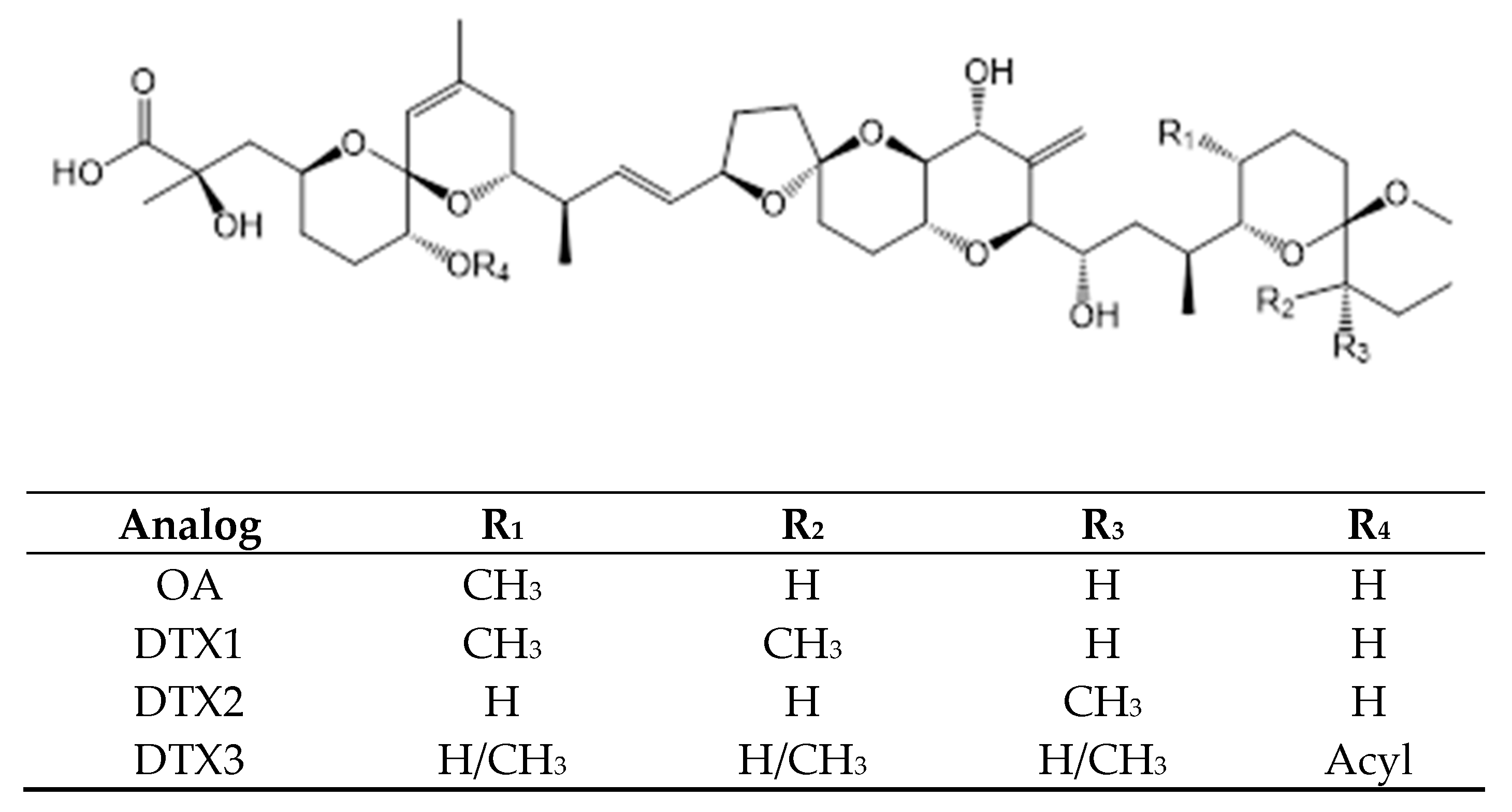
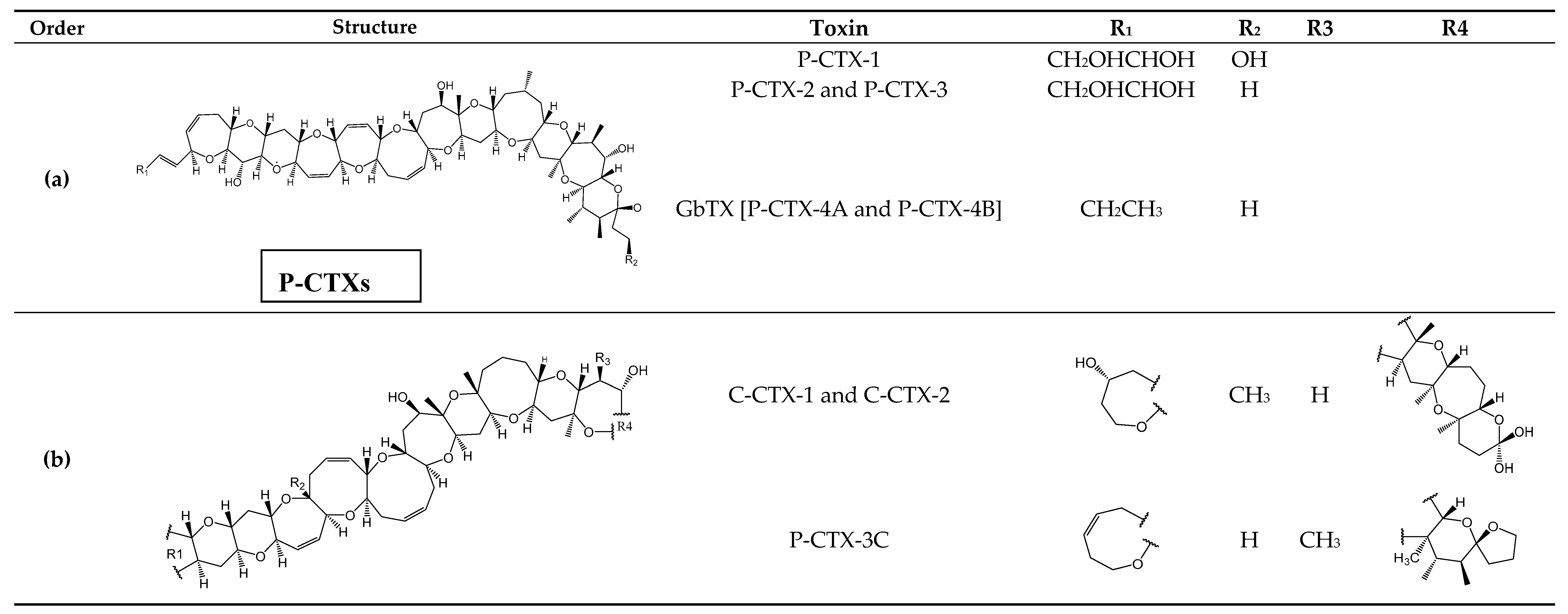
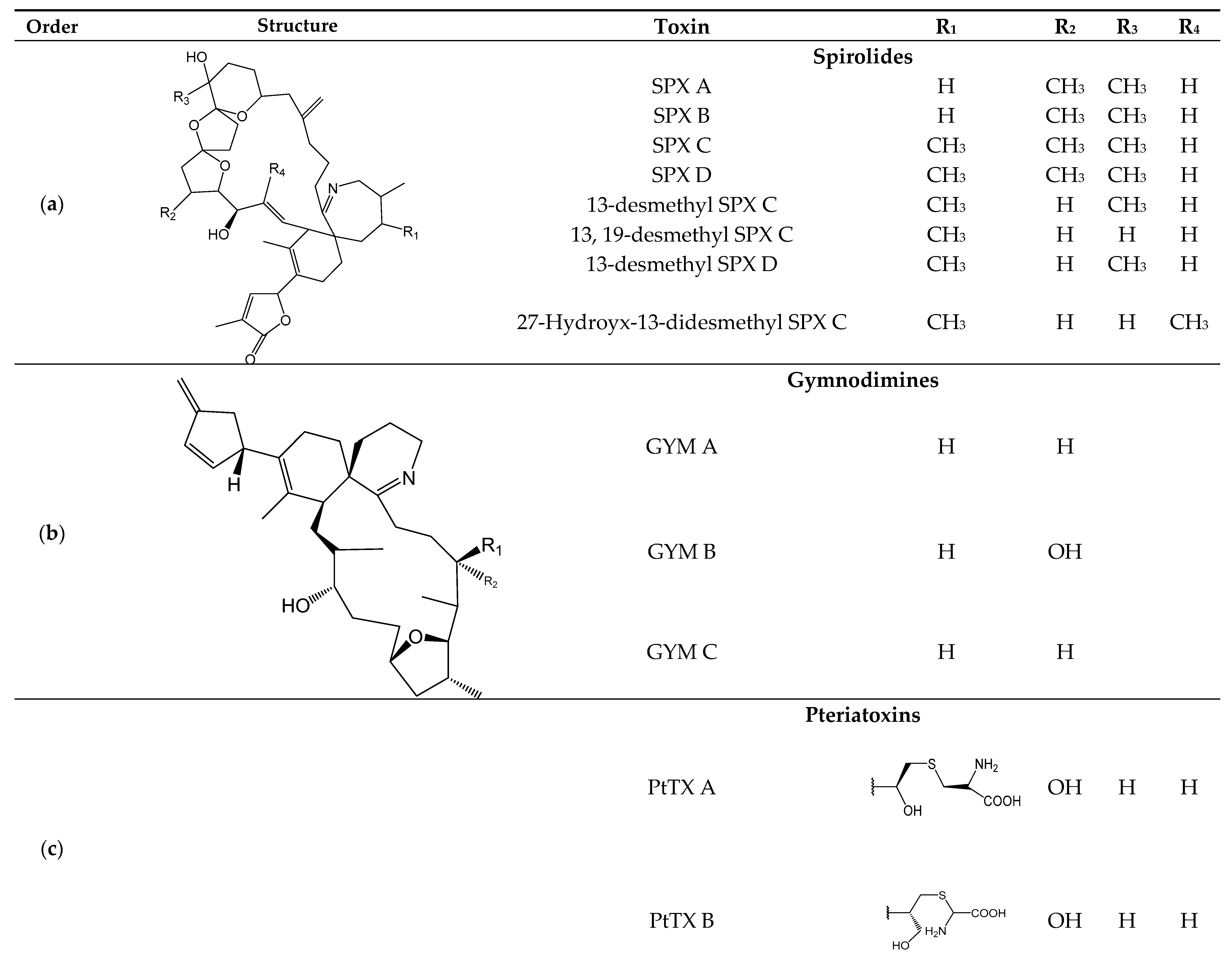
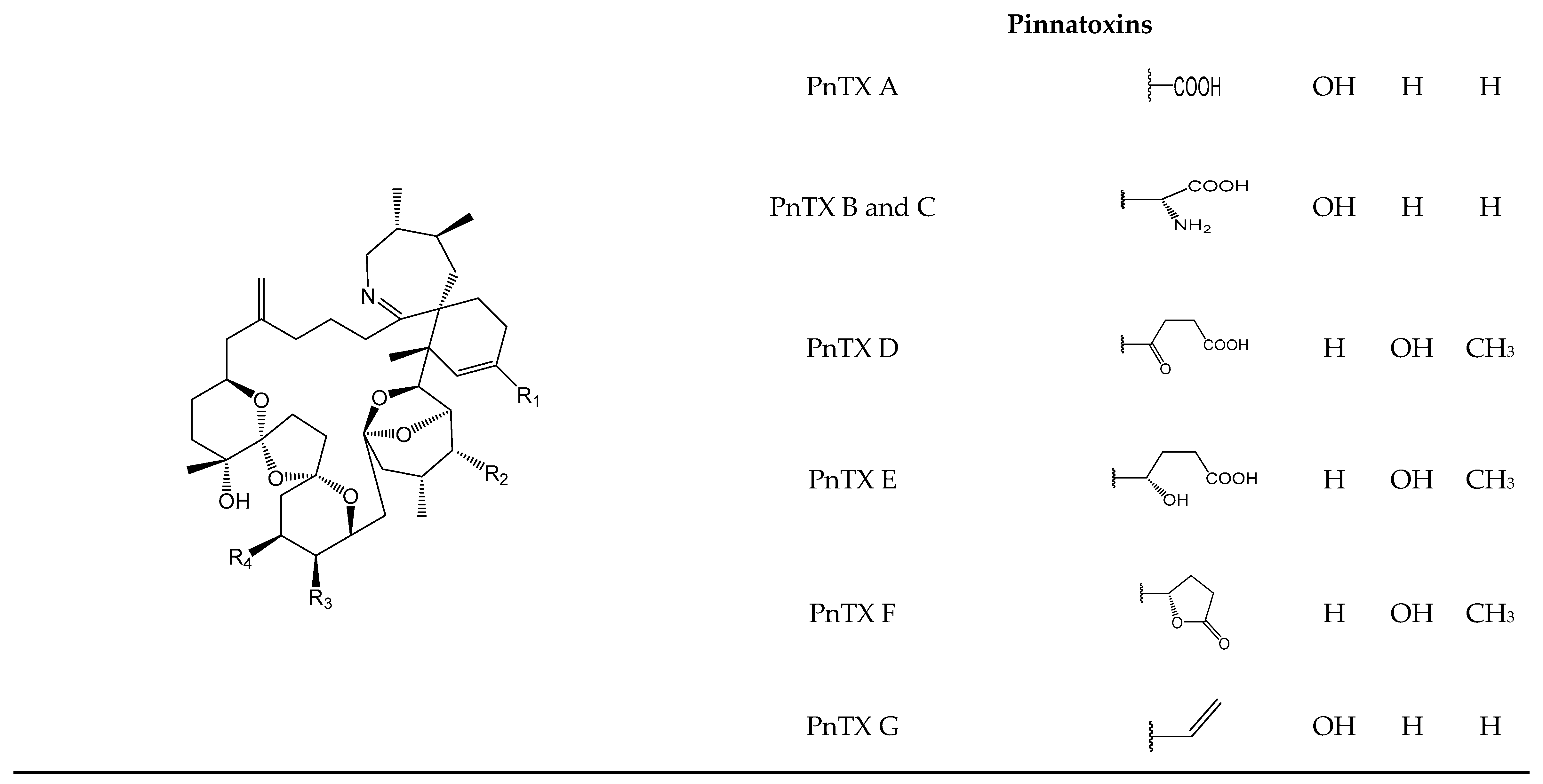
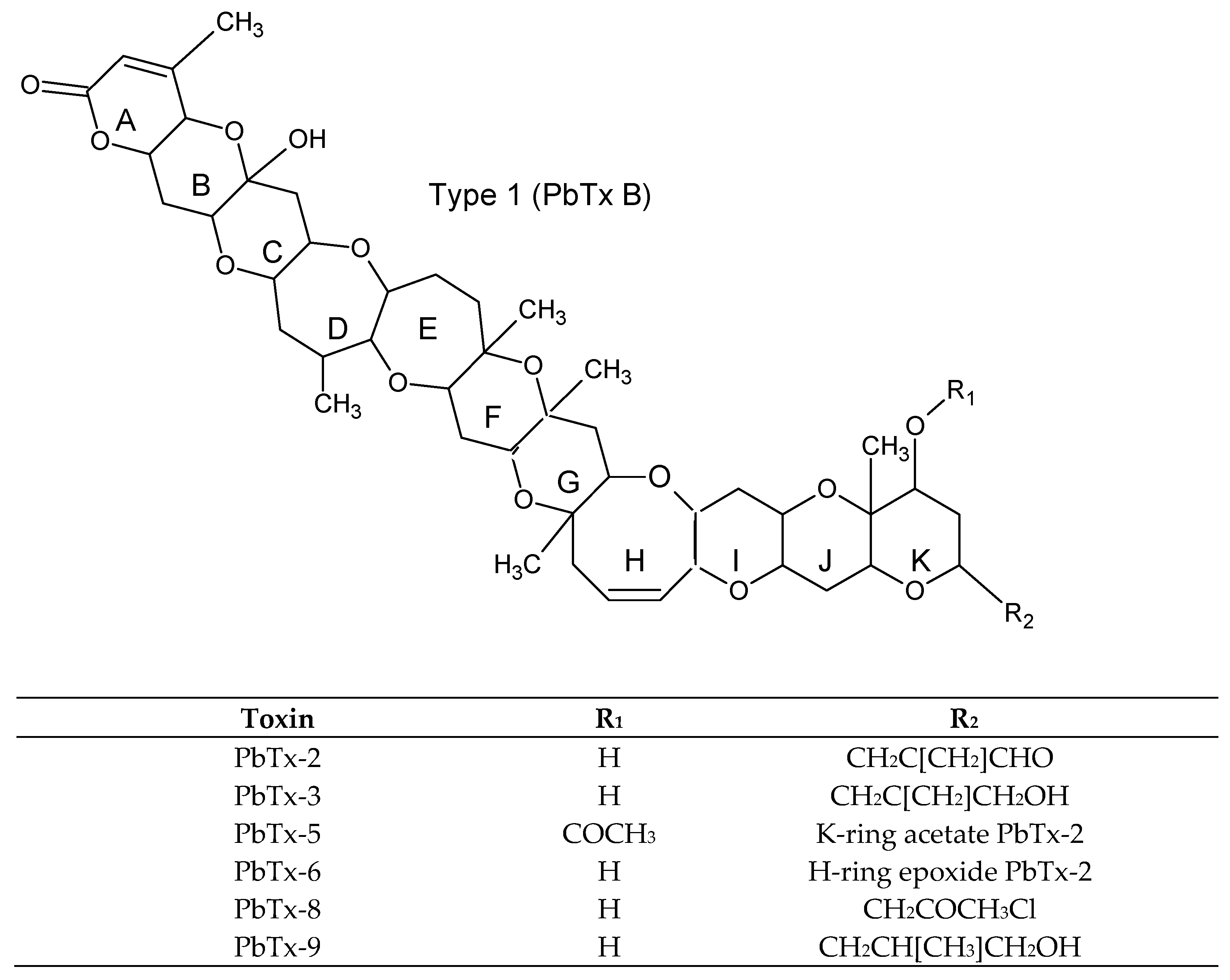
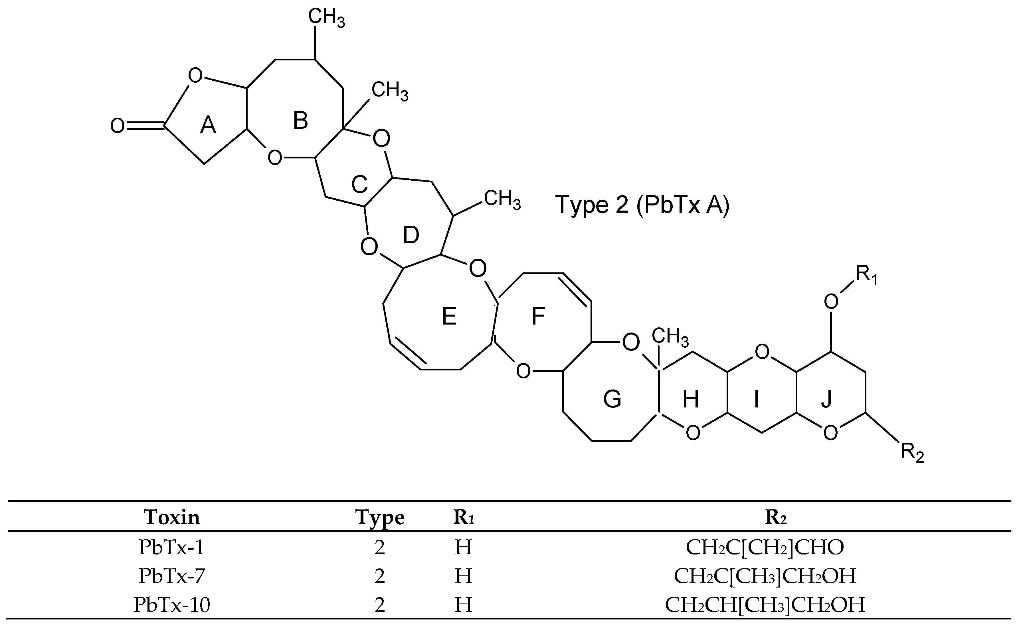
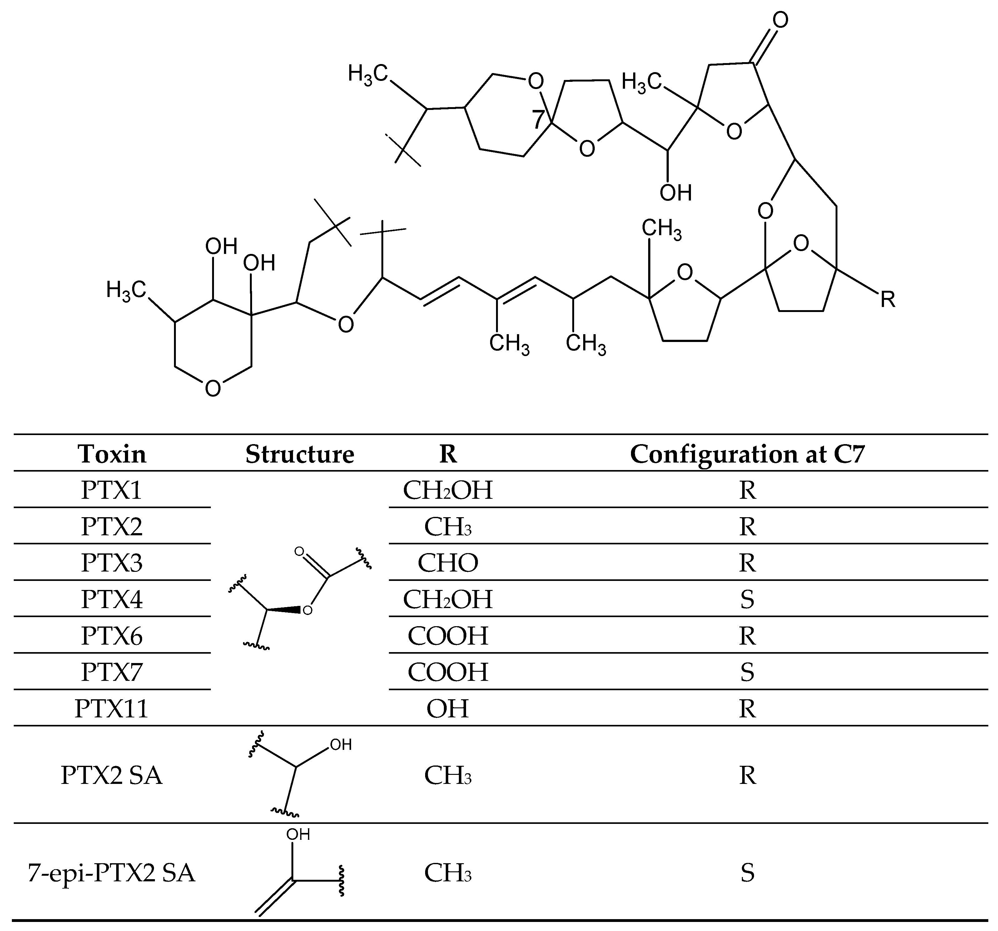
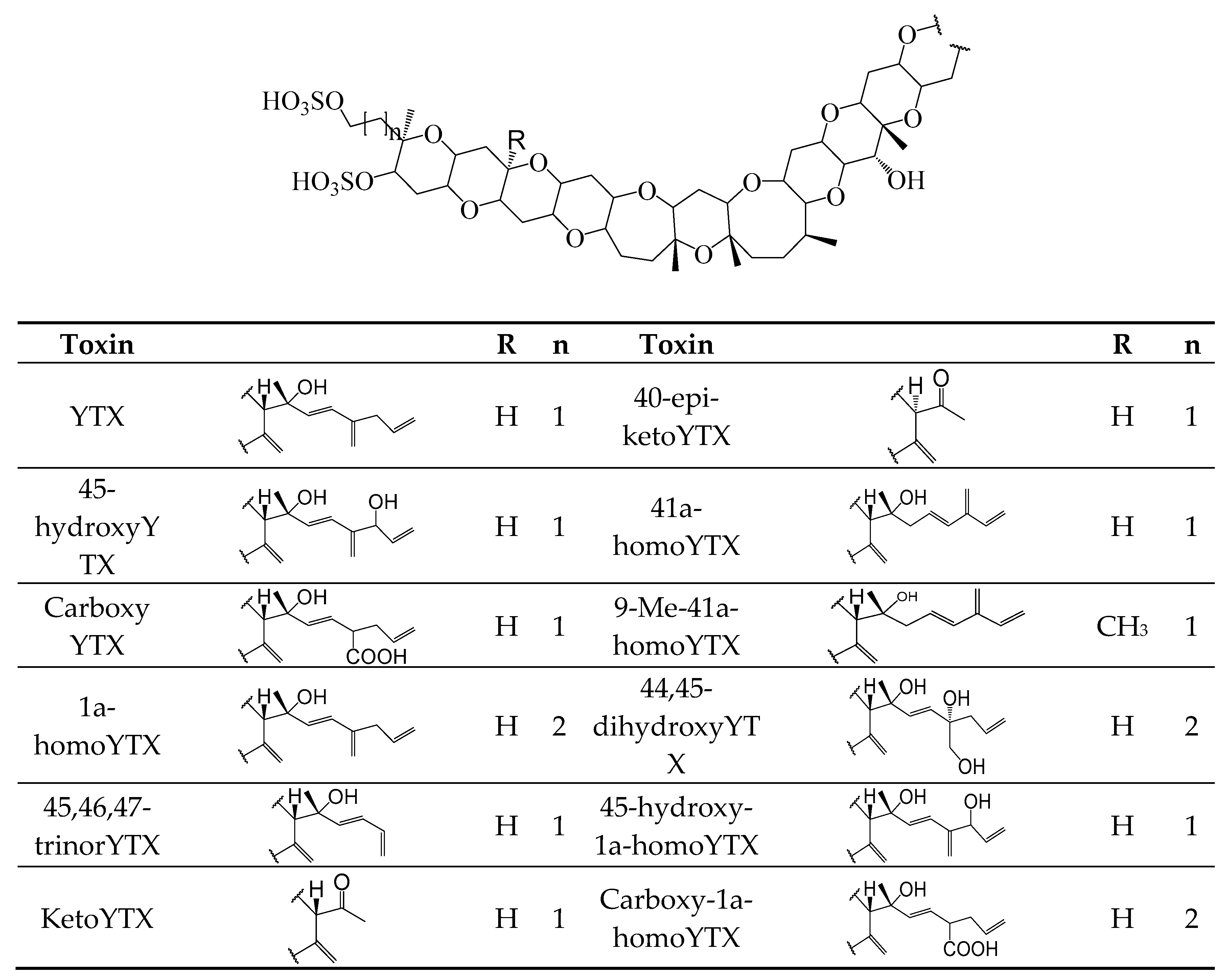
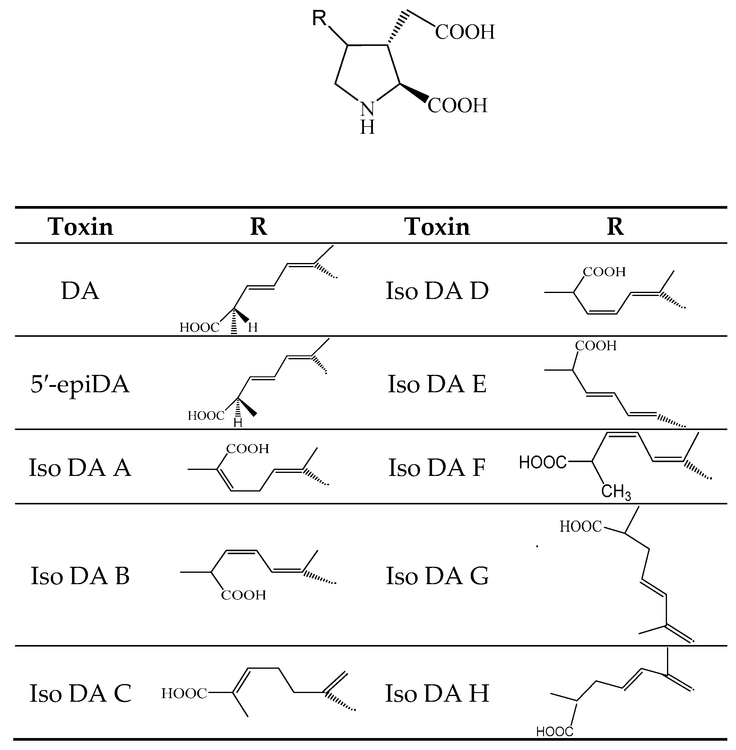
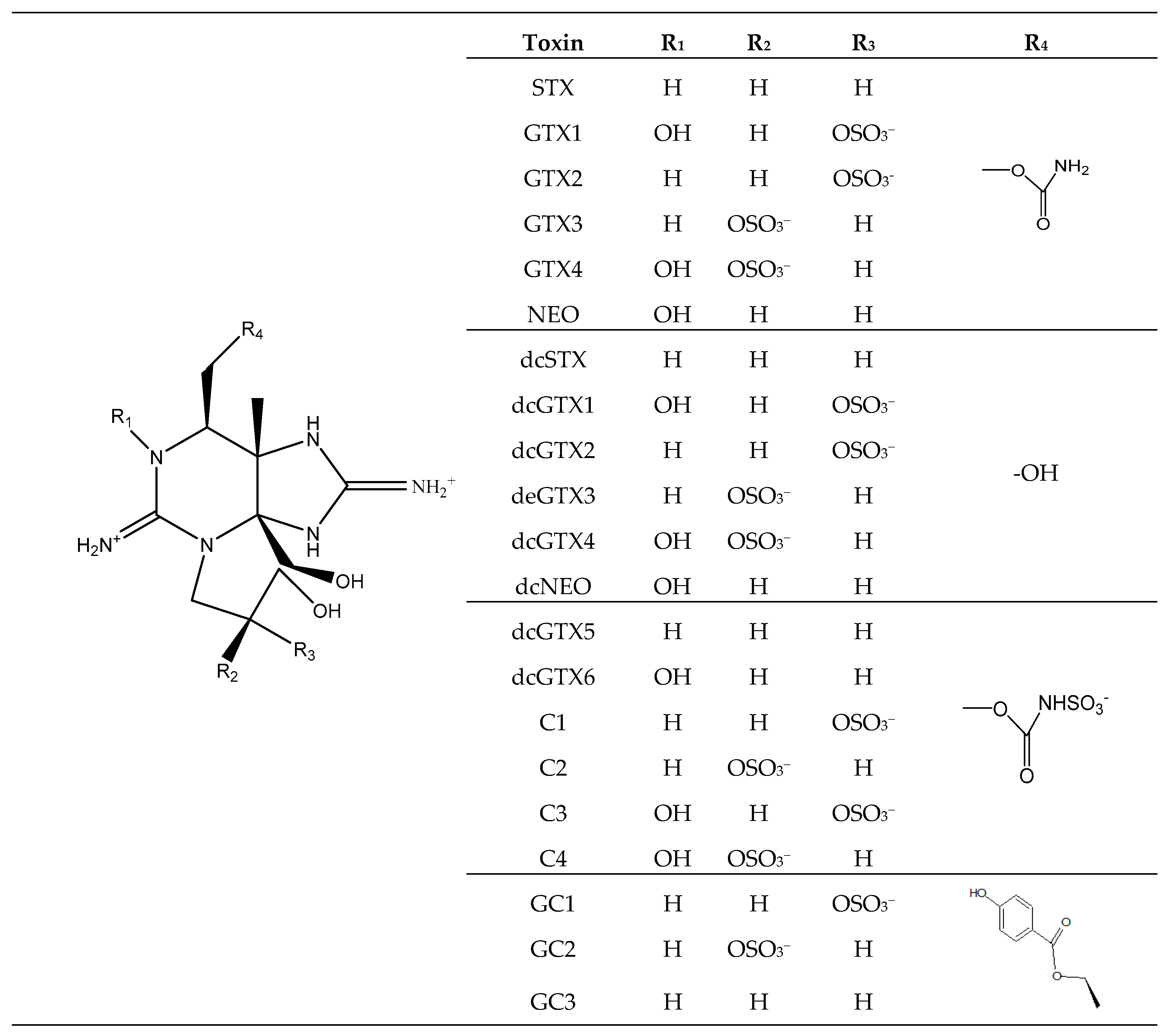
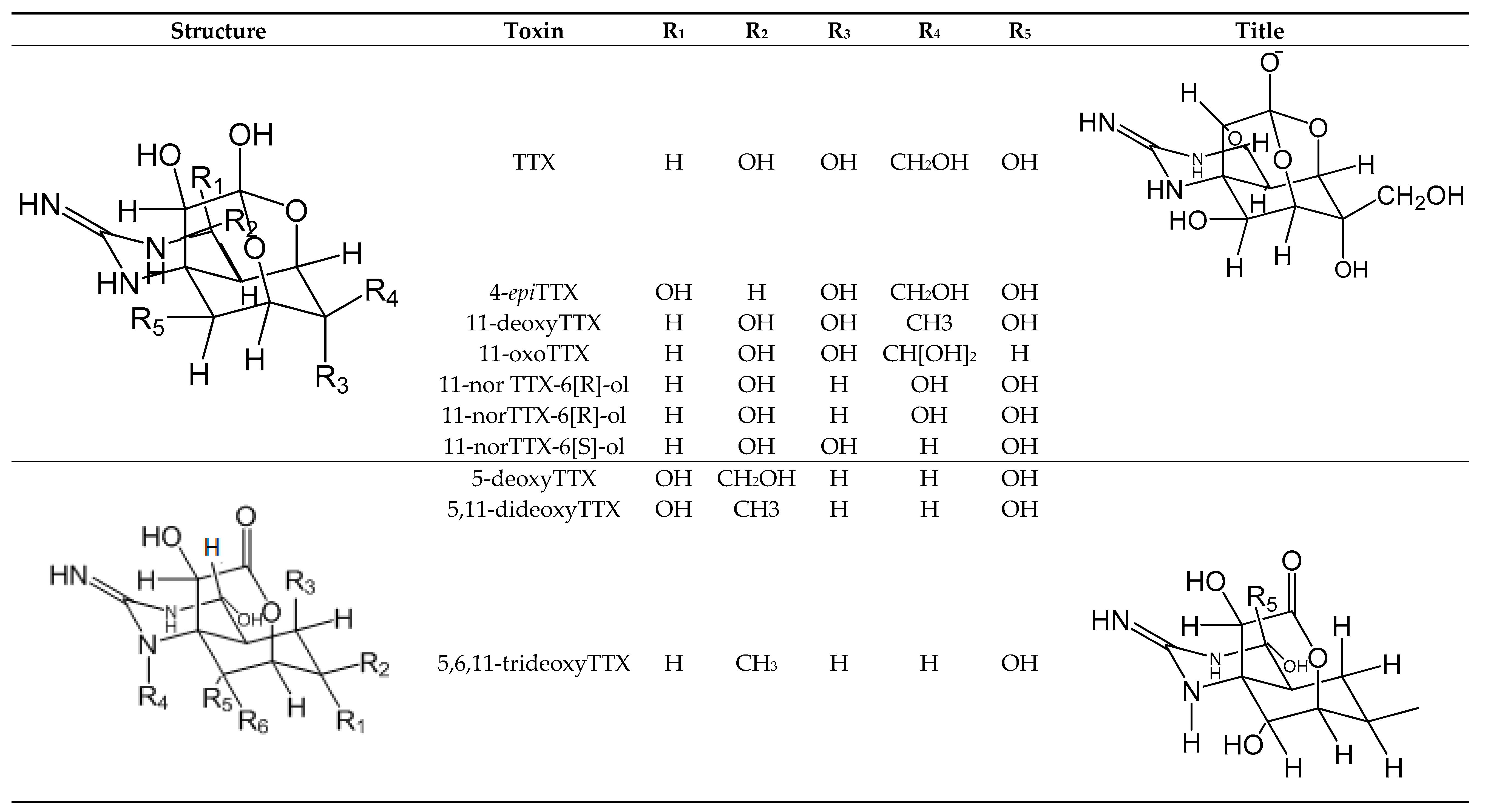
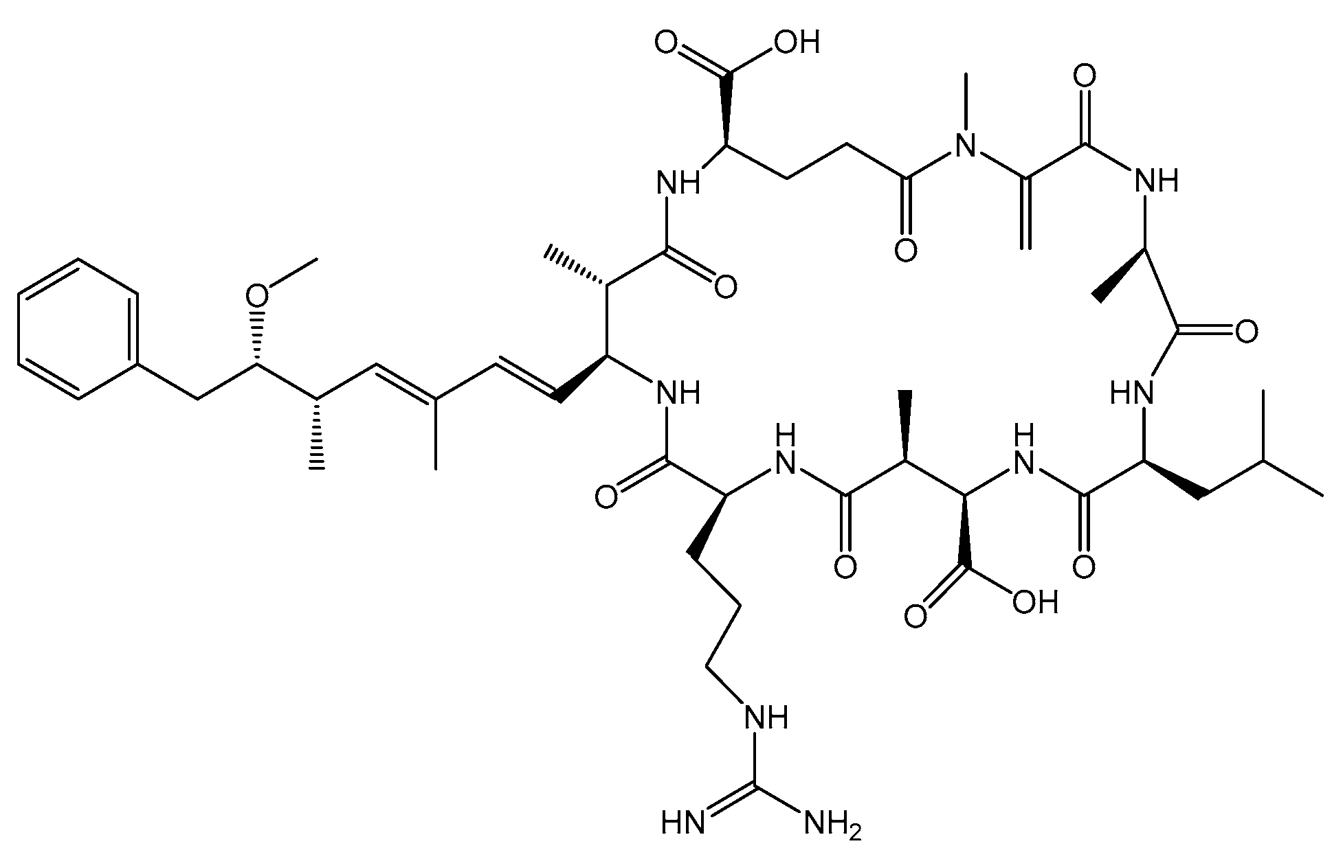
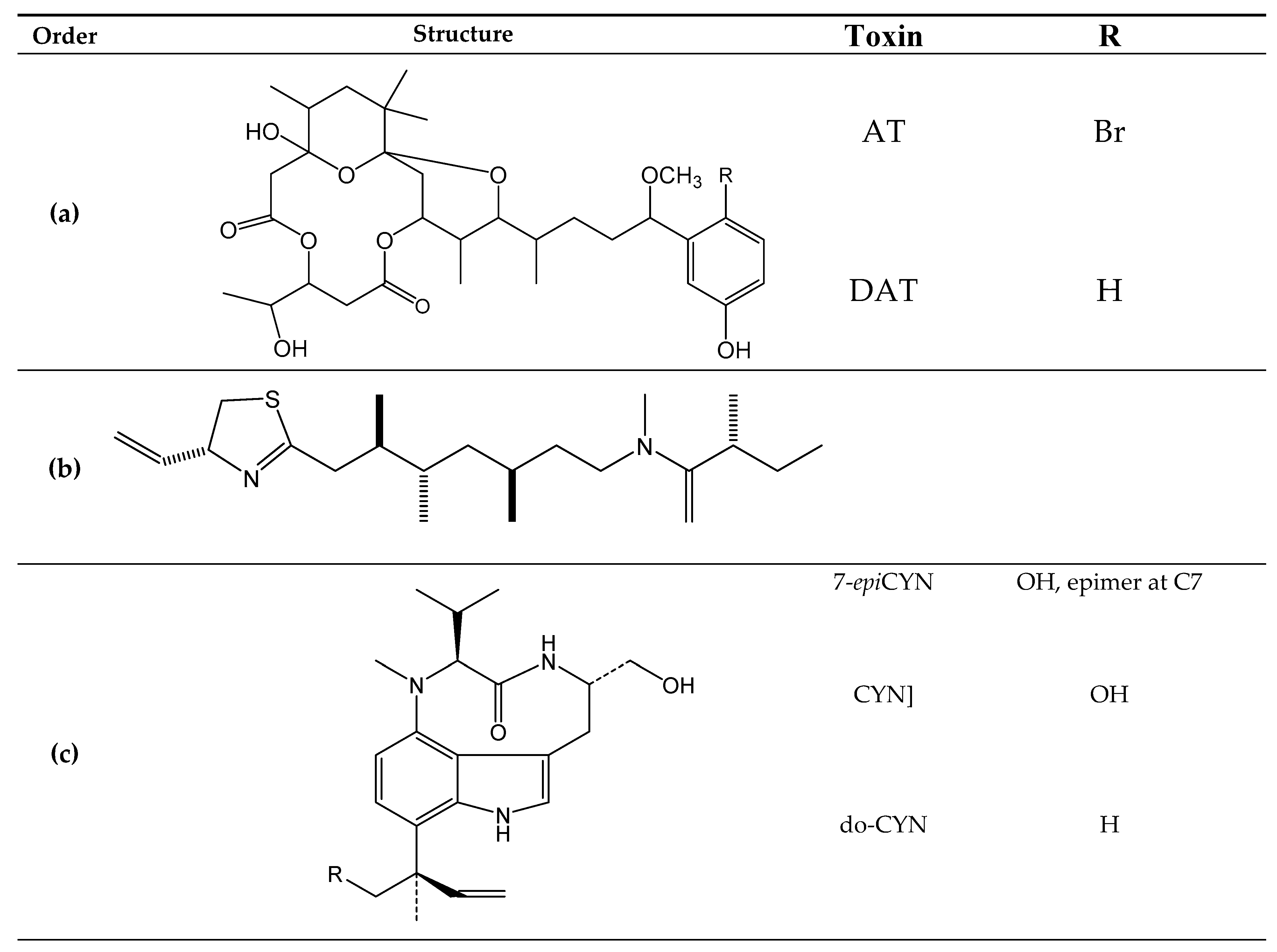
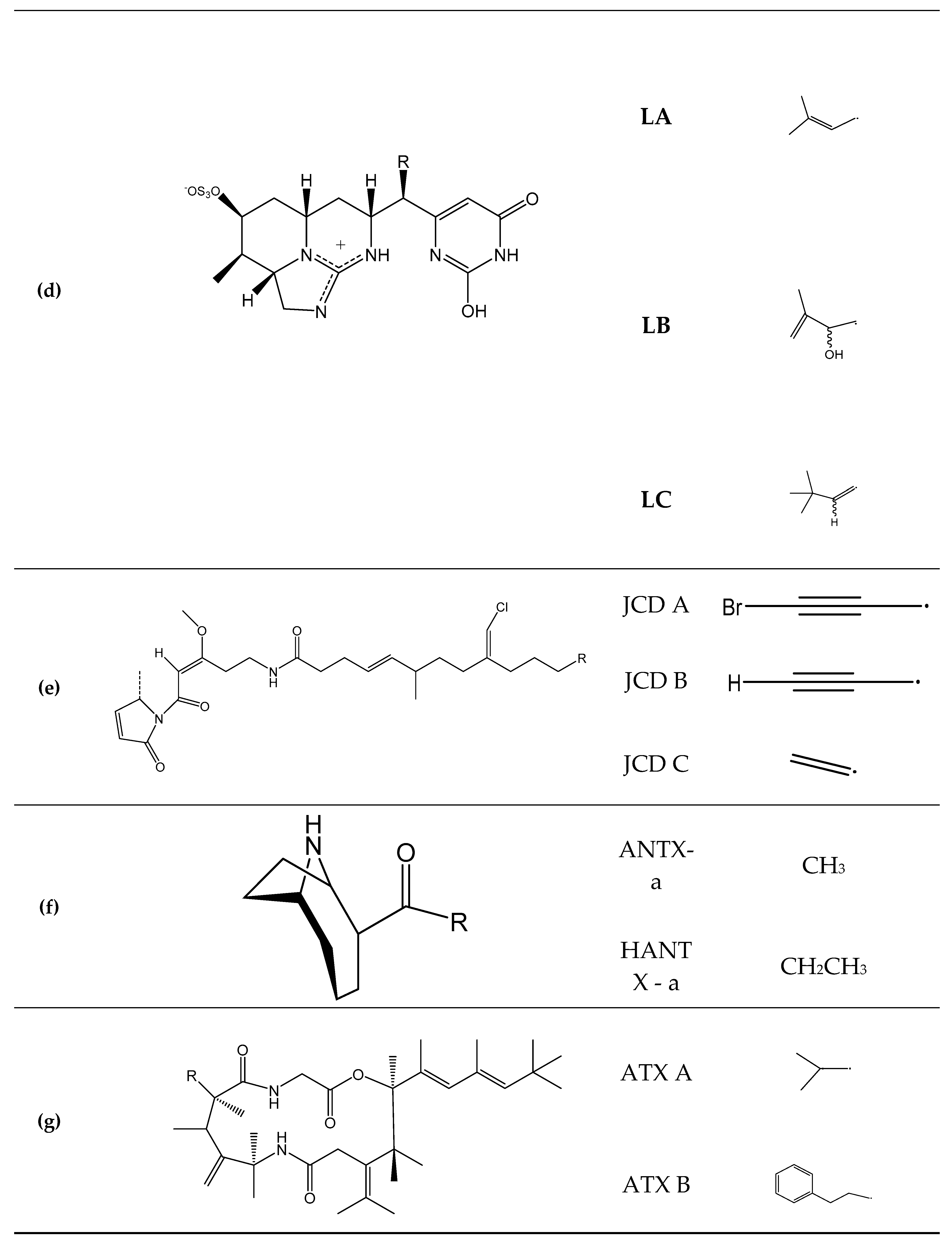
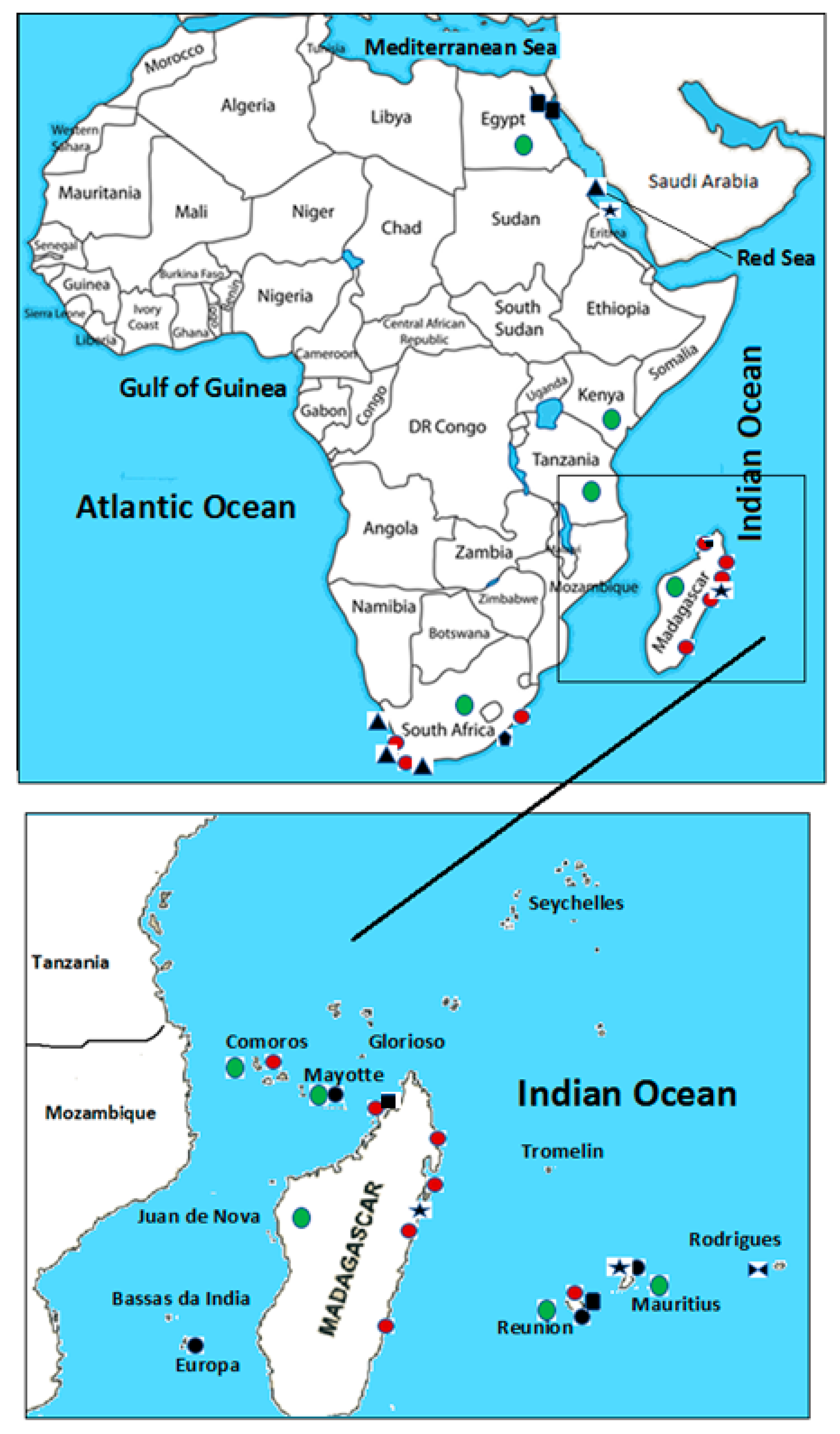
| Toxin (Syndrome) | Symptoms | Detection | Permitted Limit | Toxin (TEF) | Producer | ||
|---|---|---|---|---|---|---|---|
| Methods | LOD, μgKg−1 | LOQ, μgKg−1 | |||||
| OA and analogs (DSP) | diarrhea, nausea, vomiting, abdominal pain and tumor formation in the digestive system [50] | BA [180,181] | 160 | 0.16mg OA equivalents/Kg shellfish meat in EU region [182] | OA[1.0] | Dinoflagellates: Prorocentrum spp. [8], Dinophysis spp. [2,6,9,10,15,53,54] and Phalacroma rotundatum [55] | |
| DTX1[1.0] | |||||||
| EIA [183,184,185,186] | 10–26 | 3–41 | |||||
| DTX2 [0.6] | |||||||
| LC-MS [183], -UVD [187] | 15–30 | 1–50 | |||||
| DTX3 [1.0; 1; 0.6] | |||||||
| CTXs and analogs (CFP) | vomiting, diarrhea, nausea, tingling, itching, hypotension, bradycardia. In extreme cases, death through respiratory failure in 30 min and 48 h after fish consumption [50] | BA [188,189] | 0.16–0.560 P-CTX [190] | 0.01 μg P-CTX-1 equivalents/kg of fish in USA [191] | P-CTX-1[1.0] | Dinoflagellates: Gambierdiscus toxicus, Ostreopsis siamensis and Prorocentrum lima [59] | |
| CTA [192,193,194] | ~106 - 0.039 C-CTX | P-CTX-2[0.3] | |||||
| 2,3-dihydroxy P-CTX-3C[1.0] | |||||||
| EIA [72,189,195,196,197,198,199] | -0.032 P-CTX | ||||||
| LC-MS/MS [67,70,71,74,200], -UVD [62,201,202] | C-CTX-1[0.1] | ||||||
| CIs | non-specific symptoms such as gastric distress and tachycardia in humans [82] | BA | 5.6–77 PnTXE | Not regulated | 13-desmethyl SPX C[1.0] | Dinoflagellates: SPXs: Alexandrium spp. [1,76], GYMs: Gymnodium spp. [77], PnTXs: Vulcanodinium rugosum [78] and PtTXs: biotransformation from PnTXs via metabolic and hydrolytic transformation in shellfish [1,5,77,78,79] | |
| FPA [203] | 80–85 13-SPXC | ||||||
| LC-MS/MS [79,204], - UVD [205] | 0.8–20 13-SPXC/GYMA | ||||||
| PbTxs and analogs (NSP) | nausea, vomiting, diarrhea, paresthesia, cramps, bronchoconstriction, paralysis, seizures in 30 min to 3 h [87] | BA [206] | 800 μg BTX-2 equivalents/kg shellfish in USA [98], New Zealand, and Australia [99,100] | BTX-2, BTX-3, BTX2-B2 and S-deoxy-BTX-B2 [same TEF] | Dinoflagellate: Karenia spp. [4,16,87] | ||
| CTA [192] | 250 BTX-1 | ||||||
| RB [108] | 30BTX-3 | ||||||
| EIA [207,208] | 1 BTXs and | 25 BTXs | |||||
| LC – MS/MS [209] | 0.2 – 2 BTXs | ||||||
| PTX and analogs | No specific symptoms | MBA | - | 160 µg OA equivalents./kg shellfish meat in EU region [210] | PTX [1,2,3,4,6 and 11][1.0] | Dinoflagellate: Dinophysis acuta [101] | |
| EIA [207] | - | ||||||
| PTX [7,8,9 and 2SA] and 7-epiPTX2 SA [<<10] | |||||||
| LC – MS/MS [211,212] | 1 | ||||||
| YTX and analogs | No specific symptoms | BA | 3.75 mg YTX equivalents/Kg shellfish meat in EU region [124] | YTX[1.0] | Dinoflagellate: Protoceratium reticuatum [4,109], Lingulodinium polyedrum [4] and Gonyaulax polyhedral [4] | ||
| EIA [213] | 1a-homoYTX[1.0] | ||||||
| 45-hydroxyYTX[1.0] | |||||||
| LC-MS/MS [111] | 0.017 | ||||||
| 45-hydroxy-1a-homoYTX[0.5] | |||||||
| AZA and analogs (AZP) | nausea, vomiting, diarrhea and decreased reaction to stomach cramps, deep pain, dizziness, hallucinations, confusion, short-term memory loss, seizure [214] | BA [181] | 0.05 | 0.16 mg AZA1equivalents/Kg shellfish in EU region [210] | AZA1[1.0] | Dinoflagellates: Azadinium spinosum [117] and Protoperidinum crassipes [118] | |
| AZA2[1.8] | |||||||
| LC-MS/MS | |||||||
| AZA3[1.4] | |||||||
| AZA4[0.4] | |||||||
| AZA5[0.2] | |||||||
| STX and analogs (PSP) | Numbness in the face and neck; headache, dizziness, nausea, vomiting, diarrhea, muscular paralysis; pronounced respiratory difficulty; death through respiratory paralysis [215] | BA [216,217] | 0.8 mg STX equivalent/Kg shellfish in EU region [210] | STX[1.0] | Dinoflagellates: Alexandrium spp. [2,3,7], Gymnodinium catenatum [3], Pyrodinium bahamense [3] and cyanobacteria Trichodesmium erythraeum [131] | ||
| NSTX[1.0] | |||||||
| SBA [218] | GTX1[1.0] | ||||||
| GTX2[0.4] | |||||||
| GTX3[0.6] | |||||||
| CTA [192,219] | GTX4[0.7] | ||||||
| GTX5[0.1] | |||||||
| Antibodies Assay [220,221,222,223,224] | GTX[0.1] | ||||||
| C2[0.1] | |||||||
| Eletrophoresis [225] | C4[0.1] | ||||||
| de-STX[1.0] | |||||||
| LC-MS/MS [226,227,228,229] | 23–42 STX | de-GTX3[0.2] | |||||
| de-NSTX2[0.2] | |||||||
| de-GTX3[0.4] | |||||||
| 11-hydroxy-STX[0.3] | |||||||
| DA and analogs (ASP) | nausea, vomiting, diarrhea or abdominal cramps] within 24 h of consuming DA contaminated shellfish and/or neurological symptoms or signs [confusion, loss of memory or other serious signs such as seizure or coma] occurring within 48 h | BA [230] | 40 | 20 mg DA equivalents/Kg shellfish in EU region [210] | Diatoms: Pseudo-nitzschia spp. [126] and red algae: Chondria armata [127]. | ||
| (a) ASP- EIA [184,231] | 0.003 | 0.01 | |||||
| SPR [232] | 20 | ||||||
| RB [233,234,235] | 20 | ||||||
| Capillary electrophoresis [236,237,238] | 0.15 -1 | ||||||
| LC -MS/MS [211,239,240], UVD [241,242] | 0.015 | ||||||
| TLC [243] | 10 | ||||||
| TTX and analogs | Vomiting, strong headache, muscle weakness, respiratory failure, hypotension and even death in hours [244] | BA [144,245,246,247] | 1.1 [247] | 2 mg TTX equivalents/Kg shellfish in Japan [248] | S/R 11-norTTX-[6]-ol[0.19/0.17] | Bacteria: Serratia marcescens, Vibrio spp. [83], V. Aeromonas sp. [138], Microbacterium, arabinogalactanolyticum [139], Pseudomonas sp. [140], Shewanella putrefaciens [141], Alteromonas sp. [142], Pseudoalteromonas sp. [143], and Nocardiopsis dassonvillei [144] | |
| RB [249] | 2–4.10−3TTX | ||||||
| 4-epiTTX[0.16] | |||||||
| EIA [245,246,247,250,251,252,253,254,255,256] | 0.002/mL [255], 0. 0001/mL [253] | ||||||
| TLC [139,257] | 2 [257] | 4,9-anhydroTTX[0.02] | |||||
| GC-MS [28,258,259] | 500 | 1000 [258] | |||||
| 5,6,11-deoxyTTX[0.01] | |||||||
| LC-MS/MS [260,261,262,263,264] – FLD [265] | 0. 00009?-24.5 [260,261,262,263,264] | 40 [265] – 100 [265] | |||||
| PlTX | Vasoconstriction, hemorrhage, myalgia, ataxia, muscle weakness, ventricular fibrillation, ischemia and death [266,267] and rhabdomyolysis [268] | BA | Not regulated toxin but proposed value is 0.25mg PlTX equivalent/Kg shellfish in EU region [269] | PlTX[1.0] | Zoanthids: Palythoa spp. anddinoflagellates: Ostreopsis ovata. [153,154,155] and possibly cyanobacteria: Trichodesmium sp. [156] | ||
| Hemolysis assay [270] | 1.6 | ||||||
| CTA [107] | 50 | ||||||
| streocin-D[0.4–1.0] | |||||||
| EIA [254] | 1/mL | ||||||
| LC-MS/MS [204,271]–FLD and–UVD [272] | 2,5.10−5–0, 50.10−5 | ||||||
| MC | liver hemorrhage within a few hours of an acute dose and death [273] | LC-MS [167,274,275,276] and EIA [277] | Tolerable daily intake: 0.04 μg/kg of MC body weight/day [278] | Cyanobacteriaof genus: Pseudoanabaena, Phormidium, Spirilia [164], Leptolyngbya, Oscillatoria, Geitlerinema [165], Trichodesmium [166] and Synechococcus [167] | |||
| ANTX and HANTX | Hypersalivation, diarrhea, shaking and nasal mucus discharge [279], respiratory arrest and death [280] | RB and GC/MS [281,282] | Cyanobacteria: Hydrocoleum lyngbyaceum [177] | ||||
| AT and DAT | Contact dermal: dermatitis initiating with erythema and burning sensations, appearing a few hours after exposure, gave way to blister formation and deep desquamation, lasting up to several days [283,284] and consumption of contaminated seafood; burningsensation in the mouth and throat, vomiting and diarrhea [285] | LC-MS/MS [286] | Algae Gracilaria coronopifolia [172] and cyanobacteria Lyngbya majuscula [171] | ||||
| LA, LB, and LC | Cyanobacteria Lyngbya majuscule [174] | ||||||
| ATX and analogs | No specific symptoms | LC [287] | Cyanobacteria: Lyngbya majuscula [179] | ||||
| JCD and analogs | No specific symptoms | LC, TLC and [288] | Cyanobacteria: Lyngbya majuscula [176] | ||||
| KTX and analogs | No specific symptoms | LC [173] | Cyanobacteria: Lyngbya majuscula [173] | ||||
| CYN and analogs | Gastroenteritis [289] | LC-MS/MS [290],–PDAD [291] | 1 [292]–200 [293] | Cyanobacteria: Cylindrospermopsis raciborskii [175] | |||
| EIA [294] | |||||||
| Country | Monitored MT | Permitted Limit, mgKg−1 Shellfish | Detection | Laboratories for Toxin Analysis | Reference |
|---|---|---|---|---|---|
| South Africa | PST | 0.8 STX | Research centers and Universities | [324] | |
| OA, DTX1-2, PTX1-2 | 0.16 mg OA | LC-MS/MS | |||
| YTX, 45 OH YTX, homo YTX, and 45 OH homo YTX | 8 mg YTX | LC-MS/MS | |||
| AST | 20 mg DA | ||||
| AZA1-3 | 0.16 mg OA | LC-MS/MS | |||
| Mozambique | N.D. | N.D. | N.D. | N.D. | N.D. |
| Tanzania | CTX, TTX, AST | N.D. | Symptomology and vectors | N.D. | [325] |
| Kenya | MT producers [HAB] | N.D. | N.D. | Mombasa Research Center | [326] |
| Madagascar | N.D. | N.D. | Educational programmes | Researches centers and Universities | [327] |
| French Islands | N.D. | N.D. | N.D. | Researches centers | [35,328] |
| Mauritius | N.D. | N.D. | N.D. | [324] | |
| Comoros | N.D. | N.D. | N.D. | N.D. | |
| Somalia and Seychelles | N.D. | N.D. | N.D. | N.D. | |
| Eygpt | N.D. | N.D. | N.D. | Poison Control Center, Ain Shams University | [329,330] |
| Djibouti | N.D. | N.D. | N.D. | N.D. | |
| Eritrea | N.D. | N.D. | N.D. | N.D. | |
| Sudan | N.D. | N.D. | N.D. | N.D. |
| Toxin | Date | Location | Toxin Producer | Determination Method | Toxin Vector | TX Concentration, (mg TX Equivalents per Kg Shellfish Meat) | Cell/Extract Toxicity | Reference |
|---|---|---|---|---|---|---|---|---|
| PSTs | 1999 | South Africa | Alexandrium catenella | AOAC mouse bioassay | Haliotis midae | 0. 01609 STX | [22] | |
| 1998–2002 | South Africa: Yzerfontein, | Alexandrium catenella | HPLC-FLD | - | - | 4.8 pg STX eq cell−1 | [334] | |
| Alexandrium tamiyavanichi | 0.14 pg STX eq cell−1 | |||||||
| 2003–2004 | South Africa: Cape Town | Alexandrium minutum | LC-FD and HILIC-MS/MS | - | - | 0.65 pg GTX cell−1 | [309] | |
| 2012–2014 | Central Red Sea | Pyrodinium bahamense, Ceratium sp., Alexandrium sp. and Protoperidinium spp. | ELISA | - | - | >> 0.4 ng mL1 | [349] | |
| DSTs | 2000 | Europa Island Mozambic channel, France] | Prorocentrum arenarium | FR3T3 fibroblast | - | - | IC50 = 0,1 µg OA ml−1 and 50 µg extract ml−1 | [11] |
| PPIA | ||||||||
| HPLC-FD | ||||||||
| HPLC-MS | 22 ng OA/mg of extract | |||||||
| 2001 | Lagoons of La Reunion Mayotte and Mauritius Islands | Prorocentrum lima | PPIA | - | - | IC50 1.3–25 mg/mL onon fibroblast;6261.3 ± 156.5 − 128.3±17.2 ng eq OA/mg crudeextract | [328] | |
| 2002–2018 | South Africa:Abalgold | - | - | Haliotis asinina | - | - | [324] | |
| 2008 | South Africa: Saldanha Bay and Lambert’s Bay | Dinophysis acuminata | LC-MS/MS | Crassostrea gigas | 0.267 OA | |||
| Choromytilus meridionalis | 0.012 OA | |||||||
| CTXs | 2001 | Mauritius: Nazareth, Saya de Malha and Soudan | - | HPLC-MS/RLB, Mongoose feeding test, and MBA | Lutjanus sebae and Lutjanus Lab | Qualitative analysis | - | [71] |
| 2002 | North of the Republic of Mauritius, Banks fishery | - | HPLC-MS/RLB | Lutjanus sebae | - | [70] | ||
| 2012–2013 | Central Red Sea | Gambierdiscus belizeanus and Ostreopsis spp. | Mouse neuroblastoma cell-based assay | - | - | 6,50–1,14.10 −5 pg P-CTX−1 eq. cell−1 | [350] | |
| 2013 | Madagascar: district of Fenoarivo Atsinanana | Gambierdiscus spp. | CBA | Carcharhinus leucas | 0.083 P-CTX-1 | - | [20] | |
| MBA | 0. 09272 P-CTX-1 | |||||||
| LC-ESI-HRMS | 0. 01628 P-CTX-1 | |||||||
| MBA | 752 MU/g | |||||||
| PlTXs | 1994 | Madagascar: Antalaha District | Ostreopsis siamensis | MBA | Herklotsichthys quadrimaculatus | 0. 00045 PTXs/fish [head and esophagus] | [18] | |
| Hemolysis assays | 0. 00002 PTXs/fish [head and esophagus] | |||||||
| Cytotoxicity tests | 0. 00000005/fish [head and esophagus] | |||||||
| MS | ||||||||
| 1996 | Mauritius: Rodrigues Island | Ostreopsis mascarenensis | HPLC-diode array detector, Nanoelectrospray ionization quadrupole time-of-flight and HPLC-ESI-MS/MS analysis | - | - | [14,160] | ||
| Hemolysis assays | 8.00 ± 0.01 ng PTX mL−1 | |||||||
| Cytotoxicity Assay | IC50 = 10 μM against human H460 lung cancer cells | |||||||
| 2008 | South Africa: Saldanha Bay and Lambert’s Bay | Dinophysis acuminata | LC-MS/MS | Crassostrea gigas | 0.267 OA | |||
| Choromytilus meridionalis | 0.012 OA | |||||||
| DA cultures | 2012 | South Africa: Algoa Bay | Pseudo-nitzschia multiseries | ELISA | - | - | 0.076 pg DA cell−1–0.098 pg DA cell–1 | [12] |
| LC/MS–MS | 0.086 pg DA cell–1–0.086 pg DA cell–1 | |||||||
| TTXs | 1990–1991 | Egypt: Suez City, in the northwestern part of the Red Sea | TLC, electrophoresis, UV, GC–MS | Pleuranacanthus sceleratus | [316] | |||
| 752 MU/g | ||||||||
| MBA | ||||||||
| 1998 | Madagascar: Nosy Be Island - | - | MBA | 16 MU/g | [41] | |||
| 2002–2003 | Egypt: Gulf of Suez | MBA | Lagocephalus sceleratus | 3950 MU/g | [351] | |||
| 2013 | Reunion Island | MBA and LC-MS/MS | Lagocephalus sceleratus | 17 TTX | - | [35] |
| Local | Date | Seafood | Observed Effects/Symptoms | TX | Detection Method | TX Concentration, (mg TX Equivalents/Kg Shellfish Meat) | Victim Number | Reference |
|---|---|---|---|---|---|---|---|---|
| Comoros islands: Ndrondroni | 24 December 2012 | Eretmochelys imbricata (turtle) | Itching, Asthenia, Vomiting, Abdominal pain, Rash Myalgia Shortness of breath, Nausea Itching of the mouth/throat, Fever, Diarrhea Vertigo, Paresthesia, Dysphagia Mouth burn Sore throat, Erectile dysfunction | - | - | - | 49 suspected cases and 8 probable cases, age range [0–40 years], 1 death | [26] |
| North-eastern coast of Madagascar | December 1994 | Turtle | Nausea, vomiting, dysphagia, acute stomatitis | - | - | - | 60 persons with poisoning attack rate were 48% with a lethality of 7.7% | [47], |
| Madagascar: district of Fenoarivo Atsinanana | November 2013 | Carcharhinus leucas (shark) | Paresthesia of the extremities, dysesthesia, and reversing sensitivity of hot and cold accompanied by a headache, dizziness, and arthralgia between 2 and 12h after ingestion | CTXs | MBA | 0.083 P-CTX-1 | 124 people, 9% deaths | [20] |
| CBA | 0. 09272 P-CTX-1 | |||||||
| Madagascar: Antalaha District | January 1994 | Herklotsichthys quadrimaculatus (Fish) | Malaise, uncontrollable vomiting, diarrhea, tinglings of extremities, delirium and death | PlTXs | MBA | 0. 00045 PTXs/fish [head and esophagus] | Death of one adult | [18] |
| Hemolysis assays | 0. 00002 PTXs/fish (head and esophagus) | |||||||
| Cytotoxicity tests | 0. 00000005/fish (head and esophagus) | |||||||
| Mass spectroscopy | - | |||||||
| Madagascar: Nosy Be Island | July 1998 | - | - | TTXs | MBA | 16 MU/g (no data to covert to mg/Kg) | 4 people, one death | [41] |
| Madagascar: Manakara district | November 1993 | Carcharhinus amboinensis [shark] | Deep coma and death, body rigidity due to loss of cerebral function, myosis, mydriasis, convulsions, Respiratory distress due to acute pulmonary edema, cardiovascular collapse, bradycardia, gengivorrhagia Dehydration, paresthesia on fingertips and toes, dizziness, pruritus, narcosis, faintness, hyperthermia, ataxia asthenia, dehydration, cephalalgia, diarrhea, epigastralgia, laryngeal distress | CTXs | Ciguatera poisoning Symptomology | - | 500 people, 20% deaths | [21] |
| South Africa: Cape Town | May 1978 | Choromytilus meridionlis [Mussel] | Paraesthesia of en fingers/hands, Circumoral paresthesia, paranesthesia of toes/feet, Vertigo, Floating sensation, Ataxia, Weakness of upper, Weakness of lower limbs and Dysarthria A headache | PSTs | MBA | 72.83 STX | 17 people, no deaths | [39] |
| South Africa: Natal coast | December 1957 | Mytilus meridionalis [Mussel] | peculiar lightness of the body, with a tingling around mouth, finger, and toes; no moving; feeble inarticulate noise; | PSTs | MBA | 0.04 STX | 5 people and one cat | [40] |
| South Africa: Table and False Bays | 1888 | Donax serra [Mussel] | - | - | - | - | - | [37] |
| South Africa: Cape Town | April 1948 | Donax serra [Mussel] and Chloromytilus meridionalis [Mussel] | - | - | - | - | One death | |
| South Africa: Natal coast | December 1957 | Perna perna [Mussel] | - | - | - | - | 5 people, one death | |
| South Africa: Cape Town a | May 1958 | Chloromytilus meridionalis [Mussel] | - | - | - | One death | ||
| Reunion Island | September 10th, 2013 | Lagocephalus sceleratus [fish] | peri-oral paresthesia, weakness of both lower limbs, paresthesia all over the body, headache, dyspnea, nausea and vomiting, blurring of vision, and vertigo | TTX | MBA | Liver: 17 TTX Flesh: 5 TTX | 10 people | [35] |
| Toxin | Syndrome | Permitted Limit, mgKg−1 | To be adopted from |
|---|---|---|---|
| STX | PSP | 0.8 STXeq | EU region |
| CTX | CFP | 0.00001 P-CTX-1eq | USA |
| YTX | - | 3.75 YTXeq | EU region |
| PTX | - | 0.16 OAeq | EU region |
| TTX | - | 2 TTeq | Japan |
| DA | ASP | 20 DAeq | EU region |
| OA | DSP | 0.16 OAeq | EU region |
| AZA | AZP | 0.16 AZAeq | EU region |
| PlTX | - | 0.25 PlTXeq * | EU region |
| PbTx | NSP | 0.8 TX-2 eq | USA, New Zealand, and Australia |
© 2019 by the authors. Licensee MDPI, Basel, Switzerland. This article is an open access article distributed under the terms and conditions of the Creative Commons Attribution (CC BY) license (http://creativecommons.org/licenses/by/4.0/).
Share and Cite
Tamele, I.J.; Silva, M.; Vasconcelos, V. The Incidence of Marine Toxins and the Associated Seafood Poisoning Episodes in the African Countries of the Indian Ocean and the Red Sea. Toxins 2019, 11, 58. https://0-doi-org.brum.beds.ac.uk/10.3390/toxins11010058
Tamele IJ, Silva M, Vasconcelos V. The Incidence of Marine Toxins and the Associated Seafood Poisoning Episodes in the African Countries of the Indian Ocean and the Red Sea. Toxins. 2019; 11(1):58. https://0-doi-org.brum.beds.ac.uk/10.3390/toxins11010058
Chicago/Turabian StyleTamele, Isidro José, Marisa Silva, and Vitor Vasconcelos. 2019. "The Incidence of Marine Toxins and the Associated Seafood Poisoning Episodes in the African Countries of the Indian Ocean and the Red Sea" Toxins 11, no. 1: 58. https://0-doi-org.brum.beds.ac.uk/10.3390/toxins11010058