Article

Fusarium graminearum FgCWM1 Encodes a Cell Wall Mannoprotein Conferring Sensitivity to Salicylic Acid and Virulence to Wheat

by
Ya-Zhou Zhang
1,2,†,
Qing Chen
2,†,
Cai-Hong Liu
2,
Lu Lei
2,
Yang Li
2,
Kan Zhao
2,
Mei-Qiao Wei
2,
Zhen-Ru Guo
2,
Yan Wang
2,
Bin-Jie Xu
2,
Yun-Feng Jiang
2,
Li Kong
2,
Yan-Lin Liu
2,
Xiu-Jin Lan
2,
Qian-Tao Jiang
1,2,
Jian Ma
2,
Ji-Rui Wang
1,2,
Guo-Yue Chen
1,2,
Yu-Ming Wei
1,2,*,
You-Liang Zheng
1,2 and
Peng-Fei Qi
2,*
add Show full author list remove Hide full author list
1
State Key Laboratory of Crop Gene Exploration and Utilization in Southwest China Sichuan Agricultural University, Chengdu 611130, Sichuan, China
2
Triticeae Research Institute, Sichuan Agricultural University, Chengdu 611130, Sichuan, China
*
Authors to whom correspondence should be addressed.
Contributed equally to this paper.
Submission received: 11 September 2019 / Revised: 23 October 2019 / Accepted: 24 October 2019 / Published: 29 October 2019
(This article belongs to the Special Issue Mycotoxins and Related Fungi in Crops)

Abstract

:
Fusarium graminearum causes Fusarium head blight (FHB), a devastating disease of wheat. Salicylic acid (SA) is involved in the resistance of wheat to F. graminearum. Cell wall mannoprotein (CWM) is known to trigger defense responses in plants, but its role in the pathogenicity of F. graminearum remains unclear. Here, we characterized FgCWM1 (FG05_11315), encoding a CWM in F. graminearum. FgCWM1 was highly expressed in wheat spikes by 24 h after initial inoculation and was upregulated by SA. Disruption of FgCWM1FgCWM1) reduced mannose and protein accumulation in the fungal cell wall, especially under SA treatment, and resulted in defective fungal cell walls, leading to increased fungal sensitivity to SA. The positive role of FgCWM1 in mannose and protein accumulation was confirmed by its expression in Saccharomyces cerevisiae. Compared with wild type (WT), ΔFgCWM1 exhibited reduced pathogenicity toward wheat, but it produced the same amount of deoxynivalenol both in culture and in spikes. Complementation of ΔFgCWM1 with FgCWM1 restored the WT phenotype. Localization analyses revealed that FgCWM1 was distributed on the cell wall, consistent with its structural role. Thus, FgCWM1 encodes a CWM protein that plays an important role in the cell wall integrity and pathogenicity of F. graminearum.
Key Contribution: We identified a CWM gene, FgCWM1, in Fusarium graminearum and clarified its contribution to fungal cell wall integrity and pathogenicity in F. graminearum.

1. Introduction

Common wheat (Triticum aestivum L.) is one of the most important cereal crops worldwide. Fusarium head blight (FHB) leads to dramatic yield losses and mycotoxin (deoxynivalenol, DON) contamination of wheat seeds, which threatens human and animal health [1,2]. The filamentous fungus Fusarium graminearum is the main causal agent of FHB in wheat [3]. Despite the major economic and health impacts of FHB, there is no efficient strategy to manage this disease, partly because we have not understood the biology of F. graminearum well [4,5].
The cell wall is an important component of fungi. It is not only involved in biofilm formation, cell wall biogenesis, and protection against environmental factors, but is also the first point of contact with the host. Thus, it plays an important role in fungi/host interactions [6,7,8]. Fungal cell walls have a layered structure [6,9,10] (Figure 1a), including the inner, electron-transparent layer and the outer, electron-dense layer. The outer, electron-dense layer is mainly composed of cell wall mannoproteins (CWMs), and CWMs play important roles in fungal vegetative growth and pathogenicity [11,12,13,14]. In Saccharomyces cerevisiae, the knock-out of CWP1 and CWP2 genes encoding CWMs increased its sensitivity to abiotic stresses and antimicrobial peptides, and CWP2 was shown to play a key role in normal cell wall formation [15,16,17]. In Aspergillus fumigatus, AfMnn9 encodes a β-1,6-mannosyltransferase involved in CWM production. Deletion of AfMnn9 reduced CWM production and cell wall integrity and increased sensitivity to calcofluor white, Congo red, and hygromycin B [18,19]. Camp65p encodes a putative CWM adhesin in Candida albicans; this protein has a dual role in hyphal cell wall construction and virulence and affects the host’s immune response to C. albicans [20]. To date, there are no reports on the role of CWMs in F. graminearum.
Salicylic acid (SA) is an important plant hormone that is involved in the defense response of wheat against F. graminearum infection [21,22,23,24]. Previous studies have shown that infection of wheat heads with F. graminearum results in a significant increase in SA accumulation [25] and that SA signaling is critical for FHB resistance in wheat [26,27]. Moreover, SA treatments have been shown to decrease the germination efficiency and mycelial growth of F. graminearum and decrease DON production [22]. In our previous study, we found that SA downregulated the expression of FgLAI12 (linoleic acid isomerase gene) and FgCHS8 (chitin synthase gene) in F. graminearum; these genes encode components of the fungal cell membrane and cell wall, respectively, and are essential for the fungal response to stress conditions, including SA [28,29]. However, F. graminearum can efficiently export and metabolize SA [23,24,30] to reduce its toxicity. The data known are still not enough to explain the molecular mechanisms of F. graminearum in response to SA [22].
In this study, we characterized FgCWM1 (FG05_11315), encoding a CWM in F. graminearum. FgCWM1 expression was found to be upregulated by SA and was strongly induced in wheat heads during F. graminearum infection [31]. The objective of this study was to understand the role of this CWM in F. graminearum by analyzing the function of FgCWM1 and to clarify the role of FgCWM1 in fungal pathogenicity. These findings will be helpful for understanding the mechanism of F. graminearum in response to wheat endogenous SA and the role of CWM in the wheat/F. graminearum interaction.

2. Results

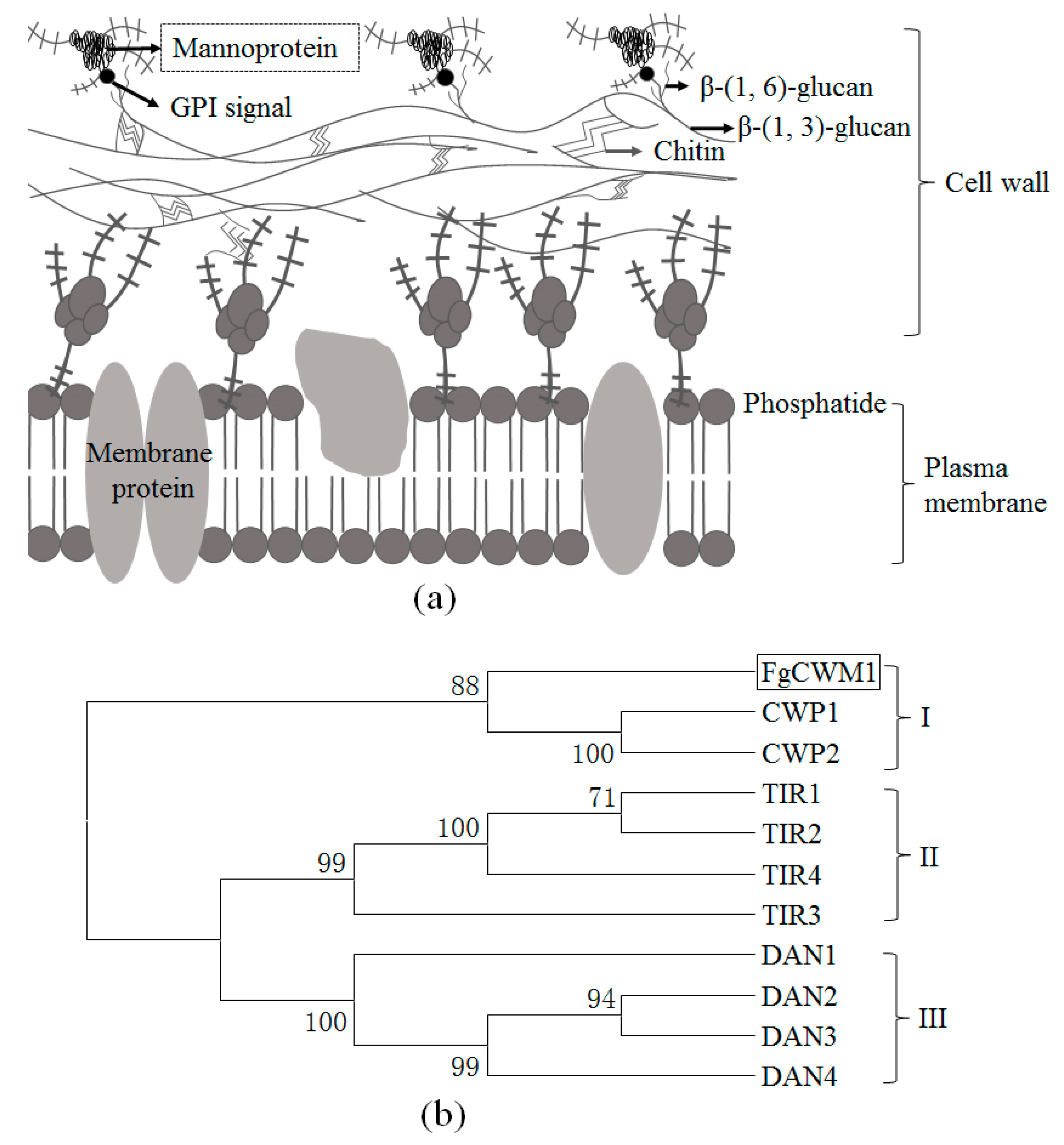
2.1. Sequence Analysis

Sequence analyses revealed that the FgCWM1 gene is 1313 bp in length, with two exons and one intron (part I of Figure 2a), and its full open reading frame is 1260 bp long. FgCWM1 encodes a putative cell wall mannoprotein (CWM), which is supported by a neighbor-joining tree of the deduced amino acid sequences of CWMs from S. cerevisiae (Figure 1b). CWMs can be divided into three groups, i.e., groups I, II, and III. FgCWM1 falls into group I with CWP1 and CWP2, which encode CWMs in S. cerevisiae [11,15,16].

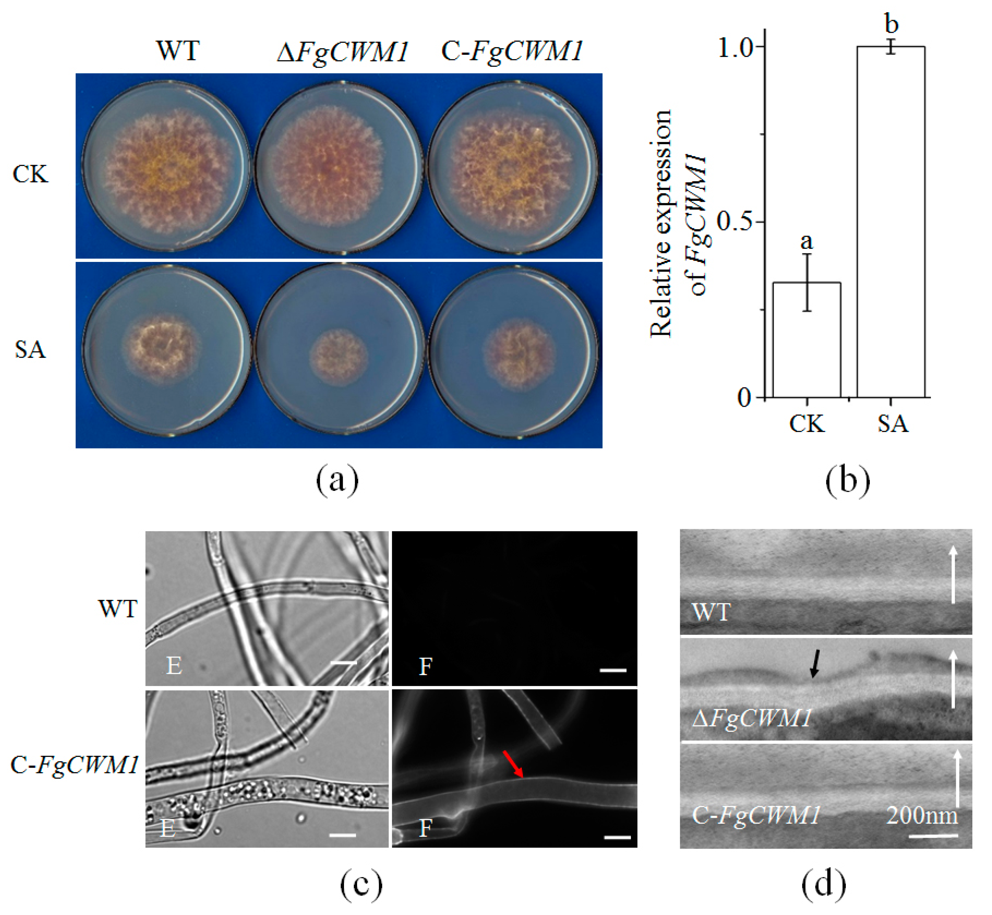
2.2. Creation of ΔFgCWM1 and C-FgCWM1

To disrupt the function of FgCWM1 in F. graminearum, the flanking regions (left border homologous arm (LBHA) and right border homologous arm (RBHA)) of the FgCWM1 gene were amplified from genomic DNA of F. graminearum, and then inserted into the pRF-HU2 vector to prepare a disruption plasmid (part I of Figure 2a). The deletion mutants (ΔFgCWM1) were created by replacing the entire FgCWM1 gene with the target selectable marker hygromycin (HPH) through homologous recombination (part II of Figure 2a). To ensure that the construct had integrated at the intended homologous site, primer pairs P5 + P6 and P7 + P8 (part III of Figure 2a) were used to detect the construct in ΔFgCWM1. The two primer pairs amplified products with the expected size from ΔFgCWM1 (Figure 2b). Seven ΔFgCWM1 mutants were generated and were verified by sequencing (data not shown).
To create complementation mutants (C-FgCWM1), the open reading frame of FgCWM1 was introduced into ΔFgCWM1. Six C-FgCWM1 mutants were used. The WT (wild type), ΔFgCWM1, and C-FgCWM1 strains were verified by RT (reverse transcription)-PCR, using the primer pair RJ-FgCWM1-F + RJ-FgCWM1-R (Table 1). FgCWM1 was expressed normally in C-FgCWM1 as in WT but was not expressed at all in ΔFgCWM1 (Figure 2c). These results demonstrated that FgCWM1 was correctly removed from the genome of F. graminearum and was successfully re-expressed in C-FgCWM1.

2.3. Effect of FgCWM1 on Mycelial Growth

To observe the changes in the growth phenotype of F. graminearum caused by disruption of FgCWM1, mycelial growth of the WT, ΔFgCWM1, and C-FgCWM1 strains was compared on mSNA (modified Synthetischer Nährstoffarmer Agar) plates with or without 0.9 mmolL−1 SA (Figure 3a). ΔFgCWM1 grew slower than WT and C-FgCWM1 under SA treatment, while their mycelia grew similarly under control conditions (Figure 3a). Consistent with its positive role in response to SA stress, FgCWM1 expression was induced by SA (Figure 3b). These observations imply that FgCWM1 participates in the F. graminearum response to SA stress.

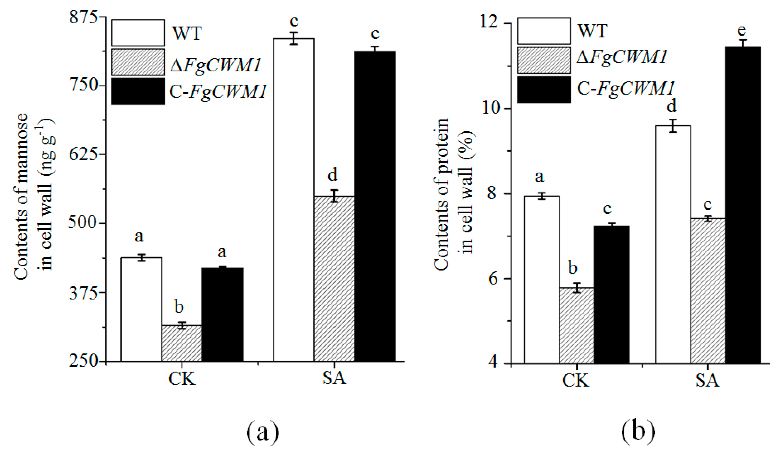
2.4. Effect of FgCWM1 on Fungal Cell Wall Development

The green fluorescent protein (GFP) was tagged to the C-terminal of FgCWM1 in C-FgCWM1 mutants, which were used to investigate the subcellular localization of FgCWM1 protein. Microscopic observations showed that the FgCWM1 protein is localized on the cell wall (Figure 3c), suggesting that it plays a structural role in the cell wall. To determine the effect of FgCWM1 on the cell wall, hyphae of WT, ΔFgCWM1, and C-FgCWM1 strains were observed under a transmission electron microscope (TEM). An obvious deficiency in the outer cell wall was observed in the hypha of ΔFgCWM1, whereas the cell walls had smooth surfaces in the hyphae of WT and C-FgCWM1 (Figure 3d).

2.5. FgCWM1 Encodes a Cell Wall Mannoprotein

Considering that FgCWM1 encodes a putative CWM (Figure 1b), the contents of mannose and protein in the cell wall of WT, ΔFgCWM1, and C-FgCWM1 strains were compared. As expected, ΔFgCWM1 showed significantly lower contents of mannose and protein, as compared with those in WT and C-FgCWM1 strains (Figure 4a,b). Consistent with the increased expression level of FgCWM1 under SA treatment, the accumulation of mannose and protein was also increased under SA treatment (Figure 4a,b). To confirm its function, FgCWM1 was expressed in S. cerevisiae (S-FgCWM1). The contents of mannose and protein in the P-FgCWM1 strain were significantly higher than those in the control strain (transformed with empty vector; S-control) (Figure 5a,b).

2.6. Effect of FgCWM1 on Pathogenicity

To determine whether FgCWM1 participates in the pathogenicity of F. graminearum, two fully developed florets of a central spikelet were point-inoculated with conidial suspensions of WT, ΔFgCWM1, and C-FgCWM1, respectively. Spikes inoculated with ΔFgCWM1 showed much milder disease symptoms and less fungal biomass, as compared with those inoculated with WT and C-FgCWM1 (Figure 6a–c). However, the DON contents in the liquid culture medium and in wheat spikes did not differ significantly between ΔFgCWM1 and WT (Figure 6d,e).
Considering that FgCWM1 was induced by SA and was highly expressed by as early as 24 h after initial inoculation in wheat heads [31], and that FgCWM1 was found to influence the fungal response to SA stress (Figure 3a), we compared the SA contents between spikes inoculated with WT and those inoculated with ΔFgCWM1 (Figure 6e). The spikes inoculated with ΔFgCWM1 accumulated more SA than did those inoculated with WT.

3. Discussion

In fungi, CWM, which is located on the outer cell wall, is an important component of the cell wall (Figure 1a). The CWM is essential for fungal vegetative growth and pathogenicity [11,12,13]. In F. graminearum, FgCWM1, which encodes a CWM, was found to affect mycelial growth under SA stress (Figure 3a). As expected, localization analyses showed that the FgCWM1 protein is localized on the cell wall (Figure 3c). Consistent with the distribution of CWMs in fungal cell wall (Figure 1a), the absence of FgCWM1 led to reduced contents of mannose and protein in the cell wall, resulting in a defective outer cell wall (Figure 3d), and consequently, dramatically reduced pathogenicity in wheat (Figure 6a,b). Our results demonstrate that FgCWM1 encodes a CWM that plays an important role in the host–pathogen interaction between F. graminearum and wheat.
There are nine CWM genes in the S. cerevisiae genome, which are divided into three classes. FgCWM1 is in group I (Figure 1b). In our unpublished transcriptome data, FgCWM1 was the only CWM gene upregulated by SA. However, as shown in Figure 4, SA was able to significantly induce the accumulation of mannose and protein in the cell wall of F. graminearum, even in ΔFgCWM1, which lacked the FgCWM1 that contributes to the accumulation of mannose and protein. This result suggests that some other CWM gene(s) are present in the genome of F. graminearum.
Fungal cell walls have a layered structure, and CWM and chitin are the two major components [6,9,10] (Figure 1a). Chitin and CWM are distributed in the inner and outer layers of the cell wall, respectively. Reduction of chitin biosynthesis by deleting the gene encoding chitin synthase resulted in a different and almost invisible inner layer in cell walls of Fusarium asiaticum [32]. In the present study, deletion of FgCWM1 resulted in an obvious deficiency in the outer cell wall of F. graminearum (Figure 3d). In F. graminearum, chitin is synthesized by chitin synthase, including FgCHS8 (chitin synthase gene) [28]. Considering the importance of CWM and chitin in fungal cell wall integrity, and the toxicity of SA to F. graminearum [22], we compared the expression of FgCHS8 and FgCWM1 under SA stress. Treatment with SA resulted in downregulation of FgCHS8 and upregulation of FgCWM1 [28] (Figure 3b), suggesting that F. graminearum can overcome the toxicity of exogenous SA by enhancing the accumulation of CWM in the outer cell wall, even though SA is able to weaken the inner cell wall structure.
FgCWM1 is a valuable gene target for controlling FHB disease. The recently cloned Fhb1 gene can lead to a substantial reduction in the severity of visual FHB disease symptoms in wheat spikes [33,34], and Fhb1 has been widely and successfully used for wheat breeding in China for a long time. In this study, spikes inoculated with ΔFgCWM1 showed mild and non-spreading FHB disease symptoms and much lower fungal biomass, as compared with those inoculated with WT and C-FgCWM1 (Figure 6a–c). Host-induced gene silencing (HIGS) is a promising way to inhibit F. graminearum infection of wheat [35,36]. Considering its significance in fungal pathogenicity, FgCWM1 is a promising gene target for enhancing wheat resistance against FHB by HIGS.
In wheat, SA is involved in resistance against FHB [21,23,24,37,38]. This plant defense hormone triggers systemic acquired resistance and induces the expression of a set of defense-related genes [26,27]. The SA content in wheat spikes was found to be significantly increased in response to F. graminearum infection [25]. SA directly affects F. graminearum, resulting in decreased DON production, lower germination efficiency, and reduced mycelial growth [22]. In this study, ΔFgCWM1 grew slower than WT and C-FgCWM1 under SA stress, while SA induced the expression of FgCWM1 (Figure 3b), suggesting that FgCWM1 is not a target of SA during the inhibition of mycelial growth. Although the SA content is increased in infected spikes, F. graminearum has the ability to export and metabolize SA to avoid SA toxicity [22,23,24,30]. Furthermore, we observed that F. graminearum is able to strengthen the outer cell wall by upregulating the expression of FgCWM1 (Figure 3b) under SA stress. This is despite the SA-induced downregulation of FgLAI12 (linoleic acid isomerase gene) and FgCHS8 in F. graminearum, which encode important components of the fungal cell membrane and cell wall, respectively, and are essential for the fungal response to SA [28,29]. Compared with spikes inoculated with the WT strain, those inoculated with ΔFgCWM1 accumulated more SA (Figure 6f), confirming the key role of FgCWM1 in the fungal response to wheat endogenous SA. The wheat defense response against F. graminearum also involves SA signaling [21,26,27]. However, Qi et al. [39] reported that exogenous application of SA at 1 mmolL−1 to wheat flowering heads induced only two genes. Therefore, we speculate that the direct effect of SA, rather than SA signaling, plays a more important role in wheat resistance against FHB disease.
Grain contamination by mycotoxin released by F. graminearum is a significant threat to the health of animals and humans [1,2]. Because SA significantly decreases the production of DON, it is a promising phytohormone for reducing mycotoxin contamination (Figure 6d). However, the gene controlling DON production that is targeted by SA remains unclear. In FHB, the severity of visual disease symptoms is not strongly related to the amount of mycotoxin in grains. Therefore, resistance to spreading and resistance to toxins are recognized as two different types of resistance in wheat [40]. We found that the expression level of FgCWM1 was strongly related to the severity of visual FHB symptoms (Figure 6a), but not to DON production (Figure 6d) under these experimental conditions. Therefore, FgCWM1 is not involved in regulating the biosynthesis of DON in F. graminearum.

4. Materials and Methods

4.1. Materials and Growth Conditions

The F. graminearum isolate DAOM180378 (Canadian Fungal Culture Collection, Agriculture and Agri-Food Canada, Ottawa, ON, Canada), which is highly virulent in wheat, was used in all experiments. To produce conidia, the fungus was cultured in carboxymethyl cellulose (CMC) medium at 28 °C, with shaking (180 rpm) for five days [41]. F. graminearum was cultured on modified SNA (mSNA; 1 g KH2PO4, 1 g KNO3, 0.5 g MgSO4, 0.5 g KCl, 1 g glucose, 1 g sucrose, and 20 g agar per liter) plates at 25 °C. Each plate was inoculated with 1 × 10³ conidia of F. graminearum and all inoculated plates were incubated in a dark cabinet at 28 °C. A stock solution of SA (1 molL−1) was prepared in methanol and added to media after autoclaving. Agrobacterium tumefaciens strain AGL-1, used for transforming F. graminearum, was grown at 28 °C in yeast extract broth (YEB; 5 g nutrient broth, 1 g yeast extract, 5 g peptone, 5 g sucrose, and 0.2 g MgSO4 per liter; pH 7.4). S. cerevisiae strain AH109 was grown on yeast peptone dextrose adenine (YPDA) medium (10 g yeast extract, 20 g dific-peptone, 20 g glucose, and 0.03 g adenine sulfate per liter). Unless specifically noted, all chemicals were purchased from Sigma-Aldrich (St. Louis, MO, USA).
Wheat (Triticum aestivum cv. ‘Roblin’) plants were grown in a greenhouse under a 16/8 h (day/night) photoperiod at 23/18 °C. Plants were watered as needed and fertilized before planting with 15–15–15 (N–P–K) compound fertilizer. ‘Roblin’ is highly susceptible to F. graminearum infection.

4.2. Sequence Analysis

The gene sequence of FgCWM1 (FG05_11315) was downloaded from the Ensemble database (http://fungi.ensembl.org/index.html). Primer Premier (version 5.0; Premier Bio soft, Palo Alto, CA, USA) was used to design PCR primers (Table 1). Reported CWM protein sequences were downloaded from NCBI (National Center for Biotechnology Information; http://www.ncbi.nlm.nih.gov). Their deduced amino acid sequences were aligned using MEGA version 5 [42]. Neighbor-joining trees (10,000 replicates) for classification of deduced proteins were constructed using MEGA software, with Poisson correction and complete deletion of gaps (Figure 1b).

4.3. Construction of Deletion and Complementation Mutants

Genomic DNA was extracted from mycelia cultured on mSNA plates for 5 d at 28 °C, by the CTAB (Cetyltrimethylammonium bromide) method [43]. The deletion of FgCWM1 from the genome of F. graminearum is illustrated in Figure 2a. The pRF-HU2 vector was used for targeted gene replacement in F. graminearum through A. tumefaciens-mediated transformation [44,45]. Transformation of F. graminearum was carried out as described elsewhere [46]. For complementation, the coding region of FgCWM1 was ligated into pCAMBIA1302 vector (with the green fluorescent protein gene (mGFP5) tag) to make the FgCWM1::mGFP5 fusion construct, which was then transformed into ΔFgCWM1 (Figure 2b).

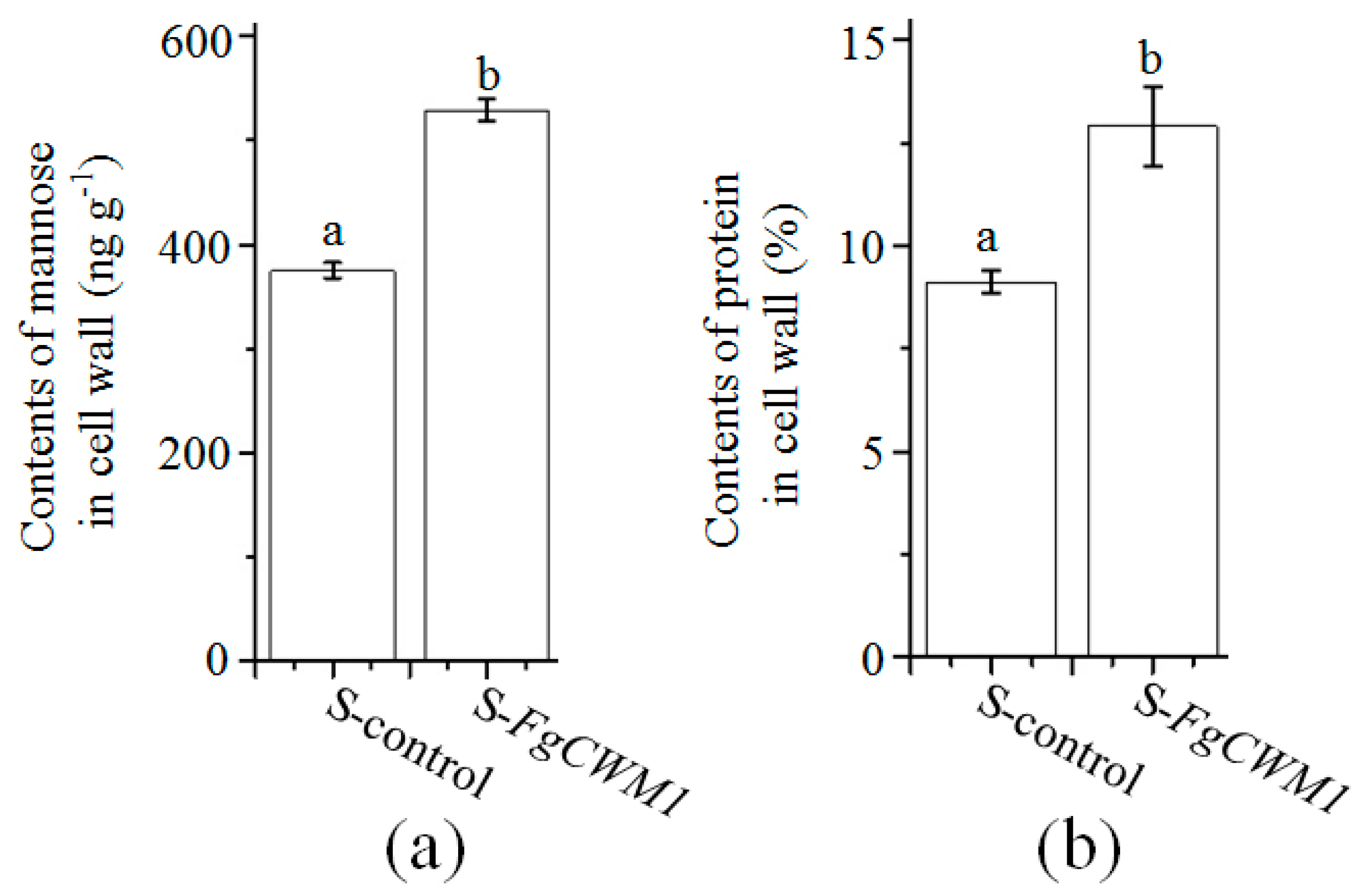
4.4. FgCWM1 Expression in S. cerevisiae

To express FgCWM1 in S. cerevisiae, the full open reading frame of FgCWM1 (without the termination codon; amplified by the primer pair SS-FgCWM1-F + SS-FgCWM1-R) was inserted into the pYC54 vector, and transformed into S. cerevisiae strain AH109 (TIANDZ, Beijing, China) following the manufacturer’s instructions.

4.5. Determination of Mannose and Protein Contents in Fungal Cell Wall

Cell walls were prepared and extracted as per Kollár et al. [47] with some modifications. Fresh mycelia of F. graminearum in Figure 3a (0.2 g) and cells of S. cerevisiae (0.2 g) were washed three times with 0.9% NaCl, and then suspended in 2 mL Tris-HCl buffer (50 mmolL−1; pH = 7.5) with 0.5 g glass beads (0.5 mm diameter, Sigma-Aldrich, St. Louis, USA). Mycelia samples were shaken at 30 s−1 for 3 min (Retsch MM400, Haan, Germany). Cell wall material was pelleted by centrifugation (1500× g) for 10 min and then washed three times with Tris-HCl buffer at room temperature.
Mannose was extracted from the cell wall pellet as described by Cameron et al. [48], with some modifications. The collected cell wall pellet was resuspended in 2 mL 0.1 molL−1 citrate buffer solution (pH = 6.6; 0.1 molL−1 citric acid and 0.1 molL−1 sodium citrate) and incubated in a sterilization pan (Sanyo MLS-3780, Tokyo, Japan) at 121 °C for 3 h. The solution was separated by centrifugation (900 g) for 20 min at 4 °C. The supernatant was transferred into a 10 mL centrifuge tube and mixed with triploid precooled ethanol (with 1% acetic acid) at 4 °C for 12 h. Mannose was obtained by centrifugation (900 g) and then suspended in 200 μL water. The concentration of mannose was assayed using an enzyme-linked immune response kit, following the manufacturer’s instructions (Jin Yibai Biological Technology Company, Nanjing, China). The protein contents in the cell wall were determined as in [49].

4.6. Microscopic Assay

For optical microscopic and fluorescence microscopic assays, 1000 conidia of WT and C-FgCWM1 were respectively inoculated into 3 mL mSNA liquid medium (1 g KH2PO4, 1 g KNO3, 0.5 g MgSO4, 0.5 g KCl, 1 g glucose, and 1 g sucrose) and cultured at 28 °C on an orbital shaker at 120 rpm for two days. Mycelia were collected and observed under a Nikon-80i fluorescence microscope (Nikon, Tokyo, Japan) to determine the subcellular localization of FgCWM1.
For transmission electron microscope (TEM) observations, mycelia of WT, ∆FgCWM1, and C-FgCWM1 were harvested as above and fixed [50]. Cells were permeated with 812 epoxy resin monomers (SPI-Pon™ 812, West Chester, USA), cut using a Leica UC7 microtome (Leica microsystems, Wetzlar, Germany), and analyzed and photographed using an Hitachi HT7700 TEM (Hitachi, Tokyo, Japan).

4.7. Virulence Assay and DON Measurement

To determine the effect of FgCWM1 on the pathogenicity of F. graminearum in wheat heads, two flowering florets of a central spikelet of one head were each inoculated with 1 × 10³ conidia. The inoculated heads were wrapped in moist plastic wrap and incubated for 48 h at 25 °C. The FHB symptoms were assessed 2–12 days after inoculation at 25 °C. There are three biological replicates per treatment, and each replicate contains at least five heads.
A two-stage protocol was used to test whether FgCWM1 is related to the production of DON in liquid media [22,51]. The effect of FgCWM1 on DON production in wheat heads was determined as described elsewhere [24]. The wheat samples were collected on the sixth day after inoculation. There are three biological replicates (with at least two heads) for each treatment. The amount of DON was measured using a DON ELISA kit (Beacon, ME, USA) and a Multiskan Spectrum instrument (Thermo Fisher Scientific, Waltham, MA, USA).

4.8. Gene Expression Analysis

Total RNA was extracted from fresh powdered material (mycelia or wheat spikelets ground in liquid nitrogen) using the E.Z.N.A.® Total RNA Kit I (Omega Bio-Tek, Norcross, GA, USA) according to the manufacturer’s instructions. Then, RNA was reverse transcribed using the PrimeScript™ RT Reagent Kit with genomic DNA Eraser (Takara, Dalian, China) following the manufacturer’s protocol.
The primer pair Rj-FgCWM1-F + Rj-FgCWM1-R was used to measure the expression level of FgCWM1 in F. graminearum. The glyceraldehyde 3-phosphate dehydrogenase (FgGAPDH, FG05_06257), β-tubulin (FG05_09530), and elongation factor 1 (FG05_08811) genes were used as reference genes when performing qPCR for F. graminearum samples [22]. The relative amount of F. graminearum was estimated by measuring the expression level of FgGAPDH in wheat spike tissue by qPCR, with normalization against three wheat reference genes (w-GAPDH, NCBI UniGene Ta.66461; Aox, Ta.6172; hn-RNP-Q, Ta.10105) [22]. The qPCRs were performed using a MyiQ Real-Time PCR Detection System (Bio-Rad, Hercules, CA, USA). All of the primers mentioned above are listed in Table 1.

4.9. Quantification of SA in Wheat Spikes

To prepare wheat spike samples, two florets from each fully developed spikelet in a whole spike at the mid-anthesis stage were inoculated with 1 × 10³ conidia or water. The inoculated wheat plants were treated as described above. At 24 h after inoculation, the spikes were harvested and ground to a fine powder in liquid nitrogen. Three biological replicates per treatment were analyzed. The quantification of SA was conducted as described by Siciliano et al. [52].

4.10. Statistical Analysis

Student’s t-test (implemented in DPS (Data Procession System) version 12.01 software (Zhejiang University, Hangzhou, China); [53]) was used to test the significance of differences among average values of cell wall mannose content, cell wall protein content, relative expression levels of genes, DON content, and disease level.

Author Contributions

Y.-Z.Z., Q.C., C.-H.L., L.L., Y.L., K.Z., M.-Q.W., Z.-R.G., Y.W., B.-J.X., Y.-F.J., L.K., Y.-L.L., and P.-F.Q. performed all the experiments and analyzed the data; P.-F.Q. and Y.-M.W. designed the experiment; X.-J.L., Q.-T.J., J.M., J.-R.W., G.-Y.C., and Y.-L.Z. provided key advice. P.-F.Q., Y.-Z.Z., and Q.C. wrote the paper; P.-F.Q., Y.-Z.Z., and Q.C. prepared the figures.

Funding

This research was funded by the National Natural Science Foundation of China (31901961 and 31671677), and Innovative training Program for students in Sichuan Agricultural University (201610626082).

Acknowledgments

The authors thank Thérèse Ouellet of Agriculture and Agri-Food Canada for providing materials, and Jennifer Smith, from Liwen Bianji, Edanz Group China (www.liwenbianji.cn/ac), for editing the English text of a draft of this manuscript.

Conflicts of Interest

The authors declare that there is no conflict of interest.

References

  1. Goswami, R.S.; Kistler, H.C. Heading for disaster: Fusarium graminearum on cereal crops. Mol. Plant Pathol. 2004, 5, 515–525. [Google Scholar] [CrossRef] [PubMed]
  2. Desjardins, A.E.; Hohn, T.M.; McCormick, S.P. Trichothecene biosynthesis in Fusarium: Chemistry, genetics, and significance. Microbiol. Rev. 1993, 57, 595–604. [Google Scholar] [PubMed]
  3. Fernando, W.G.; Paulitz, T.C.; Seaman, W.L.; Dutilleul, P.; Miller, J.D. Head blight gradients caused by Gibberella zeae from area sources of inoculum in wheat field plots. Phytopathology 1997, 87, 414–421. [Google Scholar] [CrossRef] [PubMed]
  4. McMullen, M.; Bergstrom, G.; De Wolf, E.; Dill-Macky, R.; Hershman, D.; Shaner, G.; Van Sanford, D. A unified effort to fight an enemy of wheat and barley: Fusarium Head Blight. Plant Dis. 2012, 96, 1712–1728. [Google Scholar] [CrossRef]
  5. Legrand, F.; Picot, A.; Cobo-Díaz, J.F.; Chen, W.; Le Floch, G. Challenges facing the biological control strategies for the management of Fusarium Head Blight of cereals caused by F. graminearum. Biol. Control 2017, 113, 26–38. [Google Scholar] [CrossRef]
  6. De Groot, P.W.; Ram, A.F.; Klis, F.M. Features and functions of covalently linked proteins in fungal cell walls. Fungal Genet. Biol. 2005, 42, 657–675. [Google Scholar] [CrossRef]
  7. Lionetti, V.; Giancaspro, A.; Fabri, E.; Giove, S.L.; Reem, N.; Zabotina, O.A.; Blanco, A.; Gadaleta, A.; Bellincampi, D. Cell wall traits as potential resources to improve resistance of durum wheat against Fusarium graminearum. BMC Plant Biol. 2015, 15, 6. [Google Scholar] [CrossRef]
  8. Chow, J.; Notaro, M.; Prabhakar, A.; Free, S.J.; Cullen, P.J. Impact of fungal MAPK pathway targets on the Cell Wall. J. Fungi 2018, 4, 93. [Google Scholar] [CrossRef]
  9. Schoffelmeer, E.A.; Klis, F.M.; Sietsma, J.H.; Cornelissen, B.J. The cell wall of Fusarium oxysporum. Fungal Genet. Biol. 1999, 27, 275–282. [Google Scholar] [CrossRef]
  10. Karkowska-Kuleta, J.; Kozik, A. Cell wall proteome of pathogenic fungi. Acta. Biochim. Pol. 2015, 62, 339–351. [Google Scholar] [CrossRef] [Green Version]
  11. Abramova, N.; Sertil, O.; Mehta, S.; Lowry, C.V. Reciprocal regulation of anaerobic and aerobic cell wall mannoprotein gene expression in Saccharomyces cerevisiae. J. Bacteriol. 2001, 183, 2881–2887. [Google Scholar] [CrossRef] [PubMed]
  12. Klis, F.M.; de Groot, P.; Hellingwerf, K. Molecular organization of the cell wall of Candida albicans. Med. Mycol. 2001, 39, 1–8. [Google Scholar] [CrossRef] [PubMed]
  13. Klis, F.M.; Mol, P.; Hellingwerf, K.; Brul, S. Dynamics of cell wall structure in Saccharomyces cerevisiae. FEMS Microbiol. Rev. 2002, 26, 239–256. [Google Scholar] [CrossRef] [PubMed]
  14. Francois, J.M. Cell surface interference with plasma membrane and transport processes in yeasts. Adv. Exp. Med. Biol. 2016, 892, 11–31. [Google Scholar] [CrossRef]
  15. Van der Vaart, J.M.; Caro, L.H.; Chapman, J.W.; Klis, F.M.; Verrips, C.T. Identification of three mannoproteins in the cell wall of Saccharomyces cerevisiae. J. Bacteriol. 1995, 177, 3104–3110. [Google Scholar] [CrossRef]
  16. Dielbandhoesing, S.K.; Zhang, H.; Caro, L.H.; van der Vaart, J.M.; Klis, F.M.; Verrips, C.T.; Brul, S. Specific cell wall proteins confer resistance to nisin upon yeast cells. Appl. Environ. Microbiol. 1998, 64, 4047–4052. [Google Scholar]
  17. Zhang, M.; Liang, Y.P.; Zhang, X.H.; Xu, Y.; Dai, H.P.; Xiao, W. Deletion of yeast CWP genes enhances cell permeability to genotoxic agents. Toxicol. Sci. 2008, 103, 68–76. [Google Scholar] [CrossRef]
  18. Henry, C.; Fontaine, T.; Heddergott, C.; Robinet, P.; Aimanianda, V.; Beau, R.; Beauvais, A.; Mouyna, I.; Prevost, M.C.; Fekkar, A.; et al. Biosynthesis of cell wall mannan in the conidium and the mycelium of Aspergillus fumigatus. Cell. Microbiol. 2016, 18, 1881–1891. [Google Scholar] [CrossRef]
  19. Du, T.; Ouyang, H.; Voglmeir, J.; Wilson, I.B.H.; Jin, C. Aspergillus fumigatus Mnn9 is responsible for mannan synthesis and required for covalent linkage of mannoprotein to the cell wall. Fungal Genet. Biol. 2019, 128, 20–28. [Google Scholar] [CrossRef]
  20. Sandini, S.; La Valle, R.; De Bernardis, F.; Macri, C.; Cassone, A. The 65 kDa mannoprotein gene of Candida albicans encodes a putative beta-glucanase adhesin required for hyphal morphogenesis and experimental pathogenicity. Cell. Microbiol. 2007, 9, 1223–1238. [Google Scholar] [CrossRef]
  21. Makandar, R.; Essig, J.S.; Schapaugh, M.A.; Trick, H.N.; Shah, J. Genetically engineered resistance to Fusarium head blight in wheat by expression of Arabidopsis NPR1. Mol. Plant Microbe Interact. 2006, 19, 123–129. [Google Scholar] [CrossRef] [PubMed]
  22. Qi, P.F.; Johnston, A.; Balcerzak, M.; Rocheleau, H.; Harris, L.J.; Long, X.Y.; Wei, Y.M.; Zheng, Y.L.; Ouellet, T. Effect of salicylic acid on Fusarium graminearum, the major causal agent of fusarium head blight in wheat. Fungal Biol. 2012, 116, 413–426. [Google Scholar] [CrossRef] [PubMed]
  23. Qi, P.F.; Zhang, Y.Z.; Liu, C.H.; Zhu, J.; Chen, Q.; Guo, Z.R.; Wang, Y.; Xu, B.J.; Zheng, T.; Jiang, Y.F.; et al. Fusarium graminearum ATP-Binding cassette transporter gene FgABCC9 is required for its transportation of salicylic acid, fungicide resistance, mycelial growth and pathogenicity towards wheat. Int. J. Mol. Sci. 2018, 19, 2351. [Google Scholar] [CrossRef] [PubMed]
  24. Qi, P.F.; Zhang, Y.Z.; Liu, C.H.; Chen, Q.; Guo, Z.R.; Wang, Y.; Xu, B.J.; Jiang, Y.F.; Zheng, T.; Gong, X.; et al. Functional analysis of FgNahG clarifies the contribution of salicylic acid to wheat (Triticum aestivum) resistance against Fusarium Head Blight. Toxins 2019, 11, 59. [Google Scholar] [CrossRef] [PubMed]
  25. Qi, P.F.; Balcerzak, M.; Rocheleau, H.; Leung, W.; Wei, Y.M.; Zheng, Y.L.; Ouellet, T. Jasmonic acid and abscisic acid play important roles in host-pathogen interaction between Fusarium graminearum and wheat during the early stages of fusarium head blight. Physiol. Mol. Plant. Pathol. 2016, 93, 39–48. [Google Scholar] [CrossRef]
  26. Uknes, S.; Mauch-Mani, B.; Moyer, M.; Potter, S.; Williams, S.; Dincher, S.; Chandler, D.; Slusarenko, A.; Ward, E.; Ryals, J. Acquired resistance in Arabidopsis. Plant Cell 1992, 4, 645–656. [Google Scholar] [CrossRef]
  27. Sorahinobar, M.; Niknamet, V.; Ebrahimzadeh, H.; Soltanloo, H.; Behmanesh, M.; Enferadi, S.T. Central role of salicylic acid in resistance of wheat against Fusarium graminearum. J. Plant Growth Regul. 2016, 35, 477–491. [Google Scholar] [CrossRef]
  28. Zhang, Y.Z.; Chen, Q.; Liu, C.H.; Liu, Y.B.; Yi, P.; Niu, K.X.; Wang, Y.Q.; Wang, A.Q.; Yu, H.Y.; Pu, Z.E.; et al. Chitin synthase gene FgCHS8 affects virulence and fungal cell wall sensitivity to environmental stress in Fusarium graminearum. Fungal Biol. 2016, 120, 764–774. [Google Scholar] [CrossRef]
  29. Zhang, Y.Z.; Wei, Z.Z.; Liu, C.H.; Chen, Q.; Xu, B.J.; Guo, Z.R.; Cao, Y.L.; Wang, Y.; Han, Y.N.; Chen, C.; et al. Linoleic acid isomerase gene FgLAI12 affects sensitivity to salicylic acid, mycelial growth and virulence of Fusarium graminearum. Sci. Rep. 2017, 7, 46129. [Google Scholar] [CrossRef]
  30. Rocheleau, H.; Al-harthi, R.; Ouellet, T. Degradation of salicylic acid by Fusarium graminearum. Fungal Biol. 2019, 123, 77–86. [Google Scholar] [CrossRef]
  31. Harris, L.J.; Balcerzak, M.; Johnston, A.; Schneiderman, D.; Ouellet, T. Host-preferential Fusarium graminearum gene expression during infection of wheat, barley, and maize. Fungal Biol. 2016, 120, 111–123. [Google Scholar] [CrossRef] [PubMed]
  32. Xu, Y.B.; Li, H.P.; Zhang, J.B.; Song, B.; Chen, F.F.; Duan, X.J.; Xu, H.Q.; Liao, Y.C. Disruption of the chitin synthase gene CHS1 from Fusarium asiaticum results in an altered structure of cell walls and reduced virulence. Fungal Genet. Biol. 2010, 47, 205–215. [Google Scholar] [CrossRef] [PubMed]
  33. Li, G.Q.; Zhou, J.Y.; Jia, H.Y.; Gao, Z.X.; Fan, M.; Luo, Y.J.; Zhao, P.T.; Xue, S.; Li, N.; Yuan, Y.; et al. Mutation of a histidine-rich calcium-binding-protein gene in wheat confers resistance to Fusarium head blight. Nat. Genet. 2019, 51, 1106–1112. [Google Scholar] [CrossRef] [PubMed]
  34. Su, Z.Q.; Bernardo, A.; Tian, B.; Chen, H.; Wang, S.; Ma, H.X.; Cai, S.B.; Liu, D.T.; Zhang, D.D.; Li, T.; et al. A deletion mutation in TaHRC confers Fhb1 resistance to Fusarium head blight in wheat. Nat. Genet. 2019, 51, 1099–1105. [Google Scholar] [CrossRef] [PubMed]
  35. Koch, A.; Kumar, N.; Weber, L.; Keller, H.; Imani, J.; Kogel, K.H. Host-induced gene silencing of cytochrome P450 lanosterol C14 α-demethylase-encoding genes confers strong resistance to Fusarium species. Proc. Natl. Acad. Sci. USA 2013, 110, 19324–19329. [Google Scholar] [CrossRef] [PubMed]
  36. Cheng, W.; Song, X.S.; Li, H.P.; Cao, L.H.; Sun, K.; Qiu, X.L.; Xu, Y.B.; Yang, P.; Huang, T.; Zhang, J.B.; et al. Host-induced gene silencing of an essential chitin synthase gene confers durable resistance to Fusarium head blight and seedling blight in wheat. Plant Biotechnol. J. 2015, 13, 1335–1345. [Google Scholar] [CrossRef] [PubMed]
  37. Ding, L.; Xu, H.; Yi, H.; Yang, L.; Kong, Z.; Zhang, L.; Xue, S.; Jia, H.; Ma, Z. Resistance to hemi-biotrophic F. graminearum infection is associated with coordinated and ordered expression of diverse defense signaling pathways. PLoS ONE 2011, 6, e19008. [Google Scholar] [CrossRef]
  38. Ameye, M.; Audenaert, K.; De Zutter, N.; Steppe, K.; Van Meulebroek, L.; Vanhaecke, L.; De Vleesschauwer, D.; Haesaert, G.; Smagghe, G. Priming of wheat with the green leaf volatile Z-3-hexenyl acetate enhances defense against Fusarium graminearum but boosts deoxynivalenol production. Plant Physiol. 2015, 167, 1671–1684. [Google Scholar] [CrossRef]
  39. Qi, P.F.; Jiang, Y.F.; Guo, Z.R.; Chen, Q.; Ouellet, T.; Zong, L.J.; Wei, Z.Z.; Wang, Y.; Zhang, Y.Z.; Xu, B.J.; et al. Transcriptional reference map of hormone responses in wheat spikes. BMC Genom. 2019, 20, 390. [Google Scholar] [CrossRef]
  40. Mesterházy, A. Types and components of resistance to Fusarium head blight of wheat. Plant Breed. 1995, 114, 377–386. [Google Scholar] [CrossRef]
  41. Cappellini, R.A.; Peterson, J.L. Macroconidium formation in submerged cultures by a nonsporulating strain of Gibberella zeae. Mycologia 1965, 57, 962–966. [Google Scholar] [CrossRef]
  42. Tamura, K.; Peterson, D.; Peterson, N.; Stecher, G.; Nei, M.; Kumar, S. MEGA5: Molecular evolutionary genetics analysis using maximum likelihood, evolutionary distance, and maximum parsimony methods. Mol. Biol. Evol. 2011, 28, 2731–2739. [Google Scholar] [CrossRef] [PubMed]
  43. Lodhi, M.A.; Ye, G.N.; Weeden, N.F.; Reisch, B.I. A simple and efficient method for DNA extraction from grapevine cultivars and Vitis species. Plant Mol. Biol. Rep. 1994, 12, 6–13. [Google Scholar] [CrossRef]
  44. Frandsen, R.J.; Andersson, J.A.; Kristensen, M.B.; Giese, H. Efficient four fragment cloning for the construction of vectors for targeted gene replacement in filamentous fungi. BMC Mol. Biol. 2008, 9, 70. [Google Scholar] [CrossRef]
  45. Frandsen, R.J.; Frandsen, M.; Giese, H. Targeted gene replacement in fungal pathogens via Agrobacterium tumefaciens-mediated transformation. Methods Mol. Biol. 2012, 835, 17–45. [Google Scholar] [CrossRef]
  46. Maier, F.J.; Malz, S.; Losch, A.P.; Lacour, T.; Schafer, W. Development of a highly efficient gene targeting system for Fusarium graminearum using the disruption of a polyketide synthase gene as a visible marker. FEMS Yeast Res. 2005, 5, 653–662. [Google Scholar] [CrossRef]
  47. Kollár, R.; Petrakova, E.; Ashwell, G.; Robbins, P.W.; Cabib, E. Architecture of the yeast cell wall, the linkage between chitin and β(1→3)-glucan. J. Biol. Chem. 1995, 270, 1170–1178. [Google Scholar] [CrossRef]
  48. Cameron, D.R.; Cooper, D.G.; Neufeld, R.J. The mannoprotein of Saccharomyces cerevisiae is an effective bioemulsifier. Appl. Environ. Microbiol. 1988, 54, 1420–1425. [Google Scholar]
  49. Jung, S.; Rickert, D.A.; Deak, N.A.; Aldin, E.D.; Recknor, J.; Johnson, L.A.; Murphy, P.A. Comparison of kjeldahl and dumas methods for determining protein contents of soybean products. J. Am. Oil Chem. Soc. 2003, 80, 1169. [Google Scholar] [CrossRef]
  50. Martin-Urdiroz, M.; Roncero, M.I.; Gonzalez-Reyes, J.A.; Ruiz-Roldan, C. ChsVb, a class VII chitin synthase involved in septation, is critical for pathogenicity in Fusarium oxysporum. Eukaryot. Cell 2008, 7, 112–121. [Google Scholar] [CrossRef]
  51. Miller, D.; Blackwell, B.A. Biosynthesis of 3-acetyldeoxynivalenol and other metabolites by Fusarium culmorum HLK 1503 in a stirred jar fermentor. Can. J. Bot. 1986, 64, 1–5. [Google Scholar] [CrossRef]
  52. Siciliano, I.; Amaral Carneiro, G.; Spadaro, D.; Garibaldi, A.; Gullino, M.L. Jasmonic acid, abscisic acid, and salicylic acid are involved in the phytoalexin responses of rice to Fusarium fujikuroi, a high gibberellin producer pathogen. J. Agric. Food Chem. 2015, 63, 8134–8142. [Google Scholar] [CrossRef] [PubMed]
  53. Tang, Q.Y.; Zhang, C.X. Data Processing System (DPS) software with experimental design, statistical analysis and data mining developed for use in entomological research. Insect Sci. 2013, 20, 254–260. [Google Scholar] [CrossRef] [PubMed]
Figure 1. (a) Model of fungal cell wall with a layered structure [6,9,10]. The inner, electron-transparent layer is mainly composed of chitin, β glucan (β-(1,3)-glucan and β-(1,6)-glucan). The β-(1,3)-glucan is in contact with chitin, and β-(1,6)-glucan is in contact with glycosylphosphatidyl inositol (GPI) residues. The outer, electron-dense layer is composed of cell wall mannoproteins (CWMs) that are in contact with GPI. (b) Neighbor-joining tree of FgCWM1 protein and nine CWMs in Saccharomyces cerevisiae [11,15,16]. Genbank accession numbers for CWP1, CWP2, TIR1, TIR2, TIR3, TIR4, DAN1, DAN2, DAN3, and DAN4 are NP_012827, NP_012826, KZV11760, AJU12981, KZV10607, KZV07893, NP_012684, EGA81792, EGA56320, and KZV10416, respectively.
Toxins 11 00628 g001
Figure 2. Creation of ΔFgCWM1 and C-FgCWM1 mutants. (a) Schematic for disruption of FgCWM1FgCWM1). SacI, ApaI, SpeI, and HindIII are restriction enzymes used. Black arrow represents targeted location of primers; black lines show amplified sequences. LBHA, left border homologous arm. RBHA, right border homologous arm. (b) Verification of ΔFgCWM1. Primer pairs P5 + P6 and P7 + P8 were respectively positioned upstream and downstream of inserted T-DNA sequence of ΔFgCWM1. (c) Reverse transcription PCR (RT-PCR) verification of FgCWM1 expression using primer pair RJ-FgCWM1-F + RJ-FgCWM1-R. FgGAPDH gene was used as reference. For PCR primers, see Table 1. All PCR products were verified by sequencing (Qingke, Chengdu, China).
Toxins 11 00628 g002
Figure 3. Effect of FgCWM1 on fungal biology. (a) Comparison of mycelial growth on mSNA (modified Synthetischer Nährstoffarmer Agar) plates with salicylic acid (SA, 0.9 mmolL1) and without SA (control; CK) on d 5 after inoculation with 1 × 10³ conidia (five biological replicates per treatment). (b) Relative expression of FgCWM1 in mycelia under SA and CK treatments in wild-type (WT) strain. Mycelia were collected from plates shown in Figure 3a. Different small letters above columns indicate significant difference (n = 5; p < 0.05). (c) Subcellular localization of FgCWM1 protein, by using the FgCWM1:GFP (green fluorescent protein) fusion protein. E, optical micrograph; F, fluorescence micrograph. Red arrow marks fluorescent protein signal. Scale bar, 10 µm. (d) TEM (transmission electron microscope) images of cell walls. White arrows indicate extracellular material. Black arrow marks defective site on cell wall.
Toxins 11 00628 g003
Figure 4. Measurement of mannose and protein contents in cell wall with SA (0.9 mmolL−1) and without SA (control; CK). (a) Mannose contents in cell wall. (b) Protein contents in cell wall. Mycelia were collected from mSNA plates in Figure 3a. Values are average ± standard deviation of three biological replicates per treatment. Different small letters above each box indicate significant difference (n = 3; p < 0.05).
Toxins 11 00628 g004
Figure 5. Expression of FgCWM1 increased accumulation of mannose and protein in S. cerevisiae. (a) Mannose contents in cell wall. (b) Protein contents in cell wall. Values are average ± standard deviation of three biological replicates per treatment. Different small letters above each box indicate significant difference (n = 3; p < 0.05).
Toxins 11 00628 g005
Figure 6. Effect of FgCWM1 on fungal pathogenicity. (a) Wheat heads inoculated with conidial suspensions of wild-type (WT), ΔFgCWM1, and C-FgCWM1 strains. Infected wheat heads were photographed on day 8 after initial inoculation. Black points on spikelets indicate inoculation sites. (b) Numbers of infected and bleached spikelets on d 4, 8, and 12 after inoculation. (c) Relative expression of FgGAPDH in wheat spikes at 24 h after initial inoculation. (d) Contents of DON in liquid medium with SA (0.9 mmolL−1) and without SA (control; CK). There are three biological replicates for each fungal strain under both CK and SA treatments. (e) Contents of DON in wheat ears. (f) Levels of SA in spikes inoculated with water (CK), WT, and ΔFgCWM1 at 24 h after inoculation. FW, fresh weight. Values in (b–f) are average ± standard deviation of three biological replicates per treatment. Different small letters above each box indicate significant difference (n = 3; p < 0.05).
Toxins 11 00628 g006
Table 1. Primers used in this study.
PrimerSequence (5′– 3′)Reference
FgCWM1-Up-FGCGGGCCCTACTCAGGGTAACGGAAAGGThis study
FgCWM1-Up-RGCGAGCTCACAATGCAGCTCAATGTCG
FgCWM1-Down-FGGAAGCTTCAACCCAGACCTACCCAThis study
FgCWM1-Down-RGGACTAGTGAAGAGCAGCGAACCAG
P5TGATAATAATGTCCTCGTTCCThis study
P6TGACGAACTGTAAGTCGGATA
P7ACCGAACTTCAAGACACCAThis study
P8CAACGGCCTCAACCTACT
R-FgCWM1-FAACCATGGATGAAGTTCTCCGCTGCThis study
R-FgCWM1-RGGCCATGGAAGCATCCTTCAGAAGAGGT
SS-FgCWM1-FGCGAGCTCGCATGAAGTTCTCCGCTGCThis study
SS-FgCWM1-RGGGGATCCAAGCATCCTTCAGAAGAGGT
Fg-GAPDH-FTGACTTGACTGTTCGCCTCGAGAA[22]
Fg-GAPDH-RATGGAGGAGTTGGTGTTGCCGTTA
Fg-β-tubulin-FGTTGATCTCCAAGATCCGTG[22]
Fg-β-tubulin-RCATGCAAATGTCGTAGAGGG
Fg-Factor1-FCCTCCAGGATGTCTACAAGA[22]
Fg-Factor1-RCTCAACGGACTTGACTTCAG
RJ-FgCWM1-FGCTGGTGCCGAGGCTGTTThis study
RJ-FgCWM1-RGGTCATCGCAGGTGTTTCCA
Aox-FGACTTGTCATGGTAGATGCCTG[22]
Aox-RCAGGACGAGCATAACCATTCTC
w-GAPDH-FAACTGTTCATGCCATCACTGCCAC[22]
w-GAPDH-RAGGACATACCAGTGAGCTTGCCAT
hn-RNP-Q-FTCACCTTCGCCAAGCTCAGAACTA[22]
hn-RNP-Q-RAGTTGAACTTGCCCGAAACATGCC
Restriction enzyme cut sites are underlined.

