Article

Physiological Effects on Coexisting Microalgae of the Allelochemicals Produced by the Bloom-Forming Cyanobacteria Synechococcus sp. and Nodularia Spumigena

by
Sylwia Śliwińska-Wilczewska
1,*,
Aldo Barreiro Felpeto
2,
Katarzyna Możdżeń
3,
Vitor Vasconcelos
2,4 and
Adam Latała
1
1
Division of Marine Ecosystems Functioning, Institute of Oceanography, University of Gdansk, Avenue Piłsudskiego 46, P-81-378 Gdynia, Poland
2
Interdisciplinary Center of Marine and Environmental Research-CIMAR/CIIMAR, University of Porto, Avenida General Norton de Matos s/n, PT-4450-208 Matosinhos, Portugal
3
Institute of Biology, Pedagogical University of Cracow, Podchorążych 2 St., P-30-084 Kraków, Poland
4
Department of Biology, Faculty of Sciences, Porto University, Rua do Campo Alegre, PT-4069-007 Porto, Portugal
*
Author to whom correspondence should be addressed.
Submission received: 5 November 2019 / Revised: 3 December 2019 / Accepted: 4 December 2019 / Published: 6 December 2019
(This article belongs to the Special Issue Biological Role of Cyanotoxins: Experimental and In-Field Evidence)

Abstract

:
Only a few studies have documented the physiological effects of allelopathy from cyanobacteria against coexisting microalgae. We investigated the allelopathic ability of the bloom-forming cyanobacteria Synechococcus sp. and Nodularia spumigena filtrates on several aspects related to the physiology of the target species: population growth, cell morphology, and several indexes of photosynthesis rate and respiration. The target species were the following: two species of green algae (Oocystis submarina, Chlorella vulgaris) and two species of diatoms (Bacillaria paxillifer, Skeletonema marinoi). These four species coexist in the natural environment with the employed strains of Synechococcus sp. and N. spumigena employed. The tests were performed with single and repeated addition of cyanobacterial cell-free filtrate. We also tested the importance of the growth phase in the strength of the allelopathic effect. The negative effects of both cyanobacteria were the strongest with repeated exudates addition, and generally, Synechococcus sp. and N. spumigena were allelopathic only in the exponential growth phase. O. submarina was not negatively affected by Synechococcus filtrates in any of the parameters studied, while C. vulgaris, B. paxillifer, and S. marinoi were affected in several ways. N. spumigena was characterized by a stronger allelopathic activity than Synechococcus sp., showing a negative effect on all target species. The highest decline in growth, as well as the most apparent cell physical damage, was observed for the diatom S. marinoi. Our findings suggest that cyanobacterial allelochemicals are associated with the cell physical damage, as well as a reduced performance in respiration and photosynthesis system in the studied microalgae which cause the inhibition of the population growth. Moreover, our study has shown that some biotic factors that increase the intensity of allelopathic effects may also alter the ratio between bloom-forming cyanobacteria and some phytoplankton species that occur in the same aquatic ecosystem.
Key Contribution: The article concerns the allelopathic activity of the bloom-forming cyanobacteria Synechococcus sp. and Nodularia spumigena on growth, cell morphology, and photosynthesis performance of some phytoplankton species that occur in the same aquatic ecosystem under different biotic factors.

1. Introduction

Cyanobacteria are capable of producing a variety of metabolites for which toxicity to aquatic and terrestrial organisms has been demonstrated [1,2,3,4,5,6]. Some of these compounds might function as allelochemicals that inhibit the growth of competitors. Allelopathy is regarded as one of the factors that promote and maintain massive phytoplankton blooms in both freshwater reservoirs and oceanic habitats [7,8,9,10,11]. Therefore, more effort is needed to study the mechanisms causing cyanobacterial blooms, like, potentially, their allelopathic activity on coexisting phytoplankton species.
The mode of action of allelopathic compounds depends on the interrelationship between the different organisms, and the activity of released allelochemicals. The mechanism and exact mode of action of many of these chemical compounds are still insufficiently understood [12]. In the natural environment, the chemical functionality of allelopathic compounds is highly diverse, and donor species can affect target organisms in many different ways. In most cases, allelopathic compounds are released into the environment [13]. Allelopathic compounds secreted by cyanobacteria can inhibit photosynthesis process [14,15,16,17]. Allelochemicals may also inhibit the enzyme’s activity, RNA synthesis, and DNA replication. There were also observed changes in cell morphology, temporary cyst formation, and block of cell motility and division [13,18,19]. However, it is worth noting here that the biological role of cyanobacterial metabolites remains yet to be fully elucidated.
The goal of this work was to examine mechanisms of action, at the level of cell main physiological processes of allelochemicals from the bloom-forming cyanobacteria Synechococcus sp. and N. spumigena. Also, we studied some biotic factors that could affect the strength of allelopathy, such as specificity of donor and target organisms and growth phase of the donor species. The allelopathic ability was tested on the growth, cell morphology, photosynthetic performance, and respiration rate of a set of species of green algae (O. submarina and C. vulgaris) and diatoms (B. paxillifer and S. marinoi) that coexist with the employed strain of cyanobacteria in the natural environment.

2. Results

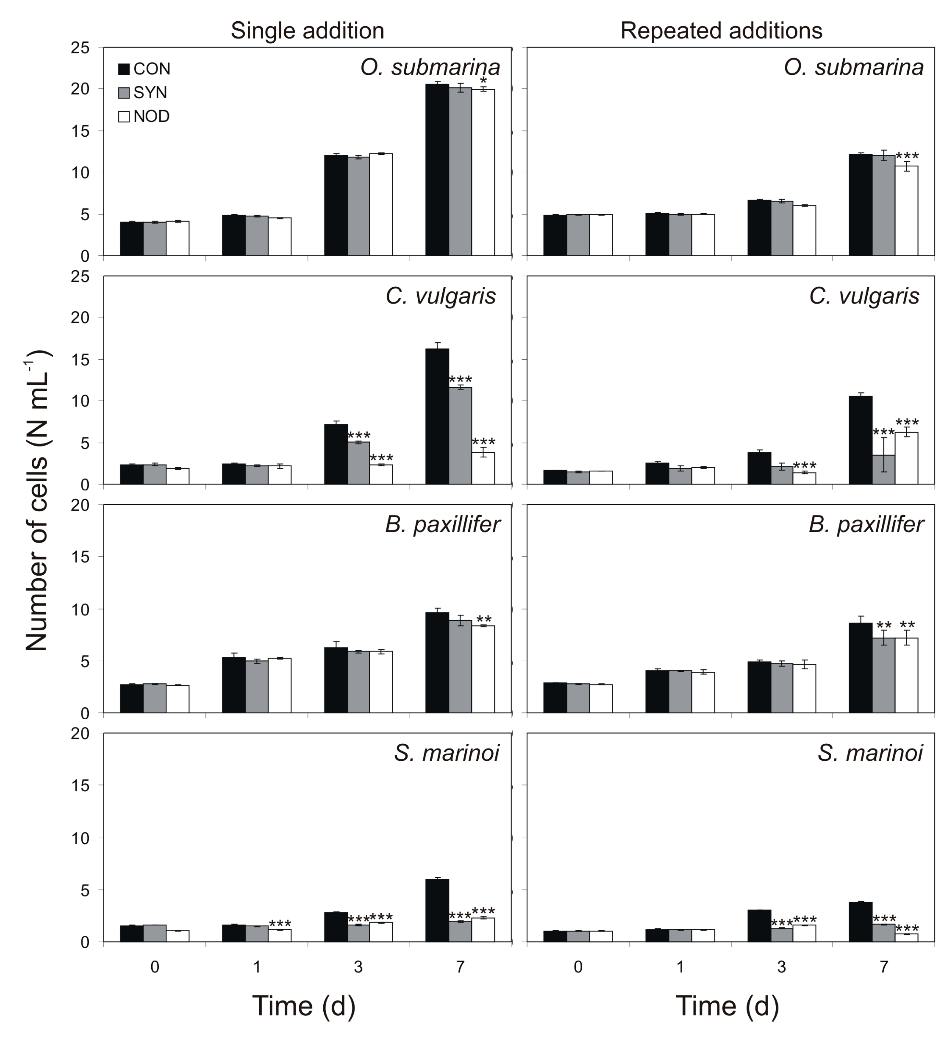
2.1. Effect of Cyanobacterial Exudates on Growth

The addition of exudates obtained from Synechococcus sp. from the exponential growth phase significantly decreased the growth of green alga C. vulgaris, as well as diatoms B. paxillifer and S. marinoi, but not green alga O. submarina. N. spumigena, however, showed a negative effect on all the species tested (Table 1, Figure 1).
The growth of C. vulgaris was also affected already on the third day by Synechococcus sp. filtrate, being, relative to the control, 70% (Tukey HSD test; p < 0.001) and 55% (Tukey HSD test; p < 0.001) for single and multiple exudates additions, respectively. On the seventh day, the growth in this microalga was, relative to the control, 72% (Tukey HSD test; p < 0.001) and 33% (Tukey HSD test; p < 0.001) for single and repeated filtrate addition, respectively. B. paxillifer growth was not so strongly inhibited as the other two species, showing a single significant effect for the repeated filtrate addition in the seventh day, being 83% relative to the control (Tukey HSD test; p < 0.01). The growth of S. marinoi was affected by Synechococcus sp. filtrate already at day 3, being, relative to the control, 57% (Tukey HSD test; p < 0.001) and 43% (Tukey HSD test; p < 0.001) with single and repeated additions of the filtrate, respectively. On the seventh day, the growth was reduced by 32% (Tukey HSD test; p < 0.001) and 44% (Tukey HSD test; p < 0.001) with single and repeated additions of the filtrate, respectively. O. submarina growth was not significantly affected by Synechococcus sp. filtrate.
N. spumigena showed a greater negative impact on tested microalgae than Synechococcus sp. filtrate. On the first, third, and seventh day, the growth of S. marinoi was reduced by 70% (Tukey HSD test; p < 0.001), 65% (Tukey HSD test; p < 0.001) and 39% (Tukey HSD test; p < 0.001) with single additions of filtrate, and by 52% (Tukey HSD test; p < 0.001) and 19% (Tukey HSD test; p < 0.001) with repeated exudates additions on the third and seventh day of the experiment. In C. vulgaris, the highest decrease in growth was observed, for the single and repeated filtrate addition, after 7 days of the experiment, being 24% and 59%, respectively, relative to the control (Tukey HSD test; p < 0.001 for both). In this same species, the single and repeated addition experiment showed a significant reduction of growth in the third day, for which the growth was, relative to the control, 33% (Tukey HSD test; p < 0.001) and 37% (Tukey HSD test; p < 0.001), respectively. The growth of B. paxillifer and O. submarina were affected already at the seventh day by N. spumigena filtrate, being, relative to the control, 87% (Tukey HSD test; p < 0.01) and 97% (Tukey HSD test; p < 0.05) for single filtrate additions and 83% (Tukey HSD test; p < 0.01) and 89% (Tukey HSD test; p < 0.001) for repeated exudates additions, respectively.
Surprisingly, the filtrate of the Synechococcus sp. and N. spumigena cultures in the stationary phase did not have a negative allelopathic effect on tested microalgae (Tukey HSD test; NS, p > 0.05 for all, Table 2). The only exception was the inhibition of the growth of the most sensitive species: C. vulgaris and S. marinoi. In C. vulgaris, for the experiment with Synechococcus sp. filtrate addition, the negative effect on growth was detected on the seventh day of exposure. The values were, relative to the control, 75% (Tukey HSD test; p < 0.001). Furthermore, the growth of S. marinoi was affected on the seventh day by N. spumigena exudates, being, relative to the control, 85% (Tukey HSD test; p < 0.01) for filtrate additions.

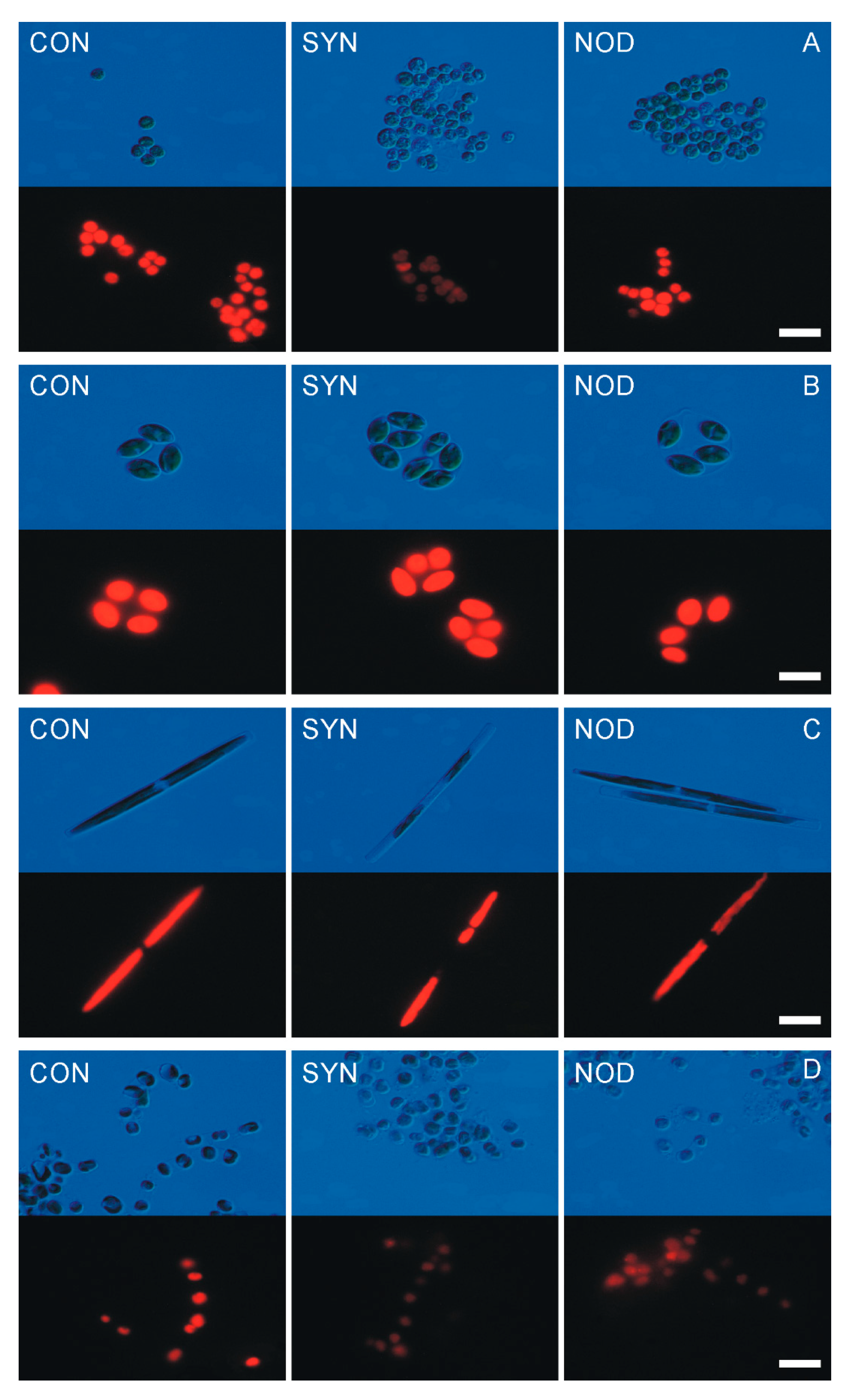
2.2. Allelopathic Effects of Cyanobacterial Exudates on the Cell Morphology

The same species whose growth was the most affected by filtrates (C. vulgaris, B. paxillifer, and S. marinoi) also experienced abnormal morphological changes (Figure 2). Cell bleaching and deformation were observed with light microscopy, and under epifluorescence microscopy, degeneration of thylakoids. In the assay with repeated addition of filtrate, the controls of C. vulgaris, B. paxillifer and S. marinoi showed, on average, 98%, 96% and 96%, of healthy cells, respectively, whereas the treatment showed the proportions of 71% (Mann–Whitney test; p < 0.001), 84% (Mann–Whitney test; p < 0.001) and 64% (Mann–Whitney test; p < 0.001), respectively for Synechococcus sp. and 68% (Mann–Whitney test; p < 0.001), 81% (Mann–Whitney test; p < 0.001) and 58% (Mann–Whitney test; p < 0.001), respectively for N. spumigena. No negative effect was observed against O. submarina.

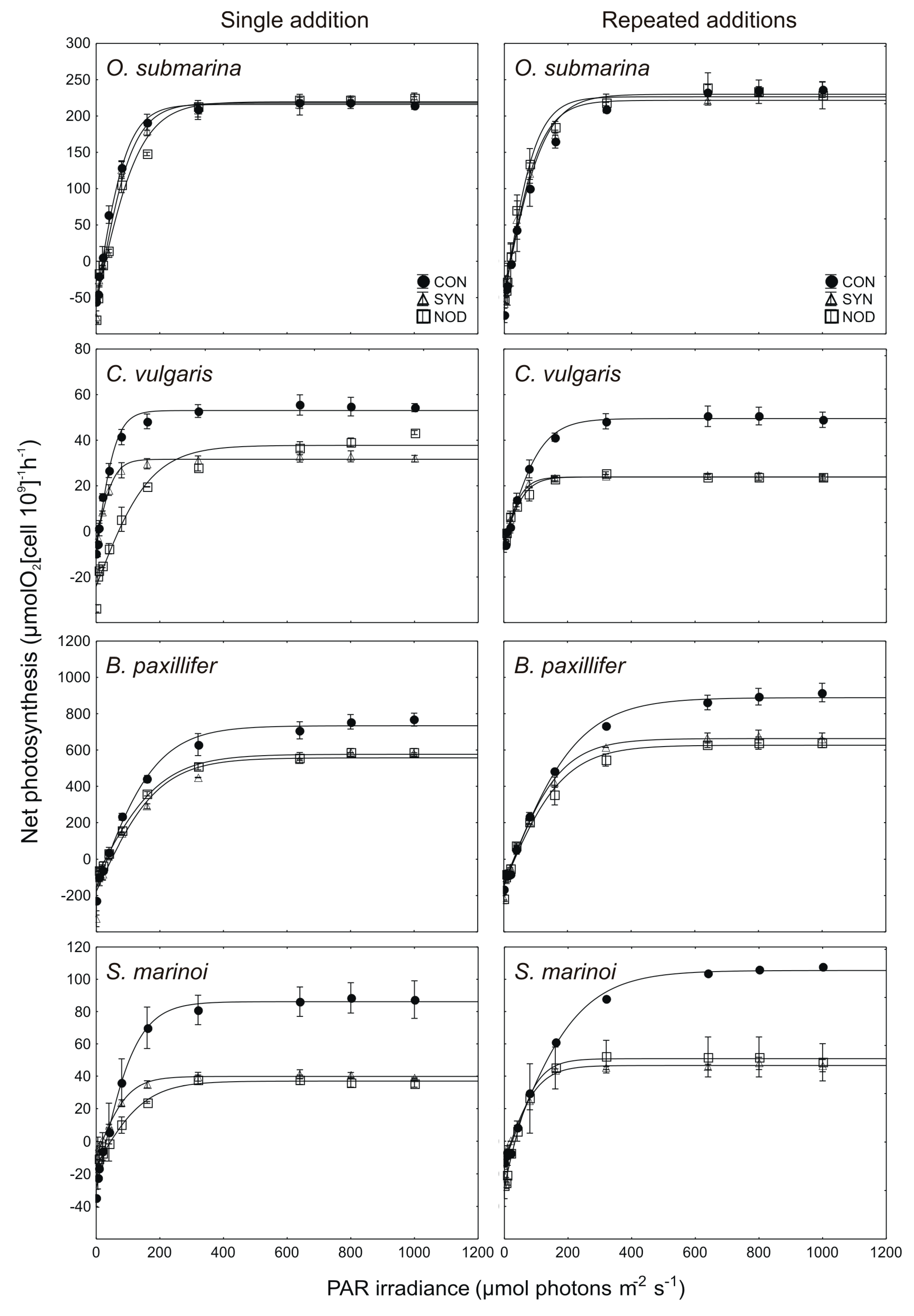
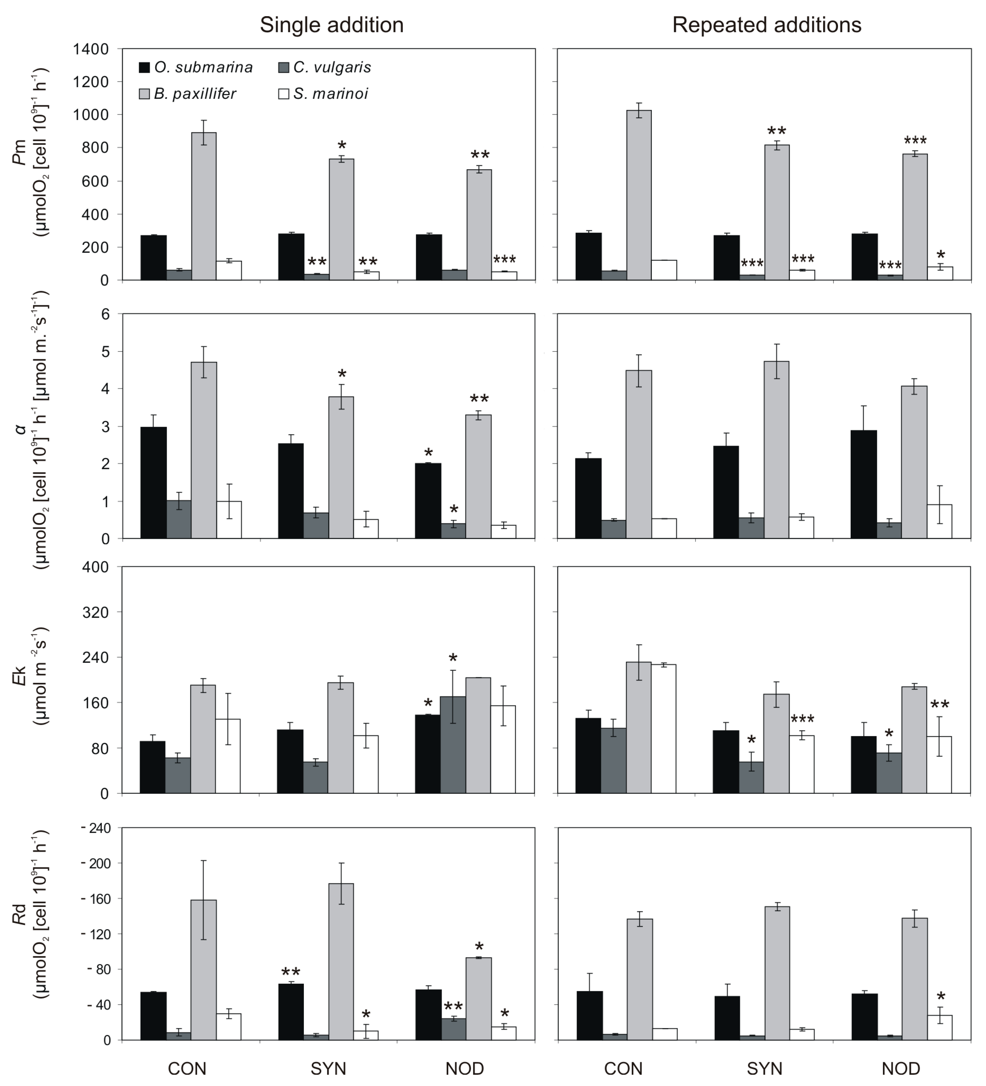
2.3. Allelopathic Effects of Cyanobacterial Exudates on P–E Curves and Photosynthetic Parameters

The effects of single and repeated addition of exudates from cyanobacterial cultures on the P–E curves and photosynthetic parameters pm, α, Ek, and Rd are shown in Figure 3 and Figure 4.
For single filtrate addition acquired from Synechococcus sp., the values of pm in C. vulgaris, B. paxillifer, and S. marinoi constituted, respectively, 61% (t-test; p < 0.01), 83% (t-test; p < 0.05), and 44% (t-test; p < 0.01), relative to the control. Furthermore, for the N. spumigena filtrate the significant negative effect was found for two tested diatoms: B. paxillifer and S. marinoi in the single filtrate addition experiment, for which it constituted 75% (t-test; p > 0.01) and 45% (t-test; p > 0.001), respectively, of the control. For this same parameter, in the repeated addition experiments, the values of pm for C. vulgaris, B. paxillifer, and S. marinoi were significantly different from the control by the seventh day, when they constituted, relative to the control, 52% (t-test; p < 0.001), 80% (t-test; p < 0.01), and 50% (t-test; p < 0.001), respectively for Synechococcus sp. exudates and 51% (t-test, p < 0.001), 75% (t-test; p < 0.001), and 67% (t-test; p < 0.05), respectively for N. spumigena filtrate. There was no significant effect of cyanobacterial filtrate on the pm value of O. submarina, (t-test; NS, p > 0.05 for all comparisons).
Regarding the α parameter, for B. paxillifer, in the single addition assay, it was 80% relative to the control (t-test; p < 0.05). For this same species, it was not found a significant effect in the repeated addition experiment (t-test; NS, p > 0.05). For single filtrate addition acquired from N. spumigena, the values of pm in O. submarina, C. vulgaris, and B. paxillifer constituted, respectively, 92% (t-test; p < 0.05), 42% (t-test; p < 0.05), and 70% (t-test; p < 0.01), relative to the control. There was no significant effect of repeated filtrate addition obtained from Synechococcus sp. and N. spumigena on the α parameter of the tested microalgal species (t-test, NS, p > 0.05).
The single addition of Synechococcus sp. exudates did not cause a significant effect in the parameter Ek for any of the species tested (t-test; NS, p > 0.05, for all comparisons). However, for single filtrate addition acquired from N. spumigena, the values of Ek in O. submarina and C. vulgaris constituted, respectively, 102% (t-test; p < 0.05) and 280% (t-test; p < 0.05), relative to the control. In the repeated exudates addition assay, the Ek values of C. vulgaris and S. marinoi were, relative to the control 49% (t-test; p < 0.05) and 45% (t-test; p < 0.001), respectively for Synechococcus sp. exudates and 63% (t-test; p < 0.05) and 44% (t-test; p < 0.01), respectively, for N. spumigena filtrate. There was no significant effect of repeated filtrate addition acquired from Synechococcus sp. and N. spumigena on the Ek of the O. submarina and B. paxillifer (t-test; NS, p > 0.05 for all).
For the dark respiration (parameter Rd) the only significant negative effect of Synechococcus sp. filtrate was found for S. marinoi in the single exudate addition assay, for which it constituted 38% of the control (t-test; p < 0.05). In the same single filtrate addition experiment, in O. submarina, the effect was significantly positive, being 17% higher than the control (t-test; p < 0.01). Repeated addition of Synechococcus sp. exudates did not cause a significant effect in the parameter Rd for any of the species tested (t-test; NS, p > 0.05, for all comparisons). Regarding the Rd parameter, for the diatoms B. paxillifer and S. marinoi, in the single addition experiment with exudates from N. spumigena, the reduction was 62% (t-test; p < 0.05) and 55% (t-test; p < 0.05) relative to the control. In the same experiment, in C. vulgaris, the effect was significantly positive, being 264% higher than the control (t-test; p < 0.01). A significant effect of N. spumigena in the multiple exudates addition treatment was found only for S. marinoi, for which it constituted 217% of the control (t-test; p < 0.05).

3. Discussion

3.1. Effect of the Selection of the Donor and Target Organisms

In the present work, we showed how several coexisting microalgae responded differently to cyanobacterial allelopathy. Coevolution and specific traits (cell wall thickness, surface-area relationship, etc.) are hypothesized to be the main reasons explaining the differences in susceptibility to allelochemicals [20]. These differences in susceptibility are found between major taxonomic groups, but also within them [21,22,23]. This differential effect on microalgae species suggests that cyanobacterial allelochemicals could have an important role in structuring phytoplankton communities.
We detected that population growth was the most negatively affected variable in the green alga Chlorella vulgaris and the diatom Skeletonema marinoi. Previous studies also reported detrimental allelopathic impact of cyanobacteria on green algae [16,24,25,26,27] and diatoms [17,20,27,28]. However, some species of green algae co-occurred with toxic and bloom-forming cyanobacteria during the summer period in the Baltic Sea [29,30,31]. Therefore, resistance to Synechococcus sp. and N. spumigena allelochemicals may differ between species within this group. The susceptibility of C. vulgaris and S. marinoi could be a consequence of its small cell size (less than 5 µm), and hence, relatively large surface to volume relationship, which enhances the efficiency of uptake of extracellular molecules [32]. Conversely, Synechococcus sp. exudates had no allelopathic impact on the abundance of Oocystis submarina. This could be due, at least in part, to the formation of colonies with thin, hyaline mucilaginous envelope. However, N. spumigena was characterized by a stronger allelopathic activity than Synechococcus sp., showing a negative effect on all tested species. This may be due to the release of higher amounts of allelochemicals or more potent compounds than those secreted by unicellular picocyanobacterium Synechococcus sp.

3.2. Effect of Single and Repeated Addition of Cyanobacterial Exudates

A greater amount of allelopathic compounds present in the filtrate should have a stronger effect on target organisms [20,33,34]. We also showed that negative effects against the population growth of tested microalgae were amplified by multiple exudates additions. These findings could be explained simply by the renewal of cyanobacterial allelochemicals, which naturally degrade in several ways, like bacterial activity or light [33,35].
We examined the allelopathic ability of Synechococcus sp. and N. spumigena using non-axenic cultures. Heterotrophic bacteria may mediate allelopathic interactions between different species by altering cyanobacterial allelochemicals or releasing chemical substances on their own [36]. However, Suikkanen et al. [20] revealed that exudates containing only allelochemicals released by heterotrophic bacteria did not affect target microalgae species, which would reduce the potential influence of accompanying bacteria in our work.

3.3. Comparison of Cyanobacterial Cell-Free Filtrate in Exponential and Stationary Growth Phases

A relevant biotic factor influencing allelopathic interactions is the growth phase of the donor organism. The release of allelopathic compounds by donor organisms could depend on their growth phase [7]. In this work, we demonstrated that Synechococcus sp. and N. spumigena were more allelopathic in exponential than in the stationary growth phase. Suikkanen et al. [20] and Kubanek et al. [7] also showed that the culture exudates acquired from N. spumigena and K. brevis, respectively, from the exponential phase of growth, had a detrimental allelopathic effect, whereas the exudates from stationary phase did not show this same effect. Schmidt and Hansen [37] concluded that cultures in the exponential growth phase exhibited greater allelopathic effects than cultures in stationary phase. However, this finding is not generalizable to all allelopathic phytoplankton species [38]. Authors that observed a greater effect for cultures from the exponential growth phase, suggested that allelopathic activity could be more related to the production of a complex of allelopathic compounds than to specific, single substances, i.e., toxins [39].

3.4. Mode of Action of Allelochemicals

Among the allelopathic effects detected in our study, there was physical damage to cell structures in three species, as observed by light and epifluorescence microscope. Other authors have described as well morphological changes or physical damage to cells caused by allelopathic activity [40,41,42].
Population growth and physical damage to cell structures could be end-of-chain (distal) effects. As a consequence of allelochemicals effects at the metabolic or physiological scale (proximal), general cell processes would be negatively affected, and the ultimate consequence could be physical cell damage and growth inhibition. However, it is also possible that cell physical damage was the proximal effect of allelochemicals, and then would come improper functioning of metabolism and physiological processes. This would be the case if allelochemicals were some sort of cytolytic compounds, not rare among phytoplankton [43,44].
We also tested the photosynthetic and dark respiration rates, to evaluate the effects on physiological processes in more detail. The inhibition of photosynthesis via the secretion of allelopathic compounds may be another effective strategy for cyanobacteria, as demonstrated by Ma et al. [16]. It is worth noting that the reduced performance of respiration and PS system might be due to the indirect consequence of the allelopathic mediation rather than a direct effect of secreted metabolites. In this work, we showed that the photosynthetic parameters of our tested organisms were significantly altered. pm was shown to be the most sensitive parameter to detect allelopathic effects. The parameter Ek is a photosynthetic characteristic that is independent of algal biomass [45,46]. Moreover, Light and Beardall [46] suggested that reduction of both the Ek and pm values, and a minimal change in α, indicates that agents other than main pigment concentration are impacting the photosynthetic performance of light-harvesting and energy transduction in microalgae. In our work, we assumed that this factor could be cyanobacterial allelopathy. Besides, the α parameter was also significantly reduced. The α parameter is the maximum light utilization (assimilation of CO2) coefficient. Allelochemicals may inhibit the CO2 assimilation rate by modifying the stomata function, as suggested by Zhou and Yu [47]. Much of the early work on the allelochemical modes of action considered their effects on respiration [48]. In this work, both a decrease and an increase in Rd parameters were observed. Respiration process can be directly disadvantaged through many modes of action. Secondary metabolites can both uncouple oxidative phosphorylation and inhibit mitochondrial electron transport.
One of the most unfinished tasks in the study of allelopathy is to identify the compounds responsible for the observed effects, where only a few works exist that have resolved this issue [2]. Cyanobacterial toxins have been thoroughly studied from the perspective of several fields of biology, but our study showed that non-toxic strains are also able to secrete compounds that inhibit co-occurring species. Our findings suggest that cyanobacterial allelochemicals are associated with the cell physical damage, as well as a reduced performance in respiration and photosynthesis system in the studied microalgae which cause the inhibition of the population growth. Moreover, our study has shown that some biotic factors such as specificity of donor and target organisms and growth phase of the donor species, that increase the intensity of allelopathic effects may also alter the ratio between bloom-forming cyanobacteria and some phytoplankton species that occur in the same aquatic ecosystem. The exact compounds involved in the observed effects were not studied and it remains to be investigated whether the observed effects result from the action of one particular metabolite or rather a mixture of different compounds. The identification of chemical structures of cyanobacterial metabolites and further studies focusing only on the isolated compounds are important steps toward elucidating their biological role.

4. Materials and Methods

4.1. Culture Conditions

The strains of Synechococcus sp. and Nodularia spumigena employed were CCBA-124 and CCBA-15, respectively, and the target microalgae were the green algae Oocystis submarina CCBA-01 and Chlorella vulgaris CCBA-80, and the diatoms Bacillaria paxillifer CCBA-14 and Skeletonema marinoi CCBA-98. All these strains were isolated from the Baltic Sea and were maintained as unispecies cultures in the Culture Collection of Baltic Algae (CCBA).
These strains were grown in f/2 culture medium [49] in 25-mL glass Erlenmeyer flasks that were stirred daily during the experiment. The f/2 medium was performed with Baltic Sea water filtered through glass fiber membranes (Whatman GF/C, Saint Louis, MO, USA) and autoclaved. Salinity was 8 PSU, measured with salinometer (inoLab Cond Level 1, Weilheim in Oberbayern, Germany). Cyanobacterial cultures were kept in a culture room at 20 °C with a 16:8 h light:dark cycle. The intensity of photosynthetically sctive radiation (PAR) was 190 μmol photons m−2s−1, measured with a quantum-meter (LI-COR; Lincoln, NE, USA) with a cosine collector. All the target cultures employed in the experiments were kept under the same conditions at least 7 days prior to the experiments.

4.2. Cell-Free Filtrate Experiments

Allelopathic effects were tested in conformity with the methods from Śliwińska-Wilczewska and Latała [27] with certain modifications. Allelopathic ability was identified by adding exudates acquired from donor cyanobacteria cultures to the target green algae and diatom species. The donor Synechococcus sp. and N. spumigena cultures were gently filtered using a 0.45-µm filter (Macherey-Nagel MN GF-5; Düren, Germany). The cell abundance in the donor cultures was about 107 mL−1 for Synechococcus sp. and 106 mL−1 for N. spumigena. Prior to the experiments, the concentrations of nutrients (N-NO3 and P-PO4) in the cyanobacterial cultures were measured using DR6000 Spectrophotometer (Hach-Lange, Loveland, CO, USA) according to Grasshoff [50]. Then, the concentrations of major nutrients in the controls and allelopathic assays were adjusted to the same level as in the f/2 medium. The filtrates obtained were analyzed on an epifluorescence microscope (Nikon Eclipse 80i; Tokyo, Japan) to verify the absence of donor cyanobacteria cells.
We performed two kind of experiments: (a) single addition (at the beginning of the experiments) of cell-free culture filtrate from the cyanobacteria; and (b) repeated addition (daily) of culture filtrate. The last experiments were aimed to simulate continuous release of allelochemicals. Treatments were executed by adding the cell-free filtrate (V = 2 mL) to 25-mL Erlenmeyer flasks containing cell suspensions of the target species (V = 20 mL). Controls consisted in the addition of filtered f/2 medium (V = 2 mL) to 25-mL Erlenmeyer flasks containing cell suspensions of the same microalgae species (V = 20 mL). Separate treatments were prepared with exudates from cyanobacterial cultures in exponential and stationary phases of growth. Synechococcus sp. and N. spumigena cultures from exponential growth phase were kept in a culture room at 8 PSU, 20 °C and 190 μmol photons m−2s−1 with a 16:8 h light:dark cycle for one week, while cyanobacterial cultures from stationary growth phases were kept in the same conditions for three weeks. The cyanobacterial growth phases were determined by monitoring cell abundances. In the repeated filtrate addition experiments, the filtrate was daily renewed by replacing 2-mL of cell suspension from the experimental flasks with an equal volume of fresh filtrate (for the experimental assay) or filtered f/2 medium (for the controls). All the experimental treatments had three replicates. The initial concentration of the experimental cultures was 0.8 µg Chl a mL−1. In the case of cultures obtained from the stationary growth phase, the Synechococcus sp. and N. spumigena were first diluted with the f/2 medium to obtain a suitable concentration of Chl a and then filtered to obtain a cell-free filtrate. This concentration was low enough to represent relevant environmental conditions and high enough to be measured properly. The experimental duration was 7 days.

4.3. Examination of Cell Abundances

Cell abundances were assessed by measuring optical density (OD) at 750 nm with a Multiskan GO spectrophotometer (Thermo Scientific, Waltham, MA, USA) and then transforming these values to cell abundances with a previously fitted linear regression model between cell abundance (N mL−1) and optical density (OD). In order to fit this model, cyanobacterial and microalgal cells were counted under the microscope in a Bürker counting chamber [51]. OD was measured in the same samples, and these data were used to fit a linear regression model between the variables N mL−1 and OD. The linear regression models for Synechococcus sp. and N. spumigena were chosen based on the model proposed by Barreiro Felpeto et al. [19]. The linear regression models for tested microalgae were described in Śliwińska-Wilczewska and Latała [27]. Cell abundances in the experiments were measured at times zeroth (1h) and first, third, and seventh day of the allelopathic assay.

4.4. Examination of Cell Morphology

Cell morphology of target species was examined in samples from the seventh day of the experiment with repeated additions of cell-free filtrate. The analysis followed the method from Barreiro Felpeto et al. [19]. The samples were determined using a microscope (Nikon Eclipse 80i, Tokyo, Japan). It would be best to observe the same cell under light and epifluorescence microscope. It is, however, not possible to do so as using epifluorescence microscopy requires filtering the material on the polycarbonate filter (Whaman Nuclepore Track-Etched Membranes, Saint Louis, MO, USA) and therefore, the same single cell cannot be viewed under a light microscope.

4.5. Measurements of the Rate of Photosynthesis

Oxygen production of target species were examined in the experiments with single and repeated additions of Synechococcus sp. and N. spumigena cell-free filtrate in exponential growth phases. Measurements of oxygen production were carried out on the seventh day of the experiment with a Clark-type oxygen electrode (Hansatech Chlorolab 2, King’s Lynn, UK). Experimental data were fitted to the photosynthesis irradiance response curves (P–E curves) using the equation from Jassby and Platt [52] with software Statistica® 13.1 (StatSoft Polska, Kraków, Poland). Following Sakshaug et al. [53], the parameters estimated were: photosynthetic capacity—pm, maximum light utilization coefficient—α, light saturation index—Ek, and dark respiration—Rd.

4.6. Statistical Analyses

Repeated measures ANOVA, with time (days) as covariate, was used to test the effect of cyanobacterial filtrate on growth of the tested microalgae. A post-hoc Tukey’s HSD test was used to determine significant differences between control and allelopathic assay. Differences in the abundance of the recognized morphological types of cells were analyzed with a Mann–Whitney test. The t-tests were used to determine whether the photosynthetic parameters of the target microalgae species, when treated with cyanobacterial exudates, differed from the control on the last day of the allelopathic assay. Data are showed as means ± standard deviations (SD). Levels of significance were: * p < 0.05; ** p < 0.01; *** p < 0.001. The statistical analyses were executed using Statistica® 13.1. (StatSoft Polska, Kraków, Poland).

Author Contributions

Methodology, K.M.; Supervision, V.V. and A.L.; Writing—original draft, S.Ś.-W. and A.B.F. All the authors were involved in the preparation of this manuscript.

Funding

This research were funded by BMN grant number 538-G245-B568-17 and FCT Project UID/Multi/04423/2019. The APC was funded by DS 530-G245-D717-18.

Acknowledgments

The authors would like to thank the anonymous Reviewers and Academic Editor for their valuable comments and suggestions to improve the quality of the paper.

Conflicts of Interest

The authors declare no conflict of interest.

References

  1. Pearson, L.; Mihali, T.; Moffitt, M.; Kellmann, R.; Neilan, B. On the chemistry, toxicology and genetics of the cyanobacterial toxins, microcystin, nodularin, saxitoxin and cylindrospermopsin. Mar. Drugs 2010, 8, 1650–1680. [Google Scholar] [CrossRef] [Green Version]
  2. Leão, P.N.; Engene, N.; Antunes, A.; Gerwick, W.H.; Vasconcelos, V. The chemical ecology of cyanobacteria. Nat. Prod. Rep. 2012, 29, 372–391. [Google Scholar] [CrossRef] [Green Version]
  3. Almeida, J.R.; Freitas, M.; Cruz, S.; Vasconcelos, V.; Cunha, I. Acetylcholinesterase in Biofouling Species: Characterization and Mode of Action of Cyanobacteria-Derived Antifouling Agents. Toxins 2015, 7, 2739–2756. [Google Scholar] [CrossRef] [PubMed] [Green Version]
  4. Poniedziałek, B.; Rzymski, P.; Kokociński, M. Cylindrospermopsin: Water-linked potential threat to human health in Europe. Environ. Toxicol. Pharmacol. 2012, 34, 651–660. [Google Scholar] [CrossRef] [PubMed]
  5. Poniedziałek, B.; Rzymski, P.; Kokociński, M.; Karczewski, J. Toxic potencies of metabolite(s) of non-cylindrospermopsin producing Cylindrospermopsis raciborskii isolated from temperate zone in human white cells. Chemosphere 2015, 120, 608–614. [Google Scholar] [CrossRef] [PubMed]
  6. Testai, E.; Scardala, S.; Vichi, S.; Buratti, F.M.; Funari, E. Risk to human health associated with the environmental occurrence of cyanobacterial neurotoxic alkaloids anatoxins and saxitoxins. Crit. Rev. Toxicol. 2016, 46, 385–419. [Google Scholar] [CrossRef]
  7. Kubanek, J.; Hicks, M.K.; Naar, J.; Villareal, T.A. Does the red tide dinoflagellate Karenia brevis use allelopathy to outcompete other phytoplankton? Limnol. Oceanogr. 2005, 50, 883–895. [Google Scholar] [CrossRef]
  8. Figueredo, C.C.; Giani, A.; Bird, D.F. Does allelopathy contribute to Cylindrospermopsis raciborskii (cyanobacteria) bloom occurrence and geographic expansion? J. Phycol. 2007, 43, 256–265. [Google Scholar] [CrossRef]
  9. Prince, E.K.; Myers, T.L.; Kubanek, J. Effects of harmful algal blooms on competitors: Allelopathic mechanisms of the red tide dinoflagellate Karenia brevis. Limnol. Oceanogr. 2008, 53, 531–541. [Google Scholar] [CrossRef]
  10. Hattenrath-Lehmann, T.K.; Gobler, C.J. Allelopathic inhibition of competing phytoplankton by North American strains of the toxic dinoflagellate, Alexandrium fundyense: Evidence from field experiments, laboratory experiments, and bloom events. Harmful Algae 2011, 11, 106–116. [Google Scholar] [CrossRef]
  11. Franzè, G.; Pierson, J.J.; Stoecker, D.K.; Lavrentyev, P. Diatom-produced allelochemicals trigger trophic cascades in the planktonic food web. Limnol. Oceanogr. 2017, 63, 1093–1108. [Google Scholar] [CrossRef] [Green Version]
  12. Rodríguez-Ramos, T.; Lorenzo, P.; González, L. Marine Allelopathy: Principles and perspectives. Thalassas 2007, 23, 39–49. [Google Scholar]
  13. Granéli, E.; Hansen, P.J. Allelopathy in Harmful Algae: A mechanism to compete for resources? In Ecology of Harmful Algae; Granéli, E., Turner, J., Eds.; Series: Ecological Studies 189; Springer: Berlin, Germany, 2006; pp. 189–201. [Google Scholar]
  14. Von Elert, E.; Jüttner, F. Phosphorus limitation and not light controls the extracellular release of allelopathic compounds by Trichormus doliolum (Cyanobacteria). Limnol. Oceanogr. 1997, 42, 1796–1802. [Google Scholar] [CrossRef] [Green Version]
  15. Smith, G.D.; Doan, D.T. Cyanobacterial metabolites with bioactivity against photosynthesis in cyanobacteria, algae and higher plants. J. Appl. Phycol. 1999, 11, 337–344. [Google Scholar] [CrossRef]
  16. Ma, Z.L.; Fang, T.X.; Thring, R.W.; Li, Y.B.; Yu, H.G.; Zhou, Q.; Zhao, M. Toxic and non-toxic strains of Microcystis aeruginosa induce temperature dependent allelopathy toward growth and photosynthesis of Chlorella vulgaris. Harmful Algae 2015, 48, 21–29. [Google Scholar] [CrossRef]
  17. Śliwińska-Wilczewska, S.; Pniewski, F.; Latała, A. Allelopathic activity of the picocyanobacterium Synechococcus sp. under varied light, temperature and salinity conditions. Int. Rev. Hydrobiol. 2016, 101, 69–77. [Google Scholar] [CrossRef]
  18. Gross, E.M. Allelopathy of aquatic autotrophs. Crit. Rev. Plant Sci. 2003, 22, 313–339. [Google Scholar] [CrossRef] [Green Version]
  19. Barreiro Felpeto, A.; Śliwińska-Wilczewska, S.; Złoch, I.; Vasconcelos, V. Light-dependent cytolysis in the allelopathic interaction between picoplanktic and filamentous cyanobacteria. J. Plankton Res. 2018, 40, 165–177. [Google Scholar] [CrossRef] [Green Version]
  20. Suikkanen, S.; Fistarol, G.O.; Granéli, E. Allelopathic effects of the Baltic cyanobacteria Nodularia spumigena, Aphanizomenon flos-aquae and Anabaena lemmermannii on algal monocultures. J. Exp. Mar. Biol. Ecol. 2004, 308, 85–101. [Google Scholar] [CrossRef]
  21. Barreiro, A.; Hairston, N.G., Jr. The influence of resource limitation on the allelopathic effect of Chlamydomonas reinhardtii on other unicellular freshwater planktonic organisms. J. Plankton Res. 2013, 35, 1339–1344. [Google Scholar] [CrossRef] [Green Version]
  22. Barreiro Felpeto, A.; Vasconcelos, V.M. Interactions between allelopathic properties and growth kynetics in four freshwater phytoplankton species studied by model simulations. Aquat. Ecol. 2014, 48, 191–205. [Google Scholar] [CrossRef]
  23. Barreiro Felpeto, A.; Roy, S.; Vasconcelos, V.M. Allelopathy prevents competitive exclusion and promotes phytoplankton biodiversity. Oikos 2017, 127, 85–98. [Google Scholar] [CrossRef] [Green Version]
  24. Antunes, J.T.; Leão, P.N.; Vasconcelos, V.M. Influence of Biotic and Abiotic Factors on the Allelopathic Activity of the Cyanobacterium Cylindrospermopsis raciborskii Strain LEGE 99043. Microb. Ecol. 2012, 64, 584–592. [Google Scholar] [CrossRef] [PubMed]
  25. Bittencourt-Oliveira, M.D.C.; Chia, M.A.; Oliveira, H.S.B.D.; Araújo, M.K.C.; Molica, R.J.R.; Dias, C.T.S. Allelopathic interactions between microcystin-producing and non-microcystin-producing cyanobacteria and green microalgae: Implications for microcystins production. J. Appl. Phycol. 2014, 27, 275–284. [Google Scholar] [CrossRef]
  26. Żak, A.; Kosakowska, A. The influence of extracellular compounds produced by selected Baltic cyanobacteria, diatoms and dinoflagellates on growth of green algae Chlorella vulgaris. Estuar. Coast. Shelf Sci. 2015, 167, 113–118. [Google Scholar] [CrossRef]
  27. Śliwińska-Wilczewska, S.; Latała, A. Allelopathic activity of the bloom-forming picocyanobacterium Synechococcus sp. on the coexisting microalgae: The role of eutrophication. Int. Rev. Hydrobiol. 2018, 103, 37–47. [Google Scholar] [CrossRef]
  28. Wang, L.; Zi, J.; Xu, R.; Hilt, S.; Hou, X.; Chang, X. Allelopathic effects of Microcystis aeruginosa on green algae and a diatom: Evidence from exudates addition and co-culturing. Harmful Algae 2017, 61, 56–62. [Google Scholar] [CrossRef]
  29. Suikkanen, S.; Laamanen, M.; Huttunen, M. Long-term changes in summer phytoplankton communities of the open northern Baltic Sea. Estuar. Coast. Shelf Sci. 2007, 71, 580–592. [Google Scholar] [CrossRef]
  30. Suikkanen, S.; Pulina, S.; Engström-Öst, J.; Lehtiniemi, M.; Lehtinen, S.; Brutemark, A. Climate Change and Eutrophication Induced Shifts in Northern Summer Plankton Communities. PLoS ONE 2013, 8, e66475. [Google Scholar] [CrossRef]
  31. Śliwińska-Wilczewska, S.; Maculewicz, J.; Tuszer, J.; Dobosz, K.; Kalusa, D.; Latała, A. First record of allelopathic activity of the picocyanobacterium Synechococcus sp. on a natural plankton community. Ecohydrol. Hydrobiol. 2017, 17, 227–234. [Google Scholar] [CrossRef]
  32. Lyczkowski, E.R.; Karp-Boss, L. Allelopathic effects of Alexandrium fundyense (Dinophyceae) on Thalassiosira cf. gravida (Bacillariophyceae): A matter of size. J. Phycol. 2014, 50, 376–387. [Google Scholar] [CrossRef] [PubMed]
  33. Fistarol, G.O.; Legrand, C.; Granéli, E. Allelopathic effect on a nutrient-limited phytoplankton species. Aquat. Microb. Ecol. 2005, 41, 153–161. [Google Scholar] [CrossRef] [Green Version]
  34. Śliwińska-Wilczewska, S.; Maculewicz, J.; Barreiro Felpeto, A.; Vasconcelos, V.; Latała, A. Allelopathic activity of the picocyanobacterium Synechococcus sp. on filamentous cyanobacteria. J. Exp. Mar. Biol. Ecol. 2017, 496, 16–21. [Google Scholar] [CrossRef]
  35. Christoffersen, K.; Lyck, S.; Winding, A. Microbial activity and bacterial community structure during degradation of microcystins. Aquat. Microb. Ecol. 2002, 27, 125–136. [Google Scholar] [CrossRef] [Green Version]
  36. Cole, J.J. Interactions between bacteria and algae in aquatic ecosystems. Annu. Rev. Ecol. Evol. Syst. 1982, 13, 291–314. [Google Scholar] [CrossRef]
  37. Schmidt, L.E.; Hansen, P.J. Allelopathy in the prymnesiophyte Chrysochromulina polylepis: Effect of cell concentration, growth phase and pH. Mar. Ecol. Prog. Ser. 2001, 216, 67–81. [Google Scholar] [CrossRef]
  38. Issa, A.A. Antibiotic production by the cyanobacteria Oscillatoria angustissima and Calothrix parietina. Environ. Toxicol. Pharmacol. 1999, 8, 33–37. [Google Scholar] [CrossRef]
  39. Arzul, G.; Seguel, M.; Guzman, L.; Denn, E.E. Comparison of allelopathic properties in three toxic Alexandrium species. J. Exp. Mar. Biol. Ecol. 1999, 232, 285–295. [Google Scholar] [CrossRef]
  40. Valdor, R.; Aboal, M. Effects of living cyanobacteria, cyanobacterial extracts and pure microcystins on growth and ultrastructure of microalgae and bacteria. Toxicon 2007, 49, 769–779. [Google Scholar] [CrossRef]
  41. Gantar, M.; Berry, J.P.; Thomas, S.; Wang, M.; Perez, R.; Rein, K.S. Allelopathic activity among cyanobacteria and microalgae isolated from Florida freshwater habitats. FEMS Microbiol. Ecol. 2008, 64, 55–64. [Google Scholar] [CrossRef] [Green Version]
  42. Pichierri, S.; Accoroni, S.; Pezzolesi, L.; Guerrini, F.; Romagnoli, T.; Pistocchi, R.; Totti, C. Allelopathic effects of diatom filtrates on the toxic benthic dinoflagellate Ostreopsis cf. ovata. Mar. Environ. Res. 2017, 131, 116–122. [Google Scholar] [CrossRef] [PubMed]
  43. Tillmann, U. Kill and eat your predator: A winning strategy of the marine flagellate Prymnesium parvum. Aquat. Microb. Ecol. 2003, 32, 73–84. [Google Scholar] [CrossRef] [Green Version]
  44. Tillmann, U.; John, U.; Cembella, A. On the allelochemical potency of the marine dinoflagellate Alexandrium ostenfeldi agains heterotrophic and autotrophic protists. J. Plankton Res. 2007, 29, 527–543. [Google Scholar] [CrossRef]
  45. Prézelin, B.B. Light reactions in photosynthesis. In Physiological Bases of Phytoplankton Ecology; Platt, T., Ed.; Canadian Journal of Fisheries and Aquatic Sciences, 210; Canadian Government Publishing: Ottawa, QC, Canada, 1981; pp. 1–46. [Google Scholar]
  46. Light, B.R.; Beardall, J. Photosynthetic characteristics of sub-tidal benthic microalgal populations from a temperate, shallow water marine ecosystem. Aquat. Bot. 2001, 70, 9–27. [Google Scholar] [CrossRef]
  47. Zhou, Y.H.; Yu, J.Q. Allelochemicals and photosynthesis. In Allelopathy: A Physiological Process with Ecological Implications; Reigosa, M.J., Pedrol, N., González, L., Eds.; Springer: Dordrecht, The Netherlands, 2006; pp. 127–139. [Google Scholar]
  48. Duke, S.O.; Dayan, F.E. Modes of action of phytotoxins from plants. In Allelopathy: A Physiological Process with Ecological Implications; Reigosa, M.J., Pedrol, N., González, L., Eds.; Springer: Dordrecht, The Netherlands, 2006; pp. 511–536. [Google Scholar]
  49. Guillard, R.R.L. Culture of phytoplankton for feeding marine invertebrates. In Culture of Marine Invertebrate Animals; Smith, W.L., Chanley, M.H., Eds.; Plenum Press: New York, NY, USA, 1975. [Google Scholar]
  50. Grasshoff, K. Methods of Seawater Analysis; Verlag Chemie Weinheim: New York, NY, USA, 1976. [Google Scholar]
  51. Guillard, R.R.; Sieracki, M.S. Counting cells in cultures with the light microscope. In Algal Culturing Techniques; Andersen, R.A., Ed.; Elsevier Academic Press: Burlington, MA, USA, 2005; pp. 239–252. [Google Scholar]
  52. Jassby, A.D.; Platt, T. Mathematical formulation of the relationship between photosynthesis and light for phytoplankton. Limnol. Oceanogr. 1976, 21, 540–547. [Google Scholar] [CrossRef] [Green Version]
  53. Sakshaug, E.; Bricaud, A.; Dandonneau, Y.; Falkowski, P.G.; Kiefer, D.A.; Legendre, L.L.; Morel, A.; Parslow, J.; Takahashi, M. Parameters of photosynthesis: Definitions, theory and interpretation of results. J. Plankton Res. 1997, 19, 1637–1670. [Google Scholar] [CrossRef] [Green Version]
Figure 1. Number of cells (N) for O. submarina (105 cell mL−1), C. vulgaris (106 cell mL−1), B. paxillifer (105 cell mL−1), and S. marinoi (106 cell mL−1) for controls (CON) and treatments with single and repeated additions of exudates acquired from Synechococcus sp. (SYN) and N. spumigena (NOD) cultures in exponential growth phases (n = 3, mean ± SD). * signify statistically significant difference between allelopathic treatment and control (ANOVA and Tukey’s post hoc test: * p < 0.05; ** p < 0.01; *** p < 0.001).
Toxins 11 00712 g001
Figure 2. The cells morphology of C. vulgaris (A), O. submarina (B), B. paxillifer (C), and S. marinoi (D) for control (CON) and treatments with repeated additions of the exudates acquired from Synechococcus sp. (SYN) and N. spumigena (NOD) cultures in exponential growth phases after 7 days of exposure under a light microscope (top panel) and under an epifluorescence microscope (bottom panel). Scale bar = 10 µm.
Toxins 11 00712 g002
Figure 3. P–E curves for tested microalgae for controls (CON) and experiments with single and multiple additions of exudates acquired from Synechococcus sp. (SYN) and N. spumigena (NOD) cultures in exponential growth phases after 7 days of exposure (n = 3, mean ± SD).
Toxins 11 00712 g003
Figure 4. Photosynthetic parameters: pm, a, Ek, and Rd for target species for controls (CON) and treatments with a single and multiple additions of the exudates acquired from Synechococcus sp. (SYN) and N. spumigena (NOD) cultures in exponential growth phases after 7 days of exposure (n = 3, mean ± SD). * signify statistically significant difference between allelopathic treatment and control (ANOVA and Tukey’s post hoc test: * p < 0.05; ** p < 0.01; *** p < 0.001).
Toxins 11 00712 g004
Table 1. Results of two-way factorial ANOVA for single and repeated additions of exudates acquired from cyanobacteria cultures in exponential growth phases (Df—degrees of freedom; Ss—sum of squares; Mss—mean sum of squares; F—Fisher's F-test statistic; * p < 0.05; ** p < 0.01; *** p < 0.001).
Response Variable (Number of Cells)Source of VariationDfSsMssFSsMssF
Synechococcus sp.N. spumigena
Single Additions of Cell-Free Filtrate
O. submarinafiltrate10.1840.1842.8960.1630.1634.595 *
time31039.170346.3905459.262 ***1037.309345.7709726.294 ***
filtrate time *30.1350.0450.7080.5430.1815.090 *
error161.0150.063 0.5690.036
C. vulgarisfiltrate117.83917.839138.156 ***119.410119.410721.685 ***
time3535.876178.6251383.420 ***242.59780.866488.730 ***
filtrate time *320.0646.68851.798 ***144.63448.211291.376 ***
error162.0660.129 2.6470.165
B. paxilliferfiltrate10.8130.8136.688 *1.2161.21613.770 **
time3129.40343.134355.008 ***119.79039.930452.322 ***
filtrate time *30.4090.1361.1231.3460.4495.083 *
error161.9440.122 1.4120.088
S. marinoifiltrate110.58810.5881341.763 ***11.72411.7241517.087 ***
time322.6327.544955.952 ***31.07810.3591340.444 ***
filtrate time *315.9645.321674.305 ***10.3153.438444.896 ***
error160.1260.008 0.1240.008
Repeated Additions of Cell-Free Filtrate
O. submarinafiltrate10.0290.0290.3871.5001.50026.882 ***
time3204.98968.330898.485 ***170.11656.7051016.222 ***
filtrate time *30.0250.0080.1081.8440.61511.018 ***
error161.2170.076 0.8930.056
C. vulgarisfiltrate134.01634.01656.588 ***19.96019.960238.473 ***
time3109.67236.55760.816 ***178.84559.615712.265 ***
filtrate time *345.66115.22025.320 ***17.2625.75468.748 ***
error169.6180.601 1.3390.084
B. paxilliferfiltrate11.0961.0968.723 **1.4561.45610.204 **
time385.71228.571227.319 ***86.87128.957202.981 ***
filtrate time *32.0090.6705.328 *1.7640.5884.121 *
error162.0110.126 2.2830.143
S. marinoifiltrate15.7505.7502175.294 ***7.9157.9153161.742 ***
time311.7303.9101479.173 ***8.2532.7511098.877 ***
filtrate time *35.6181.873708.431***9.5513.1841271.713 ***
error160.0420.003 0.0400.003
Table 2. Effects of stationary phase culture filtrates of Synechococcus sp. and N. spumigena on target species, expressed as the number of cells (N): O. submarina, B. paxillifer—105 cell mL−1; C. vulgaris, S. marinoi—106 cell mL−1, and the percentage of intact cells in the treated microalgal cultures in relation to the control (n = 3, mean ± SD) and results of two-way factorial ANOVA for additions of exudates acquired from cyanobacterial cultures (Df—degrees of freedom; Ss—sum of squares; Mss —mean sum of squares; F—Fisher's F-test statistic; * p < 0.05; ** p < 0.01; *** p < 0.001).
Target SpeciesDayNumber of Cells (N)% of ControlSource of VariationDfSsMssF
Synechococcus sp.
O. submarina00.619 ± 0.007100.3 ± 1.5filtrate10.0040.0045.045 *
10.649 ± 0.01598.2 ± 1.6time31.5710.524651.147 ***
30.815 ± 0.03297.1 ± 2.5filtrate time *30.0040.0011.816
71.227 ± 0.05494.6 ± 1.9error160.0130.001
C. vulgaris02.635 ± 0.09899.9 ± 9.6filtrate12.8852.88515.230 **
12.545 ± 0.06196.4 ± 12.3time378.56626.189138.273 ***
33.692 ± 0.20588.4 ± 5.4filtrate time *34.2821.4277.536 **
75.994 ± 0.24774.7 ± 11.3error163.0300.189
B. paxillifer00.267 ± 0.01299.1 ± 2.7filtrate10.0010.0011.809
10.254 ± 0.00287.9 ± 2.3time30.3120.104359.793 ***
30.405 ± 0.022101.1 ± 3.6filtrate time *30.0010.0001.605
70.543 ± 0.03699.1 ± 5.0error160.0050.000
S. marinoi01.379 ± 0.06399.8 ± 2.4filtrate10.0810.0814.915 *
11.757 ± 0.09997.4 ± 4.2time368.18722.7291380.932 ***
32.822 ± 0.06298.6 ± 4.6filtrate time *30.1290.0432.621
75.508 ± 0.20593.7 ± 2.4error160.2630.016
N. spumigena
O. submarina00.621 ± 0.009100.7 ± 1.1filtrate10.0020.0022.496
10.661 ± 0.009100.0 ± 0.9time31.6230.541562.660 ***
30.799 ± 0.03095.2 ± 2.3filtrate time *30.0030.0011.015
71.253 ± 0.06796.6 ± 3.5error160.0150.001
C. vulgaris01.903 ± 0.07394.6 ± 8.5filtrate11.1571.15710.760 **
12.179 ± 0.02890.3 ± 13.9time332.58010.860100.966 ***
32.391 ± 0.11291.9 ± 4.4filtrate time *31.0410.3473.226
74.335 ± 0.60979.8 ± 16.2error161.7210.108
B. paxillifer00.273 ± 0.012101.2 ± 2.5filtrate10.0030.00313.622 **
10.254 ± 0.00587.9 ± 1.5time30.2620.087350.093 ***
30.386 ± 0.02696.3 ± 4.0filtrate time *30.0020.0013.151
70.499 ± 0.02791.1 ± 3.6error160.0040.000
S. marinoi01.083 ± 0.016104.0 ± 1.4filtrate10.0860.0869.304 **
11.192 ± 0.016100.0 ± 0.0time38.0202.673290.170 ***
31.825 ± 0.00895.0 ± 0.0filtrate time *30.1990.0667.218 **
72.294 ± 0.08384.7 ± 5.4error160.1470.009

