Article

Potential of Matrix Metalloproteinase Inhibitors for the Treatment of Local Tissue Damage Induced by a Type P-I Snake Venom Metalloproteinase

by
Lina María Preciado
1,
Jaime Andrés Pereañez
1,* and
Jeffrey Comer
2,*
1
Programa de Ofidismo/Escorpionismo, Facultad de Ciencias Farmacéuticas y Alimentarias, Universidad de Antioquia UdeA, Calle 70 No. 52-21, Medellín 050010, Colombia
2
Institute of Computational Comparative Medicine, Kansas State University, Manhattan, KS 66506, USA
*
Authors to whom correspondence should be addressed.
Submission received: 30 October 2019 / Revised: 7 December 2019 / Accepted: 12 December 2019 / Published: 20 December 2019
(This article belongs to the Special Issue Novel Strategies for the Diagnosis and Treatment of Snakebites)

Abstract

:
Snake bite envenoming is a public health problem that was recently included in the list of neglected tropical diseases of the World Health Organization. In the search of new therapies for the treatment of local tissue damage induced by snake venom metalloproteinases (SVMPs), we tested the inhibitory activity of peptidomimetic compounds designed as inhibitors of matrix metalloproteinases on the activities of the SVMP Batx-I, from Bothrops atrox venom. The evaluated compounds show great potential for the inhibition of Batx-I proteolytic, hemorrhagic and edema-forming activities, especially the compound CP471474, a peptidomimetic including a hydroxamate zinc binding group. Molecular dynamics simulations suggest that binding of this compound to the enzyme is mediated by the electrostatic interaction between the hydroxamate group and the zinc cofactor, as well as contacts, mainly hydrophobic, between the side chain of the compound and amino acids located in the substrate binding subsites S1 and S1 . These results show that CP471474 constitutes a promising compound for the development of co-adjuvants to neutralize local tissue damage induced by snake venom metalloproteinases.
Key Contribution: The peptidomimetic compound CP471474 was identified as a promising candidate for the inhibition of proteolytic, hemorrhagic and edema-forming activities of a P-I type snake venom metalloproteinase. Molecular dynamics simulations coupled with the adaptive biasing force method revealed that the hydroxamate group of this compound coordinates the zinc cofactor of the metalloproteinase through strong electrostatic interactions, while the compound’s hydrophobic groups occupy the S1 and S1′ sites.

Graphical Abstract

1. Introduction

Snake bite envenoming is a major public health problem, which the World Health Organization has recognized as a Category A neglected tropical disease since 2017 [1]. Envenoming causes mortality and morbidity mainly in tropical and subtropical areas, such as sub-Saharan Africa, South and Southeast Asia, Papua New Guinea and Latin America [2], with annual deaths estimated at 100,000 and sequelae in more than 400,000 cases [3]. In Latin America, recent estimates suggest about 57,500 annual envenomings, with an incidence of 6.2 per 100,000 people [2]. In Colombia specifically, 5434 cases were reported in 2018, with an incidence of 10.9 per 100,000 people [4]. It is estimated that snakes of the Bothrops genus (Viperidae), B. asper and B. atrox, cause about 70–90% of all envenomings in Colombia.
Envenoming by Bothrops species is characterized by prominent local tissue damage, which causes pain, edema, blistering, hemorrhage, and myonecrosis [5]. In moderate and severe cases, there are also systemic manifestations characterized mainly by hemorrhage, coagulopathies, acute renal failure and cardiovascular shock [6]. These effects are induced by the main toxins of the venom: snake venom metalloproteinases (SVMPs), serine proteinases and phospholipases A 2 [7].
The SVMPs are zinc-dependent endopeptidases, with a molecular masses between 20 and 100 kDa. These enzymes are members of the subfamily of proteinases known as metzincins. They contain a consensus zinc binding sequence, HEXXHXXGXX, and a 1–4 turn containing a highly conserved methionine residue that underlies the three histidine residues and zinc cofactor involved in the catalysis [8]. SVMPs are classified as type P-I, P-II, or P-III, depending on domain organization. P-I SVMPs consist only of a proteinase domain, while P-II SVMPs possess proteinase and disintegrin domains. P-III SVMPs have proteinase, disintegrin, and cysteine rich domains [9]. BaP1, a P-I metalloproteinase isolated from the venom of B. asper inhabiting the Pacific region of Costa Rica, has been extensively studied since its crystallographic structure was reported [10]. P-I type metalloproteinases constitute about 30% of Pacific B. asper venom proteins [11]. The structure of the BaP1 is shown in Figure 1. When a polypeptide is hydrolyzed by the SVMP, its scissile peptide bond is positioned near the zinc cofactor of the metalloproteinase and the residues preceding and following this bond make contact, respectively, with the S1 and S1 subsites (Figure 1A) [12,13]. The residues of BaP1 in the active site, as well as the catalytic water molecule, are highlighted in Figure 1B,C.
The primary biological activity of SVMPs is local and systemic hemorrhage, mediated by proteolytic degradation of extracelullar matrix proteins of capillary vessels, such as type IV collagen, nidogen, laminin and perlecan [14]. Additionally, these enzymes induce myotoxicity, inflammation, edema and blistering [15], effects that are difficult to neutralize by antivenoms due to their rapid onset, especially if there is a delay in serotherapy administration [16]. In May 2019, the World Health Organization launched a plan of work with the goal of reducing incidence of snakebite death and disability by half, through strengthening of health systems and the provision of safe and effective treatments [17]. This plan directly promotes research and development for small-molecule SVMP inhibitors that could be injected in the field.
To the date, the most promising inhibitor of SVMPs is a synthetic hydroxamate peptidomimetic known as batimastat. This compound was initially developed as a matrix metalloproteinase (MMP) inhibitor to treat cancer [18] and later demonstrated neutralization of the biological activities of the snake venom metalloproteinase BaP1, from B. asper venom. Specifically, batimastat inhibited the proteolytic, hemorrhagic, dermonecrotic, and edema forming activities of BaP1 when it was injected intramuscularly at the site of BaP1 injection immediately following injection of the latter. However, this inhibition was reduced when there was a time lag between the injection of BaP1 and the injection of batimastat [19]. Similarly, it was determined that batimastat inhibits B. asper venom lethality when a venom challenge dose of double the LD 50 was administered by intraperitoneal and intravenous routes, with ED 50 values of 250 and 22 μ M, respectively. In addition, this compound exerted a significant inhibition of in vitro coagulant and in vivo defibrinogenating activities of the venom, demonstrating its ability to neutralize the systemic effects induced by B. asper venom [20].
Recently, the peptidomimetic hydroxamate molecules batimastat and marimastat were shown to inhibit local and pulmonary hemorrhagic, coagulant, defibrinogenating, and proteinase activities of Echis ocellatus venom in pre-incubation assays [21], demonstrating the potential of synthetic matrix metalloproteinase inhibitors as therapeutic tools against snake bite envenoming.
Therefore, in this study we evaluated the potential of four commercial MMP inhibitors, 4-aminobenzoyl-Gly-Pro-D-Leu-D-Ala hydroxamic acid (4AM), ARP-100 (ARP), CP471474 (CP) and phosphoramidon (PH) [22,23,24,25] selected from previous molecular docking studies, for the inhibition of proteolytic, hemorrhagic and edema forming activities of the metalloproteinase Batx-I isolated from B. atrox venom from Colombia. The mechanisms of inhibition were studied by the computational techniques molecular docking and molecular dynamics.
ARP-100 (ARP) is a hydroxamate peptidomimetic with a sulfonamide group and a biphenyl moiety that has been reported as a MMP-2-preferring inhibitor with an IC 50 value of 12 nM [23]. 4-aminobenzoyl-Gly-Pro-D-Leu-D-Ala hydroxamic acid (4AM) is a tetrapeptidyl hydroxamic acid; it is alternatively called MMP Inhibitor I (IC 50 = 1.0 μ M). It also inhibits MMP-8 (IC 50 = 1.0 μ M) and MMP-9 (IC 50 = 50 μ M) [22,26]. CP 471474 (CP) is a pyran-containing sulfonamide hydroxamate that was developed by Pfizer. It is a broad spectrum MMP inhibitor (IC 50 values are 0.7, 0.9, 13, 16 and 1170 nM for MMP-2, MMP-13, MMP-9, MMP-3 and MMP-1, respectively) [27]. This compound also inhibits cigarette smoke-induced lung inflammation and the progression of emphysema in guinea pig models [28]. The fourth compound considered in this work, phosphoramidon (PH), lacks a hydroxamate group: it possesses instead a phosphonate, which may serve a similar role as an anionic group that can coordinate the catalytic metal ion of metalloproteinases. It was initially isolated from Streptomyces tanashiensis cultures, and it has been reported as an inhibitor of the metalloendopeptidase thermolysin [29] and membrane-bound zinc metalloproteinases, neprilysin (NEP) and NEP2, with a K i of 2 nM for both enzymes [30]. The structures of the inhibitors considered in this work are shown in Figure 2.

2. Results and Discussion

2.1. Inhibition of Proteolytic Activity

Snake venom metalloproteinases are the most abundant active component of viperid venoms [7]. These enzymes play a fundamental role in the pathogenesis of local tissue damage characteristic of viperid envenomations [31]. Specifically the metalloproteinase Batx-I from B. atrox venom exhibits weak hemorrhagic activity and lacks coagulant and defibrinating activity, but it is able to induce a mild myonecrosis. This metalloproteinase may contribute to the hemorrhage and local tissue damage observed in patients envenomed by B. atrox from Colombia [32,33], since it constitutes about the 45% of venom proteins. Batx-I hemorrhagic activity is similar to the activity reported for other P-I type SVMPs [34], but with less potency compared to P-III type SVMPs.
The delay between envenomation and antivenom injection means that antivenoms are often unable to prevent serious local tissue damage [16]. Thus, the administration of synthetic metalloproteinase inhibitors that could be administered in the field directly at the site of venom injection may represent a valid alternative to confront this difficult problem.
In this study, all the tested compounds inhibited B. atrox venom and Batx-I proteolytic activity in a dose-dependent fashion. Initially, all the peptidomimetics were tested at concentrations 250, 125 and 62.5 μ M. The most active compounds were ARP and CP, which inhibited more than 70% enzyme activity at a concentration 62.5 μ M, as shown in Figure S1 of the Supplementary Information (SI). The IC 50 dose-response curve for the inhibition of Batx-I proteolytic activity is shown in Figure S2 of the SI. The obtained IC 50 values for the inhibition of B. atrox venom and Batx-I proteolytic activity are shown in Table 1 and Table 2, respectively. The peptidomimetic CP, which has a hydroxamate zinc-binding group and a sulfonamide group was the most active for the inhibition of the proteolytic activity, followed by ARP, 4AM and PH. Together with the computational study described below, these results suggest that peptidomimetic compounds with a hydroxamate group (CP, ARP, 4AM) have a greater affinity for the metalloproteinase Batx-I, compared to compounds with a phosphonate group (PH).

2.2. Inhibition of Hemorrhagic Activity

Hemorrhagic activity of Batx-I was diminished by 4AM, ARP, CP and PH with IC 50 values of 92.9, 168.3, 2.5 and 321.1 μ M, respectively, for pre-incubation assays Table 2. These values are consistent with the IC 50 values obtained for inhibition of proteolytic activity without pre-incubation. Notably, CP was the most active compound for both assays.
For studies with independent injection, all the studied compounds exhibited statistically significant inhibition of hemorrhagic lesions when they were administered immediately after metalloproteinase injection, in concentrations equivalent to 3 and 5 times the IC 50 value obtained for inhibition of hemorrhagic activity with pre-incubation (Figure 3A)). When the time lapse between metalloproteinase injection and peptidomimetics administration was increased to 5 min, inhibition was significant for 4AM (p value < 0.05) and CP (p value < 0.01), using concentrations equivalent to 5 times the IC 50 value (Figure 3B). Thus, the peptidomimetic CP appeared to be the most effective compound for neutralizing hemorrhagic activity of Batx-I when administered after metalloproteinase injection.
Previous studies with the peptidomimetic batimastat and the metalloproteinase BaP1 demonstrated significant inhibition of hemorrhage activity in independent injection assays only when this compound was administered at a concentration of 200 μ M at intervals of 0 or 1 min after toxin injection [19]. Although the results obtained with CP suggest a greater inhibitory potential of this compound with respect to batimastat, the methodologies to estimate hemorrhage are different and the results should not be directly compared.
In all cases, the concentrations we used for the in vivo assays were higher than those used in studies to test the potential of these MMP inhibitors to treat cancer. For instance, ARP has been tested at concentrations of 13 nM and 25 μ M [35,36] and CP471474 at 100 nM [37,38]. We did not perform cytotoxicity assays for the evaluated compounds, but all the experiments had negative controls with the inhibitors at the highest tested concentrations. No evidence of toxicity was observed in the negative controls, suggesting some level of safety for the inhibitor concentrations used. However, acute and chronic toxicity assays would need to be performed to verify the safety of these inhibitors at therapeutic concentrations.

2.3. Inhibition of Edema-Forming Activity

Assays for inhibition of Batx-I edema-forming activity were performed by measuring the plantar pad thickness of mice injected with 30 μ g of toxin and the inhibitors at various time intervals. The concentration evaluated was equivalent to three times the IC 50 value for inhibition of hemorrhagic activity with pre-incubation. Peptidomimetic compounds demonstrated activity against Batx-I edema-forming activity. All the evaluated compounds reduced significantly Batx-I edema-forming activity at all the evaluated time intervals Figure 4. The most active compound was CP, which inhibited edema formation with p value < 0.01 at times of 1, 2 and 4.5 h.
These compounds’ inhibition of Batx-I edema-forming activity is consistent with their inhibition hemorrhagic activity, as well as their previously reported anti-inflammatory activity for different pathologies [39,40].
In addition, it has been described that different endogeneous MMPs, especially the latent forms of MMP-9 and MMP-2, are expressed in muscle injected with the metalloproteinase BaP1 [41]. Hence, SVMPs may activate these endogenous MMPs, which in turn may modulate various aspects of inflammation [42,43]. However, it is necessary to perform further studies to determine the effect of Batx-I on the release and activation of endogenous inflammation mediators such as MMPs and interleukin-1 β and interleukin-6 and the inhibitory potential of studied peptidomimetics.

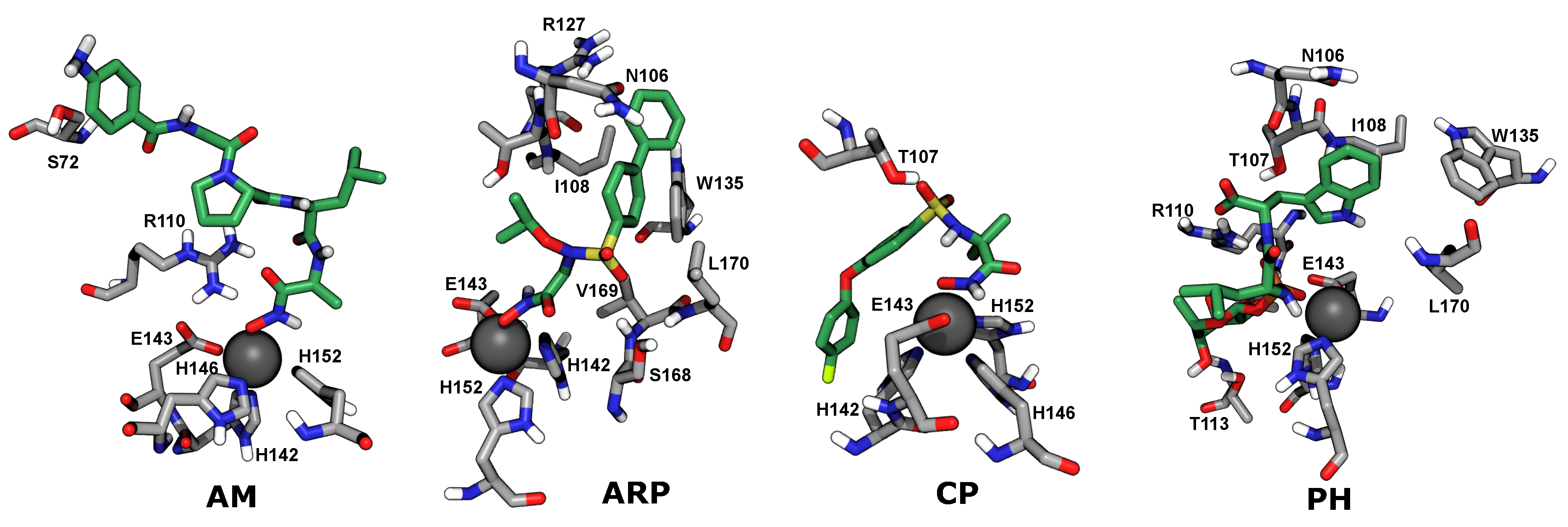
2.4. Computational Analysis of Inhibitor–BaP1 Interactions

To better understand the role of the hydroxamate, phosphonate, sulfonamide, and hydrophobic groups of the inhibitors in SVMP binding and zinc coordination, we applied computational techniques using an available x-ray crystal structure of BaP1 [10]. Molecular docking with AutoDock Vina did not show the expected contacts between the zinc binding groups (hydroxamate or phosphonate) and the Zn 2 + cofactor. Therefore, we turned to explicit-solvent molecular dynamics simulation using the CHARMM force field [44,45] to study the interactions between the compounds and the metalloproteinases in a more realistic manner. An enhanced sampling technique [46] was employed to obtain the free energy (potential of mean force) as a function of the distance between the hydroxamate or phosphonate group of each compound and the Zn 2 + ion. The resulting potential of mean force functions are shown in Figure 5.
The potential of mean force for each compound exhibits a single free-energy minimum. In all cases, the simulations showed that binding of these compounds to BaP1 is mainly mediated by the strong electrostatic interaction between hydroxamate or phosphonate groups and the catalytic Zn 2 + cofactor (Figure 6). Also, hydrophobic contacts between the bulky aromatic groups of each compound and amino acids and amino acid side chains located in substrate binding subsites S1 and S1 appeared to reinforce the binding.
The hydroxamate group appears to make a major contribution to the affinity of the studied peptidomimetics for the metalloproteinase BaP1. This zinc binding group is a bidentate anion that binds the Zn 2 + cofactor creating a distorted trigonal bipyramidal geometry around it. This geometry was previously described for a peptidomimetic inhibitor possessing a zinc-binding hydroxamate group, co-crystallized with the metalloproteinase BaP1 from the B. asper venom (PDB code 2W15) [10]. In addition, the effectiveness of this group is mediated by the possibility of hydrogen bond formation between the heteroatoms of the hydroxamate (specifically the NH group and the deprotonated OH group) with the side chain of Glu143 or the carbonyl of Ala111, amino acids highly conserved in all metalloproteinases of the metzincin family [47].
In the design of sulfonamide hydroxamates, such as CP and ARP, the sulfonamide group was incorporated to form hydrogen bonds and improve the enzyme–inhibitor binding by directing the hydrophobic substituent into the S1 pocket [48]. Consistent with this, we observe hydrogen bonding between the phosphonate oxygen atoms of CP and the hydroxy group Thr107, as exemplified in Figure 6, CP. Furthermore, we also find contacts between sulfonamide-attached hydrophobic groups and side chains of residues making up the S1 subsite, such as Ser168, Leu170, and Trp135. An example of these contacts is seen in Figure 6, ARP.

3. Conclusions

We found that the peptidomimetics considered here inhibited proteolytic and hemorrhagic activities of a P-I type SVMP in a dose-dependent manner. The hemorrhagic activity was reduced when the inhibitors were independently injected 5 min after the injection of the metalloproteinase. Peptidomimetics bearing a hydroxamate group (4AM, ARP, CP) were found to be more active than the compound PH, which carried a phosphonate instead. Molecular dynamics simulations supported the supposition that the hydroxamate groups of 4AM, ARP, and CP coordinated the Zn 2 + cofactor by strong electrostatic interactions. The phosphonate of PH also coordinated the Zn 2 + cofactor, but more weakly, which was partially responsible for the weaker predicted BaP1-binding affinity of PH as compared to the 4AM, ARP, and CP.

4. Materials and Methods

4.1. Venom

A venom pool was obtained by manual extraction from ten B. atrox adult specimens from the department of Meta (southwest region of Colombia) kept in captivity at the University of Antioquia serpentarium (Medellin, Colombia). The venom was centrifuged at 800× g for 15 min and the supernatant was lyophilized and stored at −70 C until use.

4.2. Chemicals and Reagents

The compounds 4-aminobenzoyl-Gly-Pro-D-Leu-D-Ala hydroxamic acid, ARP100, CP471474 and phosphoramidon, were purchased from Santa Cruz Biotechnology, Dallas, TX, USA. They were diluted in 1% DMSO in buffer. Other reagents used in this work were of the highest purity available from MilliporeSigma, St. Louis, MO, USA.

4.3. Animals

Swiss Webster mice, 18–20 g body weight, were used for the in vivo assays. All experiments were conducted in accordance with guidelines of the Universidad de Antioquia Ethics Committee (Acta No. 90, August 2014).

4.4. Purification of Metalloproteinase Batx-I

The metalloproteinase Batx-I was isolated from B. atrox venom. The toxin was purified following established methods [32]. The toxin purity was judged by RP-HPLC and SDS-PAGE [49]. Batx-I was lyophilized and stored at −20 C until its use.

4.5. Inhibition of Proteolytic Activity

Proteolytic activity was assayed on fluorescein conjugates of gelatin using the EnzCheck Gelatinase/Collagenase assay kit (Molecular Probes Inc., Eugene, OR, USA) following the protocol described by [33]. Aliquots of 80 μ L of the compounds at different concentrations dissolved in buffer (0.05 M Tris-HCl, 0.15 M NaCl, 5 mM CaCl 2 , 0.2 mM sodium azide) were added to each well of a 96-well plate. Then, 20 μ L of substrate (6.25 μ g/ μ L) followed by 100 μ L of the active enzyme (1 μ g/ μ L) or venom (2 μ g/ μ L) was added and the fluorescence intensity was measured by a Synergy HT Multi-Mode Microplate Reader (BioTekInstruments, Inc.; Winooski, VT, USA) for excitation at 485 nm and emission detection at 515 nm at each minute for 60 min. Initially, inhibitor concentrations were 250, 125, and 62.5 μ M were tested. Subsequently, to better delineate the dose–response curve, we tested a few further concentration values specific to each inhibitor (see Figure S2 of the SI) Each reaction was performed in quadruplicate. The decrease in fluorescence compared with the enzyme activity (positive control) was recorded to determine percent inhibition.

4.6. Inhibition of Hemorrhagic Activity

Hemorrhagic activity was quantitatively determined following the method adapted by [50]. Groups of four mice were injected intradermally with different concentrations of each inhibitor and the metalloproteinase (30 μ g) previously pre-incubated at 37 C for 30 min. The metalloproteinase alone was used as positive control and each compound alone at the highest evaluated concentration were used as negative controls. After 2 h, the animals were euthanized by CO 2 inhalation, the skin was removed and diameters of the hemorrhagic lesions were measured. The evaluated concentrations were in the interval 25–400 μ M for 4AM and ARP, 0.625–20 μ M for CP, and 25–500 μ M for PH, following a series of two-fold dilutions in these ranges.
For independent injection experiments, groups of four mice were injected intradermally with 30 μ g of Batx-I and, subsequently, the compounds were injected at time intervals of 0 and 5 min in concentrations equivalent to three and five times the IC 50 value obtained for experiments with pre-incubation.

4.7. Inhibition of Edema-Forming Activity

Edema was quantified in the footpad of mice by measuring the increase of thickness with a caliper. The metallopoteinase Batx-I (30 μ g) was pre-incubated for 30 min at 37 C with a concentration equivalent to 3 times the IC 50 value for the inhibition of hemorrhagic activity with preincubation. Later, 50 μ L of the toxin-inhibitor mixture was injected subcutaneously into the right footpads of mice. The same controls described above were included. Edema was assessed after 1, 2, 4 and 6 h by measuring the footpad thickness with a caliper and expressed as percent of edema relative to the diameter of left footpad injected with saline solution. Each experiment was performed by quadruplicate.

4.8. Molecular Models

The software Avogadro 1.90.0 was used to build the compounds structure and to optimize its conformation by an energy minimization process based on the MMF94 force field. The structure of the metalloproteinase BaP1 from B. asper venom from Costa Rica (PDB code 2W15) [10] was used for computational studies since the 3D structure of Batx-I is not available. However, these SVMPs have comparable biological activities and portions of their sequences are identical [32]. The software AutoDock Vina (Scripps Research Institute, San Diego, CA, USA) was used to dock the studied peptidomimetics into BaP1 active site.

4.9. Molecular Dynamics

The compounds were parameterized with the CHARMM General Force Field using the ParamChem web interface [44,51]. The structure of each compound was built according to its protonation state at physiological pH. Thus, the oxygen atom of hydroxyl group of hydroxamate group was deprotonated (ionized). The metalloproteinase was represented in the simulations by the CHARMM36m force field for proteins [45,52] and constructed with the CHARMM-GUI server [53,54]. Conventional molecular dynamics simulations were performed with NAMD 2.13 [55] and analyzed with VMD 1.9.4a37 [56]. Lennard-Jones interactions were calculated with a 12 Å cutoff. The pressure was maintained at 101.325 kPa using the Langevin piston method. The temperature was maintained at 310.15 K using a Langevin thermostat with a damping parameter of 1 ps 1 . All simulations were performed with mass repartitioning of ligand and protein hydrogen atoms [57]. The mass distribution of water molecules was not altered. Electrostatic interactions were calculated using the particle-mesh Ewald method [58] with a grid spacing <1.2. Water molecules were represented by the TIP3P model [59]. Sodium and chloride ions (0.15 M NaCl) were added to the aqueous phase. Additional ions were added to obtain charge neutrality.

4.10. Free Energy Calculations

The potentials of mean force were calculated by a hybrid of metadynamics and extended adaptive biasing force (meta-eABF) [46] using the implementation provided in the Colvars module of NAMD [60]. This algorithm uses the extended ABF method [61,62,63] to reduce free-energy barriers and calculate free energies (by integrating the potential of mean force), while a history-dependent potential term (metadynamics) [64] drives evolution of the system into less well-sampled regions of the transition coordinate.
To calculate the binding free energy of the peptidomimetics to the metalloproteinase BaP1 active site, the radial transition coordinate r was defined as r = ( X X 0 ) 2 + ( Y Y 0 ) 2 + ( Z Z 0 ) 2 where ( X , Y , Z ) is the center of mass of the hydroxamate group for AM, ARP and CP, or phosphoramidate group for PH and ( X 0 , Y 0 , Z 0 ) is the center of mass of the Zn + 2 ion. Meta-eABF was applied along the transition coordinate on the interval 1.8 r 20.0 Å using a bin size of 0.05 Å. A cylindrical restraint was applied to define the unbinding pathway, similar to the restraint used in funnel metadynamics [65]. The standard binding free energy was calculated by:
Δ G = k T ln π R 0 2 C 0 d r exp [ β Δ G ( r ) ]
where β = 1 k B T is the inverse thermal energy, R 0 is the radius of the restraint cylinder and C 0 is the standard concentration (1/(1660.5389 Å 3 ) ) . Four ABF independent calculations were performed using the simulation conditions previously described. Statistical uncertainties in free energies were inferred from the differences between the four independent calculations [33].

Supplementary Materials

The following are available online at https://0-www-mdpi-com.brum.beds.ac.uk/2072-6651/12/1/8/s1, Figure S1: Fluorescence intensity in the experiment to measure the proteolytic activity of Batx-I in the presence of the inhibitors, Figure S2: Inhibition of Batx-I proteolytic activity as a function of concentration.

Author Contributions

Conceptualization, L.M.P., J.A.P. and J.C.; Formal analysis, L.M.P., J.A.P. and J.C.; Resources, J.A.P. and J.C.; Supervision, J.A.P. and J.C.; Writing—original draft, L.M.P. Writing—review & editing, L.M.P., J.A.P. and J.C.; Funding acquisition, J.A.P. and J.C. All authors have read and agreed to the published version of the manuscript.

Funding

Comite para el desarrollo de la investigación (CODI-CIQF-217), Universidad de Antioquia (UdeA) and Kansas State University have financed this work. Computing for this project was performed on the Beocat Research Cluster at Kansas State University, which is funded in part by NSF grants CNS-1006860, EPS-1006860, EPS-0919443, and CHE-1726332. This work was partially supported by the Kansas Bioscience Authority funds to the Institute of Computational Comparative Medicine (ICCM) and to the Nanotechnology Innovation Center of Kansas State University (NICKS).

Acknowledgments

This project was developed with the permission to access genetic resources and their derived products from Ministerio de Ambiente y Desarrollo Sostenible, Colombia [No. 152 of 2017, resolution 2405].

Conflicts of Interest

The authors declare no conflict of interest.

Abbreviations

The following abbreviations are used in this manuscript:
ABFAdaptive biasing force
ColvarsCollective variables
eABFExtended adaptive biasing force
meta-eABFExtended adaptive biasing force with sampling enhanced by metadynamics
MMPsMatrix metalloproteinases
SVMPsSnake venom metalloproteinases

References

  1. Chippaux, J.P. Snakebite envenomation turns again into a neglected tropical disease! J. Venom. Anim. Toxins Incl. Trop. Dis. 2017, 23, 38. [Google Scholar] [CrossRef]
  2. Chippaux, J.P. Incidence and mortality due to snakebite in the Americas. PLoS Negl. Trop. Dis. 2017, 11, e0005662. [Google Scholar] [CrossRef]
  3. Gutiérrez, J.M.; Calvete, J.J.; Habib, A.G.; Harrison, R.A.; Williams, D.J.; Warrell, D.A. Snakebite envenoming. Nat. Rev. Dis. Primers 2017, 3, 17063. [Google Scholar] [CrossRef]
  4. Informe Final de Accidente ofídico Colombia año 2018. Available online: https://www.ins.gov.co/buscador-eventos/Informesdeevento/ACCIDENTE%20OF%C3%8DDICO_2018.pdf (accessed on 16 December 2019).
  5. Otero-Patiño, R. Epidemiological, clinical and therapeutic aspects of Bothrops asper bites. Toxicon 2009, 54, 998–1011. [Google Scholar] [CrossRef]
  6. Gutiérrez, J.M.; Rucavado, A.; Chaves, F.; Díaz, C.; Escalante, T. Experimental pathology of local tissue damage induced by Bothrops asper snake venom. Toxicon 2009, 54, 958–975. [Google Scholar] [CrossRef]
  7. Georgieva, D.; Arni, R.K.; Betzel, C. Proteome analysis of snake venom toxins: Pharmacological insights. Exp. Rev. Proteomic 2008, 5, 787–797. [Google Scholar] [CrossRef]
  8. Bode, W.; Gomis-Rüth, F.X.; Stöckler, W. Astacins, serralysins, snake venom and matrix metalloproteinases exhibit identical zinc-binding environments (HEXXHXXGXXH and Met-turn) and topologies and should be grouped into a common family, the ‘metzincins’. FEBS Lett. 1993, 331, 134–140. [Google Scholar] [CrossRef] [Green Version]
  9. Fox, J.W.; Serrano, S.M. Insights into and speculations about snake venom metalloproteinase (SVMP) synthesis, folding and disulfide bond formation and their contribution to venom complexity. FEBS J. 2008, 275, 3016–3030. [Google Scholar] [CrossRef]
  10. Lingott, T.; Schleberger, C.; Gutiérrez, J.M.; Merfort, I. High-resolution crystal structure of the snake venom metalloproteinase BaP1 complexed with a peptidomimetic: Insight into inhibitor binding. Biochemistry 2009, 48, 6166–6174. [Google Scholar] [CrossRef]
  11. Alape-Girón, A.; Sanz, L.; Escolano, J.; Flores-Diaz, M.; Madrigal, M.; Sasa, M.; Calvete, J.J. Snake venomics of the lancehead pitviper Bothrops asper: Geographic, individual, and ontogenetic variations. J. Proteome Res. 2008, 7, 3556–3571. [Google Scholar] [CrossRef]
  12. Berger, A.; Schechter, I. Mapping the active site of papain with the aid of peptide substrates and inhibitors. Proc. R. Soc. Lond. B. (Biol. Sci.) 1970, 257, 249–264. [Google Scholar] [CrossRef]
  13. Gupta, S.P.; Patil, V.M. Specificity of binding with matrix metalloproteinases. In Matrix Metalloproteinase Inhibitors; Springer: Berlin, Germany, 2012; pp. 35–56. [Google Scholar]
  14. Gutiérrez, J.; Escalante, T.; Rucavado, A.; Herrera, C. Hemorrhage caused by snake venom metalloproteinases: A journey of discovery and understanding. Toxins 2016, 8, 93. [Google Scholar] [CrossRef] [Green Version]
  15. Gutiérrez, J.M.; Rucavado, A. Snake venom metalloproteinases: Their role in the pathogenesis of local tissue damage. Biochimie 2000, 82, 841–850. [Google Scholar] [CrossRef]
  16. Gutiérrez, J.M.; León, G.; Rojas, G.; Lomonte, B.; Rucavado, A.; Chaves, F. Neutralization of local tissue damage induced by Bothrops asper (terciopelo) snake venom. Toxicon 1998, 36, 1529–1538. [Google Scholar] [CrossRef]
  17. Lancet. Snakebite—Emerging from the shadows of neglect. Lancet 2019, 393, 2175. [Google Scholar] [CrossRef]
  18. Rasmussen, H.S. Batimastat and Marimastat in Cancer. In Antiangiogenic Agents in Cancer Therapy; Springer: Berlin, Germany, 1999; pp. 399–405. [Google Scholar]
  19. Escalante, T.; Franceschi, A.; Rucavado, A.; Gutiérrez, J.M. Effectiveness of batimastat, a synthetic inhibitor of matrix metalloproteinases, in neutralizing local tissue damage induced by BaP1, a hemorrhagic metalloproteinase from the venom of the snake Bothrops asper. Biochem. Pharmacol. 2000, 60, 269–274. [Google Scholar] [CrossRef]
  20. Rucavado, A.; Escalante, T.; Gutiérrez, J. Effect of the metalloproteinase inhibitor batimastat in the systemic toxicity induced by Bothrops asper snake venom: Understanding the role of metalloproteinases in envenomation. Toxicon 2004, 43, 417–424. [Google Scholar] [CrossRef]
  21. Arias, A.S.; Rucavado, A.; Gutiérrez, J.M. Peptidomimetic hydroxamate metalloproteinase inhibitors abrogate local and systemic toxicity induced by Echis ocellatus (saw-scaled) snake venom. Toxicon 2017, 132, 40–49. [Google Scholar] [CrossRef]
  22. Odake, S.; Morita, Y.; Morikawa, T.; Yoshida, N.; Hori, H.; Nagai, Y. Inhibition of matrix metalloproteinases by peptidyl hydroxamic acids. Biochem. Biophys. Res. Commun. 1994, 199, 1442–1446. [Google Scholar] [CrossRef]
  23. Rossello, A.; Nuti, E.; Orlandini, E.; Carelli, P.; Rapposelli, S.; Macchia, M.; Minutolo, F.; Carbonaro, L.; Albini, A.; Benelli, R.; et al. New N-arylsulfonyl-N-alkoxyaminoacetohydroxamic acids as selective inhibitors of gelatinase A (MMP-2). Bioorg. Med. Chem. 2004, 12, 2441–2450. [Google Scholar] [CrossRef]
  24. Rohde, L.E.; Ducharme, A.; Arroyo, L.H.; Aikawa, M.; Sukhova, G.H.; Lopez-Anaya, A.; McClure, K.F.; Mitchell, P.G.; Libby, P.; Lee, R.T. Matrix metalloproteinase inhibition attenuates early left ventricular enlargement after experimental myocardial infarction in mice. Circulation 1999, 99, 3063–3070. [Google Scholar] [CrossRef] [Green Version]
  25. Umezawa, H. Structures and activities of protease inhibitors of microbial origin. In Methods in Enzymology; Elsevier: Amsterdam, The Netherlands, 1976; Volume 45, pp. 678–695. [Google Scholar]
  26. Odake, S.; Okayama, T.; Obata, M.; Morikawa, T.; Hattori, S.; HORI, H.; Nagai, Y. Vertebrate collagenase inhibitor. II. Tetrapeptidyl hydroxamic acids. Chem. Pharm. Bull. 1991, 39, 1489–1494. [Google Scholar] [CrossRef] [Green Version]
  27. Jain, P.; Saravanan, C.; Singh, S.K. Sulphonamides: Deserving class as MMP inhibitors? Eur. J. Med. Chem. 2013, 60, 89–100. [Google Scholar] [CrossRef]
  28. Selman, M.; Cisneros-Lira, J.; Gaxiola, M.; Ramirez, R.; Kudlacz, E.M.; Mitchell, P.G.; Pardo, A. Matrix metalloproteinases inhibition attenuates tobacco smoke-induced emphysema in guinea pigs. Chest 2003, 123, 1633–1641. [Google Scholar] [CrossRef]
  29. Van den Burg, B.; Eijsink, V. Thermolysin and related Bacillus metallopeptidases. In Handbook of Proteolytic Enzymes; Elsevier: Amsterdam, The Netherlands, 2004; pp. 374–387. [Google Scholar]
  30. Suda, H.; Aoyago, T.; Takeuchi, T.; Umezawa, H. A thermolysin inhibitor produced by actinomycetes: Phosphoramidon. J. Antibiot. 1973, 26, 621–623. [Google Scholar] [CrossRef] [Green Version]
  31. Bjarnason, J.B.; Fox, J.W. Hemorrhagic metalloproteinases from snake venoms. Pharmacol. Therapeut. 1994, 62, 325–372. [Google Scholar] [CrossRef]
  32. Patiño, A.C.; Pereañez, J.A.; Núñez, V.; Benjumea, D.M.; Fernandez, M.; Rucavado, A.; Sanz, L.; Calvete, J.J. Isolation and biological characterization of Batx-I, a weak hemorrhagic and fibrinogenolytic PI metalloproteinase from Colombian Bothrops atrox venom. Toxicon 2010, 56, 936–943. [Google Scholar] [CrossRef]
  33. Preciado, L.M.; Rey-Suarez, P.; Henao, I.C.; Pereañez, J.A. Betulinic, oleanolic and ursolic acids inhibit the enzymatic and biological effects induced by a PI snake venom metalloproteinase. Chem. Biol. Int. 2018, 279, 219–226. [Google Scholar] [CrossRef]
  34. Gutiérrez, J.; Romero, M.; Díaz, C.; Borkow, G.; Ovadia, M. Isolation and characterization of a metalloproteinase with weak hemorrhagic activity from the venom of the snake Bothrops asper (terciopelo). Toxicon 1995, 33, 19–29. [Google Scholar] [CrossRef]
  35. Loffek, S.; Ullrich, N.; Gorgens, A.; Murke, F.; Eilebrecht, M.; Menne, C.; Giebel, B.; Schadendorf, D.; Singer, B.; Helfrich, I. CEACAM1-4L Promotes Anchorage-Independent Growth in Melanoma. Front. Oncol. 2015, 5, 234. [Google Scholar] [CrossRef] [Green Version]
  36. Higuchi, T.; Takeuchi, A.; Munesue, S.; Yamamoto, N.; Hayashi, K.; Kimura, H.; Miwa, S.; Inatani, H.; Shimozaki, S.; Kato, T.; et al. Anti-tumor effects of a nonsteroidal anti-inflammatory drug zaltoprofen on chondrosarcoma via activating peroxisome proliferator-activated receptor gamma and suppressing matrix metalloproteinase-2 expression. Can. Med. 2018, 7, 1944–1954. [Google Scholar] [CrossRef] [Green Version]
  37. Ulasov, I.; Thaci, B.; Sarvaiya, P.; Yi, R.; Guo, D.; Auffinger, B.; Pytel, P.; Zhang, L.; Kim, C.K.; Borovjagin, A.; et al. Inhibition of MMP 14 potentiates the therapeutic effect of temozolomide and radiation in gliomas. Can. Med. 2013, 2, 457–467. [Google Scholar] [CrossRef]
  38. Meng, H.; Xing, G.; Blanco, E.; Song, Y.; Zhao, L.; Sun, B.; Li, X.; Wang, P.C.; Korotcov, A.; Li, W.; et al. Gadolinium metallofullerenol nanoparticles inhibit cancer metastasis through matrix metalloproteinase inhibition: Imprisoning instead of poisoning cancer cells. Nanomed. Nanotechnol. Biol. Med. 2012, 8, 136–146. [Google Scholar] [CrossRef] [Green Version]
  39. Le Quément, C.; Lagente, V.; Guénon, I.; Muzio, V.; Gillon, J.Y.; Boichot, E. Anti-inflammatory properties of MMP inhibitors in experimental models of chronic obstructive pulmonary disease and lung inflammation. In Matrix Metalloproteinases in Tissue Remodelling and Inflammation; Springer: Berlin, Germany, 2008; pp. 57–69. [Google Scholar]
  40. Aerts, J.; Vandenbroucke, R.; Dera, R.; Balusu, S.; Van Wonterghem, E.; Moons, L.; Libert, C.; Dehaen, W.; Arckens, L. Synthesis and validation of a hydroxypyrone-based, potent, and specific matrix metalloproteinase-12 inhibitor with anti-inflammatory activity in vitro and in vivo. Mediat. Inflamm. 2015, 2015. [Google Scholar] [CrossRef]
  41. Rucavado, A.; Escalante, T.; Teixeira, C.F.; Fernándes, C.M.; Díaz, C.; Gutiérrez, J.M. Increments in cytokines and matrix metalloproteinases in skeletal muscle after injection of tissue-damaging toxins from the venom of the snake Bothrops asper. Mediat. Inflamm. 2002, 11, 121–128. [Google Scholar] [CrossRef] [Green Version]
  42. Nissinen, L.; Kähäri, V.M. Matrix metalloproteinases in inflammation. Biochim. Biophys. Acta Gen. Subj. 2014, 1840, 2571–2580. [Google Scholar] [CrossRef]
  43. Manicone, A.M.; McGuire, J.K. Matrix metalloproteinases as modulators of inflammation. In Seminars in Cell & Developmental Biology; Elsevier: Amsterdam, The Netherlands, 2008; Volume 19, pp. 34–41. [Google Scholar]
  44. Vanommeslaeghe, K.; MacKerell, A.D., Jr. Automation of the CHARMM General Force Field (CGenFF) I: Bond perception and atom typing. J. Chem. Inf. Model. 2012, 52, 3144–3154. [Google Scholar] [CrossRef]
  45. Huang, J.; Rauscher, S.; Nawrocki, G.; Ran, T.; Feig, M.; de Groot, B.L.; Grubmüller, H.; MacKerell, A.D., Jr. CHARMM36m: An improved force field for folded and intrinsically disordered proteins. Nat. Meth. 2017, 14, 71. [Google Scholar] [CrossRef] [Green Version]
  46. Fu, H.; Zhang, H.; Chen, H.; Shao, X.; Chipot, C.; Cai, W. Zooming across the Free-Energy Landscape: Shaving Barriers, and Flooding Valleys. J. Chem. Theory Comput. 2018, 9, 4738–4745. [Google Scholar] [CrossRef]
  47. Rao, B.G. Recent developments in the design of specific matrix metalloproteinase inhibitors aided by structural and computational studies. Curr. Pharm. Des. 2005, 11, 295–322. [Google Scholar] [CrossRef]
  48. Cheng, X.C.; Wang, Q.; Fang, H.; Xu, W.F. Role of sulfonamide group in matrix metalloproteinase inhibitors. Curr. Med. Chem. 2008, 15, 368–373. [Google Scholar] [CrossRef] [PubMed]
  49. Laemmli, U.K. Cleavage of structural proteins during the assembly of the head of bacteriophage T4. Nature 1970, 227, 680. [Google Scholar] [CrossRef] [PubMed]
  50. Gutiérrez, J.; Gené, J.; Rojas, G.; Cerdas, L. Neutralization of proteolytic and hemorrhagic activities of Costa Rican snake venoms by a polyvalent antivenom. Toxicon 1985, 23, 887–893. [Google Scholar] [CrossRef]
  51. Vanommeslaeghe, K.; Raman, E.P.; MacKerell, A.D., Jr. Automation of the CHARMM General Force Field (CGenFF) II: Assignment of bonded parameters and partial atomic charges. J. Chem. Inf. Model. 2012, 52, 3155–3168. [Google Scholar] [CrossRef] [Green Version]
  52. Best, R.B.; Zhu, X.; Shim, J.; Lopes, P.E.; Mittal, J.; Feig, M.; MacKerell, A.D., Jr. Optimization of the additive CHARMM all-atom protein force field targeting improved sampling of the backbone ϕ, ψ and side-chain χ1 and χ2 dihedral angles. J. Chem. Theory Comput. 2012, 8, 3257–3273. [Google Scholar] [CrossRef] [Green Version]
  53. Jo, S.; Kim, T.; Iyer, V.G.; Im, W. CHARMM-GUI: A web-based graphical user interface for CHARMM. J. Comput. Chem. 2008, 29, 1859–1865. [Google Scholar] [CrossRef]
  54. Lee, J.; Cheng, X.; Swails, J.M.; Yeom, M.S.; Eastman, P.K.; Lemkul, J.A.; Wei, S.; Buckner, J.; Jeong, J.C.; Qi, Y.; et al. CHARMM-GUI input generator for NAMD, GROMACS, AMBER, OpenMM, and CHARMM/OpenMM simulations using the CHARMM36 additive force field. J. Chem. Theory Comput. 2015, 12, 405–413. [Google Scholar] [CrossRef]
  55. Phillips, J.C.; Braun, R.; Wang, W.; Gumbart, J.; Tajkhorshid, E.; Villa, E.; Chipot, C.; Skeel, R.D.; Kale, L.; Schulten, K. Scalable molecular dynamics with NAMD. J. Comput. Chem. 2005, 26, 1781–1802. [Google Scholar] [CrossRef] [Green Version]
  56. Humphrey, W.; Dalke, A.; Schulten, K. VMD—Visual Molecular Dynamics. J. Mol. Graphics 1996, 14, 33–38. [Google Scholar] [CrossRef]
  57. Hopkins, C.W.; Le Grand, S.; Walker, R.C.; Roitberg, A.E. Long-time-step molecular dynamics through hydrogen mass repartitioning. J. Chem. Theory Comput. 2015, 11, 1864–1874. [Google Scholar] [CrossRef]
  58. Darden, T.A.; York, D.M.; Pedersen, L.G. Particle mesh Ewald: An N log N method for Ewald sums in large systems. J. Chem. Phys. 1993, 98, 10089–10092. [Google Scholar] [CrossRef] [Green Version]
  59. Jorgensen, W.L.; Chandrasekhar, J.; Madura, J.D.; Impey, R.W.; Klein, M.L. Comparison of Simple Potential Functions for Simulating Liquid Water. J. Chem. Phys. 1983, 79, 926–935. [Google Scholar] [CrossRef]
  60. Fiorin, G.; Klein, M.L.; Hénin, J. Using collective variables to drive molecular dynamics simulations. Math. Probl. Eng. 2013, 111, 3345–3362. [Google Scholar] [CrossRef]
  61. Darve, E.; Pohorille, A. Calculating Free Energies Using Average Force. J. Chem. Phys. 2001, 115, 9169–9183. [Google Scholar] [CrossRef] [Green Version]
  62. Comer, J.; Roux, B.; Chipot, C. Achieving Ergodic Sampling Using Replica-exchange Free-energy Calculations. Mol. Simul. 2014, 40, 218–228. [Google Scholar] [CrossRef]
  63. Fu, H.; Shao, X.; Chipot, C.; Cai, W. Extended adaptive biasing force algorithm. An on-the-fly implementation for accurate free-energy calculations. J. Chem. Theory Comput. 2016, 12, 3506–3513. [Google Scholar] [CrossRef]
  64. Bussi, G.; Laio, A.; Parrinello, M. Equilibrium free energies from nonequilibrium metadynamics. Phys. Rev. Lett. 2006, 96, 090601. [Google Scholar] [CrossRef] [Green Version]
  65. Limongelli, V.; Bonomi, M.; Parrinello, M. Funnel metadynamics as accurate binding free-energy method. Proc. Natl. Acad. Sci. USA 2013, 110, 6358–6363. [Google Scholar] [CrossRef] [Green Version]
Figure 1. Structure of the metalloproteinase BaP1. (A) Location of the catalytic zinc cofactor and S1 and S1 subsites in BaP1. The zinc cofactor is shown as a gray sphere. (B) Secondary structure representation for the same orientation of BaP1 shown in panel A. Residues in the active site are explicitly represented. (C) Detailed structure of the metalloproteinase active site. The zinc cofactor is tetrahedrally coordinated by the N ϵ 2 atoms of three histidine residues (H142, H146, H152) and the oxygen atom of the catalytic water molecule (w). This water molecule is coordinated by the highly conserved residue E143. H, C, N, and O atoms are shown in white, aquamarine, blue, and red, respectively. The zinc cofactor is represented as a gray sphere. (PDB code 2W15).
Toxins 12 00008 g001
Figure 2. Chemical structures of the peptidomimetic compounds studied in this work at physiological pH.
Toxins 12 00008 g002
Figure 3. Inhibition of hemorrhagic activity induced by Batx-I with independent injection of peptidomimetics. Toxin (30 μ g) was injected intradermally into the abdominal ventral region of mice and then, at two time intervals (immediately and 5 min), mice were injected at the same site of toxin injection with two different peptidomimetics concentrations, equivalent to 3 and 5 times the IC 50 value obtained for inhibition of hemorrhagic activity with pre-incubation. Control mice were injected with toxin alone (positive control) or buffer/peptidomimetics (negative control). Two hours after toxin injection, the mice were sacrificed and their skin was removed to measure the hemorrhagic area. The results are shown as hemorrhagic area (mm 2 ) (mean ± SEM). (A) Injection interval 0 min. (B) Injection interval 5 min. *** Represents statistical differences with respect to positive control with p value < 0.001. ** p < 0.01. * p < 0.05.
Toxins 12 00008 g003
Figure 4. Inhibition of edema-forming activity of Batx-I by peptidomimetic compounds. 30 μ g of Batx-I was pre-incubated for 30 min at 37 C with concentrations of compounds equivalent to three times the IC 50 value for the inhibition of hemorrhagic activity with preincubation. Then aliquots of 50 μ L were injected subcutaneously into the right footpads of mice. Controls included mice injected with the compounds alone. Edema was assessed at various time intervals by measuring the increase in footpad thickness with a caliper. Results are presented as means ± SEM ( n = 4 ). The compounds alone did not induce edema (data not shown).
Toxins 12 00008 g004
Figure 5. Potential of mean force as a function of distance between the hydroxamate (4AM, ARP, CP) or phosphonate group (PH) and the Zn 2 + ion obtained from adaptive biasing force calculations.
Toxins 12 00008 g005
Figure 6. Conformations of peptidomimetic compounds bound to BaP1 associated with the free-energy minima in molecular dynamics simulations.
Toxins 12 00008 g006
Table 1. Inhibition of B. atrox venom proteolytic activity. a IC 50 values for the inhibition of B. atrox venom proteolytic activity on gelatin in assays (see Figure S1 of the SI) without pre-incubation. CI corresponds to the confidence interval.
CompoundIC 50 ( μ M) a CI (95%) ( μ M)
4AM408.3328.8–507.1
ARP55.443.1–71.1
CP8.97.6–10.5
PH793.2450.6–1396.0
Table 2. Inhibition of Batx-I proteolytic and hemorrhagic activities. a IC 50 for the inhibition of Batx-I proteolytic activity on gelatin without pre-incubation. b IC 50 for the inhibition of Batx-I hemorrhagic activity with pre-incubation.
CompoundIC 50 ( μ M) a CI (95%) ( μ M)IC 50 ( μ M) b CI (95%) ( μ M)
4AM87.172.6–104.492.979.3–108.8
ARP36.529.5–45.2163.8156.1–171.8
CP11.69.3–11.32.53.1–3.0
PH358.3327.0–392.0321.1212.7–484.9

