Communication

Study of the Bacillus thuringiensis Cry1Ia Protein Oligomerization Promoted by Midgut Brush Border Membrane Vesicles of Lepidopteran and Coleopteran Insects, or Cultured Insect Cells

by
Ayda Khorramnejad
1,2,
Mikel Domínguez-Arrizabalaga
3,
Primitivo Caballero
3,
Baltasar Escriche
1 and
Yolanda Bel
1,*
1
Departamento de Genética/ERI BioTecMed, Universitat de València, Burjassot, 46100 València, Spain
2
Department of Plant Protection, College of Agriculture and Natural Resources, University of Tehran, Karaj 31578-77871, Alborz, Iran
3
Departamento de Agronomía, Biotecnología y Alimentación, Universidad Pública de Navarra, Pamplona, 31006 Navarra, Spain
*
Author to whom correspondence should be addressed.
Submission received: 16 December 2019 / Revised: 31 January 2020 / Accepted: 19 February 2020 / Published: 21 February 2020

Abstract

:
Bacillus thuringiensis (Bt) produces insecticidal proteins that are either secreted during the vegetative growth phase or accumulated in the crystal inclusions (Cry proteins) in the stationary phase. Cry1I proteins share the three domain (3D) structure typical of crystal proteins but are secreted to the media early in the stationary growth phase. In the generally accepted mode of action of 3D Cry proteins (sequential binding model), the formation of an oligomer (tetramer) has been described as a major step, necessary for pore formation and subsequent toxicity. To know if this could be extended to Cry1I proteins, the formation of Cry1Ia oligomers was studied by Western blot, after the incubation of trypsin activated Cry1Ia with insect brush border membrane vesicles (BBMV) or insect cultured cells, using Cry1Ab as control. Our results showed that Cry1Ia oligomers were observed only after incubation with susceptible coleopteran BBMV, but not following incubation with susceptible lepidopteran BBMV or non-susceptible Sf21 insect cells, while Cry1Ab oligomers were persistently detected after incubation with all insect tissues tested, regardless of its host susceptibility. The data suggested oligomerization may not necessarily be a requirement for the toxicity of Cry1I proteins.
Key Contribution: The paper studies the oligomer formation of trypsin activated Cry1I in vitro, after incubation with insect BBMV or insect cultured cells. The results show that Cry1Ia oligomers are only visualized after incubation with coleopteran susceptible BBMV. This could suggest that oligomerization may not be a limiting step in the mode of action of Cry1I protein.

1. Introduction

The entomopathogenic Gram-positive bacterium, Bacillus thuringiensis (Bt), is the most successful bioinsecticide commercialized to date. It generates a wide variety of insecticidal proteins that can be produced at different growth stages. Cry, Cyt and parasporin proteins are synthetized during the stationary growth phase, Cry1I proteins are secreted in the initial phase of sporulation and Vip and Sip proteins are secreted during the vegetative phase of bacterial growth [1,2,3,4,5]. The success of Bt-based insecticides is due to its narrow spectrum of activity, environmental safety and because it is harmless to animals and plants [6]. To date, the most studied Bt entomopathogenic proteins are the three domain (3D) Cry proteins such as Cry1 and Cry2 toxins. Their mode of action is not completely known, but it is commonly accepted that specific binding to insect midgut receptors is essential to exert their toxicity [7,8]. The current models of Cry toxin action include the “signal transduction model” that claims that the toxicity is mediated by intracellular pathways [9], and the “sequential mode of action”—the most accepted model so far, that is based on the sequential binding of Bt toxin to several midgut receptors, the promotion of a pre-pore oligomer, the insertion of pre-pore oligomer into the midgut membrane, pore formation, osmotic imbalance, midgut epithelium disruption, septicemia and insect death [10]. Regarding this model, some authors have stated that the formation of the oligomer prior to toxin insertion into membrane is a major step in the toxicity process, and that the oligomeric (tetrameric) structure, is necessary for the final pore formation; also, it has been claimed that the oligomer formation is a conserved mechanism in the mode of action of the Cry proteins [11,12,13,14,15,16].
Oligomerization has been studied in several wild type and mutant toxins such as Cry1Aa, Cry1Ab, Cry1Ac, Cry1Ca, Cry1Da, Cry1Ea, Cry1Fa, Cry2Ab, Cry3Aa, Cry3Ba, Cry3Ca, Cry4Ba, Cry11Aa and Cry46Aa1 [15,17,18,19,20,21,22,23,24,25]. Oligomerization has also been detected in other Bt toxins such as Cyt [26] and Vip [27]. However, regarding the Cry1I protein family, only one study has described the possible oligomerization of this protein in solution, in the absence of insect midgut proteins [28]. So far, the promotion of Cry1I oligomer formation after incubation with insect tissues or insect cell-derived models has never been experimentally addressed.
Cry1I proteins are included within the Cry1 family (3D Cry proteins) because of their sequence similarities and structural characteristics. However, Cry1I proteins display several unique, specific and remarkable characteristics, that include their secretion early in the stationary growth phase of Bt (instead of forming part of the Bt crystals), their unusual protoxin molecular weight (80 kDa), and the dual insecticidal activity against lepidopteran and coleopteran pests [29,30]. This, together with the lack of cross-resistance with other Cry1A and Cry1F insecticidal proteins [31,32,33], make Cry1I proteins interesting for developing new insecticidal products and indeed, they have been recently introduced in Bt crops [34,35].
Understanding the mode of action of Bt toxins is critical for enhancing and sustaining their efficacy against pests. In this context, in the present work, for the first time, the oligomer formation of Cry1Ia toxins has been examined after incubation with midgut insect tissues, trying to clarify its relevance in the Cry1I mode of action. Cry1Ab, for which oligomerization has been reported in several studies [36,37,38,39], has been used as control. In this study, midgut brush border membrane vesicles (BBMV) of different insect species from Lepidoptera (Ostrinia nubilalis and Lobesia botrana) and Coleoptera (Leptinotarsa decemlineata), as well as a cell line derived from Spodoptera frugiperda (Sf21) were used to promote the Cry oligomer formation. This selection covered both susceptible and tolerant insect species for Cry1Ia and Cry1Ab. Based on this, the oligomer formation of Cry1Ia toxin compared to Cry1Ab, and its possible correlation with insecticidal activity, have been examined.

2. Results

2.1. Toxicity of Cry1Ia and Cry1Ab against Lepidopteran and Coleopteran Species

The toxicity of Cry1Ia and Cry1Ab protoxins towards the insects’ species used in the present study were assessed by performing bioassays. The results are summarized in Table 1. The Cry1Ia protein was found to be toxic for the two selected lepidopteran species, L. botrana, and O. nubilalis, with LC50 values of 80 and 273 ng/cm2 respectively, as well as for the coleopteran L. decemlineata (LC50 = 22 µg/mL). On the other hand, Cry1Ab was only toxic for the lepidopteran insect species.

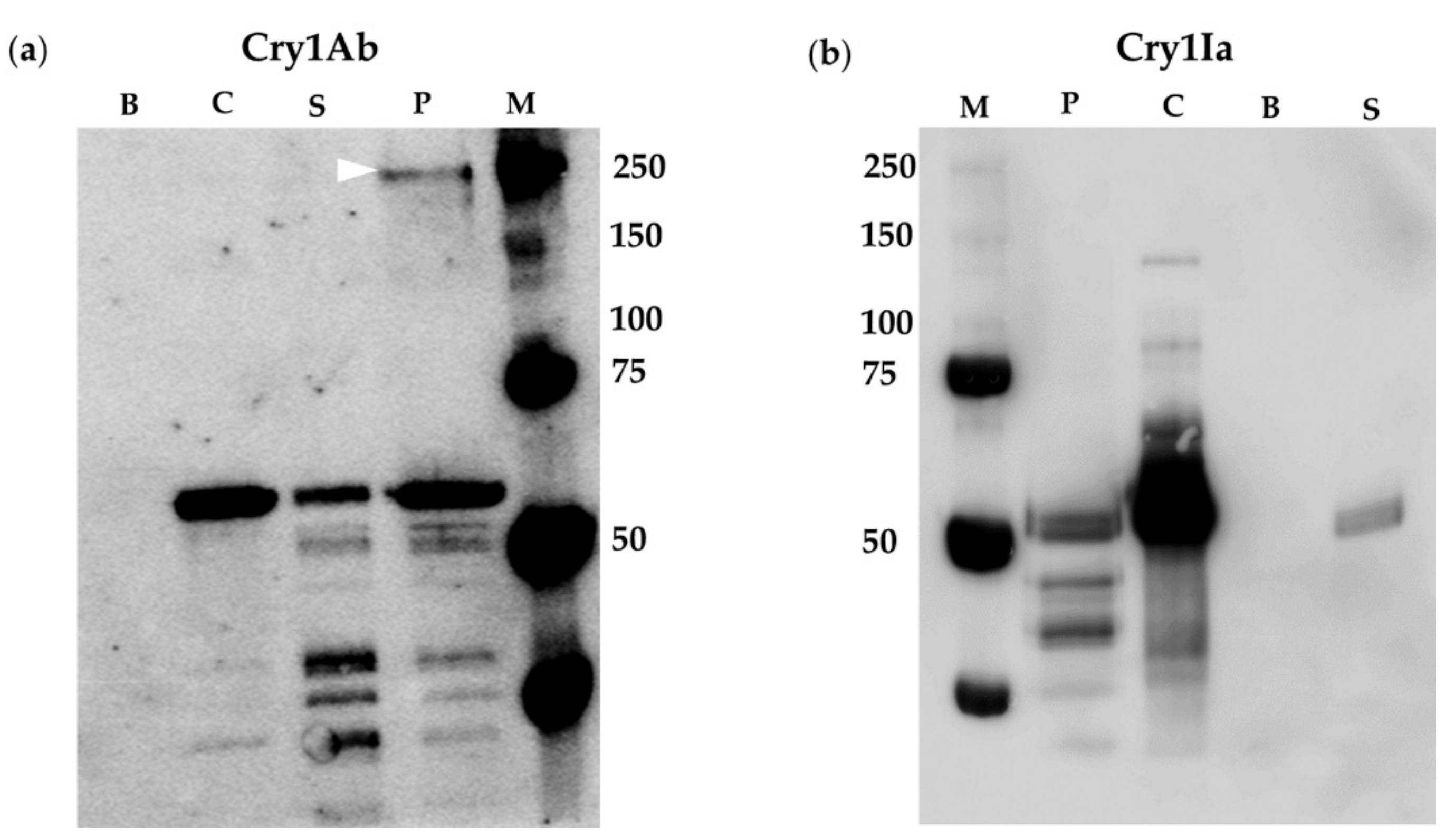
2.2. BBMV of Susceptible Lepidopteran Insects Promoted Oligomerization of Cry1Ab But Not of Cry1Ia

The oligomer formation of Cry1Ab and Cry1Ia was studied by incubating the proteins with lepidopteran BBMV from O. nubilalis and L. botrana, susceptible hosts (Table 1). In order to favor oligomer detection, the milder SDS-PAGE denaturing conditions used in the bibliography to observe Cry1 oligomers were employed (see Section 4.8 in Materials and Methods). The results showed that Cry1Ab toxin was able to form oligomers after incubation with BBMV from O. nubilalis, but this incubation did not promote Cry1Ia oligomer formation (Figure 1). The oligomers observed (band of about 250 kDa) were associated to the O. nubilalis BBMV fraction (Figure 1a, lane P); these oligomeric structures could be inserted into the membranes or just bound to the surface of the BBMV due to interaction with specific membrane proteins. For Cry1Ab, the Western blot revealed another band with a molecular weight of approximately 60 kDa, corresponding to the monomeric form of Cry1Ab (Figure 1a, lane P). Nevertheless, the Cry1Ia protein associated with O. nubilalis BBMV was detected as a single band of about 50 kDa, corresponding to Cry1Ia monomers, and no band corresponding to Cry1Ia oligomers was detected (Figure 1b, lane P). The Cry1Ab and Cry1Ia monomeric proteins were also recovered in the supernatants, as bands of about 60 and 50 kDa, respectively (Figure 1, lanes S). Controls of Cry1Ab or Cry1Ia proteins without BBMV, but subjected to the same process as the rest of the samples, were conducted to assess the possible spontaneous formation of oligomers. The results (Figure 1, lanes C) showed the presence of Cry1Ab and Cry1Ia monomers mostly while the oligomers (tetramers) were not detected in these lanes, pointing to the fact that Cry1Ab and Cry1Ia tetramers did not form spontaneously in the solution in the absence of insect BBMV. In the case of Cry1Ia some minor bands of MW about 65 and 90 kDa were detected, which were most probably traces of Cry1Ia protoxin and partially trypsinized products; a third minor band of about 130 kDa (MW that does not match with the size of Cry1Ia dimers or trimers) was also observed.
The oligomer formation was also tested using BBMV from L. botrana. The results obtained were similar to the ones found with O. nubilalis BBMV. The incubation of Cry1Ia with BBMV from L. botrana, did not render bands with molecular weight consistent with Cry1Ia oligomeric structures (Figure 2b, lane P), whilst, after incubation of Cry1Ab with L. botrana BBMV, a clear band corresponding to a Cry1Ab oligomer (tetramer) was observed (Figure 2, lane P). On the other hand, monomers of both proteins were detected in the corresponding supernatants recovered after the BBMV-protein incubation assays (Figure 2, lanes S). In the Cry1Ia experiments, minor bands of MW of about 65 and 90 kDa were observed in the supernatant (S) and control (C) lanes that probably represent a minor fraction of partially trypsinized Cry1Ia.

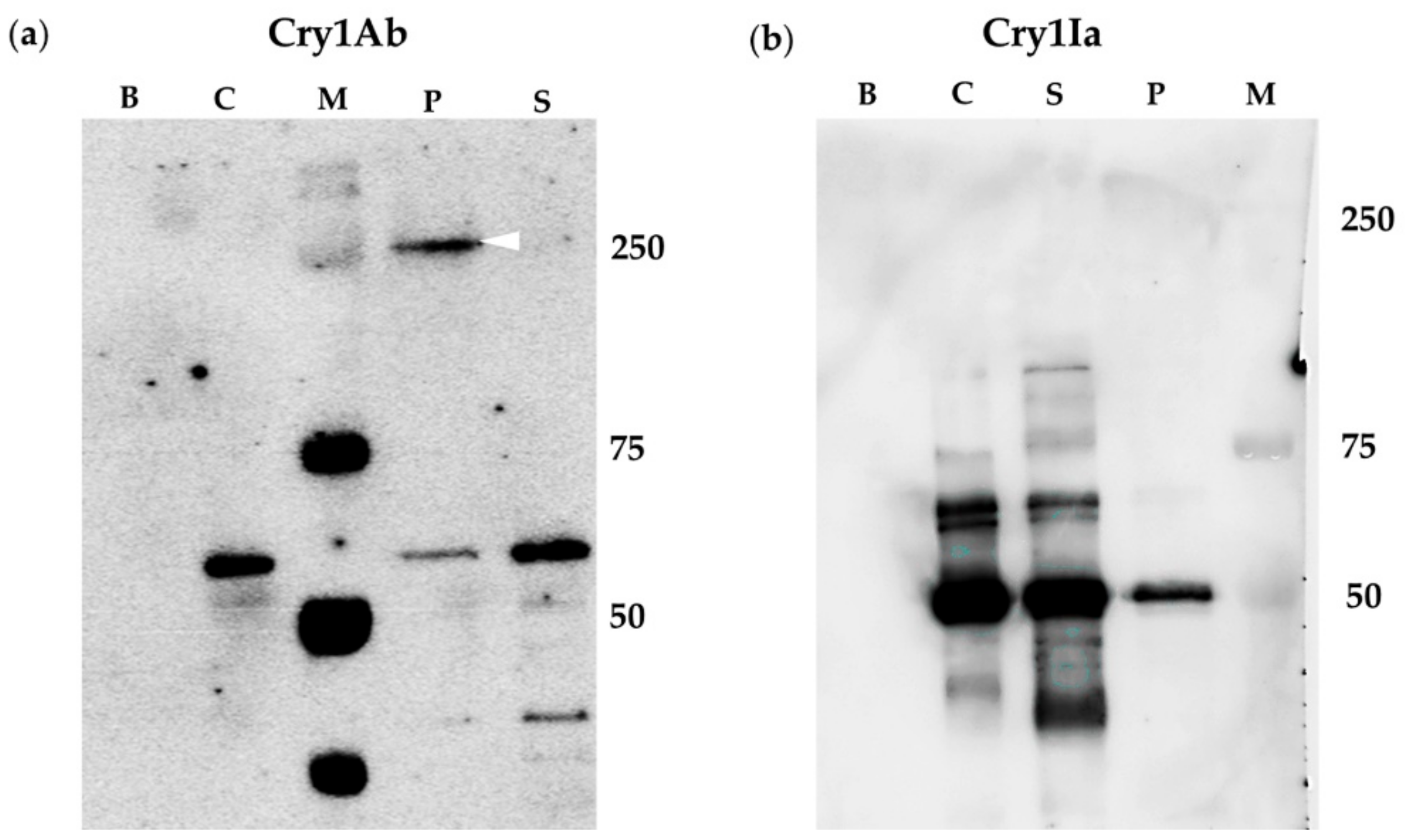
2.3. Oligomerization of Cry1Ia Was Promoted by BBMV from L. decemlineata

Cry1Ia exhibits a dual toxic activity towards lepidopteran and coleopteran hosts, whereas Cry1Ab is only toxic to lepidopteran species. In this work, L. decemlineata BBMV was employed as a coleopteran Cry1Ia susceptible host tissue (Table 1) to study Cry1Ia oligomer promotion as well to study if these BBMV promoted the oligomerization of the Cry1Ab protein, non-toxic for this insect species (Table 1).
The results, summarized in Figure 3, showed that the incubation of either Cry1Ab or Cry1Ia toxins with the coleopteran BBMV provided bands of a molecular weight of about 250 kDa for both Cry proteins, corresponding to the respective oligomers. Interestingly, the oligomers were detected in both, pelleted (BBMV) and supernatant fractions (Figure 3, lanes P and S respectively). Bands corresponding to the monomers of both proteins were also observed in both fractions (pelleted BBMV and supernatant) for both Cry proteins. Incubation with Cry1Ab rendered also a band of about 150 kDa that could correspond to a dimer. It is worth highlighting that the band corresponding to the Cry1Ia oligomer had a molecular weight of about 250 kDa, which would indicate that Cry1Ia oligomer could be composed by more than four subunits, since the Cry1Ia monomer has a size of about 50 kDa. Moreover, a strong band of molecular weight higher than 250 kDa was also observed as being associated to the BBMV (Figure 3, lane P) that could correspond to Cry1Ia aggregates with high number of units.

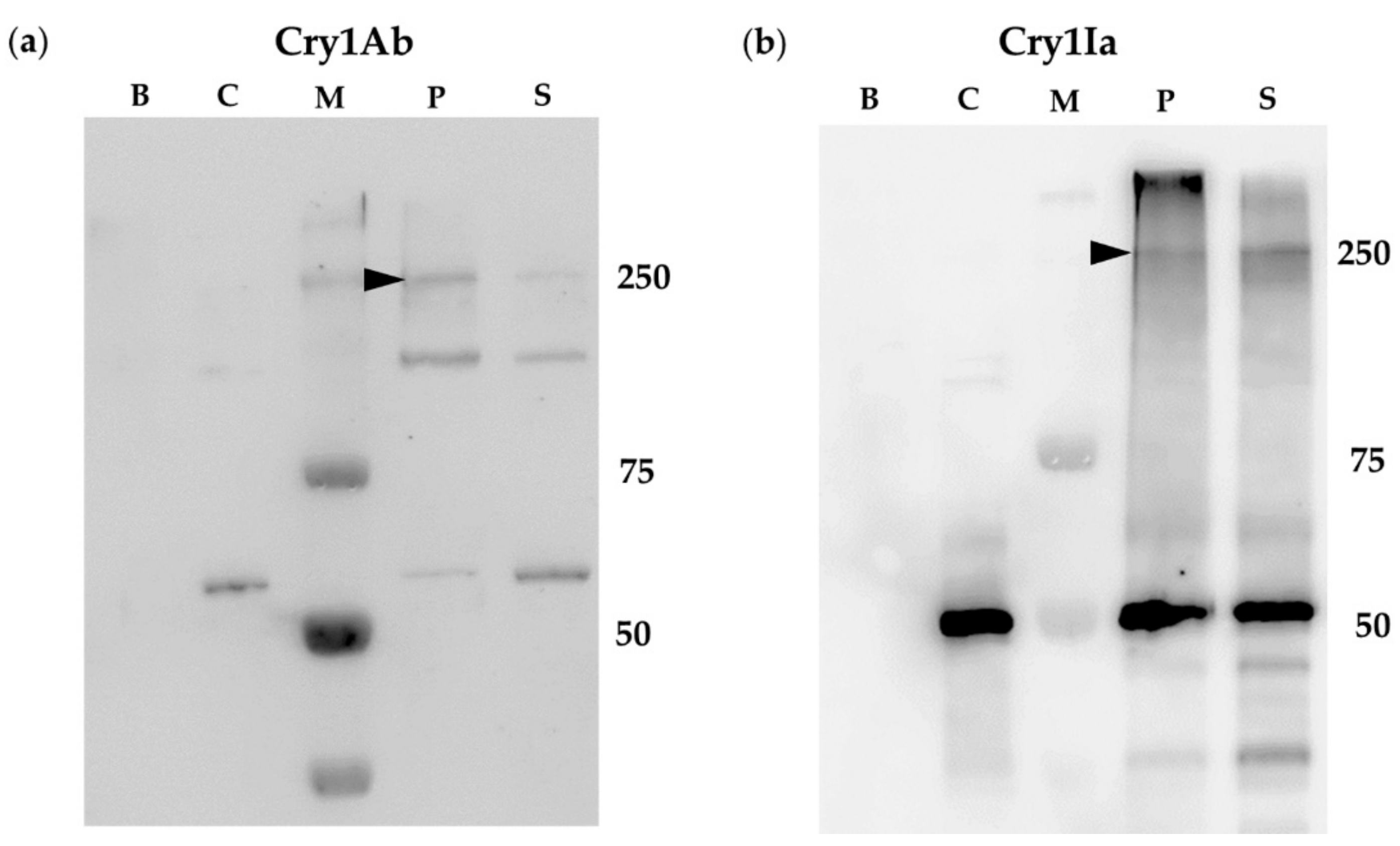
2.4. Oligomerization Promoted by Sf21 Insect Cells

It has been described that Sf21 cells (insect cultured cells derived from S. frugiperda ovaries) are tolerant to both Cry1Ab and Cry1Ia proteins [40,41]. These insect cells were selected in order to check if oligomer formation could be promoted by tolerant insect tissues. Figure 4 shows the results of the incubation of Sf21 cells with each one of these proteins. In the case of Cry1Ab, the incubation of the protein with the insect cells resulted in the detection of two main bands of approximately 60 and 250 kDa associated with the cell fraction, corresponding to Cry1Ab monomers and oligomers, respectively (Figure 4a, lane P). Other minor bands detected were also present in the control lanes of Sf21 cells that had been incubated without Cry proteins, showing that these bands probably correspond to natural biotinylated proteins present in the cells (Figure 4, lanes B). In the case of Sf21 cells incubated with Cry1Ia, only a band with the molecular weight of the monomer (50 kDa) was found to be associated with the cells (Figure 4b, lane P). In summary, results pointed out the absence of Cry1Ia oligomer formation after incubation with Sf21 cells, in contrast to what is observed after incubation with Cry1Ab. It is worth noting that in the supernatants, only a main band with the molecular weight of the Cry1Ab or Cry1Ia monomers, was detected (Figure 4, lanes S).

3. Discussion

Cry1I proteins share sequence and structural similarities to the most-known three domain Cry proteins present in the parasporal crystal of Bt [10,29,42]. Therefore, so far, it has been assumed that their mode of action is similar to its crystal protein counterparts, despite their special features such as that the Cry1I proteins do not form crystals [5,43], their protoxin MW is smaller (about 81 kDa [29]), and they show dual toxic activity against lepidopteran and coleopteran insect pests [29,44].
The mode of action of the Cry proteins accumulated in the crystals is not completely understood [10,12]. It is commonly accepted that includes the solubilization of the crystals in the insect midgut to yield the protoxin form, and a proteolytic processing to produce the activated forms. Then, the “sequential binding model”, suggests that the activated proteins bind to several midgut membrane receptors to finally form an oligomeric structure that inserts in the midgut membrane and forms pores, leading to cellular osmotic imbalance, cell lysis, septicaemia and eventually insect death [13,14]. In support of this model, several studies have reported that oligomerization plays a crucial role in the insecticidal activity of B. thuringiensis Cry toxins [18,38,45,46]. Moreover, Cry protein mutants that did not form oligomers, showed severely decreased toxicity [17,24,45]. Likewise, it has been shown that some Cry1A mutants that had lost their toxicity, were unable to oligomerize and to form pores [38,47]. However, despite these studies, the sequential binding to several insect membrane proteins as well as the oligomer formation and insertion events prior to pore formation are not clearly defined yet [12]. On the other hand, as an alternative to the “sequential binding model”, the “signaling pathway model” has been proposed. This model claims that the Cry toxicity can be due to the activation of intracellular cell death pathways [9]. Indeed, some authors claim that both mechanisms could coexist [48].
In this context, the occurrence of an oligomerization step in the mode of action of Cry1Ia protein, and the study of Cry1Ia oligomerization promotion by membranes of susceptible and tolerant insect BBMV or insect cells, have been the main goals of this research.
In 2009, the occurrence of a spontaneous oligomerization of trypsinized Cry1Ie in solution was reported [28]. According to the mentioned research, the oligomer fraction contained a small amount of dimer and a large amount of aggregates larger than tetramers. The toxicity of the oligomers against Plutella xylostella was about 70 times lower than the toxicity of the monomer or the toxicity of the non-trypsinized Cry1Ie protein, which led the authors to claim that the Cry1Ie spontaneous aggregation most likely differ from the oligomers occur by the insect midgut membranes. In the present work, the oligomerization of Cry1Ia protein after incubation with susceptible insect BBMV or with non-susceptible cultured insect cells, has been determined for the first time.
To properly select the insect species for this study, the insecticidal activity of Cry1Ia and Cry1Ab protoxins towards two lepidopteran species (O. nubilalis and L. botrana) and a coleopteran insect (L. decemlineta) has been assessed. The toxicity data obtained were expected based on the published data [29,44,49,50]. Moreover, Sf21 cells were also used in this study, on the bases that Cry1Ia and Cry1Ab are not toxic for them [40,41].
So far, Cry oligomerization studies have focused on the incubation of the Cry proteins with BBMV of susceptible and resistant populations of the same insect, and, as a result, an association has been found between resistance and reduced oligomerization [51,52]. In the present work, we have used Cry1Ab as an oligomeric control protein [36,37,38,39], and have been able to clearly detect Cry1Ab oligomers in the expected tetramer form (about 250 kDa molecular weight size) after incubation with all tested insect tissues, regardless to their susceptibility. Thus, oligomers have been observed after incubation of the trypsinized Cry1Ab protein with O. nubilalis or L. botrana BBMV (susceptible insects), but also with L. decemlineata BBMV (non-susceptible insect) and Sf21 cells (non-susceptible cultured insect cells). The oligomeric structures were found associated to the insect BBMV or to the insect cells, indicating that they were either inserted or bound to the membrane proteins. The finding of Cry1Ab oligomers after incubation with tolerant insect BBMV or associated with tolerant insect cells could be explained by an improper insertion of oligomers into the membranes (and therefore inefficiency to produce damages), or by the inability to induce the post-pore subsequent events in the cells (e.g., no triggering of cell death mechanisms).
In this study, the Cry1Ab oligomer was mainly found to be associated to the membrane fractions in accordance with previous reports, but oligomers were also observed in the supernatants after incubation with BBMV from the non-susceptible insect L. decemlineata. This suggested that the Cry1Ab oligomers (tetramers and dimers), promoted by the coleopteran BBMV, are not only associated to the BBMV (whether inserted or not) but also free in the supernatant (Figure 3a, lane S). The results obtained resemble the ones shown by Rodríguez-Almazán et al. [47], with the Cry1Ab helix α-4 mutants which had a mutation in domain I, involved in membrane insertion and pore formation, and had lost drastically their insecticidal activity towards M. sexta larvae. Similarly, in our results, the incubation of Cry1Ab with BBMV from L. decemlineata rendered a relatively high amount of oligomeric structures (dimers and tetramers) in the supernatant, apparently indicating that these oligomers were not able to insert into the membranes, resulting in the absence of toxicity. Nevertheless, in this study, after the incubation of the Cry1Ia protein with L. decemlineata BBMV, a high proportion of Cry1Ia oligomers were also observed in the supernatants (Figure 3b, laneS), and in this case, the protein showed a high toxicity against this coleopteran pest (Table 1).
The Cry1Ia oligomers were observed after incubation of the protein with BBMV from the susceptible coleopteran L. decemlineata. The molecular weights of the bands that correspond to the Cry1Ia oligomers (about 250 kDa) point out that the Cry1Ia oligomer could be composed of more than four units.
After incubation of Cry1Ia with BBMV of susceptible lepidopterans (O. nubilalis and L. botrana) or with insect Sf21 cells, no oligomers were detected, suggesting that the Cry1Ia proteins associated to the lepidopteran membranes could be mainly in monomeric forms. In this case, oligomers would not be a limiting step in the toxicity of Cry1Ia proteins, and the toxicity could be mediated by intracellular signaling pathways [9]. Moreover, other hypotheses could be mentioned to explain the reason for the absence of Cry1Ia oligomers. Firstly, it can be suggested that the observed monomers come from disassembled oligomers produced due to the SDS-PAGE technique conditions, similarly to what was proposed by Ocelotl et al. [51] working with Cry1Ab oligomers. However, this reasoning would be in conflict with the observation of oligomers after the incubation of Cry1I with coleopteran BBMV, which were detected using the same SDS-PAGE conditions. Secondly, it could be considered that the biotinlylation of Cry1I could interfere with oligomerization. This hypothesis was examined following the incubation of the biotin labelled Cry1Ab with O. nubilalis BBMV. The results showed no influence of biotin in oligomerization (Figure S1), similarly to what had been already claimed by other authors showing that biotinylation of Cry proteins does not prevent their oligomerization, binding and insecticidal activity [12,53,54]. Thirdly, it has to be noticed that, in the present study, the oligomer formation experiments were performed with trypsinized Cry1Ab and Cry1Ia proteins to mimic the in vivo conditions. It has been described that Cry1Ie protoxin and trypsinized protein have the same toxicity [28]. However, other Cry1I proteins such as Cry1Ia1 have shown some differences in toxicity amongst protoxins (more active) and trypsinized proteins [49]. We can speculate that in vitro trypsinization of Cry1Ia could alter the toxicity by impairing oligomer formation, maybe provoking a flawed ability to form oligomers when they are promoted by lepidopteran BBMV.
The spontaneous formation of Cry1Ab and Cry1Ia oligomers in solution has been questioned in this study. Cry1Ab submitted to the same experimental situation of treatments, but without being in contact with insect BBMV or cells, did not oligomerize (Figure 1, Figure 2, Figure 3 and Figure 4a, lanes C). Regarding Cry1Ia, some bands of smaller sizes than the expected tetramer, and with MW sizes that were not multiples of 50 kDa (monomer size) were observed (Figure 1, Figure 2, Figure 3 and Figure 4b, lanes C). Most probably, these bands are residual incomplete trypsinized Cry1Ia forms, dragged through the toxin purification process. In conclusion, although some studies have reported the spontaneous formation of Cry protein oligomers in solution without being in contact with the membrane-like environment (i.e., Cry4Ba [55] or Cry2Ab [24]), in our study neither Cry1Ab nor Cry1Ia formed oligomers without being exposed to insect BBMV or cultured insect cells.
In summary, our findings indicated that the oligomers of a classical 3D crystal forming Cry protein of Bt such as Cry1Ab were promoted and could be detected after incubation of activated Cry1Ab with susceptible or non-susceptible insect midgut BBMV and with non- susceptible Sf21 cells. In contrast, in the same assay conditions, Cry1Ia oligomers were detected only after incubation with BBMV of L. decemlineata (coleopteran susceptible host), but no Cry1Ia oligomeric structures were found following incubation of Cry1Ia with lepidopteran BBMV or with the Sf21 cells, regardless of its host susceptibility. Hence, our results, using trypsin processed Cry1Ab and Cry1Ia as an in vitro model of what might occur in vivo, suggest that: (1) The promotion of oligomers can occur by incubation of the Cry toxin with susceptible insect BBMV but also with non-susceptible insect tissues, and (2) The oligomerization may not be a determining step in the toxicity of Cry1I proteins.

4. Materials and Methods

4.1. Production and Purification of Cry Proteins

The Cry1Ab protein used for oligomer formation was obtained from a recombinant Escherichia coli strain GG094-208 (kindly supplied by Dr. R.A. de Maagd, Wageningen University, The Netherlands). Protein expression, inclusion bodies purification, solubilization and protoxin activation by trypsin, were performed as described previously [56]. The activated Cry1Ab was purified by anion-exchange chromatography using Äkta 100 explorer system (GE Healthcare, Amersham, UK) following Crava et al. [57]. The eluted fractions from the column were individually analysed by sodium dodecyl sulphate-12% polyacrylamide gel electrophoresis (SDS-PAGE).
The cry1Ia7 gene was cloned and expressed in E. coli, BL21(DE3) cells [40]. The purification of the protein by affinity chromatography using a HisTrapTM FF crude column (GE Healthcare Bio-Sciences, Upsala, Sweden), protein dialysis and protoxin activation by trypsin, were performed as reported by Khorramnejad et al. [40]. The activated Cry1Ab and Cry1Ia were visualized after SDS-PAGE, and their concentration was estimated by densitometry using TotalLab Quant program version 12.3 (Newcastle, UK), employing bovine serum albumin as standard.
The Cry1Ab protein used in bioassays (protoxin) was obtained from a recombinant Bt strain that produced a crystal composed solely of the Cry1Ab protein, kindly supplied by Dr. Colin Berry, Cardiff University, Cardiff, UK. This Bt strain was grown in CCY medium [58] supplemented with erythromycin and the crystal formation was observed daily. When at least 95% of the cells were lysed, the fermentation process was stopped and then spores and crystals were collected by centrifugation (8600× g, 4 °C), washed with a saline solution (NaCl 1M, EDTA 10mM) and resuspended in KCl (10 mM). For Cry1Ia7 protoxin production, the recombinant E. coli BL21 (DE3) was grown and purified as described above. Both Cry1Ab and Cry1Ia7 protoxins were quantified by the method of Bradford [59] using BSA as standard, and kept at 4 °C until used.

4.2. Insect Rearing

Two different lepidopteran species, L. botrana (Lep: Tortricide) and O. nubilalis Hübner (Lep.: Crambidae), and one coleopteran species, L. decemlineata (Col.: Chrysomelidae) (the Colorado potato beetle, CPB) were used in this study. Both lepidopteran pests were maintained in the insectary of the Universidad Pública de Navarra (UPNA, Pamplona, Spain) at 25 °C ± 1 °C with 70% ± 5% RH and a photoperiod of 16/8 h (light/dark) on the artificial diet described by MacIntosh et al. [60]. The population of beetles was raised on potato plants (Desiré variety) grown throughout the year in a phytotron to provide a continuous supply of food substrate. This population was refreshed 1–2 times every year by the introduction of wild adults collected in the field during the spring-summer.

4.3. Insect Cell Line

The lepidopteran cell line Sf21, from ovaries of fall armyworm S. frugiperda (Lep.: Noctuidea), was obtained from Wageningen University (Wageningen, The Netherlands). The Sf21 cells were maintained at 25 °C on 1X Grace’s medium (Gibco® Life technologiesTM, Carslbad, CA, USA) supplemented with 10% heat-inactivated fetal bovine serum (FBS) in 25 cm2 cell culture flasks. Cells were passaged weekly. Cell concentrations were measured by using an automatic cell counter (Countess Automated Cell Counter from Invitrogen, Carlsbad, CA, USA).

4.4. Insect Bioassays

The bioassays for the three insect species were performed with first instar larvae. Five different protoxin concentrations, ranging from 0.39 to 100 μg/mL, were prepared to determine the concentration-mortality responses in order to calculate the mean lethal concentration (LC50). For the lepidopteran insect species, O. nubilalis and L. botrana, the diet surface contamination assay was used [61], and for L. decemlineata, the leaf dip bioassay described by Iriarte et al. [62], was performed. For each bioassay, several protein concentrations were tested, using 28 larvae per concentration. Each bioassay was repeated at least three times. Total insect mortality was recorded after 7 days for lepidopteran insects, and after 4 days for L. decemlineata. The concentration-mortality data obtained for each insect species were analyzed after transformation of the concentration-response curve to fit a linear model using POLO-PC program (LeOra Software, Berkeley, CA, USA, 1987), based on the Probit analysis [63].

4.5. Midgut Isolation and BBMV Preparation

Midguts were dissected from fifth-instar larvae of O. nubilalis and forth-instar larvae of L. decemlineata. The dissected midguts were rinsed in ice-cold MET buffer (0.3 M mannitol, 5 mM EGTA, 17 mM Trsi-HCl, pH 7.5), snap frozen in liquid nitrogen and kept at −80 °C until use.
Brush border membrane vesicles (BBMV) were obtained from the dissected midguts of O. nubilalis and L. decemlineata and from the whole last instar larvae of L. botrana, following the differential magnesium precipitation method [64,65]. Proteins in the purified BBMV were quantified following Bradford protein assay [59] and stored at −80 °C.

4.6. Biotin Labelling

Trypsin activated Cry1Ia protein was biotinylated by using the protein biotinylation kit from GE Healthcare (GE Healthcare, Little Chalfont, UK), as described elsewhere [66]. The eluted fractions were quantified by NanoDrop 2000 spectrophotometer (ThermoFisher Scientific, Waltham, MA, USA), analyzed by 12% SDS-PAGE and verified by Western blot. The protein fractions were concentrated by using an Amicon Ultra-4 10K centrifugal filter device (Merck Millipore, Tullagreen, Ireland) and stored at 4 °C.
The interference of biotin in oligomerization was tested following incubation of the biotin labelled Cry1Ab with O. nubilalis BBMV. The detection of biotin labelled Cry1Ab oligomerization was performed following the same protocol than has been described for Cry1Ia. The results showed no influence of biotin in oligomerization (Figure S1), as had been already claimed by other authors that have used biotin labelled proteins to detect oligomers [12,20,53,54].

4.7. Oligomerization Assays with Sf21 Cells

The confluent monolayer growing Sf21 cells were suspended in fresh Grace’s medium without FBS. The cell concentration was measured (by using the Countess Automated Cell Counter from Invitrogen, Carlsbad, California, USA), and 100 µL of cell suspension at a concentration of 2 × 10⁶ cells/mL were seeded into 96-well plates. The oligomerization assays were performed as described by Portugal et al. [38] with slight modifications. In short, cells were incubated at 25 °C for at least 30 min. Later, 4 µg of activated toxins (biotinylated Cry1Ia or unlabeled Cry1Ab) were added to the cells (final concentration of 0.03 µg protein/µL) except in controls, which received 50 mM carbonate buffer pH 10.5. The plates were incubated for 3 h at 25 °C. After the incubation, the treated cells were collected and pelleted by centrifugation at 16,200× g, 4 °C for 15 min. The supernatants containing unbound proteins were kept for further analysis. The controls (proteins alone, without cells) were submitted to the same experimental conditions as treatments. After centrifugation of the controls, as there was no pellet due to the absence of cells, a dilution of supernatant (containing 200 ng of selected protein) was analyzed in the gel. The Sf21 cells in the pellet were washed once with 200 µL of 50 mM carbonate buffer pH 10.5, and recovered by centrifugation (45 min, 18,800× g). The final pellet was resuspended in 10 µL of buffer and heated at 50 °C for 3 min. The proteins present in the sample were separated by SDS-PAGE 10% and electrotransferred onto polyvinylidene difluoride (PVDF) Western Blotting membrane (Roche Diagnostics GmbH, Mannheim, Germany). The membrane was incubated overnight in blocking buffer (PBST; 0.1% Tween 20 in phosphate-buffered saline, supplemented with 5% skimmed milk) with gentle shaking, and washed three times with PBST, before incubation with the corresponding antibodies. Cry1Ab protein was detected with polyclonal rabbit anti-Bt Cry1Ab/1Ac (1:10,000; 60 min) from Abraxis (Warminster, PA, USA) followed by secondary antibody (1:20,000; 60 min) coupled with horseradish peroxidase (HRP) (Sigma-Aldrich, Saint Louis, MO, USA), whereas biotinylated Cry1Ia protein was detected by streptavidin-conjugated horseradish peroxidase (1:2000; 60 min) (GE Healthcare, Amersham, UK). Both Cry1Ab and Cry1Ia proteins were visualized by chemiluminescence using ECLTM prime western blotting detection reagent (GE Healthcare, Little Chalfont, UK) using an ImageQuant LAS400 image analyzer (GE Healthcare Bio-Sciences, Upsala, Sweden). The molecular weight marker used was Precision Plus Protein™ Dual Color Standard (Bio-Rad, Carlsbad, CA, USA). Each oligomerization assay was repeated at least three times.

4.8. Oligomerization Assays with BBMV

The BBMV were centrifuged for 10 min at 16,000× g and suspended in 50 mM carbonate buffer pH 10.5. The oligomerization protocol was set up after reviewing the procedures described in the literature for Cry1 proteins [17,18,37,38,39,45,47,51,52,54]. Finally, the oligomerization assays with BBMV were performed following Ocelotl et al. [51] who employed the milder SDS-PAGE denaturing conditions (heating the samples 3 min at 50 °C), with some modifications. Briefly, 2 µg of biotin labelled activated Cry1Ia and activated Cry1Ab toxins were incubated for one hour with 5 µg of L. botrana or L. decemlineata BBMV, or with 20 µg of O. nubilalis BBMV, at 37 °C, in a final volume of 50 μL. Activated proteins incubated in the absence of BBMV and samples containing only BBMV were used as controls. Then, phenylmethylsulfoyl fluoride (PMSF) was added (final concentration 1 mM) and BBMV were recovered by centrifugation at 18,400× g for 45 min at 4 °C. The supernatant containing unbound protein was recovered and stored. The controls (samples with proteins and without BBMV) went through the same experimental conditions as treatments. After centrifugation of the controls, a dilution of the supernatant (containing 200 ng of selected protein) was analyzed in the gel to avoid the observation of the over saturated signal in the membrane. In the samples containing BBMV, the pellet was washed once with 100 μL ice-cold buffer. The final BBMV pellets were resuspended in 10 µl of the buffer. After incubating the samples for 3 min at 50 °C, the proteins were separated by 10% SDS-PAGE and blotted onto PVDF Western Blot membranes (Roche Diagnostics GmbH, Mannheim, Germany). After Western blot, Cry1Ab was detected with polyclonal rabbit anti-Bt Cry1Ab/1Ac, and Cry1Ia was detected by streptavidin-conjugated horseradish peroxidase as has been described in the previous section. The experiments were repeated, at least, three times.

Supplementary Materials

The following is available online at https://0-www-mdpi-com.brum.beds.ac.uk/2072-6651/12/2/133/s1, Figure S1: Biotin labelled Cry1Ab oligomer formation promoted by O. nubilalis BBMV.

Author Contributions

B.E. and Y.B. contributed to the design of the study. A.K. and M.D.-A. performed the experiments. A.K., Y.B., B.E., P.C. and M.D.-A. analyzed the data. A.K., Y.B. and M.D.-A. wrote the manuscript. All authors have read and agreed to the published version of the manuscript.

Funding

This work was supported by grants from the Spanish Ministry of Science, Innovation and Universities, the State Research Agency of Spain and the European FEDER founds (Refs. AGL2015-70584-C2 and RTI2018-095204-B-C21), and by the Generalitat Valenciana (GVPROMETEOII-2015-001). M. Domínguez received a predoctoral fellowship from the Universidad Pública de Navarra, Spain.

Acknowledgments

We are deeply grateful to Patricia Hernández-Martínez for her valuable comments. We thank Rosa Maria González-Martínez and Óscar Marin Vázquez for their support in laboratory assistance.

Conflicts of Interest

The authors declare no conflict of interest.

References

  1. Van Frankenhuyzen, K. Insecticidal activity of Bacillus thuringiensis crystal proteins. J. Invertebr. Pathol. 2009, 101, 1–16. [Google Scholar] [CrossRef]
  2. Ohba, M.; Mizuki, E.; Uemori, A. Parasporin, a new anticancer protein group from Bacillus thuringiensis. Anticancer Res. 2009, 29, 427–433. [Google Scholar] [PubMed]
  3. Palma, L.; Muñoz, D.; Berry, C.; Murillo, J.; Caballero, P. Bacillus thuringiensis toxins: An overview of their biocidal activity. Toxins 2014, 6, 3296–3325. [Google Scholar] [CrossRef] [PubMed] [Green Version]
  4. Chakroun, M.; Banyuls, N.; Bel, Y.; Escriche, B.; Ferré, J. Bacterial vegetative insecticidal proteins (Vip) from entomophatogenic bacteria. Microbiol. Mol. Biol. Rev. 2016, 80, 329–350. [Google Scholar] [CrossRef] [PubMed] [Green Version]
  5. Kostichka, K.; Warren, G.W.; Mullins, M.; Mullins, A.D.; Craig, J.A.; Koziel, M.G.; Estruch, A.D. Cloning of a cryV-type insecticidal protein gene from Bacillus thuringiensis: The cryV-encoded protein is expressed early in stationary phase. J. Bacteriol. 1996, 178, 2141–2144. [Google Scholar] [CrossRef] [Green Version]
  6. Jurat-Fuentes, J.L.; Jackson, T.A. Bacterial entomopathoges. In Insect Pathology, 2nd ed.; Vega, F.E., Kaya, H.K., Eds.; Academic Press: San Diego, CA, USA, 2012; pp. 265–349. [Google Scholar] [CrossRef]
  7. Hoffman, C.; Vanderbruggen, H.; Höfte, H.; Van Rie, G.; Jansens, S.; Van Mellaert, H. Specificity of Bacillus thuringiensis delta-endotoxins is correlated with the presence of high-affinity binding sites in the brush border membrane of target insect midguts. Proc. Natl. Acad. Sci. USA 1988, 85, 7844–7848. [Google Scholar] [CrossRef] [Green Version]
  8. Ferre, J.; VanRie, J. Biochemistry and genetics of insect resistance to Bacillus thuringiensis. Annu. Rev. Entomol. 2002, 47, 501–533. [Google Scholar] [CrossRef]
  9. Zhang, X.; Candas, M.; Griko, N.B.; Taussig, R.; Bulla, L.A., Jr. A mechanism of cell death involving an adenylyl cyclase/PKA signaling pathway is induced by the Cry1Ab toxin of Bacillus thuringiensis. Proc. Natl. Acad. Sci. USA 2006, 103, 9897–9902. [Google Scholar] [CrossRef] [Green Version]
  10. Adang, M.J.; Crickmore, N.; Jurat-Fuentes, J.L. Diversity of Bacillus thuringiensis crystal toxins and mechanism of action. Adv. Insect Physiol. 2014, 47, 39–87. [Google Scholar] [CrossRef]
  11. Arenas, I.; Bravo, A.; Soberón, M.; Gómez, I. Role of alkaline phosphatase from Manduca sexta in the mechanism of action of Bacillus thuringiensis Cry1Ab toxin. J. Biol. Chem. 2010, 285, 12497–12503. [Google Scholar] [CrossRef] [Green Version]
  12. Vachon, V.; Raynald, L.; Schwartz, J.L. Current models of the mode of action of Bacillus thuringiensis insecticidal crystal proteins: A critical review. J. Invertebr. Pathol. 2012, 111, 1–12. [Google Scholar] [CrossRef] [PubMed]
  13. Pardo-López, L.; Soberón, M.; Bravo, A. Bacillus thuringiensis insecticidal three-domain Cry toxins: Mode of action, insect resistance and consequences for crop protection. FEMS Microbiol. Rev. 2013, 37, 3–22. [Google Scholar] [CrossRef] [PubMed] [Green Version]
  14. Gomez, I.; Sanchez, J.; Miranda, R.; Bravo, A.; Soberon, M. Cadherin-like receptor binding facilitates proteolytic cleavage of helix alpha-1 in domain I and oligomer pre-pore formation of Bacillus thuringiensis Cry1Ab toxin. FEBS Lett. 2002, 513, 242–246. [Google Scholar] [CrossRef] [Green Version]
  15. Rausell, C.; García-Robles, I.; Sánchez, J.; Muñoz-Garay, C.; Martínez-Ramírez, A.C.; Real, M.D.; Bravo, A. Role of toxin activation on binding and pore formation activity of the Bacillus thuringiensis Cry3 toxins in membranes of Leptinotarsa decemlineata (Say). Biochim. Biophys. Acta 2004, 1660, 99–105. [Google Scholar] [CrossRef] [Green Version]
  16. Muñoz-Garay, C.; Portugal, L.; Pardo-López, L.; Jiménez-Juárez, N.; Arenas, I.; Gómez, I.; Sánchez-López, R.; Arroyo, R.; Holzenburg, A.; Savva, C.G.; et al. Characterization of the mechanism of action of the genetically modified Cry1AbMod toxin that is active against Cry1Ab-resistant insects. Biochim. Biophys. Acta 2009, 1788, 2229–2237. [Google Scholar] [CrossRef] [Green Version]
  17. Tigue, N.; Jacoby, J.; Ellar, D.J. The α-helix residue, Asn135, is involved in the oligomerization of Cry1Ac1 and Cry1Ab5 Bacillus thuringiensis toxins. Appl. Environ. Microbiol. 2001, 67, 5715–5720. [Google Scholar] [CrossRef] [Green Version]
  18. Herrero, S.; González-Cabrera, J.; Ferré, J.; Bakker, P.L.; de Maagd, R.A. Mutations in the Bacillus thuringiensis Cry1Ca toxin demonstrate the role of domains II and III in specificity towards Spodoptera exigua larvae. Biochem. J. 2004, 384, 507–513. [Google Scholar] [CrossRef] [Green Version]
  19. Pérez, C.; Muñoz-Garay, C.; Portugal, L.; Sánchez, J.; Gill, S.S.; Soberón, M.; Bravo, A. Bacillus thuringiensis spp. israelensis Cyt1Aa enhances activity of Cry11Aa toxin by facilitating the formation of a pre-pore oligomeric structure. Cell Microbiol. 2007, 9, 2931–2937. [Google Scholar] [CrossRef] [Green Version]
  20. Fabrick, J.; Oppert, C.; Lorenzen, M.D.; Morris, K.; Oppert, B.; Jurat-Fuentes, J.L. A novel Tenebrio molitor cadherin is a functional receptor for Bacillus thuringiensis Cry3Aa toxin. J. Biol. Chem. 2009, 284, 18401–18410. [Google Scholar] [CrossRef] [Green Version]
  21. Groulx, N.; McGuire, H.; Laprade, R.; Schwartz, J.L.; Blunck, R. Single molecule fluorescence study of the Bacillus thuringiensis toxin Cry1Aa reveals tetramerization. J. Biol. Chem. 2011, 286, 42274–42282. [Google Scholar] [CrossRef] [Green Version]
  22. Carmona, D.; Rodríguez-Almazán, C.; Muñoz-Garay, C.; Portugal, L.; Pérez, C.; de Maagd, R.A.; Bakker, P.; Soberón, M.; Bravo, A. Dominant negative phenotype of Bacillus thuringiensis Cry1Ab, Cry11Aa and Cry4Ba mutants suggest hetero-oligomer formation among different Cry toxins. PLoS ONE 2011, 6, e19952. [Google Scholar] [CrossRef] [PubMed] [Green Version]
  23. Khomkhum, N.; Leetachewa, S.; Angsuthanasombat, C.; Moonsom, S. Functional assembly of 260-kDa oligomers required for mosquito-larvicidal activity of the Bacillus thuringiensis Cry4Ba toxin. Peptides 2015, 68, 183–189. [Google Scholar] [CrossRef] [PubMed]
  24. Xu, L.; Pan, Z.; Zhang, J.; Niu, L.; Li, J.; Chen, Z.; Liu, B.; Zhu, Y.; Chen, Q. Exposures of helices α4 and α5 is required for insecticidal activity of Cry2Ab by prompting assembly of prepore oligomeric structure. Cell. Microbiol. 2018, e12827. [Google Scholar] [CrossRef] [PubMed]
  25. Abe, Y.; Inoue, H.; Ashida, H.; Maeda, Y.; Kinoshita, T.; Kitada, S. Glycan region of GPI anchored-protein is required for cytocidal oligomerization of an anticancer parasporin-2, Cry46Aa1 protein, from Bacillus thuringiensis strain A1547. J. Invertebr. Pathol. 2017, 142, 71–81. [Google Scholar] [CrossRef]
  26. López-Diaz, J.; Cantón, P.E.; Gill, S.S.; Soberón, M.; Bravo, A. Oligomerization is a key step in Cyt1Aa membrane insertion and toxicity but not necessary to synergize Cry11Aa toxicity in Aedes aegypti larvae. Environ. Microbiol. 2013, 15, 3030–3039. [Google Scholar] [CrossRef] [Green Version]
  27. Palma, L.; Scott, D.J.; Harris, G.; Din, S.; Williams, T.L.; Reoberts, O.J.; Young, M.T.; Caballero, P.; Berry, C. The Vip3Ag4 insecticidal protoxins from Bacillus thuringiensis adopts a tetrameric configuration that is maintained on proteolysis. Toxins 2017, 9, 165. [Google Scholar] [CrossRef] [PubMed]
  28. Guo, S.; Zhang, Y.; Song, F.; Zhang, J.; Huang, D. Protease-resistant core form of Bacillus thuringiensis Cry1Ie: Monomeric and oligomeric forms in solution. Biotechnol. Lett. 2009, 31, 1769–1774. [Google Scholar] [CrossRef]
  29. Tailor, R.; Tippett, J.; Gibb, G.; Pells, S.; Pike, D.; Jordan, L.; Ely, S. Identification and characterization of a novel Bacillus thuringiensis delta-endotoxin entomocidal to coleopteran and lepidopteran larvae. Mol. Microbiol. 1992, 6, 1211–1217. [Google Scholar] [CrossRef]
  30. Choi, S.K.; Shin, B.S.; Kong, E.M.; Rho, H.M.; Park, S.H. Cloning of a new Bacillus thuringiensis cry1I-type crystal protein gene. Curr. Microbiol. 2000, 41, 65–69. [Google Scholar] [CrossRef]
  31. Xu, L.; Ferry, N.; Wang, Z.; Zhang, J.; Edwards, M.G.; Gatehouse, A.M.R.; He, K. A proteomic approach to study the mechanism of tolerance to Bt toxins in Ostrinia furnacalis larvae selected for resistance to Cry1Ab. Transgenic Res. 2013, 22, 1155–1166. [Google Scholar] [CrossRef]
  32. Zhang, T.; He, M.; Gatehouse, A.M.R.; Wang, Z.; Edwards, M.G.; Li, Q.; He, K. Inheritance patterns, dominance and cross-resistance of Cry1Ab- and Cry1Ac-selected Ostrinia furnacalis (Guenée). Toxins 2014, 6, 2694–2707. [Google Scholar] [CrossRef] [PubMed] [Green Version]
  33. Xu, L.; Wang, Z.; Zhang, J.; He, K.; Ferry, N.; Gatehouse, A.M.R. Cross-resistance of Cry1Ab-selected Asian corn borer to other Cry toxins. J. Appl. Entomol. 2010, 134, 429–438. [Google Scholar] [CrossRef]
  34. Guo, J.; He, K.; Hellmich, R.L.; Bai, S.; Zhang, T.; Liu, Y.; Ahmed, T.; Wang, Z. Field trials to evaluate the effects of transgenic cry1Ie maize on the community characteristics of arthropod natural enemies. Sci. Rep. 2016, 6, 22102. [Google Scholar] [CrossRef] [PubMed]
  35. Fan, C.; Wu, F.; Dong, J.; Wang, B.; Yin, J.; Song, X. No impact of transgenic cry1Ie maize on the diversity, abundance and composition of soil fauna in a 2-year field trial. Sci. Rep. 2019, 9, 10333. [Google Scholar] [CrossRef] [Green Version]
  36. Rausell, C.; Muñoz-Garay, C.; Miranda-CasoLuengo, R.; Gómez, I.; Rudiño-Piñera, E.; Soberón, M.; Bravo, A. Tryptophan spectroscopy studies and black lipid bilayer analysis indicate that the oligomeric structure of Cry1Ab toxin from Bacillus thuringiensis is the membrane-insertion intermediate. Biochem. J. 2004, 43, 166–174. [Google Scholar] [CrossRef]
  37. Gómez, I.; Sánchez, J.; Muñoz-Garay, C.; Matus, V.; Gill, S.S.; Soberón, M.; Bravo, A. Bacillus thuringiensis Cry1A toxins are versatile proteins with multiple modes of action: Two distinct pre-pores are involved in toxicity. Biochem. J. 2014, 459, 383–396. [Google Scholar] [CrossRef] [Green Version]
  38. Portugal, L.; Gringorten, J.L.; Caputo, G.F.; Soberón, M.; Muñoz-Garay, C.; Bravo, A. Toxicity and mode of action of insecticidal Cry1A proteins from Bacillus thuringiensis in an insect cell line, CF-1. Peptides 2014, 53, 292–299. [Google Scholar] [CrossRef]
  39. Nair, M.S.; Dean, D.H. Composition of the putative prepore complex of Bacillus thuringiensis Cry1Ab toxin. Adv. Biol. Chem. 2015, 5, 179–188. [Google Scholar] [CrossRef] [Green Version]
  40. Khorramnejad, A.; Bel, Y.; Hernández-Martínez, P.; Talaei-Hassanloui, R.; Escriche, B. Insecticidal activity and cytotoxicity of Bacillus thuringiensis Cry1Ia7 protein. In GMOs in Integrated Plant Protection; Meissle, M., De Schrijver, A., Smagghe, G., Eds.; IOBC-WPRS Bulletin: Ghent, Belgium, 2018; Volume 131, pp. 56–63. [Google Scholar]
  41. Martínez-Solis, M.; Pinos, D.; Endo, H.; Portugal, L.; Sato, R.; Ferré, F.; Herrero, S.; Hernández-Martínez, P. Role of Bacillus thuringiensis Cry1A toxins domains in the binding to the ABCC2 receptor from Spodoptera exigua. Insect. Biochem. Mol. Biol. 2018, 101, 47–56. [Google Scholar] [CrossRef]
  42. Crickmore, N.; Zeigler, D.R.; Feitelson, J.; Schnepf, E.; Van Rie, J.; Lereclus, D.; Baum, J.; Dean, D.H. Revision of the nomenclature for the Bacillus thuringiensis pesticidal crystal proteins. Microbiol. Mol. Biol. Rev. 1998, 62, 807–813. [Google Scholar] [CrossRef] [Green Version]
  43. Masson, L.; Erlandson, M.; Puzstai-Carey, M.; Brousseau, R.; Juárez-Pérez, V.; Frutos, R. A holistic approach for determining the entomopathogenic potential of Bacillus thuringiensis strains. Appl. Environ. Microbiol. 1998, 46, 4782–4788. [Google Scholar] [CrossRef] [Green Version]
  44. Ruiz de Escudero, I.; Estela, A.; Porcar, M.; Martínez, C.; Oguiza, J.A.; Escriche, B.; Ferré, J.; Caballero, P. Molecular and insecticidal characterization of a Cry1I protein toxic to insects of the families Noctuidae, Tortricidae, Plutellidae and Chrysomelidae. Appl. Environ. Microbiol. 2006, 72, 4796–4804. [Google Scholar] [CrossRef] [PubMed] [Green Version]
  45. Jiménez-Juárez, N.; Muñoz-Garay, C.; Gómez, I.; Saab-Rincon, G.; Damian-Almazo, J.Y.; Gill, S.S.; Soberón, M.; Bravo, A. Bacillus thuringiensis Cry1Ab mutants affecting oligomer formation are not-toxic to Manduca sexta larvae. J. Biol. Chem. 2007, 282, 21222–21229. [Google Scholar] [CrossRef] [Green Version]
  46. Likitvivatanavong, S.; Katzenmeier, G.; Andsuthanasombat, C. Asn183 in α5 is essential for oligomerization and toxicity of the Bacillus thuringiensis Cry4Ba toxin. Arch. Biochem. Biophys. 2005, 445, 46–55. [Google Scholar] [CrossRef]
  47. Rodríguez-Almazán, C.; Zavala, L.E.; Muñoz-Garay, C.; Jiménez-Juárez, N.; Pacheco, S.; Masson, L.; Soberón, M.; Bravo, A. Dominant negative mutants of Bacillus thuringiensis Cry1Ab toxin function as anti-toxins: Demonstration of the role of oligomerization in toxicity. PLoS ONE 2009, 4, e5545. [Google Scholar] [CrossRef] [PubMed]
  48. Jurat Fuentes, J.L.; Adang, M.J. Cry toxin mode of action in susceptible and resistant Heliothis virescens larvae. J. Invertebr. Pathol. 2006, 92, 166–171. [Google Scholar] [CrossRef]
  49. Sekar, V.; Held, B.; Tippet, J.; Amirhusin, B.; Robeff, P.; Wang, K.; Wilson, H.M. Biochemical and Molecular Characterization of the insecticidal fragment of CryV. Appl. Environ. Microbiol. 1997, 63, 2798–2801. [Google Scholar] [CrossRef] [Green Version]
  50. Crava, C.M.; Bel, Y.; Escriche, B. Bacillus thuringiensis susceptibility variation among Ostrinia nubilalis populations. In Future Research and Development in the Use of Microbial Agents and Nematodes for Biological Insect Control; Ehlers, R.U., Crickmore, N., Enkerli, J., Glazer, I., Lopez-Ferber, M., Tkaczuk, C., Eds.; IOBC-WPRS Bulletin: Pamplona, Spain, 2009; Volume 45, pp. 171–174. [Google Scholar]
  51. Ocelotl, J.; Sánchez, J.; Arroyo, R.; Gracía-Gómez, B.I.; Gómez, I.; Unnithan, G.C.; Tabashnik, B.E.; Bravo, A.; Soberón, M. Binding and oligomerization of modified and native Bt toxins in resistance and susceptible pink bollworm. PLoS ONE 2015, 10. [Google Scholar] [CrossRef]
  52. Ocelotl, J.; Sánchez, J.; Gómez, I.; Tabashnik, B.E.; Bravo, A.; Soberón, M. ABCC2 is associated with Bacillus thuringiensis Cry1Ac toxin oligomerization and membrane insertion in diamondback moth. Sci. Rep. 2017, 7, 2386. [Google Scholar] [CrossRef] [Green Version]
  53. Padilla, C.; Pardo-López, L.; de la Riva, G.; Gómez, I.; Sánchez, J.; Hernandez, G.; Nuñez, M.E.; Carey, M.P.; Dean, D.H.; Alzate, O.; et al. Role of tryptophan residues in toxicity of Cry1Ab toxin from Bacillus thuringiensis. Appl. Environ. Microbiol. 2006, 72, 901–907. [Google Scholar] [CrossRef] [Green Version]
  54. Chakroun, M.; Sellami, S.; Ferré, J.; Tounsi, S.; Rouis, S. Ephestia kuehniella tolerance to Bacillus thuringiensis Cry1Aa associated with reduced oligomer formation. Biochem. Biophys. Res. Commun. 2017, 482, 808–813. [Google Scholar] [CrossRef] [PubMed]
  55. Rodríguez-Almazán, C.; Reyes, E.Z.; Zúñiga-Navarrete, F.; Muñoz-Garay, C.; Gómez, I.; Evans, A.M.; Likitvivatanavong, S.; Bravo, A.; Gill, S.S.; Soberón, M. Cadherin binding is not a limiting step for Bacillus thuringiensis subsp. israelensis Cry4Ba toxicity to Aedes aegypti larvae. Biochem. J. 2012, 443, 711–717. [Google Scholar] [CrossRef] [PubMed] [Green Version]
  56. Sayyed, A.H.; Gatsi, R.; Ibiza-Palacios, M.; Escriche, B.; Wright, D.J.; Crickmore, N. Common, but complex, mode of resistance of Plutella xylostella to Bacillus thuringiensis toxins Cry1Ab and Cry1Ac. Appl. Environ. Microbiol. 2005, 11, 6863–6868. [Google Scholar] [CrossRef] [PubMed] [Green Version]
  57. Crava, C.M.; Bel, Y.; Jakubowska, A.K.; Ferré, J.; Escriche, B. Midgut aminopeptidase N isoforms from Ostrinia nubilalis: Activity characterization and differential binding to Cty1Ab and Cry1Fa proteins from Bacillus thuringiensis. Insect. Biochem. Mol. Biol. 2013, 43, 924–935. [Google Scholar] [CrossRef] [PubMed]
  58. Stewart, G.S.; Johnstone, K.; Hagelberg, E.; Ellar, D.J. Commitment of bacterial spores to germinate. A measure of the trigger reaction. Biochem. J. 1981, 196, 101–106. [Google Scholar] [CrossRef] [PubMed]
  59. Bradford, M.M. A rapid and sensitive method for the quantification of microgram quantities of protein using the principle of dye-binding. Anal. Biochem. 1976, 72, 248–254. [Google Scholar] [CrossRef]
  60. MacIntosh, S.C.; Stone, T.B.; Sims, S.R.; Hunst, P.L.; Greenplate, J.T.; Marrone, P.G.; Perlak, F.J.; Fischhoff, D.A.; Fuchs, R.L. Specificity and efficacy of purified Bacillus thuringiensis proteins against agronomically important insects. J. Invertebr. Pathol. 1990, 56, 258–266. [Google Scholar] [CrossRef]
  61. Beegle, C.C. Bioassay methods for quantification of Bacillus thuringiensis δ-endotoxin. In Analytical Chemistry of Bacillus Thuringiensis, 1st ed.; Hickle, L.A., Fitch, W.L., Eds.; American Chemical Society: Washington, DC, USA, 1990; pp. 14–21. [Google Scholar] [CrossRef]
  62. Iriarte, J.; Bel, Y.; Ferrandis, M.D.; Andrew, R.; Murillo, J.; Ferré, J.; Caballero, P. Environmental distribution and diversity of Bacillus thuringiensis in Spain. Syst. Appl. Microbiol. 1998, 21, 97–106. [Google Scholar] [CrossRef]
  63. Finney, D.J. Probit Analysis, 3rd ed.; Cambridge University Press: Cambridge, UK, 1971. [Google Scholar] [CrossRef]
  64. Wolfersberger, M.G.; Luethy, P.; Maurer, A.; Parenti, P.; Sacchi, F.V.; Giordana, B.; Hanozet, G.M. Preparation and partial characterization of amino acid transporting brush border membrane vesicles from the larval midgut of the cabbage butterfly (Pieris brassicae). Comp. Biochem. Physiol. A Physiol. 1987, 86, 301–308. [Google Scholar] [CrossRef]
  65. Escriche, B.; Silva, F.S.; Ferré, J. Testing suitability of brush border membrane vesicles prepared from whole larvae from small insects for binding studies with Bacillus thuringiensis CrylA(b) crystal protein. J. Invertebr. Pathol. 1995, 65, 318–320. [Google Scholar] [CrossRef]
  66. Hernández-Martínez, P.; Vera-Velasco, N.M.; Martínez-Solís, M.; Ghislain, M.; Ferré, J.; Escriche, B. Shared binding sites for the Bacillus thuringiensis proteins Cry3Bb, Cry3Ca, and Cry7Aa in the African sweet potato pest Cylas puncticollis (Brentidae). Appl. Environ. Microbiol. 2014, 80, 7545–7550. [Google Scholar] [CrossRef] [PubMed] [Green Version]
Figure 1. Cry1Ab and Cry1Ia oligomer formation promoted by O. nubilalis BBMV: (a) Cry1Ab; (b) Cry1Ia. Lanes B: O. nubilalis BBMV incubated without Cry1Ab or Cry1Ia proteins. Lanes C: Control of Cry1Ab or Cry1Ia proteins, incubated without BBMV. Lanes S: Supernatant obtained after incubation of Cry1Ab or Cry1Ia proteins, with the BBMV. Lanes P: Pellet obtained after incubation of Cry1Ab or Cry1Ia with the BBMV. Lanes M: Molecular weight marker. The arrowhead points to the Cry1Ab oligomer (about 250 kDa).
Toxins 12 00133 g001
Figure 2. Cry1Ab and Cry1Ia oligomer formation promoted by L. botrana BBMV: (a) Cry1Ab; (b) Cry1Ia. Lanes B: L. botrana BBMV incubated without Cry1Ab or Cry1Ia proteins. Lanes C: Controls of Cry1Ab or Cry1Ia proteins incubated without BBMV. Lanes P: Pellet obtained after incubation of the respective protein with the L. botrana BBMV. Lanes S: Supernatant obtained after incubation of the respective protein with the L. botrana BBMV. Lanes M: molecular weight marker. The arrowhead points to the Cry1Ab oligomer (about 250 kDa).
Toxins 12 00133 g002
Figure 3. Cry1Ab and Cry1Ia oligomer formation promoted by L. decemlineata BBMV: (a) Cry1Ab; (b) Cry1Ia. Lanes B: L. decemlineata BBMV incubated without Cry1Ab or Cry1Ia proteins. Lanes C: Controls of Cry1Ab or Cry1Ia proteins incubated without BBMV. Lanes P: Pellet obtained after incubation of the respective protein with the L. decemlineata BBMV. Lanes S: Supernatant obtained after incubation of the respective protein with the L. decemlineata BBMV. Lanes M: molecular weight marker. The arrowheads in panels (a,b) point to the Cry1Ab (about 250 kDa) and the Cry1Ia (about 250 kDa) oligomer bands respectively.
Toxins 12 00133 g003
Figure 4. Cry1Ab and Cry1Ia oligomer formation promoted by Sf21 cells: (a) Cry1Ab; (b) Cry1Ia. Lanes B: Sf21 cells incubated without Cry1Ab or Cry1Ia proteins. Lanes C: Controls of Cry1Ab or Cry1Ia proteins incubated without Sf21 cells. Lanes P: Pellet obtained after incubation of the respective protein with the Sf21 cells. Lanes S: Supernatant obtained after incubation of the respective protein with the Sf21cells. Lanes M: molecular weight marker. The arrowhead in Cry1Ab panel points to the Cry1Ab oligomer.
Toxins 12 00133 g004
Table 1. Toxicity parameters of Cry1Ab and Cry1Ia protoxins.
Insect SpeciesBt ToxinsLC50Fiducial Limits (95%)Regression Line
LowerUpperSlope ± SEa ± SE
L. botranaCry1Ab1531062171.15 ± 0.132.48 ± 0.3
Cry1Ia80561081.28 ± 0.132.56 ± 0.31
O. nubilalisCry1Ab6947971.06 ± 0.113.04 ± 0.23
Cry1Ia2738810111.50 ± 0.151.34 ± 0.14
L. decemlineataCry1AbNT----
Cry1Ia2212530.51 ± 0.074.29 ± 0.09
LC50 values are expressed as ng/cm2 for L. botrana and O. nubilalis, and in μg/mL for L. decemlineata. NT: non-toxic at 100 µg/mL.

