Shiga Toxin-Associated Hemolytic Uremic Syndrome: A Narrative Review
by
 ,
,
Adrien Joseph
1 ,
,
Aurélie Cointe
2,
Patricia Mariani Kurkdjian
2,
Cédric Rafat
1 and
Alexandre Hertig
3,* 1
Department of Nephrology, AP-HP, Hôpital Tenon, F-75020 Paris, France
2
Department of Microbiology, AP-HP, Hôpital Robert Debré, F-75019 Paris, France
3
Department of Renal Transplantation, Sorbonne Université, AP-HP, Hôpital Pitié Salpêtrière, F-75013 Paris, France
*
Author to whom correspondence should be addressed.
Toxins 2020, 12(2), 67; https://0-doi-org.brum.beds.ac.uk/10.3390/toxins12020067
Submission received: 29 December 2019
/
Revised: 13 January 2020
/
Accepted: 17 January 2020
/
Published: 21 January 2020
(This article belongs to the Special Issue Molecular Basis and the Pathogenesis of Enterohemorrhagic Escherichia coli Infections)
Abstract
:The severity of human infection by one of the many Shiga toxin-producing Escherichia coli (STEC) is determined by a number of factors: the bacterial genome, the capacity of human societies to prevent foodborne epidemics, the medical condition of infected patients (in particular their hydration status, often compromised by severe diarrhea), and by our capacity to devise new therapeutic approaches, most specifically to combat the bacterial virulence factors, as opposed to our current strategies that essentially aim to palliate organ deficiencies. The last major outbreak in 2011 in Germany, which killed more than 50 people in Europe, was evidence that an effective treatment was still lacking. Herein, we review the current knowledge of STEC virulence, how societies organize the prevention of human disease, and how physicians treat (and, hopefully, will treat) its potentially fatal complications. In particular, we focus on STEC-induced hemolytic and uremic syndrome (HUS), where the intrusion of toxins inside endothelial cells results in massive cell death, activation of the coagulation within capillaries, and eventually organ failure.
Key Contribution: We review here the current understanding of the mechanisms of virulence of Shiga toxin-producing Escherichia coli; in particular how infection can lead to thrombotic microangiopathy and acute kidney injury. A modern procedure for differential diagnosis is also provided.
1. Introduction
Shiga toxin-producing Escherichia coli-associated hemolytic uremic syndrome (STEC-HUS) belongs to the body of thrombotic microangiopathies [1], a heterogeneous group of diseases characterized by a triad of features: thrombocytopenia, mechanical hemolytic anemia with schistocytosis, and ischemic organ damage. It is caused by gastrointestinal infection by a Shiga toxin-producing E. coli (and occasionally other pathogens) and is also called “typical” HUS, as opposed to “atypical” HUS, which results from alternative complement pathway dysregulation, and “secondary” HUS, caused by various co-existing conditions (see [2,3] and Figure 1).
1.1. Historical Perspective
Swiss pediatric hematologist Conrad von Gasser introduced in a paper published in 1955 the term “hemolytic uremic syndrome” [5], but it was not until 1983 that Karmali and colleagues linked the sporadic post-diarrheal HUS of hitherto unknown origin to a toxin produced by specific strains of E. coli that they found in the stools of affected children. This toxin was toxic to Vero cells (a line of renal epithelial cells isolated from the African green monkey), hence the name Verotoxin [6]. The same year, Dr. O’Brien and colleagues purified a lethal toxin from the E. coli O157:H7 strain, which structurally resembled that of Shigella dysenteriae type 1, and termed it Shiga toxin [7]. Both terms still apply to describe the disease, which accounts for an estimated 2,801,000 acute illnesses annually and leads to 3890 cases of HUS [8]. The unprecedented German outbreak of 2011, which led to 3816 cases, including 845 HUS and 54 deaths caused by the emergence of hypervirulent O104:H4, recently acted as a grim reminder of the potentially devastating consequences of STEC-HUS [9].
1.2. Purpose of the Review
In this review, we summarize epidemiology, pathophysiology, diagnostic, and treatment measures of STEC-HUS. We emphasize key messages derived from recent outbreaks and advances in the understanding of the pathogenesis that have uncovered potential avenues for future therapies. Other Shiga toxin-producing bacteria (S. dysenteriae [10], S. flexneri [11,12], S. sonnei [13], and Citrobacter freundii [14]) and neuraminidase-producing bacteria [15,16] (Clostridium perfringens and Streptococcus pneumoniae), responsible for rare cases of enteropathic and non-enteropathic infection-induced HUS, are described elsewhere and are beyond the scope of this review.
2. Epidemiology and Microbiology
Since the first recorded outbreaks in 1983 [17,18], significant efforts have been made to understand the epidemiology, microbiology and mode of transmission of Shiga toxin-producing E. coli. Such efforts concretized in the creation of surveillance networks such as Foodnet in North America, the European Center for Disease Prevention and Control (ECDC), or PulseNet, a global network dedicated to laboratory-based surveillance for food-borne diseases in 85 countries [19].
2.1. The Infectious Agent
2.1.1. Nomenclature: Shiga Toxin, Vero Toxin-Producing, or Enterohemorrhagic E. coli
The term Shiga toxin-producing Escherichia coli (STEC) refers to an E. coli strain that acquired the capacity to produce a Shiga toxin, through transfer of gene by means of a Shiga-toxin (Stx) phage. However, not all STEC can infect humans, and only a subset of these are responsible for human disease and belong to the pathovar called enterohemorrhagic E. coli (EHEC) [20]. Shiga toxins are also commonly referred to as Verotoxins, a synonym which will not be used in this review. Most EHEC harbor a chromosomal pathogenicity island called locus of enterocyte effacement (LEE), encoding, in particular, a type III secretion system (T3SS), an adhesin called intimin, and its receptor Tir. Intimin encoded by the eae gene allows for intimate attachment of the bacteria to the intestinal epithelium causing characteristic attaching and effacing lesions and shared with enteropathogenic E. coli (EPEC) strains. Enterohemorrhagic E. coli harboring LEE are referred to as typical EHEC and those which do not as atypical EHEC. Atypical EHEC possess other adhesion factors such as the STEC autoagglutinating adhesin (Saa) or the AggR transcriptional regulator, which is characteristic of enteroaggregative E. coli (EAEC) and were present in the epidemic O104:H4 EHEC involved in the German outbreak [21]. The presence of the intimin (eae) gene is associated with human disease and evolution towards hemorrhagic colitis and HUS [22,23]. Several classifications of Shiga toxin-producing E. coli have been proposed. Karmali et al. divided STEC into five seropathotypes (A through E) according to their pathogenicity in humans [24], whereas Kobayashi et al. individualized eight clusters based on virulence gene profiles [25]. Nomenclature of E. coli and thrombotic microangiopathies is schematized in Figure 1.
2.1.2. Evolution of E. coli and Phage Acquisition of Stx Gene
Enterohemorrhagic E. coli constitutes a homogeneous pathotype but consists of various phylogenies that have acquired virulence factors (VFs) independently [26]. For example, E. coli O157:H7 is believed to have evolved in a series of steps from O55:H7, a recent ancestor of the enteropathogenic serotype associated with infantile diarrhea [27,28]. Unlike S. dysenteriae type 1, the capacity of STEC to produce Shiga toxins results from the integration of the genome encoded in various bacteriophages related to phage lambda, called Stx phages [29], in a process known as transduction. These bacteriophages can be cryptic during their lysogenic phase, duplicating with every subsequent cell division of its host, or active and propagate from one receptive enterobacteria to another during their lytic phase [30]. A single STEC strain may carry up to six Shiga toxin-encoding genes [30,31,32]. Shiga toxin is under the control of the phage’s late genetic circuitry and upstream of the lysis cassette. During the lysogenic phase, the expression of most phage genes is inhibited. Certain triggers, in particular SOS-inducing agents such as some antibiotics [33], have the potential to derepress the transcription of phage genes, including Stx, hence switching cells from a lysogenic to lytic phase (induction) [34]. Stx is then released in the extracellular milieu when phage-mediated bacterial lysis occurs [35].
2.1.3. EHEC: Microbiological Characteristics of Classic O157:H7 and Emergent Non-O157 Serotypes
Hundreds of STEC serotypes have been described based on their somatic (O) and flagellar (H) antigens, dozens of which are implicated in human diseases [25]. The first ever to be described was O157:H7 and it remains the predominant serotype to this day, responsible for more than one million cases of diarrhea and an estimated 2000 cases of STEC-HUS worldwide annually [8]. Classic O157:H7 E. coli lost its capacity to ferment sorbitol [36], contrary to most commensal and other pathogenic E. coli. However, the existence of a sorbitol-fermenting, nonmotile strain has been identified and incriminated in several outbreaks in central Europe. This strain displays enhanced virulence with a greater risk of HUS (30%), requirement for dialysis, and higher case-fatality (11%) [37,38,39,40]. More recently, there is a growing awareness that non-O157 serogroups can also cause severe diseases thanks to the increased availability of immunoassay and molecular tools that allow their detection [41,42]. In northern America and Europe, non-O157 serogroups are increasingly associated with post-STEC-HUS and since the 2010s have even exceeded the number of O157:H7 infections [4,41,43]. Moreover, an unusual serogroup, O80, is currently emerging in France [44,45,46] and Europe [47,48,49]. Several epidemiological and clinical features differentiate O157 and non-O157 serogroups. As a whole, non-O157 serogroups are less associated with outbreaks, are more strongly connected to international travel, and appear to be less prone to elicit STEC-HUS (1% versus 11% risk of STEC-HUS, p < 0.001) [41,43,50]. Yet, non-O157 serogroups represent a heterogeneous group, the O104:H4 epidemic serving as an example that these serotypes may also have dreadful consequences [9]. The O104:H4 serotype stands out as one of the most virulent strains responsible for HUS in history (Figure 2). During the German outbreak in 2011, 855 patients suffered from O104:H4-associated STEC-HUS, and over 100 were admitted to intensive care units [51] with more than 50 fatalities recorded [9]. From a microbiological point of view, the O104:H4 serotype also displays unique features. First, despite glaring evidence of its noxious clinical impact, it lacked the canonical VFs encoded in the locus of enterocyte effacement of other EHEC (see Section 3.1). Second, genome-wide comparisons suggested that this strain derived from an enteroaggregative E. coli, which acquired a prophage-encoding Shiga toxin 2 and a distinct set of additional virulence and antibiotic-resistance factors via horizontal genetic exchange [52]. It thus combined pathogenic features from enteroaggregative E. coli, the capacity to produce Shiga toxin, and an extended-spectrum β-lactamase phenotype. The lack of previous immunity may have acted as an additional factor in the severity of this outbreak. The O26:H11 serotype has emerged as the most common non-O157 serotype causing human disease in Europe [4,53] and North America [42,43]. More specifically, a strain harboring Stx2 accounted for approximately 50% of all Stx2a-harboring EHEC O26 strains isolated between 1996 and 2012 in Europe [54]. This serotype has been most commonly found among young children [53] and does not differ from the O157 serotype in terms of severity of disease.
2.2. Shiga Toxins: Structure and Nomenclature
Shiga toxins are named after the Japanese microbiologist Kiyoshi Shiga who in 1898 described the bacteria S. dysenteriae [55,56]. This bacterium produces a toxin structurally and antigenically identical to E. coli-produced Stx1. Shiga toxins are an AB5 toxin type consisting of a monomeric, enzymatically active A subunit non-covalently linked to a pentameric B subunit responsible for binding to the glycosphingolipid globotriaosylceramide (Gb3, also known as CD77 or Pk blood group antigen), a specific receptor on the cell surface [57]. Functionally, the Shiga toxins belong to the family of ribosome-inactivating proteins [58]. This AB class of bacterial toxins also includes the pertussis and diphtheria toxins, as well as the cholera toxin family, with which Shiga toxins seem to share a distant evolutionary relationship [59,60]. In addition to their ribosome-modifying properties, Shiga toxins exert various other cellular effects, detailed in Section 3.2. Shiga toxins exist as two immunologically distinct types, Stx1 and Stx2, that share the same structure and function but are not cross-neutralized with heterologous antibodies because of their only 50% homology and 10 subtypes (Stx1a, Stx1c, Stx1d, and Stx2a to Stx2g). Each subtype is then divided into variants that differ from the prototype by one or more amino acids [61]. This nomenclature reflects both the phylogeny and origin of the toxin as well as its pathogenicity. For example, the presence of Stx2 is strongly associated with hemorrhagic colitis and HUS compared to Stx1 or to the presence of both genes [22,23,62,63]. Within the Stx2 type, Stx2a (formerly named Stx2), Stx2c, and Stx2dactivable [64] are associated with a higher risk for human disease [22,65]. Conversely, Stx2e is mostly associated with pig edema disease [66], and Stx2f was first isolated from the feces of feral pigeons [67] and, until recently, rarely reported in human illness [68,69,70,71]. Occurrences of Stx2e or Stx2f in human disease are thought to be extremely rare [72]. Nevertheless, Stx2f-producing EHEC infections are more common than expected [73,74,75].
3. STEC-HUS as a Zoonosis: Reservoirs, Sources, and Modes of Transmission
The importance of cattle as the primary reservoir for STEC has been hypothesized since the first outbreaks associated with undercooked hamburgers [17]. Occasionally, sheep [76] or goats [77] have been reported as sources of outbreaks. Cattle are asymptomatic carriers of STEC: after internalization in bovine epithelial cells, Shiga toxin is excluded from the endoplasmic reticulum and localizes to lysosomes, where its cytotoxicity is abrogated [78]. Reported prevalence in farm and slaughterhouse studies varies widely, but a recent meta-analysis yielded an estimated prevalence of E. coli O157:H7 in North America of 10.68% (95% CI: 9.17%–12.28%) in fed beef, 4.65% (95% CI: 3.37%–6.10%) in adult beef, and 1.79% (95% CI: 1.20%–2.48%) in adult dairy. In winter months, the prevalence was nearly 50% lower than that recorded in the summer months [79], consistent with the seasonality observed in human infections [80]. Contamination by EHEC decreases during processing of the meat [81], but some authors reported that salt at concentrations used for this process may in fact enhance Stx production [82]. Among animals positive for STEC, the term “super-shedder” is applied to cattle that shed concentrations of E. coli O157:H7 ≥ 10⁴ colony-forming units/g feces. This population of animals, which includes calves after weaning, could be responsible for the spread of the pathogen in the hide and the environment and, therefore, represent potential targets for veterinary interventions such as vaccination, bacteriophage therapy, probiotics, or dietary measures [83]. No difference was observed between organic and conventional farms [84], but antibiotic growth promoters may contribute to the expansion of STEC by triggering the bacterial SOS (see Individual Level in Section 6.1.2) response system [85]. Transmission to humans may occur through various routes: consumption of meat and dairy products (foodborne), contamination of crops or drinking water (waterborne) by animal waste, or direct person to person transmission due to a very low infective dose [86]. Rarely, transmission from cattle to farmer has been implicated [87]. The role of ground beef as a vehicle for STEC seems to be decreasing, and recent outbreaks have been associated with raw milk products, spinach [88], municipal drinking water [89], or fenugreek [90]. In a retrospective analysis of 350 outbreaks in the USA between 1982 and 2002, Rangel and colleagues found that 52% of outbreaks were foodborne (including 21% for which ground beef was the transmission route), 14% resulted from person to person transmission, and 6% from recreational water. The transmission route remained unknown after investigation in 21% of outbreaks [91]. It is noteworthy that E. coli can survive for months in the environment, potentially leading to the contamination of fresh produce [92].
3.1. Global Burden, Spatial and Temporal Distribution of STEC-HUS Cases
Hemorrhagic colitis and STEC-HUS represent serious health issues, although the global burden remains unclear, chiefly because of the lack of diagnostic tools that are easy to use in routine and a loose surveillance network in many countries. Nonetheless, it has been estimated that STEC accounts for 2.8 million acute illnesses and 3890 HUS cases annually [8], with a slight decrease in its incidence since 2000 [8,93]. The estimated cost of STEC-associated diseases could exceed US$400 million [94,95]. STEC-HUS is one of the most common diseases requiring emergency renal replacement therapy in children [96] and is responsible for 2%–5% of mortality worldwide during the acute phase [97]. In global terms, the incidence of STEC-associated diseases varies widely, mainly in relation to environmental and agricultural factors such as stockbreeding, with Argentina having the highest prevalence worldwide: 12.2 cases per 100,000 children younger than 5 years old, approximately 10-fold higher than that in other industrialized countries [8,98,99]. New Zealand reports an annual infection rate of 3.3 per 100,000 persons, whereas neighboring Australia only reports 0.4 cases per 100,000 persons [100]. Rural areas also tend to be more affected than urban ones [101,102], and cases occur predominantly during summer months [99,103]. Incidence rates of HUS vary greatly depending on the age of the patient and have peaked to 3.3 cases per 100,000 children-years in children aged 6 months to 2 years, for example in France [80]. Contrary to common belief, most cases of STEC-HUS are sporadic [99,104], and the incidence of STEC-HUS has been fairly steady since its recognition in the 1980s, with only a slight decrease after 2000 [8], despite public and industry efforts to reduce the risk of food and water contamination [105].
3.2. Propensity to Develop STEC-HUS
Approximately 5%–10% of infected patients will develop STEC-HUS about a week after the onset of digestive signs. The propensity to develop the disease varies according to microbiological and individual characteristics, although the determinants of the disease are not fully elucidated. First, the risk of HUS is greater for O157:H7 E. coli (≈10%) and Stx2v-harboring strains than for non-O157 serotypes and Stx1-harboring strains (≈1%) [22,23,41,43,50,62,63]. Since the first documented outbreaks in the 1980s, the O157:H7 strain has genetically diversified and concurrently acquired enhanced virulence due to bacteriophage-related insertions, deletions, and duplications [106]. Second, age is also an important risk factor for HUS, with peak incidence below 5 years and above 65 years [99,103,107]. Gastric acidity is an important barrier to ingested pathogens, and the use of anti-acid medications has been suggested as a risk factor [93]. Behavioral and environmental factors such as eating undercooked meat, contact with farm animals, and consumption of raw milk or well water have been described as risk factors in case control studies [93]. Some authors also reported that female sex [108] and a higher socio-economic status [109] are associated with a higher risk of developing STEC-related disease. Genetic factors, like erythrocyte and serum Gb3 level [110,111] or presence of the platelet glycoprotein 1b alpha 145M allele [112], could also influence the susceptibility to HUS.
4. Pathogenesis
EHEC ranks among the most dreaded enteric pathogens in temperate countries. Following ingestion of contaminated food or water, EHEC displays a sophisticated molecular machinery consisting of a dual strategy: colonization of the bowel and Shiga toxin production. Recent progress in the understanding of HUS mechanisms has highlighted the role of the complement pathway in endothelial damage and gone a long way in deciphering the intracellular trafficking of Shiga toxin. However, most studies have focused on the O157:H7 serotype, and whether the mechanisms uncovered in the setting of O157:H7 infections apply to non-O157 strains, or whether specific mechanisms are involved, is speculative. Another shortcoming has long been the absence of a reliable animal model. Briefly, until recently, murine models did not fully recapitulate the features of STEC-HUS as a result of predominant expression of Gb3 on mouse tubular cells [113], as opposed to glomerular endothelial cells in humans [114,115]. Previous mouse models also relied on the co-injection of lipopolysaccharide (LPS) in order to boost cytotoxicity [116,117,118,119,120], thus obscuring the significance of the results considering the uncertainty regarding the implication of LPS in this pathology in humans. Indeed, even though the LPS-binding protein has been reported to be elevated in STEC-HUS patients [121], the role of endotoxinemia has never been properly demonstrated, as opposed to HUS resulting from Shigellosis [122]. More recently, new models have been created with refined Stx2 injection strategies and without the need for LPS injections. These models exhibit a wider range of the pathomechanisms expected in HUS [123]. Primate models have sometimes provided conflicting results, as exemplified in Section 4.4 in complement pathway research.
4.1. Colonization of the Bowel: The Attaching and Effacing Phenotype
Prior to adhering to the enterocytes, EHEC must first penetrate the thick mucus layer that protects the enterocytes. It accomplishes this by secreting the StcE metalloprotease, which reduces the inner mucus layer, thus allowing EHEC to access the intestinal epithelium [124]. Like the enteropathogenic pathovar [20], typical EHEC harbor the LEE [125], which includes the type 3 secretion system, a protein appendage capable of translocating a wide repertoire of effector proteins into the cytoplasm of the target cell in the distal ileum. Among these, the translocated intimin receptor (Tir), once injected into the host cell, allows for the attachment of the bacterium. Once expressed on the surface of the enterocyte it acts as the receptor for intimin (eae) and consolidates the attachment of E. coli to mucosal surfaces initiated by their flagella [126] and pili [127]. Stx plays a role in reinforcing E. coli adherence to the epithelium by increasing the expression of nucleolin, another surface receptor for intimin [128]. Tir also links the extracellular bacterium to the cytoskeleton of the host cell via a Tir-cytoskeleton coupling protein (Tccp, also known as EspF(U)), in the presence of a host protein insulin receptor substrate protein of 53 kDa (IRSp53) [129], in a process termed “pedestal formation”. Tccp, in turn, activates the actin nucleation-promoting factor WASP/N-WASP, enabling E. coli to literally seize control of the eukaryotic cytoskeletal machinery [130,131]. However, EHEC are not tissue invasive and, if it was not due to Shiga toxins, their pathological effect would be identical to enteropathogenic E. coli (i.e., invasion of the colon, disruption of tight junctions, and effacement of microvilli, resulting in watery diarrhea) [20].
4.2. Shiga Toxin Production and Effect: Gb3 Fixation and Trafficking
After bacterial lysis, Shiga toxins are released into the intestinal lumen, and its B subunit binds to its receptor globotriaosylceramide (Gb3) (see Section 2.2). Normal enterocytes (at variance with colon cancer cells [132]) do not express Gb3. Thus, it is believed that Stx translocates across the intestinal epithelium tight junction by binding to Gb3 expressed on Paneth cells, which are seated in the deep crypts of the small intestine [133]. Stx does so either by paracellular transport during neutrophil (PMN) transmigration, or by Gb3-independent transcytosis and macropinocytosis [134,135] before being released into the bloodstream. The mechanisms governing the circulation of Stx from the intestines to the target organs are still debated (reviewed in [136]). Some authors point to the role of polymorphonuclear leucocytes as potential carriers [137,138], but these results have yet to be replicated [139,140], and Stx possibly only binds to mature polymorphonuclear cells [141]. In any case, the estimated half-life of Stx in serum is less than 5 min, as it rapidly diffuses to affected tissues [142]. It is thus likely that by the time patients develop HUS, Stx has disappeared from the serum [140,143]. Expression of Gb3 in humans is restricted to podocytes, microvascular endothelial cells (the highest content being found on microvascular glomeruli) [144,145], platelets [146], germinal center B lymphocytes [147], erythrocytes (where it constitutes the rare Pk antigen), and neurons [148]. The physiological role of this glycosphingolipid and the reasons behind its specific distribution in human tissues are unknown. A Gb3 knock-out mouse model resulted in no apparent phenotype, except for the loss of sensitivity to Shiga toxins [149]. In Gb3-positive cells, the Stx-Gb3 complex induces membrane invagination [150] that facilitates endocytosis. Importantly, this initial process of Stx endocytosis is highly dependent on the close connection of Gb3 and lipid rafts [151] stemming from animal cell membranes. Indeed, lipid rafts contain caveolin where polymerization provides the platform on which to form early endosomes. The mobilization of microtubular units bring into play both clathrin-dependent [150,152] and clathrin-independent pathways [153,154]. Next, the Stx-Gb3 complex is addressed from early endosomes to the endoplasmic reticulum though retrograde transport, making it possible for Stx Gb3 to escape lysosomal degradation [155]. During transport [155], the catalytic A subunit is cleaved by the protease furin into two fragments: A1 and A2. In the endoplasmic reticulum, the disulfide bound between the two fragments is reduced [156], and the A1 fragment translocates into the cytoplasm (anterograde transport) where it is free to exert its cytotoxic effects by removing an adenine base at the N-glycosidic bond from the 28S rRNA of the 60S ribosome [157], thus inhibiting protein synthesis leading to cell death [57,158]. The mechanism allowing Shiga toxins to bypass late endosomes and lysosomes is only partially known, but is thought to involve cycling Golgi protein GPP130, which is susceptible to degradation by physiological concentrations of manganese, yielding hope for a future therapeutic application [159]. The pathophysiology of Shiga toxin trafficking and intracellular action is schematized in Figure 3.
4.3. Mechanisms of Shiga Toxin Cytotoxicity
Inhibition of protein translation by ribotoxic stress is the prominent mechanism of Stx cytotoxicity and a major gateway to apoptosis. The processed A1 fragment cleaves one adenine residue from the 28S RNA of the 60S ribosomal subunit, thus inhibiting protein translation and triggering the ribotoxic and endoplasmic reticulum stress responses, which in turn paves the way for cell apoptosis through p38 mitogen-activated protein kinase (p38 MAPK) activation [160,161] and various apoptotic pathways depending on the infected cell type [162]. In addition to its ribotoxic effect, Shiga toxin activates multiple stress signaling and apoptotic pathways, and it is responsible for the production of inflammatory cytokines by target cells. On the cell surface of monocytes, Gb3 surface expression is not associated with lipid rafts, which means that Stx is routed towards the lysosomal pathway [163,164,165]. This results in the production of a high amount of TNF-α, GM-CSF, and IL-8 by monocytes in response to Stx, enhancing endothelial dysfunction and organ damage in patients with HUS [166,167,168,169] along a ribotoxic-independent route [170]. Stx can also be found in Gb3-negative intestinal cells (probably after internalization by macropinocytosis/transcytosis), where it can modulate the immune response by inhibiting the PI3K/NF-ϰB pathway [171].
4.4. Activation of Complement Pathways: Culprit or Innocent Bystander?
By unraveling the role of alternative pathway dysregulation in atypical HUS [172,173,174,175,176,177], investigators have initiated the use of eculizumab, a terminal C5 inhibitor, which is now established as a mainstay in the management of patients with atypical HUS [178,179,180,181,182]. Evidence has also been garnered suggesting the participation of an alternative pathway in STEC-HUS [183]. Plasma levels of Bb and C5b-9, two complement pathway products [184], and C3-bearing microparticles from platelets and monocytes [185,186], were found to be elevated in patients suffering from STEC-HUS. Both decreased at recovery but were not associated with disease severity. Recent in vitro studies demonstrated that Stx is capable of directly activating complement, in addition to its cytotoxic effects. Stx2 binds to complement factor H and its regulators [187,188]. Furthermore, Stx2 induces the expression of P-selectin on the human microvascular endothelial cell surface, which binds and activates C3 via the alternative pathway, leading to thrombi formation in a murine model of STEC-HUS [189]. Recently, serological and genetic complement alterations were reported in 28% of STEC-HUS children [190]. Nevertheless, these intriguing results have been diminished by the inability to replicate the findings in nonhuman primate models [191]. The absence of C4d or C5b9 by immunochemistry in biopsies from 11 patients during the O104:H4 outbreak is also a source of concern [192]. Lastly, mice lacking the lectin-like domain of thrombomodulin, an endothelial glycoprotein with anticoagulant, anti-inflammatory, and cytoprotective properties, show higher glomerular C3 deposits and a higher mortality after intraperitoneal injection of Stx2 + LPS [193], and a deficiency of this protein has been implied in rare cases of atypical HUS [194]. Although preliminary, these results could provide the rationale for the use of ART-123, a human recombinant thrombomodulin tested in the setting of disseminated intravascular coagulation (without improvement of all-cause mortality) [195] and acute exacerbations of idiopathic pulmonary fibrosis (ongoing, NCT02739165), in STEC-HUS. Published results from three patients are encouraging [196].
4.5. Endothelial Damage: From Stx Cytotoxicity to Thrombotic Microangiopathy
Once released into the bloodstream, Stx reach target organs [197] and bind Gb3 on microvascular endothelial cells. Differences in Gb3 expression distribution across various vascular beds are the basis for differential organ susceptibility to Stx [144,198,199]. Vascular dysfunction is both a hallmark of Shiga toxin pathophysiology and an early harbinger of negative clinical outcomes [200,201]. Damage to the vascular bed can broadly be categorized as (1) direct cytotoxicity to the endothelium; (2) disturbance of the hemostatic pathway; (3) enhanced release of chemokines; and (4) alternative pathway activation [198,201]. Shiga toxins induce a profound remodeling of the gene expression repertoire of endothelial cells rather than prompting cell death, provided that vascular cells are subjected to sublethal concentrations of Shiga toxin [199,202]. The net effect is that endothelial cells adopt a prothrombogenic phenotype by expressing increased levels of tissue factor (TF) [203], releasing augmented levels of von Willebrand factor [204,205], and activating platelets [206] via the CXCR4/CXCR7/SDF-1 pathway [202]. In addition, Stx stimulates the expression of adhesion molecules [207] and inflammatory chemokines [208], thereby potentiating the cytotoxity of Stx [209] and promoting the adhesion of leucocytes to endothelial cells, which in turn exacerbate thrombosis and tissue damage. At higher concentrations, Stx trigger endothelial apoptosis and cell detachment, exposing the subendothelial bed rich with prothrombogenic tissue factor and collagen [209,210,211]. Finally, Stx elicits the formation of C3- and/or C9-coated microvesicles derived from platelets or red blood cells [185,186,212]. Complement fraction C3a is believed to activate microvascular thrombosis by mobilizing P-selectin on the surface of endothelial cells [189]. As a result, Stx-mediated changes in the endothelial phenotype result in a prothrombogenic environment, demonstrated by higher median plasma concentrations of prothrombin fragments, tissue plasminogen activator (t-PA), and D-dimer in children in whom STEC-HUS develops, compared to those with uncomplicated infection [200].
5. Diagnosis
5.1. Clinical Presentation
STEC-related diseases display a wide range of severity, from asymptomatic carriage to lethal HUS. Rapid identification of symptoms compatible with EHEC infection is indispensable, both to allow for appropriate patient care and for epidemic control. Yet, clinicians are often bewildered by the misleading clinical presentations of STEC-HUS, especially in adults, and deceived by misconceptions regarding the disease (Table 1), namely that it essentially occurs as part of large outbreaks and that ground beef represents its main vector [213,214]. Importantly, most investigations have focused on the clinical presentation in children. However, symptoms at presentation can differ greatly with age [215] and, with the exception of a few large outbreaks that have been the subject of extensive study [9,216], there is a dearth of data regarding the clinical presentation in adults.
5.2. From Colitis to HUS
The proportion of patients exposed to EHEC who will develop colitis (attack rate) varies considerably, from 14% [222] to 33% [216], depending on individual (age) [99,103,107], immunological factors, and strain characteristics [41]. The infective dose is probably very low, with less than one E. coli O111 per 10 g of fermented sausage in the 1996 Australian outbreak [223] and a median 68 E. coli O157:H7 per hamburger patty in the 1993 outbreak in western USA [224]. After a median incubation of 4 (1 to 10) d [225,226] following ingestion of the inoculum, patients usually present with painful diarrhea and abdominal cramping. Vomiting (20%–30%) and fever (10%–40%) are less frequent, the disease being usually limited to the colon and not prone to bacteremia. Bloody diarrhea only occurs in a second stage, between one to five days after the onset of the symptoms [227]. Of note, bloody diarrhea is not a defining feature of STEC-HUS and it may never occur in 20%–30% of patients [41,218]. Exceptionally, hemorrhagic colitis can be severe and necessitate bowel resection [228], or result in rectal prolapse [229]. Patients infected with non-O157 EHEC usually have a milder disease severity, and 95%–99% will heal spontaneously within seven days [230]. In contrast, the proportion of patients whose course is complicated by HUS is approximately 10% for O157:H7 infection [41,91], and once again hinges on the characteristics of both patient and strain (see Section 2.1.3). Timeframe and evolution from colitis to STEC-HUS, along with the theoretical window for diagnostic tests, are depicted in Figure 4.
5.3. Clinical Predictors of Evolution Towards HUS
The lack of factors that reliably predict the occurrence of HUS is still a significant obstacle for clinicians facing patients infected with EHEC. Discrepancies between studies can be explained by inter alia strain variability, that is, a predictor identified during a specific outbreak may not necessarily be relevant in other cases. Clinical predictors include dehydration [231,232], fever [233], vomiting [221,234], visible blood in the stool, older [234] or younger age [235], and use of antimotility agents in the first three days of illness [236]. The use of certain antibiotics could also be associated with the development of STEC-HUS (see Individual Level in Section 6.1.2).
5.4. Renal Involvement
Acute kidney injury (AKI) in STEC-HUS patients ranges from asymptomatic urine sediment abnormalities to severe renal failure and end-stage renal disease. Proteinuria is usually mild and has been described in 30% of patients, combined with hematuria in 6.6% and leukocyturia in 26% [237]. Between 30% [221] and 61% [218,238] of STEC-HUS patients require renal replacement therapy (RRT) during the course of the disease, with a mean duration of oliguria or RRT of 9–10 d [239,240], and 15% of children develop hypertension [237,238]. In addition to blood urea nitrogen and creatinine levels, neutrophil gelatinase-associated lipocalin (NGAL) could be a useful biomarker for the diagnosis of acute kidney injury and in predicting the need for RRT [241].
5.5. Extra-Renal Involvement
The kidney and the brain are the organs most vulnerable to STEC-HUS [242], but several other organ involvements have been described and need to be considered when evaluating patients with STEC-HUS.
5.5.1. Neurologic Involvement
Neurologic involvement is one of the most dreaded complications of STEC-HUS. It is responsible for the majority of patient deaths [243] and is an important contributor to the morbidity of the disease. Approximately 25% [238] of STEC-HUS patients develop neurologic symptoms after a median delay of four days following the onset of HUS [244]. The two most common neurologic manifestations are coma and seizures, but various focal defects, pyramidal or extrapyramidal syndromes have been described [245,246,247]. During the O104:H4 outbreak, in which half of the patients developed neurologic symptoms, epileptic seizures were seen in 20% and cognitive impairment or aphasia in 67.3% [244]. In addition, older patients are prone to psychiatric symptoms [248]. Fatal outcome is recorded in around 20% of patients with neurologic involvement, and severe sequelae is observed in about 27% of these patients [245,246]. Magnetic resonance imaging (MRI) and histopathological studies have pointed out that basically every structure of the central nervous system can be affected, consistent with the ubiquitous distribution of Gb3 in neurons [148], although astrogliosis and microgliosis are especially prominent in the thalamus and the cortex [244]. Multiple resonance imaging with apparent diffusion coefficient is almost always abnormal when patients present with neurologic symptoms, but these early findings do not seem to reinforce clinical prediction nor to correlate with symptoms [249,250]. Lastly, neurologic complications often parallel renal failure and are exceedingly rare in the absence of AKI. Neurologic symptoms as the unique manifestation of thrombotic microangiopathy should prompt clinicians to consider the diagnosis of thrombotic thrombocytopenic purpura (TTP) rather than STEC-HUS.
5.5.2. Cardiac Involvement
Although rarer, acute myocardial infarction is another potentially life-threatening complication of STEC-HUS [251]. Its incidence has not been properly evaluated, and even though histologic lesions have been identified in 30% of autopsied cases [252], clinical manifestations (cardiac ischemia, rhythm disorders, cardiac arrest) seem to occur in less than 10% of STEC-HUS pediatric patients [253,254,255]. Pericardial involvement with cardiac tamponade has also been recorded [256].
5.5.3. STEC-HUS and Diabetes Mellitus
Biological pancreatitis, as well as elevated liver enzymes, occur in 20% of STEC-HUS patients [237] but do not commonly result in organ failure. Nevertheless, around 3% of patients have hyperglycemia during the acute phase [257], and survivors of STEC-HUS (but not uncomplicated EHEC infection) have a significantly increased incidence of diabetes [258], possibly as a consequence of thrombosis of vessels supplying the islets of Langerhans as evidenced in autopsy series. Diabetes may be transient, yet the partial reduction in the stock of Langerhans islets may translate to the re-emergence of diabetes after a variable delay [252,258,259].
5.6. Recurrence
STEC-HUS does not usually recur, and a second bout of the disease should lead to the suspicion of alternative complement pathway dysregulation [260]. Patients develop antibodies that may, in part, be protective [101], but in the case of repeated exposition to Stx, recurrence has been described [261,262], for example with the atypical O80 serogroup [46].
5.7. Unusual Invasive Infections
Unusual extra-intestinal infections such as bacteremia or deep abscesses have recently been described for the emerging O80 serogroup EHEC, whereas EHEC is generally known to be a strictly intestinal pathogen [45,46]. Other rare cases of bacteremia due to EHEC strains have been described, in particular following urinary tract infections [263,264,265,266,267]. Nevertheless, for the O80 serogroup, additional extra-intestinal VFs characteristic of the plasmid pS88 are consistently associated with classical EHEC intestinal VF (eae, stx, ehxA). pS88 is a key determinant of extraintestinal E. coli (ExPEC) virulence, involved in neonatal meningitis [268,269].
5.8. Paraclinical Signs
5.8.1. Thrombotic Microangiopathy
HUS is a type of thrombotic microangiopathy and is, therefore, defined by the triad of Coombs-negative anemia with erythrocyte fragmentation (as seen on a peripheral blood smear by the presence of schistocytes), thrombocytopenia, and ischemic organ failure [1]. Anemia is often severe and sudden, requiring red blood cell transfusion in more than 80% of cases. Thrombocytopenia can also be profound, with a reported mean nadir of 37 G/L [238], and the risk of platelet transfusion is discussed in Section 6.2.4. Other features of hemolysis include elevated Lactate deshydrogenase (LDH) elevated indirect bilirubin levels, and undetectable haptoglobin. The severity of thrombocytopenia does not correlate with kidney injury or outcome, but median peak LDH levels are higher in patients requiring RRT, and these patients require more red blood cell transfusions [270]. Along with the decrease in LDH, the rise of the platelet count is one of the first signs signifying recovery from STEC-HUS, usually within 10 to 14 d after disease onset. At variance, anemia can persist for a longer period, in particular in the presence of prolonged renal failure, and may necessitate the prescription of erythropoiesis stimulating agents (ESA) to alleviate blood transfusion requirements [271,272].
5.8.2. Inflammatory Features and Coagulation Activation
STEC-HUS patients display inflammatory features with elevated leucocyte count (often more than 15 G/L [238,273]), C-reactive protein, and fibrinogen. Plasma concentrations of prothrombin, fragment 1+2 tissue plasminogen activator (t-PA) antigen, t-PA–plasminogen-activator inhibitor type 1 (PAI-1) complex, and D-dimer are also elevated, providing further evidence of the disequilibrium between enhanced thrombin generation and inhibited fibrinolysis. These prothrombotic coagulation markers precede HUS and may, therefore, herald its occurrence [200].
5.8.3. Biological Predictors of Evolution Towards HUS
Along with clinical predictors (Section 5.3), the degree of systemic inflammation seems to correlate with the evolution from colitis to STEC-HUS. In particular, higher leucocyte count [221,233,235,236] and C-reactive protein level >1.2 mg/dL [233] are independent risk factors for STEC-HUS, consistent with the role of cytokine production in the development of the disease. Proteinuria has also been occasionally described as associated with the onset of STEC-HUS [235].
5.8.4. Histopathology
Renal biopsy is only performed in the context of STEC-HUS in the case of diagnostic uncertainty, which makes its histopathological description rare and potentially biased. Nevertheless, STEC-HUS patients seem to display unspecific features of thrombotic microangiopathy, such as glomerular capillary thromboses with a widened subendothelial space, endothelial swelling, and congested glomeruli. Necrosis of capillary walls, with luminal narrowing and thrombosis, is also characteristic. Cortical infarcts can be seen in severe and fatal cases [115], and superimposed acute tubular damage is commonplace [192]. Immunochemistry for C1q, C3, C4d, or C5b-9 does not seem to detect complement deposition in the glomeruli [192].
5.9. Microbiology
A prompt and accurate etiological diagnosis is needed in the face of a thrombotic microangiopathy syndrome in order to tailor the initial treatment that is specific to each etiology [1]. Likewise, during a diarrhea outbreak, rapid identification of EHEC allows for timely epidemiological investigations and isolation measures that will prevent further spreading of the pathogen, as well as avoidance of antibiotic therapy and antimotility agents in cases of STEC-related disease [214]. Early clinical and biological signs do not easily permit the distinction of STEC-HUS from other thrombotic microangiopathy syndromes or STEC-associated colitis from enteropathogenic agent colitis (see Section 4.4). Thus, the importance of a rapid diagnosis relies on microbiological tools, first of all on detection of Shiga toxin by molecular diagnosis or immunoassay. Isolation of a Shiga toxin-producing E. coli is also crucial for epidemiological surveillance, such as within the PulseNet International network [274]. Therefore, current US guidelines recommend plating stools from patients with acute community-acquired diarrhea on a selective medium, in combination with an assay that detects the Shiga toxins or the genes encoding these toxins [219]. In addition, every patient presenting with a thrombotic microangiopathy syndrome, irrespective of the presence of inaugural bloody diarrhea or neurologic symptoms, should be investigated for Shiga toxin and STEC. A sequential two-step strategy with a non-culture assay, followed by culture for STEC in the event of positive Shiga toxin, represent an unacceptable delay for the isolation of STEC strains. Selective testing on the basis of a patient’s age or season of the year is also inappropriate, and it has been shown that the prevalence of Stx was identical whether routine screening is implemented or if the analysis is based on physician’s request [63]. In 2000, 68% of US clinical laboratories reported routinely testing stool specimens for E. coli O157:H7 with stool culture, an immunoassay, or both [275]. Shedding of EHEC is transient, and its isolation is highly dependent on obtaining stool cultures or rectal swabs within six days of onset of diarrhea [276], prior to any antibiotic therapy. The amount of free fecal Shiga toxin is low, and the likelihood of identifying Shiga toxin decreases dramatically over the time course of the disease [277]. Factors associated with success in identifying STEC in pediatric post-diarrheal HUS include testing less than 4 d after onset of symptoms, patient age older than 12 months, cases related to an outbreak of STEC-HUS, patients presenting with bloody diarrhea during the summer months, high leucocyte count, and moderate anemia [220].
5.9.1. Identification of EHEC: Culture and Characterization
E. coli O157:H7 lost its capacity to ferment sorbitol during its evolution [27]. Therefore, culture on a sorbitol-containing selective medium, such as sorbitol–MacConkey agar (SMAC) [278], facilitates identification of E. coli O157:H7, which appears colorless after incubation for 16 to 24 h. The addition of cefixime tellurite (CT-SMAC) or bile salts can suppress the growth of irrelevant flora and increase the sensitivity of the culture [279]. Other species can occasionally carry the O157 antigen, and confirmation that the isolated colony consists of E. coli is warranted [280]. However, sorbitol fermenting O157 and non-O157 strains go undetected on the McConkey agar medium. The use of a chromogenic medium has recently been designed for detecting both O157 and non-O157 STEC from clinical samples [281]. Sorbitol fermenting O157 is a pathogen of great virulence and has been repeatedly incriminated in deadly outbreaks across Europe [37,38,282]. This fact combined with the low yield of stool cultures after four days [220,283,284] makes a strong case for the concurrent use of nonculture-based assays.
5.9.2. Identification of Shiga Toxin: Non-Culture Assays
The emergence of non-culture assays has facilitated the diagnosis of STEC-HUS and highlighted the importance of non-O157 strains in the epidemiology of STEC-related diseases. Non-culture assays are quicker and have the potential to detect all serotypes of EHEC potentially involved in STEC-related diseases. Its main drawback is that the infective organism is not isolated, thus restricting the clinical relevance of the result and the potential for public health interventions. Another pitfall when relying exclusively on assays detecting Shiga toxins is the potential loss of toxin production by EHEC during the course of the infection, which significantly hampers the sensitivity of this technique in the later stages of the disease [285,286].
Molecular Biology
Diagnosis relies on the detection and distinction of genes encoding Shiga toxins (stx1 and/or stx2) by polymerase chain reaction (PCR) following stool enrichment to maximize the sensitivity. The results are then available within 12 to 24 h. Depending on the primer used, PCR can also detect stx subtypes; virulence-associated genes such as eae encoding for intimin, ehxA encoding enterohemolysin, and aggR encoding for aggregative adherence fimbria I; or the specific O group of the pathogen [287]. All these features may detect risk factors for HUS evolution. Multiplex PCR [288] and real-time PCR [254] have been developed and allow an earlier diagnosis (less than 24 h) compared to traditional methods. PCR found Stx in the stools of infected patients during a median of 20 d (1–256 d) after onset of symptoms [63]. Early isolation and characterization of STEC strains enable epidemiological surveillance and cluster detection by performing molecular analyses such as whole-genome sequencing [289]. The determination of serotype (O and H antigens), virulence genes (stx and their subtypes eae, ehxA, saa, aggR and subA genes), acquired resistance genes, and multilocus sequence typing (MLST) are performed using tools available at the Center for Genomic Epidemiology (https://cge.cbs.dtu.dk/services/) [290]. Phylogenetic analysis is performed by single nucleotide polymorphisms (SNPs), and a core genome MLST (cgMLST) analysis is integrated into Enterobase [291]. These tools are crucial to rapidly detect clusters of STEC strains and, thus, to identify outbreaks and take preventive measures.
Immunological Tests
Immunoassays (reviewed in [219,292]) are based on enzymatic, optical, magnetic, or immunochromatographic tests to detect Shiga toxin 1, 2 or both. All necessitate overnight enrichment in broth cultures. Comparisons of the diagnostic performances of the different immunoassays available are currently lacking, but sensitivity is usually lower compared to PCR [284,293,294], and a negative result in the presence of strong clinical suspicion of HUS requires confirmation using the PCR method.
Serodiagnosis
Serodiagnosis can be useful in cases where isolation of EHEC or Shiga toxin could not be performed, or was negative despite strong clinical suspicion, but is barely made at present, except for a few serotypes and with poor discriminative value. Detection of antibodies directed against LPS (O-groups) seems to be of greatest diagnostic value: IgM appear soon after the infection and peak at day 9, whereas IgG appear from day 8 [295] and persist several weeks after infection [296]. Repeated serology after two to three weeks may demonstrate an increase in antibody titers. The combination of serology with standard fecal diagnostic tests could be specifically useful when patients present late in the course of the disease and at the time of HUS [283], or for epidemiological purposes [101]. Important caveats remain about the possibility of cross-reactions with other bacterial strains belonging to different genera (Salmonella, Yersinia, Citrobacter), with which E. coli O157 shares epitopes, and in the lack of sensitivity for non-O157 serotypes.
5.10. Differential Diagnosis
At the early stage of acute bloody diarrhea, Shiga toxin-producing E. coli is hardly distinguishable from other pathogens (Campylobacter, Salmonella, Yersinia, Shigella, Clostridium difficile, and other pathogenic serovars of E. coli) and non-infectious causes (appendicitis, intussusception, colorectal cancer, and ulcerative and ischemic colitis) based on the general clinical and biological criteria [297]. Fever is rare, compared to entero-invasive pathogens, and occurrence of diarrhea during hospitalization and antibiotic therapy are atypical. Patient history should include recent travels and food consumption. Computed tomography can help to rule out ischemic colitis in adults [298], but the added value in acute bloody diarrhea is otherwise scant, and imaging studies are not required [299]. On the contrary, prompt investigations for STEC and Shiga toxin in patients with community-acquired diarrhea are mandatory. In the pediatric setting, STEC-HUS represents the vast majority of thrombotic microangiopathy cases (>80%) [2], and additional biological testing, such as monitoring the protease ADAMTS13 activity or complement investigations, are mostly decided depending on atypical clinical presentation or after exclusion of STEC infection. In adults, the initial etiological approach should be grounded in the patient’s clinical setting and existing conditions; bone marrow or solid organ transplantation, drugs, HIV infection, malignant hypertension, or metastatic malignancy suggest a secondary thrombotic microangiopathy syndrome. The next step consists of discriminating STEC-HUS from atypical HUS, and TTP and represents a far greater challenge if only for the inconsistent presence of hemorrhagic diarrhea that is found in a substantial proportion of patients with TTP and atypical HUS [300]. Dysregulation of the alternative complement pathway, ADAMTS13 activity, and the presence of Shiga toxin-producing E. coli should be examined in every patient, bearing in mind that these investigations require delays irreconcilable with the necessity for prompt targeted therapies. Most studies, to date, have focused on identifying features that differentiate TTP and HUS, on one hand [301,302,303,304], or TTP with other thrombotic microangiopathies on the other [305]. TTP patients typically display lower platelet counts, higher reticulocyte count, and lower creatinine and blood urea nitrogen levels. The presence of antinuclear antibodies provides another clue for diagnosis. Clinical and general biological features do not reliably distinguish STEC-related from atypical HUS [300]; therefore, stool culture combined with an assay that detects Shiga toxins should be performed each time a patient presents with thrombotic microangiopathy.
6. Treatment
STEC-HUS stands at the crossroads between veterinary medicine, public health, and acute care, and this multidisciplinarity is reflected in its treatment, which implies veterinary and industrial preventive measures, epidemiological interventions when an outbreak occurs, as well as hospital-based and sometimes intensive care for STEC-HUS patients.
6.1. Prevention
6.1.1. Primary Prevention
Individual Level
Hygiene: Modifiable individual risk factors for contamination with a STEC include consumption of raw beef, raw milk products, vegetables or sprouts [93], in particular for young children. Thus, proper hand [306] and food hygiene [307] are the main preventive measures. Cooking thoroughly, pasteurizing, or irradiating the food remove all EHEC, but supplementary precautions should be taken when visiting farms or for individuals working in contact with ruminants.
Farm and Industry Level
Prevention of animal carriage, reviewed in [314], can be categorized as follows: (1) Animal vaccination, at variance with human vaccination, has proven its efficacy [315] in reducing the shedding of E. coli O157. (2) Some dietary manipulations, including probiotics [316], especially Lactobacillus acidophilus, and changing diet from grain to forage before slaughter [317] also decreases fecal E. coli in cattle. (3) Lastly, farm practices can be improved by providing dry bedding, keeping animals in the same herd groupings [318], and solarization of soil in feedlot pens [319].
Slaughterhouse Hygiene and Meat Processing
The importance of slaughterhouse hygiene and meat processing cannot be highlighted enough, considering a study published in 2000 reported a contamination by EHEC O157 in 87% of lots tested pre-evisceration, 57% post-evisceration, and this proportion dropped to 17% after processing (including antimicrobial treatment) [81]. The sanitary procedures implemented in slaughterhouses are, therefore, of tremendous importance. Guidelines for safe food handling and processing are provided by the United States Department of Agriculture Food Safety and Inspection Service [320].
6.1.2. Secondary Prevention
Community Level
Once an outbreak is recognized, in addition to measures described in Section 6.1.1, exclusion from work or school and separation of pediatric patients from their siblings should be advised [321], whilst hospitalization of confirmed cases is recommended [214]. Recommendations summarized in the British guidelines [322] include identification of associated cases and vulnerable contacts and source-specific control measures. Prompt notification to public health authorities and recognition of the outbreak is of the utmost importance because it determines identification of the source. Outbreaks are recognized two or three weeks after contamination, and trading networks are becoming increasingly complex, which makes interviews and case control studies more challenging, as highlighted by the O104:H4 outbreak in Germany [90].
Individual Level
Prevention measures also include interventions designed to impede the transition from colitis to HUS in infected patients.
Antimicrobial agents in the setting of HUS have sparked an ongoing controversy [323]. They were originally devised as a positive intervention to eliminate STEC, thereby diminishing Stx production and the risk of HUS. It was also argued that antibiotics could reduce fecal carriage of STEC and, thus, help to thwart the dissemination of the strain after early effective antimicrobial therapy for S. dysenteriae type 1 infection in Bangladesh [324]. Case control studies [107,221] and one prospective study [325] have dealt a blow to these arguments by connecting antibiotic therapy to the development of HUS. The debate was further fueled by two Japanese studies that reported a preventive effect of fosfomycin on the risk of STEC-HUS when given within the first three days of the illness [326,327]. Furthermore, during the O104:H4 outbreak, treatment with azithromycin was associated with a lower frequency of long-term STEC carriage in one center [328], and aggressive antibiotic treatment with ciprofloxacin and meropenem shortened STEC excretion in another center [329]. Treatment with azithromycin is currently being evaluated in a French clinical trial (ZITHROSHU, NCT02336516). Two meta-analyses [330,331] were performed to address this issue and concluded that antibiotics neither decreased nor increased the likelihood of STEC-HUS. However, a more recent meta-analysis [332] found that the association between antibiotic treatment and the risk of STEC-HUS did exist, provided studies at high risk for bias were excluded (12 out of 17). The single randomized trial designed to address this issue also found no significant effect of trimethoprim-sulfamethoxazole on progression of symptoms, fecal pathogen excretion, or the incidence of HUS, although its methodology has been called into question [333]. The explanation for these conflicting results lies, in great part, in the class-specific ability of certain antibiotics to induce phage replication and Shiga toxin release. Bacterial SOS response genes are expressed together with Stx phage genes, and fluoroquinolones, trimethoprim-sulfamethoxazole, and ß-lactams, which are SOS-inducing antimicrobial agents, are associated with Stx2 expression in vitro, whereas fosfomycin, rifampicin, gentamicin, doxycycline, erythromycin [33], and rifaximin [334] are not. In vivo models replicated these results with enhanced free fecal Shiga toxin and lethality in mouse or gnotobiotic piglet models of STEC-HUS treated with ciprofloxacin, and no effect of treatment was found with fosfomycin or azithromycin, respectively [335,336]. The response of E. coli O157 isolates to subinhibitory concentrations of antibiotics could also be dependent on the nature of the strain involved [337]. Clinical studies that segregated the role of specific antibiotic classes have demonstrated that ß-lactams [338], metronidazole, and trimethoprim-sulfamethoxazole [221] were associated with the most significant risk for HUS, whereas azithromycin [221] and aminoglycosides were protective against HUS development [338]. These results led some authors to advocate the use of antibiotic treatment with protein and cell wall synthesis inhibitors for STEC-HUS patients in specific situations [339]. Japanese guidelines for STEC-HUS and the French Haut Comité de Santé Publique (HCSP) [340] do not provide a definitive statement on the effectiveness of antibiotics in preventing HUS, but they consider treating asymptomatic carriers to prevent shedding of the pathogen [341]. In contrast, British guidelines [342] and the Infectious Disease Society of America (IDSA) caution against the use of antibiotics [343]. At any rate, their use should be weighed against the risk of aggravating the patient’s course through the induction of bacterial SOS response and Shiga toxin release and the potential toxicity in case of renal failure of dehydration. Despite evidence that antibiotics can aggravate the course of STEC-HUS, adult patients still receive unwarranted antimicrobial treatment in more than half of the cases, fluoroquinolones ranking as the most prescribed class. These results underscore the need for greater awareness among clinicians [344].
A small prospective cohort study showed in 2005 that the amount of sodium infused was associated with protection against developing oligoanuric HUS [231]. Since then, evidence has accumulated, and a recent meta-analysis demonstrated that a hematocrit value ≤ 23% was associated with an odds of 2.38 of developing oligoanuric renal failure [345]. Even though the risk of developing HUS was not assessed as an outcome variable per se, this potentially makes intravenous fluid expansion the first effective individual measure to prevent STEC-HUS and improve prognosis, and it represents a paradigm shift since, until recently, fluid restriction was the mainstay of treatment [346]. The hypothetic mechanisms of volume expansion consist in improving renal perfusion, counteracting the consequences of thrombi formation, avoiding ischemic organ damage, and maintaining tubular flow. Protocols are not well-established; most studies used isotonic saline, and the volume of intravenous fluid infused should be based on clinical assessment of intravascular volume in order to avoid fluid overload. Echocardiography or invasive hemodynamic monitoring in intensive care patients has not been evaluated in this context but may be of help in the hands of experienced clinicians. Monitoring of fluid intake and excreta is warranted.
During the O104:H4 outbreak, one center proposed daily intestinal lavage with polyethylene glycol (PEG) as a prevention for HUS in EHEC-infected patients, and they found this strategy to be efficient [347], but this finding remains to be validated on a wider scale.
6.2. Supportive Therapy
Supportive therapy is the cornerstone of the treatment of STEC-HUS patients once the disease is established, and it is mainly responsible for the improved prognosis in recent years [105]. Hospitalization is mandatory, preferably in specialized centers, and intensive care is often required [51]. Unspecific preventive and dialytic management of AKI will not be addressed in this review, and the reader is referred to the Kidney Disease: Improving Global Outcomes (KDIGO) guidelines [348], but specific measures, most of them relying on low grade evidence, are detailed below.
6.2.1. Volume, Electrolytic Balance, and Nutrition
In addition to its protective effect on the development of oligoanuric renal failure in STEC-infected children (see Individual Level in Section 6.1.2), intravenous fluid expansion up to, and including, the day of STEC-HUS diagnosis has also proven to lessen the need for renal replacement therapy (RRT) (OR = 0.26, 95% CI 0.11–0.60) and reduce central nervous system-associated complications (OR = 0.26, 95% CI 0.07–0.91), as dehydration has been associated with mortality (OR = 5.13, 95% CI 1.50–17.57) [345]. Early volume expansion is therefore mandatory, but when AKI is established, it should be balanced against the risk of fluid overload. Nutrition, administered parenterally if necessary, is of special importance in toddlers and children [349], but its benefit may be extended to acutely ill patients of all ages [350].
6.2.2. Blood Pressure Control
Hypertension is common in STEC-HUS patients, occurring in 15% of children [238], and is believed to result from fluid overload or renin–angiotensin system activation. No trial has ever compared the impact of assigning different blood pressure targets in STEC-HUS specifically, and thrombotic microangiopathies in general, but hypertension is a well-established contributor to thrombotic microangiopathy lesions [351] and could also partly account for the occurrence of posterior reversible encephalopathy syndrome (PRES) [352]. It should, therefore, be managed with appropriate medication, such as calcium receptor blockers or diuretics in the case of fluid overload. Angiotensin-converting enzyme inhibitors may be used, preferably after the acute phase [353].
6.2.3. Renal Replacement Therapy (RRT)
Half of STEC-HUS patients will require RRT. If hemodialysis is the preferred renal replacement modality in adult patients, children are frequently treated with acute peritoneal dialysis. The choice of the method and its indication rely on centers’ protocols and general guidelines [348], and it has not been evaluated on a larger scale. In case of severe thrombocytopenia, regional citrate anticoagulation is advised [354,355].
6.2.4. Transfusion
Packed red blood cell transfusion is required in most STEC-HUS patients [218]. Importantly, restrictive thresholds of 7 g/dL, advocated in the recent American Association of Blood Banks (AABB) guidelines, do not apply to patients with severe thrombocytopenia [356], and indications rely on individual patient characteristics and symptoms. Concerns regarding platelet transfusion in STEC-HUS are driven by the hypothetical fear that, by furnishing the cells needed for extensive microthrombi, platelet transfusion may exacerbate the disease. This concept is partly corroborated by data derived from other platelet-consumptive disorders such as heparin-induced thrombocytopenia [357] and TTP [358,359]. However, the risk has not been substantiated by two series of 77 pediatric STEC-HUS patients [360] and 44 adults [361]. Hemorrhagic complications are a rare event during the course of STEC-HUS, as highlighted by the fact that peritoneal and central venous catheter placement and removal can be accomplished safely in most cases without platelet transfusion [362]. Caution is advised, and indications should be restricted to active bleeding and invasive surgical procedures. In addition, allo-immunization may be an issue in patients with severe renal impairment who may need renal transplantation.
6.2.5. Detrimental Effect of Antimotility Agents
Use of antimotility agents has been associated with an excess risk of HUS development in children infected with EHEC [236,247,363] and, in accordance with the 2014 recommendations of the European Society for Pediatric Gastroenterology for acute gastroenteritis [364], should therefore be discouraged. In spite of these recommendations, 21% of children and 43% of adults received antimotility agents during the course of the disease [344].
6.3. Specific Therapies
Over 30 years after the description of Shiga toxins and Shiga toxin E. coli-associated HUS [6], the quest for a specific treatment remains elusive, despite major achievements in the understanding of the pathophysiology of the disease.
6.3.1. Plasma Exchange and Immunoadsorption
Soon after the original description of STEC-HUS, randomized trials tried to evaluate the indication of plasma exchange, based on the rationale that therapeutic plasma exchange could remove the toxin or circulating factors that damage the endothelium [365,366] and its efficacy in TTP [367]. These studies yielded contradictory results. The procedure was later evaluated in adults during the Lanarkshire outbreak [368] and in the O104:H4 epidemic, again yielding conflicting results. A small cohort study (n = 5) showed recovery of a normal neurological status in each patient at day 7 with early plasma exchange [369]. In another noncontrolled study performed on 12 severe cases, the initiation of immunoadsorption seemed to correlate with a favorable course of the neurological symptoms, prompting authors to envisage neurological complications in STEC-HUS as antibody-mediated [370]. However, a more methodologically sound case control study did not confirm the benefit of plasma exchange [329]. Pending randomized trials, the American Society for Apheresis and the Japanese Study Group for Hemolytic Uremic Syndrome both suggest the use of plasma therapy in a selected subgroup of STEC-HUS adult patients with severe neurological involvement [341,371] (weak and not graded recommendations, respectively).
6.3.2. Complement Blockade Therapy
The first report of three STEC-HUS children treated with the humanized monoclonal antibody medication eculizumab was published in May 2011 and showed dramatic improvement in neurological status and discontinuation of dialysis after the first infusion [372]. Only a few days after this publication, the O104:H4 outbreak provided an unprecedented basis for clinical investigation, and many patients were treated with complement blockade therapy. If the analysis of the German registry [373] did not support the use of eculizumab in adult STEC-HUS cases, early treatment was associated with a rapid and efficient recovery in French patients [374] and children with central nervous system involvement [375]. Based on the conflicting results of these uncontrolled studies, a randomized multicenter controlled trial is currently under way (ECULISHU, NCT02205541).
6.3.3. Gb3 Receptor Analogues, Shiga Toxin-Binding Agents, and Monoclonal Antibodies
Multiple efforts have been made to develop an agent that can bind and neutralize the Shiga toxin and thereby protect the endothelium. To date, this seemingly enticing prospect has failed to find its way in the modern armamentarium against STEC-HUS. Oral receptor analogues have been devised in a bid to bind the Shiga toxin before it translocates into the bloodstream and to entrap it in the gut. Several have been developed and tested in mice models [376,377,378], but so far only one has reached the clinical trial stage. SYNSORB Pk is an oral Shiga toxin binding agent that has been tested in an ambitious multicenter randomized trial including 150 children [273], which was prematurely stopped for futility after an interim analysis. The rapid development of endothelial insult following contact with Shiga toxins combined with its disappearance from patient stools and bloodstream by the time HUS develops [140,143] probably hinders the usefulness of such a strategy. Injectable Shiga toxin competitive inhibitors have also been developed in order to neutralize Shiga toxins once they cross the mucosal barrier. Named SUPER TWIG [379], STARFISH [380], PC7-30 [381], TF-1 [382], or DAISY [383], they have yet to translate into clinical applications, partly because of their synthetic complexity. Using a similar rationale, Paton et al. designed a recombinant bacterium expressing a Shiga toxin receptor mimic on its surface capable of adsorbing and neutralizing Shiga toxins, thus protecting mice from an otherwise lethal dose of STEC [384]. To date, this innovative approach has not been tested in humans, and its application to a piglet model failed to prove any clinical effect [385]. Monoclonal antibodies represent a powerful tool in modern biochemistry and drug development. Urtoxazumab is a humanized monoclonal antibody directed against Shiga toxin 2, which proved its efficacy in a gnotobiotic piglet model [386] and its tolerance in a phase 1 study in 2010 [387]. A randomized placebo-controlled study was conducted at the same time, but the results are still awaiting publication. Shigamabs® comprises two chimeric monoclonal antibodies against Stx1 and Stx2. Its development was stopped after phase 1 [388] (phase 2 NCT01252199, unpublished). Several other monoclonal antibodies are under scrutiny, including single-chain antibodies [389,390], but they may face the same issue: a narrow time window for meaningful intervention because of the fleeting presence of circulating Shiga toxins.
6.3.4. Manganese
Mukhopadhyay reported in 2012 that the widely available metal manganese was capable of blocking endosome-to-Golgi trafficking of Shiga toxins by targeting the cycling Golgi protein GPP130, thus causing its degradation in lysosomes [159]. Furthermore, mice perfused with manganese became completely resistant to a lethal Shiga toxin challenge. Even though the results have not been replicated by another team [391], and caution is advised regarding its potential neurological toxicity, manganese deserves further clinical evaluation owing to its low cost.
6.3.5. Other Abandoned Therapies
7. Prognosis
Despite the relatively low lethality of pediatric STEC-HUS, which has fallen below 3% in recent years [105,218,346,396], EHEC remains a public health threat, as mortality can rise to 15%–33% in adult and fragile populations [107,108,215,397], and long-term sequelae have been increasingly described, affecting about one-third of patients [398].
7.1. Renal Sequelae
The kidneys bear the brunt of most of the long-term sequelae. According to a meta-analysis published in 2003 (including studies up to 2001), 12% of STEC-HUS patients included died or had end-stage renal disease, and 25% of patients had a glomerular filtration rate < 80 mL/min/1.73 m2, hypertension, or proteinuria after a mean 4.4 years of follow-up [399]. Prognosis seems to have improved, and a more recent study has recorded a lowered incidence of 1.4% of the patients receiving long-term RRT or transplantation. Nine percent had a glomerular filtration rate < 80 mL/min/1.73 m2 after a 5-year follow-up [240]. Compared with age-matched controls, this could represent an average decrease of 10 mL/min/1.73 m2 [400]. Mild renal abnormalities, namely proteinuria and hypertension, are described in 19% and 9% of the patients [240]. These are of particular concern, as they may contribute to chronic kidney disease and renal failure decades after the initial insult in otherwise healthy children, and they may have been underestimated in studies with only short and intermediate follow-up. Three years after the O104:H4 outbreak, chronic kidney disease, hypertension, and proteinuria were present in 4%, 19%, and 28% of the patients included in the German Pediatric HUS Registry, respectively [401]. In adults, chronic kidney disease, de novo hypertension, and proteinuria were observed in 47%, 25%, and 27% of the patients included in a single-center study [402]. The pathophysiology of chronic kidney disease after STEC-HUS is believed to be connected to the burden of hyperfiltration affecting the reduced pool of functional nephrons (rather than recurrence or persistence of the microangiopathic process) and progressive scarring [403]. This view is consistent with the development of focal segmental glomerulosclerosis and hyalinosis, which has been described [404]. This outcome is not specific to STEC-HUS, as the risk of chronic kidney disease following any given cause of AKI is increased in adult patients [405]. At any rate, these results stress the need for a long-term follow-up of STEC-HUS patients for a minimum of 5 years, including in cases of apparent full recovery [237,240]. For patients with renal sequelae after STEC-HUS, a low-sodium diet, early restriction of protein intake, and angiotensin-converting enzyme inhibitors in the event of hypertension or proteinuria seem to slow down the progression of chronic kidney disease [406,407]. If end-stage renal disease develops, renal transplantation in STEC-HUS patients appears to be safe, and the previously described recurrences after transplantation may have been linked to unrecognized complement mutations [408]. Two retrospective studies performed in Argentina, where STEC-HUS represent 15% of the causes leading to terminal renal failure in children who received transplantation [403], found excellent survival of patients and grafts, without evidence of recurrence in any patient [409,410]. As progression to end-stage renal disease is a rare event in the course of STEC-HUS, and given the risk of recurrence of atypical HUS [411], it seems reasonable to screen patients for complement mutations before renal transplantation.
7.2. Extra-Renal Sequelae
Next to renal sequelae, the most feared long-term complications after STEC-HUS are related to the central nervous system. Even if intellectual performance does not seem to be impaired in children [412], subtle neuromotor impairment has been described in most of them, independently of acute central nervous system involvement [413]. Neuropsychological symptoms, including fatigue, headache, and attention deficits, were present in 70% of adult patients 19 months after the O104:H4 outbreak [414]. Gastrointestinal complications, such as colonic stricture [415], are rare after the acute phase. Recurrence of diabetes warrants long-term screening of STEC-HUS survivors [258].
7.3. Predicting the Risk of Long-Term Sequelae
If 70% of EHEC-infected patients will fully recover, identification of the ones at risk for fatal outcome or long-term sequelae is of critical importance. Risk factors for death include young or old age [107], dehydration [243], elevated white blood cell count [243], and, perhaps most importantly, the presence of neurological symptoms [243,246]. Need and duration of dialysis are seemingly the most reliable predictors of poor renal outcome, and the occurrence of chronic sequelae increases stepwise with the duration of anuria [237,239,416,417,418]; an approximate 5% increase in the odds of renal sequelae with each supplementary day in dialysis has been reported [240]. Likewise, patients suffering from anuria for longer than 10 d are particularly vulnerable since normal renal function will not return in more than half of them. Higher leucocyte count and presence of hypertension may also be associated with poorer renal outcomes [112,240,418]. Extra-renal manifestations have sometimes been associated with a poorer long-term prognosis, probably as a reflection of the severity of the microangiopathic process [419]. Lastly, genetic factors also likely influence the course of the disease, and the 1166C allele of the angiotensin II type 1 receptor was found to have a significant protective effect [112]. Patients with normal renal function, normal blood pressure, and no proteinuria one year after the acute stage had an excellent prognosis, without occurrence of renal sequelae during further long-term follow-up [399].
8. Conclusions
In conclusion, Shiga toxin-associated HUS remains a global health concern. Our review emphasizes that data regarding adult patients are limited, and this scarcity probably prevents prompt recognition and implementation of the best standard of care for these patients. The emergence of new and more virulent pathogens such as the O104:H4 strain reminds us that, despite major improvements in the understanding of the pathophysiology and the encouraging results in preclinical models and ongoing clinical trials, a specific treatment is still absent, and concerns about sequelae make scientific and clinical research into this pathology a public health priority.
Author Contributions
Writing, A.J.; Bacteriological content, A.C.; Supervision, C.R., A.H., and P.M.K. All authors have read and agreed to the published version of the manuscript.
Funding
This research received no external funding.
Conflicts of Interest
A.H. has received speaker honorarium from Alexion Pharma®.
References
- George, J.N.; Nester, C.M. Syndromes of thrombotic microangiopathy. N. Engl. J. Med. 2014, 371, 654–666. [Google Scholar] [CrossRef] [PubMed] [Green Version]
- Loirat, C.; Fakhouri, F.; Ariceta, G.; Besbas, N.; Bitzan, M.; Bjerre, A.; Coppo, R.; Emma, F.; Johnson, S.; Karpman, D.; et al. An international consensus approach to the management of atypical hemolytic uremic syndrome in children. Pediatr. Nephrol. Berl. Ger. 2016, 31, 15–39. [Google Scholar] [CrossRef] [PubMed]
- Scully, M.; Cataland, S.; Coppo, P.; Rubia, J.; de la Friedman, K.D.; Hovinga, J.K.; Lämmle, B.; Matsumoto, M.; Pavenski, K.; Sadler, E.; et al. Consensus on the standardization of terminology in thrombotic thrombocytopenic purpura and related thrombotic microangiopathies. J. Thromb. Haemost. 2016, 15, 312–322. [Google Scholar] [CrossRef] [Green Version]
- Shigatoxin/Verocytotoxin-Producing Escherichia coli (STEC/VTEC) Infection—AER. 2016. Available online: https://ecdc.europa.eu/en/STEC/AER2016 (accessed on 23 March 2018).
- Gasser, C.; Gautier, E.; Steck, A.; Siebenmann, R.E.; Oechslin, R. [Hemolytic-uremic syndrome: Bilateral necrosis of the renal cortex in acute acquired hemolytic anemia]. Schweiz Med. Wochenschr. 1955, 85, 905–909. [Google Scholar] [PubMed]
- Karmali, M.A.; Steele, B.T.; Petric, M.; Lim, C. Sporadic cases of haemolytic-uraemic syndrome associated with faecal cytotoxin and cytotoxin-producing Escherichia coli in stools. Lancet Lond. Engl. 1983, 1, 619–620. [Google Scholar] [CrossRef]
- O’Brien, A.D.; Lively, T.A.; Chang, T.W.; Gorbach, S.L. Purification of Shigella dysenteriae 1 (Shiga)-like toxin from Escherichia coli O157:H7 strain associated with haemorrhagic colitis. Lancet Lond. Engl. 1983, 2, 573. [Google Scholar] [CrossRef]
- Majowicz, S.E.; Scallan, E.; Jones-Bitton, A.; Sargeant, J.M.; Stapleton, J.; Angulo, F.J.; Yeung, D.H.; Kirk, M.D. Global incidence of human Shiga toxin-producing Escherichia coli infections and deaths: A systematic review and knowledge synthesis. Foodborne Pathog. Dis. 2014, 11, 447–455. [Google Scholar] [CrossRef] [Green Version]
- Frank, C.; Werber, D.; Cramer, J.P.; Askar, M.; Faber, M.; an der Heiden, M.; Bernard, H.; Fruth, A.; Prager, R.; Spode, A.; et al. Epidemic profile of Shiga-toxin-producing Escherichia coli O104:H4 outbreak in Germany. N. Engl. J. Med. 2011, 365, 1771–1780. [Google Scholar] [CrossRef] [Green Version]
- Butler, T. Haemolytic uraemic syndrome during shigellosis. Trans. R. Soc. Trop. Med. Hyg. 2012, 106, 395–399. [Google Scholar] [CrossRef] [PubMed]
- Gray, M.D.; Lampel, K.A.; Strockbine, N.A.; Fernandez, R.E.; Melton-Celsa, A.R.; Maurelli, A.T. Clinical Isolates of Shiga Toxin 1a–Producing Shigella flexneri with an Epidemiological Link to Recent Travel to Hispañiola. Emerg. Infect. Dis. CDC 2014, 20, 1669–1677. [Google Scholar] [CrossRef]
- Gray, M.D.; Lacher, D.W.; Leonard, S.R.; Abbott, J.; Zhao, S.; Lampel, K.A.; Prothery, E.; Gouali, M.; Weill, F.-X.; Maurelli, A.T.; et al. Prevalence of Shiga toxin-producing Shigella species isolated from French travellers returning from the Caribbean: An emerging pathogen with international implications. Clin. Microbiol. Infect. 2015, 21, 765.e9–765.e14. [Google Scholar] [CrossRef] [PubMed] [Green Version]
- Lamba, K.; Nelson, J.A.; Kimura, A.C.; Poe, A.; Collins, J.; Kao, A.S.; Cruz, L.; Inami, G.; Vaishampayan, J.; Garza, A.; et al. Shiga Toxin 1–Producing Shigella sonnei Infections, California, United States, 2014–2015. Emerg. Infect. Dis. CDC 2016, 22, 679–686. [Google Scholar] [CrossRef] [PubMed] [Green Version]
- Schmidt, H.; Montag, M.; Bockemühl, J.; Heesemann, J.; Karch, H. Shiga-like toxin II-related cytotoxins in Citrobacter freundii strains from humans and beef samples. Infect. Immun. 1993, 61, 534–543. [Google Scholar] [CrossRef] [PubMed] [Green Version]
- Constantinescu, A.R.; Bitzan, M.; Weiss, L.S.; Christen, E.; Kaplan, B.S.; Cnaan, A.; Trachtman, H. Non-enteropathic hemolytic uremic syndrome: Causes and short-term course. Am. J. Kidney Dis. 2004, 43, 976–982. [Google Scholar] [CrossRef]
- Spinale, J.M.; Ruebner, R.L.; Kaplan, B.S.; Copelovitch, L. Update on Streptococcus pneumoniae associated hemolytic uremic syndrome. Curr. Opin. Pediatr. 2013, 25, 203–208. [Google Scholar] [CrossRef]
- Riley, L.W.; Remis, R.S.; Helgerson, S.D.; McGee, H.B.; Wells, J.G.; Davis, B.R.; Hebert, R.J.; Olcott, E.S.; Johnson, L.M.; Hargrett, N.T.; et al. Hemorrhagic colitis associated with a rare Escherichia coli serotype. N. Engl. J. Med. 1983, 308, 681–685. [Google Scholar] [CrossRef]
- Johnson, W.M.; Lior, H.; Bezanson, G.S. Cytotoxic Escherichia coli O157:H7 associated with haemorrhagic colitis in Canada. Lancet Lond. Engl. 1983, 1, 76. [Google Scholar] [CrossRef]
- Swaminathan, B.; Gerner-Smidt, P.; Ng, L.-K.; Lukinmaa, S.; Kam, K.-M.; Rolando, S.; Gutiérrez, E.P.; Binsztein, N. Building PulseNet International: An interconnected system of laboratory networks to facilitate timely public health recognition and response to foodborne disease outbreaks and emerging foodborne diseases. Foodborne Pathog. Dis. 2006, 3, 36–50. [Google Scholar] [CrossRef] [Green Version]
- Croxen, M.A.; Finlay, B.B. Molecular mechanisms of Escherichia coli pathogenicity. Nat. Rev. Microbiol. 2010, 8, 26–38. [Google Scholar] [CrossRef]
- Bielaszewska, M.; Mellmann, A.; Zhang, W.; Köck, R.; Fruth, A.; Bauwens, A.; Peters, G.; Karch, H. Characterisation of the Escherichia coli strain associated with an outbreak of haemolytic uraemic syndrome in Germany, 2011: A microbiological study. Lancet Infect. Dis. 2011, 11, 671–676. [Google Scholar] [CrossRef] [Green Version]
- Boerlin, P.; McEwen, S.A.; Boerlin-Petzold, F.; Wilson, J.B.; Johnson, R.P.; Gyles, C.L. Associations between virulence factors of Shiga toxin-producing Escherichia coli and disease in humans. J. Clin. Microbiol. 1999, 37, 497–503. [Google Scholar] [CrossRef] [PubMed] [Green Version]
- Byrne, L.; Vanstone, G.L.; Perry, N.T.; Launders, N.; Adak, G.K.; Godbole, G.; Grant, K.A.; Smith, R.; Jenkins, C. Epidemiology and microbiology of Shiga toxin-producing Escherichia coli other than serogroup O157 in England, 2009–2013. J. Med. Microbiol. 2014, 63, 1181–1188. [Google Scholar] [CrossRef] [PubMed] [Green Version]
- Karmali, M.A.; Mascarenhas, M.; Shen, S.; Ziebell, K.; Johnson, S.; Reid-Smith, R.; Isaac-Renton, J.; Clark, C.; Rahn, K.; Kaper, J.B.; et al. Association of genomic O island 122 of Escherichia coli EDL 933 with verocytotoxin-producing Escherichia coli seropathotypes that are linked to epidemic and/or serious disease. J. Clin. Microbiol. 2003, 41, 4930–4940. [Google Scholar] [CrossRef] [PubMed] [Green Version]
- Kobayashi, N.; Lee, K.-I.; Yamazaki, A.; Saito, S.; Furukawa, I.; Kono, T.; Maeda, E.; Isobe, J.; Sugita-Konishi, Y.; Hara-Kudo, Y.; et al. Virulence gene profiles and population genetic analysis for exploration of pathogenic serogroups of Shiga toxin-producing Escherichia coli. J. Clin. Microbiol. 2013, 51, 4022–4028. [Google Scholar] [CrossRef] [PubMed] [Green Version]
- Ogura, Y.; Ooka, T.; Iguchi, A.; Toh, H.; Asadulghani, M.; Oshima, K.; Kodama, T.; Abe, H.; Nakayama, K.; Kurokawa, K.; et al. Comparative genomics reveal the mechanism of the parallel evolution of O157 and non-O157 enterohemorrhagic Escherichia coli. Proc. Natl. Acad. Sci. USA 2009, 106, 17939–17944. [Google Scholar] [CrossRef] [PubMed] [Green Version]
- Wick, L.M.; Qi, W.; Lacher, D.W.; Whittam, T.S. Evolution of genomic content in the stepwise emergence of Escherichia coli O157:H7. J. Bacteriol. 2005, 187, 1783–1791. [Google Scholar] [CrossRef] [Green Version]
- Feng, P.; Lampel, K.A.; Karch, H.; Whittam, T.S. Genotypic and phenotypic changes in the emergence of Escherichia coli O157:H7. J. Infect. Dis. 1998, 177, 1750–1753. [Google Scholar] [CrossRef]
- Newland, J.W.; Strockbine, N.A.; Miller, S.F.; O’Brien, A.D.; Holmes, R.K. Cloning of Shiga-like toxin structural genes from a toxin converting phage of Escherichia coli. Science 1985, 230, 179–181. [Google Scholar] [CrossRef]
- Krüger, A.; Lucchesi, P.M.A. Shiga toxins and stx phages: Highly diverse entities. Microbiology Read. Engl. 2015, 161 Pt 3, 451–462. [Google Scholar] [CrossRef] [Green Version]
- Mauro, S.A.; Koudelka, G.B. Shiga toxin: Expression, distribution, and its role in the environment. Toxins 2011, 3, 608–625. [Google Scholar] [CrossRef]
- Krüger, A.; Lucchesi, P.M.A.; Parma, A.E. Verotoxins in Bovine and Meat Verotoxin-Producing Escherichia coli Isolates: Type, Number of Variants, and Relationship to Cytotoxicity. Appl. Environ. Microbiol. 2011, 77, 73–79. [Google Scholar] [CrossRef] [PubMed] [Green Version]
- Kimmitt, P.T.; Harwood, C.R.; Barer, M.R. Toxin gene expression by shiga toxin-producing Escherichia coli: The role of antibiotics and the bacterial SOS response. Emerg. Infect. Dis. 2000, 6, 458–465. [Google Scholar] [CrossRef] [PubMed] [Green Version]
- Wagner, P.L.; Waldor, M.K. Bacteriophage control of bacterial virulence. Infect. Immun. 2002, 70, 3985–3993. [Google Scholar] [CrossRef] [PubMed] [Green Version]
- Wagner, P.L.; Livny, J.; Neely, M.N.; Acheson, D.W.K.; Friedman, D.I.; Waldor, M.K. Bacteriophage control of Shiga toxin 1 production and release by Escherichia coli. Mol. Microbiol. 2002, 44, 957–970. [Google Scholar] [CrossRef] [PubMed]
- Feng, P.C.H.; Monday, S.R.; Lacher, D.W.; Allison, L.; Siitonen, A.; Keys, C.; Eklund, M.; Nagano, H.; Karch, H.; Keen, J.; et al. Genetic diversity among clonal lineages within Escherichia coli O157:H7 stepwise evolutionary model. Emerg. Infect. Dis. 2007, 13, 1701–1706. [Google Scholar] [CrossRef] [PubMed]
- Werber, D.; Bielaszewska, M.; Frank, C.; Stark, K.; Karch, H. Watch out for the even eviler cousin-sorbitol-fermenting E coli O157. Lancet 2011, 377, 298–299. [Google Scholar] [CrossRef]
- Alpers, K.; Werber, D.; Frank, C.; Koch, J.; Friedrich, A.W.; Karch, H.; HEIDEN, M.A.; PRAGER, R.; FRUTH, A.; BIELASZEWSKA, M.; et al. Sorbitol-fermenting enterohaemorrhagic Escherichia coli O157:H- causes another outbreak of haemolytic uraemic syndrome in children. Epidemiol. Infect. 2009, 137, 389–395. [Google Scholar] [CrossRef] [Green Version]
- Ammon, A.; Petersen, L.R.; Karch, H. A large outbreak of hemolytic uremic syndrome caused by an unusual sorbitol-fermenting strain of Escherichia coli O157:H-. J. Infect. Dis. 1999, 179, 1274–1277. [Google Scholar] [CrossRef] [Green Version]
- Orth, D.; Grif, K.; Zimmerhackl, L.B.; Würzner, R. Sorbitol-fermenting Shiga toxin-producing Escherichia coli O157 in Austria. Wien. Klin. Wochenschr. 2009, 121, 108–112. [Google Scholar] [CrossRef] [PubMed]
- Gould, L.H.; Mody, R.K.; Ong, K.L.; Clogher, P.; Cronquist, A.B.; Garman, K.N.; Lathrop, S.; Medus, C.; Spina, N.L.; Webb, T.H.; et al. Increased recognition of non-O157 Shiga toxin-producing Escherichia coli infections in the United States during 2000-2010: Epidemiologic features and comparison with E. coli O157 infections. Foodborne Pathog. Dis. 2013, 10, 453–460. [Google Scholar] [CrossRef]
- Brooks, J.T.; Sowers, E.G.; Wells, J.G.; Greene, K.D.; Griffin, P.M.; Hoekstra, R.M.; Strockbine, N.A. Non-O157 Shiga Toxin–Producing Escherichia coli Infections in the United States, 1983–2002. J. Infect. Dis. 2005, 192, 1422–1429. [Google Scholar] [CrossRef] [PubMed] [Green Version]
- Hedican, E.B.; Medus, C.; Besser, J.M.; Juni, B.A.; Koziol, B.; Taylor, C.; Smith, K.E. Characteristics of O157 versus Non-O157 Shiga Toxin-Producing Escherichia coli Infections in Minnesota, 2000–2006. Clin. Infect. Dis. 2009, 49, 358–364. [Google Scholar] [CrossRef] [PubMed] [Green Version]
- Cointe, A.; Birgy, A.; Mariani-Kurkdjian, P.; Liguori, S.; Courroux, C.; Blanco, J.; Delannoy, S.; Fach, P.; Loukiadis, E.; Bidet, P.; et al. Emerging Multidrug-Resistant Hybrid Pathotype Shiga Toxin-Producing Escherichia coli O80 and Related Strains of Clonal Complex 165, Europe. Emerg. Infect. Dis. 2018, 24, 2262–2269. [Google Scholar] [CrossRef] [PubMed] [Green Version]
- Soysal, N.; Mariani-Kurkdjian, P.; Smail, Y.; Liguori, S.; Gouali, M.; Loukiadis, E.; Fach, P.; Bruyand, M.; Blanco, J.; Bidet, P.; et al. Enterohemorrhagic Escherichia coli Hybrid Pathotype O80:H2 as a New Therapeutic Challenge. Emerg. Infect. Dis. 2016, 22, 1604–1612. [Google Scholar] [CrossRef] [Green Version]
- Mariani-Kurkdjian, P.; Lemaître, C.; Bidet, P.; Perez, D.; Boggini, L.; Kwon, T.; Bonacorsi, S. Haemolytic-uraemic syndrome with bacteraemia caused by a new hybrid Escherichia coli pathotype. New Microbes New Infect. 2014, 2, 127–131. [Google Scholar] [CrossRef] [Green Version]
- Fierz, L.; Cernela, N.; Hauser, E.; Nüesch-Inderbinen, M.; Stephan, R. Characteristics of Shigatoxin-Producing Escherichia coli Strains Isolated during 2010-2014 from Human Infections in Switzerland. Front. Microbiol. 2017, 8, 1471. [Google Scholar] [CrossRef]
- Wijnsma, K.L.; Schijvens, A.M.; Rossen, J.W.A.; Kooistra-Smid, A.M.D.M.; Schreuder, M.F.; van de Kar, N.C.A.J. Unusual severe case of hemolytic uremic syndrome due to Shiga toxin 2d-producing E. coli O80:H2. Pediatr. Nephrol. 2017, 32, 1263–1268. [Google Scholar] [CrossRef] [Green Version]
- Nüesch-Inderbinen, M.; Cernela, N.; Wüthrich, D.; Egli, A.; Stephan, R. Genetic characterization of Shiga toxin producing Escherichia coli belonging to the emerging hybrid pathotype O80:H2 isolated from humans 2010–2017 in Switzerland. Int. J. Med. Microbiol. IJMM 2018, 308, 534–538. [Google Scholar]
- Hadler, J.L.; Clogher, P.; Hurd, S.; Phan, Q.; Mandour, M.; Bemis, K.; Marcus, R. Ten-Year Trends and Risk Factors for Non-O157 Shiga Toxin–Producing Escherichia coli Found Through Shiga Toxin Testing, Connecticut, 2000–2009. Clin. Infect. Dis. 2011, 53, 269–276. [Google Scholar] [CrossRef]
- Braune, S.A.; Wichmann, D.; von Heinz, M.C.; Nierhaus, A.; Becker, H.; Meyer, T.N.; Meyer, G.P.; Müller-Schulz, M.; Fricke, J.; De Weerth, A.; et al. Clinical features of critically ill patients with Shiga toxin-induced hemolytic uremic syndrome. Crit. Care Med. 2013, 41, 1702–1710. [Google Scholar] [CrossRef]
- Rasko, D.A.; Webster, D.R.; Sahl, J.W.; Bashir, A.; Boisen, N.; Scheutz, F.; Paxinos, E.E.; Sebra, R.; Chin, C.-S.; Iliopoulos, D.; et al. Origins of the E. coli strain causing an outbreak of hemolytic-uremic syndrome in Germany. N. Engl. J. Med. 2011, 365, 709–717. [Google Scholar] [CrossRef] [Green Version]
- Zimmerhackl, L.-B.; Rosales, A.; Hofer, J.; Riedl, M.; Jungraithmayr, T.; Mellmann, A.; Bielaszewska, M.; Karch, H. Enterohemorrhagic Escherichia coli O26:H11-Associated Hemolytic Uremic Syndrome: Bacteriology and Clinical Presentation. Semin. Thromb. Hemost. 2010, 36, 586–593. [Google Scholar] [CrossRef]
- Bielaszewska, M.; Mellmann, A.; Bletz, S.; Zhang, W.; Köck, R.; Kossow, A.; Prager, R.; Fruth, A.; Orth-Höller, D.; Marejková, M.; et al. Enterohemorrhagic Escherichia coli O26:H11/H-: A new virulent clone emerges in Europe. Clin. Infect. Dis. 2013, 56, 1373–1381. [Google Scholar] [CrossRef] [PubMed]
- Felsenfeld, O.K. Shiga, Bacteriologist. Science 1957, 126, 113. [Google Scholar] [CrossRef] [PubMed]
- Shiga, K. Ueber den Disenteriebacillus (Bacillus dysenteriae). Zentralblat Fuer Bakteriol Parasitenkd Infekt Erste Abt. 1898, 24, 913–918. [Google Scholar]
- Johannes, L.; Römer, W. Shiga toxins—From cell biology to biomedical applications. Nat. Rev. Microbiol. 2010, 8, 105–116. [Google Scholar] [CrossRef] [PubMed]
- Walsh, M.J.; Dodd, J.E.; Hautbergue, G.M. Ribosome-inactivating proteins: Potent poisons and molecular tools. Virulence 2013, 4, 774–784. [Google Scholar] [CrossRef] [Green Version]
- Ling, H.; Boodhoo, A.; Hazes, B.; Cummings, M.D.; Armstrong, G.D.; Brunton, J.L.; Read, R.J. Structure of the shiga-like toxin I B-pentamer complexed with an analogue of its receptor Gb3. Biochemistry 1998, 37, 1777–1788. [Google Scholar] [CrossRef]
- Stein, P.E.; Boodhoo, A.; Tyrrell, G.J.; Brunton, J.L.; Read, R.J. Crystal structure of the cell-binding B oligomer of verotoxin-1 from E. coli. Nature 1992, 355, 748–750. [Google Scholar] [CrossRef]
- Scheutz, F.; Teel, L.D.; Beutin, L.; Piérard, D.; Buvens, G.; Karch, H.; Mellmann, A.; Caprioli, A.; Tozzoli, R.; Morabito, S.; et al. Multicenter evaluation of a sequence-based protocol for subtyping Shiga toxins and standardizing Stx nomenclature. J. Clin. Microbiol. 2012, 50, 2951–2963. [Google Scholar] [CrossRef] [Green Version]
- Ostroff, S.M.; Tarr, P.I.; Neill, M.A.; Lewis, J.H.; Hargrett-Bean, N.; Kobayashi, J.M. Toxin genotypes and plasmid profiles as determinants of systemic sequelae in Escherichia coli O157:H7 infections. J. Infect. Dis. 1989, 160, 994–998. [Google Scholar] [CrossRef] [PubMed]
- Matussek, A.; Einemo, I.-M.; Jogenfors, A.; Löfdahl, S.; Löfgren, S. Shiga Toxin-Producing Escherichia coli in Diarrheal Stool of Swedish Children: Evaluation of Polymerase Chain Reaction Screening and Duration of Shiga Toxin Shedding. J. Pediatr. Infect. Dis. Soc. 2016, 5, 147–151. [Google Scholar] [CrossRef] [PubMed]
- Bielaszewska, M.; Friedrich, A.W.; Aldick, T.; Schürk-Bulgrin, R.; Karch, H. Shiga toxin activatable by intestinal mucus in Escherichia coli isolated from humans: Predictor for a severe clinical outcome. Clin. Infect. Dis. 2006, 43, 1160–1167. [Google Scholar] [CrossRef] [PubMed] [Green Version]
- Orth, D.; Grif, K.; Khan, A.B.; Naim, A.; Dierich, M.P.; Würzner, R. The Shiga toxin genotype rather than the amount of Shiga toxin or the cytotoxicity of Shiga toxin in vitro correlates with the appearance of the hemolytic uremic syndrome. Diagn. Microbiol. Infect. Dis. 2007, 59, 235–242. [Google Scholar] [CrossRef] [PubMed]
- Marques, L.R.M.; Peiris, J.S.M.; Cryz, S.J.; O’Brien, A.D. Escherichia coli strains isolated from pigs with edema disease produce a variant of Shiga-like toxin II. FEMS Microbiol. Lett. 1987, 44, 33–38. [Google Scholar] [CrossRef]
- Schmidt, H.; Scheef, J.; Morabito, S.; Caprioli, A.; Wieler, L.H.; Karch, H. A new Shiga toxin 2 variant (Stx2f) from Escherichia coli isolated from pigeons. Appl. Environ. Microbiol. 2000, 66, 1205–1208. [Google Scholar] [CrossRef] [PubMed] [Green Version]
- Seto, K.; Taguchi, M.; Kobayashi, K.; Kozaki, S. Biochemical and molecular characterization of minor serogroups of Shiga toxin-producing Escherichia coli isolated from humans in Osaka prefecture. J. Vet. Med. Sci. 2007, 69, 1215–1222. [Google Scholar] [CrossRef] [Green Version]
- Etoh, Y.; Murakami, K.; Ichihara, S.; Sera, N.; Hamasaki, M.; Takenaka, S.; Horikawa, K.; Kawano, K.; Takeishi, T.; Kuwana, Y.; et al. Isolation of Shiga toxin 2f-producing Escherichia coli (O115:HNM) from an adult symptomatic patient in Fukuoka Prefecture, Japan. Jpn. J. Infect. Dis. 2009, 62, 315–317. [Google Scholar]
- Sonntag, A.-K.; Zenner, E.; Karch, H.; Bielaszewska, M. Pigeons as a possible reservoir of Shiga toxin 2f-producing Escherichia coli pathogenic to humans. Berl. Munch. Tierarztl. Wochenschr. 2005, 118, 464–470. [Google Scholar]
- Jenkins, C.; Willshaw, G.A.; Evans, J.; Cheasty, T.; Chart, H.; Shaw, D.J.; Dougan, G.; Frankel, G.; Smith, H.R. Subtyping of virulence genes in verocytotoxin-producing Escherichia coli (VTEC) other than serogroup O157 associated with disease in the United Kingdom. J. Med. Microbiol. 2003, 52 Pt 11, 941–947. [Google Scholar] [CrossRef] [Green Version]
- Pierard, D.; Huyghens, L.; Lauwers, S.; Lior, H. Diarrhoea associated with Escherichia coli producing porcine oedema disease verotoxin. Lancet Lond. Engl. 1991, 338, 762. [Google Scholar] [CrossRef]
- Friesema, I.; Zwaluw, K.; van der Schuurman, T.; Kooistra-Smid, M.; Franz, E.; van Duynhoven, Y.; van Pelt, W. Emergence of Escherichia coli encoding Shiga toxin 2f in human Shiga toxin-producing E. coli (STEC) infections in the Netherlands, January 2008 to December 2011. Eurosurveillance 2014, 19. [Google Scholar] [CrossRef] [Green Version]
- De Rauw, K.; Jacobs, S.; Piérard, D. Twenty-seven years of screening for Shiga toxin-producing Escherichia coli in a university hospital. Brussels, Belgium, 1987–2014. PLoS ONE 2018, 13, e0199968. [Google Scholar] [CrossRef] [PubMed]
- Prager, R.; Fruth, A.; Siewert, U.; Strutz, U.; Tschäpe, H. Escherichia coli encoding Shiga toxin 2f as an emerging human pathogen. Int. J. Med. Microbiol. IJMM 2009, 299, 343–353. [Google Scholar] [CrossRef] [PubMed]
- Ogden, I.D.; Hepburn, N.F.; MacRae, M.; Strachan, N.J.C.; Fenlon, D.R.; Rusbridge, S.M.; Pennington, T.H. Long-term survival of Escherichia coli O157 on pasture following an outbreak associated with sheep at a scout camp. Lett. Appl. Microbiol. 2002, 34, 100–104. [Google Scholar] [CrossRef] [Green Version]
- Bielaszewska, M.; Janda, J.; Bláhová, K.; Minaríková, H.; Jíková, E.; Karmali, M.A.; Laubova, J.; Scikulova, J.; Preston, M.A.; Khakhria, R.; et al. Human Escherichia coli O157:H7 infection associated with the consumption of unpasteurized goat’s milk. Epidemiol. Infect. 1997, 119, 299–305. [Google Scholar] [CrossRef]
- Hoey, D.E.E.; Sharp, L.; Currie, C.; Lingwood, C.A.; Gally, D.L.; Smith, D.G.E. Verotoxin 1 binding to intestinal crypt epithelial cells results in localization to lysosomes and abrogation of toxicity. Cell Microbiol. 2003, 5, 85–97. [Google Scholar] [CrossRef]
- Ekong, P.S.; Sanderson, M.W.; Cernicchiaro, N. Prevalence and concentration of Escherichia coli O157 in different seasons and cattle types processed in North America: A systematic review and meta-analysis of published research. Prev. Vet. Med. 2015, 121, 74–85. [Google Scholar] [CrossRef]
- Bruyand, M.; Mariani-Kurkdjian, P.; Hello, S.L.; King, L.-A.; Cauteren, D.V.; Lefevre, S.; Gouali, M.; Silva, N.J.-D.; Mailles, A.; Donguy, M.-P.; et al. Paediatric haemolytic uraemic syndrome related to Shiga toxin-producing Escherichia coli, an overview of 10 years of surveillance in France, 2007 to 2016. Eurosurveillance 2019, 24. [Google Scholar] [CrossRef] [Green Version]
- Elder, R.O.; Keen, J.E.; Siragusa, G.R.; Barkocy-Gallagher, G.A.; Koohmaraie, M.; Laegreid, W.W. Correlation of enterohemorrhagic Escherichia coli O157 prevalence in feces, hides, and carcasses of beef cattle during processing. Proc. Natl. Acad. Sci. USA 2000, 97, 2999–3003. [Google Scholar] [CrossRef]
- Harris, S.M.; Yue, W.-F.; Olsen, S.A.; Hu, J.; Means, W.J.; McCormick, R.J.; Du, M.; Zhu, M.-J. Salt at concentrations relevant to meat processing enhances Shiga toxin 2 production in Escherichia coli O157:H7. Int. J. Food Microbiol. 2012, 159, 186–192. [Google Scholar] [CrossRef] [PubMed]
- Munns, K.D.; Selinger, L.B.; Stanford, K.; Guan, L.; Callaway, T.R.; McAllister, T.A. Perspectives on super-shedding of Escherichia coli O157:H7 by cattle. Foodborne Pathog. Dis. 2015, 12, 89–103. [Google Scholar] [CrossRef] [PubMed]
- Smith-Spangler, C.; Brandeau, M.L.; Hunter, G.E.; Bavinger, J.C.; Pearson, M.; Eschbach, P.J.; Sundaram, V.; Liu, H.; Schirmer, P.; Stave, C.; et al. Are organic foods safer or healthier than conventional alternatives: A systematic review. Ann. Intern. Med. 2012, 157, 348–366. [Google Scholar] [CrossRef] [PubMed] [Green Version]
- Kim, J.-C.; Chui, L.; Wang, Y.; Shen, J.; Jeon, B. Expansion of Shiga Toxin–Producing Escherichia coli by Use of Bovine Antibiotic Growth Promoters. Emerg. Infect. Dis. 2016, 22, 802–809. [Google Scholar] [CrossRef] [PubMed] [Green Version]
- Naylor, S.W.; Gally, D.L.; Low, J.C. Enterohaemorrhagic E. coli in veterinary medicine. Int. J. Med. Microbiol. IJMM 2005, 295, 419–441. [Google Scholar] [CrossRef] [PubMed]
- Louie, M.; Read, S.; Louie, L.; Ziebell, K.; Rahn, K.; Borczyk, A.; Lior, H. Molecular typing methods to investigate transmission of Escherichia coli O157:H7 from cattle to humans. Epidemiol. Amp. Infect. 1999, 123, 17–24. [Google Scholar] [CrossRef]
- Centers for Disease Control and Prevention (CDC). Importance of culture confirmation of shiga toxin-producing Escherichia coli infection as illustrated by outbreaks of gastroenteritis—New York and North Carolina, 2005. MMWR Morb. Mortal. Wkly. Rep. 2006, 55, 1042–1045. [Google Scholar]
- Salvadori, M.I.; Sontrop, J.M.; Garg, A.X.; Moist, L.M.; Suri, R.S.; Clark, W.F. Factors that led to the Walkerton tragedy. Kidney Int. Suppl. 2009, S33–S34. [Google Scholar] [CrossRef] [Green Version]
- Buchholz, U.; Bernard, H.; Werber, D.; Böhmer, M.M.; Remschmidt, C.; Wilking, H.; Deleré, Y.; an der Heiden, M.; Adlhoch, C.; Dreesman, J.; et al. German outbreak of Escherichia coli O104:H4 associated with sprouts. N. Engl. J. Med. 2011, 365, 1763–1770. [Google Scholar] [CrossRef]
- Rangel, J.M.; Sparling, P.H.; Crowe, C.; Griffin, P.M.; Swerdlow, D.L. Epidemiology of Escherichia coli O157:H7 Outbreaks, United States, 1982–2002. Emerg. Infect. Dis. 2005, 11, 603–609. [Google Scholar] [CrossRef]
- Park, S.; Szonyi, B.; Gautam, R.; Nightingale, K.; Anciso, J.; Ivanek, R. Risk Factors for Microbial Contamination in Fruits and Vegetables at the Preharvest Level: A Systematic Review. J. Food Prot. 2012, 75, 2055–2081. [Google Scholar] [CrossRef] [PubMed]
- Rivas, M.; Chinen, I.; Miliwebsky, E.; Masana, M. Risk Factors for Shiga Toxin-Producing Escherichia coli-Associated Human Diseases. Microbiol. Spectr. 2014, 2. [Google Scholar] [CrossRef] [Green Version]
- Frenzen, P.D.; Drake, A.; Angulo, F.J. Emerging Infections Program FoodNet Working Group. Economic cost of illness due to Escherichia coli O157 infections in the United States. J. Food Prot. 2005, 68, 2623–2630. [Google Scholar] [CrossRef] [PubMed]
- Buzby, J.C.; Roberts, T. The economics of enteric infections: Human foodborne disease costs. Gastroenterology 2009, 136, 1851–1862. [Google Scholar] [CrossRef]
- Williams, D.M.; Sreedhar, S.S.; Mickell, J.J.; Chan, J.C.M. Acute kidney failure: A pediatric experience over 20 years. Arch. Pediatr. Adolesc. Med. 2002, 156, 893–900. [Google Scholar] [CrossRef] [Green Version]
- Thorpe, C.M. Shiga toxin-producing Escherichia coli infection. Clin. Infect. Dis. 2004, 38, 1298–1303. [Google Scholar] [CrossRef] [Green Version]
- Rivas, M.; Miliwebsky, E.; Chinen, I.; Roldán, C.D.; Balbi, L.; García, B.; Fiorilli, G.; Sosa-Estani, S.; Kincaid, J.; Rangel, J.; et al. Characterization and epidemiologic subtyping of Shiga toxin-producing Escherichia coli strains isolated from hemolytic uremic syndrome and diarrhea cases in Argentina. Foodborne Pathog. Dis. 2006, 3, 88–96. [Google Scholar] [CrossRef] [Green Version]
- Elliott, E.J.; Robins-Browne, R.M.; O’Loughlin, E.V.; Bennett-Wood, V.; Bourke, J.; Henning, P.; Hogg, G.G.; Knight, J.; Powell, H.; Redmond, D.; et al. Nationwide study of haemolytic uraemic syndrome: Clinical, microbiological, and epidemiological features. Arch. Dis. Child. 2001, 85, 125–131. [Google Scholar] [CrossRef] [Green Version]
- Vally, H.; Hall, G.; Dyda, A.; Raupach, J.; Knope, K.; Combs, B.; Desmarchelier, P. Epidemiology of Shiga toxin producing Escherichia coli in Australia, 2000–2010. BMC Public Health 2012, 12, 63. [Google Scholar] [CrossRef] [Green Version]
- Karmali, M.A.; Mascarenhas, M.; Petric, M.; Dutil, L.; Rahn, K.; Ludwig, K.; Arbus, G.S.; Michel, P.; Sherman, P.M.; Wilson, J.; et al. Age-Specific Frequencies of Antibodies to Escherichia coli Verocytotoxins (Shiga Toxins) 1 and 2 among Urban and Rural Populations in Southern Ontario. J. Infect. Dis. 2003, 188, 1724–1729. [Google Scholar] [CrossRef] [Green Version]
- Kistemann, T.; Zimmer, S.; Vågsholm, I.; Andersson, Y. GIS-supported investigation of human EHEC and cattle VTEC O157 infections in Sweden: Geographical distribution, spatial variation and possible risk factors. Epidemiol. Infect. 2004, 132, 495–505. [Google Scholar] [CrossRef] [PubMed]
- Griffin, P.M.; Tauxe, R.V. The Epidemiology of Infections Caused by Escherichia coli O157: H7, Other Enterohemorrhagic, E. coli, and the Associated Hemolytic Uremic Syndrome. Epidemiol. Rev. 1991, 13, 60–98. [Google Scholar] [CrossRef] [PubMed]
- Nielsen, E.M.; Scheutz, F.; Torpdahl, M. Continuous Surveillance of Shiga Toxin–Producing Escherichia coli Infections by Pulsed-Field Gel Electrophoresis Shows That Most Infections Are Sporadic. Foodborne Pathog. Dis. 2006, 3, 81–87. [Google Scholar] [CrossRef] [PubMed]
- Grisaru, S.; Midgley, J.P.; Hamiwka, L.A.; Wade, A.W.; Samuel, S.M. Diarrhea-associated hemolytic uremic syndrome in southern Alberta: A long-term single-centre experience. Paediatr. Child Health 2011, 16, 337–340. [Google Scholar] [CrossRef] [PubMed] [Green Version]
- Manning, S.D.; Motiwala, A.S.; Springman, A.C.; Qi, W.; Lacher, D.W.; Ouellette, L.M.; Mladonicky, J.M.; Somsel, P.; Rudrik, J.T.; Dietrich, S.E.; et al. Variation in virulence among clades of Escherichia coli O157:H7 associated with disease outbreaks. Proc. Natl. Acad. Sci. USA 2008, 105, 4868–4873. [Google Scholar] [CrossRef] [Green Version]
- Dundas, S.; Todd, W.T.; Stewart, A.I.; Murdoch, P.S.; Chaudhuri, A.K.; Hutchinson, S.J. The central Scotland Escherichia coli O157:H7 outbreak: Risk factors for the hemolytic uremic syndrome and death among hospitalized patients. Clin. Infect. Dis. 2001, 33, 923–931. [Google Scholar] [CrossRef] [Green Version]
- Gould, L.H.; Demma, L.; Jones, T.F.; Hurd, S.; Vugia, D.J.; Smith, K.; Shiferaw, B.; Segler, S.; Palmer, A.; Zansky, S.; et al. Hemolytic uremic syndrome and death in persons with Escherichia coli O157:H7 infection, foodborne diseases active surveillance network sites, 2000–2006. Clin. Infect. Dis. 2009, 49, 1480–1485. [Google Scholar] [CrossRef] [Green Version]
- Whitney, B.M.; Mainero, C.; Humes, E.; Hurd, S.; Niccolai, L.; Hadler, J.L. Socioeconomic Status and Foodborne Pathogens in Connecticut, USA, 2000–2011(1). Emerg. Infect. Dis. 2015, 21, 1617–1624. [Google Scholar] [CrossRef] [Green Version]
- Newburg, D.S.; Chaturvedi, P.; Lopez, E.L.; Devoto, S.; Fayad, A.; Cleary, T.G. Susceptibility to hemolytic-uremic syndrome relates to erythrocyte glycosphingolipid patterns. J. Infect. Dis. 1993, 168, 476–479. [Google Scholar] [CrossRef]
- Watarai, S.; Yokota, K.; Kishimoto, T.; Kanadani, T.; Taketa, K.; Oguma, K. Relationship between susceptibility to hemolytic-uremic syndrome and levels of globotriaosylceramide in human sera. J. Clin. Microbiol. 2001, 39, 798–800. [Google Scholar] [CrossRef] [Green Version]
- Taranta, A.; Gianviti, A.; Palma, A.; De Luca, V.; Mannucci, L.; Procaccino, M.A.; Ghiggeri, G.M.; Caridi, G.; Fruci, D.; Ferracuti, S.; et al. Genetic risk factors in typical haemolytic uraemic syndrome. Nephrol. Dial. Transplant. 2009, 24, 1851–1857. [Google Scholar] [CrossRef] [PubMed] [Green Version]
- Fujii, Y.; Numata, S.; Nakamura, Y.; Honda, T.; Furukawa, K.; Urano, T.; Wiels, J.; Uchikawa, M.; Ozaki, N.; Matsuo, S.; et al. Murine glycosyltransferases responsible for the expression of globo-series glycolipids: cDNA structures, mRNA expression, and distribution of their products. Glycobiology 2005, 15, 1257–1267. [Google Scholar] [CrossRef] [PubMed]
- Argyle, J.C.; Hogg, R.J.; Pysher, T.J.; Silva, F.G.; Siegler, R.L. A clinicopathological study of 24 children with hemolytic uremic syndrome. Pediatr. Nephrol. 1990, 4, 52–58. [Google Scholar] [CrossRef]
- Inward, C.D.; Howie, A.J.; Fitzpatrick, M.M.; Rafaat, F.; Milford, D.V.; Taylor, C.M. Renal histopathology in fatal cases of diarrhoea-associated haemolytic uraemic syndrome. Pediatr. Nephrol. 1997, 11, 556–559. [Google Scholar] [CrossRef]
- Keepers, T.R.; Psotka, M.A.; Gross, L.K.; Obrig, T.G. A Murine Model of HUS: Shiga Toxin with Lipopolysaccharide Mimics the Renal Damage and Physiologic Response of Human Disease. J. Am. Soc. Nephrol. 2006, 17, 3404–3414. [Google Scholar] [CrossRef] [Green Version]
- Keepers, T.R.; Gross, L.K.; Obrig, T.G. Monocyte Chemoattractant Protein 1, Macrophage Inflammatory Protein 1α, and RANTES Recruit Macrophages to the Kidney in a Mouse Model of Hemolytic-Uremic Syndrome. Infect. Immun. 2007, 75, 1229–1236. [Google Scholar] [CrossRef] [Green Version]
- Roche, J.K.; Keepers, T.R.; Gross, L.K.; Seaner, R.M.; Obrig, T.G. CXCL1/KC and CXCL2/MIP-2 Are Critical Effectors and Potential Targets for Therapy of Escherichia coli O157:H7-Associated Renal Inflammation. Am. J. Pathol. 2007, 170, 526–537. [Google Scholar] [CrossRef] [Green Version]
- Mayer, C.L.; Leibowitz, C.S.; Kurosawa, S.; Stearns-Kurosawa, D.J. Shiga toxins and the pathophysiology of hemolytic uremic syndrome in humans and animals. Toxins 2012, 4, 1261–1287. [Google Scholar] [CrossRef] [Green Version]
- Mohawk, K.L.; O’Brien, A.D. Mouse models of Escherichia coli O157:H7 infection and shiga toxin injection. J. Biomed. Biotechnol. 2011, 2011, 258185. [Google Scholar] [CrossRef] [Green Version]
- Proulx, F.; Seidman, E.; Mariscalco, M.M.; Lee, K.; Caroll, S. Increased Circulating Levels of Lipopolysaccharide Binding Protein in Children with Escherichia coli O157:H7 Hemorrhagic Colitis and Hemolytic Uremic Syndrome. Clin. Diagn. Lab. Immunol. 1999, 6, 773. [Google Scholar] [CrossRef] [Green Version]
- Koster, F.; Levin, J.; Walker, L.; Tung, K.S.; Gilman, R.H.; Rahaman, M.M.; Majid, M.A.; Islam, S.; Williams, J.R. Hemolytic-uremic syndrome after shigellosis. Relation to endotoxemia and circulating immune complexes. N. Engl. J. Med. 1978, 298, 927–933. [Google Scholar] [CrossRef] [PubMed]
- Dennhardt, S.; Pirschel, W.; Wissuwa, B.; Daniel, C.; Gunzer, F.; Lindig, S.; Medyukhina, A.; Kiehntopf, M.; Rudolph, W.W.; Zipfel, P.F.; et al. Modeling Hemolytic-Uremic Syndrome: In-Depth Characterization of Distinct Murine Models Reflecting Different Features of Human Disease. Front. Immunol. 2018, 9. [Google Scholar] [CrossRef] [PubMed] [Green Version]
- Hews, C.L.; Tran, S.-L.; Wegmann, U.; Brett, B.; Walsham, A.D.S.; Kavanaugh, D.; Ward, N.J.; Juge, N.; Schüller, S. The StcE metalloprotease of enterohaemorrhagic Escherichia coli reduces the inner mucus layer and promotes adherence to human colonic epithelium ex vivo. Cell. Microbiol. 2017, 19. [Google Scholar] [CrossRef] [PubMed] [Green Version]
- McDaniel, T.K.; Jarvis, K.G.; Donnenberg, M.S.; Kaper, J.B. A genetic locus of enterocyte effacement conserved among diverse enterobacterial pathogens. Proc. Natl. Acad. Sci. USA 1995, 92, 1664–1668. [Google Scholar] [CrossRef] [PubMed] [Green Version]
- Erdem, A.L.; Avelino, F.; Xicohtencatl-Cortes, J.; Girón, J.A. Host Protein Binding and Adhesive Properties of H6 and H7 Flagella of Attaching and Effacing Escherichia coli. J. Bacteriol. 2007, 189, 7426–7435. [Google Scholar] [CrossRef] [Green Version]
- Xicohtencatl-Cortes, J.; Monteiro-Neto, V.; Ledesma, M.A.; Jordan, D.M.; Francetic, O.; Kaper, J.B.; Puente, J.L.; Girón, J.A. Intestinal adherence associated with type IV pili of enterohemorrhagic Escherichia coli O157:H7. J. Clin. Investig. 2007, 117, 3519–3529. [Google Scholar] [CrossRef] [Green Version]
- Robinson, C.M.; Sinclair, J.F.; Smith, M.J.; O’Brien, A.D. Shiga toxin of enterohemorrhagic Escherichia coli type O157:H7 promotes intestinal colonization. Proc. Natl. Acad. Sci. USA 2006, 103, 9667–9672. [Google Scholar] [CrossRef] [Green Version]
- Weiss, S.M.; Ladwein, M.; Schmidt, D.; Ehinger, J.; Lommel, S.; Städing, K.; Beutling, U.; Disanza, A.; Frank, R.; Jänsch, L.; et al. IRSp53 links the enterohemorrhagic E. coli effectors Tir and EspFU for actin pedestal formation. Cell Host Microbe 2009, 5, 244–258. [Google Scholar] [CrossRef] [Green Version]
- Garmendia, J.; Phillips, A.D.; Carlier, M.-F.; Chong, Y.; Schüller, S.; Marches, O.; Dahan, S.; Oswald, E.; Shaw, R.K.; Knutton, S.; et al. TccP is an enterohaemorrhagic Escherichia coli O157:H7 type III effector protein that couples Tir to the actin-cytoskeleton. Cell Microbiol. 2004, 6, 1167–1183. [Google Scholar] [CrossRef]
- Cheng, H.-C.; Skehan, B.M.; Campellone, K.G.; Leong, J.M.; Rosen, M.K. Structural mechanism of WASP activation by the enterohaemorrhagic E. coli effector EspF(U). Nature 2008, 454, 1009–1013. [Google Scholar] [CrossRef] [Green Version]
- Kovbasnjuk, O.; Mourtazina, R.; Baibakov, B.; Wang, T.; Elowsky, C.; Choti, M.A.; Kane, A.; Donowitz, M. The glycosphingolipid globotriaosylceramide in the metastatic transformation of colon cancer. Proc. Natl. Acad. Sci. USA 2005, 102, 19087–19092. [Google Scholar] [CrossRef] [PubMed] [Green Version]
- Schüller, S.; Heuschkel, R.; Torrente, F.; Kaper, J.B.; Phillips, A.D. Shiga toxin binding in normal and inflamed human intestinal mucosa. Microbes Infect. 2007, 9, 35–39. [Google Scholar] [CrossRef]
- Malyukova, I.; Murray, K.F.; Zhu, C.; Boedeker, E.; Kane, A.; Patterson, K.; Peterson, J.R.; Donowitz, M.; Kovbasnjuk, O. Macropinocytosis in Shiga toxin 1 uptake by human intestinal epithelial cells and transcellular transcytosis. Am. J. Physiol. Gastrointest. Liver Physiol. 2009, 296, G78–G92. [Google Scholar] [CrossRef] [PubMed] [Green Version]
- Schüller, S. Shiga Toxin Interaction with Human Intestinal Epithelium. Toxins 2011, 3, 626–639. [Google Scholar] [CrossRef] [Green Version]
- Brigotti, M. The Interactions of Human Neutrophils with Shiga Toxins and Related Plant Toxins: Danger or Safety? Toxins 2012, 4, 157–190. [Google Scholar] [CrossRef] [Green Version]
- Te Loo, D.M.; van Hinsbergh, V.W.; van den Heuvel, L.P.; Monnens, L.A. Detection of verocytotoxin bound to circulating polymorphonuclear leukocytes of patients with hemolytic uremic syndrome. J. Am. Soc. Nephrol. JASN 2001, 12, 800–806. [Google Scholar]
- Brigotti, M.; Caprioli, A.; Tozzi, A.E.; Tazzari, P.L.; Ricci, F.; Conte, R.; Carnicelli, D.; Procaccino, M.A.; Minelli, F.; Ferretti, A.V.S.; et al. Shiga toxins present in the gut and in the polymorphonuclear leukocytes circulating in the blood of children with hemolytic-uremic syndrome. J. Clin. Microbiol. 2006, 44, 313–317. [Google Scholar] [CrossRef] [Green Version]
- Geelen, J.M.; van der Velden, T.J.A.M.; Te Loo, D.M.W.M.; Boerman, O.C.; van den Heuvel, L.P.W.J.; Monnens, L.A.H. Lack of specific binding of Shiga-like toxin (verocytotoxin) and non-specific interaction of Shiga-like toxin 2 antibody with human polymorphonuclear leucocytes. Nephrol. Dial. Transplant. 2007, 22, 749–755. [Google Scholar] [CrossRef] [Green Version]
- Flagler, M.J.; Strasser, J.E.; Chalk, C.L.; Weiss, A.A. Comparative analysis of the abilities of Shiga toxins 1 and 2 to bind to and influence neutrophil apoptosis. Infect. Immun. 2007, 75, 760–765. [Google Scholar] [CrossRef] [Green Version]
- Brigotti, M.; Carnicelli, D.; Ravanelli, E.; Barbieri, S.; Ricci, F.; Bontadini, A.; Tozzi, A.E.; Scavia, G.; Caprioli, A.; Tazzari, P.L.; et al. Interactions between Shiga toxins and human polymorphonuclear leukocytes. J. Leukoc. Biol. 2008, 84, 1019–1027. [Google Scholar] [CrossRef] [Green Version]
- Richardson, S.E.; Rotman, T.A.; Jay, V.; Smith, C.R.; Becker, L.E.; Petric, M.; Olivieri, N.F.; Karmali, M.A. Experimental verocytotoxemia in rabbits. Infect. Immun. 1992, 60, 4154–4167. [Google Scholar] [CrossRef] [Green Version]
- Brigotti, M.; Tazzari, P.L.; Ravanelli, E.; Carnicelli, D.; Rocchi, L.; Arfilli, V.; Scavia, G.; Minelli, F.; Ricci, F.; Pagliaro, P.; et al. Clinical relevance of shiga toxin concentrations in the blood of patients with hemolytic uremic syndrome. Pediatr. Infect. Dis. J. 2011, 30, 486–490. [Google Scholar] [CrossRef] [PubMed]
- Obrig, T.G.; Louise, C.B.; Lingwood, C.A.; Boyd, B.; Barley-Maloney, L.; Daniel, T.O. Endothelial heterogeneity in Shiga toxin receptors and responses. J. Biol. Chem. 1993, 268, 15484–15488. [Google Scholar] [PubMed]
- Khan, F.; Proulx, F.; Lingwood, C.A. Detergent-resistant globotriaosyl ceramide may define verotoxin/glomeruli-restricted hemolytic uremic syndrome pathology. Kidney Int. 2009, 75, 1209–1216. [Google Scholar] [CrossRef] [Green Version]
- Cooling, L.L.W.; Walker, K.E.; Gille, T.; Koerner, T.A.W. Shiga Toxin Binds Human Platelets via Globotriaosylceramide (Pk Antigen) and a Novel Platelet Glycosphingolipid. Infect. Immun. 1998, 66, 4355–4366. [Google Scholar] [PubMed]
- Mangeney, M.; Richard, Y.; Coulaud, D.; Tursz, T.; Wiels, J. CD77: An antigen of germinal center B cells entering apoptosis. Eur. J. Immunol. 1991, 21, 1131–1140. [Google Scholar] [CrossRef]
- Obata, F.; Tohyama, K.; Bonev, A.D.; Kolling, G.L.; Keepers, T.R.; Gross, L.K.; Nelson, M.T.; Sato, S.; Obrig, T.G. Shiga Toxin 2 Affects the Central Nervous System through Receptor Globotriaosylceramide Localized to Neurons. J. Infect. Dis. 2008, 198, 1398–1406. [Google Scholar] [CrossRef] [Green Version]
- Okuda, T.; Tokuda, N.; Numata, S.; Ito, M.; Ohta, M.; Kawamura, K.; Wiels, J.; Urano, T.; Tajima, O.; Furukawa, K.; et al. Targeted disruption of Gb3/CD77 synthase gene resulted in the complete deletion of globo-series glycosphingolipids and loss of sensitivity to verotoxins. J. Biol. Chem. 2006, 281, 10230–10235. [Google Scholar] [CrossRef] [Green Version]
- Römer, W.; Berland, L.; Chambon, V.; Gaus, K.; Windschiegl, B.; Tenza, D.; Aly, M.R.E.; Fraisier, V.; Florent, J.-C.; Perrais, D.; et al. Shiga toxin induces tubular membrane invaginations for its uptake into cells. Nature 2007, 450, 670–675. [Google Scholar] [CrossRef]
- Müthing, J.; Schweppe, C.H.; Karch, H.; Friedrich, A.W. Shiga toxins, glycosphingolipid diversity, and endothelial cell injury. Thromb. Haemost. 2009, 101, 252–264. [Google Scholar]
- Torgersen, M.L.; Lauvrak, S.U.; Sandvig, K. The A-subunit of surface-bound Shiga toxin stimulates clathrin-dependent uptake of the toxin. FEBS J. 2005, 272, 4103–4113. [Google Scholar] [CrossRef] [PubMed]
- Villysson, A.; Tontanahal, A.; Karpman, D. Microvesicle Involvement in Shiga Toxin-Associated Infection. Toxins 2017, 9, 376. [Google Scholar] [CrossRef] [Green Version]
- Johannes, L.; Parton, R.G.; Bassereau, P.; Mayor, S. Building endocytic pits without clathrin. Nat. Rev. Mol. Cell Biol. 2015, 16, 311–321. [Google Scholar] [CrossRef]
- Sandvig, K.; Garred, O.; Prydz, K.; Kozlov, J.V.; Hansen, S.H.; van Deurs, B. Retrograde transport of endocytosed Shiga toxin to the endoplasmic reticulum. Nature 1992, 358, 510–512. [Google Scholar] [CrossRef]
- Garred, O.; Dubinina, E.; Polesskaya, A.; Olsnes, S.; Kozlov, J.; Sandvig, K. Role of the disulfide bond in Shiga toxin A-chain for toxin entry into cells. J. Biol. Chem. 1997, 272, 11414–11419. [Google Scholar] [CrossRef] [Green Version]
- Endo, Y.; Tsurugi, K.; Yutsudo, T.; Takeda, Y.; Ogasawara, T.; Igarashi, K. Site of action of a Vero toxin (VT2) from Escherichia coli O157:H7 and of Shiga toxin on eukaryotic ribosomes. RNA N-glycosidase activity of the toxins. Eur. J. Biochem. 1988, 171, 45–50. [Google Scholar] [CrossRef]
- Kavaliauskiene, S.; Dyve Lingelem, A.B.; Skotland, T.; Sandvig, K. Protection against Shiga Toxins. Toxins 2017, 9, 44. [Google Scholar] [CrossRef]
- Mukhopadhyay, S.; Linstedt, A.D. Manganese Blocks Intracellular Trafficking of Shiga Toxin and Protects Against Shiga Toxicosis. Science 2012, 335, 332–335. [Google Scholar] [CrossRef] [Green Version]
- Cherla, R.P.; Lee, S.Y.; Mees, P.L.; Tesh, V.L. Shiga toxin 1-induced cytokine production is mediated by MAP kinase pathways and translation initiation factor eIF4E in the macrophage-like THP-1 cell line. J. Leukoc. Biol. 2006, 79, 397–407. [Google Scholar] [CrossRef]
- Jandhyala, D.M.; Ahluwalia, A.; Schimmel, J.J.; Rogers, A.B.; Leong, J.M.; Thorpe, C.M. Activation of the Classical Mitogen-Activated Protein Kinases Is Part of the Shiga Toxin-Induced Ribotoxic Stress Response and Contribute to Shiga Toxin-Induced Inflammation. Infect. Immun. 2016, 84, 138–148. [Google Scholar] [CrossRef] [Green Version]
- Tesh, V.L. Activation of cell stress response pathways by Shiga toxins. Cell Microbiol. 2011, 14, 1–9. [Google Scholar] [CrossRef] [Green Version]
- Van Setten, P.A.; Monnens, L.A.; Verstraten, R.G.; van den Heuvel, L.P.; van Hinsbergh, V.W. Effects of verocytotoxin-1 on nonadherent human monocytes: Binding characteristics, protein synthesis, and induction of cytokine release. Blood 1996, 88, 174–183. [Google Scholar] [CrossRef] [Green Version]
- Ramegowda, B.; Tesh, V.L. Differentiation-associated toxin receptor modulation, cytokine production, and sensitivity to Shiga-like toxins in human monocytes and monocytic cell lines. Infect. Immun. 1996, 64, 1173–1180. [Google Scholar] [CrossRef] [PubMed] [Green Version]
- Falguières, T.; Mallard, F.; Baron, C.; Hanau, D.; Lingwood, C.; Goud, B.; Salamero, J.; Johannes, L. Targeting of Shiga toxin B-subunit to retrograde transport route in association with detergent-resistant membranes. Mol. Biol. Cell 2001, 12, 2453–2468. [Google Scholar] [CrossRef] [PubMed] [Green Version]
- Eisenhauer, P.B.; Chaturvedi, P.; Fine, R.E.; Ritchie, A.J.; Pober, J.S.; Cleary, T.G.; Newburg, D.S. Tumor necrosis factor alpha increases human cerebral endothelial cell Gb3 and sensitivity to Shiga toxin. Infect. Immun. 2001, 69, 1889–1894. [Google Scholar] [CrossRef] [PubMed] [Green Version]
- Stricklett, P.K.; Hughes, A.K.; Kohan, D.E. Inhibition of p38 mitogen-activated protein kinase ameliorates cytokine up-regulated shigatoxin-1 toxicity in human brain microvascular endothelial cells. J. Infect. Dis. 2005, 191, 461–471. [Google Scholar] [CrossRef] [PubMed]
- Stone, M.K.; Kolling, G.L.; Lindner, M.H.; Obrig, T.G. p38 mitogen-activated protein kinase mediates lipopolysaccharide and tumor necrosis factor alpha induction of shiga toxin 2 sensitivity in human umbilical vein endothelial cells. Infect. Immun. 2008, 76, 1115–1121. [Google Scholar] [CrossRef] [PubMed] [Green Version]
- Matussek, A.; Lauber, J.; Bergau, A.; Hansen, W.; Rohde, M.; Dittmar, K.E.J.; Gunzer, M.; Mengel, M.; Gatzlaff, P.; Hartmann, M.; et al. Molecular and functional analysis of Shiga toxin–induced response patterns in human vascular endothelial cells. Blood 2003, 102, 1323–1332. [Google Scholar] [CrossRef] [PubMed]
- Lee, M.-S.; Koo, S.; Jeong, D.G.; Tesh, V.L. Shiga Toxins as Multi-Functional Proteins: Induction of Host Cellular Stress Responses, Role in Pathogenesis and Therapeutic Applications. Toxins 2016, 8, 77. [Google Scholar] [CrossRef] [Green Version]
- Gobert, A.P.; Vareille, M.; Glasser, A.-L.; Hindré, T.; de Sablet, T.; Martin, C. Shiga toxin produced by enterohemorrhagic Escherichia coli inhibits PI3K/NF-kappaB signaling pathway in globotriaosylceramide-3-negative human intestinal epithelial cells. J. Immunol. 2007, 178, 8168–8174. [Google Scholar]
- Sellier-Leclerc, A.-L.; Fremeaux-Bacchi, V.; Dragon-Durey, M.-A.; Macher, M.-A.; Niaudet, P.; Guest, G.; Boudailliez, B.; Bouissou, F.; Deschenes, G.; Gie, S.; et al. Differential impact of complement mutations on clinical characteristics in atypical hemolytic uremic syndrome. J. Am. Soc. Nephrol. JASN 2007, 18, 2392–2400. [Google Scholar] [CrossRef]
- Noris, M.; Caprioli, J.; Bresin, E.; Mossali, C.; Pianetti, G.; Gamba, S.; Daina, E.; Fenili, C.; Castelletti, F.; Sorosina, A.; et al. Relative role of genetic complement abnormalities in sporadic and familial aHUS and their impact on clinical phenotype. Clin. J. Am. Soc. Nephrol. CJASN 2010, 5, 1844–1859. [Google Scholar] [CrossRef]
- Frémeaux-Bacchi, V.; Miller, E.C.; Liszewski, M.K.; Strain, L.; Blouin, J.; Brown, A.L.; Moghal, N.; Kaplan, B.S.; Weiss, R.A.; Lhotta, K.; et al. Mutations in complement C3 predispose to development of atypical hemolytic uremic syndrome. Blood 2008, 112, 4948–4952. [Google Scholar] [CrossRef] [Green Version]
- Fremeaux-Bacchi, V.; Dragon-Durey, M.-A.; Blouin, J.; Vigneau, C.; Kuypers, D.; Boudailliez, B.; Loirat, C.; Rondeau, E.; Fridman, W.H. Complement factor I: A susceptibility gene for atypical haemolytic uraemic syndrome. J. Med. Genet. 2004, 41, e84. [Google Scholar] [CrossRef] [PubMed] [Green Version]
- De Jorge, E.G.; Harris, C.L.; Esparza-Gordillo, J.; Carreras, L.; Arranz, E.A.; Garrido, C.A.; Lopez-Trascasa, M.; Sanchez-Corral, P.; Morgan, B.P.; de Cordoba, S.R.; et al. Gain-of-function mutations in complement factor B are associated with atypical hemolytic uremic syndrome. Proc. Natl. Acad. Sci. USA 2007, 104, 240–245. [Google Scholar] [CrossRef] [PubMed] [Green Version]
- Dragon-Durey, M.-A.; Loirat, C.; Cloarec, S.; Macher, M.-A.; Blouin, J.; Nivet, H.; Weiss, L.; Fridman, W.H.; Frémeaux-Bacchi, V. Anti–Factor H Autoantibodies Associated with Atypical Hemolytic Uremic Syndrome. J. Am. Soc. Nephrol. 2005, 16, 555–563. [Google Scholar] [CrossRef] [Green Version]
- Noris, M.; Remuzzi, G. Atypical hemolytic-uremic syndrome. N. Engl. J. Med. 2009, 361, 1676–1687. [Google Scholar] [CrossRef]
- Fakhouri, F.; Zuber, J.; Frémeaux-Bacchi, V.; Loirat, C. Haemolytic uraemic syndrome. Lancet Lond. Engl. 2017, 390, 681–696. [Google Scholar] [CrossRef]
- Legendre, C.M.; Licht, C.; Muus, P.; Greenbaum, L.A.; Babu, S.; Bedrosian, C.; Bingham, C.; Cohen, D.J.; Delmas, Y.; Douglas, K.; et al. Terminal complement inhibitor eculizumab in atypical hemolytic-uremic syndrome. N. Engl. J. Med. 2013, 368, 2169–2181. [Google Scholar] [CrossRef] [Green Version]
- Licht, C.; Greenbaum, L.A.; Muus, P.; Babu, S.; Bedrosian, C.L.; Cohen, D.J.; Delmas, Y.; Douglas, K.; Furman, R.R.; Gaber, O.A.; et al. Efficacy and safety of eculizumab in atypical hemolytic uremic syndrome from 2-year extensions of phase 2 studies. Kidney Int. 2015, 87, 1061–1073. [Google Scholar] [CrossRef] [Green Version]
- Fakhouri, F.; Hourmant, M.; Campistol, J.M.; Cataland, S.R.; Espinosa, M.; Gaber, A.O.; Menne, J.; Minetti, E.E.; Provôt, F.; Rondeau, E.; et al. Terminal Complement Inhibitor Eculizumab in Adult Patients With Atypical Hemolytic Uremic Syndrome: A Single-Arm, Open-Label Trial. Am. J. Kidney Dis. 2016, 68, 84–93. [Google Scholar] [CrossRef] [Green Version]
- Noris, M.; Mescia, F.; Remuzzi, G. STEC-HUS, atypical HUS and TTP are all diseases of complement activation. Nat. Rev. Nephrol. 2012, 8, 622–633. [Google Scholar] [CrossRef]
- Thurman, J.M.; Marians, R.; Emlen, W.; Wood, S.; Smith, C.; Akana, H.; Holers, V.M.; Lesser, M.; Kline, M.; Hoffman, C.; et al. Alternative pathway of complement in children with diarrhea-associated hemolytic uremic syndrome. Clin. J. Am. Soc. Nephrol. CJASN 2009, 4, 1920–1924. [Google Scholar] [CrossRef] [Green Version]
- Ståhl, A.; Sartz, L.; Karpman, D. Complement activation on platelet-leukocyte complexes and microparticles in enterohemorrhagic Escherichia coli-induced hemolytic uremic syndrome. Blood 2011, 117, 5503–5513. [Google Scholar] [CrossRef] [Green Version]
- Ge, S.; Hertel, B.; Emden, S.H.; Beneke, J.; Menne, J.; Haller, H.; von Vietinghoff, S. Microparticle generation and leucocyte death in Shiga toxin-mediated HUS. Nephrol. Dial. Transplant. 2012, 27, 2768–2775. [Google Scholar] [CrossRef] [PubMed] [Green Version]
- Orth, D.; Khan, A.B.; Naim, A.; Grif, K.; Brockmeyer, J.; Karch, H.; Joannidis, M.; Clark, S.J.; Day, A.J.; Fidanzi, S.; et al. Shiga toxin activates complement and binds factor H: Evidence for an active role of complement in hemolytic uremic syndrome. J. Immunol. 2009, 182, 6394–6400. [Google Scholar] [CrossRef] [Green Version]
- Poolpol, K.; Orth-Höller, D.; Speth, C.; Zipfel, P.F.; Skerka, C.; de Córdoba, S.R.; Brockmeyer, J.; Bielaszewska, M.; Würzner, R. Interaction of Shiga toxin 2 with complement regulators of the factor H protein family. Mol. Immunol. 2014, 58, 77–84. [Google Scholar] [CrossRef] [PubMed]
- Morigi, M.; Galbusera, M.; Gastoldi, S.; Locatelli, M.; Buelli, S.; Pezzotta, A.; Pagani, C.; Noris, M.; Gobbi, M.; Stravalaci, M.; et al. Alternative pathway activation of complement by Shiga toxin promotes exuberant C3a formation that triggers microvascular thrombosis. J. Immunol. 2011, 187, 172–180. [Google Scholar] [CrossRef] [Green Version]
- Westra, D.; Volokhina, E.B.; van der Molen, R.G.; van der Velden, T.J.A.M.; Jeronimus-Klaasen, A.; Goertz, J.; Gracchi, V.; Dorresteijn, E.M.; Bouts, A.H.M.; Keijzer-Veen, M.G.; et al. Serological and genetic complement alterations in infection-induced and complement-mediated hemolytic uremic syndrome. Pediatr. Nephrol. 2017, 32, 297–309. [Google Scholar] [CrossRef] [Green Version]
- Lee, B.C.; Mayer, C.L.; Leibowitz, C.S.; Stearns-Kurosawa, D.J.; Kurosawa, S. Quiescent complement in nonhuman primates during E coli Shiga toxin-induced hemolytic uremic syndrome and thrombotic microangiopathy. Blood 2013, 122, 803–806. [Google Scholar] [CrossRef] [Green Version]
- Porubsky, S.; Federico, G.; Müthing, J.; Jennemann, R.; Gretz, N.; Büttner, S.; Obermüller, N.; Jung, O.; Hauser, I.A.; Gröne, E.; et al. Direct acute tubular damage contributes to Shigatoxin-mediated kidney failure. J. Pathol. 2014, 234, 120–133. [Google Scholar] [CrossRef] [PubMed] [Green Version]
- Zoja, C.; Locatelli, M.; Pagani, C.; Corna, D.; Zanchi, C.; Isermann, B.; Remuzzi, G.; Conway, E.M.; Noris, M. Lack of the lectin-like domain of thrombomodulin worsens Shiga toxin-associated hemolytic uremic syndrome in mice. J. Immunol. 2012, 189, 3661–3668. [Google Scholar] [CrossRef] [Green Version]
- Delvaeye, M.; Noris, M.; De Vriese, A.; Esmon, C.T.; Esmon, N.L.; Ferrell, G.; Del-Favero, J.; Plaisance, S.; Claes, B.; Lambrechts, D.; et al. Thrombomodulin mutations in atypical hemolytic-uremic syndrome. N. Engl. J. Med. 2009, 361, 345–357. [Google Scholar] [CrossRef] [Green Version]
- Vincent, J.L.; Francois, B.; Zabolotskikh, I.; Daga, M.K.; Lascarrou, J.B.; Kirov, M.Y.; Pettilä, V.; Wittebole, X.; Meziani, F.; Mercier, E.; et al. Effect of a Recombinant Human Soluble Thrombomodulin on Mortality in Patients With Sepsis-Associated Coagulopathy: The SCARLET Randomized Clinical Trial. JAMA 2019, 321, 1993–2002. [Google Scholar] [CrossRef] [PubMed] [Green Version]
- Honda, T.; Ogata, S.; Mineo, E.; Nagamori, Y.; Nakamura, S.; Bando, Y.; Ishii, M. A novel strategy for hemolytic uremic syndrome: Successful treatment with thrombomodulin α. Pediatrics 2013, 131, e928–e933. [Google Scholar] [CrossRef]
- Hughes, A.K.; Ergonul, Z.; Stricklett, P.K.; Kohan, D.E. Molecular Basis for High Renal Cell Sensitivity to the Cytotoxic Effects of Shigatoxin-1: Upregulation of Globotriaosylceramide Expression. J. Am. Soc. Nephrol. 2002, 13, 2239–2245. [Google Scholar] [CrossRef] [Green Version]
- Zoja, C.; Buelli, S.; Morigi, M. Shiga toxin-associated hemolytic uremic syndrome: Pathophysiology of endothelial dysfunction. Pediatr. Nephrol. 2010, 25, 2231–2240. [Google Scholar] [CrossRef]
- Petruzziello-Pellegrini, T.N.; Moslemi-Naeini, M.; Marsden, P.A. New insights into Shiga toxin-mediated endothelial dysfunction in hemolytic uremic syndrome. Virulence 2013, 4, 556–563. [Google Scholar] [CrossRef] [Green Version]
- Chandler, W.L.; Jelacic, S.; Boster, D.R.; Ciol, M.A.; Williams, G.D.; Watkins, S.L.; Igarashi, T.; Tarr, P.I. Prothrombotic coagulation abnormalities preceding the hemolytic-uremic syndrome. N. Engl. J. Med. 2002, 346, 23–32. [Google Scholar] [CrossRef]
- Goldberg, R.J.; Nakagawa, T.; Johnson, R.J.; Thurman, J.M. The role of endothelial cell injury in thrombotic microangiopathy. Am. J. Kidney Dis. 2010, 56, 1168–1174. [Google Scholar] [CrossRef] [Green Version]
- Petruzziello-Pellegrini, T.N.; Yuen, D.A.; Page, A.V.; Patel, S.; Soltyk, A.M.; Matouk, C.C.; Wong, D.K.; Turgeon, P.J.; Fish, J.E.; Ho, J.J.D.; et al. The CXCR4/CXCR7/SDF-1 pathway contributes to the pathogenesis of Shiga toxin-associated hemolytic uremic syndrome in humans and mice. J. Clin. Investig. 2012, 122, 759–776. [Google Scholar] [CrossRef] [PubMed] [Green Version]
- Nestoridi, E.; Tsukurov, O.; Kushak, R.I.; Ingelfinger, J.R.; Grabowski, E.F. Shiga toxin enhances functional tissue factor on human glomerular endothelial cells: Implications for the pathophysiology of hemolytic uremic syndrome*. J. Thromb. Haemost. 2005, 3, 752–762. [Google Scholar] [CrossRef] [PubMed]
- Huang, J.; Haberichter, S.L.; Sadler, J.E. The B subunits of Shiga-like toxins induce regulated VWF secretion in a phospholipase D1-dependent manner. Blood 2012, 120, 1143–1149. [Google Scholar] [CrossRef] [PubMed] [Green Version]
- Liu, F.; Huang, J.; Sadler, J.E. Shiga toxin (Stx)1B and Stx2B induce von Willebrand factor secretion from human umbilical vein endothelial cells through different signaling pathways. Blood 2011, 118, 3392–3398. [Google Scholar] [CrossRef] [Green Version]
- Karpman, D.; Papadopoulou, D.; Nilsson, K.; Sjögren, A.C.; Mikaelsson, C.; Lethagen, S. Platelet activation by Shiga toxin and circulatory factors as a pathogenetic mechanism in the hemolytic uremic syndrome. Blood 2001, 97, 3100–3108. [Google Scholar] [CrossRef] [Green Version]
- Morigi, M.; Micheletti, G.; Figliuzzi, M.; Imberti, B.; Karmali, M.A.; Remuzzi, A.; Remuzzi, G.; Zoja, C. Verotoxin-1 promotes leukocyte adhesion to cultured endothelial cells under physiologic flow conditions. Blood 1995, 86, 4553–4558. [Google Scholar] [CrossRef] [Green Version]
- Zoja, C.; Angioletti, S.; Donadelli, R.; Zanchi, C.; Tomasoni, S.; Binda, E.; Imberti, B.; te Loo, M.; Monnens, L.; Remuzzi, G.; et al. Shiga toxin-2 triggers endothelial leukocyte adhesion and transmigration via NF-kappaB dependent up-regulation of IL-8 and MCP-1. Kidney Int. 2002, 62, 846–856. [Google Scholar] [CrossRef] [Green Version]
- Pijpers, A.H.; van Setten, P.A.; van den Heuvel, L.P.; Assmann, K.J.; Dijkman, H.B.; Pennings, A.H.; Monnens, L.A.; van Hinsbergh, V.W. Verocytotoxin-induced apoptosis of human microvascular endothelial cells. J. Am. Soc. Nephrol. JASN 2001, 12, 767–778. [Google Scholar]
- Karpman, D.; Håkansson, A.; Perez, M.T.; Isaksson, C.; Carlemalm, E.; Caprioli, A.; Svanborg, C. Apoptosis of renal cortical cells in the hemolytic-uremic syndrome: In vivo and in vitro studies. Infect. Immun. 1998, 66, 636–644. [Google Scholar] [CrossRef] [Green Version]
- Owens, A.P.; Mackman, N. Tissue factor and thrombosis: The clot starts here. Thromb. Haemost. 2010, 104, 432–439. [Google Scholar] [CrossRef]
- Arvidsson, I.; Ståhl, A.-L.; Hedström, M.M.; Kristoffersson, A.-C.; Rylander, C.; Westman, J.S.; Storry, J.R.; Olsson, M.L.; Karpman, D. Shiga toxin-induced complement-mediated hemolysis and release of complement-coated red blood cell-derived microvesicles in hemolytic uremic syndrome. J. Immunol. 2015, 194, 2309–2318. [Google Scholar] [CrossRef] [Green Version]
- Clogher, P.; Hurd, S.; Hoefer, D.; Hadler, J.L.; Pasutti, L.; Cosgrove, S.; Segler, S.; Tobin-D’Angelo, M.; Nicholson, C.; Booth, H.; et al. Assessment of physician knowledge and practices concerning Shiga toxin-producing Escherichia coli infection and enteric illness, 2009, Foodborne Diseases Active Surveillance Network (FoodNet). Clin. Infect. Dis. 2012, 54 (Suppl. 5), S446–S452. [Google Scholar] [CrossRef] [Green Version]
- Tarr, P.I.; Gordon, C.A.; Chandler, W.L. Shiga-toxin-producing Escherichia coli and haemolytic uraemic syndrome. Lancet 2005, 365, 1073–1086. [Google Scholar] [CrossRef]
- Reiss, G.; Kunz, P.; Koin, D.; Keeffe, E.B. Escherichia coli O157:H7 infection in nursing homes: Review of literature and report of recent outbreak. J. Am. Geriatr. Soc. 2006, 54, 680–684. [Google Scholar] [CrossRef] [PubMed]
- Carter, A.O.; Borczyk, A.A.; Carlson, J.A.; Harvey, B.; Hockin, J.C.; Karmali, M.A.; Krishnan, C.; Korn, D.A.; Lior, H. A severe outbreak of Escherichia coli O157:H7--associated hemorrhagic colitis in a nursing home. N. Engl. J. Med. 1987, 317, 1496–1500. [Google Scholar] [CrossRef] [PubMed]
- McDonough, M.A.; Butterton, J.R. Spontaneous tandem amplification and deletion of the Shiga toxin operon in Shigella dysenteriae 1. Mol. Microbiol. 1999, 34, 1058–1069. [Google Scholar] [CrossRef]
- Mody, R.K.; Gu, W.; Griffin, P.M.; Jones, T.F.; Rounds, J.; Shiferaw, B.; Tobin-D’Angelo, M.; Smith, G.; Spina, N.; Hurd, S.; et al. Postdiarrheal hemolytic uremic syndrome in United States children: Clinical spectrum and predictors of in-hospital death. J. Pediatr. 2015, 166, 1022–1029. [Google Scholar] [CrossRef]
- Gould, L.H. Update: Recommendations for Diagnosis of Shiga Toxin-Producing Escherichia coli Infections by Clinical Laboratories. Clin. Microbiol. Newsl. 2012, 34, 75–83. [Google Scholar] [CrossRef]
- Mody, R.K.; Luna-Gierke, R.E.; Jones, T.F.; Comstock, N.; Hurd, S.; Scheftel, J.; Lathrop, S.; Smith, G.; Palmer, A.; Strockbine, N.; et al. Infections in pediatric postdiarrheal hemolytic uremic syndrome: Factors associated with identifying shiga toxin-producing Escherichia coli. Arch. Pediatr. Adolesc. Med. 2012, 166, 902–909. [Google Scholar] [CrossRef] [Green Version]
- Wong, C.S.; Mooney, J.C.; Brandt, J.R.; Staples, A.O.; Jelacic, S.; Boster, D.R.; Watkins, S.L.; Tarr, P.I. Risk Factors for the Hemolytic Uremic Syndrome in Children Infected With Escherichia coli O157:H7: A Multivariable Analysis. Clin. Infect. Dis. 2012, 55, 33–41. [Google Scholar] [CrossRef] [Green Version]
- King, L.A.; Nogareda, F.; Weill, F.-X.; Mariani-Kurkdjian, P.; Loukiadis, E.; Gault, G.; Jourdan-DaSilva, N.; Bingen, E.; Macé, M.; Thevenot, D.; et al. Outbreak of Shiga toxin-producing Escherichia coli O104:H4 associated with organic fenugreek sprouts, France, June 2011. Clin. Infect. Dis. 2012, 54, 1588–1594. [Google Scholar] [CrossRef] [PubMed] [Green Version]
- Paton, A.W.; Ratcliff, R.M.; Doyle, R.M.; Seymour-Murray, J.; Davos, D.; Lanser, J.A.; Paton, J.C. Molecular microbiological investigation of an outbreak of hemolytic-uremic syndrome caused by dry fermented sausage contaminated with Shiga-like toxin-producing Escherichia coli. J. Clin. Microbiol. 1996, 34, 1622–1627. [Google Scholar] [CrossRef] [PubMed] [Green Version]
- Tuttle, J.; Gomez, T.; Doyle, M.P.; Wells, J.G.; Zhao, T.; Tauxe, R.V.; Griffin, P.M. Lessons from a large outbreak of Escherichia coli O157:H7 infections: Insights into the infectious dose and method of widespread contamination of hamburger patties. Epidemiol. Infect. 1999, 122, 185–192. [Google Scholar] [CrossRef]
- Keene, W.E.; McAnulty, J.M.; Hoesly, F.C.; Williams, L.P.; Hedberg, K.; Oxman, G.L.; Barrett, T.J.; Pfaller, M.A.; Fleming, D.W. A swimming-associated outbreak of hemorrhagic colitis caused by Escherichia coli O157:H7 and Shigella sonnei. N. Engl. J. Med. 1994, 331, 579–584. [Google Scholar] [CrossRef]
- Fukushima, H.; Hashizume, T.; Morita, Y.; Tanaka, J.; Azuma, K.; Mizumoto, Y.; Kaneno, M.; Matsuura, M.; Konma, K.; Kitani, T. Clinical experiences in Sakai City Hospital during the massive outbreak of enterohemorrhagic Escherichia coli O157 infections in Sakai City, 1996. Pediatr. Int. 1999, 41, 213–217. [Google Scholar] [CrossRef]
- Tarr, P.I. Shiga toxin-associated hemolytic uremic syndrome and thrombotic thrombocytopenic purpura: Distinct mechanisms of pathogenesis. Kidney Int. 2009, 75, S29–S32. [Google Scholar] [CrossRef] [Green Version]
- Rahman, R.C.; Cobeñas, C.J.; Drut, R.; Amoreo, O.R.; Ruscasso, J.D.; Spizzirri, A.P.; Suarez, A.D.C.; Zalba, J.H.; Ferrari, C.; Gatti, M.C. Hemorrhagic colitis in postdiarrheal hemolytic uremic syndrome: Retrospective analysis of 54 children. Pediatr. Nephrol. 2012, 27, 229–233. [Google Scholar] [CrossRef]
- De Buys Roessingh, A.S.; de Lagausie, P.; Baudoin, V.; Loirat, C.; Aigrain, Y. Gastrointestinal Complications of Post-Diarrheal Hemolytic Uremic Syndrome. Eur. J. Pediatr. Surg. 2007, 17, 328–334. [Google Scholar] [CrossRef]
- Ostroff, S.M.; Kobayashi, J.M.; Lewis, J.H. Infections with Escherichia coli 0157:H7 in Washington State: The First Year of Statewide Disease Surveillance. JAMA 1989, 262, 355–359. [Google Scholar] [CrossRef]
- Ake, J.A.; Jelacic, S.; Ciol, M.A.; Watkins, S.L.; Murray, K.F.; Christie, D.L.; Klein, E.J.; Tarr, P.I. Relative Nephroprotection During Escherichia coli O157:H7 Infections: Association With Intravenous Volume Expansion. Pediatrics 2005, 115, e673–e680. [Google Scholar] [CrossRef] [Green Version]
- Balestracci, A.; Martin, S.M.; Toledo, I.; Alvarado, C.; Wainsztein, R.E. Dehydration at admission increased the need for dialysis in hemolytic uremic syndrome children. Pediatr. Nephrol. 2012, 27, 1407–1410. [Google Scholar] [CrossRef] [PubMed]
- Ikeda, K.; Ida, O.; Kimoto, K.; Takatorige, T.; Nakanishi, N.; Tatara, K. Predictors for the development of haemolytic uraemic syndrome with Escherichia coli O157:H7 infections: With focus on the day of illness. Epidemiol. Infect. 2000, 124, 343–349. [Google Scholar] [CrossRef] [PubMed]
- Zoufaly, A.; Cramer, J.P.; Vettorazzi, E.; Sayk, F.; Bremer, J.P.; Koop, I.; de Weerth, A.; Schmiedel, S.; Jordan, S.; Fraedrich, K.; et al. Risk Factors for Development of Hemolytic Uremic Syndrome in a Cohort of Adult Patients with STEC 0104:H4 Infection. PLoS ONE 2013, 8, e59209. [Google Scholar] [CrossRef]
- Tserenpuntsag, B.; Chang, H.-G.; Smith, P.F.; Morse, D.L. Hemolytic Uremic Syndrome Risk and Escherichia coli O157:H7. Emerg. Infect. Dis. 2005, 11, 1955–1957. [Google Scholar] [CrossRef]
- Bell, B.P.; Griffin, P.M.; Lozano, P.; Christie, D.L.; Kobayashi, J.M.; Tarr, P.I. Predictors of Hemolytic Uremic Syndrome in Children During a Large Outbreak of Escherichia coli O157:H7 Infections. Pediatrics 1997, 100, e12. [Google Scholar] [CrossRef] [Green Version]
- Siegler, R.L.; Milligan, M.K.; Burningham, T.H.; Christofferson, R.D.; Chang, S.Y.; Jorde, L.B. Long-term outcome and prognostic indicators in the hemolytic-uremic syndrome. J. Pediatr. 1991, 118, 195–200. [Google Scholar] [CrossRef]
- Gerber, A.; Karch, H.; Allerberger, F.; Verweyen, H.M.; Zimmerhackl, L.B. Clinical Course and the Role of Shiga Toxin–Producing Escherichia coli Infection in the Hemolytic-Uremic Syndrome in Pediatric Patients, 1997–2000, in Germany and Austria: A Prospective Study. J. Infect. Dis. 2002, 186, 493–500. [Google Scholar] [CrossRef] [Green Version]
- Oakes, R.S.; Kirkhamm, J.K.; Nelson, R.D.; Siegler, R.L. Duration of oliguria and anuria as predictors of chronic renal-related sequelae in post-diarrheal hemolytic uremic syndrome. Pediatr. Nephrol. 2008, 23, 1303–1308. [Google Scholar] [CrossRef]
- Rosales, A.; Hofer, J.; Zimmerhackl, L.-B.; Jungraithmayr, T.C.; Riedl, M.; Giner, T.; Strasak, A.; Orth-Höller, D.; Würzner, R.; Karch, H.; et al. Need for long-term follow-up in enterohemorrhagic Escherichia coli-associated hemolytic uremic syndrome due to late-emerging sequelae. Clin. Infect. Dis. 2012, 54, 1413–1421. [Google Scholar] [CrossRef] [Green Version]
- Lukasz, A.; Beneke, J.; Menne, J.; Vetter, F.; Schmidt, B.M.W.; Schiffer, M.; Haller, H.; Kümpers, P.; Kielstein, J.T. Serum neutrophil gelatinase-associated lipocalin (NGAL) in patients with Shiga toxin mediated haemolytic uraemic syndrome (STEC-HUS). Thromb. Haemost. 2014, 111, 365–372. [Google Scholar]
- Trachtman, H.; Austin, C.; Lewinski, M.; Stahl, R.A.K. Renal and neurological involvement in typical Shiga toxin-associated HUS. Nat. Rev. Nephrol. 2012, 8, 658–669. [Google Scholar] [CrossRef] [PubMed]
- Oakes, R.S.; Siegler, R.L.; McReynolds, M.A.; Pysher, T.; Pavia, A.T. Predictors of Fatality in Postdiarrheal Hemolytic Uremic Syndrome. Pediatrics 2006, 117, 1656–1662. [Google Scholar] [CrossRef] [PubMed] [Green Version]
- Magnus, T.; Röther, J.; Simova, O.; Meier-Cillien, M.; Repenthin, J.; Möller, F.; Gbadamosi, J.; Panzer, U.; Wengenroth, M.; Hagel, C.; et al. The neurological syndrome in adults during the 2011 northern German E. coli serotype O104:H4 outbreak. Brain 2012, 135, 1850–1859. [Google Scholar] [CrossRef] [Green Version]
- Eriksson, K.J.; Boyd, S.G.; Tasker, R.C. Acute neurology and neurophysiology of haemolytic-uraemic syndrome. Arch. Dis. Child. 2001, 84, 434–435. [Google Scholar] [CrossRef] [PubMed] [Green Version]
- Nathanson, S.; Kwon, T.; Elmaleh, M.; Charbit, M.; Launay, E.A.; Harambat, J.; Brun, M.; Ranchin, B.; Bandin, F.; Cloarec, S.; et al. Acute neurological involvement in diarrhea-associated hemolytic uremic syndrome. Clin. J. Am. Soc. Nephrol. CJASN 2010, 5, 1218–1228. [Google Scholar] [CrossRef] [PubMed] [Green Version]
- Cimolai, N.; Morrison, B.J.; Carter, J.E. Risk factors for the central nervous system manifestations of gastroenteritis-associated hemolytic-uremic syndrome. Pediatrics 1992, 90, 616–621. [Google Scholar] [PubMed]
- Kleimann, A.; Toto, S.; Eberlein, C.K.; Kielstein, J.T.; Bleich, S.; Frieling, H.; Sieberer, M. Psychiatric symptoms in patients with Shiga toxin-producing E. coli O104:H4 induced haemolytic-uraemic syndrome. PLoS ONE 2014, 9, e101839. [Google Scholar] [CrossRef] [Green Version]
- Donnerstag, F.; Ding, X.; Pape, L.; Bültmann, E.; Lücke, T.; Zajaczek, J.; Hoy, L.; Das, A.M.; Lanfermann, H.; Ehrich, J.; et al. Patterns in early diffusion-weighted MRI in children with haemolytic uraemic syndrome and CNS involvement. Eur. Radiol. 2012, 22, 506–513. [Google Scholar] [CrossRef]
- Weissenborn, K.; Donnerstag, F.; Kielstein, J.T.; Heeren, M.; Worthmann, H.; Hecker, H.; Schmitt, R.; Schiffer, M.; Pasedag, T.; Schuppner, R.; et al. Neurologic manifestations of E coli infection-induced hemolytic-uremic syndrome in adults. Neurology 2012, 79, 1466–1473. [Google Scholar] [CrossRef]
- Thomas, N.J.; Messina, J.J.; DeBruin, W.J.; Carcillo, J.A. Cardiac failure in hemolytic uremic syndrome and rescue with extracorporeal life support. Pediatr. Cardiol. 2005, 26, 104–106. [Google Scholar] [CrossRef]
- Gallo, G.E.; Gianantonio, C.A. Extrarenal involvement in diarrhoea-associated haemolytic-uraemic syndrome. Pediatr. Nephrol. 1995, 9, 117–119. [Google Scholar] [CrossRef] [PubMed]
- Siegler, R.L.; Christofferson, R.D.; Milligan, M.K.; Pavia, A.T. A 20-Year Population-Based Study of Postdiarrheal Hemolytic Uremic Syndrome in Utah. Pediatrics 1994, 94, 35–40. [Google Scholar] [PubMed]
- Jenssen, G.R.; Vold, L.; Hovland, E.; Bangstad, H.-J.; Nygård, K.; Bjerre, A. Clinical features, therapeutic interventions and long-term aspects of hemolytic-uremic syndrome in Norwegian children: A nationwide retrospective study from 1999–2008. BMC Infect. Dis. 2016, 16, 285. [Google Scholar] [CrossRef] [PubMed] [Green Version]
- Thayu, M.; Chandler, W.L.; Jelacic, S.; Gordon, C.A.; Rosenthal, G.L.; Tarr, P.I. Cardiac ischemia during hemolytic uremic syndrome. Pediatr. Nephrol. 2003, 18, 286–289. [Google Scholar] [CrossRef]
- Mohammed, J.; Filler, G.; Price, A.; Sharma, A.P. Cardiac tamponade in diarrhoea-positive haemolytic uraemic syndrome. Nephrol. Dial. Transplant. 2009, 24, 679–681. [Google Scholar] [CrossRef]
- Suri, R.S.; Clark, W.F.; Barrowman, N.; Mahon, J.L.; Thiessen-Philbrook, H.R.; Rosas-Arellano, M.P.; Zarnke, K.; Garland, J.S.; Garg, A.X. Diabetes during diarrhea-associated hemolytic uremic syndrome: A systematic review and meta-analysis. Diabetes Care 2005, 28, 2556–2562. [Google Scholar] [CrossRef] [Green Version]
- Suri, R.S.; Mahon, J.L.; Clark, W.F.; Moist, L.M.; Salvadori, M.; Garg, A.X. Relationship between Escherichia coli O157:H7 and diabetes mellitus. Kidney Int. Suppl. 2009, 75, S44–S46. [Google Scholar] [CrossRef] [Green Version]
- Grodinsky, S.; Telmesani, A.; Robson, W.L.; Fick, G.; Scott, R.B. Gastrointestinal manifestations of hemolytic uremic syndrome: Recognition of pancreatitis. J. Pediatr. Gastroenterol. Nutr. 1990, 11, 518–524. [Google Scholar] [CrossRef]
- Caillaud, C.; Zaloszyc, A.; Licht, C.; Pichault, V.; Frémeaux-Bacchi, V.; Fischbach, M. CFH gene mutation in a case of Shiga toxin-associated hemolytic uremic syndrome (STEC-HUS). Pediatr. Nephrol. 2016, 31, 157–161. [Google Scholar] [CrossRef]
- Siegler, R.L.; Griffin, P.M.; Barrett, T.J.; Strockbine, N.A. Recurrent Hemolytic Uremic Syndrome Secondary to Escherichia coli 0157:H7 Infection. Pediatrics 1993, 91, 666–668. [Google Scholar]
- Commereuc, M.; Weill, F.-X.; Loukiadis, E.; Gouali, M.; Gleizal, A.; Kormann, R.; Ridel, C.; Frémeaux-Bacchi, V.; Rondeau, E.; Hertig, A.; et al. Recurrent Hemolytic and Uremic Syndrome Induced by Escherichia Coli. Medicine 2016, 95, e2050. [Google Scholar] [CrossRef] [PubMed]
- Buvens, G.; De Rauw, K.; Roisin, S.; Vanfraechem, G.; Denis, O.; Jacobs, F.; Scheutz, F.; Piérard, D. Verocytotoxin-producing Escherichia coli O128ab:H2 bacteremia in a 27-year-old male with hemolytic-uremic syndrome. J. Clin. Microbiol. 2013, 51, 1633–1635. [Google Scholar] [CrossRef] [Green Version]
- Chiurchiu, C.; Firrincieli, A.; Santostefano, M.; Fusaroli, M.; Remuzzi, G.; Ruggenenti, P. Adult nondiarrhea hemolytic uremic syndrome associated with Shiga toxin Escherichia coli O157:H7 bacteremia and urinary tract infection. Am. J. Kidney Dis. 2003, 41, e4.1–e4.4. [Google Scholar] [CrossRef] [PubMed]
- Lienemann, T.; Salo, E.; Rimhanen-Finne, R.; Rönnholm, K.; Taimisto, M.; Hirvonen, J.J.; Tarkka, E.; Kuusi, M.; Siitonen, A. Shiga toxin-producing Escherichia coli serotype O78:H(-) in family, Finland, 2009. Emerg. Infect. Dis. 2012, 18, 577–581. [Google Scholar] [CrossRef] [PubMed]
- Nguyen, Q.-V.; Hochstrasser, L.; Chuard, C.; Hächler, H.; Regamey, C.; Descombes, E. Adult hemolytic-uremic syndrome associated with urosepsis due to Shigatoxin-producing Escherichia coli O138:H-. Ren. Fail. 2007, 29, 747–750. [Google Scholar] [CrossRef]
- Starr, M.; Bennett-Wood, V.; Bigham, A.K.; de Koning-Ward, T.F.; Bordun, A.M.; Lightfoot, D.; Bettelheim, K.A.; Jones, C.L.; Robins-Browne, R.M. Hemolytic-uremic syndrome following urinary tract infection with enterohemorrhagic Escherichia coli: Case report and review. Clin. Infect. Dis. 1998, 27, 310–315. [Google Scholar] [CrossRef] [Green Version]
- Bonacorsi, S.; Clermont, O.; Houdouin, V.; Cordevant, C.; Brahimi, N.; Marecat, A.; Tinsley, C.; Nassif, X.; Lange, M.; Bingen, E.; et al. Molecular analysis and experimental virulence of French and North American Escherichia coli neonatal meningitis isolates: Identification of a new virulent clone. J. Infect. Dis. 2003, 187, 1895–1906. [Google Scholar] [CrossRef] [PubMed]
- Peigne, C.; Bidet, P.; Mahjoub-Messai, F.; Plainvert, C.; Barbe, V.; Médigue, C.; Frapy, E.; Nassif, X.; Denamur, E.; Bingen, E.; et al. The plasmid of Escherichia coli strain S88 (O45:K1:H7) that causes neonatal meningitis is closely related to avian pathogenic E. coli plasmids and is associated with high-level bacteremia in a neonatal rat meningitis model. Infect. Immun. 2009, 77, 2272–2284. [Google Scholar] [CrossRef] [Green Version]
- Cobeñas, C.J.; Bresso, P.S.; Lombardi, L.L.; Amoreo, O.R.; Ruscasso, J.D.; Spizzirri, A.P.; del Suarez, Â.C.; Zalba, J.H.; Rahman, R.C.; Risso, P.; et al. Relationship between red blood cell transfusion requirements and severity of renal disease during the acute stage of hemolytic uremic syndrome. Pediatr. Nephrol. 2015, 30, 2115–2119. [Google Scholar] [CrossRef]
- Pape, L.; Ahlenstiel, T.; Kreuzer, M.; Drube, J.; Froede, K.; Franke, D.; Ehrich, J.H.H.; Haubitz, M. Early erythropoietin reduced the need for red blood cell transfusion in childhood hemolytic uremic syndrome—A randomized prospective pilot trial. Pediatr. Nephrol. 2009, 24, 1061–1064. [Google Scholar] [CrossRef]
- Balestracci, A.; Martin, S.M.; Toledo, I.; Alvarado, C.; Wainsztein, R.E. Early erythropoietin in post-diarrheal hemolytic uremic syndrome: A case–control study. Pediatr. Nephrol. 2015, 30, 339–344. [Google Scholar] [CrossRef] [PubMed]
- Trachtman, H.; Cnaan, A.; Christen, E.; Gibbs, K.; Zhao, S.; Acheson, D.W.K.; Weiss, R.; Kaskel, F.J.; Spitzer, A.; Hirschman, G.H.; et al. Effect of an Oral Shiga Toxin–Binding Agent on Diarrhea-Associated Hemolytic Uremic Syndrome in Children: A Randomized Controlled Trial. JAMA 2003, 290, 1337–1344. [Google Scholar] [CrossRef] [PubMed]
- Nadon, C.; Van Walle, I.; Gerner-Smidt, P.; Campos, J.; Chinen, I.; Concepcion-Acevedo, J.; Gilpin, B.; Smith, A.M.; Kam, K.M.; Perez, E.; et al. PulseNet International: Vision for the implementation of whole genome sequencing (WGS) for global food-borne disease surveillance. Eurosurveillance 2017, 22. [Google Scholar] [CrossRef] [PubMed]
- Voetsch, A.C.; Angulo, F.J.; Rabatsky-Ehr, T.; Shallow, S.; Cassidy, M.; Thomas, S.M.; Swanson, E.; Zansky, S.M.; Hawkins, M.A.; Jones, T.F.; et al. Laboratory Practices for Stool-Specimen Culture for Bacterial Pathogens, Including Escherichia coli O157:H7, in the FoodNet Sites, 1995–2000. Clin. Infect. Dis. 2004, 38 (Suppl. 3), S190–S197. [Google Scholar] [CrossRef] [PubMed] [Green Version]
- Tarr, P.I.; Neill, M.A.; Clausen, C.R.; Watkins, S.L.; Christie, D.L.; Hickman, R.O. Escherichia coli O157:H7 and the hemolytic uremic syndrome: Importance of early cultures in establishing the etiology. J. Infect. Dis. 1990, 162, 553–556. [Google Scholar] [CrossRef] [PubMed]
- Cornick, N.A.; Jelacic, S.; Ciol, M.A.; Tarr, P.I. Escherichia coli O157:H7 infections: Discordance between filterable fecal shiga toxin and disease outcome. J. Infect. Dis. 2002, 186, 57–63. [Google Scholar] [CrossRef]
- March, S.B.; Ratnam, S. Sorbitol-MacConkey medium for detection of Escherichia coli O157:H7 associated with hemorrhagic colitis. J. Clin. Microbiol. 1986, 23, 869–872. [Google Scholar] [CrossRef] [Green Version]
- Hussein, H.S.; Bollinger, L.M. Influence of Selective Media on Successful Detection of Shiga Toxin–Producing Escherichia coli in Food, Fecal, and Environmental Samples. Foodborne Pathog. Dis. 2008, 5, 227–244. [Google Scholar] [CrossRef]
- Bettelheim, K.A.; Evangelidis, H.; Pearce, J.L.; Sowers, E.; Strockbine, N.A. Isolation of a Citrobacter freundii strain which carries the Escherichia coli O157 antigen. J. Clin. Microbiol. 1993, 31, 760–761. [Google Scholar] [CrossRef] [Green Version]
- Zelyas, N.; Poon, A.; Patterson-Fortin, L.; Johnson, R.P.; Lee, W.; Chui, L. Assessment of commercial chromogenic solid media for the detection of non-O157 Shiga toxin-producing Escherichia coli (STEC). Diagn. Microbiol. Infect. Dis. 2016, 85, 302–308. [Google Scholar] [CrossRef]
- Pollock, K.G.J.; Locking, M.E.; Beattie, T.J.; Maxwell, H.; Ramage, I.; Hughes, D.; Cowieson, J.; Allison, L.; Hanson, M.; Cowden, J.M.; et al. Sorbitol-fermenting Escherichia coli O157, Scotland. Emerg. Infect. Dis. 2010, 16, 881–882. [Google Scholar] [CrossRef] [PubMed]
- Wijnsma, K.L.; van Bommel, S.A.M.; van der Velden, T.; Volokhina, E.; Schreuder, M.F.; van den Heuvel, L.P.; van de Kar, N.C.A.J. Fecal diagnostics in combination with serology: Best test to establish STEC-HUS. Pediatr. Nephrol. 2016, 31, 2163–2170. [Google Scholar] [CrossRef] [PubMed] [Green Version]
- Vallières, E.; Saint-Jean, M.; Rallu, F. Comparison of Three Different Methods for Detection of Shiga Toxin-Producing Escherichia coli in a Tertiary Pediatric Care Center. J. Clin. Microbiol. 2013, 51, 481–486. [Google Scholar] [CrossRef] [PubMed] [Green Version]
- Bielaszewska, M.; Prager, R.; Köck, R.; Mellmann, A.; Zhang, W.; Tschäpe, H.; Tarr, P.I.; Karch, H. Shiga Toxin Gene Loss and Transfer In Vitro and In Vivo during Enterohemorrhagic Escherichia coli O26 Infection in Humans. Appl. Environ. Microbiol. 2007, 73, 3144–3150. [Google Scholar] [CrossRef] [Green Version]
- Bielaszewska, M.; Köck, R.; Friedrich, A.W.; von Eiff, C.; Zimmerhackl, L.B.; Karch, H.; Mellmann, A. Shiga Toxin-Mediated Hemolytic Uremic Syndrome: Time to Change the Diagnostic Paradigm? PLoS ONE 2007, 2, e1024. [Google Scholar] [CrossRef]
- Scientific Opinion on VTEC-seropathotype and scientific criteria regarding pathogenicity assessment. EFSA J. 2013, 11, 3138. [CrossRef]
- Sharma, V.K.; Dean-Nystrom, E.A. Detection of enterohemorrhagic Escherichia coli O157:H7 by using a multiplex real-time PCR assay for genes encoding intimin and Shiga toxins. Vet. Microbiol. 2003, 93, 247–260. [Google Scholar] [CrossRef]
- Holmes, A.; Allison, L.; Ward, M.; Dallman, T.J.; Clark, R.; Fawkes, A.; Murphy, L.; Hanson, M. Utility of Whole-Genome Sequencing of Escherichia coli O157 for Outbreak Detection and Epidemiological Surveillance. J. Clin. Microbiol. 2015, 53, 3565–3573. [Google Scholar] [CrossRef] [PubMed] [Green Version]
- Joensen, K.G.; Tetzschner, A.M.M.; Iguchi, A.; Aarestrup, F.M.; Scheutz, F. Rapid and Easy In Silico Serotyping of Escherichia coli Isolates by Use of Whole-Genome Sequencing Data. J. Clin. Microbiol. 2015, 53, 2410–2426. [Google Scholar] [CrossRef] [Green Version]
- Zhou, Z.; Alikhan, N.-F.; Mohamed, K.; Achtman, M.; Fan, Y. The Agama Study Group. The user’s guide to comparative genomics with EnteroBase. Three case studies: Micro-clades within Salmonella enterica serovar Agama, ancient and modern populations of Yersinia pestis, and core genomic diversity of all Escherichia. bioRxiv 2019, 10, 18. [Google Scholar] [CrossRef] [Green Version]
- Karch, H.; Bielaszewska, M.; Bitzan, M.; Schmidt, H. Epidemiology and diagnosis of Shiga toxin-producing Escherichia coli infections. Diagn. Microbiol. Infect. Dis. 1999, 34, 229–243. [Google Scholar] [CrossRef]
- Chui, L.; Patterson-Fortin, L.; Kuo, J.; Li, V.; Boras, V. Evaluation of enzyme immunoassays and real-time PCR for detecting Shiga toxin-producing Escherichia coli in Southern Alberta, Canada. J. Clin. Microbiol. 2015, 53, 1019–1023. [Google Scholar] [CrossRef] [PubMed] [Green Version]
- Qin, X.; Klein, E.J.; Galanakis, E.; Thomas, A.A.; Stapp, J.R.; Rich, S.; Buccat, A.M.; Tarr, P.I. Real-Time PCR Assay for Detection and Differentiation of Shiga Toxin-Producing Escherichia coli from Clinical Samples. J. Clin. Microbiol. 2015, 53, 2148–2153. [Google Scholar] [CrossRef] [PubMed] [Green Version]
- Chart, H.; Perry, N.T.; Cheasty, T.; Wright, P.A. The kinetics of antibody production to antigens of Escherichia coli O157 in a pregnant woman with haemolytic uraemic syndrome. J. Med. Microbiol. 2002, 51, 522–525. [Google Scholar] [CrossRef] [Green Version]
- Chart, H.; Jenkins, C. The serodiagnosis of infections caused by Verocytotoxin-producing Escherichia coli. J. Appl. Microbiol. 1999, 86, 731–740. [Google Scholar] [CrossRef]
- Holtz, L.R.; Neill, M.A.; Tarr, P.I. Acute bloody diarrhea: A medical emergency for patients of all ages. Gastroenterology 2009, 136, 1887–1898. [Google Scholar] [CrossRef]
- Trotter, J.M.; Hunt, L.; Peter, M.B. Ischaemic colitis. BMJ 2016, 355, i6600. [Google Scholar] [CrossRef]
- Miller, F.H.; Ma, J.J.; Scholz, F.J. Imaging Features of Enterohemorrhagic Escherichia coli Colitis. Am. J. Roentgenol. 2001, 177, 619–623. [Google Scholar] [CrossRef]
- Joseph, A.; Rafat, C.; Zafrani, L.; Mariani-Kurkdjian, P.; Veyradier, A.; Hertig, A.; Rondeau, E.; Mariotte, E.; Azoulay, E. Early Differentiation of Shiga Toxin-Associated Hemolytic Uremic Syndrome in Critically Ill Adults With Thrombotic Microangiopathy Syndromes. Crit. Care Med. 2018, 46, e904–e911. [Google Scholar] [CrossRef]
- Coppo, P.; Schwarzinger, M.; Buffet, M.; Wynckel, A.; Clabault, K.; Presne, C.; Poullin, P.; Malot, S.; Vanhille, P.; Azoulay, E.; et al. Predictive features of severe acquired ADAMTS13 deficiency in idiopathic thrombotic microangiopathies: The French TMA reference center experience. PLoS ONE 2010, 5, e10208. [Google Scholar] [CrossRef]
- Bentley, M.J.; Lehman, C.M.; Blaylock, R.C.; Wilson, A.R.; Rodgers, G.M. The utility of patient characteristics in predicting severe ADAMTS13 deficiency and response to plasma exchange. Transfusion (Paris) 2010, 50, 1654–1664. [Google Scholar] [CrossRef] [PubMed]
- Mannucci, P.M.; Cugno, M. The complex differential diagnosis between thrombotic thrombocytopenic purpura and the atypical hemolytic uremic syndrome: Laboratory weapons and their impact on treatment choice and monitoring. Thromb. Res. 2015, 136, 851–854. [Google Scholar] [CrossRef] [PubMed]
- Cataland, S.R.; Wu, H.M. How I treat: The clinical differentiation and initial treatment of adult patients with atypical hemolytic uremic syndrome. Blood 2014, 123, 2478–2484. [Google Scholar] [CrossRef] [PubMed] [Green Version]
- Bendapudi, P.K.; Hurwitz, S.; Fry, A.; Marques, M.B.; Waldo, S.W.; Li, A.; Sun, L.; Upadhyay, V.; Hamdan, A.; Brunner, A.M.; et al. Derivation and external validation of the PLASMIC score for rapid assessment of adults with thrombotic microangiopathies: A cohort study. Lancet Haematol. 2017, 4, e157–e164. [Google Scholar] [CrossRef]
- Ejemot, R.I.; Ehiri, J.E.; Meremikwu, M.M.; Critchley, J.A. Hand washing for preventing diarrhoea. Cochrane Database Syst. Rev. 2008, CD004265. [Google Scholar] [CrossRef] [Green Version]
- WHO. The Five Keys to Safer Food Programme. Available online: http://www.who.int/foodsafety/areas_work/food-hygiene/5keys/en/ (accessed on 20 January 2020).
- Ahmed, A.; Li, J.; Shiloach, Y.; Robbins, J.B.; Szu, S.C. Safety and immunogenicity of Escherichia coli O157 O-specific polysaccharide conjugate vaccine in 2-5-year-old children. J. Infect. Dis. 2006, 193, 515–521. [Google Scholar] [CrossRef] [Green Version]
- Smith, M.J.; Teel, L.D.; Carvalho, H.M.; Melton-Celsa, A.R.; O’Brien, A.D. Development of a hybrid Shiga holotoxoid vaccine to elicit heterologous protection against Shiga toxins types 1 and 2. Vaccine 2006, 24, 4122–4129. [Google Scholar] [CrossRef]
- Wen, S.X.; Teel, L.D.; Judge, N.A.; O’Brien, A.D. A plant-based oral vaccine to protect against systemic intoxication by Shiga toxin type 2. Proc. Natl. Acad. Sci. USA 2006, 103, 7082–7087. [Google Scholar] [CrossRef] [Green Version]
- Gao, X.; Cai, K.; Shi, J.; Liu, H.; Hou, X.; Tu, W.; Xiao, L.; Wang, Q.; Wang, H. Immunogenicity of a novel Stx2B-Stx1B fusion protein in a mice model of Enterohemorrhagic Escherichia coli O157:H7 infection. Vaccine 2009, 27, 2070–2076. [Google Scholar] [CrossRef]
- Bentancor, L.V.; Bilen, M.; Brando, R.J.F.; Ramos, M.V.; Ferreira, L.C.S.; Ghiringhelli, P.D.; Palermo, M.S. A DNA Vaccine Encoding the Enterohemorragic Escherichia coli Shiga-Like Toxin 2 A2 and B Subunits Confers Protective Immunity to Shiga Toxin Challenge in the Murine Model. Clin. Vaccine Immunol. 2009, 16, 712–718. [Google Scholar] [CrossRef] [Green Version]
- Szu, S.C.; Ahmed, A. Clinical Studies of Escherichia coli O157:H7 Conjugate Vaccines in Adults and Young Children. Microbiol. Spectr. 2014, 2, EHEC-0016-2013. [Google Scholar] [CrossRef] [PubMed] [Green Version]
- Thomas, D.E.; Elliott, E.J. Interventions for preventing diarrhea-associated hemolytic uremic syndrome: Systematic review. BMC Public Health 2013, 13, 799. [Google Scholar] [CrossRef] [PubMed] [Green Version]
- Varela, N.P.; Dick, P.; Wilson, J. Assessing the existing information on the efficacy of bovine vaccination against Escherichia coli O157:H7—A systematic review and meta-analysis. Zoonoses Public Health 2013, 60, 253–268. [Google Scholar] [CrossRef] [PubMed]
- Sargeant, J.M.; Amezcua, M.R.; Rajic, A.; Waddell, L. Pre-harvest Interventions to Reduce the Shedding of E. coli O157 in the Faeces of Weaned Domestic Ruminants: A Systematic Review. Zoonoses Public Health 2007, 54, 260–277. [Google Scholar] [CrossRef]
- Callaway, T.R.; Elder, R.O.; Keen, J.E.; Anderson, R.C.; Nisbet, D.J. Forage Feeding to Reduce Preharvest Escherichia coli Populations in Cattle, a Review. J. Dairy Sci. 2003, 86, 852–860. [Google Scholar] [CrossRef] [Green Version]
- Ellis-Iversen, J.; Smith, R.P.; Winden, S.V.; Paiba, G.A.; Watson, E.; Snow, L.C.; Cook, A.J.C. Farm practices to control E. coli O157 in young cattle—A randomised controlled trial. Vet. Res. 2008, 39, 3–12. [Google Scholar] [CrossRef] [Green Version]
- Berry, E.D.; Wells, J.E. Soil Solarization Reduces Escherichia coli O157:H7 and Total Escherichia coli on Cattle Feedlot Pen Surfaces. J. Food Prot. 2012, 75, 7–13. [Google Scholar] [CrossRef]
- E. coli O157:H7 and STEC. Available online: https://www.fsis.usda.gov/wps/portal/fsis/topics/food-safety-education/get-answers/food-safety-fact-sheets/foodborne-illness-and-disease/escherichia-coli-o157h7/ct_index (accessed on 20 January 2020).
- Werber, D.; Mason, B.W.; Evans, M.R.; Salmon, R.L. Preventing household transmission of Shiga toxin-producing Escherichia coli O157 infection: Promptly separating siblings might be the key. Clin. Infect. Dis. 2008, 46, 1189–1196. [Google Scholar] [CrossRef] [Green Version]
- Shiga Toxin-Producing Escherichia coli: Guidance, Data and Analysis—GOV.UK. Available online: https://www.gov.uk/government/collections/vero-cytotoxin-producing-escherichia-coli-vtec-guidance-data-and-analysis (accessed on 20 January 2020).
- Mor, M.; Ashkenazi, S. The dilemma of antimicrobial treatment of Shiga toxin-producing Escherichia coli. Pediatr. Infect. Dis. J. 2014, 33, 979–981. [Google Scholar] [CrossRef]
- Bennish, M.L.; Khan, W.A.; Begum, M.; Bridges, E.A.; Ahmed, S.; Saha, D.; Salam, M.A.; Acheson, D.; Ryan, E.T. Low risk of hemolytic uremic syndrome after early effective antimicrobial therapy for Shigella dysenteriae type 1 infection in Bangladesh. Clin. Infect. Dis. 2006, 42, 356–362. [Google Scholar] [CrossRef] [PubMed]
- Wong, C.S.; Jelacic, S.; Habeeb, R.L.; Watkins, S.L.; Tarr, P.I. The Risk of the Hemolytic–Uremic Syndrome after Antibiotic Treatment of Escherichia coli O157:H7 Infections. N. Engl. J. Med. 2000, 342, 1930–1936. [Google Scholar] [CrossRef] [PubMed] [Green Version]
- Ikeda, K.; Ida, O.; Kimoto, K.; Takatorige, T.; Nakanishi, N.; Tatara, K. Effect of early fosfomycin treatment on prevention of hemolytic uremic syndrome accompanying Escherichia coli O157:H7 infection. Clin. Nephrol. 1999, 52, 357–362. [Google Scholar] [PubMed]
- Tajiri, H.; Nishi, J.; Ushijima, K.; Shimizu, T.; Ishige, T.; Shimizu, M.; Tanaka, H.; Brooks, S. A role for fosfomycin treatment in children for prevention of haemolytic–uraemic syndrome accompanying Shiga toxin-producing Escherichia coli infection. Int. J. Antimicrob. Agents 2015, 5, 586–589. [Google Scholar] [CrossRef]
- Nitschke, M.; Sayk, F.; Härtel, C.; Roseland, R.T.; Hauswaldt, S.; Steinhoff, J.; Fellermann, K.; Derad, I.; Wellhöner, P.; Büning, J.; et al. Association Between Azithromycin Therapy and Duration of Bacterial Shedding Among Patients With Shiga Toxin–Producing Enteroaggregative Escherichia coli O104:H4. JAMA 2012, 307, 1046–1052. [Google Scholar] [CrossRef] [Green Version]
- Menne, J.; Nitschke, M.; Stingele, R.; Abu-Tair, M.; Beneke, J.; Bramstedt, J.; Bremer, J.P.; Brunkhorst, R.; Busch, V.; Dengler, R.; et al. Validation of treatment strategies for enterohaemorrhagic Escherichia coli O104:H4 induced haemolytic uraemic syndrome: Case-control study. BMJ 2012, 345, e4565. [Google Scholar] [CrossRef] [Green Version]
- Panos, G.Z.; Betsi, G.I.; Falagas, M.E. Systematic review: Are antibiotics detrimental or beneficial for the treatment of patients with Escherichia coli O157:H7 infection? Aliment. Pharmacol. Ther. 2006, 24, 731–742. [Google Scholar] [CrossRef]
- Safdar, N.; Said, A.; Gangnon, R.E.; Maki, D.G. Risk of Hemolytic Uremic Syndrome After Antibiotic Treatment of Escherichia coli O157:H7 Enteritis: A Meta-analysis. JAMA 2002, 288, 996–1001. [Google Scholar] [CrossRef]
- Freedman, S.B.; Xie, J.; Neufeld, M.S.; Hamilton, W.L.; Hartling, L.; Tarr, P.I.; Nettel-Aguirre, A.; Chuck, A.; Lee, B.; Johnson, D.; et al. Shiga Toxin–Producing Escherichia coli Infection, Antibiotics, and Risk of Developing Hemolytic Uremic Syndrome: A Meta-analysis. Clin. Infect. Dis. 2016, 62, 1251–1258. [Google Scholar] [CrossRef] [Green Version]
- Proulx, F.; Turgeon, J.P.; Delage, G.; Lafleur, L.; Chicoine, L. Randomized, controlled trial of antibiotic therapy for Escherichia coli O157:H7 enteritis. J. Pediatr. 1992, 121, 299–303. [Google Scholar] [CrossRef]
- Ochoa, T.J.; Chen, J.; Walker, C.M.; Gonzales, E.; Cleary, T.G. Rifaximin Does Not Induce Toxin Production or Phage-Mediated Lysis of Shiga Toxin-Producing Escherichia coli. Antimicrob. Agents Chemother. 2007, 51, 2837–2841. [Google Scholar] [CrossRef] [Green Version]
- Zhang, X.; McDaniel, A.D.; Wolf, L.E.; Keusch, G.T.; Waldor, M.K.; Acheson, D.W.K. Quinolone Antibiotics Induce Shiga Toxin-Encoding Bacteriophages, Toxin Production, and Death in Mice. J. Infect. Dis. 2000, 181, 664–670. [Google Scholar] [CrossRef] [PubMed]
- Zhang, Q.; Donohou-Rolfe, A.; Krautz-Peterson, G.; Sevo, M.; Parry, N.; Abeijon, C.; Tzipori, S. Gnotobiotic piglet infection model for evaluating the safe use of antibiotics against Escherichia coli O157:H7 infection. J. Infect. Dis. 2009, 199, 486–493. [Google Scholar] [CrossRef] [PubMed] [Green Version]
- Grif, K.; Dierich, M.P.; Karch, H.; Allerberger, F. Strain-specific differences in the amount of Shiga toxin released from enterohemorrhagic Escherichia coli O157 following exposure to subinhibitory concentrations of antimicrobial agents. Eur. J. Clin. Microbiol. Infect. Dis. 1998, 17, 761–766. [Google Scholar] [CrossRef] [PubMed]
- Smith, K.E.; Wilker, P.R.; Reiter, P.L.; Hedican, E.B.; Bender, J.B.; Hedberg, C.W. Antibiotic treatment of Escherichia coli O157 infection and the risk of hemolytic uremic syndrome, Minnesota. Pediatr. Infect. Dis. J. 2012, 31, 37–41. [Google Scholar] [CrossRef]
- Agger, M.; Scheutz, F.; Villumsen, S.; Mølbak, K.; Petersen, A.M. Antibiotic treatment of verocytotoxin-producing Escherichia coli (VTEC) infection: A systematic review and a proposal. J. Antimicrob. Chemother. 2015, 70, 2440–2446. [Google Scholar] [CrossRef] [Green Version]
- HCSP. Gastroentérites à Escherichia coli Entérohémorragique. Conduite à Tenir; Haut Conseil de la Santé Publique: Paris, France, 2015; Available online: https://www.hcsp.fr/explore.cgi/avisrapportsdomaine?clefr=494 (accessed on 20 January 2020).
- Garashi, T.; Ito, S.; Sako, M.; Saitoh, A.; Hataya, H.; Mizuguchi, M.; Morishima, T.; Ohnishi, K.; Kawamura, N.; Kitayama, H.; et al. Guidelines for the management and investigation of hemolytic uremic syndrome. Clin. Exp. Nephrol. 2014, 18, 525–557. [Google Scholar] [CrossRef]
- Shiga Toxin-Producing Escherichia coli (STEC): Symptoms, How to Avoid, How to Treat. Available online: https://www.gov.uk/government/publications/vero-cytotoxin-producing-escherichia-coli-symptoms-how-to-avoid-how-to-treat/vero-cytotoxin-producing-escherichia-coli-symptoms-how-to-avoid-how-to-treat (accessed on 20 January 2020).
- Shane, A.L.; Mody, R.K.; Crump, J.A.; Tarr, P.I.; Steiner, T.S.; Kotloff, K.; Langley, J.M.; Warren, C.A.; Cheng, A.C.; Cantey, J.; et al. 2017 Infectious Diseases Society of America Clinical Practice Guidelines for the Diagnosis and Management of Infectious Diarrhea. Clin. Infect. Dis. 2017, 65, e45–e80. [Google Scholar] [CrossRef] [Green Version]
- Nelson, J.M.; Griffin, P.M.; Jones, T.F.; Smith, K.E.; Scallan, E. Antimicrobial and antimotility agent use in persons with shiga toxin-producing Escherichia coli O157 infection in FoodNet Sites. Clin. Infect. Dis. 2011, 52, 1130–1132. [Google Scholar] [CrossRef] [Green Version]
- Grisaru, S.; Xie, J.; Samuel, S.; Hartling, L.; Tarr, P.I.; Schnadower, D.; Freedman, S.B.; Alberta Provincial Pediatric Enteric Infection Team. Associations Between Hydration Status, Intravenous Fluid Administration, and Outcomes of Patients Infected With Shiga Toxin-Producing Escherichia coli: A Systematic Review and Meta-analysis. JAMA Pediatr. 2017, 171, 68–76. [Google Scholar] [CrossRef]
- Ardissino, G.; Tel, F.; Possenti, I.; Testa, S.; Consonni, D.; Paglialonga, F.; Salardi, S.; Borsa-Ghiringhelli, N.; Salice, P.; Tedeschi, S.; et al. Early Volume Expansion and Outcomes of Hemolytic Uremic Syndrome. Pediatrics 2016, 137, e20152153. [Google Scholar] [CrossRef] [PubMed] [Green Version]
- Lüth, S.; Fründt, T.W.; Rösch, T.; Schlee, C.; Lohse, A.W. Prevention of hemolytic uremic syndrome with daily bowel lavage in patients with Shiga toxin-producing enterohemorrhagic Escherichia coli O104:H4 infection. JAMA Intern. Med. 2014, 174, 1003–1005. [Google Scholar] [CrossRef] [PubMed] [Green Version]
- Kellum, J.A.; Lameire, N.; Aspelin, P.; Barsoum, R.S.; Burdmann, E.A.; Goldstein, S.L.; Herzog, C.A.; Joannidis, M.; Kribben, A.; Levey, A.S.; et al. Kidney disease: Improving global outcomes (KDIGO) acute kidney injury work group. KDIGO clinical practice guideline for acute kidney injury. Kidney Int. Suppl. 2012, 2, 1–138. [Google Scholar]
- Mehta, N.M.; Skillman, H.E.; Irving, S.Y.; Coss-Bu, J.A.; Vermilyea, S.; Farrington, E.A.; McKeever, L.; Hall, A.M.; Goday, P.S.; Braunschweig, C.; et al. Guidelines for the Provision and Assessment of Nutrition Support Therapy in the Pediatric Critically Ill Patient: Society of Critical Care Medicine and American Society for Parenteral and Enteral Nutrition. JPEN J. Parenter. Enter. Nutr. 2017, 41, 706–742. [Google Scholar] [CrossRef] [Green Version]
- Reintam Blaser, A.; Starkopf, J.; Alhazzani, W.; Berger, M.M.; Casaer, M.P.; Deane, A.M.; Fruhwald, S.; Hiesmayr, M.; Ichai, C.; Jakob, S.M.; et al. Early enteral nutrition in critically ill patients: ESICM clinical practice guidelines. Intensive Care Med. 2017, 43, 380–398. [Google Scholar] [CrossRef]
- Mathew, R.O.; Nayer, A.; Asif, A. The endothelium as the common denominator in malignant hypertension and thrombotic microangiopathy. J. Am. Soc. Hypertens. JASH 2016, 10, 352–359. [Google Scholar] [CrossRef]
- Gómez-Lado, C.; Martinón-Torres, F.; Alvarez-Moreno, A.; Eirís-Puñal, J.; Carreira-Sande, N.; Rodriguez-Nuñez, A.; Castro-Gago, M. Reversible posterior leukoencephalopathy syndrome: An infrequent complication in the course of haemolytic-uremic syndrome. Rev. Neurol. 2007, 44, 475–478. [Google Scholar]
- Dyck, M.V.; Proesmans, W. Renoprotection by ACE inhibitors after severe hemolytic uremic syndrome. Pediatr. Nephrol. 2004, 19, 688–690. [Google Scholar] [CrossRef]
- Davis, T.K.; Neumayr, T.; Geile, K.; Doctor, A.; Hmeil, P. Citrate anticoagulation during continuous renal replacement therapy in pediatric critical care. Pediatr. Crit. Care Med. 2014, 15, 471–485. [Google Scholar] [CrossRef]
- Morabito, S.; Pistolesi, V.; Tritapepe, L.; Fiaccadori, E. Regional citrate anticoagulation for RRTs in critically ill patients with AKI. Clin. J. Am. Soc. Nephrol. CJASN 2014, 9, 2173–2188. [Google Scholar] [CrossRef] [Green Version]
- Carson, J.L.; Guyatt, G.; Heddle, N.M.; Grossman, B.J.; Cohn, C.S.; Fung, M.K.; Gernsheimer, T.; Holcomb, J.B.; Kaplan, L.J.; Katz, L.M.; et al. Clinical Practice Guidelines From the AABB: Red Blood Cell Transfusion Thresholds and Storage. JAMA 2016, 316, 2025–2035. [Google Scholar] [CrossRef]
- Goel, R.; Ness, P.M.; Takemoto, C.M.; Krishnamurti, L.; King, K.E.; Tobian, A.A.R. Platelet transfusions in platelet consumptive disorders are associated with arterial thrombosis and in-hospital mortality. Blood 2015, 125, 1470–1476. [Google Scholar] [CrossRef] [Green Version]
- Benhamou, Y.; Baudel, J.-L.; Wynckel, A.; Galicier, L.; Azoulay, E.; Provôt, F.; Pène, F.; Mira, J.-P.; Presne, C.; Poullin, P.; et al. Are platelet transfusions harmful in acquired thrombotic thrombocytopenic purpura at the acute phase? Experience of the French thrombotic microangiopathies reference center. Am. J. Hematol. 2015, 90, E127–E129. [Google Scholar] [CrossRef] [PubMed]
- Duffy, S.M.; Coyle, T.E. Platelet transfusions and bleeding complications associated with plasma exchange catheter placement in patients with presumed thrombotic thrombocytopenic purpura. J. Clin. Apher. 2013, 28, 356–358. [Google Scholar] [CrossRef]
- Balestracci, A.; Martin, S.M.; Toledo, I.; Alvarado, C.; Wainsztein, R.E. Impact of platelet transfusions in children with post-diarrheal hemolytic uremic syndrome. Pediatr. Nephrol. 2013, 28, 919–925. [Google Scholar] [CrossRef]
- Beneke, J.; Sartison, A.; Kielstein, J.T.; Haller, H.; Nitschke, M.; Kunzendorf, U.; Loos, S.; Kemper, M.J.; Stahl, R.A.K.; Menne, J.; et al. Clinical and Laboratory Consequences of Platelet Transfusion in Shiga Toxin-Mediated Hemolytic Uremic Syndrome. Transfus. Med. Rev. 2017, 31, 51–55. [Google Scholar] [CrossRef]
- Weil, B.R.; Andreoli, S.P.; Billmire, D.F. Bleeding risk for surgical dialysis procedures in children with hemolytic uremic syndrome. Pediatr. Nephrol. 2010, 25, 1693–1698. [Google Scholar] [CrossRef]
- Cimolai, N.; Basalyga, S.; Mah, D.G.; Morrison, B.J.; Carter, J.E. A continuing assessment of risk factors for the development of Escherichia coli O157:H7-associated hemolytic uremic syndrome. Clin. Nephrol. 1994, 42, 85–89. [Google Scholar]
- Guarino, A.; Ashkenazi, S.; Gendrel, D.; Lo Vecchio, A.; Shamir, R.; Szajewska, H. European Society for Pediatric Gastroenterology, Hepatology, and Nutrition/European Society for Pediatric Infectious Diseases evidence-based guidelines for the management of acute gastroenteritis in children in Europe: Update 2014. J. Pediatr. Gastroenterol. Nutr. 2014, 59, 132–152. [Google Scholar] [CrossRef]
- Loirat, C.; Sonsino, E.; Hinglais, N.; Jais, J.P.; Landais, P.; Fermanian, J. Treatment of the childhood haemolytic uraemic syndrome with plasma. A multicentre randomized controlled trial. The French Society of Paediatric Nephrology. Pediatr. Nephrol. 1988, 2, 279–285. [Google Scholar] [CrossRef]
- Rizzoni, G.; Claris-Appiani, A.; Edefonti, A.; Facchin, P.; Franchini, F.; Gusmano, R.; Imbasciati, E.; Pavanello, L.; Perfumo, F.; Remuzzi, G.; et al. Plasma infusion for hemolytic-uremic syndrome in children: Results of a multicenter controlled trial. J. Pediatr. 1988, 112, 284–290. [Google Scholar] [CrossRef]
- Rock, G.A.; Shumak, K.H.; Buskard, N.A.; Blanchette, V.S.; Kelton, J.G.; Nair, R.C.; Spasoff, R.A. Comparison of Plasma Exchange with Plasma Infusion in the Treatment of Thrombotic Thrombocytopenic Purpura. N. Engl. J. Med. 1991, 325, 393–397. [Google Scholar] [CrossRef] [PubMed]
- Dundas, S.; Murphy, J.; Soutar, R.L.; Jones, G.A.; Hutchinson, S.J.; Todd, W.T. Effectiveness of therapeutic plasma exchange in the 1996 Lanarkshire Escherichia coli O157:H7 outbreak. Lancet 1999, 354, 1327–1330. [Google Scholar] [CrossRef]
- Colic, E.; Dieperink, H.; Titlestad, K.; Tepel, M. Management of an acute outbreak of diarrhoea-associated haemolytic uraemic syndrome with early plasma exchange in adults from southern Denmark: An observational study. Lancet 2011, 378, 1089–1093. [Google Scholar] [CrossRef]
- Greinacher, A.; Friesecke, S.; Abel, P.; Dressel, A.; Stracke, S.; Fiene, M.; Ernst, F.; Selleng, K.; Weissenborn, K.; Schmidt, B.M.W.; et al. Treatment of severe neurological deficits with IgG depletion through immunoadsorption in patients with Escherichia coli O104:H4-associated haemolytic uraemic syndrome: A prospective trial. Lancet Lond. Engl. 2011, 378, 1166–1173. [Google Scholar] [CrossRef]
- Schwartz, J.; Padmanabhan, A.; Aqui, N.; Balogun, R.A.; Connelly-Smith, L.; Delaney, M.; Dunbar, N.M.; Witt, V.; Wu, Y.; Shaz, B.H. Guidelines on the Use of Therapeutic Apheresis in Clinical Practice-Evidence-Based Approach from the Writing Committee of the American Society for Apheresis: The Seventh Special Issue. J. Clin. Apher. 2016, 31, 149–162. [Google Scholar] [CrossRef]
- Lapeyraque, A.-L.; Malina, M.; Fremeaux-Bacchi, V.; Boppel, T.; Kirschfink, M.; Oualha, M.; Proulx, F.; Clermont, M.-J.; Le Deist, F.; Niaudet, P.; et al. Eculizumab in severe Shiga-toxin-associated HUS. N. Engl. J. Med. 2011, 364, 2561–2563. [Google Scholar] [CrossRef]
- Kielstein, J.T.; Beutel, G.; Fleig, S.; Steinhoff, J.; Meyer, T.N.; Hafer, C.; Kuhlmann, U.; Bramstedt, J.; Panzer, U.; Vischedyk, M.; et al. Best supportive care and therapeutic plasma exchange with or without eculizumab in Shiga-toxin-producing E. coli O104:H4 induced haemolytic–uraemic syndrome: An analysis of the German STEC-HUS registry. Nephrol. Dial. Transplant. 2012, 27, 3807–3815. [Google Scholar] [CrossRef] [Green Version]
- Delmas, Y.; Vendrely, B.; Clouzeau, B.; Bachir, H.; Bui, H.-N.; Lacraz, A.; Hélou, S.; Bordes, C.; Reffet, A.; Llanas, B.; et al. Outbreak of Escherichia coli O104:H4 haemolytic uraemic syndrome in France: Outcome with eculizumab. Nephrol. Dial. Transplant. 2014, 29, 565–572. [Google Scholar] [CrossRef] [Green Version]
- Pape, L.; Hartmann, H.; Bange, F.C.; Suerbaum, S.; Bueltmann, E.; Ahlenstiel-Grunow, T. Eculizumab in Typical Hemolytic Uremic Syndrome (HUS) With Neurological Involvement. Medicine 2015, 94, e1000. [Google Scholar] [CrossRef]
- Watanabe-Takahashi, M.; Sato, T.; Dohi, T.; Noguchi, N.; Kano, F.; Murata, M.; Hamabata, T.; Natori, Y.; Nishikawa, K. An Orally Applicable Shiga Toxin Neutralizer Functions in the Intestine to Inhibit the Intracellular Transport of the Toxin. Infect. Immun. 2010, 78, 177–183. [Google Scholar] [CrossRef] [PubMed] [Green Version]
- Nishikawa, K.; Watanabe, M.; Kita, E.; Igai, K.; Omata, K.; Yaffe, M.B.; Natori, Y. A multivalent peptide library approach identifies a novel Shiga toxin inhibitor that induces aberrant cellular transport of the toxin. FASEB J. 2006, 20, 2597–2599. [Google Scholar] [CrossRef] [PubMed]
- Tsutsuki, K.; Watanabe-Takahashi, M.; Takenaka, Y.; Kita, E.; Nishikawa, K. Identification of a Peptide-Based Neutralizer That Potently Inhibits Both Shiga Toxins 1 and 2 by Targeting Specific Receptor-Binding Regions. Infect. Immun. 2013, 81, 2133–2138. [Google Scholar] [CrossRef] [PubMed] [Green Version]
- Nishikawa, K.; Matsuoka, K.; Kita, E.; Okabe, N.; Mizuguchi, M.; Hino, K.; Miyazawa, S.; Yamasaki, C.; Aoki, J.; Takashima, S.; et al. A therapeutic agent with oriented carbohydrates for treatment of infections by Shiga toxin-producing Escherichia coli O157:H7. Proc. Natl. Acad. Sci. USA 2002, 99, 7669–7674. [Google Scholar] [CrossRef] [Green Version]
- Kitov, P.I.; Sadowska, J.M.; Mulvey, G.; Armstrong, G.D.; Ling, H.; Pannu, N.S.; Read, R.J.; Bundle, D.R. Shiga-like toxins are neutralized by tailored multivalent carbohydrate ligands. Nature 2000, 403, 669–672. [Google Scholar] [CrossRef]
- Bernedo-Navarro, R.A.; Miyachiro, M.M.; Da, M.S.; Reis, C.F.; Conceição, R.A.; Gatti, M.S.; Yano, T. Peptides derived from phage display libraries as potential neutralizers of Shiga toxin-induced cytotoxicity in vitro and in vivo. J. Appl. Microbiol. 2014, 116, 1322–1333. [Google Scholar] [CrossRef]
- Li, T.; Tu, W.; Liu, Y.; Zhou, P.; Cai, K.; Li, Z.; Liu, X.; Ning, N.; Huang, J.; Wang, S.; et al. A potential therapeutic peptide-based neutralizer that potently inhibits Shiga toxin 2 in vitro and in vivo. Sci. Rep. 2016, 6, 21837. [Google Scholar] [CrossRef] [Green Version]
- Mulvey, G.L.; Marcato, P.; Kitov, P.I.; Sadowska, J.; Bundle, D.R.; Armstrong, G.D. Assessment in Mice of the Therapeutic Potential of Tailored, Multivalent Shiga Toxin Carbohydrate Ligands. J. Infect. Dis. 2003, 187, 640–649. [Google Scholar] [CrossRef]
- Paton, A.W.; Morona, R.; Paton, J.C. A new biological agent for treatment of Shiga toxigenic Escherichia coli infections and dysentery in humans. Nat. Med. 2000, 6, 265–270. [Google Scholar] [CrossRef]
- Hostetter, S.J.; Helgerson, A.F.; Paton, J.C.; Paton, A.W.; Cornick, N.A. Therapeutic use of a receptor mimic probiotic reduces intestinal Shiga toxin levels in a piglet model of hemolytic uremic syndrome. BMC Res. Notes 2014, 7, 331. [Google Scholar] [CrossRef]
- Moxley, R.A.; Francis, D.H.; Tamura, M.; Marx, D.B.; Santiago-Mateo, K.; Zhao, M. Efficacy of Urtoxazumab (TMA-15 Humanized Monoclonal Antibody Specific for Shiga Toxin 2) Against Post-Diarrheal Neurological Sequelae Caused by Escherichia coli O157:H7 Infection in the Neonatal Gnotobiotic Piglet Model. Toxins 2017, 9, 49. [Google Scholar] [CrossRef] [PubMed] [Green Version]
- López, E.L.; Contrini, M.M.; Glatstein, E.; González Ayala, S.; Santoro, R.; Allende, D.; Ezcurra, G.; Teplitz, E.; Koyama, T.; Matsumoto, Y.; et al. Safety and pharmacokinetics of urtoxazumab, a humanized monoclonal antibody, against Shiga-like toxin 2 in healthy adults and in pediatric patients infected with Shiga-like toxin-producing Escherichia coli. Antimicrob. Agents Chemother. 2010, 54, 239–243. [Google Scholar] [CrossRef] [PubMed] [Green Version]
- Bitzan, M.; Poole, R.; Mehran, M.; Sicard, E.; Brockus, C.; Thuning-Roberson, C.; Rivière, M. Safety and pharmacokinetics of chimeric anti-Shiga toxin 1 and anti-Shiga toxin 2 monoclonal antibodies in healthy volunteers. Antimicrob. Agents Chemother. 2009, 53, 3081–3087. [Google Scholar] [CrossRef] [PubMed] [Green Version]
- Tremblay, J.M.; Mukherjee, J.; Leysath, C.E.; Debatis, M.; Ofori, K.; Baldwin, K.; Boucher, C.; Peters, R.; Beamer, G.; Sheoran, A.; et al. A single VHH-based toxin-neutralizing agent and an effector antibody protect mice against challenge with Shiga toxins 1 and 2. Infect. Immun. 2013, 81, 4592–4603. [Google Scholar] [CrossRef] [Green Version]
- Mejías, M.P.; Hiriart, Y.; Lauché, C.; Fernández-Brando, R.J.; Pardo, R.; Bruballa, A.; Ramos, M.V.; Goldbaum, F.A.; Palermo, M.S.; Zylberman, V.; et al. Development of camelid single chain antibodies against Shiga toxin type 2 (Stx2) with therapeutic potential against Hemolytic Uremic Syndrome (HUS). Sci. Rep. 2016, 6, 24913. [Google Scholar] [CrossRef] [Green Version]
- Gaston, M.A.; Pellino, C.A.; Weiss, A.A. Failure of Manganese to Protect from Shiga Toxin. PLoS ONE 2013, 8, e69823. [Google Scholar] [CrossRef] [Green Version]
- Perez, N.; Spizzirri, F.; Rahman, R.; Suarez, A.; Larrubia, C.; Lasarte, P. Steroids in the hemolytic uremic syndrome. Pediatr. Nephrol. 1998, 12, 101–104. [Google Scholar] [CrossRef]
- Loirat, C.; Beaufils, F.; Sonsino, E.; Schlegel, N.; Guesnu, M.; Pillion, G.; André, J.L.; Broyer, M.; Guyot, C.; Habib, R.; et al. Treatment of childhood hemolytic-uremic syndrome with urokinase. Cooperative controlled trial. Arch. Fr. Pediatr. 1984, 41, 15–19. [Google Scholar]
- Bergstein, J.M.; Edson, J.R.; Michael, A.F. Fibrinolytic treatment of the haemolytic-uraemic syndrome. Lancet 1972, 1, 448–449. [Google Scholar] [CrossRef]
- Van Damme-Lombaerts, R.; Proesmans, W.; Van Damme, B.; Eeckels, R.; Binda ki Muaka, P.; Mercieca, V.; Vlietinck, R.; Vermylen, J. Heparin plus dipyridamole in childhood hemolytic-uremic syndrome: A prospective, randomized study. J. Pediatr. 1988, 113, 913–918. [Google Scholar] [CrossRef]
- Cobeñas, C.J.; Alconcher, L.F.; Spizzirri, A.P.; Rahman, R.C. Long-term follow-up of Argentinean patients with hemolytic uremic syndrome who had not undergone dialysis. Pediatr. Nephrol. 2007, 22, 1343–1347. [Google Scholar] [CrossRef]
- Schieppati, A.; Ruggenenti, P.; Cornejo, R.P.; Ferrario, F.; Gregorini, G.; Zucchelli, P.; Rossi, E.; Remuzzi, G. Renal function at hospital admission as a prognostic factor in adult hemolytic uremic syndrome. The Italian Registry of Haemolytic Uremic Syndrome. J. Am. Soc. Nephrol. JASN 1992, 2, 1640–1644. [Google Scholar]
- Spinale, J.M.; Ruebner, R.L.; Copelovitch, L.; Kaplan, B.S. Long-term outcomes of Shiga toxin hemolytic uremic syndrome. Pediatr. Nephrol. 2013, 28, 2097–2105. [Google Scholar] [CrossRef] [PubMed]
- Garg, A.X.; Suri, R.S.; Barrowman, N.; Rehman, F.; Matsell, D.; Rosas-Arellano, M.P.; Salvadori, M.; Haynes, R.B.; Clark, W.F. Long-term Renal Prognosis of Diarrhea-Associated Hemolytic Uremic Syndrome: A Systematic Review, Meta-analysis, and Meta-regression. JAMA 2003, 290, 1360–1370. [Google Scholar] [CrossRef] [PubMed]
- Garg, A.X.; Salvadori, M.; Okell, J.M.; Thiessen-Philbrook, H.R.; Suri, R.S.; Filler, G.; Moist, L.; Matsell, D.; Clark, W.F. Albuminuria and estimated GFR 5 years after Escherichia coli O157 hemolytic uremic syndrome: An update. Am. J. Kidney Dis. 2008, 51, 435–444. [Google Scholar] [CrossRef] [PubMed]
- Loos, S.; Aulbert, W.; Hoppe, B.; Ahlenstiel-Grunow, T.; Kranz, B.; Wahl, C.; Staude, H.; Humberg, A.; Benz, K.; Krause, M.; et al. Intermediate Follow-up of Pediatric Patients With Hemolytic Uremic Syndrome During the 2011 Outbreak Caused by E. coli O104:H4. Clin. Infect. Dis. 2017, 64, 1637–1643. [Google Scholar] [CrossRef] [PubMed] [Green Version]
- Derad, I.; Obermann, B.; Katalinic, A.; Eisemann, N.; Knobloch, J.K.-M.; Sayk, F.; Wellhöner, P.; Lehnert, H.; Solbach, W.; Süfke, S.; et al. Hypertension and mild chronic kidney disease persist following severe haemolytic uraemic syndrome caused by Shiga toxin-producing Escherichia coli O104:H4 in adults. Nephrol. Dial. Transplant. 2016, 31, 95–103. [Google Scholar] [CrossRef] [Green Version]
- Repetto, H.A. Long-term course and mechanisms of progression of renal disease in hemolytic uremic syndrome. Kidney Int. Suppl. 2005, 68, S102–S106. [Google Scholar] [CrossRef] [Green Version]
- Caletti, M.G.; Gallo, G.; Gianantonio, C.A. Development of focal segmental sclerosis and hyalinosis in hemolytic uremic syndrome. Pediatr. Nephrol. 1996, 10, 687–692. [Google Scholar] [CrossRef]
- Lo, L.J.; Go, A.S.; Chertow, G.M.; McCulloch, C.E.; Fan, D.; Ordoñez, J.D.; Hsu, C. Dialysis-requiring acute renal failure increases the risk of progressive chronic kidney disease. Kidney Int. 2009, 76, 893–899. [Google Scholar] [CrossRef] [Green Version]
- Caletti, M.G.; Lejarraga, H.; Kelmansky, D.; Missoni, M. Two different therapeutic regimes in patients with sequelae of hemolytic-uremic syndrome. Pediatr. Nephrol. 2004, 19, 1148–1152. [Google Scholar] [CrossRef] [PubMed]
- Caletti, M.G.; Missoni, M.; Vezzani, C.; Grignoli, M.; Piantanida, J.J.; Repetto, H.A.; Exeni, R.; Rasse, S.M. Effect of diet, enalapril, or losartan in post-diarrheal hemolytic uremic syndrome nephropathy. Pediatr. Nephrol. 2011, 26, 1247–1254. [Google Scholar] [CrossRef] [PubMed]
- Alberti, M.; Valoti, E.; Piras, R.; Bresin, E.; Galbusera, M.; Tripodo, C.; Thaiss, F.; Remuzzi, G.; Noris, M. Two patients with history of STEC-HUS, posttransplant recurrence and complement gene mutations. Am. J. Transplant. 2013, 13, 2201–2206. [Google Scholar] [CrossRef] [PubMed] [Green Version]
- Bassani, C.E.; Ferraris, J.; Gianantonio, C.A.; Ruiz, S.; Ramirez, J. Renal transplantation in patients with classical haemolytic-uraemic syndrome. Pediatr. Nephrol. 1991, 5, 607–611. [Google Scholar] [CrossRef] [PubMed]
- Ferraris, J.R.; Ramirez, J.A.; Ruiz, S.; Caletti, M.G.; Vallejo, G.; Piantanida, J.J.; Araujo, J.L.; Sojo, E.T. Shiga toxin-associated hemolytic uremic syndrome: Absence of recurrence after renal transplantation. Pediatr. Nephrol. 2002, 17, 809–814. [Google Scholar] [CrossRef]
- Loirat, C.; Niaudet, P. The risk of recurrence of hemolytic uremic syndrome after renal transplantation in children. Pediatr. Nephrol. 2003, 18, 1095–1101. [Google Scholar] [CrossRef]
- Schlieper, A.; Orrbine, E.; Wells, G.A.; Clulow, M.; McLaine, P.N.; Rowe, P.C. Neuropsychological sequelae of haemolytic uraemic syndrome. Investigators of the HUS Cognitive Study. Arch. Dis. Child. 1999, 80, 214–220. [Google Scholar] [CrossRef]
- Buder, K.; Latal, B.; Nef, S.; Neuhaus, T.J.; Laube, G.F.; Spartà, G. Neurodevelopmental long-term outcome in children after hemolytic uremic syndrome. Pediatr. Nephrol. 2015, 30, 503–513. [Google Scholar] [CrossRef]
- Schuppner, R.; Maehlmann, J.; Dirks, M.; Worthmann, H.; Tryc, A.B.; Sandorski, K.; Bahlmann, E.; Kielstein, J.T.; Giesemann, A.M.; Lanfermann, H.; et al. Neurological Sequelae in Adults After E coli O104: H4 Infection-Induced Hemolytic-Uremic Syndrome. Medicine 2016, 95, e2337. [Google Scholar] [CrossRef]
- Masumoto, K.; Nishimoto, Y.; Taguchi, T.; Tsutsumi, Y.; Kanemitsu, S.; Hara, T.; Suita, S. Colonic stricture secondary to hemolytic uremic syndrome caused by Escherichia coli O-157. Pediatr. Nephrol. 2005, 20, 1496–1499. [Google Scholar] [CrossRef]
- Spizzirri, F.D.; Rahman, R.C.; Bibiloni, N.; Ruscasso, J.D.; Amoreo, O.R. Childhood hemolytic uremic syndrome in Argentina: Long-term follow-up and prognostic features. Pediatr. Nephrol. 1997, 11, 156–160. [Google Scholar] [CrossRef] [PubMed]
- Small, G.; Watson, A.R.; Evans, J.H.; Gallagher, J. Hemolytic uremic syndrome: Defining the need for long-term follow-up. Clin. Nephrol. 1999, 52, 352–356. [Google Scholar] [PubMed]
- Hüseman, D.; Gellermann, J.; Vollmer, I.; Ohde, I.; Devaux, S.; Ehrich, J.H.H.; Filler, G. Long-term prognosis of hemolytic uremic syndrome and effective renal plasma flow. Pediatr. Nephrol. 1999, 13, 672–677. [Google Scholar] [CrossRef] [PubMed]
- Lopez, E.L.; Devoto, S.; Fayad, A.; Canepa, C.; Morrow, A.L.; Cleary, T.G. Association between severity of gastrointestinal prodrome and long-term prognosis in classic hemolytic-uremic syndrome. J. Pediatr. 1992, 120, 210–215. [Google Scholar] [CrossRef]
Figure 1.
Nomenclature of thrombotic microangiopathies and pathogenic Escherichia coli, including distribution of serotypes in reported cases in 2012–2014 in Europe. Abbreviations—TMAs: thrombotic microangiopathies; HELLP: hemolysis, elevated liver enzymes and low platelets syndrome; TTP: thrombotic thrombocytopenic purpura; HUS: hemolytic uremic syndrome; aHUS: atypical hemolytic uremic syndrome; STEC-HUS: Shiga toxin Escherichia coli-associated hemolytic uremic syndrome; ST+: Shiga toxin-producing bacteria; EHEC: enterohemorrhagic E. coli (represent STEC serotypes pathogenic to humans); LEE+: locus of enterocyte effacement-expressing bacteria, E. coli expressing both ST and LEE genes (“typical STEC”); AEEC: attaching and effacing E. coli; EPEC: enteropathogenic E. coli. The distribution of EHEC serotypes corresponds to the reported cases in Europe between 2012 and 2014 [4].

Figure 2.
Proportional circles map of major outbreaks of enterohemorrhagic E. coli O157 and non-O157 reported in the literature (1985–2017). Published reports of outbreaks including more than 5 STEC-HUS cases are represented. The sizes of violet and red circles are proportional to the numbers of cases of diarrhea (when available) and hemolytic uremic syndrome reported in each outbreak, respectively, using perceptual scaling. Dashed circles represent outbreaks caused by non-O157 strains.

Figure 3.
Intracellular trafficking and cytotoxicity of Shiga toxin. A simplified depiction of Shiga toxin intracellular trafficking and mechanisms of toxicity. 1: Shiga toxins consist of a monomeric enzymatically active A subunit, non-covalently linked to a pentameric B subunit. The B subunit binds to the glycosphingolipid globotriaosylceramide (Gb3), present in lipid rafts on the surface of the target cell. 2: Shiga toxin and its receptor are internalized (endocytosis), and Shiga toxin is activated through cleavage of the A subunit into 2 fragments by the protease furin (represented by a blue crescent). Disulfide bonds keep the 2 fragments together in the endosome. 3: Shiga toxin avoids the lysosomal pathway and is directed towards the endoplasmic reticulum (retrograde transport) where the disulfide bound is reduced. 4: The A1 subunit translocates into the cytoplasm (anterograde transport) where it can exert its cytotoxic effects. 5: The processed A1 fragment cleaves one adenine residue from the 28S RNA of the 60S ribosomal subunit, thus inhibiting protein synthesis and triggering the ribotoxic and endoplasmic reticulum stress responses. 6: In addition to its ribotoxic effect, Shiga toxin activates multiple stress signaling and apoptotic pathways, and is responsible for the production of inflammatory cytokines by target cells.

Figure 4.
Timeframe of the development and evolution of STEC-HUS, with the theoretical window of diagnostic tests. The timeframe and proportions represented here are based on median values and are highly variable, depending on strain, epidemiological and individual patient characteristics.
Figure 4.
Timeframe of the development and evolution of STEC-HUS, with the theoretical window of diagnostic tests. The timeframe and proportions represented here are based on median values and are highly variable, depending on strain, epidemiological and individual patient characteristics.


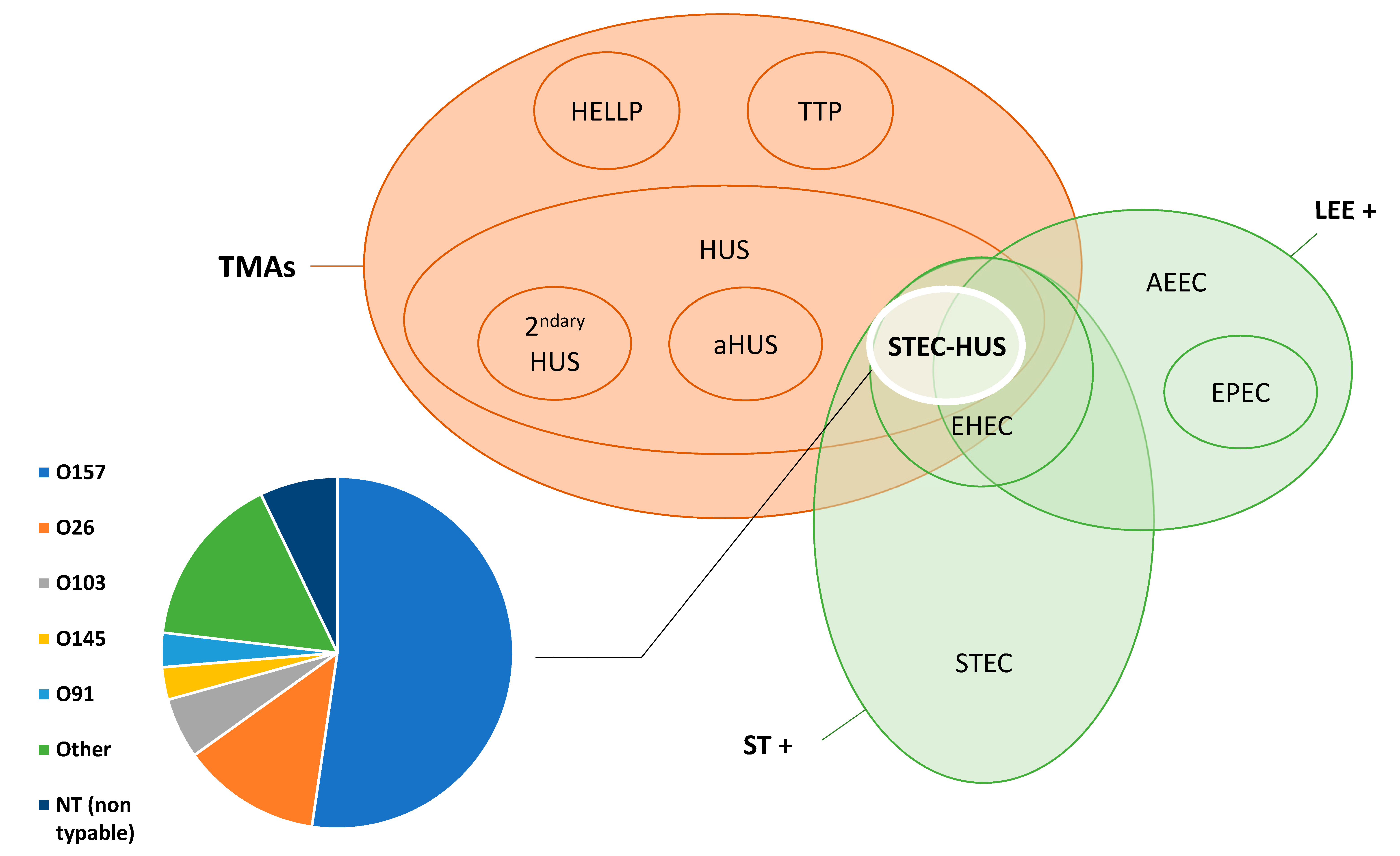
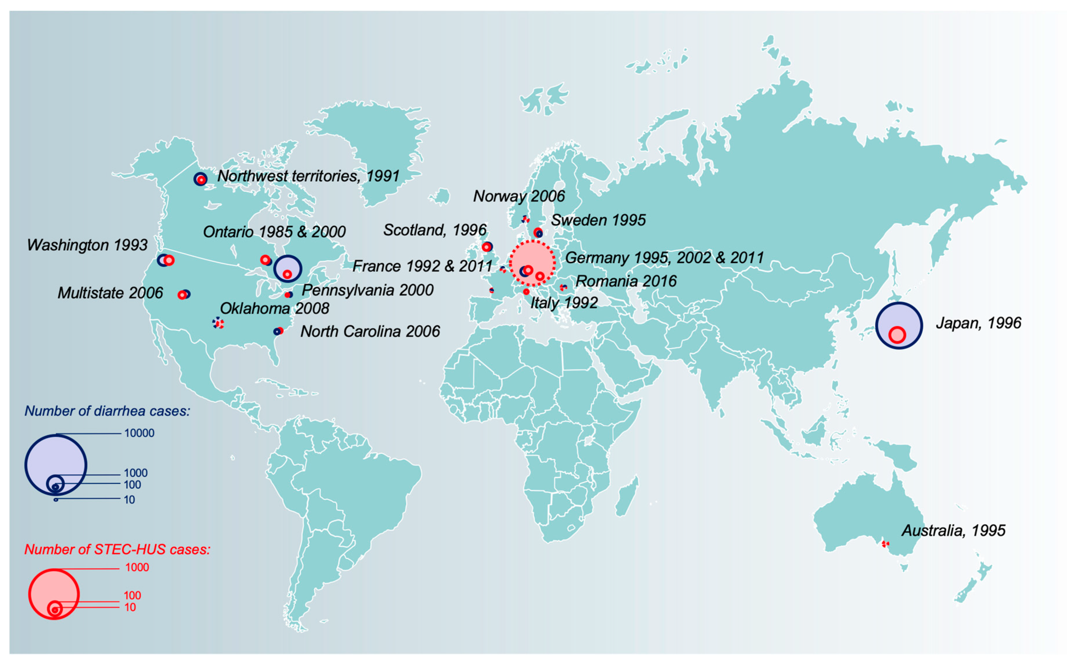
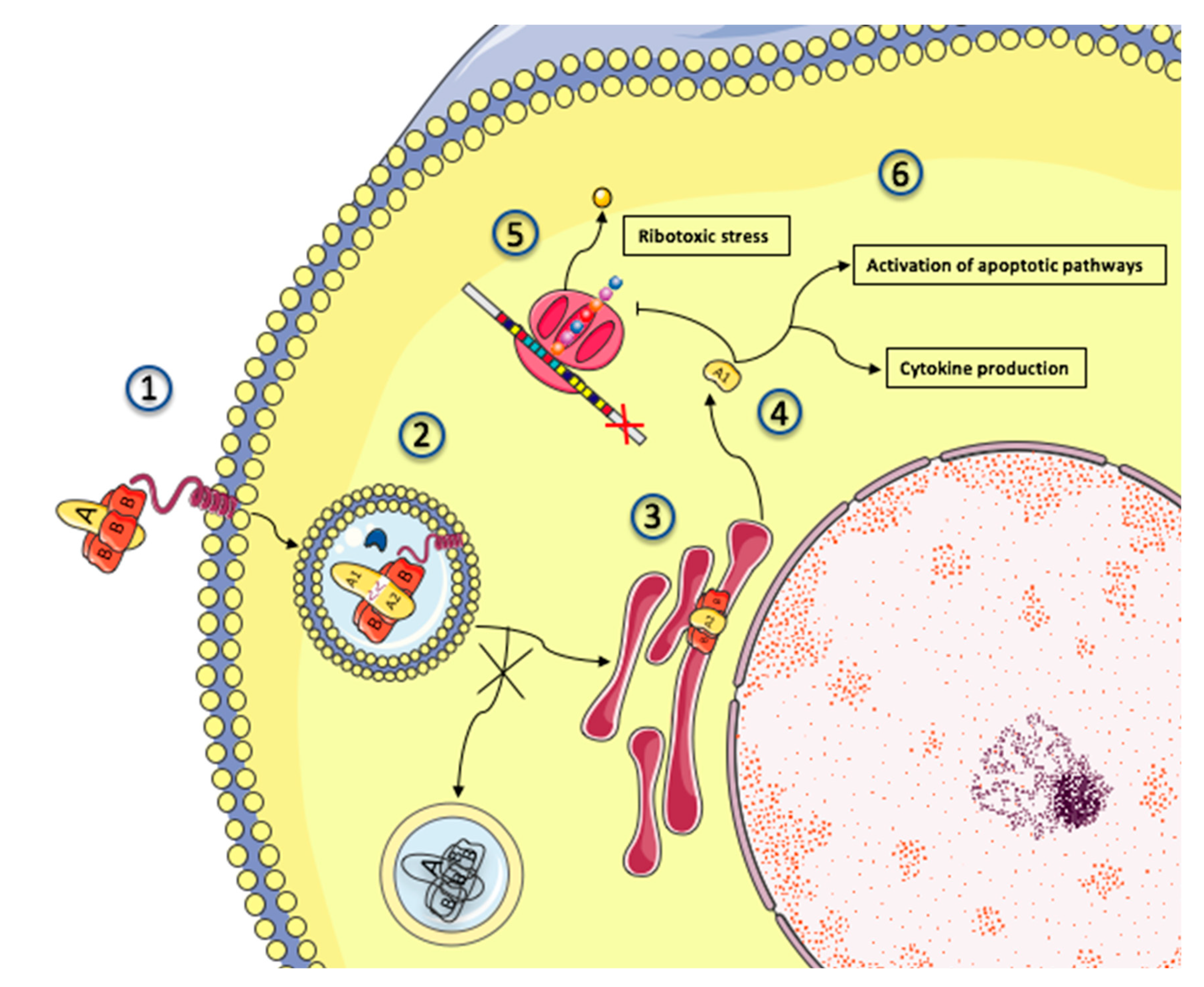
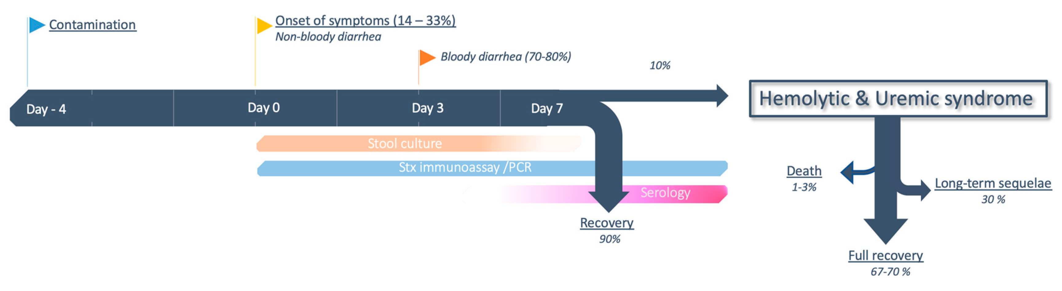
Table 1.
Common misconceptions about STEC-HUS.
| STEC-HUS mainly occurs through large outbreaks |
| Despite sensational publications about large outbreaks, most STEC-HUS cases (≈75%) are actually sporadic, judging by nationwide studies [99] and surveillance networks [104]. |
| Ground beef is the cause of the majority of vehicle-born transmissions |
| Cattle are a major reservoir for E. coli. Ground beef was responsible for the first outbreaks reported [6,7] and currently represents around 33% of cases [91]. |
| E. coli is the only bacteria that produces Shiga toxin |
| Shigella dysenteriae type 1 produces a chromosomally encoded toxin almost identical to Stx1 [217]. In addition, Stx phages can occasionally be found in other gram-negative bacteria (Citrobacter, Salmonella). |
| Community-acquired nonbloody diarrhea does not suggest investigation for STEC |
| If digestive symptoms are the rule in STEC infections, the proportion of bloody diarrhea can vary between 65%–80%, and is usually lower in non-O157 infections [41,218]. Investigations for STEC can be ordered for community-acquired diarrheas irrespective of the presence of blood [219]. |
| Complement is involved in the pathophysiology of atypical HUS, not STEC-HUS |
| Although the breakthrough discovery of alternative complement pathway dysregulation in aHUS is not paralleled in STEC-HUS, recent publications highlighted a potential role in the pathophysiology of STEC-HUS [183], providing hope for potential clinical applications. |
| HUS with a negative stool culture is probably atypical |
| Stool culture sensitivity is insufficient to exclude STEC-HUS. The diagnostic strategy must include both culture and nonculture-based assays to detect Shiga toxins or the genes encoding it [219]. Additionally, by the time of HUS, enterohemorrhagic E. coli is less likely to be found in stool cultures [220]. |
| Antibiotics are detrimental during STEC infection |
| Antibiotics are not recommended for STEC infection. Nevertheless, an important distinction has to be made between antibiotics capable of triggering bacterial SOS response and the release of Stx (fluoroquinolones, B-lactams) and others (azithromycin, fosfomycin) which do not [33,221]. The potential beneficial effects of the latter agents are currently being evaluated. |
| O157:H7 is responsible for the majority of STEC infections throughout the world |
| A shift in epidemiology occurred in the 2000s, and thanks to new diagnostic techniques, non-0157 serotypes are now more commonly found than 0157:H7 in Europe and North America [41,50]. However, 0157:H7 is still responsible for the majority of cases in Latin America [93]. |
© 2020 by the authors. Licensee MDPI, Basel, Switzerland. This article is an open access article distributed under the terms and conditions of the Creative Commons Attribution (CC BY) license (http://creativecommons.org/licenses/by/4.0/).
Share and Cite
MDPI and ACS Style
Joseph, A.; Cointe, A.; Mariani Kurkdjian, P.; Rafat, C.; Hertig, A. Shiga Toxin-Associated Hemolytic Uremic Syndrome: A Narrative Review. Toxins 2020, 12, 67. https://0-doi-org.brum.beds.ac.uk/10.3390/toxins12020067
AMA Style
Joseph A, Cointe A, Mariani Kurkdjian P, Rafat C, Hertig A. Shiga Toxin-Associated Hemolytic Uremic Syndrome: A Narrative Review. Toxins. 2020; 12(2):67. https://0-doi-org.brum.beds.ac.uk/10.3390/toxins12020067
Chicago/Turabian StyleJoseph, Adrien, Aurélie Cointe, Patricia Mariani Kurkdjian, Cédric Rafat, and Alexandre Hertig. 2020. "Shiga Toxin-Associated Hemolytic Uremic Syndrome: A Narrative Review" Toxins 12, no. 2: 67. https://0-doi-org.brum.beds.ac.uk/10.3390/toxins12020067
Note that from the first issue of 2016, this journal uses article numbers instead of page numbers. See further details here.