Article

Vaccination with VLPs Presenting a Linear Neutralizing Domain of S. aureus Hla Elicits Protective Immunity

1
Department of Pharmaceutical Sciences, College of Pharmacy, University of New Mexico Health Sciences Center, Albuquerque, NM 87131, USA
2
Department of Molecular Genetics and Microbiology, School of Medicine, University of New Mexico Health Sciences Center, Albuquerque, NM 87131, USA
*
Author to whom correspondence should be addressed.
Submission received: 11 June 2020 / Revised: 8 July 2020 / Accepted: 9 July 2020 / Published: 11 July 2020
(This article belongs to the Special Issue Staphylococcus aureus Toxins: Promoter or Handicap during Infection)

Abstract

:
The pore-forming cytotoxin α-hemolysin, or Hla, is a critical Staphylococcus aureus virulence factor that promotes infection by causing tissue damage, excessive inflammation, and lysis of both innate and adaptive immune cells, among other cellular targets. In this study, we asked whether a virus-like particle (VLP)-based vaccine targeting Hla could attenuate S. aureus Hla-mediated pathogenesis. VLPs are versatile vaccine platforms that can be used to display target antigens in a multivalent array, typically resulting in the induction of high titer, long-lasting antibody responses. In the present study, we describe the first VLP-based vaccines that target Hla. Vaccination with either of two VLPs displaying a 21 amino-acid linear neutralizing domain (LND) of Hla protected both male and female mice from subcutaneous Hla challenge, evident by reduction in lesion size and neutrophil influx to the site of intoxication. Antibodies elicited by VLP-LND vaccination bound both the LND peptide and the native toxin, effectively neutralizing Hla and preventing toxin-mediated lysis of target cells. We anticipate these novel and promising vaccines being part of a multi-component S. aureus vaccine to reduce severity of S. aureus infection.
Key Contribution: This work demonstrates that virus-like particle-based vaccines presenting a neutralizing epitope of S. aureus α-hemolysin may prove useful as part of a combination vaccine to limit pathogenesis in humans resulting from S. aureus infection.

1. Introduction

Staphylococcus aureus α-hemolysin (Hla) is an important secreted bacterial virulence factor whose loci is found in 99% of clinical isolates. Hla mediates invasive infection and promotes pathogenesis associated with both primary and recurrent skin and soft tissue infection (SSTI), pneumonia (PNA), peritoneal infections, and sepsis, among others [1,2,3,4,5,6,7,8,9]. In SSTI models, S. aureus mutants lacking Hla are attenuated [10] and are more rapidly cleared by the host [3]. Hla binds to a zinc metalloprotease, ADAM10, on host cells to form a heptameric pore and initiate breach of epithelial barriers [6,9,11]. The importance of Hla to various S. aureus infections likely stems from the broad cellular distribution of ADAM10 [7]. Therefore, Hla is a major toxin target for vaccines and therapeutics to limit S. aureus infections.
Several Hla vaccines have been tested in preclinical animal models including (i) a full length nontoxigenic Hla (HlaH35L), (ii) the N-terminal 50 amino acids of Hla fused to glutathione S-transferase (GST) (GST-Hla1-50), (iii) a structurally designed vaccine consisting of 62 non-contiguous Hla amino acids, and (iv) Hla engineered to lack the predicted membrane-spanning stem domain (HlaPSGS) [10,12,13,14]. Despite some successes in animal models, no S. aureus or Hla vaccine has been successful in clinical trials. This, together with the burden of disease caused by S. aureus, suggests that novel approaches to vaccine development are urgently needed.
Virus-like particles (VLPs) are a versatile nanoparticle vaccine platform for displaying practically any epitope in a multivalent format that virtually guarantees the ability to elicit high titer and long-lasting epitope-specific Abs [15]. While many VLP platforms can be used to display target antigens, VLPs derived from spherical RNA bacteriophage, such as Qß and AP205, have many advantages, including the ability to express and purify large amounts of VLPs in E. coli, and the multiple flexible approaches for displaying antigenic targets on their surface. Several Qß VLP-based vaccines have been evaluated in clinical trials, and these vaccines were shown to be safe and immunogenic [16]. Although VLP-based vaccines targeting S. aureus toxins have yet to be developed, their successful utilization against other pathogens suggests their potential for vaccine protection of humans against Hla-mediated pathogenesis. We developed active VLP-based vaccines by displaying a 21 amino-acid Hla linear neutralizing domain (LND), first identified by Oscherwitz and Cease as the target of an Hla-inactivating mAb [17]. The LND domain is involved in heptamerization of the Hla (Figure 1A), and it has been shown that an antibody against this epitope can neutralize Hla activity. We postulated that vaccination with VLPs displaying this peptide would elicit a neutralizing antibody (NAb) response and provide active protection in a mouse model of S. aureus Hla challenge.
To test our postulate, we vaccinated mice with two different VLPs displaying the Hla-LND and assessed vaccine efficacy using a murine skin challenge model. Here, we demonstrate that vaccination with LND-VLPs induces Hla-reactive antibodies that provide protection against lesion formation upon subcutaneous challenge with recombinant Hla in both male and female mice. In addition, these Abs prevented Hla-mediated lysis of Jurkat cells in an in vitro neutralization assay. Together, our findings demonstrate the efficacy of VLP-based vaccines displaying the Hla-LND and suggest that these vaccines could contribute to a multi-component vaccine to prevent S. aureus pathogenesis and infection.

2. Results

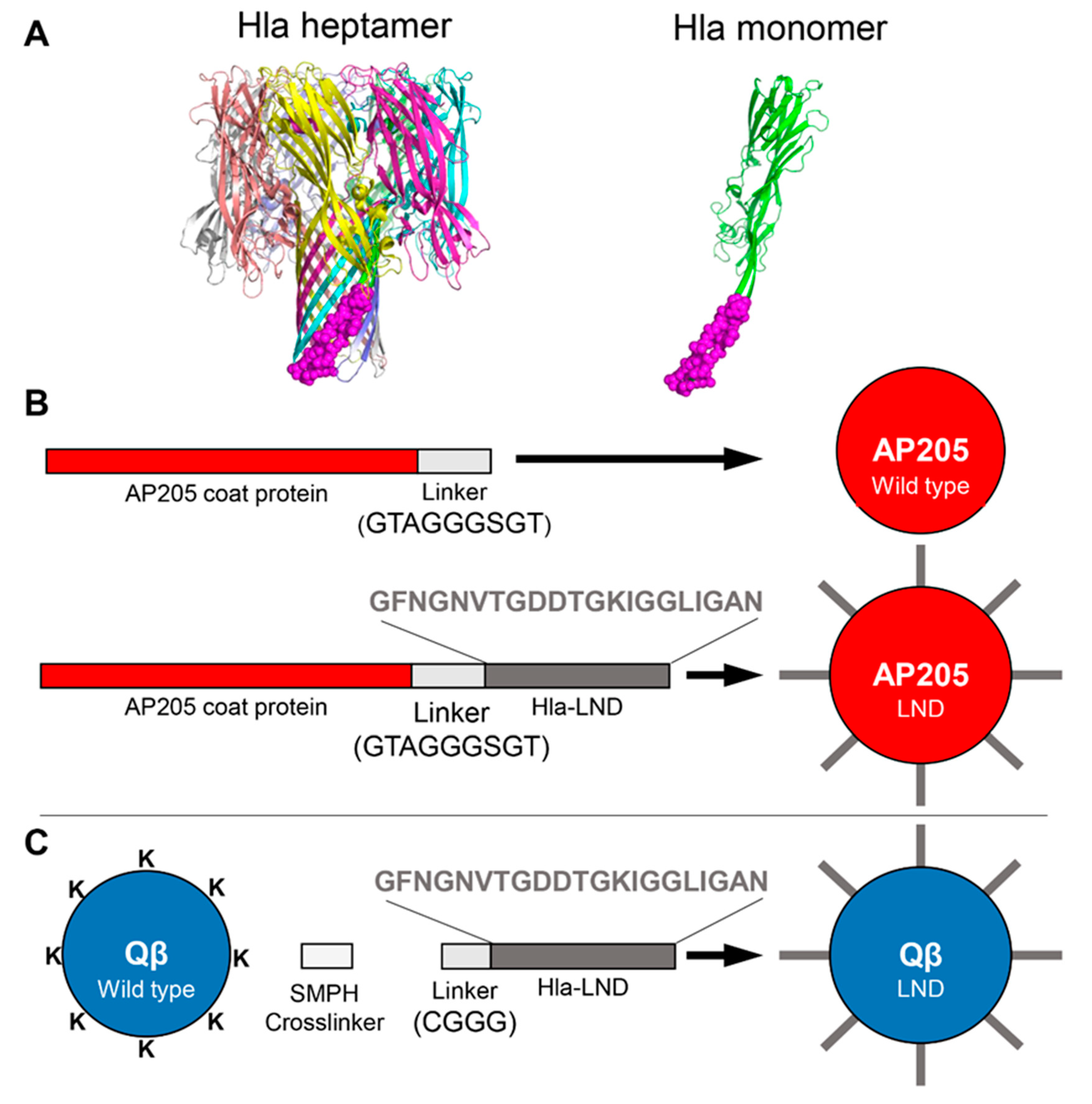
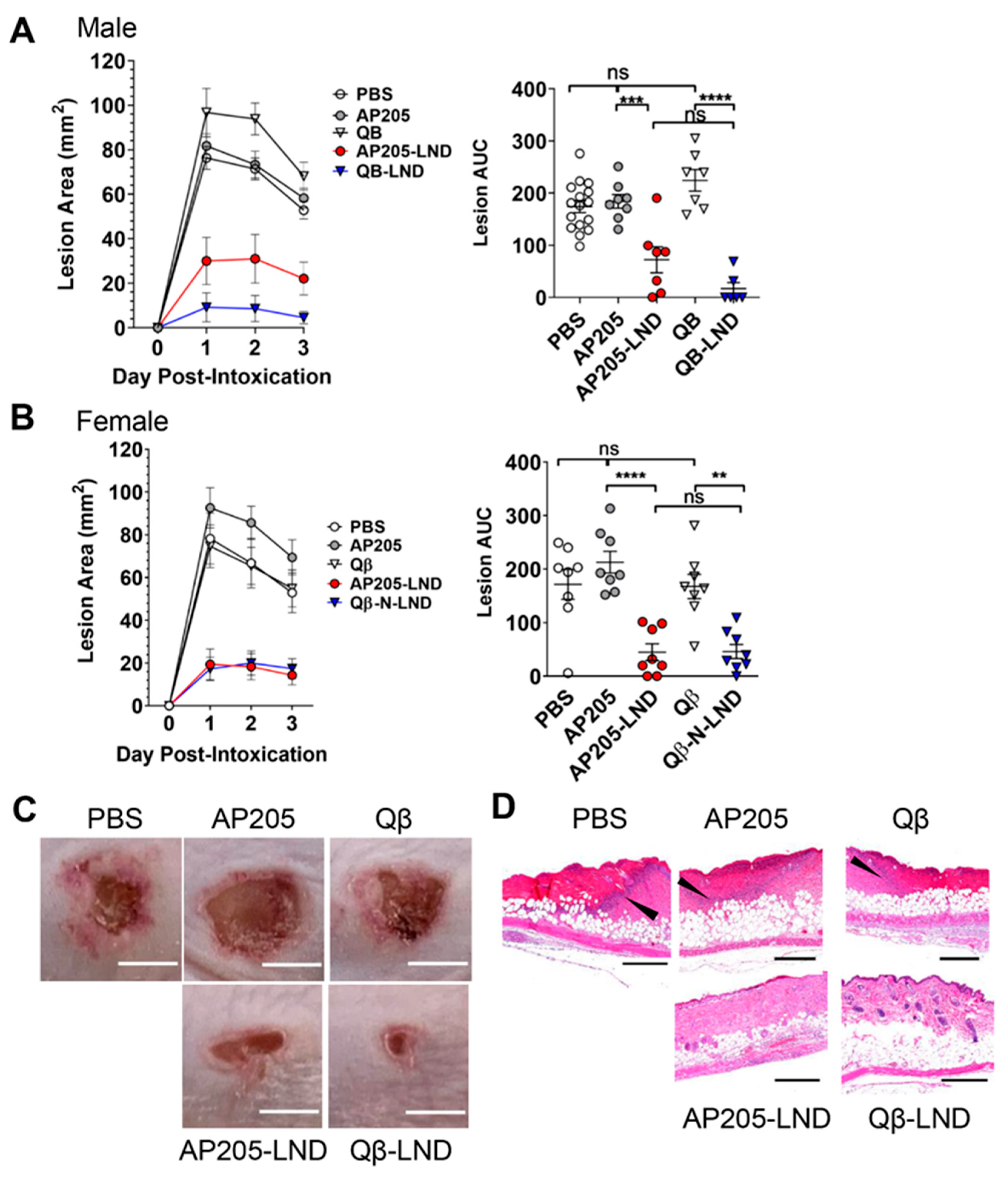
2.1. Vaccination with VLPs Displaying LND Protect against Hla Challenge

We used two different techniques for displaying the 21 amino acid Hla-LND epitope (Figure 1A) [17] on VLPs. First, we produced recombinant VLPs by genetically fusing this epitope to the C-terminus of the bacteriophage AP205 coat protein (AP205-LND; Figure 1B). This technique has been used previously to generate recombinant AP205 VLPs [18]. AP205 VLPs consist of 180 copies of the viral coat protein; thus, each VLP displays 180 copies of Hla-LND. As an alternative strategy, we chemically conjugated a synthetic peptide representing LND to Qß VLPs using a bifunctional crosslinker (Figure 1C). Formation of intact VLPs, purity of the preparations, and conjugation efficiency was determined by agarose gel electrophoresis and SDS-PAGE (Supplemental Methods and Figure S1). Based on the efficiency of LND conjugation, we determined that Qβ VLPs display an average of approximately 190 copies of the LND peptide per particle.
To assess the efficacy of our VLP-LND vaccines, we used a well-established mouse model of S. aureus skin infection/intoxication, where formation of a necrotic lesion is Hla-dependent [10,19,20,21]. In this model, the necrotic lesion visibly forms on the skin surface and lesion size is easily measurable [22,23,24,25,26], allowing straightforward assessment of Hla vaccine efficacy. Mice were immunized with two doses of AP205-LND or Qβ-LND VLPs. As controls, mice were immunized with similar doses of wild type, unmodified VLPs or were mock vaccinated with PBS. Eight weeks post-boost, mice were challenged by subcutaneous injection of rHla. The ability of vaccination to protect mice was evaluated by measuring lesion area for three days after Hla injection and by the calculated area under the curve (AUC) for the three-day challenge. Male and female mice were analyzed separately because female mice are more resistant to Hla compared to males [23], and different concentrations of Hla were subcutaneously injected in effort to achieve similar size lesions between the sexes. Hla-induced lesion size was significantly reduced in both AP205-LND and Qβ-LND vaccinated male (~60% and 94%, respectively) and female mice (~79% and 73%, respectively) compared to control mice (Figure 2A–C). The two VLP-based vaccines provided statistically similar protection from lesion formation in both sexes.
Along with reduced lesion size, we predicted that in vivo Hla challenge of VLP-LND-vaccinated mice would result in reduced local neutrophil influx and reduced inflammation. To assess inflammatory responses, we conducted histological analysis of lesions collected from vaccinated mice on day three post-Hla challenge. While control-vaccinated mice displayed a neutrophilic barrier crossing the dermis and epidermis surrounding the intoxication site, lesions from VLP-LND-vaccinated mice exhibited reduced numbers of neutrophils influx and decreased tissue damage (Figure 2D). This indicates that VLP-LND vaccination limits the pro-inflammatory response to Hla challenge.

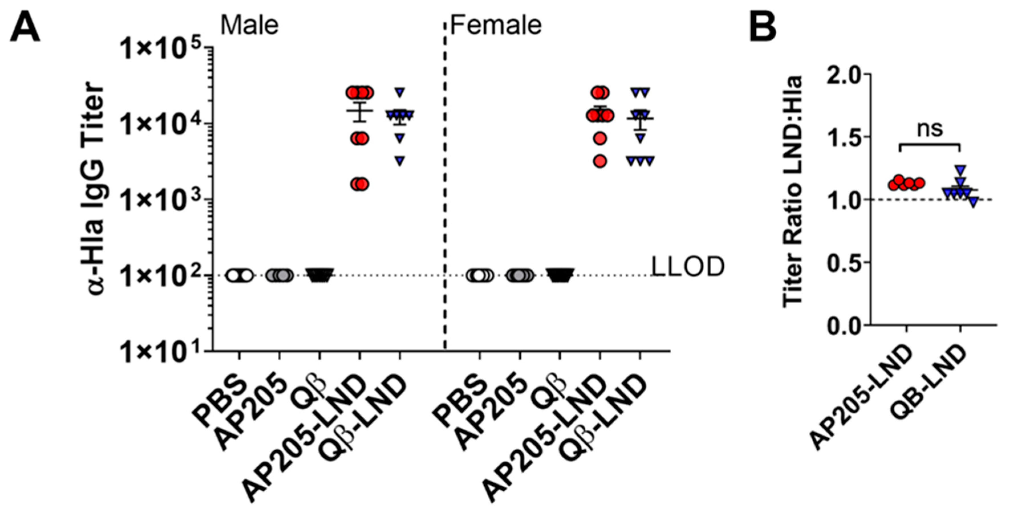
2.2. Antibodies Induced by VLP-LND Vaccination Bind to Intact Hla

In order to assess the mechanism(s) of in vivo protection, we asked whether serum Abs elicited by VLP-LND vaccination could bind Hla protein in vitro. As shown in Figure 3, vaccination with AP205-LND and Qβ-LND VLPs elicited high titer anti- rHla IgG antibodies, whereas no rHla-binding was detected in control mice (Figure 3A). These data indicate that both VLP-LND vaccines induce Ab recognizing Hla, suggesting that the conformation of the LND epitope displayed on the surface of the VLP is sufficiently similar to that of the target domain of the full-length toxin.
To investigate the molecular mimicry of the LND peptide in the context of AP205 and Qβ VLPs to the target domain of the full-length toxin, we also compared the ability of mouse VLP-LND antisera to bind to a synthetic LND peptide versus full-length rHla. The ratio of IgG Ab titers to the LND peptide versus rHla was not statistically different between mice vaccinated with AP205-LND or Qβ-LND, with ratios averaging 1.11 versus 1.13 respectively (Figure 3B). These findings support our hypothesis that the LND epitope displayed on either VLP platform exhibits molecular mimicry to the target epitope in Hla.

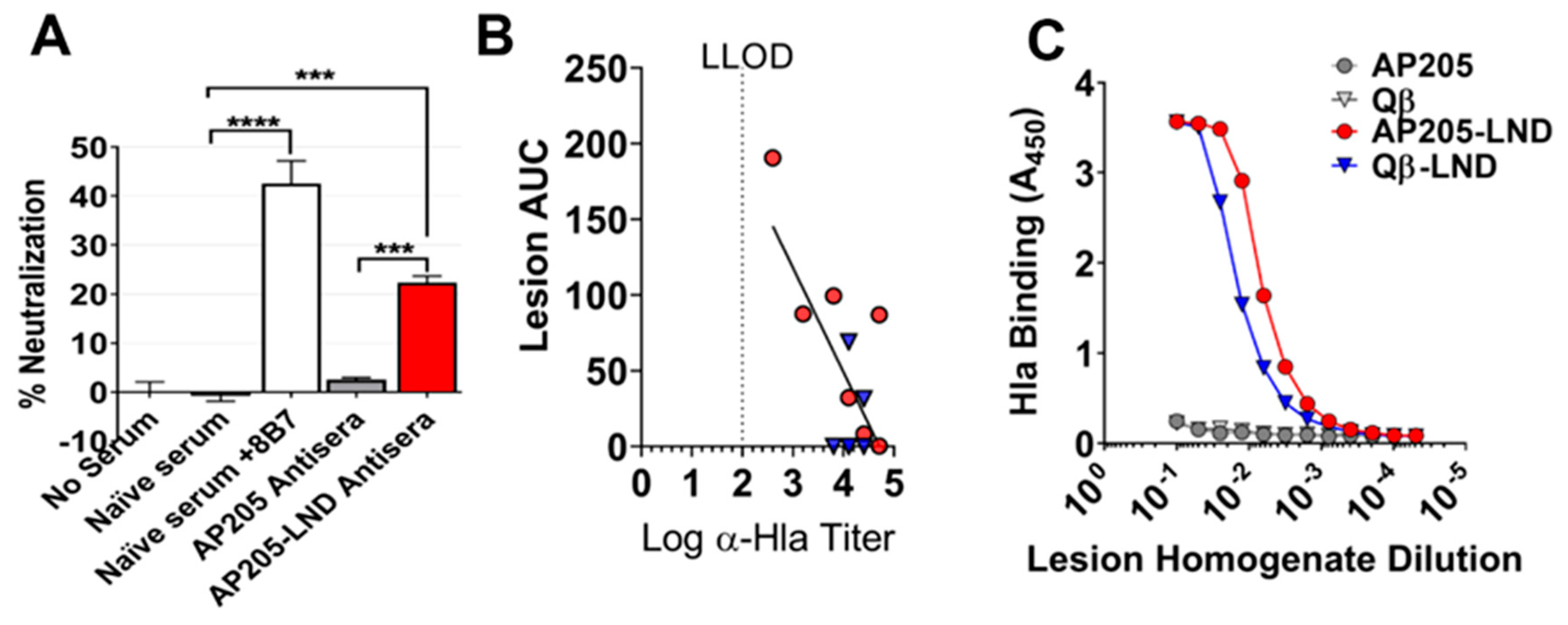
2.3. AP205-LND Vaccination Induces Hla-Neutralizing Antibodies

To determine the Hla-neutralizing ability of Ab elicited by VLP-LND vaccination, we performed a toxin neutralization assay as previously described [17]. Sera collected from vaccinated mice was tested for its ability to prevent lysis of susceptible Jurkat cells, a human T-cell line. AP205-LND antisera, but not sera raised against wild type AP205 VLPs, potently inhibited Jurkat cell lysis (Figure 4A) indicating that vaccination elicited Abs that neutralize Hla activity.
We next asked whether lesion size in individual Hla-challenged mice inversely correlated with Hla antibody titer. To test this, we performed a Hla ELISA on individual sera collected from vaccinated, Hla challenged mice at the time of sacrifice. Lesion area inversely correlated with the log of α-Hla titer for individual mice vaccinated with VLP-LND (p < 0.007; Figure 4B). For example, the individual mouse with the lowest anti-Hla antibody titer also had the largest legion after Hla-challenge. Finally, we tested whether antibodies were present at the site of Hla-challenge by detecting the presence of anti-Hla IgG in lesion homogenates by ELISA (Figure 4C). Anti-Hla IgG antibodies were readily detectable at the lesion site of VLP-LND immunized mice, but not controls, indicating that antibodies are present at the site of challenge. Together, these findings point to an in vivo mechanism of action whereby VLP-LND vaccination elicits an antibody response to S. aureus Hla, infiltration of anti-Hla Ab to the site of intoxication, and neutralization of Hla function, thus providing protection against Hla-mediated pathogenesis.

3. Discussion

Oscherwitz and Cease [17] previously demonstrated the ability of a linear 21 amino-acid region of Hla, termed the linear neutralizing domain (LND), to elicit Hla NAbs. We utilized this region to develop VLP-based vaccines that protect against Hla challenge in a murine model of SSTI. VLP-based vaccines, including some already in clinical use, have historically been developed against viral pathogens [16], making their utilization to protect against bacterial toxins a novel strategy. We used two strategies to display the LND epitope on VLPs; genetic fusion to AP205 and chemically conjugation to Qß. Both VLPs displayed the LND peptide at a similar valency (180–190 copies per VLP) and in a relatively unconstrained format on the surface of the VLP. Correspondingly, both VLP-based vaccines elicited high-titer antibodies to both the LND and to intact Hla and conferred protection against Hla skin challenge in a murine model. Furthermore, AP205-LND-elicited Abs neutralized Hla function and prevented lysis of cultured Jurkat cells. Finally, Hla-targeting Abs were found at the site of Hla challenge in the mouse model and the Hla titer inversely correlated with lesion size. Together, these findings point to an in vivo mechanism of action whereby VLP-LND vaccination elicits an antibody response capable of neutralizing S. aureus Hla, thus providing protection against Hla-mediated pathogenesis.
Although opsonizing antibodies presumably have the potential to clear S. aureus infection [27,28,29], rational design of S. aureus vaccines aimed at generating opsonizing antibodies have so far been clinically unsuccessful [30]. These failures suggest that an alternative approach that targets virulence factors like Hla could be effective at reducing pathogenesis or could be effective in the context of a combination vaccine. Hla and other S. aureus pore-forming toxins are capable of lysing host immune cells [7] and contribute to several types of S. aureus infection [1,2,3,9]. Neutralization of pore-forming toxins could therefore help preserve phagocytic cells such as neutrophils and monocytes/macrophages, thus secondarily enhancing bacterial clearance [30]. The importance of preserving phagocyte numbers is evidenced by the higher incidence of S. aureus infection in patients with functional defects in these cells [31,32,33]. In addition, human clinical data indicate that a high Ab titer against Hla correlates with decreased severity of S. aureus sepsis [34] and reduced recurrence of S. aureus SSTI in children [35].
Hla promotes invasive infection and is therefore a target for therapeutic and prophylactic interventions. Although Hla-targeting vaccines have shown promise in animal models of pneumonia [12,13,36], SSTI [10,17,37,38], and bacteremia [13], even vaccines inducing opsonophagocytic Abs show efficacy in animal models, but subsequently failed in human clinical trials [39]. The VLP-based vaccines reported here focus on a targeted, essential domain of Hla and thus direct the immune response towards a vulnerable epitope while avoiding non-neutralizing regions present in the native toxin. It is worth noting that many of the other pore forming toxins secreted by S. aureus also encode what appears to be linear neutralizing domains. Similar to what we have shown here regarding the Hla-LND, it may be possible to target the LNDs of other S. aureus pore-forming toxins with VLP-based vaccines that could have efficacy against a variety of S. aureus infections. Therefore, vaccines that elicit NAbs against pore-forming toxins, such as Hla, have the potential to limit pathogenesis and enhance bacterial clearance by sparing host immune cells.

4. Conclusions

Novel treatment and prevention strategies are urgently needed to combat infections caused by antibiotic resistant S. aureus. Here, we describe two candidate VLP-based vaccines, AP205-LND and Qβ-LND, with demonstrated efficacy in the prevention of Hla-mediated pathogenesis in a murine skin intoxication model. Although further studies are required, the efficacy of these vaccines and the clinical utility of VLP-based vaccines in general suggests that they could contribute to a multi-component S. aureus vaccine.

5. Materials and Methods

5.1. Ethics Statement

Animal experiments described in this study were approved (3/25/2020) by the Institutional Animal Care and Use Committee (IACUC) of the University of New Mexico Health Sciences Center (Animal Welfare Assurance number D16-00228) and conducted in strict accordance with recommendations in the Guide for Care and Use of Laboratory Animals [40] the Animal Welfare Act and U.S. federal law.

5.2. VLP Cloning, Expression, and Purification

The pAPKP plasmid encoding the AP205 coat protein and C-terminal linker (GTAGGGSGT) was used for generation of AP205-LND. The pAPKP plasmid was derived from pBAD-thio-TOPO-AP205, provided by Kaspars Tars of the Latvian Biomedical Research and Study Centre, Riga. It is based on pBAD-thio-TOPO (ThermoFisher, Grand Island, NY, USA) and expresses AP205 coat protein from the E. coli arabinose promoter. We constructed pAPKP by modifying pBAD-thio-TOPO-AP205 to encode a linker peptide at the coat protein C-terminus similar to that described by Tissot et al. in 2010 [18]. Briefly, the E. coli optimized Hla-LND sequence was synthesized as a mini-gene in IDT’s pSMART plasmid (Integrated DNA Technologies, Coralville, IA, USA). We fused foreign peptides to coat protein through the linker by insertion of their coding sequences between the KpnI and PstI sites of pAPKP and the LND-insert. Complete plasmid sequences are available upon request. The pAPKP-Hla-LND was transformed into E. coli 5αF’Iq for transcription followed by transformation into E. coli BL21*DE3 for expression.
Wild type Qβ was expressed from plasmid pETQCT and purified from Escherichia coli (E. coli) as previously described [41,42]. The synthetic Hla-LND peptide (Hla 119-131; GFNGNVTGDDTGKIGGLIGAN), including an N-terminal cysteine residue followed by a three-glycine-spacer sequence (CGGG-LND), was synthesized by GenScript (Piscataway, NJ, USA) then conjugated to Qβ’s surface-exposed lysine residues using the bifunctional crosslinker succinimidyl 6-[(beta-maleimidopropionamido) hexanoate] (SMPH; 22363; Thermo Scientific, Waltham, MA, USA) [43].
Wild type AP205 and AP205-LND were expressed and purified from BL21*DE3. Overnight cultures with 50 ug/mL kanamycin (Sigma-Aldrich, Burlington, MA, USA) were grown in 2xYT (16 g/L tryptone, 10 g/L yeast extract, 5 g/L NaCl; BD Biosciences, Franklin Lakes, NJ, USA). Expression strains of both AP205 and AP205-LND were grown overnight on media with kanamycin and used to inoculate a 50 to 500 mL culture with shaking. When OD600 nm surpassed 0.6, the culture was induced with 0.2% w/v arabinose and incubated for an additional 3 h. The cells were pelleted by centrifugation and purified via FPLC and confirmed for purity as previously described [25].

5.3. Mouse Immunizations

Four-week-old male and female BALB/c mice (Jackson Laboratories, Bar Harbor, ME, USA) were immunized by injection of 50–60 μL vaccine mixture in the caudal thigh muscle. Vaccines contained 10 μg VLP suspended in sterile PBS, mixed 1:1 in Incomplete Freund’s Adjuvant (Invivogen, San Diego, CA, USA), or mock vaccinated with PBS alone. Four weeks later, mice were given an identical vaccination (boost). Serum for ELISAs was collected by retro-orbital bleed without euthanizing or at the time of euthanasia by cardiac puncture. Challenge experiments were performed eight weeks after boosting.

5.4. Hla and LND Peptide ELISA

Vaccine-elicited murine serum antibody binding to Hla was determined by ELISA. ELISA plates were prepared by coating Ultra Cruz ELISA High-Binding plates (Santa Cruz Biotechnology, Santa Cruz, CA, USA) with 125 ng per well of rHla in 50 μL carbonate-bicarbonate buffer (pH 9.5) and incubated overnight (approximately 16–18 h) with mild shaking at 4 °C. After incubation, liquid was removed, and wells were blocked with PBS containing 1% casein (Thermo Scientific, Waltham, MA, USA) + 0.05% Tween-20 and then incubated for 2 h at 22 °C with shaking. Sera from vaccinated mice were serially diluted 2-fold or 4-fold for control samples, added onto the Hla-coated wells for antibody binding to Hla, and incubated at 22 °C for 1 h with shaking. After serum Ab binding, excess liquid was removed, and plates were washed three times using PBS containing 0.05% Tween-20 (PBS-T). Murine Ab bound to Hla was detected using polyclonal goat anti-mouse IgG secondary antibody linked to HRP (32230, Invitrogen), and assays were developed using 1-StepTM Ultra TMP-ELISA according to manufacturer’s directions. Hla titer was calculated as the reciprocal of the dilution value of the furthest diluted well with absorbance greater than two times the average of background (naïve sera). The lower limit of detection (LLOD) was a titer of 100. For detection of anti-Hla IgG Ab in Hla lesions, lesion homogenates collected from Hla-challenged mice, as reported below, were used in place of serum in the Hla ELISA.
For the LND peptide ELISA, streptavidin was coated to Immulon II ELISA plates (Thermo Scientific, Waltham, MA, USA) at 500 ng per well in pH 7.4 PBS overnight at 4 °C. Unbound streptavidin was removed by washing with PBS, and then, SMPH (22363; Thermo Scientific, Waltham, MA, USA) was added at 1 µg per well and incubated for 1 h at room temperature with shaking. After washing with PBS, 1 µg of synthetic CGGG-LND peptide (Genscript, Piscataway, NJ, USA) was added to each well and incubated for 1 h at room temperature with shaking. Unconjugated peptide was removed by washing with PBS, plates were blocked with PBS + 0.5% non-fat dry milk, and the ELISA was conducted as described above.

5.5. Toxin Neutralization Assay

Vaccine-elicited Hla-neutralizing Ab were detected in vitro by toxin neutralization assay, similar to that described by Oscherwitz and Cease [17] using the Jurkat human T cell line (TIB-152, ATCC, Manassas, VA, USA). Jurkat cells were grown in RPMI 1640 (Life Technologies, Grand Island, NY, USA) supplemented with 10% fetal bovine serum and 50 μM BME (complete medium) in a humidified 5% CO2 incubator at 37 °C. Fifty μL of vaccinated mouse sera diluted 1:12 in complete medium was plated in triplicate in a 96-well polystyrene plate (Corning Incorporated, Corning, NY, USA) and incubated with 50 μL rHla at 0.8 μg/μL for 30 min at 37 °C. Approximately 100,000 Jurkat cells in 100 μL complete medium were combined with the sera-rHla mixture and incubated at 37 °C. After 2 h incubation, 100 μL of 2,3-bis-(2-methoxy-4-nitro-5-sulfophenyl)-2H-tetrazolium-5-carboxanilide (XTT; Sigma-Aldrich, St. Louis, MO, USA) at 0.3 mg/mL and phenazine methosulfate (PMS; TCI America, Portland, OR, USA) at 0.015 mg/mL diluted in complete medium was added to the wells. After 18 h incubation, absorbance at 450 nm was measured using a SpectraMax 340 microplate reader (Molecular Devices, San Jose, CA, USA). As a positive control, 8B7 Hla-neutralizing antibody (0210-001, IBT, Rockville, MD, USA) was added to naïve mouse sera.

5.6. Mouse Skin Intoxication Model

The murine model for Hla-mediated lesion formation was essentially performed as previously described [19]. Briefly, the right flank of vaccinated male or female BALB/c mice was cleared of hair by shaving and depilation two days prior to intoxication. Because female mice are more resistant to Hla compared to males [23], in an effort to achieve similar size lesions between the sexes, male mice were challenged with 1.25 µg of Hla while females were challenged with 2 µg. On the day of intoxication, rHla was diluted in 50 μL saline (Braun, Irvine, CA, USA). Mice were anesthetized by isoflurane inhalation and injected subcutaneously with rHla in saline. Mice were weighed on the day of injection and daily thereafter until sacrifice. The site of intoxication was photographed daily, and lesion size was determined by analysis with ImageJ11. Three days after intoxication, mice were sacrificed by CO2 asphyxiation, and a 225-mm2 skin section containing the lesion was excised and stored in Hank’s Balanced Salt Solution (HBSS; Life Technologies, Carlsbad, CA, USA) containing 10 mM HEPES. Lesions were homogenized by mechanical disruption, and the clarified fraction was stored at −80 °C with 1x HaltTM protease inhibitor (Thermo Scientific, Waltham, MA, USA) for further analysis.

5.7. Histology

Histological evaluation was performed as previously reported [23], with skin samples collected on day 3 post-Hla intoxication. A 225-mm2 piece of skin containing the site of Hla injection was flattened on cardboard, fixed for 24 h in neutral buffered formalin, and then stored in 70% ethanol. Fixed skin was bisected through the center of the injection site and processed for embedding in paraffin, sectioned at 5 μm thickness, and stained with hematoxylin and eosin (H&E). Slides of H&E stained skin sections were scanned by an Aperio CS2 scanner (Leica Biosystems, Buffalo Grove, IL, USA).

5.8. Statistical analyses

Statistical analyses were performed using GraphPad Prism 8.4.2. Differences were considered statistically significant at p < 0.05.

Supplementary Materials

The following are available online at https://0-www-mdpi-com.brum.beds.ac.uk/2072-6651/12/7/450/s1. Supplemental Methods and Figure S1: Validation of AP205-LND and Qβ-LND VLPs.

Author Contributions

Conceptualization, P.R.H., B.C., D.S.P., S.M.D., J.A.J.; formal analysis, J.A.J., P.R.H.; investigation and methodology, J.A.J., S.M.D., J.P., K.D.T., S.P., B.O.E., D.A.; Writing—original draft preparation, J.A.J., P.R.H., S.M.D.; writing—manuscript editing and revision, B.C., D.S.P., P.R.H., J.A.J., S.M.D., B.O.E., K.D.T., S.P. project administration, PRH; funding acquisition, PRH. All authors have read and agreed to the published version of the manuscript.

Funding

This research was funded by research grants from the National Institutes of Health (AI145324, AI131098,) to PRH. JAJ was supported by an NIH supplement to AI145324. PRH was also supported in part by funds from the Autophagy, Inflammation and Metabolism (AIM) Center of Biomedical Research Excellence (CoBRE) supported by NIH P20 GM121176. In addition, this work utilized services and facilities provided through the NIH-funded University of New Mexico Comprehensive Cancer Center (P30 CA118100).

Conflicts of Interest

The sponsors had no role in the design, execution, interpretation, or writing of the study. BC and DSP have an equity stake in Agilvax, Inc.

References

  1. Rauch, S.; DeDent, A.C.; Kim, H.K.; Bubeck Wardenburg, J.; Missiakas, D.M.; Schneewind, O. Abscess Formation and Alpha-Hemolysin Induced Toxicity in a Mouse Model of Staphylococcus aureus Peritoneal Infection. Infect. Immun. 2012, 80, 3721–3732. [Google Scholar] [CrossRef] [Green Version]
  2. Bubeck Wardenburg, J.; Bae, T.; Otto, M.; Deleo, F.R.; Schneewind, O. Poring over Pores: Alpha-Hemolysin and Panton-Valentine Leukocidin in Staphylococcus aureus Pneumonia. Nat. Med. 2007, 13, 1405–1406. [Google Scholar] [CrossRef] [PubMed]
  3. Kobayashi, S.D.; Malachowa, N.; Whitney, A.R.; Braughton, K.R.; Gardner, D.J.; Long, D.; Bubeck Wardenburg, J.; Schneewind, O.; Otto, M.; DeLeo, F.R. Comparative Analysis of USA300 Virulence Determinants in a Rabbit Model of Skin and Soft Tissue Infection. J. Infect. Dis. 2011, 204, 937–941. [Google Scholar] [CrossRef]
  4. Powers, M.E.; Kim, H.K.; Wang, Y.; Bubeck Wardenburg, J. Adam10 Mediates Vascular Injury Induced by Staphylococcus aureus Alpha-Hemolysin. J. Infect. Dis. 2012, 206, 352–356. [Google Scholar] [CrossRef] [PubMed]
  5. Ragle, B.E.; Bubeck Wardenburg, J. Anti-Alpha-Hemolysin Monoclonal Antibodies Mediate Protection against Staphylococcus aureus Pneumonia. Infect. Immun. 2009, 77, 2712–2718. [Google Scholar] [CrossRef] [Green Version]
  6. Inoshima, N.; Wang, Y.; Bubeck Wardenburg, J. Genetic Requirement for ADAM10 in Severe Staphylococcus aureus Skin Infection. J. Investig. Dermatol. 2012, 132, 1513–1516. [Google Scholar] [CrossRef] [PubMed] [Green Version]
  7. Berube, B.J.; Bubeck Wardenburg, J. Staphylococcus aureus Alpha-Toxin: Nearly a Century of Intrigue. Toxins 2013, 5, 1140–1166. [Google Scholar] [CrossRef] [Green Version]
  8. Sampedro, G.R.; DeDent, A.C.; Becker, R.E.; Berube, B.J.; Gebhardt, M.J.; Cao, H.; Bubeck Wardenburg, J. Targeting Staphylococcus aureus Alpha-Toxin as a Novel Approach to Reduce Severity of Recurrent Skin and Soft-Tissue Infections. J. Infect. Dis. 2014, 210, 1012–1018. [Google Scholar] [CrossRef] [Green Version]
  9. Inoshima, I.; Inoshima, N.; Wilke, G.A.; Powers, M.E.; Frank, K.M.; Wang, Y.; Bubeck Wardenburg, J. A Staphylococcus aureus pore-forming toxin subverts the activity of ADAM10 to cause lethal infection in mice. Nat. Med. 2011, 17, 1310–1314. [Google Scholar] [CrossRef] [Green Version]
  10. Kennedy, A.D.; Bubeck Wardenburg, J.; Gardner, N.J.; Long, D.; Whitney, A.R.; Braughton, K.R.; Schneewind, O.; DeLeo, F.R. Targeting of alpha-hemolysin by active or passive immunization decreases severity of USA300 skin infection in a mouse model. J. Infect. Dis. 2010, 202, 1050–1058. [Google Scholar] [CrossRef] [Green Version]
  11. Wilke, G.A.; Bubeck Wardenburg, J. Role of a Disintegrin and Metalloprotease 10 in Staphylococcus aureus Alpha-Hemolysin-Mediated Cellular Injury. Proc. Natl. Acad. Sci. USA 2010, 107, 13473–13478. [Google Scholar] [CrossRef] [PubMed] [Green Version]
  12. Bubeck Wardenburg, J.; Schneewind, O. Vaccine protection against Staphylococcus aureus pneumonia. J. Exp. Med. 2008, 205, 287–294. [Google Scholar] [CrossRef]
  13. Adhikari, R.P.; Karauzum, H.; Sarwar, J.; Abaandou, L.; Mahmoudieh, M.; Boroun, A.R.; Vu, H.; Nguyen, T.; Devi, V.S.; Shulenin, S.; et al. Novel Structurally Designed Vaccine for S. aureus α-Hemolysin: Protection against Bacteremia and Pneumonia. PLoS ONE 2012, 7, e38567. [Google Scholar] [CrossRef] [PubMed]
  14. Fiaschi, L.; Di Palo, B.; Scarselli, M.; Pozzi, C.; Tomaszewski, K.; Galletti, B.; Nardi-Dei, V.; Arcidiacono, L.; Mishra, R.P.N.; Mori, E.; et al. Auto-Assembling Detoxified Staphylococcus aureus Alpha-Hemolysin Mimicking the Wild-Type Cytolytic Toxin. Clin. Vaccine Immunol. 2016, 23, 442–450. [Google Scholar] [CrossRef] [PubMed] [Green Version]
  15. Chackerian, B. Virus-like particles: Flexible platforms for vaccine development. Expert Rev. Vaccines 2007, 6, 381–390. [Google Scholar] [CrossRef]
  16. Mohsen, M.O.; Zha, L.; Cabral-Miranda, G.; Bachmann, M.F. Major findings and recent advances in virus–like particle (VLP)-based vaccines. Semin. Immunol. 2017, 34, 123–132. [Google Scholar] [CrossRef]
  17. Oscherwitz, J.; Cease, K.B. Identification and Validation of a Linear Protective Neutralizing Epitope in the Beta-Pore Domain of Alpha Toxin. PLoS ONE 2015, 10, e0116882. [Google Scholar] [CrossRef] [PubMed]
  18. Tissot, A.C.; Renhofa, R.; Schmitz, N.; Cielens, I.; Meijerink, E.; Ose, V.; Jennings, G.T.; Saudan, P.; Pumpens, P.; Bachmann, M.F. Versatile Virus-Like Particle Carrier for Epitope Based Vaccines. PLoS ONE 2010, 5, e9809. [Google Scholar] [CrossRef] [Green Version]
  19. Malachowa, N.; Kobayashi, S.D.; Braughton, K.R.; DeLeo, F.R. Mouse Model of Staphylococcus aureus Skin Infection. Methods Mol. Biol. 2013, 1031, 109–116. [Google Scholar] [PubMed]
  20. Ridder, M.J.; Daly, S.M.; Triplett, K.D.; Seawell, N.A.; Hall, P.R.; Bose, J.L. Staphylococcus aureus fatty acid kinase FakA modulates pathogenesis during skin infection via proteases. Infect. Immun. 2020. [Google Scholar] [CrossRef]
  21. Wright, J.S.; Jin, R.; Novick, R.P. Transient interference with staphylococcal quorum sensing blocks abscess formation. Proc. Natl. Acad. Sci. USA 2005, 102, 1691–1696. [Google Scholar] [CrossRef] [PubMed] [Green Version]
  22. Castleman, M.J.; Febbraio, M.; Hall, P.R. Cd36 Is Essential for Regulation of the Host Innate Response to Staphylococcus aureus Alpha-Toxin-Mediated Dermonecrosis. J. Immunol. 2015, 195, 2294–2302. [Google Scholar] [CrossRef] [PubMed] [Green Version]
  23. Castleman, M.J.; Pokhrel, S.; Triplett, K.D.; Kusewitt, D.F.; Elmore, B.O.; Joyner, J.A.; Femling, J.K.; Sharma, G.; Hathaway, H.J.; Prossnitz, E.R.; et al. Innate Sex Bias of Staphylococcus aureus Skin Infection Is Driven by Alpha-Hemolysin. J. Immunol. 2018, 200, 657–668. [Google Scholar] [CrossRef] [Green Version]
  24. Hall, P.R. Omega-Hydroxyemodin Limits Staphylococcus aureus Quorum Sensing-Mediated Pathogenesis and Inflammation. J. Immunol. 2015, 59, 2223–2235. [Google Scholar]
  25. Daly, S.; Joyner, J.A.; Triplett, K.D.; Elmore, B.O.; Pokhrel, S.; Frietze, K.M.; Peabody, D.S.; Chackerian, B.; Hall, P.R. VLP-based vaccine induces immune control of Staphylococcus aureus virulence regulation. Sci. Rep. 2017, 7, 637. [Google Scholar] [CrossRef] [PubMed] [Green Version]
  26. Triplett, K.D.; Pokhrel, S.; Castleman, M.J.; Daly, S.M.; Elmore, B.O.; Joyner, J.A.; Sharma, G.; Herbert, G.; Campen, M.J.; Hathaway, H.J.; et al. GPER activation protects against epithelial barrier disruption by Staphylococcus aureus α-toxin. Sci. Rep. 2019, 9, 1343. [Google Scholar] [CrossRef] [Green Version]
  27. Kim, H.K.; Kim, H.-Y.; Schneewind, O.; Missiakas, D. Identifying protective antigens of Staphylococcus aureus, a pathogen that suppresses host immune responses. FASEB J. 2011, 25, 3605–3612. [Google Scholar] [CrossRef] [Green Version]
  28. Atkins, K.L.; Burman, J.D.; Chamberlain, E.S.; Cooper, J.E.; Poutrel, B.; Bagby, S.; Jenkins, A.T.A.; Feil, E.; Elsen, J.M.V.D.; Jenkins, A.T.A.S. aureus IgG-binding proteins SpA and Sbi: Host specificity and mechanisms of immune complex formation. Mol. Immunol. 2008, 45, 1600–1611. [Google Scholar] [CrossRef]
  29. Sun, Y.; Emolo, C.; Holtfreter, S.; Wiles, S.; Kreiswirth, B.; Missiakas, D.; Schneewind, O. Staphylococcal Protein A Contributes to Persistent Colonization of Mice with Staphylococcus aureus. J. Bacteriol. 2018, 200, e00735-17. [Google Scholar] [CrossRef] [Green Version]
  30. Miller, L.S.; Fowler, V.G.; Shukla, S.K.; Rose, W.E.; Proctor, R.A. Development of a vaccine against Staphylococcus aureus invasive infections: Evidence based on human immunity, genetics and bacterial evasion mechanisms. FEMS Microbiol. Rev. 2020, 44, 123–153. [Google Scholar] [CrossRef] [Green Version]
  31. Bouma, G.; Ancliff, P.J.; Thrasher, A.J.; Burns, S. Recent advances in the understanding of genetic defects of neutrophil number and function. Br. J. Haematol. 2010, 151, 312–326. [Google Scholar] [CrossRef]
  32. Miller, L.S.; Cho, J.S. Immunity against Staphylococcus aureus cutaneous infections. Nat. Rev. Immunol. 2011, 11, 505–518. [Google Scholar] [CrossRef] [PubMed] [Green Version]
  33. Andrews, T.; Sullivan, K.E. Infections in Patients with Inherited Defects in Phagocytic Function. Clin. Microbiol. Rev. 2003, 16, 597–621. [Google Scholar] [CrossRef] [PubMed] [Green Version]
  34. Adhikari, R.P.; Ajao, A.O.; Karauzum, H.; Sarwar, J.; Lydecker, A.D.; Aman, M.J.; Johnson, J.K.; Nguyen, C.; Chen, W.H.; Roghmann, M.-C. Lower Antibody Levels to Staphylococcus aureus Exotoxins Are Associated With Sepsis in Hospitalized Adults With Invasive S. aureus Infections. J. Infect. Dis. 2012, 206, 915–923. [Google Scholar] [CrossRef]
  35. Fritz, S.A.; Tiemann, K.M.; Hogan, P.G.; Epplin, E.K.; Rodriguez, M.; Al-Zubeidi, D.N.; Bubeck Wardenburg, J.; Hunstad, D.A. A Serologic Correlate of Protective Immunity against Community-Onset Staphylococcus aureus Infection. Clin. Infect. Dis. 2013, 56, 1554–1561. [Google Scholar] [CrossRef] [PubMed] [Green Version]
  36. Tran, V.G.; Venkatasubramaniam, A.; Adhikari, R.P.; Krishnan, S.; Wang, X.; Le, V.T.M.; Le, H.N.; Vu, T.T.T.; Schneider-Smith, E.; Aman, M.J.; et al. Efficacy of Active Immunization With Attenuated α-Hemolysin and Panton-Valentine Leukocidin in a Rabbit Model of Staphylococcus aureus Necrotizing Pneumonia. J. Infect. Dis. 2019, 221, 267–275. [Google Scholar] [CrossRef] [PubMed]
  37. Oscherwitz, J.; Muñoz-Planillo, R.; Yu, F.; Nunez, G.; Cease, K.B. In vivo mapping of a protective linear neutralizing epitope at the N-terminus of alpha hemolysin from Staphylococcus aureus. Mol. Immunol. 2014, 60, 62–71. [Google Scholar] [CrossRef]
  38. Adhikari, R.P.; Thompson, C.D.; Aman, M.J.; Lee, J.C. Protective efficacy of a novel alpha hemolysin subunit vaccine (AT62) against Staphylococcus aureus skin and soft tissue infections. Vaccine 2016, 34, 6402–6407. [Google Scholar] [CrossRef] [Green Version]
  39. Proctor, R.A. Immunity to Staphylococcus aureus: Implications for Vaccine Development. Microbiol. Spectr. 2019, 7, 766–775. [Google Scholar] [CrossRef]
  40. National Research Council Committee for the Update of the Guide for the Care and Use of Laboratory Animals. Guide for the Care and Use of Laboratory Animals, 8th ed.; National Academies Press: Washington, DC, USA, 2011. [Google Scholar]
  41. Crossey, E.; Amar, M.J.A.; Sampson, M.; Peabody, J.; Schiller, J.T.; Chackerian, B.; Remaley, A.T. A cholesterol-lowering VLP vaccine that targets PCSK9. Vaccine 2015, 33, 5747–5755. [Google Scholar] [CrossRef] [Green Version]
  42. Akache, B.; Weeratna, R.D.; Deora, A.; Thorn, J.M.; Champion, B.; Merson, J.R.; Davis, H.L.; McCluskie, M.J. Anti-IgE Qb-VLP Conjugate Vaccine Self-Adjuvants through Activation of TLR7. Vaccines 2016, 4, 3. [Google Scholar] [CrossRef] [PubMed] [Green Version]
  43. Chackerian, B.; Rangel, M.; Hunter, Z.; Peabody, D.S. Virus and virus-like particle-based immunogens for Alzheimer’s disease induce antibody responses against amyloid-beta without concomitant T cell responses. Vaccine 2006, 24, 6321–6331. [Google Scholar] [CrossRef] [PubMed]
Figure 1. Schematic of virus-like particles (VLPs) Displaying α-hemolysin (Hla) linear neutralizing domain (LND). (A) (Left) Ribbon depiction of Hla heptameric pore based on 3ANZ.pdb. Monomers are shown in different colors and LND region shown as purple spheres. (Right) Ribbon depiction of Hla monomer with LND region shown as described above. Figures produced using PyMOL (The PyMOL Molecular Graphics System, Version 2.0 Schrödinger, LLC.) (B) (Top left) Linear schematic depicting wild type AP205 coat protein with C-terminal linker; (top right) schematic of assembled AP205 wild type VLP; (Bottom left) linear schematic of AP205 coat protein with Hla-LND sequence genetically inserted; (bottom right) schematic of assembled AP205-LND VLP created through molecular cloning. (C) (Left) schematic of assembled Qβ wild type VLP depicting surface exposed lysines; (center) linear depiction of SMPH crosslinker and synthetic CGGG-Hla-LND prior to chemical conjugation to surface lysines; (right) schematic of assembled Qβ VLP displaying surface lysine conjugated LND peptides.
Toxins 12 00450 g001
Figure 2. Vaccination with VLPs displaying LND protects mice from lesion formation caused by subcutaneous Hla challenge. (A) (Left) Lesion area over the course of a 3-day Hla intoxication of vaccinated or mock-vaccinated male BALB/c mice (1.25 µg Hla/mouse). (Right) Calculated lesion area under the curve (AUC) for the 3-day Hla challenge. (B) (Left) Lesion area over the course of a 3-day Hla intoxication of female BALB/c mice (2 µg Hla/mouse). (Right) Calculated lesion AUC for the 3-day Hla challenge. (C) Representative images of external lesions of vaccinated Hla-challenged male BALB/c mice at day 3 post-intoxication. Scale bar = 5 mm. (D) H&E stained Hla-intoxicated skin sections collected 3-days post-intoxication from vaccinated male BALB/c mice. Arrowheads denote neutrophilic barrier around intoxication site. Scale bar = 0.5 mm. (A,B) Data are mean ± SEM for (A) n = 16, 8, 7, 7, 6 mice per group and (B) n = 8 mice/group. One-Way ANOVA p < 0.0001, Tukey’s multiple comparisons test; ns, not significant; **, p < 0.01, ***, p < 0.001, ****, p < 0.0001.
Toxins 12 00450 g002
Figure 3. Antibodies elicited by VLP-LND vaccination recognize recombinant Hla. (A) Antibody binding to Hla from serum from male (n = 5 to 16 mice/group) or female (n = 8 mice/group) BALB/c mice vaccinated with AP205-LND, Qβ-LND or controls, at two weeks post-boost, tested by ELISA. LLOD = lower limit of detection. (B) Ratio of anti-LND antibody titer over anti-Hla titer using serum from male BALB/c mice vaccinated with AP205-LND (n = 8 mice/group) or Qβ-LND (n = 7 mice/group). All antibody titers are reported as the reciprocal of the highest serum dilution with an OD450 2-fold greater than naïve sera. Data shown as mean ± SEM. ns, not significant.
Toxins 12 00450 g003
Figure 4. VLP-LND vaccination elicits antibodies that protect against Hla-mediated pathogenesis in vitro and in vivo. (A) Toxin neutralization assay measuring viability of Jurkat cells incubated with 0.8 µg/mL Hla combined with sera collected from vaccinated mice diluted 1:12. Sera include pooled naïve BALB/c serum (n = 5 mice/group), naïve serum spiked with ~9 μg/mL Hla-neutralizing mAb 8B7 (IBT #0210-001), and pooled serum from mice vaccinated with AP205wt (n = 3 mice/group) or AP205-LND (n = 3 mice/group). Assay performed in triplicate. A450 values were normalized to a total lysis control (RPMI+0.1% Triton-X114) set to 0% neutralization and a no lysis control (RPMI) set to 100% neutralization. One-way ANOVA p < 0.0001. Sidak’s multiple comparison test; ***, p < 0.001; ****, p < 0.0001. (B) Linear regression plot comparing lesion AUC for a 3-day Hla challenge of male BALB/c mice vaccinated with AP205-LND (n = 7 mice/group) or Qβ-LND (n = 6 mice/group) versus the log α-Hla titer, p = 0.0070. (C) ELISA showing Hla binding by antibodies from day 3 post-intoxication pooled lesion homogenate collected from male BALB/c mice previously vaccinated with AP205 (n = 8 mice/group), AP205-LND (n = 7 mice/group), Qβ (n = 7 mice/group), or Qβ-LND (n = 5 mice/group).
Toxins 12 00450 g004

