Are Cyanotoxins the Only Toxic Compound Potentially Present in Microalgae Supplements? Results from a Study of Ecological and Non-Ecological Products


Abstract
:1. Introduction
2. Results
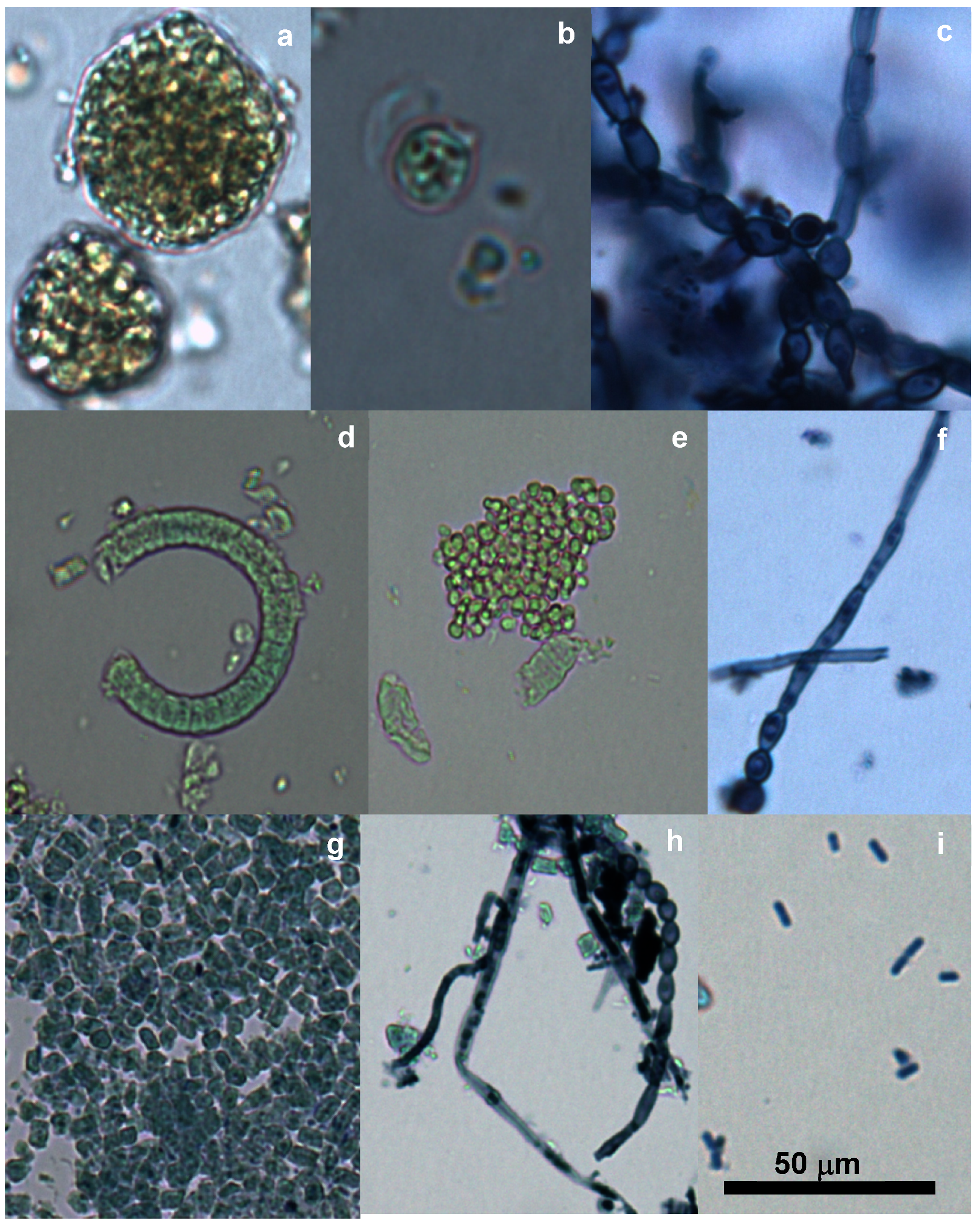
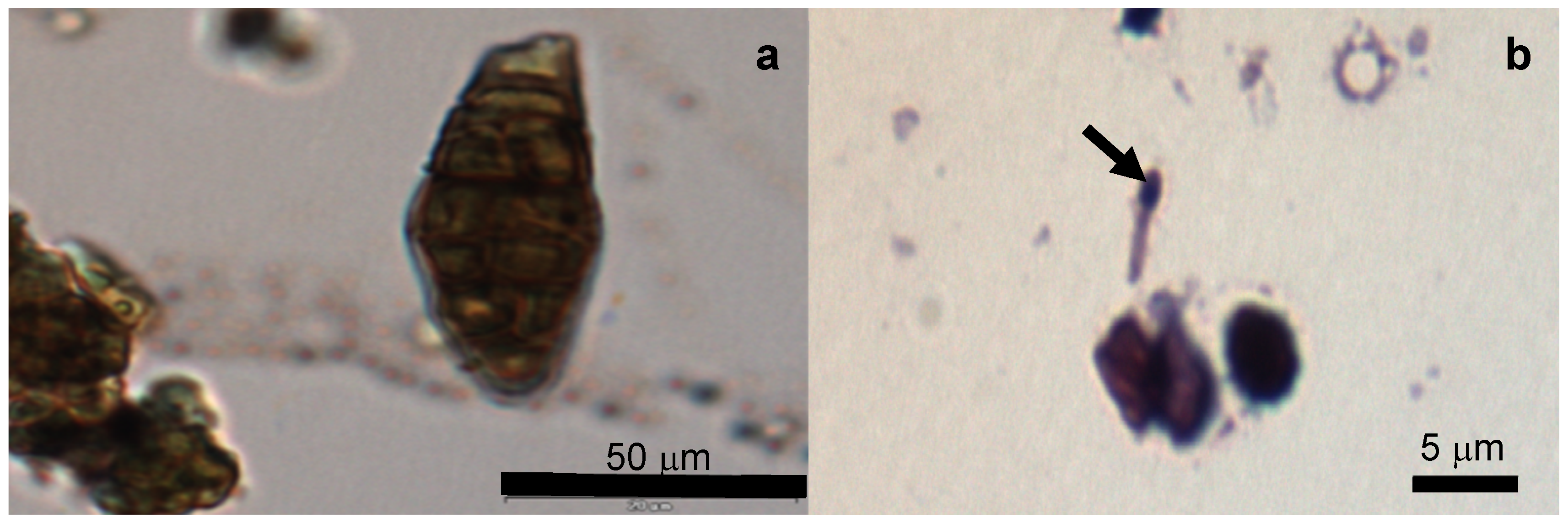
2.1. Microscopic Analysis
2.2. ELISA
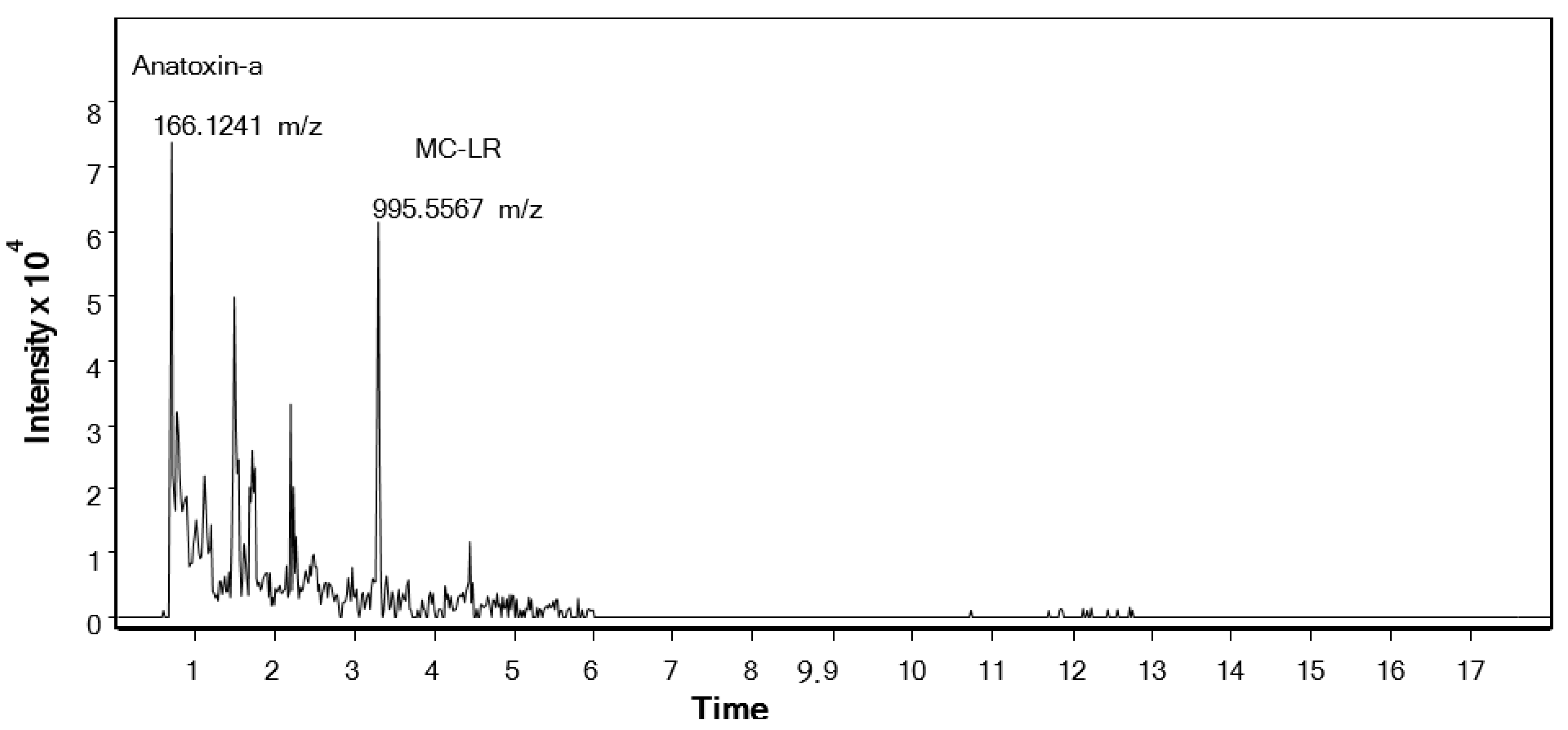
2.3. HPLC-MS
2.4. Elemental Composition
3. Discussion
4. Conclusions
5. Materials and Methods
5.1. The Analyzed Algae Supplement Products
5.2. Light Microscopy
5.3. Extraction
5.4. Cyanotoxins Characterization and Quantification
5.4.1. ELISA Test
5.4.2. HPLC-MS
5.5. Elemental Composition (ICP-OES)
Supplementary Materials
Author Contributions
Funding
Acknowledgments
Conflicts of Interest
References
- Buratti, F.M.; Manganelli, M.; Vichi, S.; Stefanelli, M.; Scardala, S.; Testai, E.; Funari, E. Cyanotoxins: Producing organisms, occurrence, toxicity, mechanism of action and human health toxicological risk evaluation. Arch. Toxicol. 2017, 91, 1049–1130. [Google Scholar] [CrossRef] [PubMed]
- Clarys, P.; Deliens, T.; Huybrechts, I.; Deriemaeker, P.; Vanaelst, B.; De Keyzer, W.; Hebbelinck, M.; Mullie, P. Comparison of nutritional quality of the vegan, vegetarian, semi-vegetarian, pesco-vegetarian and omnivorous diet. Nutrients 2014, 6, 1318–1332. [Google Scholar] [CrossRef]
- Smetana, S.; Mathys, A.; Knoch, A.; Heinz, V. Meat alternatives: Life cycle assessment of most known meat substitutes. Int. J. Life Cycle Assess 2015, 20, 1254–1267. [Google Scholar] [CrossRef]
- Kumar, P.; Chatli, M.K.; Mehta, N.; Singh, P.; Malav, O.P.; Verma, A.K. Meat analogues: Health promising sustainable meat substitutes. Crit. Rev. Food Sci. Nutr. 2017, 57, 923–932. [Google Scholar] [CrossRef] [PubMed]
- Bleakley, S.; Hayes, M. Algal proteins: Extraction, application, and challenges concerning production. Foods 2017, 6, 33. [Google Scholar] [CrossRef] [Green Version]
- Vichi, S.; Lavorini, P.; Funari, E.; Scardala, S.; Testai, E. Contamination by Microcystis and microcystins of blue-green algae food supplements (BGAS) on the Italian market and possible risk for the exposed population. Food Chem. Toxicol. 2012, 50, 4493–4499. [Google Scholar] [CrossRef]
- Hori, K.; Ishibashi, G.; Okita, T. Hypocholesterolemic effect of blue-green alga, ishikurage (Nostoc commune) in rats fed atherogenic diet. Plant Food Hum. Nutr. 1994, 45, 63–70. [Google Scholar] [CrossRef]
- Jensen, G.S.; Ginsberg, D.; Drapeau, C. Blue-green algae as an immuno-enhancer and biomodulator. JANA 2001, 3, 24–30. [Google Scholar]
- Torres-Duran, P.V.; Ferreira-Hermosillo, A.; Juarez-Oropeza, M.A. Antihyperlipemic and antihypertensive effects of Spirulina maxima in an open simple of mexican population: A preliminary report. Lipids Health Dis. 2007, 6, 33. [Google Scholar] [CrossRef] [Green Version]
- Hosseini, S.M.; Khosravi-Darani, K.; Mozafari, M.R. Nutritional and medical applications of Spirulina microalgae. Mini Rev. Med. Chem. 2013, 13, 1231–1237. [Google Scholar] [CrossRef]
- Ku, C.S.; Yang, Y.; Park, Y.; Lee, J. Health benefits of blue- green algae: Prevention of cardiovascular disease and nonalcoholic fatty liver disease. J. Med. Food 2013, 16, 103–111. [Google Scholar] [CrossRef] [PubMed] [Green Version]
- Ibrahim, A.E.; Abdel-Daim, M.M. Modulating effects of Spirulina platensis against tilmicosin-induced cardiotoxicity in mice. Cell J. 2015, 17, 137–144. [Google Scholar] [PubMed]
- Abdel-Daim, M.; El-Bialy, B.E.; Rahman, H.G.A.; Radi, A.M.; Hefny, H.A.; Hassan, A.M. Antagonistic effects of Spirulina platensis against sub-acute deltamethrin toxicity in mice: Biochemical and histopathological studies. Biomed. Pharmacother. 2016, 77, 79–85. [Google Scholar] [CrossRef] [PubMed]
- Aissaoui, O.; Amiali, M.; Bouzid, N.; Belkacemi, K.; Bitam, A. Effect of Spirulina platensis ingestion on the abnormal biochemical and oxidative stress parameters in the pancreas and liver of alloxan-induced diabetic rats. Pharm. Biol. 2017, 55, 1304–1312. [Google Scholar] [CrossRef] [Green Version]
- Olasehinde, T.A.; Olaniran, A.O.; Okoh, A.I. Therapeutic potentials of microalgae in the treatment of Alzheimer’s Disease. Molecules 2017, 22, 480. [Google Scholar] [CrossRef] [Green Version]
- Zeinalian, R.; Farhangi, M.A.; Shariat, A.; Saghfi-Asl, M. The effects of Spirulina platensis on anthropometric indices, appetite, lipid profile and serum vascular endothelial growth factor (VEGF) in obese individuals: A randomized double blinded placebo-controlled trial. BMC Complement. Altern. Med. 2017, 17, 225. [Google Scholar] [CrossRef]
- Funari, E.; Testai, E. Human health risk assessment related to cyanotoxins exposure. Crit. Rev. Toxicol. 2008, 38, 97–125. [Google Scholar] [CrossRef]
- Bautista, A.C.; Moore, C.E.; Lin, Y.; Cline, M.G.; Benitah, N.; Puschner, B. Hepatopathy following consumption of a commercially available blue-green algae dietary supplement in a dog. BMC Vet. Res. 2015, 11, 136. [Google Scholar] [CrossRef] [Green Version]
- Pulz, O.; Gross, W. Valuable products from biotechnology of microalgae. Appl. Microbiol. Biotechnol. 2004, 65, 635–648. [Google Scholar] [CrossRef]
- Santos, A.M.; González-Arechavala, Y.; Martín-Sastre, C. Uso y aplicaciones potenciales de las microalgas. Ana. Mec. Electr. 2014, 91, 20–28. [Google Scholar]
- Marsan, D.W.; Conrad, S.M.; Stutts, W.L.; Parker, C.H.; Deeds, J.R. Evaluation of microcystin contamination in blue-green algal dietary supplements using a protein phosphatase inhibition-based test kit. Heliyon 2018, 4, e00573. [Google Scholar] [CrossRef] [PubMed] [Green Version]
- Liang, S.; Liu, X.; Chen, F.; Chen, Z. Current microalgal health food R & D activities in China. Hydrobiologia 2004, 512, 45–48. [Google Scholar]
- Zouari, N.; Abid, M.; Fakhfakh, N.; Ayadi, M.A.; Zorgui, L.; Ayadi, M.; Attia, H. Blue-green algae (Arthrospira platensis) as an ingredient in pasta: Free radical scavenging activity, sensory and cooking characteristics evaluation. Int. J. Food Sci. Nutr. 2011, 62, 811–813. [Google Scholar] [CrossRef] [PubMed]
- European Commission. EU Directive 2002/46/EC of the European Parliament and of the Council of 10 June 2002 on the approximation of the law of the Member States related to food supplements. OJEC 2002, 183, 51–57. [Google Scholar]
- U.S. Food and Drug Administration. Dietary Supplement Health and Education Act of 1994 (DSHEA). Public Law 103-417; National Institutes of Health, U.S. Department of Health & Human Services: Bethesda, MD, USA, 1994. [Google Scholar]
- Jiang, Y.; Xie, P.; Chen, J.; Liang, G. Detection of the hepatotoxic microcystin in 36 kinds of cyanobacteria Spirulina food products in China. Food Addit. Contam. 2008, 25, 885–894. [Google Scholar] [CrossRef]
- Gilroy, D.J.; Kauffman, K.W.; Hall, R.A.; Huang, X.; Chu, F.S. Assessing potencial health risks from microcystin toxins in blue-green algae dietary supplements. Environ. Health Perspect. 2000, 108, 435–439. [Google Scholar] [CrossRef]
- Saker, M.L.; Jungblut, A.D.; Neilan, B.A.; Rawn, D.F.K.; Vasconcelos, V.M. Detection of microcystin synthetase genes in health food supplements containing the freshwater cyanobacterium Aphanizomenon flos aquae. Toxicon 2005, 46, 555–562. [Google Scholar] [CrossRef] [Green Version]
- Haque, F.; Banayan, S.; Yee, J.; Chiang, Y.W. Extraction and applications of cyanotoxins and other cyanobacterial secondary metabolites. Chemosphere 2017, 183, 164–175. [Google Scholar] [CrossRef]
- Heussner, A.H.; Mazija, L.; Fastner, J.; Dietrich, D.R. Toxin content and cytotoxicity of algal dietary supplements. Toxicol. Appl. Pharmacol. 2012, 265, 263–271. [Google Scholar] [CrossRef] [Green Version]
- Dietrich, D.; Hoeger, S. Guidance values for microcystins in water and cyanobacterial supplement products (blue-green algal supplements): A reasonable or misguided approach? Toxicol. Appl. Pharmacol. 2005, 203, 273–289. [Google Scholar] [CrossRef] [Green Version]
- Manganelli, M.; Scardala, S.; Stefanelli, M.; Palazzo, F.; Funari, E.; Vichi, S.; Buratti, F.M.; Testai, E. Emerging health issues of cyanobacterial blooms. Ann. Ist. Super. Sanita. 2012, 48, 415–428. [Google Scholar] [CrossRef]
- Roy-Lachapelle, A.; Solliec, M.; Bouchard, M.F.; Sauvé, S. Detection of cyanotoxins in algae dietary supplements. Toxins 2017, 9, 76. [Google Scholar] [CrossRef] [PubMed] [Green Version]
- Zegura, B.; Sedmak, B.; Filipic, M. Microcystin-LR induces oxidative DNA damage in human hepatoma cell line HepG2. Toxicon 2003, 41, 41–48. [Google Scholar] [CrossRef]
- Ortelli, D.; Edde, P.; Cognard, E.; Philippe, J. Fast screening and quantitation of microcystins in microalgae dietary supplement products and water by liquid chromatography coupled to time of flight mass spectrometry. Anal. Chim. 2008, 617, 230–237. [Google Scholar] [CrossRef]
- WHO. Guidelines for Drinking-water Quatlity. Addendum to Volume 2: Health Criteria and Other Supporting Information. 2; World Health Organisation: Geneva, Switzerland, 1998. [Google Scholar]
- Testai, E.; Buratti, F.M.; Funari, E.; Manganelli, M.; Vichi, S.; Arnich, N.; Biré, R.; Fessard, V.; Sialehaamoa, A. Review and analysis of occurrence, exposure and toxicity of cyanobacteria toxins in food. EFSA J. 2016, 13, 998E. [Google Scholar] [CrossRef]
- Rzymski, P.; Budzulak, J.; Niedzielski, P.; Klimaszyk, P.; Proch, J.; Kozak, L.; Poniedzialek, B. Essential and toxic elements in commercial microalgal food supplements. J. Appl. Phycol. 2019, 31, 3567–3579. [Google Scholar] [CrossRef] [Green Version]
- Hedegaard, R.; Rokkjaer, I.; Sloth, J.J. Total and inorganic arsenic in dietary supplements based on herbs, other botanicals and algae—A posible contributor to inorganic arsenic exposure. Anal. Bional. Chem. 2013, 405, 4429–4435. [Google Scholar] [CrossRef]
- Alshannaq, A.; Yu, J.-H. Occurrence, toxicity and analysis of major mycotoxins in food. Int. J. Environ. Res. Public Health 2017, 14, 632. [Google Scholar] [CrossRef] [Green Version]
- European Commission. Commission regulation (EC) Nº 1881/2006 of 19 December 2006 setting maximum levels for certain contaminants in foodstuffs. OJEU 2006, 364, 5–24. [Google Scholar]
- Anderson, A.C. Iron poisoning in children. Curr. Opin. Pediatr. 1994, 6, 289–294. [Google Scholar] [CrossRef]
- Scientific Committee on Food & Scientific Panel on Dietetic Products, Nutrition and Allergies. Tolerable Upper Intake Levels for Vitamins and Minerals; European Food Safety Authority (EFSA): Parma, Italy, 2006. [Google Scholar]
- Wang, S.; Rustandi, R.R.; Lancaster, C.; Hong, L.G.; Thiriot, D.S.; Xie, J.; Secore, S.; Kristopeit, A.; Wang, S.C.; Heinrichs, J.H. Toxicity assessment of Clostridium difficile toxins in rodent models and protection of vaccination. Vaccine 2016, 34, 1319–1323. [Google Scholar] [CrossRef] [PubMed]
- Lawrence, J.; Niedzwiadek, B.; Menard, C.; Lau, B.P.Y.; Lewis, D.; Kuper-Goodman, T. Comparison of liquid chromatography/mass spectrometry, ELISA and phosphatase assay for the determination of microcystins in blue-green algae products. J. AOAC Int. 2001, 84, 1035–1044. [Google Scholar] [CrossRef] [PubMed] [Green Version]
- Vinogradova, T.; Danaher, M.; Baxter, A. Rapid surface plasmon resonance immunobiosensor assay for microcystin toxins in blue-green algae food supplements. Talanta 2011, 84, 638–643. [Google Scholar] [CrossRef] [PubMed]
- Rellán, S.; Osswald, J.; Saker, M.; Gago-Martínez, A.; Vasconcelos, V. First detection of anatoxin-a in human and animal dietary supplements containing cyanobacteria. Food Chem. Toxicol. 2009, 47, 2189–2195. [Google Scholar] [CrossRef]
- Rawn, D.F.; Niedzwiadek, B.; Lau, B.P.; Saker, M. Anatoxin-a and its metabolites in blue-green algae food supplements from Canada and Portugal. J. Food Prot. 2007, 70, 776–779. [Google Scholar] [CrossRef]
- Yu, F.Y.; Liu, B.H.; Chou, H.N.; Chu, F.S. Development of a sensitive ELISA for the determination of microcystins in algae. J. Agric. Food Chem. 2002, 50, 4176–4182. [Google Scholar] [CrossRef]
- Dwyer, J.T.; Coates, P.M.; Smith, M.J. Dietary supplements: Regulating challenges and research resources. Nutrients 2018, 10, 41. [Google Scholar] [CrossRef] [Green Version]
- World Health Organization (WHO). Strengthening action to improve feeding of infants and young children 6–23 months of age in nutrition and child health programmes. In Proceedings of the World Health Organization/UNICEF Technical Meeting, Geneva, Switzerland, 6–9 October 2008. [Google Scholar]
- EFSA Panel on Contaminants in Food Chain (CONTAM). Scientific opinion on lead in food. EFSA J. 2010, 8, 1570. [Google Scholar] [CrossRef]
- Papazi, A.; Makridid, P.; Divanach, P. Harvesting Chlorella minutissima using cell coagulants. J. Appl. Phycol. 2010, 22, 349–355. [Google Scholar] [CrossRef]
- Levi, F.; Pasche, C.; Lucchini, F.; La Vecchia, C. Selected micronutrients and colorectal cancer. A case control study from Canton Vaud, Switzerland. Eur. J. Cancer 2000, 36, 2115–2119. [Google Scholar] [CrossRef]
- Kato, I.; Dnistrian, A.M.; Schwartz, M.; Toniolo, P.; Koenig, K.; Shore, R.E.; Zeleniuch-Jacquotte, A.; Akhmedkhanor, A.; Riboli, E. Iron intake, body iron stores and colorectal cancer risk in women: A nested-control study. Int. J. Cancer 1999, 80, 693–698. [Google Scholar] [CrossRef]
- Faassen, E.J.; Lürling, M. Occurrence of the microcystins MC-LW and MC-LF in Dutch surface waters and their contribution to total microcystin toxicity. Mar. Drugs 2013, 11, 2643–2654. [Google Scholar] [CrossRef] [PubMed]
- Lürling, M.; Faassen, E.J. Dog poisonings associated with a Microcystis aeruginosa bloom in Netherlands. Toxins 2013, 5, 556–567. [Google Scholar] [CrossRef] [PubMed]
- Furey, A.; Crowley, J.; Hamilton, B.; Lehane, M.; James, K.J. Strategies to avoid the miss-identification of anatoxin-a using mass spectrometry in the forensic investigation of the acute neurotoxic poisoning. J. Chomatogr. A 2005, 1082, 91–97. [Google Scholar] [CrossRef] [PubMed]


| Products Commercial Name | ||||||||||
|---|---|---|---|---|---|---|---|---|---|---|
| Contaminants | 1 Spirulina | 2 Chlorella | 3 Alga Spirulina | 4 Espirulina | 5 Spirulina | 6 Spirulina | 7 Chlorella | 8 Spirulina | 9 Klamath and Spirulina | 10 Upper Klamath Algae |
| Fungi hyphae | 3 | 3 | 3 | 2 | 2 | 2 | 2 | 2 | 3 | 3 |
| Conidia | 2 | 2 | 2 | 1 | 1 | 2 | 2 | 1 | 1 | 1 |
| Other algae | 1 | 1 | 1 | 1 | 1 | 1 | 2 | 1 | 1 | 1 |
| Bacteria | 2 | 2 | 2 | 2 | 2 | 2 | 2 | 2 | 2 | 2 |
| Bacteria endospores | - | - | - | - | - | - | 1 | - | - | - |
| Products Commercial Name | ||||||||||
|---|---|---|---|---|---|---|---|---|---|---|
| Toxins | 1 Spirulina | 2 Chlorella | 3 Alga Spirulina | 4 Espirulina | 5 Spirulina | 6 Spirulina | 7 Chlorella | 8 Spirulina | 9 Klamath and Spirulina | 10 Upper Klamath Algae |
| Absorbance 450 nm | 0.761 ± 0.051 | 1.042 ± 0.126 | 0.759 ± 0.087 | 0.868 ± 0.071 | 0.867 ± 0.048 | 0.719 ± 0.107 | 0.451 ± 0.132 | 0.960 ± 0.180 | 0.115 ± 0.004 | 0.172 ± 0.012 |
| MC-NOD (μg) | <0.15 | <0.15 | <0.15 | <0.15 | <0.15 | <0.15 | 0.813 | <0.15 | >5.00 | >5.00 |
| Dry weight (g) | 0.5 | 0.5 | 0.5 | 0.5 | 0.5 | 0.5 | 0.5 | 0.5 | 0.5 | 0.5 |
| Product | MC-LR (µg/g) | ANA-a (µg/g) | Dry Weight (g) |
|---|---|---|---|
| 7. Chlorella | <LoQ | 0.034 ± 0.002 | 1.64 |
| 9. Klamath and Spirulina | 0.008 ± 0.002 | 0.025 ± 0.006 | 1.57 |
| 10. Upper Klamath Algae | 0.002 ± 0.0001 | 0.002 ± 0.001 | 1.53 |
| Products | Al µg/g | As µg/g | Cr µg/g | Cu µg/g | Fe µg/g | Mo µg/g | Na µg/g | Ni µg/g | Pb µg/g | Sr µg/g | Ti µg/g |
|---|---|---|---|---|---|---|---|---|---|---|---|
| 1. Spirulina | 308.65 ± 15.43 | <0.01 | 2.03 ± 0.06 | 1.22 ± 0.09 | 940.55 ± 47.02 | <0.01 | 12.55 ± 0.75 | 0.70 ± 0.04 | 0.54 ± 0.04 | 23.06 ± 2.07 | 22.93 ± 0.91 |
| 2. Chlorella | 43.62 ± 1.31 | 0.13 ± 0.05 | 1.19 ± 0.06 | 3.52 ± 0.14 | 661.64 ± 46.31 | <0.01 | 0.41 ± 0.02 | 0.38 ± 0.03 | 0.26 ± 0.01 | 7.26 ± 1.09 | 9.99 ± 0.79 |
| 3. Alga Spirulina | 482.53 ± 19.30 | <0.01 | 2.74 ± 0.14 | 1.63 ± 0.03 | 1053.56 ± 84.28 | <0.01 | 8.24 ± 0.41 | 2.54 ± 0.20 | 0.87 ± 0.03 | 24.37 ± 1.70 | 35.84 ± 1.79 |
| 4. Espirulina | 39.08 ± 0.78 | <0.01 | 0.62 ± 0.03 | 0.30 ± 0.02 | 324.34 ± 9.72 | <0.01 | 1.66 ± 0.03 | 0.08 ± 0.01 | <0.01 | 15.24 ± 0.45 | 2.42 ± 0.12 |
| 5. Spirulina | 40.76 ± 1.63 | <0.01 | 0.41 ± 0.02 | 0.45 ± 0.04 | 260.45 ± 15,63 | <0.01 | 11.19 ± 0.78 | 0.20 ± 0.01 | 0.14 ± 0.01 | 12.47 ± 0.75 | 1.96 ± 0.05 |
| 6. Spirulina | 263.73 ± 18.48 | 0.34 ± 0.02 | 1.83 ± 0.11 | 0.99 ± 0.06 | 896.61 ± 44.83 | 3.15 ± 0.19 | 2.26 ± 0.14 | 0.26 ± 0.02 | 0.39 ± 0.03 | 13.27 ± 0.39 | 34.81 ± 2.44 |
| 7. Chlorella | 17.58 ± 0,88 | <0.01 | 1.06 ± 0.04 | 9.14 ± 0.73 | 123.59 ± 2.47 | <0.01 | 2.15 ± 0.13 | 0.16 ± 0.01 | <0.01 | 20.36 ± 1.42 | 1.66 ± 0.06 |
| 8. Spirulina | 77.40 ± 5.42 | <0.01 | 1.11 ± 0.08 | 0.65 ± 0.04 | 262.05 ± 20.96 | <0.01 | 8.14 ± 0.73 | 0.54 ± 0.02 | 0.14 ± 0.01 | 27.56 ± 1.10 | 3.72 ± 0.33 |
| 9. Klamath and Spirulina | 97.57 ± 6.83 | 2.07 ± 0.10 | 0.79 ± 0.04 | 2.85 ± 0.19 | 363.18 ± 21.79 | 0.36 ± 0.03 | 3.48 ± 0.28 | 0.66 ± 0.02 | <0.01 | 25.42 ± 1.52 | 6.14 ± 0,25 |
| 10. Upper Klamath Algae | 20.01 ± 1.80 | 2.20 ± 0.06 | 0.28 ± 0.02 | 3.68 ± 0.15 | 305.89 ± 12.24 | 2.03 ± 0.08 | 1.82 ± 0.11 | 0.32 ± 0.01 | <0.01 | 33.47 ± 2.67 | 2.82 ± 0.14 |
| Products | RDI G | Al mg | As µg | Cr µg | Cu µg | Fe mg | Mo µg | Na µg | Ni µg | Pb µg | Sr mg | Ti mg |
|---|---|---|---|---|---|---|---|---|---|---|---|---|
| 1. Spirulina | 60.00 | 18.51 | n.d. | 121.80 | 73.20 | 56.43 | n.d. | 753.00 | 42.00 | 32.40 | 1.38 | 1.37 |
| 2. Chlorella | 25.00 | 1.10 | 3.25 | 29.75 | 88.25 | 16.54 | n.d. | 10.25 | 9.50 | 6.50 | 0.18 | 0.25 |
| 3. Alga Spirulina | 10.00 | 4.82 | n.d. | 27.40 | 16.30 | 10.53 | n.d. | 82.40 | 25.40 | 8.70 | 0.24 | 0.36 |
| 4. Espirulina | 0.90 | 0.04 | n.d. | 0.56 | 0.27 | 0.29 | n.d. | 1.49 | 0.07 | n.d. | 0.01 | 0.002 |
| 5. Spirulina | 2.34 | 0.09 | n.d. | 0.96 | 1.05 | 0.61 | n.d. | 26.18 | 0.47 | 0.33 | 0.03 | 0.004 |
| 6. Spirulina | 3.60 | 0.95 | 1.22 | 6.59 | 3.56 | 3.22 | 11.34 | 8.14 | 0.94 | 1.40 | 0.05 | 0.12 |
| 7. Chlorella | 3.60 | 0.06 | n.d. | 3.82 | 32.90 | 0.44 | n.d. | 7.74 | 0.58 | n.d. | 0.07 | 0.005 |
| 8. Spirulina | 1.35 | 0.10 | n.d. | 1.50 | 0.88 | 0.35 | n.d. | 10.99 | 0.73 | 0.19 | 0.04 | 0.005 |
| 9. Klamath and Spirulina | 1.20 | 0.12 | 2.48 | 0.95 | 3.42 | 0.43 | 0.43 | 1.50 | 0.79 | n.d. | 0.03 | 0.007 |
| 10. Upper Klamath Algae | 0.75 | 0.01 | 1.65 | 0.21 | 2.76 | 0.23 | 1.52 | 1.37 | 0.24 | n.d. | 0.02 | 0.002 |
| Product Number | Product Name | Microalgae Composition | Producer/Seller | Ecological | Presentation | Cultivation | Country of Origin |
|---|---|---|---|---|---|---|---|
| 1 | Spirulina | 100% Spirulina | Ecolife | Yes | Powder | Outdoor ponds | China |
| 2 | Chlorella | 100% Chlorella | Ecolife | Yes | Powder | Outdoor ponds | China |
| 3 | Alga Spirulina | Arthrospira platensis | Drasanvi | Yes | Powder | - | Spain |
| 4 | Espirulina | 71% Limnospira maxima | Vive+ Salud y Vida | No | Capsules | - | Spain |
| 5 | Spirulina | Arthrospira platensis | Biogran S.L. | No | Tablets | - | - |
| 6 | Spirulina | 100% Spirulina | Raab Vitalfood | Yes | Tablets | Controlled biologic aquaculture | No EU |
| 7 | Chlorella | 100% Chlorella | Raab Vitalfood | Yes | Tablets | Controlled biologic aquaculture | No EU |
| 8 | Spirulina | Arthrospira platensis | Nature Essential | No | Tablets | - | No EU |
| 9 | Klamath and Spirulina | 50% Aphanizomenon flos-aquae 50% Arthrospira platensis | Santiveri | No | Tables | Collected in nature + ? | - |
| 10 | Upper Klamath Algae | Aphanizomenon flos-aquae | Blue Green Planet | No | Capsules | Collected in nature | USA |
| Toxin | Formula | Theoretical m/z | Standard m/z | Standard Rt(min) | Experimental m/z | Experimental Rt(min) |
|---|---|---|---|---|---|---|
| Anatoxin-a | C10H15NO | 166.1226 | 166.1221 | 0.52 | 166.1219 | 0.58 |
| Nodularin | C41H60N8O10 | 825.4505 | 825.451 | 2.39 | - | - |
| [Dhb7]-MC-LR | C48H72N10O12 | 981.5404 | 981.5404 | 3.39 | - | - |
| MC-LR | C49H74N10O12 | 995.556 | 995.5567 | 3.31 | 995.5528 | 3.3 |
| MC-LW | C54H72N8O12 | 1025.5342 | 1025.5342 | 4.71 | - | - |
| MC-RR | C49H75N13O12 | 1038.5731 | 1038.5731 | 4.70 | - | - |
| MC-YR | C52H72N10O13 | 1045.5353 | 1045.5353 | 3.16 | - | - |
© 2020 by the authors. Licensee MDPI, Basel, Switzerland. This article is an open access article distributed under the terms and conditions of the Creative Commons Attribution (CC BY) license (http://creativecommons.org/licenses/by/4.0/).
Share and Cite
Sánchez-Parra, E.; Boutarfa, S.; Aboal, M. Are Cyanotoxins the Only Toxic Compound Potentially Present in Microalgae Supplements? Results from a Study of Ecological and Non-Ecological Products. Toxins 2020, 12, 552. https://0-doi-org.brum.beds.ac.uk/10.3390/toxins12090552
Sánchez-Parra E, Boutarfa S, Aboal M. Are Cyanotoxins the Only Toxic Compound Potentially Present in Microalgae Supplements? Results from a Study of Ecological and Non-Ecological Products. Toxins. 2020; 12(9):552. https://0-doi-org.brum.beds.ac.uk/10.3390/toxins12090552
Chicago/Turabian StyleSánchez-Parra, Elisabet, Soumia Boutarfa, and Marina Aboal. 2020. "Are Cyanotoxins the Only Toxic Compound Potentially Present in Microalgae Supplements? Results from a Study of Ecological and Non-Ecological Products" Toxins 12, no. 9: 552. https://0-doi-org.brum.beds.ac.uk/10.3390/toxins12090552