Article

Whole Genome Sequencing Reveals Virulence Potentials of Helicobacter pylori Strain KE21 Isolated from a Kenyan Patient with Gastric Signet Ring Cell Carcinoma

1
Department of Medical Microbiology, Jomo Kenyatta University of Agriculture and Technology, Nairobi P.O. Box 62000-00200, Kenya
2
Department of Medical Microbiology and Parasitology, Kenyatta University, Nairobi P.O. Box 43844-00100, Kenya
3
Department of Medical Microbiology, Technical University of Kenya, Nairobi P.O. Box 52428, Kenya
4
Department of Environmental and Preventive Medicine, Faculty of Medicine, Oita University, Oita 879-5593, Japan
5
Department of Pathology and Laboratory Medicine, Aga Khan Hospital University, Nairobi P.O. Box 37002-00100, Kenya
6
Gastroenterology section, Aga Khan Hospital University, Nairobi P.O. Box 37002-00100, Kenya
7
Kenya Medical Research Institute, Nairobi P.O. Box 20778-00202, Kenya
*
Author to whom correspondence should be addressed.
Submission received: 18 July 2020 / Revised: 26 August 2020 / Accepted: 27 August 2020 / Published: 29 August 2020
(This article belongs to the Special Issue Helicobacter pylori Toxins and Pathogenesis)

Abstract

:
Helicobacter pylori (H.pylori) infection is etiologically associated with severe diseases including gastric cancer; but its pathogenicity is deeply shaped by the exceptional genomic diversification and geographic variation of the species. The clinical relevance of strains colonizing Africa is still debated. This study aimed to explore genomic features and virulence potentials of H. pylori KE21, a typical African strain isolated from a native Kenyan patient diagnosed with a gastric cancer. A high-quality circular genome assembly of 1,648,327 bp (1590 genes) obtained as a hybrid of Illumina Miseq short reads and Oxford Nanopore MinION long reads, clustered within hpAfrica1 population. This genome revealed a virulome and a mobilome encoding more than hundred features potentiating a successful colonization, persistent infection, and enhanced disease pathogenesis. Furthermore, through an experimental infection of gastric epithelial cell lines, strain KE21 showed the ability to promote interleukin-8 production and to induce cellular alterations resulting from the injection of a functional CagA oncogene protein into the cells. This study shows that strain KE21 is potentially virulent and can trigger oncogenic pathways in gastric epithelial cells. Expended genomic and clinical explorations are required to evaluate the epidemiological importance of H. pylori infection and its putative complications in the study population.
Key Contribution: This report is the first to depict typical African H. pylori isolated from a native African patient with gastric cancer. This isolate reveals genomic features evoking an enhanced virulence and displays the ability to trigger carcinogenic pathways in an experimental infection model of human gastric epithelial cells. These observations tend to contrast with the reputation of attenuated strains widely attributed to isolates circulating in Africa.

1. Introduction

Helicobacter pylori is a Gram-negative bacillus colonizing half of the world’s population [1,2] and is associated with severe gastro-duodenal diseases, including chronic gastritis, peptic ulcer disease, lymphoma of mucosa-associated lymphoid tissue (MALT lymphoma) and gastric adenocarcinoma [3]. Following several epidemiological studies establishing high incidence of gastric cancer in the H. pylori-infected population [4,5], the International Agency for Research on Cancer (IARC) of the World Health Organization (WHO) classified this bacteria as a class I carcinogen in 1994 [6]. Nonetheless, in recent years, the pathogenicity of H. pylori has been shown to be deeply shaped by the exceptional genetic diversity and wide geographic variation of the species. This seems to result from the distribution and properties of several bacterial features revealed as virulence factors that grant H. pylori clinical strains with abilities to colonize the human stomach, to persist, and to induce diseases [7]. The geographic distribution of these virulence factors has emerged which allows the mapping of the gastric cancer risk worldwide [8].
However, there is persistent incongruence between the prevalence of H. pylori infection and the incidence of gastric cancer in several regions [8]. The so-called “African enigma” of H. pylori infection has thus been evoked to refer to the contrast observed within African populations between the lowest gastric cancer incidence and the highest H. pylori prevalence [9,10]. Several hypotheses related to the human host, to the environment, and to the bacterium have been postulated to clarify this enigma [11,12]. Notably, a lower virulence of African strains has been invoked [7]. However, there is still relatively few studies supporting all these hypotheses [11]. More interestingly, accumulative data suggest that the gastric ulcer and cancer prevalence in Africa might not be as low as reported initially [11,13,14]. It remains disconcerting that the age-standardized incidence rate of gastric cancer in Africa ranges from as high as 20/100,000 in some African regions (i.e., Mali) to as low as 0.3/100,000 in other regions (i.e., Botswana) [13,15,16]. In some African countries such as Kenya, the incidence and mortality rates of gastric cancer are markedly higher than in developed nations such as United States/United Kingdom, yet very scarce published data evaluating etiology, epidemiology, prevention or management exist [13,16]. In this context, studies are needed for circumscribing the epidemiological and clinical relevance of H. pylori species colonizing different African regions. These studies have the potential to support current understanding and management of the gastroduodenal disease burden and its distribution in the continent.
This study is the first to report genomic features and virulence potentials of a H. pylori clinical isolate retrieved from a native African patient diagnosed with gastric cancer. The aim is to apply genomic analyses to explore the mobilome and the virulome encoded by the isolate and to experimentally assess its intrinsic ability to trigger gastric epithelial human cells for promoting oncogenic pathways.

2. Results

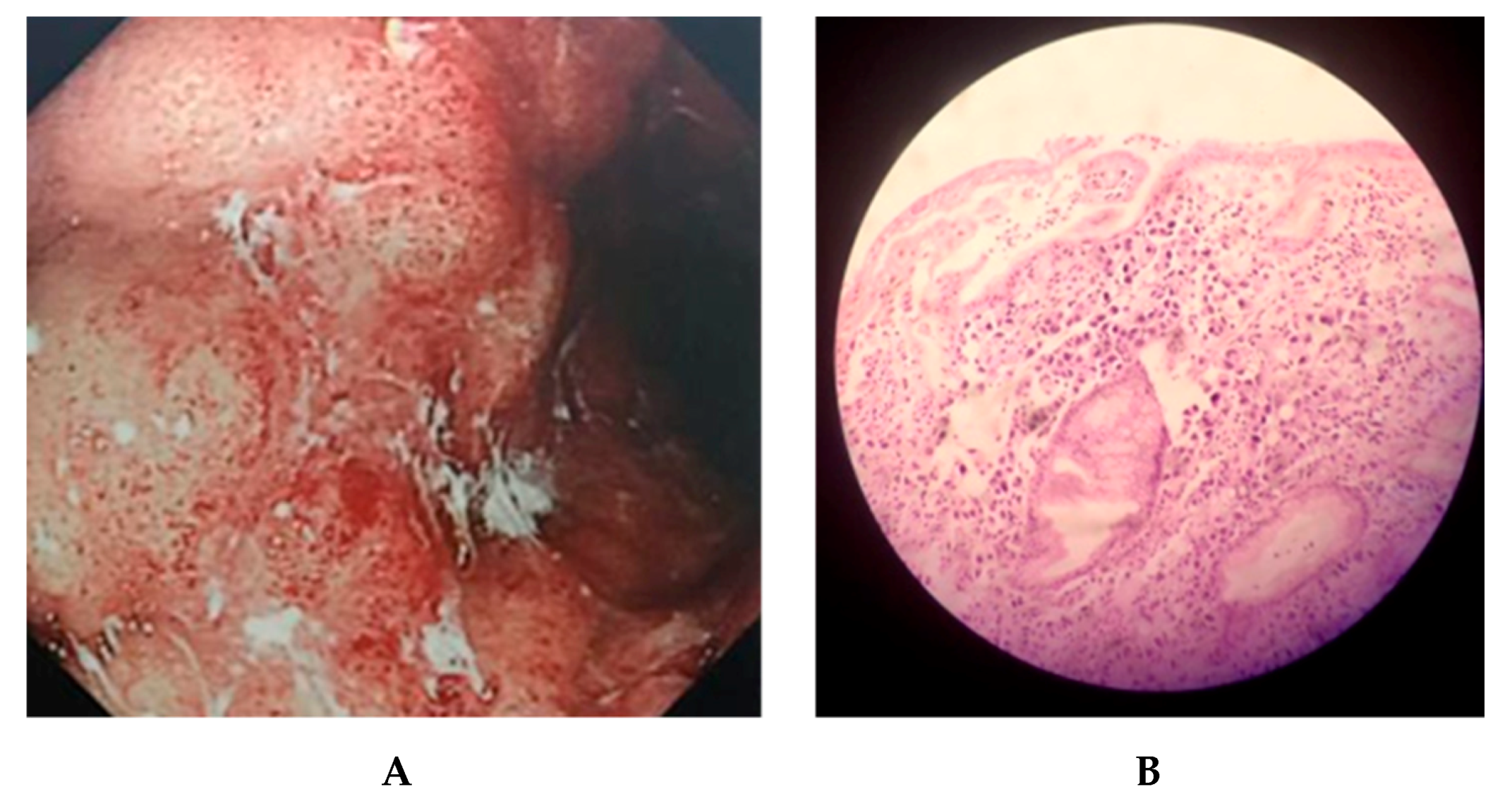
2.1. Clinical Outcomes of KE21 Clinical Isolate

H. pylori KE21 was isolated from a gastric biopsy specimen collected by endoscopy from a 61-year-old Kenyan female patient. The patient was referred to the Endoscopy unit at Aga Khan University Hospital Nairobi, for an upper gastrointestinal endoscopy given a history of severe abdominal pain and discomfort. Endoscopic examination identified seemingly non-benign and protruding gastric lesions that were later histologically identified as signet-ring cell carcinoma with depth of invasion in the lamina propria (Figure 1). Further explorations identified a pelvic metastasis of gastric carcinoma. The clinical isolate showed no in vitro resistance to tetracycline (TET) and levofloxacine (LEVO) at Minimum Inhibitory Concentrations (MICs) 0.19 and 0.25 mg/L, respectively. However, resistance was noted to clarithromycin (CLA), amoxicillin (AMX), and metronidazole(MTZ) with MICs of 4 mg/L, 2 mg/L, and ≥256 mg/L, respectively.

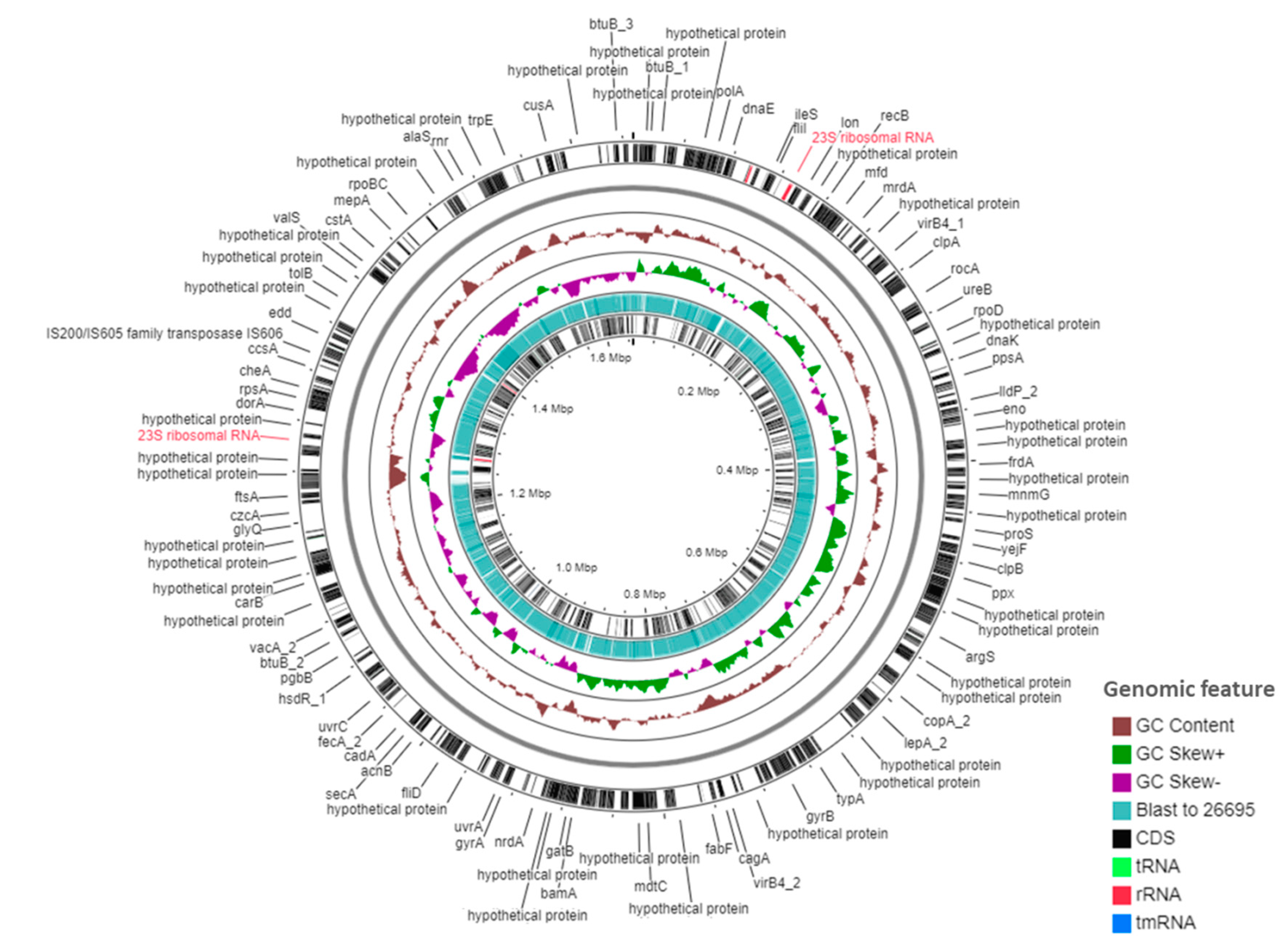
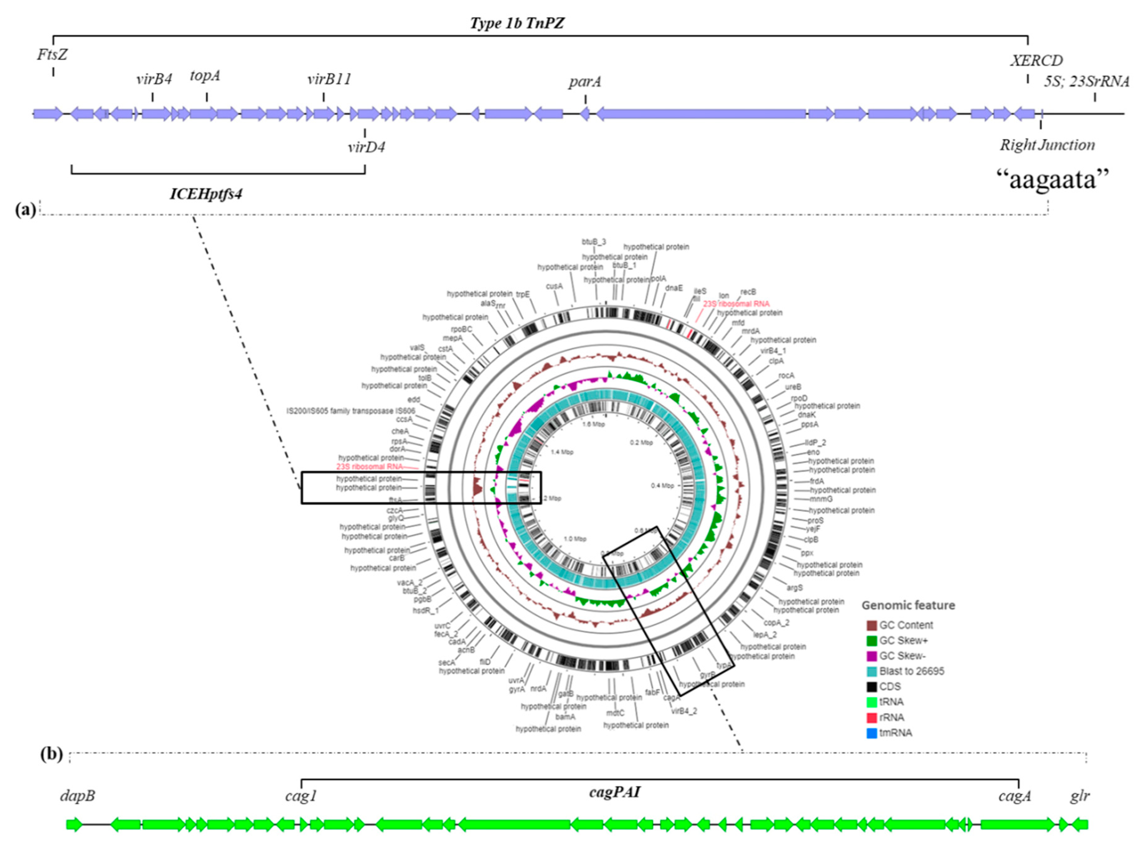
2.2. Genomic Features of KE21 Clinical Isolate

The de novo assembly of the genome of strain KE21 resulted in a single circularized chromosome displayed by one contig sequence of 1,648,327 bp length with a global GC content of 39.1% (Figure 2). Through the CheckM algorithm, the genome assembly reached a completeness of >99% with no detected genetic contamination and strain heterogeneity. The WIMP workflow of Epi2Me [17] assigned 99.9% of MinION reads to Helicobacter species. The FastANI [18] used by the DFAST Quality Control tool estimated the highest average nucleotide identity (ANI) of KE21 genome at 94.8% against H. pylori CCUG 17874 (GCA_000258845.1). These results conformed the taxonomic assignment of strain KE21 to H. pylori species. This genome revealed 1590 predicted genes including 1549 coding sequences (CDSs) through Prokka. Genes encoding thirty-six transfer RNAs (tRNAs) organized into seven clusters and 15 singletons, two separate sets of 5S-23S and 16S ribosomial RNAs (rRNAs), as well as one transfer-messenger RNA (tmRNA) could be identified in the genome. The pan-genome of orthologous genes from strain KE21 and two universal reference genomes, i.e., 26695 and J99, displayed 2508 genes of which 36.5% (916 genes) formed the core genome and 63.5% (1592 genes) formed the accessory genome in the shell (Supplementary Material, Tables S1 and S2).

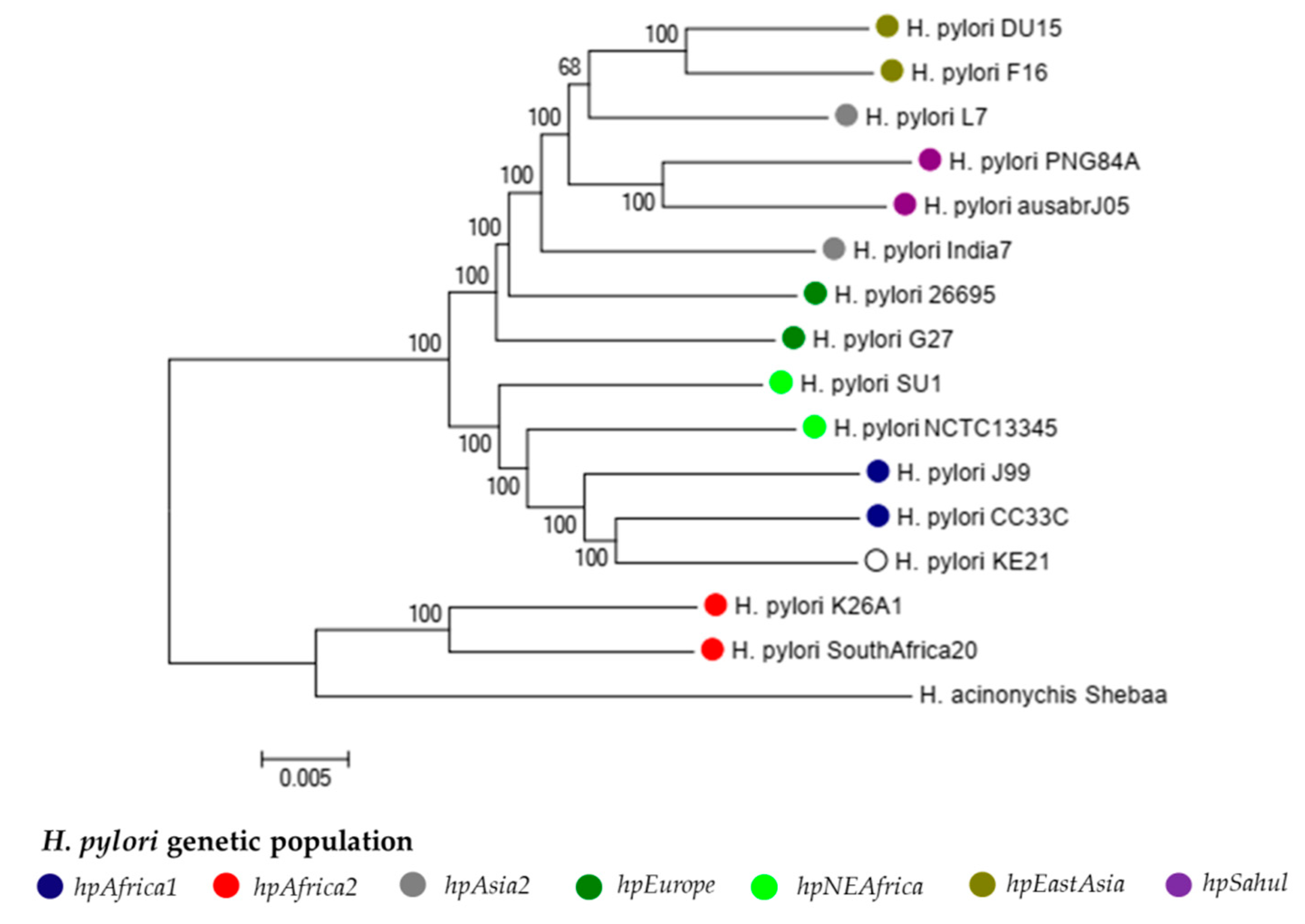
2.2.1. Population Genetics of Strain KE21

We performed a phylogenetic analysis of strain KE21 with additional 15 representative Helicobacter strains (i.e., 26695, J99, SouthAfrica20, CC33CC, NCTC1354, SU1, L7, India7, DU15, F16, G27, K26A1, PNG84A, ausabrJ05, and Shebaa) belonging to existing H. pylori genetic populations defined previously by fineSTRUCTURE and STRUCTURE approaches (Supplementary Materials, Table S3) [19,20]. This phylogenetic analysis was accomplished via neighbor-joining tree estimating core genome pairwise distance by Maximum Likelihood method. This analysis clearly classified H. pylori genomes into hpAfrica2, hpfrica1, hpNEAfrica, hpAsia2, hpEurope, and hpEastAsia phylogeographic clades. Of note, strain KE21 strain belonged distinctively to the hpAfrica1 population (Figure 3).

2.2.2. The Mobilome of Strain KE21

No prophage or plasmid could be detected in strain KE21 by using PlasmidSeeker [21] and PHASTER [22] while two genomic islands with an average lower GC content of 35.6% were identified (Figure 4). The first island comprised 46 predicted CDS spanning over 40,235 bp (coordinates: 1221910-1262145 in genome sequence) and was located between genes encoding putative FtsZ and 5S-23S rRNAs, an insertion site described previously as a “transposon, plasticity zone” (TnPZ). As shown in Figure 4, the organization of this TnPZ was typically of type 1b including a cluster of adjacent vir homologue genes encoding an integrating conjugative element type four secretion system of type 4 (ICEHptfs4) [23,24]. However, this KE21 ICEHptfs4 cannot be assigned to any of the known subtypes (Supplementary Materials, Table S4) [23]. The second genomic island had 47 predicted CDS that were inserted within a chromosomal region of 49,454 bp (coordinates: 706,360-755,813 in genome sequence) located between genes encoding the 4-hydroxy-tetrahydrodipicolinate reductase (dapB) and the glutamate racemase (glr), a region known as dg-region [25]. Remarkably, this dg-region encoded a cluster of cytotoxin-associated genes known as cag pathogenicity island (cagPAI) (Supplementary Materials, Table S5).

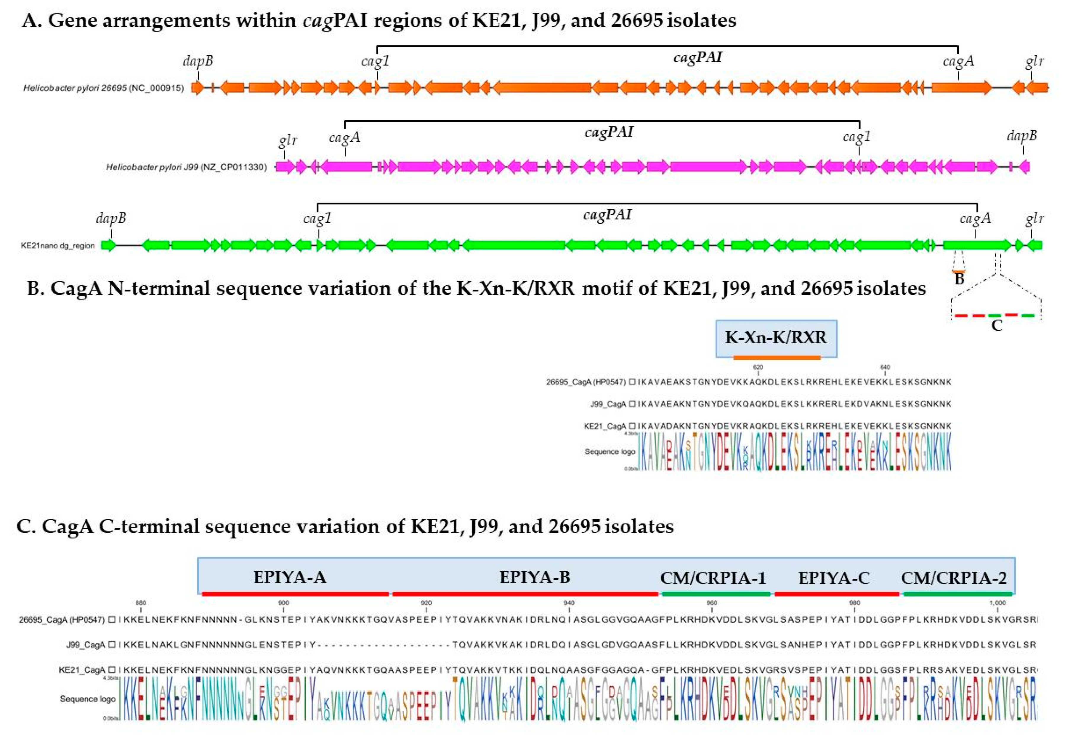
2.2.3. The Virulome of Strain KE21

By using ABRicate to screen the genome of H. pylori KE21 against a customized virulome database, we identified 147 genes that encode proteins whose function relates basically to motility, chemotaxis, cell-to-cell adherence, persistence, acid resistance, and host tissue damage (Supplementary Material, Table S6). First, we noted a cagPAI region of 36,507 bp (coordinates: 717,715–754,221) within a dg-region whose synteny and organization had been previously described as being of type-A (Figure 5A) [25]. A total of 28 CDSs were predicted in this cagPAI including genes of all known components of a syringe-like type four secretion system (T4SS)—i.e., cagζ/cag1, cagε/cag2, cagδ/cag3, cagγ/cag4, cagβ/cag5, cagα, cagZ, cagY, cagX, cagW, cagV, cagU, cagT, cagS, cagQ, cagP, cagM, cagN, cagL, cagI, cagH, cagG, cagF, cagE, cagD, cagC, and cagB—as well as cagA which represent major virulence factors with a well-acknowledged causative role in gastric cancer development [26,27]. The cagA gene-encoded CagA oncogene protein of H. pylori KE21, intended for delivery into gastric epithelial cells via the T4SS machinery, could be predicted (Figure 5). Its N-terminal sequence showed a plecstrin homology (PH) domain with a conserved K-Xn-X/RXR motif which is required for the bound of CagA to host membrane phosphatidylserine (PS) as a prerequisite for pathophysiological activities of the oncoprotein in polarized epithelial cells (Figure 5B) [28]. Furthermore, the C-terminal CagA repeat sequences comprised three Glu-Pro-IleTyr-Ala (EPIYA) motifs within segments of type ABC, previously referred to as of Western-type, which may undergo tyrosine phosphorylation to hijack carcinogenic intracellular pathways (Figure 5C) [27]. Interestingly, proximal and distal to the 34-amino-acid of the EPIYA-C segment, we identified two variants of CagA-multimerization motifs (yet to be described) also known as conserved repeat responsible for phosphorylation-independent activity motifs (CM or CRPIA motifs)—i.e., FPLKRHDKVEDLSKVG and FPLKRRSAKVEDLSKVG. Second, we identified a vacuolating activity associated gene A (vacA) that encodes VacA protein which is a pore-forming toxin of H. pylori species with pleiotropic biological effects [29]. Allelic variations of the H. pylori KE21 vacA was typically of type s1i1d1m1, a genotype associated with increased toxicity, gastric inflammation, peptic ulcer, and gastric cancer development in previous studies (Figure 6) [29,30]. Third, genes encoding more 54 putative adhesins for H. pylori KE21 interaction with surface receptors on gastric epithelial cells could be detected [31]. These include main outer membrane proteins (OMPs)—e.g., BabA (HopS), OipA (HopH), HopQ (Omp27), HomA, AlpA (HopC), AlpB (HopB), SabB (HopO) and SabA (HopP)—that are critical in the pathogenesis of H. pylori infection [31,32,33]. The characteristics of these genes are shown in Table 1 and in Supplementary Materials, Table S6 and Figures S1 and S2. Some of the outer membrane proteins (OMPs) were encoded by duplicated gene copies (e.g., babA, homA). In contrast, genes encoding few putative virulence factors (i.e., BabC or HopU, BabB or HopT, IceA, and DupA) were not detected in KE21 genome (Table 1 and Supplementary Materials, Table S6). Furthermore, a cluster of all seven urease genes (i.e., ureA/B, ureI, and ureE-H) that are required for resistance and survival in the harsh acidic environment of the stomach was detected in KE21 genome [34]. Finally, the isolate was also equipped with several genes encoding flagella components (e.g., flgE, flaA, and flaB) and lipopolysaccharides (e.g., rfaJ, rfaC) mediating bacterial motility and immune modulation while contributing to H. pylori virulence as putative bacterial endotoxins (Supplementary Materials, Table S6) [35,36]. Overall, the genetic features described above reveal potentials for successful colonization, persistent infection, and disease pathogenesis during infection with H. pylori KE21.

2.3. Experimental Virulence Assays on KE21

To further assess the virulence abilities of H. pylori KE21, we conducted an in vitro infection experiment using the AGS epithelial cells line. Consistently with an intact and functional cagPAI-related T4SS [26,27], KE21 showed ability to promote interleukin-8 (IL-8) production and to produce morphological changes called hummingbird phenotype in AGS cells. Furthermore, a phosphorylated CagA was detected in AGS cells, attesting the competence of KE21-related T4SS for translocation of a bioactive oncoprotein in human epithelial cells (Figure 7).

3. Discussion

H. pylori species is characterized by an exceptionally high genetic diversity and geographic variability driving substantial difference in clinical outcomes between different regions. Despite its well-acknowledged causative role in severe gastrointestinal diseases including peptic ulcers and gastric adenocarcinoma; the clinical relevance of African H. pylori strains is still debated given the reported contrast between the highest prevalence of the infection and the lowest incidence of gastric cancer in the continent. This discrepant epidemiological situation had been referred to as the so-called “African enigma” [14]. This study is the first to describe genomic features and virulence potential of typical African H. pylori isolate retrieved from a native African patient with gastric cancer. Through the genome of isolate KE21, we screened the expanded virulome of H. pylori species with the aim to depict its potentialities to promote gastric carcinogenesis in general and signet-ring cell carcinoma (SRCC) in particular. We thus detected tens of genes encoding factors involved in cell-to-cell adherence, acid resistance factors, cell motility and chemotaxis, immune response evasion, as well as in direct tissue damages. An experimental infection of gastric epithelial cells demonstrated the ability of strain KE21 to induce carcinogenic signals. While this report cannot establish a causality link between the isolate and the diagnosed gastric cancer in our patient, the discussion made grasps the full scale of the clinical threat this African H. pylori strain would represent during the infection.
It is notable that this clinical H. pylori strain (KE21) was isolated from a Kenyan patient diagnosed with gastric SRCC. SRCC is a unique type of gastric cancer classified as diffuse type (in contrast to intestinal type) according to Lauren’s classification, given its poorly differentiated histological aspect with the lack of the intercellular adhesion and presence of scattered cells of signet-ring morphology predisposed to diffuse invasion throughout the stroma [40,41]. This cancer is found in 8 to 30% of gastric cancers, has unfavorable prognosis while affecting more frequently women from 55 to 61 years old, consistently around 7 years younger than non-SRCC gastric cancer cases [40,42,43]. Consistently with this general profile of SRCC cases, our patient was a 61-year-old female. In contrast with the pathogenicity of intestinal-type gastric cancer that follows the Correa’s cascade consensually linked with H. pylori infection, the development of diffuse-type gastric cancer remains mostly elusive and controversial [42]. The SRCC is widely believed to arise from distinct biologic pathways involving genetic abnormalities in the host such as alterations of cell adherence factors like E-cadherin [40,42]. However, increasing epidemiological data have been also associating H. pylori infection with sporadic diffuse-type gastric cancers likely through carcinogenic pathways that are independent from gastric mucosa atrophy [44,45,46,47,48,49]. Several pathways probably exploited by the H. pylori to induce SRCC-like abnormalities in gastric epithelial cells have been reviewed recently [42]. Consequently, the etiological role of H. pylori in both intestinal-type and sporadic diffuse-type gastric cancer is currently plausible. We attempted to explore the genomic attributes of strain KE21 in light with different pathways able to trigger carcinogenic development and H. pylori-related pathogenesis in general.
Overall, the metric characteristics of the genome of strain KE21 were consistent with genomic features (e.g., size, structure, gene content) usually reported in non-African isolates [50]. This genome included a mobilome comprising no prophage or plasmid but two genomic islands (GEIs) inserted within regions previously identified as the “transposon, plasticity zone” (TnPZ) and the “dg-region” [23,24]. GEIs are syntenic blocks of genes acquired horizontally and that likely contribute to the diversification and adaptation of microorganisms, thus having a significant impact on the genome plasticity and evolution [51]. A close analysis of the KE21 TnPZ assigned its structure to the type 1b and identified an integrating conjugative element type four secretion system of type 4 (ICEHptfs4) commonly occurring in H. pylori species [23,24]. TnPZs and ICEHptfs(s) are highly conserved in H. pylori while displaying great allelic diversity. Excluding mosaic and remnant forms, TnPZ have been structurally categorized based on their gene arrangement in three types: 1, 1b, and 2; while ICEHptfs(s) have been grouped into two types with related subgroups: ICEHptfs3 and ICEHptfs4 (i.e., ICEHptfs4a, 4b, and 4c) [23,24]. Interestingly, the ICEHptfs4 identified in strain KE21 could not be assigned to any of the known subtypes, suggesting being a new ICEHptfs4 allele. The T4SSs contained in ICEHptfs(s) have been shown to contribute to bacterial virulence through both epidemiological and in vitro infection model studies [52]. However, further studies are still needed to completely elucidate the structure and function of these ICEHptfs(s)/T4SSs as well as their possible interactions with other bacterial virulence factors [52]. In contrast, the content of the dg-region, delimited by the dapB and glr genes, has been extensively explored [25]. Depending on the strain, the dg-region may carry a 40-kb DNA segment known as the cag PAI region which generally consists of 26 or 27 genes encoding a special syringe-like structure T4SS and the oncoprotein CagA, two major virulence factors in H. pylori species [53]. The cag PAI region is thought to have been introduced into the H. pylori genome via horizontal transfer from an unknown source. This island has been show as being prone to functional disruption due to various genetic rearrangements occurring within and outside the constituent genes [25,54]. The intactness or rearrangement of the cag-PAI has therefore been thought to be crucial for the progression of gastroduodenal pathology due to H. pylori [25,55]. Notably, we found that the dg-region of strain KE21 comprises an intact type-A cag PAI region, encoding a complete T4SS with a CagA oncoprotein and whose rearrangement is compatible with a biological functionality.
Among the various virulence factors of H. pylori, the oncoprotein CagA plays a central role as a scaffolding protein in the development of gastric cancer [54,56,57]. The CagA is a cellular effector whose injection into host cells by the cag PAI T4SS deregulates an impressive number of molecular signaling processes including carcinogenic pathways [54]. Some of these pathways involve the binding of the non-phosphorylated CagA to E-cadherin that ultimately trans-activates the β-catenin-dependent genes while inducing also mutational alterations (e.g., TP53) as well as aberrant DNA hypermethylation and inactivation of the CDH1 gene associated with the progression of sporadic diffuse-type gastric cancers [58,59,60,61,62]. Otherwise, the translocated CagA may also undergo a phosphorylation by host cell kinases at a conserved tyrosine residue found within the EPIYA (Glu-Pro-Ile-Tyr-Ala) motif. This allows binding the phosphorylated CagA to a SH2-domain-containing protein tyrosine phosphatase (SHP2), and thus deregulates the phosphatase activity of SHP2, a crucial step in the development of H. pylori-related intestinal-type gastric cancer [54,56,63]. The structure of the CagA oncoprotein is crucial for the virulence of H. pylori and leads to pathogenic differences. We observed that the KE21 strain comprises in its N-terminal CagA, a cluster of conserved basic residues, known as the basic patch or K-Xn-X/RXR motif, that plays an important role in the interaction of CagA with phosphatidylserine required for the biological activity of cagA [28]. In addition, the CagA C-terminal tail of KE21 is characterized by the presence of three EPIYA (Glu-ProIle-Tyr-Ala) motifs, which serve physiologically as motifs for tyrosine phosphorylation of T4SS-delivered CagA by host cell kinases such as Src-family kinases (SFKs) and c-Abl [56]. Based on the sequences flanking each of the EPIYA motifs, we concluded that the KE21 CagA was of ABC type which is termed Western CagA, as it was identified first in Western countries, in contrast to the East Asian CagA of ABD type [57]. The Western type CagA ABC is known to be competent for tyrosine phosphorylation and able to even undergo a sequence amplification of EPIYA-C motifs to more efficiently bind to SHP-2 for increased carcinogenicity [54,57]. Furthermore, the C-terminal tail of the strain KE21 CagA contains another repeatable sequence motif, originally designated as the CagA multimerization sequence motif (CM) comprising 16 amino-acid residues and located immediately distal or proximal to the last repeat of the EPIYA segments [57,64]. Whereas the CM motif sequence is highly conserved, there are several variants previously described in East Asian (CME type, FPLRRSAAVNDLSKVG), Western (CMW type, FPLKRHDKVDDLSKVG), and Amerindian H. pylori species (CMAmI and CMAmII types, SSLKRHAKVDDLSKVG and YTLKMHAGDDNLRSKVG) [65,66,67]. Aberrant pro-oncogenic signals elicited by deregulated SHP2 via the EPIYA motif, together with destruction of the gastric epithelium caused by CM-mediated PAR1 inhibition are two major pathophysiological processes that cooperatively contribute to H. pylori CagA-induced gastric oncogenesis [57]. Remarkably, we observed that the KE21 CM motifs—i.e., FPLKRHDKVEDLSKVG and FPLKRRSAKVEDLSKVG—are different from the motifs described previously in non-African populations. However, while the KE21-CagA contains two motifs as in Western type CagA, its most distal motif is very similar to East Asian type (with only two amino acid differences) which binds the most strongly to PAR1 with enhanced biological effect [57]. Given these similarities with CME and CMW types, pending further studies, we hypothesize that the KE21 CM motif is biologically functional in contrast to Amerindian CM variants (i.e., CMAmI and CMAmII) that had been shown as abrogating the ability of CagA to interact with PAR1 and substantially attenuating the CagA oncogenicity [65,66]. Further analyses exploiting an in vitro infection model using the AGS epithelial cells line, indirectly attested the competence of KE21-related T4SS for translocate a bioactive CagA oncoprotein [26,27]. Hence, H. pylori KE21 demonstrated ability to promote the production of the proinflammatory cytokine IL-8 from gastric epithelial cells which represent hallmarks of cag PAI/T4SS function [54,56]. Moreover, showed the capacity to produce morphological changes of gastric epithelial cells, referred to as “hummingbird phenotype” of AGS cells, which reflect CagA-induced carcinogenic signaling pathways resulting in cytoskeletal rearrangement, cellular motility, and elongated shape of host cells [54,56]. We noted also that the strain KE21 genome encodes a full-length gene for the high temperature requirement A (HtrA) protein. Ubiquitously, H. pylori expresses HtrA, a protein with dual function acting as a chaperone and a serine protease, which cleaves-off the ectodomain of E-cadherin and disrupts intercellular adhesions opening up the intercellular space for transmigration of bacteria [68]. Consequently, HtrA-dependent E-cadherin shedding strongly enhances CagA delivery into infected host cells via integrin β1 essential for gastric cancer development [69]. Furthermore, strain KE21 was also found with a full-length gene for the VacA, a key and ubiquitous toxin for pathogenesis in H. pylori species [30]. The VacA toxin is known for its multitude of effects on epithelial cells, varying from endosomal alterations of intraluminal pH and disruption of endocytic compartment trafficking, induction of autophagy and enhancement of mitochondrial dysfunction, which can result either from its pore-forming ability or through the activation of pro-apoptotic factors [38,39]. Four main regions of diversity in VacA sequences have been recognized, namely the signal sequence region (s)-region, the intermediate region (i)-region, the deletion (d)-region, and middle region (m)-region [37,38,39]. These result in VacA alleles containing multiple combinations of s-, i-, d-, and m-region types [37,39]. Being of the s1i1d1m1 allele, the KE21 VacA is thus a variant that has been reported with enhanced vacuolating activity, and linked to a potentially higher relative risk for development of gastric cancer or peptic ulcer disease [39].
Hence, the analysis of cagPAI/CagA, HtrA, and VacA clearly raises the potential for strain KE21 to cause tissue damage and to trigger carcinogenic pathways in epithelial cells. To explore further the full virulence potential of strain KE21, we screened the genome for the presence of genes encoding other factors that are critical in different steps of H. pylori colonization and pathogenesis. We thus observed that strain KE21 encodes a cluster of all seven urease genes (i.e., ureA/B, ureI, and ureE-H) whose activity is required for adjusting the periplasmic pH as an acid acclimation mechanism to resist and survive in the harsh acidic environment of the stomach [34]. The isolate is also equipped with several genes encoding flagella components (e.g., flgE, flaA, and flaB) and lipopolysaccharides (e.g., rfaJ, rfaC) mediating bacterial motility and immune modulation enabling colonization and persistence in the stomach [35,36]. The bacterial attachment to the epithelial cells, as an important step of the infection, is mediated by an impressive number of adhesins and OMPs in H. pylori species [32]. We identified more than 50 genes encoding putative OMPs including main proteins that have been formally implicated in the pathogenesis of H. pylori infection, e.g., BabA (HopS), OipA (HopH), HopQ (Omp27), HomA, AlpA (HopC), AlpB (HopB), SabB (HopO) and SabA (HopP) [32,33]. However, few putative virulence factors including BabC (HopU), BabB (HopT), IceA, and DupA were not detected in the KE21 genome. BabA, the best characterized of the adhesin proteins in H. pylori, mediates binding to host cells’ fucosylated Lewis b (Le(b)) blood group antigens and was encoded in two copies likely granting strain KE21 interesting potentials for host-bacterium interactions [70]. OipA may serve as an adhesin altering the host immune response but also promotes inflammation was predicted to be preserved in switched “ON” phenotype in strain KE21, suggesting preservation of functions [71]. The HopQ outer-membrane adhesin of H. pylori exhibits a high level of genetic diversity, and two families of HopQ alleles have been described (type I HopQ and type II HopQ) [72]. The strain KE21 displays HopQ gene encoding type I allele which has been documented to be present in cag PAI positive H. pylori strains and is epidemiologically associated with gastric cancer [73,74]. This protein was recently shown to bind carcinoembryonic antigen-related cell adhesion molecules (CEACAMs) including CEACAM1, an inhibitory receptor expressed mainly by activated T and NK cells and involved in cancer development and progression [73,75,76].
To cope with the need to attest the African origin of the strain, we performed phylogenetic analyses that assigned the isolate to hpAfrica1, a major genetic population of H. pylori species that is native to Africa [19,20]. H. pylori species is split into distinct bacterial populations exhibiting tight relationships with ethno-geographical distribution and history of human host [3,77]. Of these populations, three are originating from Africa (hpNEAfrica, hpAfrica1 and hpAfrica2), one from Europe (hpEurope), and three from Asia (hpEAsia, hpAsia2 and hpSahul) [3,77,78,79]. Our results suggest strongly that strain KE21 was probably not imported from outside Africa. Nevertheless, reporting on a single isolate which may not be representative of all the strains circulating in the Kenyan population constitutes the main limitation of this study. Furthermore, the experimental analyses used in this study constitute a model which only partially reflects conditions in vivo. In vivo studies, using for example animal models, would have further strengthened the validity of our observations. Obviously, further studies are needed to enhance our observation and to fully understand the epidemiological threat and the clinical implications that would result from isolates displaying similar biological properties as strain KE21 that probably are spreading in the study population.

4. Conclusions

Our results highlight substantial virulence potentials displayed by typical African H. pylori isolate, including the ability to deregulate carcinogenic pathways in epithelial cells via translocation of a functional CagA oncogene protein. It would therefore be more interesting to assess at the population level, the epidemiological distribution of strains with the similar biological characteristics and which could represent a significant risk of developing gastric cancer in African populations. This will facilitate a better understanding of the risk of gastric cancer in Africa and will contribute to the elucidation of the so-called African Enigma, which still refers, according to Agha A. et al. [14], to an epidemiological situation warranting further clarification. The message in this report does not establish strain KE21 as the cause of the SRCC diagnosed in our patient, but it is more a call for increased surveillance efforts and enhanced research, including genomic explorations, regarding H. pylori isolates circulating in Africa and related gastric cancer risk.

5. Materials and Methods

5.1. Patient and Biological Sampling

H. pylori KE21 was obtained from the gastric mucosa of a Kenyan female patient who underwent gastro-duodenal endoscopy at Aga Khan University Hospital, Nairobi. This strain was isolated through culture of two gastric biopsy specimens sampled from the gastric antrum and corpus of the patient. The culture process was performed by homogenizing the gastric biopsy specimen and inoculating on Brucella agar (BD Difco, USA) supplemented with 7% sheep blood. The culture plates were incubated under microaerophilic conditions (10% CO2, 5% O2, and 85% N2) at 37 °C for up to 7 days. H. pylori-like colonies with translucent, convex morphology grew on the plates and were identified based on biochemical properties (catalase, oxidase, and urease reactions) and microscopic morphology following a Gram staining (Gram negative bacilli). Then, they were sub-cultured for 72 h before being stored at −80 °C in a Brucella broth medium containing glycerol, until shipped in cold-chain to Oita University in Japan where the genomic sequencing was performed. In addition, two biopsy specimens were sampled from the stomach, fixed in 10% buffered formalin, and embedded in paraffin for histological examination by a clinical pathologist.

5.2. Antimicrobial Susceptibility Testing

The antimicrobial susceptibility was assessed using the E-Test® (bioMérieux) method on culture growth of H. pylori colonies isolated from gastric biopsy specimens following the Clinical and Laboratory Standards Institute protocols (Wayne, PA, USA). H. pylori culture was suspended at a turbidity equivalent to a 3.0 McFarland standard and inoculated onto Müeller–Hinton agar plates supplemented with 7% sheep blood and antibiotics (AMX, CLA, LEVO, TET and MTZ). The MICs of antibiotics were determined after 72 h of incubation. The H. pylori strain 26695 was used as a control strain. Clinical breakpoints between resistant and susceptible strains were determined following the guidelines of the European Committee on Antimicrobial Susceptibility Testing (EUCAST) available at http://www.eucast.org/.

5.3. DNA Extraction, Library Preparation, and Whole Genome Sequencing

Harvested confluent bacterial cultures expanded from a single colony of H. pylori KE21 isolate were used for the extraction of total DNA by DNeasy Blood and Tissue kit (QIAGEN Inc., Valencia, CA, USA).

5.3.1. Short-Read Illumina Sequencing

A library of 1 ng DNA was prepared for sequencing of 300-bp paired-end reads, using the Nextera XT DNA Library Preparation kit (Illumina, San Diego, CA, USA). Whole-genome sequencing was performed at 300 cycles using the Illumina Miseq platform (Illumina, Inc., San Diego, CA, USA) following the Manufacturer’s instructions. Fluorescent images were assessed with the MiSeq Control Software, and FASTQ-formatted sequence data were created with MiSeq Reporter Analysis Software. The density cluster and Q-score ≥ 30 of sequenced reads reached 1206 k/mm3 and 88%, attesting the good quality of sequencing runs.

5.3.2. Long-Read MinION Sequencing.

To produce long-read sequences of this strain, we applied 400 ng of genomic DNA on the Oxford Nanopore MinION (Oxford Nanopore Technologies, Oxford, UK) device following the Rapid Sequencing protocol (SQK-RAD004). Raw sequence reads were uploaded to the Epi2Me interface (Metrichor, Oxford, UK) for base calling and demultiplexing of MinION data. Epi2Me was used also for examining basic metrics of sequencing abundance and quality. Only base-called data passing Epi2Me quality parameters (qmean > 6) were downloaded off the cloud in FAST5 and FASTQ formats to use in further analyses. In total, 13,386 MinION reads were obtained with average length and quality score of 6963 bp and 8.3, respectively.

5.4. Bioinformatic Analyses

Low-quality bases (the quality of bases < Q30) and adapters were trimmed using Trimmomatic v0.30 [80]. Filtered High-throughput short reads were screened for the presence of known plasmids using the PlasmidSeeker tool with 26695 (NC_000915.1) as the reference genome [81]. MinION long reads were trimmed using Porechop v.0.2.4 (https://github.com/rrwick/Porechop), assembled along with Illumina Miseq short reads in a hybrid genome assembly using Unicycler v.0.4.8 [82]. The draft genome was polished by using Pilon v.1.23 [83]. The quality assessment of the obtained genome was assessed using QUAST v5.0.2 [84]. The taxonomic assignment of the genome was performed using the WIMP workflow of Epi2Me [17] and the FastANI [18] through the DFAST Quality Control tool of DFAST v1.1.5 (https://dfast.nig.ac.jp/, Tokyo, Japan, 2020). Gene sequences were annotated with Prokka v1.14.5 [85] and DFAST v1.1.5 (https://dfast.nig.ac.jp/). They were functionally characterized and clustered in subsystems by the RASTtk pipeline of Rapid Annotation using Subsystem Technology v2.0 [86].
Additionally, 14 H. pylori genomes publicly available that had been previously well-characterized, were used along with strain KE21 to infer the pan-genome and its core genome by using Roary v3.13.0 with an 80% BLASTp percentage identity cut-off [87]. The phylogenetic characterization of the strain KE21 was estimated through a bootstrapped neighbor-joining tree estimated by Maximum Likelihood method with MEGA v7 [88] based on the core genome alignment. MAUVE was used for comparing the genome of KE21 with those of J99 and 26695, two reference isolates [50,81]. Nucleotide sequences and inferred amino acid sequences were aligned with references and visually analyzed using CLC genomic Workbench v8.5.1 and in MEGA v7. Maps of single genome and genomes alignment were constructed using CGView Sever v1.0 [89]. ABRicate v1.0.1 (https://github.com/tseemann/abricate) was applied to construct the virulome of H. pylori KE21 by detecting putative virulence factors in silico through a BLAST+ against a reference database locally optimized for H. pylori species by including additional species-specific virulence genes to the VFDB repository of bacterial virulence factors from various pathogens (http://www.mgc.ac.cn/VFs/) [90]. The H. pylori virulome database included 167 non-redundant genes retrieved from H. pylori J99 and 26695 isolates.

5.5. AGS Cell Line Co-Infection with H. pylori KE21

The virulence ability of H. pylori KE21 strain was further assessed experimentally by infecting human gastric epithelial AGS cell lines as described previously [91]. The experiments were performed twice independently in triplicate. Briefly, AGS cells were seeded into 6-well plates and grown overnight in RPMI 1640 medium supplemented with 10% FBS. The plates were incubated at 37°C for the indicated periods of time in a humidified environment containing 5% CO2 and 95% air. K21 strain was harvested from an agar dish and washed twice with PBS before being added to the AGS culture wells with a bacterium-to-cell ratio of 50:1. After 24 h of co-culture, formation of the hummingbird phenotype was examined microscopically in ten randomly chosen fields. Additionally, the functionality of the cagPAI was assessed through measurement of the concentration of induced IL-8 in the supernatant of AGS cells co-cultured with H. pylori by using the CXCL8/IL-8 ELISA Kit (R & D Systems, Minneapolis, MN, USA).

5.6. Nucleotide Sequence Accession Number

The genome sequenced of H. pylori KE21 was deposited at the DNA Data Bank of Japan (DDBJ) under the accession number AP023320.

5.7. Ethical Consideration

The patient KE21 gave informed consent for the conduct of this study, in accordance with the Declaration of Helsinki. The study was approved by the Institutional Ethics Committee of the Aga Khan University Hospital (Ref N#: 2017/REC-97(vl)), the Kenyatta University Ethical Review Committee (Ref N# PKU/509/1602-PKU/447/E39), and the Oita University Ethical Review Committee (Ref N#: 1660).

Supplementary Materials

The following are available online at https://0-www-mdpi-com.brum.beds.ac.uk/2072-6651/12/9/556/s1, Figure S1: N-terminal sequence alignment of genes encoding the Outer inflammatory protein A (OipA) of H. pylori KE21 and reference strains, Figure S2: Molecular Phylogenetic analysis of genes encoding the Helicobacter outer membrane protein Q (HopQ) of KE21 and reference strains by Maximum Likelihood method, Table S1: Summary statistics of the pan-genome between H. pylori strains KE21, J99, and 26695, Table S2: The pan-genome of orthologous genes formed by KE21, J99, and 26695 strains, Table S3: Baseline characteristics of genomes used in this study, Table S4: Characterization of the transposon plasticity zone (TnPZ) and the integrating conjugative element (ICEHptfs) identified in H. pylori KE21, Table S5: Predicted dg-region and cag pathogenicity island (cagPAI) in H. pylori KE21, Table S6: The virulome of H. pylori KE21.

Author Contributions

Conceptualization, C.M. and E.T.-K.; methodology, C.M., S.N. and E.T.-K.; software, E.T.-K.; validation, Y.Y., G.R. and Z.M.; formal analysis, C.M., E.T.-K. and T.M.; investigation, C.M. and S.N.; writing—original draft preparation, C.M. and E.T.-K.; writing—review and editing, S.N., K.N., T.M., S.D., A.R., S.K., G.R., Y.Y. and Z.M.; visualization, E.T.-K.; supervision, G.R., K.N., S.K. and S.D.; project administration, G.R. and Y.Y.; funding acquisition, Y.Y. and T.M. All authors have read and agreed to the published version of the manuscript.

Funding

This research was funded by the Grants-in-Aid for Scientific Research from the Ministry of Education, Culture, Sports, Science and Technology (MEXT) of Japan (26640114, 15H02657, 16H05191, 16H06279, 18KK0266, and 19H03473 to Yoshio Yamaoka), the Special Coordination Funds for Promoting Science and Technology from the MEXT of Japan to Yoshio Yamaoka, and National Institutes of Health grants DK62813 to Yoshio Yamaoka. This work was also supported by the Japan Society for the Promotion of Science (JSPS) Institutional Program for Core-to-Core Program; B. Africa-Asia Science Platform to Yoshio Yamaoka, and the Oita University President Research Project 2019 to Takashi Matsumoto, Evariste Tshibangu-Kabamba is a doctoral student supported by the MEXT Scholarship Program founded by the Japanese Government.

Acknowledgments

We thank the Endoscopy Unit and the Department of Pathology and Laboratory Medicine at Aga Khan University Hospital Nairobi for their technical support during sample collection and initial analyses.

Conflicts of Interest

The authors declare no conflict of interest.

References

  1. Polk, D.B.; Peek, R.M., Jr. Helicobacter pylori: Gastric cancer and beyond. Nat. Rev. Cancer 2010, 10, 403–414. [Google Scholar] [CrossRef] [PubMed] [Green Version]
  2. Loughlin, M.F. Novel therapeutic targets in Helicobacter pylori. Expert Opin. Ther. Targets 2003, 7, 725–735. [Google Scholar] [CrossRef] [PubMed]
  3. Linz, B.; Balloux, F.; Moodley, Y.; Manica, A.; Liu, H.; Roumagnac, P.; Falush, D.; Stamer, C.; Prugnolle, F.; van der Merwe, S.W.; et al. An African origin for the intimate association between humans and Helicobacter pylori. Nature 2007, 445, 915–918. [Google Scholar] [CrossRef] [PubMed] [Green Version]
  4. Parsonnet, J.; Friedman, G.D.; Vandersteen, D.P.; Chang, Y.; Vogelman, J.H.; Orentreich, N.; Sibley, R.K. Helicobacter pylori infection and the risk of gastric carcinoma. N. Engl. J. Med. 1991, 325, 1127–1131. [Google Scholar] [CrossRef] [PubMed]
  5. Uemura, N.; Okamoto, S. Effect of Helicobacter pylori eradication on subsequent development of cancer after endoscopic resection of early gastric cancer in Japan. Gastroenterol. Clin. N. Am. 2000, 29, 819–827. [Google Scholar] [CrossRef]
  6. IARC Working Group on the Evaluation of Carcinogenic Risks to Humans. Schistosomes, liver flukes and Helicobacter pylori, In IARC Monographs on the Evaluation of Carcinogenic Risks to Humans; International Agency for Research on Cancer: Lyon, France, 1994; Volume 61, pp. 1–241. [Google Scholar]
  7. Yamaoka, Y. Mechanisms of disease: Helicobacter pylori virulence factors. Nat. Rev. Gastroenterol. Hepatol. 2010, 7, 629. [Google Scholar] [CrossRef] [PubMed] [Green Version]
  8. Yamaoka, Y.; Kato, M.; Asaka, M. Geographic differences in gastric cancer incidence can be explained by differences between Helicobacter pylori strains. Intern. Med. 2008, 47, 1077–1083. [Google Scholar] [CrossRef] [PubMed] [Green Version]
  9. Holcombe, C. Helicobacter pylori: The African enigma. Gut 1992, 33, 429–431. [Google Scholar] [CrossRef] [Green Version]
  10. Bray, F.; Ferlay, J.; Soerjomataram, I.; Siegel, R.L.; Torre, L.A.; Jemal, A. Global cancer statistics 2018: GLOBOCAN estimates of incidence and mortality worldwide for 36 cancers in 185 countries. CA A Cancer J. Clin. 2018, 68, 394–424. [Google Scholar] [CrossRef] [Green Version]
  11. Graham, D.Y.; Lu, H.; Yamaoka, Y. African, Asian or Indian enigma, the East Asian Helicobacter pylori: Facts or medical myths. J. Dig. Dis. 2009, 10, 77–84. [Google Scholar] [CrossRef] [Green Version]
  12. Campbell, D.I.; Warren, B.F.; Thomas, J.E.; Figura, N.; Telford, J.L.; Sullivan, P.B. The African enigma: Low prevalence of gastric atrophy, high prevalence of chronic inflammation in West African adults and children. Helicobacter 2001, 6, 263–267. [Google Scholar] [CrossRef] [PubMed]
  13. Asombang, A.W.; Rahman, R.; Ibdah, J.A. Gastric cancer in Africa: Current management and outcomes. World J. Gastroenterol. 2014, 20, 3875–3879. [Google Scholar] [CrossRef] [PubMed]
  14. Agha, A.; Graham, D.Y. Evidence-based examination of the African enigma in relation to Helicobacter pylori infection. Scand. J. Gastroenterol. 2005, 40, 523–529. [Google Scholar] [CrossRef] [PubMed]
  15. Ghoshal, U.C.; Chaturvedi, R.; Correa, P. The enigma of Helicobacter pylori infection and gastric cancer. Indian J. Gastroenterol. Off. J. Indian Soc. Gastroenterol. 2010, 29, 95–100. [Google Scholar] [CrossRef] [Green Version]
  16. Asombang, A.W.; Kelly, P. Gastric cancer in Africa: what do we know about incidence and risk factors? Trans. R. Soc. Trop. Med. Hyg. 2012, 106, 69–74. [Google Scholar] [CrossRef]
  17. Juul, S.; Izquierdo, F.; Hurst, A.; Dai, X.; Wright, A.; Kulesha, E.; Pettett, R.; Turner, D.J. What’s in my pot? Real-time species identification on the MinION™. BioRxiv 2015, 030742. [Google Scholar] [CrossRef] [Green Version]
  18. Jain, C.; Rodriguez-R, L.M.; Phillippy, A.M.; Konstantinidis, K.T.; Aluru, S. High throughput ANI analysis of 90K prokaryotic genomes reveals clear species boundaries. Nat. Commun. 2018, 9, 1–8. [Google Scholar] [CrossRef] [Green Version]
  19. Montano, V.; Didelot, X.; Foll, M.; Linz, B.; Reinhardt, R.; Suerbaum, S.; Moodley, Y.; Jensen, J.D. Worldwide population structure, long-term demography, and local adaptation of Helicobacter pylori. Genetics 2015, 200, 947–963. [Google Scholar] [CrossRef] [Green Version]
  20. Yahara, K.; Furuta, Y.; Oshima, K.; Yoshida, M.; Azuma, T.; Hattori, M.; Uchiyama, I.; Kobayashi, I. Chromosome painting in silico in a bacterial species reveals fine population structure. Mol. Biol. Evol. 2013, 30, 1454–1464. [Google Scholar] [CrossRef] [Green Version]
  21. Roosaare, M.; Puustusmaa, M.; Möls, M.; Vaher, M.; Remm, M. PlasmidSeeker: Identification of known plasmids from bacterial whole genome sequencing reads. PeerJ 2018, 6, e4588. [Google Scholar] [CrossRef]
  22. Arndt, D.; Grant, J.R.; Marcu, A.; Sajed, T.; Pon, A.; Liang, Y.; Wishart, D.S. PHASTER: A better, faster version of the PHAST phage search tool. Nucleic Acids Res. 2016, 44, W16–W21. [Google Scholar] [CrossRef] [PubMed] [Green Version]
  23. Fischer, W.; Breithaupt, U.; Kern, B.; Smith, S.I.; Spicher, C.; Haas, R. A comprehensive analysis of Helicobacter pylori plasticity zones reveals that they are integrating conjugative elements with intermediate integration specificity. BMC Genom. 2014, 15, 310. [Google Scholar] [CrossRef] [Green Version]
  24. Kersulyte, D.; Lee, W.; Dharmalingam Subramaniam, S.A.; Herrera, P.; Cabrera, L.; Balqui, J.; Barabas, O.; Kalia, A.; Gilman, R.H.; Berg, D.E. Helicobacter pylori’s plasticity zones are novel transposable elements. PLoS ONE 2009, 4, e6859. [Google Scholar] [CrossRef] [PubMed]
  25. Su, H.; Tissera, K.; Jang, S.; Choi, Y.H.; Kim, A.; Cho, Y.-J.; Li, M.; Gunawardhana, N.; Merrell, D.S.; Ge, L. Evolutionary mechanism leading to the multi-cagA genotype in Helicobacter pylori. Sci. Rep. 2019, 9, 1–13. [Google Scholar] [CrossRef] [PubMed]
  26. Tegtmeyer, N.; Wessler, S.; Backert, S. Role of the cag-pathogenicity island encoded type IV secretion system in Helicobacter pylori pathogenesis. FEBS J. 2011, 278, 1190–1202. [Google Scholar] [CrossRef] [PubMed]
  27. Knorr, J.; Ricci, V.; Hatakeyama, M.; Backert, S. Classification of Helicobacter pylori Virulence Factors: Is CagA a Toxin or Not? Trends Microbiol. 2019. [Google Scholar] [CrossRef]
  28. Murata-Kamiya, N.; Kikuchi, K.; Hayashi, T.; Higashi, H.; Hatakeyama, M. Helicobacter pylori exploits host membrane phosphatidylserine for delivery, localization, and pathophysiological action of the CagA oncoprotein. Cell Host Microbe 2010, 7, 399–411. [Google Scholar] [CrossRef] [Green Version]
  29. Isomoto, H.; Moss, J.; Hirayama, T. Pleiotropic actions of Helicobacter pylori vacuolating cytotoxin, VacA. Tohoku J. Exp. Med. 2010, 220, 3–14. [Google Scholar] [CrossRef] [Green Version]
  30. Palframan, S.L.; Kwok, T.; Gabriel, K. Vacuolating cytotoxin A (VacA), a key toxin for Helicobacter pylori pathogenesis. Front. Cell. Infect. Microbiol. 2012, 2, 92. [Google Scholar] [CrossRef] [Green Version]
  31. Voss, B.J.; Gaddy, J.A.; McDonald, W.H.; Cover, T.L. Analysis of surface-exposed outer membrane proteins in Helicobacter pylori. J. Bacteriol. 2014, 196, 2455–2471. [Google Scholar] [CrossRef] [Green Version]
  32. Alm, R.A.; Bina, J.; Andrews, B.M.; Doig, P.; Hancock, R.E. Comparative genomics of Helicobacter pylori: Analysis of the outer membrane protein families. Infect. Immun. 2000, 68, 4155–4168. [Google Scholar] [CrossRef] [PubMed] [Green Version]
  33. Yamaoka, Y.; Ojo, O.; Fujimoto, S.; Odenbreit, S.; Haas, R.; Gutierrez, O.; El-Zimaity, H.M.; Reddy, R.; Arnqvist, A.; Graham, D.Y. Helicobacter pylori outer membrane proteins and gastroduodenal disease. Gut 2006, 55, 775–781. [Google Scholar] [CrossRef] [PubMed] [Green Version]
  34. Mobley, H.; Island, M.D.; Hausinger, R.P. Molecular biology of microbial ureases. Microbiol. Mol. Biol. Rev. 1995, 59, 451–480. [Google Scholar] [CrossRef]
  35. Eaton, K.; Suerbaum, S.; Josenhans, C.; Krakowka, S. Colonization of gnotobiotic piglets by Helicobacter pylori deficient in two flagellin genes. Infect. Immun. 1996, 64, 2445–2448. [Google Scholar] [CrossRef] [PubMed] [Green Version]
  36. Altman, E.; Chandan, V.; Li, J.; Vinogradov, E. Lipopolysaccharide structures of Helicobacter pylori wild-type strain 26695 and 26695 HP0826: Kan mutant devoid of the O-chain polysaccharide component. Carbohydr. Res. 2011, 346, 2437–2444. [Google Scholar] [CrossRef] [PubMed]
  37. Thi Huyen Trang, T.; Thanh Binh, T.; Yamaoka, Y. Relationship between vacA types and development of gastroduodenal diseases. Toxins 2016, 8, 182. [Google Scholar] [CrossRef] [PubMed]
  38. Sgouras, D.; Tegtmeyer, N.; Wessler, S. Activity and functional importance of Helicobacter pylori virulence factors. In Helicobacter Pylori in Human Diseases: Advances in Microbiology, Infectious Diseases and Public Health; Springer: Berlin/Heidelberg, Germany, 2019; Volume 11, pp. 35–56. [Google Scholar]
  39. Foegeding, N.J.; Caston, R.R.; McClain, M.S.; Ohi, M.D.; Cover, T.L. An overview of Helicobacter pylori VacA toxin biology. Toxins 2016, 8, 173. [Google Scholar] [CrossRef]
  40. Yorita, N.; Ito, M.; Boda, T.; Kotachi, T.; Nagasaki, N.; Abuduwaili, M.; Hata, K.; Hiyama, Y.; Oka, S.; Yoshihara, M.; et al. Potential of Helicobacter pylori-uninfected signet ring cell carcinoma to invade the submucosal layer. J. Gastroenterol. Hepatol. 2019, 34, 1955–1962. [Google Scholar] [CrossRef]
  41. Lauren, P. The two histological main types of gastric carcinoma: Diffuse and so-called intestinal-type carcinoma: An attempt at a histo-clinical classification. Acta Pathol. Microbiol. Scand. 1965, 64, 31–49. [Google Scholar] [CrossRef]
  42. Ansari, S.; Gantuya, B.; Tuan, V.P.; Yamaoka, Y. Diffuse gastric cancer: A summary of analogous contributing factors for its molecular pathogenicity. Int. J. Mol. Sci. 2018, 19, 2424. [Google Scholar] [CrossRef] [Green Version]
  43. Pernot, S.; Voron, T.; Perkins, G.; Lagorce-Pages, C.; Berger, A.; Taieb, J. Signet-ring cell carcinoma of the stomach: Impact on prognosis and specific therapeutic challenge. World J. Gastroenterol. 2015, 21, 11428–11438. [Google Scholar] [CrossRef]
  44. Asaka, M.; Kimura, T.; Kato, M.; Kudo, M.; Miki, K.; Ogoshi, K.; Kato, T.; Tatsuta, M.; Graham, D.Y. Possible role of Helicobacter pylori infection in early gastric cancer development. Cancer 1994, 73, 2691–2694. [Google Scholar] [CrossRef]
  45. Huang, J.Q.; Sridhar, S.; Chen, Y.; Hunt, R.H. Meta-analysis of the relationship between Helicobacter pylori seropositivity and gastric cancer. Gastroenterology 1998, 114, 1169–1179. [Google Scholar] [CrossRef]
  46. Kikuchi, S.; Wada, O.; Nakajima, T.; Nishi, T.; Kobayashi, O.; Konishi, T.; Inaba, Y. Serum anti-Helicobacter pylori antibody and gastric carcinoma among young adults. Research Group on Prevention of Gastric Carcinoma among Young Adults. Cancer 1995, 75, 2789–2793. [Google Scholar] [CrossRef]
  47. Jindal, Y.; Singh, A.; Kumar, R.; Varma, K.; Misra, V.; Misra, S.P.; Dwivedi, M. Expression of alpha methylacyl CoA racemase (AMACR) in gastric adenocarcinoma and its correlation with Helicobacter pylori infection. J. Clin. Diagn. Res. JCDR 2016, 10, EC10. [Google Scholar] [CrossRef] [PubMed]
  48. Lee, J.Y.; Gong, E.J.; Chung, E.J.; Park, H.W.; Bae, S.E.; Kim, E.H.; Kim, J.; Do, Y.S.; Kim, T.H.; Chang, H.-S. The characteristics and prognosis of diffuse-type early gastric cancer diagnosed during health check-ups. Gut Liver 2017, 11, 807. [Google Scholar] [CrossRef] [PubMed]
  49. Watanabe, M.; Kato, J.; Inoue, I.; Yoshimura, N.; Yoshida, T.; Mukoubayashi, C.; Deguchi, H.; Enomoto, S.; Ueda, K.; Maekita, T. Development of gastric cancer in nonatrophic stomach with highly active inflammation identified by serum levels of pepsinogen and Helicobacter pylori antibody together with endoscopic rugal hyperplastic gastritis. Int. J. Cancer 2012, 131, 2632–2642. [Google Scholar] [CrossRef]
  50. Alm, R.A.; Ling, L.-S.L.; Moir, D.T.; King, B.L.; Brown, E.D.; Doig, P.C.; Smith, D.R.; Noonan, B.; Guild, B.C.; Dejonge, B.L. Genomic-sequence comparison of two unrelated isolates of the human gastric pathogen Helicobacter pylori. Nature 1999, 397, 176–180. [Google Scholar] [CrossRef]
  51. Hacker, J.; Kaper, J.B. Pathogenicity islands and the evolution of microbes. Annu. Rev. Microbiol. 2000, 54, 641–679. [Google Scholar] [CrossRef] [Green Version]
  52. Waskito, L.A.; Yih-Wu, J.; Yamaoka, Y. The role of integrating conjugative elements in Helicobacter pylori: A review. J. Biomed. Sci. 2018, 25, 1–10. [Google Scholar] [CrossRef]
  53. Odenbreit, S.; Püls, J.; Sedlmaier, B.; Gerland, E.; Fischer, W.; Haas, R. Translocation of Helicobacter pylori CagA into gastric epithelial cells by type IV secretion. Science 2000, 287, 1497–1500. [Google Scholar] [CrossRef]
  54. Backert, S.; Tegtmeyer, N. Type IV secretion and signal transduction of Helicobacter pylori CagA through interactions with host cell receptors. Toxins 2017, 9, 115. [Google Scholar] [CrossRef] [PubMed] [Green Version]
  55. Backert, S.; Tegtmeyer, N.; Fischer, W. Composition, structure and function of the Helicobacter pylori cag pathogenicity island encoded type IV secretion system. Future Microbiol. 2015, 10, 955–965. [Google Scholar] [CrossRef] [PubMed] [Green Version]
  56. Hatakeyama, M. Helicobacter pylori CagA and gastric cancer: A paradigm for hit-and-run carcinogenesis. Cell Host Microbe 2014, 15, 306–316. [Google Scholar] [CrossRef] [PubMed] [Green Version]
  57. Hatakeyama, M. Structure and function of Helicobacter pylori CagA, the first-identified bacterial protein involved in human cancer. Proc. Jpn. Acad. Ser. B 2017, 93, 196–219. [Google Scholar] [CrossRef] [PubMed] [Green Version]
  58. Oliveira, M.J.; Costa, A.M.; Costa, A.C.; Ferreira, R.M.; Sampaio, P.; Machado, J.C.; Seruca, R.; Mareel, M.; Figueiredo, C. CagA associates with c-Met, E-cadherin, and p120-catenin in a multiproteic complex that suppresses Helicobacter pylori–induced cell-invasive phenotype. J. Infect. Dis. 2009, 200, 745–755. [Google Scholar] [CrossRef] [PubMed] [Green Version]
  59. Kim, S.S.; Meitner, P.; Konkin, T.A.; Cho, Y.S.; Resnick, M.B.; Moss, S.F. Altered expression of Skp2, c-Myc and p27 proteins but not mRNA after H. pylori eradication in chronic gastritis. Mod. Pathol. 2006, 19, 49–58. [Google Scholar] [CrossRef] [Green Version]
  60. André, A.R.; Ferreira, M.V.P.; Mota, R.M.S.; Ferrasi, A.C.; Pardini, M.I.d.M.C.; Rabenhorst, S.H.B. Gastric adenocarcinoma and Helicobacter pylori: Correlation with p53 mutation and p27 immunoexpression. Cancer Epidemiol. 2010, 34, 618–625. [Google Scholar] [CrossRef]
  61. Yamamoto, E.; Suzuki, H.; Takamaru, H.; Yamamoto, H.; Toyota, M.; Shinomura, Y. Role of DNA methylation in the development of diffuse-type gastric cancer. Digestion 2011, 83, 241–249. [Google Scholar] [CrossRef]
  62. Perri, F.; Cotugno, R.; Piepoli, A.; Merla, A.; Quitadamo, M.; Gentile, A.; Pilotto, A.; Annese, V.; Andriulli, A. Aberrant DNA methylation in non-neoplastic gastric mucosa of H. pylori infected patients and effect of eradication. Am. J. Gastroenterol. 2007, 102, 1361–1371. [Google Scholar] [CrossRef]
  63. Jiménez-Soto, L.; Haas, R. The CagA toxin of Helicobacter pylori: abundant production but relatively low amount translocated. Sci. Rep. 2016, 6, 23227. [Google Scholar]
  64. Ren, S.; Higashi, H.; Lu, H.; Azuma, T.; Hatakeyama, M. Structural basis and functional consequence of Helicobacter pylori CagA multimerization in cells. J. Biol. Chem. 2006, 281, 32344–32352. [Google Scholar] [CrossRef] [PubMed] [Green Version]
  65. Hashi, K.; Murata-Kamiya, N.; Varon, C.; Mégraud, F.; Dominguez-Bello, M.G.; Hatakeyama, M. Natural variant of the Helicobacter pylori CagA oncoprotein that lost the ability to interact with PAR 1. Cancer Sci. 2014, 105, 245–251. [Google Scholar] [CrossRef] [PubMed]
  66. Suzuki, M.; Kiga, K.; Kersulyte, D.; Cok, J.; Hooper, C.C.; Mimuro, H.; Sanada, T.; Suzuki, S.; Oyama, M.; Kozuka-Hata, H. Attenuated CagA oncoprotein in Helicobacter pylori from Amerindians in Peruvian Amazon. J. Biol. Chem. 2011, 286, 29964–29972. [Google Scholar] [CrossRef] [Green Version]
  67. Nishikawa, H.; Hayashi, T.; Arisaka, F.; Senda, T.; Hatakeyama, M. Impact of structural polymorphism for the Helicobacter pylori CagA oncoprotein on binding to polarity-regulating kinase PAR1b. Sci. Rep. 2016, 6, 1–13. [Google Scholar] [CrossRef]
  68. Hoy, B.; Löwer, M.; Weydig, C.; Carra, G.; Tegtmeyer, N.; Geppert, T.; Schröder, P.; Sewald, N.; Backert, S.; Schneider, G. Helicobacter pylori HtrA is a new secreted virulence factor that cleaves E-cadherin to disrupt intercellular adhesion. EMBO Rep. 2010, 11, 798–804. [Google Scholar] [CrossRef] [Green Version]
  69. Tegtmeyer, N.; Wessler, S.; Necchi, V.; Rohde, M.; Harrer, A.; Rau, T.T.; Asche, C.I.; Boehm, M.; Loessner, H.; Figueiredo, C. Helicobacter pylori employs a unique basolateral type IV secretion mechanism for CagA delivery. Cell Host Microbe 2017, 22, 552–560.e5. [Google Scholar] [CrossRef] [Green Version]
  70. Kabamba, E.T.; Yamaoka, Y. Helicobacter pylori and related virulence factors for gastrointestinal diseases. In Gastric Cancer; Springer: Berlin/Heidelberg, Germany, 2019; pp. 31–50. [Google Scholar]
  71. Yamaoka, Y.; Kwon, D.H.; Graham, D.Y. A Mr 34,000 proinflammatory outer membrane protein (oipA) of Helicobacter pylori. Proc. Natl. Acad. Sci. USA 2000, 97, 7533–7538. [Google Scholar] [CrossRef] [Green Version]
  72. Leylabadlo, H.E.; Yekani, M.; Ghotaslou, R. Helicobacter pylori hopQ alleles (type I and II) in gastric cancer. Biomed. Rep. 2016, 4, 601–604. [Google Scholar] [CrossRef] [Green Version]
  73. Ohno, T.; Sugimoto, M.; Nagashima, A.; Ogiwara, H.; Vilaichone, R.K.; Mahachai, V.; Graham, D.Y.; Yamaoka, Y. Relationship between Helicobacter pylori hopQ genotype and clinical outcome in Asian and Western populations. J. Gastroenterol. Hepatol. 2009, 24, 462–468. [Google Scholar] [CrossRef]
  74. Cao, P.; Cover, T.L. Two different families of hopQ alleles in Helicobacter pylori. J. Clin. Microbiol. 2002, 40, 4504–4511. [Google Scholar] [CrossRef] [PubMed] [Green Version]
  75. Javaheri, A.; Kruse, T.; Moonens, K.; Mejías-Luque, R.; Debraekeleer, A.; Asche, C.I.; Tegtmeyer, N.; Kalali, B.; Bach, N.C.; Sieber, S.A. Helicobacter pylori adhesin HopQ engages in a virulence-enhancing interaction with human CEACAMs. Nat. Microbiol. 2016, 2, 1–13. [Google Scholar] [CrossRef] [PubMed] [Green Version]
  76. Gur, C.; Maalouf, N.; Gerhard, M.; Singer, B.B.; Emgård, J.; Temper, V.; Neuman, T.; Mandelboim, O.; Bachrach, G. The Helicobacter pylori HopQ outermembrane protein inhibits immune cell activities. Oncoimmunology 2019, 8, e1553487. [Google Scholar] [CrossRef] [PubMed]
  77. Falush, D.; Wirth, T.; Linz, B.; Pritchard, J.K.; Stephens, M.; Kidd, M.; Blaser, M.J.; Graham, D.Y.; Vacher, S.; Perez-Perez, G.I.; et al. Traces of human migrations in Helicobacter pylori populations. Science 2003, 299, 1582–1585. [Google Scholar] [CrossRef] [PubMed] [Green Version]
  78. Moodley, Y.; Linz, B. Helicobacter pylori Sequences Reflect Past Human Migrations. Genome Dyn. 2009, 6, 62–74. [Google Scholar] [CrossRef]
  79. Moodley, Y.; Linz, B.; Bond, R.P.; Nieuwoudt, M.; Soodyall, H.; Schlebusch, C.M.; Bernhoft, S.; Hale, J.; Suerbaum, S.; Mugisha, L.; et al. Age of the association between Helicobacter pylori and man. PLoS Pathog. 2012, 8, e1002693. [Google Scholar] [CrossRef] [Green Version]
  80. Bolger, A.M.; Lohse, M.; Usadel, B. Trimmomatic: A flexible trimmer for Illumina sequence data. Bioinformatics 2014, 30, 2114–2120. [Google Scholar] [CrossRef] [Green Version]
  81. Tomb, J.-F.; White, O.; Kerlavage, A.R.; Clayton, R.A.; Sutton, G.G.; Fleischmann, R.D.; Ketchum, K.A.; Klenk, H.P.; Gill, S.; Dougherty, B.A. The complete genome sequence of the gastric pathogen Helicobacter pylori. Nature 1997, 388, 539–547. [Google Scholar] [CrossRef] [Green Version]
  82. Wick, R.R.; Judd, L.M.; Gorrie, C.L.; Holt, K.E. Unicycler: Resolving bacterial genome assemblies from short and long sequencing reads. PLoS Comput. Biol. 2017, 13, e1005595. [Google Scholar] [CrossRef] [Green Version]
  83. Walker, B.J.; Abeel, T.; Shea, T.; Priest, M.; Abouelliel, A.; Sakthikumar, S.; Cuomo, C.A.; Zeng, Q.; Wortman, J.; Young, S.K. Pilon: An integrated tool for comprehensive microbial variant detection and genome assembly improvement. PLoS ONE 2014, 9, e112963. [Google Scholar] [CrossRef]
  84. Gurevich, A.; Saveliev, V.; Vyahhi, N.; Tesler, G. QUAST: Quality assessment tool for genome assemblies. Bioinformatics 2013, 29, 1072–1075. [Google Scholar] [CrossRef]
  85. Seemann, T. Prokka: Rapid prokaryotic genome annotation. Bioinformatics 2014, 30, 2068–2069. [Google Scholar] [CrossRef] [PubMed]
  86. Brettin, T.; Davis, J.J.; Disz, T.; Edwards, R.A.; Gerdes, S.; Olsen, G.J.; Olson, R.; Overbeek, R.; Parrello, B.; Pusch, G.D. RASTtk: A modular and extensible implementation of the RAST algorithm for building custom annotation pipelines and annotating batches of genomes. Sci. Rep. 2015, 5, 8365. [Google Scholar] [CrossRef] [PubMed] [Green Version]
  87. Page, A.J.; Cummins, C.A.; Hunt, M.; Wong, V.K.; Reuter, S.; Holden, M.T.; Fookes, M.; Falush, D.; Keane, J.A.; Parkhill, J. Roary: Rapid large-scale prokaryote pan genome analysis. Bioinformatics 2015, 31, 3691–3693. [Google Scholar] [CrossRef] [PubMed]
  88. Kumar, S.; Stecher, G.; Li, M.; Knyaz, C.; Tamura, K. MEGA X: Molecular evolutionary genetics analysis across computing platforms. Mol. Biol. Evol. 2018, 35, 1547–1549. [Google Scholar] [CrossRef] [PubMed]
  89. Grant, J.R.; Stothard, P. The CGView Server: A comparative genomics tool for circular genomes. Nucleic Acids Res. 2008, 36, W181–W184. [Google Scholar] [CrossRef] [PubMed]
  90. Chen, L.; Zheng, D.; Liu, B.; Yang, J.; Jin, Q. VFDB 2016: Hierarchical and refined dataset for big data analysis—10 years on. Nucleic Acids Res. 2016, 44, D694–D697. [Google Scholar] [CrossRef]
  91. Choi, I.J.; Fujimoto, S.; Yamauchi, K.; Graham, D.Y.; Yamaoka, Y. Helicobacter pylori environmental interactions: Effect of acidic conditions on H. pylori-induced gastric mucosal interleukin-8 production. Cell. Microbiol. 2007, 9, 2457–2469. [Google Scholar] [CrossRef] [PubMed] [Green Version]
Figure 1. Gastric lesion observed. The panel (A) shows endoscopic image obtained by conventional white light imaging in the patient’s stomach. An extended protruding lesion spanning over the greater curvature from the corpus to the antrum of stomach was noted, with irregular and reddened surface, which was bleeding easily on contact. The margin area of the lesion was saw-toothed onto a background mucosa marked with redness. The panel (B) shows a histological image obtained by microscopic examination (20×). This fragment of gastric mucosa was lined by dysplastic foveolar type epithelium, with a lamina propria exhibiting a poorly differentiated diffuse neoplasm and signet cells (>50%).
Toxins 12 00556 g001
Figure 2. Overview of the genome of H. pylori KE21 by circo plot with predicted genes. This circo plot displays an overview of the H. pylori KE21 genome as a circular chromosome of 1,648,327 bp length with related information shown onto concentric rings. The outermost and inner rings indicate the genes predicted and annotated by Prokka v. 1.13.3 (Carlton, VIC, Australia, 2018) in the genome. The rings for the GC content, the GC skew, and the BLAST outcomes using H. pylori 26695 as reference strain are indicated.
Toxins 12 00556 g002
Figure 3. Molecular Phylogenetic analysis of H. pylori KE21 with reference strains by Maximum Likelihood method. The neighbor-joining tree was calculated from concatenated nucleic acid sequences of 431 orthologous core genes (length 417,152 bp) between 14 H. pylori and one H. acinonychis. Each H. pylori genetic population and strains within it are indicated. The H. pylori KE21 grouped with hpAfrica1 strains namely J99 and CC33C.
Toxins 12 00556 g003
Figure 4. Genomic islands identified in KE21 isolate. This Figure depicts two genomic islands (GIs) through the H. pylori KE21, with substantially lower GC content (average ~ 35.6%) compared to the remaining genome (average ~ 39.1%). (a) The Transposon Plasticity Zone (TnPZ), delimited by genes of putative FtsZ and 5S-23S rRNAs, and encoding a cluster of vir homolog genes for an integrated conjugative element of H. pylori type 4 secretion system of type 4 (ICEHptfs4). (b) The dg-region, delimited by genes encoding the 4-hydroxy-tetrahydrodipicolinate reductase (dapB) and the glutamate racemase (glr) and described, and encoding a cag pathogenicity island (cagPAI) region.
Toxins 12 00556 g004
Figure 5. Gene arrangements within the cag pathogenecity island (cagPAI) and CagA sequence variations of strain KE21. This Figure depicts the genes arrangement withing the cagPAI of H. pylori KE21 in comparison to those in J99 and 26695 reference strains (A). The genes are drawn as filled arrows. Analysis of sequence variations of CagA indicates that strain KE21 has a well conserved K-Xn-X/RXR motif of its plecstrin homology (PH) domain (B). The C-terminal CagA has three EPIYA segments of type ABC and two CagA-multimerization motifs (CM or CRPIA motif) of type FPLKRHDKVEDLSKVG and FPLKRRSAKVEDLSKVG which stretch proximal and distal to the EPIYA-C, respectively (C).
Toxins 12 00556 g005
Figure 6. Characterization of vacA gene of strain KE21. This Figure displays aligned vacA sequences of H. pylori KE21, J99, 26695, SouthAfrica20, and K26A1. The sequences shown focus specifically on the four main regions of diversity in vacA sequences that have been recognized, namely the signal sequence region or s-region (A), the intermediate region or i-region (B), the deletion region or d-region (C), and the middle region or (m)-region (D) [37,38,39]. The vacA alleles characterized by deletions in s-, i-, and m-regions correspond to s1, i1, and m1, respectively; otherwise they are classified as to s2, i2, and m2. In contrast, the vacA allele with a large deletion in d-region corresponds to d2 type; otherwise it is classified as d1 type. Multiple combinations of s-, i-, d-, and m-region types may be observed [37,39]. Of note, the strain KE21 vacA is of s1i1d1m1 allele.
Toxins 12 00556 g006
Figure 7. H. pylori KE21-induced IL-8 secretion and hummingbird phenotype in gastric epithelial cells line. The panel (a) represents the induction of IL8 during infections of gastric epithelial AGS cell lines. AGS cells were incubated with or without various H. pylori KE21 or H. pylori 26695 (multiplicity of infection 50) for 6, 12 and 24 hrs. IL-8 secretion in the culture supernatant was measured via ELISA. Data are presented as the mean ± standard deviation of three separate experiments. AGS treated with either H. pylori KE21 or H. pylori 26695 induced high levels of IL8 compared with the untreated one. The panel (b) shows a microscopic field of human gastric epithelial AGS cells co-infected with H. pylori KE21 for 12 hrs. Red arrows point out some cells with induced hummingbird phenotype.
Toxins 12 00556 g007
Table 1. Main virulence factors encoded in the genome of H. pylori KE21*.
Virulence FactorGeneStartEnd% Coverage% IdentityCharacteristics
CagAhp0547750,676754,22199.4994.581 copy
VacAhp08871,114,6521,118,53399.6991.78s1m1
HtrAhp1018639,400640,83099.9395.531 copy
CGThp04211,272,6961,273,86199.6695.201 copy
GGThp11181,371,2881,372,991100.0095.311 copy
DupAhp0441Absent
IceAhp1209Absent
OipA/HopHhp0638852,806853,73199.8993.851 copy in “ON” status
AlpA/HopChp09121,143,1171,144,676100.0094.491 copy
AlpB/HopBhp09131,144,6981,146,28799.8194.351 copy
HopQ/Omp27hp11771,434,5141,436,42899.1788.181 copy hopQ type1 allele
BabA/HopShp12431,124,618; 1,507,2381,126,840; 1,509,40499.19; 96.6987.64; 87.322 copies
BabB/HopThp0896Absent
BabC/HopUhp0317Absent
HomAhp0710937,597; 1,173,61193,9589; 1,175,57698.03; 98.2985.96; 90.642 copies
SabA/Omp17hp0725952,657954,51197.9989.101 copy
SabB/HopOhp0722948,177949,97598.3088.061 copy
(*) Gene: the gene name as in 26695 reference isolate; Start: start nucleotide-position in the genome of strain KE21; End: the end nucleotide-position in the genome of strain KE21; % Coverage, the coverage of the query blast against the reference sequence; % Identity, the proportion of nucleotide matching between the query sequence in strain KE21 and the reference sequence.

