Article

Efficacy of Potentially Probiotic Fruit-Derived Lactobacillus fermentum, L. paracasei and L. plantarum to Remove Aflatoxin M1 In Vitro

by
Paloma Oliveira da Cruz
1,
Clarisse Jales de Matos
1,
Yuri Mangueira Nascimento
2,
Josean Fechine Tavares
2,
Evandro Leite de Souza
3,* and
Hemerson Iury Ferreira Magalhães
1
1
Laboratory of Toxicology, Department of Pharmaceutical Sciences, Health Sciences Center, Federal University of Paraíba, João Pessoa 58051-900, Brazil
2
Unity for Characterization and Analysis, Institute for Research in Pharmaceuticals and Medications, Federal University of Paraíba, João Pessoa 58051-900, Brazil
3
Laboratory of Food Microbiology, Department of Nutrition, Health Sciences Center, Federal University of Paraíba, João Pessoa 58051-900, Brazil
*
Author to whom correspondence should be addressed.
Submission received: 23 September 2020 / Revised: 28 October 2020 / Accepted: 18 November 2020 / Published: 23 December 2020
(This article belongs to the Special Issue Mycotoxins Study: Toxicology, Identification and Control)

Abstract

:
This study evaluated the efficacy of potentially probiotic fruit-derived Lactobacillus isolates, namely, L. paracasei 108, L. plantarum 49, and L. fermentum 111, to remove aflatoxin M1 (AFM1) from a phosphate buffer solution (PBS; spiked with 0.15 µg/mL AFM1). The efficacy of examined isolates (approximately 109 cfu/mL) as viable and non-viable cells (heat-killed; 100 °C, 1 h) to remove AFM1 was measured after 1 and 24 h at 37 °C. The recovery of AFM1 bound to bacterial cells after washing with PBS was also evaluated. Levels of AFM1 in PBS were measured with high-performance liquid chromatography. Viable and non-viable cells of all examined isolates were capable of removing AFM1 in PBS with removal percentage values in the range of 73.9–80.0% and 72.9–78.7%, respectively. Viable and non-viable cells of all examined Lactobacillus isolates had similar abilities to remove AFM1. Only L. paracasei 108 showed higher values of AFM1 removal after 24 h for both viable and non-viable cells. Percentage values of recovered AFM1 from viable and non-viable cells after washing were in the range of 13.4–60.6% and 10.9–47.9%, respectively. L. plantarum 49 showed the highest AFM1 retention capacity after washing. L. paracasei 108, L. plantarum 49, and L. fermentum 111 could have potential application to reduce AFM1 to safe levels in foods and feeds. The cell viability of examined isolates was not a pre-requisite for their capacity to remove and retain AFM1.
Key Contribution: Viable and non-viable cells of all examined Lactobacillus isolates removed AFM1; viable and heat-killed cells had a similar AFM1 removal capability; AFM1 retention efficacy of test isolates increased when contact time increased.

1. Introduction

Aflatoxins are fungal secondary metabolites toxic to humans and animals, causing carcinogenic, mutagenic, teratogenic, and immunosuppressive effects [1]. Aflatoxins are produced by toxigenic Aspergillus flavus, A. parasiticus, and A. nomius isolates growing in a variety of food and feed commodities [2]. These metabolites are very stable to autoclaving, pasteurization, and other food processing procedures [3].
Aflatoxin M1 (AFM1) is a 4-hydroxy derivative of aflatoxin B1 (AFB1), which, although approximately ten-fold less toxigenic than aflatoxin B1, exerts cytotoxic, genotoxic, and carcinogenic effects in a variety of species [2], being classified as belonging to group 1 (i.e., carcinogenic to humans) by the International Agency for Cancer Research [4]. AFM1 is formed in the liver and excreted through the milk of lactating animals that have consumed feed contaminated with AFB1. Approximately 0.3–6.2% of AFB1 ingested by livestock is converted to AFM1 in milk [5]. In Brazil and the USA, the maximum allowable limit of AFM1 in raw milk is 0.5 µg/L [6,7]. The European Union has set a maximum limit of AFM1 of 0.05 µg/L for raw milk, heat-treated milk, and milk used in dairy products formulation [8].
Control of aflatoxin in food and feed can be primarily achieved by a prevention of mold contamination and growth with the adoption of improved agricultural practices and control of storage conditions, as well as by the detoxification of contaminated products through chemical (e.g., ammonia, hydrogen peroxide, alkalis, and acids) or physical methods (e.g., heat, radiations, ultraviolet, and microwave) [9]. Some methods used for aflatoxins decontamination, although they have been shown to be effective to a certain extent, may have some drawbacks, such as negative impacts on nutritional and sensory characteristics of foods, production of potentially toxic by-products, or non-suitability for use in solid foods [2,9].
Use of lactic acid bacteria (LAB) has been considered a safe and environmentally friendly biological method for the detoxification of aflatoxins in foods and feeds [10,11]. Studies have found a variable capability among probiotic Lactobacillus species or isolates to bind aflatoxins [12,13,14]. These studies have mostly used commercial Lactobacillus cultures or isolates from dairy origin. Although a number of Lactobacillus isolates recovered from fruit, vegetables, or their processing by-products have shown good performance in in vitro tests for the selection of probiotics [15,16,17], none of these isolates have been examined for their capacity to remove aflatoxins. The use of select probiotic Lactobacillus isolates has been considered a promising biological tool for removing aflatoxins from foods through adsorption when compared to chemical and physical treatments. Furthermore, although still the fastest method for retaining high detoxification efficacy [18,19], many chemical agents are nonedible materials and need to be eliminated after aflatoxin decontamination [20,21], while Lactobacillus species have been usually considered safe for use in foods [16,17].
Considering the available evidence, it was expected that fruit-derived L. fermentum, L. paracasei, and L. plantarum isolates with aptitudes to be used as probiotics would be able to remove AFM1 in a prospective view for application in food and feed detoxification. To test this hypothesis, this study evaluated the efficacy of these isolates as viable and non-viable (heat-killed) cells, in the removal of AFM1 in vitro, as well as the recovery of the AFM1 bound to bacterial cells.

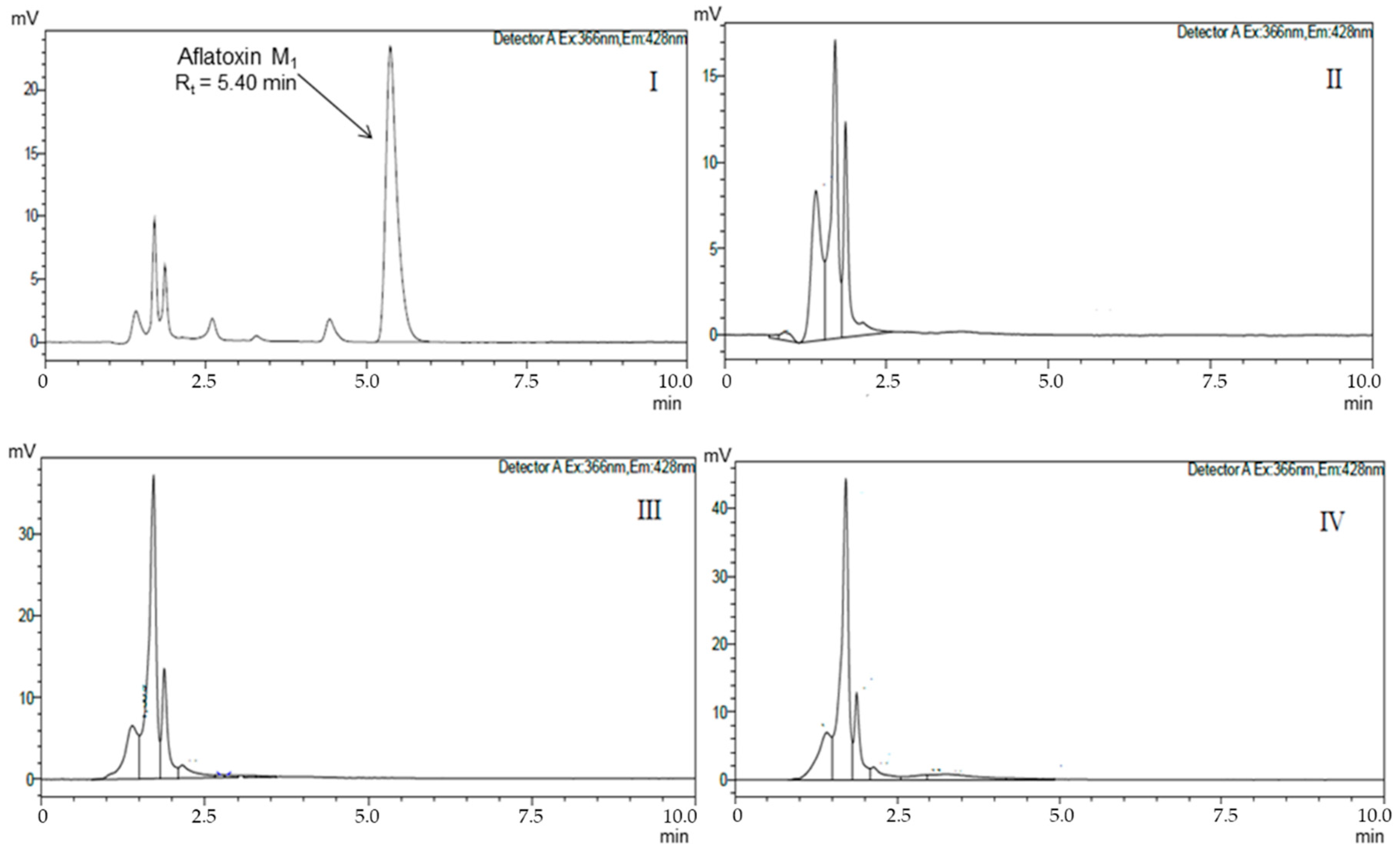
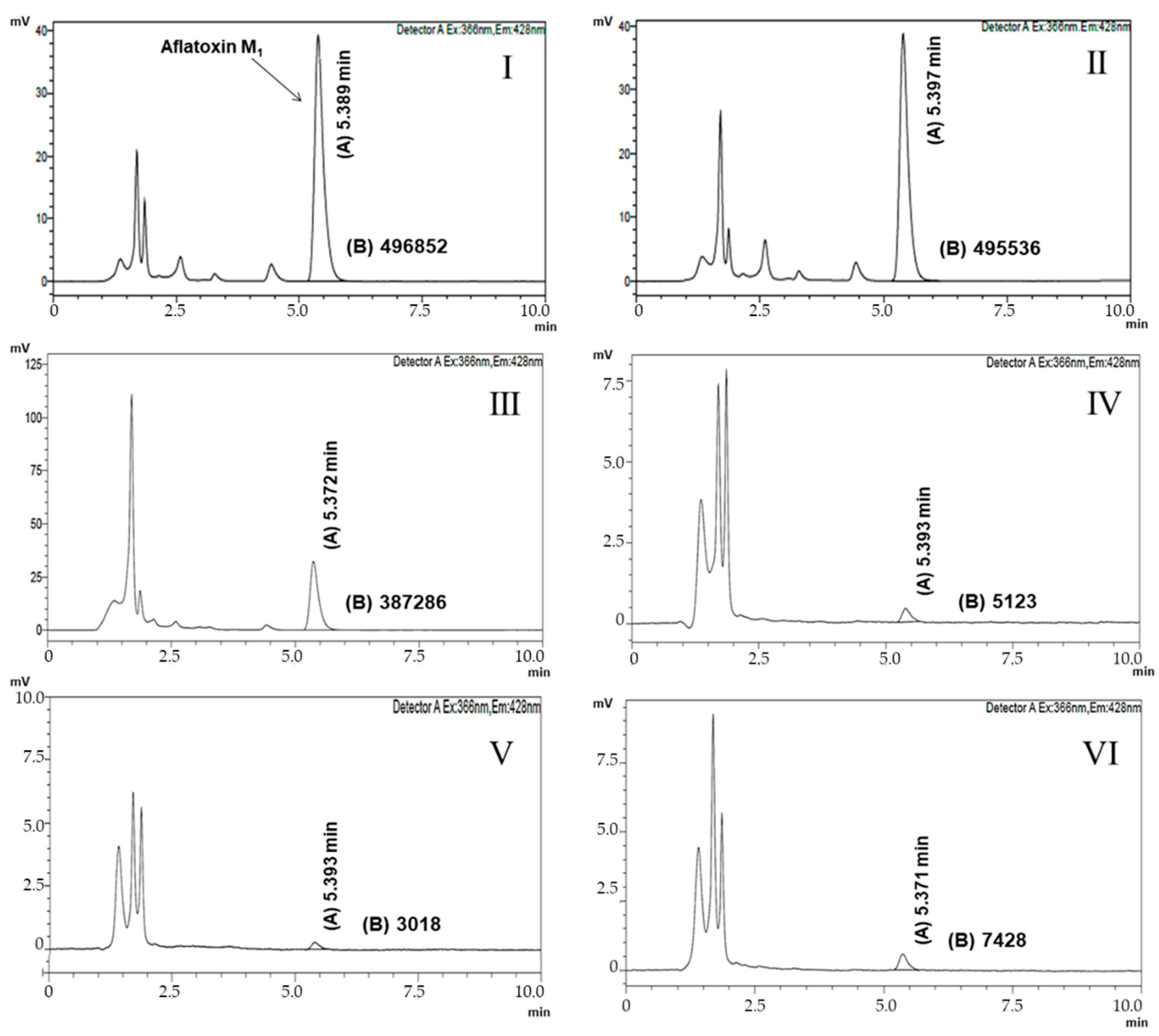
2. Results and Discussion

Chromatograms for the quantification of AFM1 in positive control, negative control, as well as in samples with viable cells of L. paracasei 108, L. plantarum 49, and L. fermentum 111 are shown in Figure 1. Chromatograms for the quantification of AFM1 in assays evaluating the recovery of AFM1 from cells after 1 h of incubation are shown in Figure 2.
Results of the capability of viable and heat-killed (non-viable) cells of L. paracasei 108, L. plantarum 49, and L. fermentum 111 for removing AFM1 in PBS are presented in Table 1. Viable and heat-killed cells of all examined Lactobacillus isolates were able to remove AFM1 in PBS, with removal percentage values in the range of 73.0 ± 1.2–80.0 ± 1.7% and 72.9 ± 1.1–78.7 ± 1.2%, respectively. Viable and heat-killed cells of the three examined isolates had similar values (p > 0.05) of AFM1 removal. Only L. paracasei 108 had higher values (p ≤ 0.05) of AFM1 removal after 24 h for both viable and heat-killed cells compared to 1 h. Higher values of AFM1 removal (p ≤ 0.05) after 1 h were found for L. plantarum 49 and L. fermentum 111, but the three examined isolates had similar values of AFM1 removal (p > 0.05) after 24 h.
Previous studies have also verified that the capacity of LAB—either as viable or non-viable cells, of binding aflatoxins (e.g., aflatoxin B1, ochratoxin, trichothecene, and AFM1) in PBS, laboratory media, or dairy matrices (e.g., milk and yoghurt)—varies in an isolate-dependent manner [2,11,22,23]. Aflatoxins bind to the surface components of LAB cells and variations in aflatoxin’s binding capacities among LAB species or isolates could be associated with differences in the bacterial cell wall and cell envelope structures [7]. Early investigations have found lower capacity of AFM1 removal by viable and/or heat-killed cells of different LAB (e.g., L. plantarum, L. acidophilus, L. reuteri, L. johnsonii, L. rhamnosus, L. bulgaricus, and Streptococcus thermophilus) [2,22,23], including probiotic L. casei [10], compared to L. paracasei 108, L. plantarum 49, and L. fermentum 111. The efficacy of AFM1 removal from PBS as high (>60%) as those found for Lactobacillus isolates examined in this study was reported to L. plantarum MON03 and L. rhamnosus GAF01 after 6 or 24 h of incubation [24].
Results of the AFM1 retention capacity of the viable and heat-killed cells of L. paracasei 108, L. plantarum 49, and L. fermentum 111 after washing with PBS are presented in Table 2. Percentage values of recovered AFM1 from viable and heat-killed cells were in the range of 13.4 ± 1.5–60.6 ± 1.6% and 10.9 ± 1.2%–47.9 ± 1.5%, respectively. The highest values of recovered AFM1 after 1 and 24 h were found for L. fermentum 111 and L. paracasei 108, respectively, for both viable and heat-killed cells. Only for L. fermentum 111 did the values of recovered AFM1 decrease after 24 h for viable and heat-killed cells; for L. paracasei 108 and L. plantarum 49, these values varied with the viability/non-viability of cells and incubation time period. Overall, L. plantarum 49 had the higher AFM1 retention capacity after washing. Variations in aflatoxin release have been linked to the differences in binding sites in different LAB isolates, or even in these binding sites being very similar. They could have minimal differences depending on each isolate [13,25,26].
For all examined isolates, the values of recovered AFM1 decreased after 24 h of incubation, indicating that AFM1 retention capacity increased when the length of the contact time increased. There was no clear association between the capability of removing AFM1, initially, and of retaining AFM1 after washing among examined isolates. Interestingly, a study with different Lactobacillus species found lower AFM1 removal values than those found in this study, although the recovery of AFM1 from bacterial cells was lower in the former [11].
Heat treatment positively affected the capability of retaining AFM1 in L. paracasei 108 after 1 h of incubation, as well as of L. plantarum 49 and L. fermentum 111 after 24 h of incubation. Heating could increase the interaction capacity of bacterial cells/aflatoxin complexes by causing an increased exposure of the cell wall components, primarily polysaccharides and peptidoglycans, which act as binding sites to aflatoxin [14]. However, the destruction of specific components of the bacterial cell wall by heating, causing the denaturation of proteins and increased cell surface hydrophobicity, has been cited to result in a decreased capability of LAB cells of binding AFM1 [7]. An increased capability of removing aflatoxin B1 was also found in L. rhamnosus after heating [27].
The recovery of the AFM1 bound to the cells of examined Lactobacillus isolates after washing indicates that the binding was not strong and could not involve a non-covalent weak bond, but probably a physical association of AFM1 with hydrophobic sites in the bacterial cell wall [13,20,25]. The lower AFM1 recovery values found for the examined isolates could be linked to the interaction of AFM1 molecules retained in the bacterial cell wall with other AFM1 molecules retained in adjacent cells, forming a type of cross-linked matrix that avoids aflatoxin release during washing [10]. Probably, the efficacy of this type of cross-linked matrix decreased over time for L. paracasei 108 and L. plantarum 49. Although some authors have reported that a part of non-recovered AFM1 might be degraded or biotransformed by a Lactobacillus metabolism [2,7], most of the available literature has indicated that aflatoxins are not removed by the metabolism of LAB, but because of a physical bound to the molecular components of bacterial cells, primarily peptidoglycans from the cell wall [19,21,25].
In agreement with available literature, the results of this study showed that the cell viability of the examined isolates is not a prerequisite for the removal and retaining of AFM1 [13,28]. Cell concentration as high as 108–109 CFU/mL of viable or non-viable LAB is typically needed to reach a level of aflatoxins removal of ≥ 50% [22,28].

3. Conclusions

Results showed that potentially probiotic L. fermentum 111, L. paracasei 108, and L. plantarum 49 isolated from fruit processing by-products are capable of binding AFM1 in vitro when assayed as either viable or non-viable cells. The recovery of AFM1 from bacterial cell complexes varied with the examined isolate and contact time. Non-viable cells had a higher capability for retaining AFM1 after 1 or 24 h of incubation. These results indicate that Lactobacillus isolates recovered from fruit with performance compatible to use as probiotics could have a satisfactory aflatoxin binding capacity, which could be exploited as a biological tool for the detoxification of foods and feeds, particularly, for the removal and restoration of AFM1 to safe levels. Further studies are needed to investigate the mechanisms involved in removal of AFM1 by these isolates and possible factors affecting the stability of formed complexes, including when exposed to conditions mimicking the human gastrointestinal tract.

4. Materials and Methods

4.1. Chemicals, Bacterial Isolates, and Inoculum Preparation

The AFM1 standard was obtained from Sigma Aldrich (St. Louis, MO, USA). High-performance liquid chromatography (HPLC) grade solvents were obtained from Merck (Darmstadt, Germany).
The isolates Lactobacillus plantarum 49, L. fermentum 111, and L. paracasei 108 were examined separately for the removal of AFM1. These isolates were recovered from fruit processing by-products, identified with a partial 16S rRNA gene sequence analysis and characterized as potential candidates for use as probiotics [17]. Stocks were stored at −20 °C in de Man, Rogosa, and Sharpe (MRS) broth (HiMedia, Mumbai, India) with glycerol (20 mL/100 mL; Sigma-Aldrich, St. Louis, MO, USA). Working cultures were maintained aerobically on MRS agar (HiMedia, Mumbai, India) at 4 °C and transferred to a new media monthly. Prior to use in assays, each isolate was cultivated anaerobically (Anaerobic System Anaerogen, Oxoid, Hampshire, UK) in MRS broth at 37 °C for 20–24 h (to reach the stationary growth phase), harvested by centrifugation (4500× g, 15 min, 4 °C), washed twice, and resuspended in phosphate buffer solution (PBS; 50 mM K2HPO4/KH2PO4; pH 6.9) to obtain cell suspensions with an optical density reading at 660 nm (OD660) of 0.5. This suspension had viable counts of approximately 1.1 × 10⁹ CFU/mL for each isolate when plated in MRS agar.

4.2. Evaluation of AFM1 Removal and Recovery of AFM1 from Bacterial Cells

The capability of examined Lactobacillus isolates to remove AFM1 in PBS was assessed with viable and non-viable bacterial cell suspensions. To obtain non-viable bacterial cells, Lactobacillus cell suspensions were inactivated by boiling at 100 °C for 1 h. No visible colonies were found when heat-treated cell suspensions (named heat-killed cells) were plated onto MRS agar and followed by anaerobic incubation (using Anaerobic System Anaerogen, Oxoid, Hampshire, UK) for 48 h. For testing the AFM1 removal capability, 1 mL of test isolate suspension (pure culture of viable and heat-killed cells) was mixed with 1.5 mL of PBS, previously spiked with 0.15 µg/mL AFM1, and incubated aerobically at 37 °C [28]. After 1 and 24 h of incubation, the mixture was centrifuged (1500× g, 15 min, 4 °C) and the AFM1 content in the supernatant was determined by HPLC, as detailed in Section 4.3.
Cell pellets collected from each monitored incubation period (contact time) were evaluated for the recovery of AFM1 from cell complexes. Obtained pellets were washed with 1.5 mL of fresh PBS, the cells were re-pelleted (1500× g, 15 min, 4 °C), and supernatant was collected for the quantification of released AFM1 [18]. For each isolate, a positive control consisting of free cells suspended in PBS with 0.15 µg/mL AFM1, and a negative control, consisting of bacterial cells (viable or heat-killed), suspended in PBS were used.

4.3. Quantification of AFM1

The quantification of AFM1 in supernatants was done with high-performance liquid chromatography (HPLC) using a Shimadzu (Prominense, Tokyo, Japan) HPLC system, equipped with an auto sampler SIL 20A HT (Prominense, Shimadzu, Tokyo, Japan), fluorescence detector RF-20A (Prominense, Shimadzu, Tokyo, Japan), an LC-20AT pump (Prominense, Shimadzu, Tokyo, Japan), oven CTO-20A (Prominense, Shimadzu, Tóquio, Japão), a CBM-20A controller (Prominense, Shimadzu, Tokyo, Japan), a CLC-ODS (M) reverse phase column (4.6 × 150 mm; Shim-Pack, Prominense, Shimadzu, Tokyo, Japan) and pre-column G-ODS-4 (1.0 × 4.0 mm; Shim-Pack, Prominense, Shimadzu, Tokyo, Japan).
Chromatographic conditions were the same as those described in a previous study [7]. Excitation and emission wavelengths were 366 and 428 nm, and the injection volume was 20 μL. The mobile phase was water:methanol:acetonitrile (6:2:2) and the flow rate was 1 mL/min. The calibration curve was constructed using six concentrations of AFM1 standard diluted in acetonitrile (20–60 ng/mL), performed in triplicate. From this analysis, the equation y = 2E+07x + 873,267 (r2 > 0.99) was obtained. The limit of detection (LOD) and limit of quantification (LOQ) were estimated based on Resolution n° 899 of the Brazilian Agency for Health Surveillance [29]. The LOD and LOQ of AFM1 were 0.20 and 0.67 ng/mL, respectively.
The percentage of AFM1 removed by each isolate was determined with the Equation (1) [22,27,30]:
100 × [1 − (peak area of chromatographic peak of sample)/area of positive control chromatographic peak)].

4.4. Statistical Analysis

Assays were done in triplicate in three independent experiments (repetitions). A Kolmogorov–Smirnov normality test was run to assess whether obtained results had normal distribution. Results (average data ± standard deviation) were submitted to a one-way analysis of variance (ANOVA), followed by Tukey’s test, considering a p value of ≤ 0.05 for significance. Statistical analyses were done with IBM SPSS Statistics 20 (Armonk, NY, USA).

Author Contributions

Conceptualization, P.O.d.C., H.I.F.M., E.L.d.S.; methodology, P.O.d.C., H.I.F.M., E.L.d.S., C.J.d.M., Y.M.N., J.F.T.; validation, P.O.d.C., H.I.F.M., C.J.d.M., Y.M.N., J.F.T.; investigation, P.O.d.C., C.J.d.M., Y.M.N.; writing—original draft preparation, P.O.d.C., H.I.F.M., E.L.d.S.; writing—review and editing; supervision, H.I.F.M.; funding acquisition, H.I.F.M., E.L.d.S., J.F.T. All authors have read and agreed to the published version of the manuscript.

Funding

This research was partially funded by CAPES (Brazil), finance code 001.

Acknowledgments

Authors thank the Coordenação de Aperfeiçoamento de Pessoal de Nível Superior (CAPES, Brazil) for partial funding of this research.

Conflicts of Interest

The authors declare no conflict of interest.

References

  1. Bhat, R.; Rai, R.V.; Karim, A.A. Mycotoxins in food and feed: Present status and future concerns. Compr. Rev. Food Sci. Food Saf. 2010, 59, 57–81. [Google Scholar] [CrossRef]
  2. Elsanhoty, R.M.; Salam, S.A.; Ramadan, M.F.; Badr, F.H. Detoxification of aflatoxin M1 in yoghurt using probiotics and lactic acid bacteria. Food Cont. 2014, 43, 129–134. [Google Scholar] [CrossRef]
  3. Koppen, R.; Koch, M.; Slegel, D. Determination of mycotoxins in foods: Current state of analytical methods and limitations. Appl. Microbiol. Biotechnol. 2010, 86, 1595–1612. [Google Scholar] [CrossRef] [PubMed]
  4. International Agency for Research on Cancer. Some traditional herbal medicine, some mycotoxins and styrene. In Monographs on the Evaluation of Carginogenic Risks to Humans; International Agency for Research on Cancer: Lyon, France, 2002. [Google Scholar]
  5. Ayar, A.; Sert, D.; Lon, A.H. A study on the occurrence of aflatoxin in raw milk due to feeds. J. Food Saf. 2007, 27, 199–207. [Google Scholar] [CrossRef]
  6. Ministry of Health; National Agency for Health Surveillance; Brazilian Legislation. On the Maximum Tolerated Limit (MTL) for Mycotoxins in Foods; Resolution No. 7 of 18 February 2011; National Agency for Health Surveillance: Brasilia, Brazil, 2011. [Google Scholar]
  7. Corassin, C.H.; Bovo, F.; Rossim, R.E.; Oliveira, C.A.F. Efficiency of Saccharomyces cerevisiae and lactic acid bacteria strains to bind aflatoxin M1 in UHT skim milk. Food Cont. 2013, 31, 80–83. [Google Scholar] [CrossRef] [Green Version]
  8. European Commission. Commission Regulation (EC) No 401/2006 of 23 February 2006 laying down the methods of sampling and analysis for the official control of the levels of mycotoxins in foodstuffs. Off. J. Eur. Union 2006, 70, 12–34. [Google Scholar]
  9. Wang, B.; Mahoney, N.E.; Pan, Z.; Khir, R.; Wu, B.; Ma, H.; Zhao, L. Effectiveness of pulsed light treatment for degradation and detoxification of aflatoxin B1 and B2 in rough rice and rice bran. Food Cont. 2016, 59, 461–467. [Google Scholar] [CrossRef]
  10. Hernandez-Mendoza, A.; Garcia, H.S.; Steele, J.L. Screening of Lactobacillus casei strains for their ability to bind aflatoxin B1. Food Chem. Toxicol. 2009, 47, 1064–1068. [Google Scholar] [CrossRef] [PubMed]
  11. Serrano-Niño, J.C.; Cavazos-Garduño, A.; Hernandez-Mendoza, A.; Applegate, B.; Ferruzzi, M.C.; Martin-González, M.F.S.; García, H.S. Assessment of probiotic strains ability to reduce the bioaccessibility of aflatoxin M1 in artificially contaminated milk using an in vitro digestive model. Food Cont. 2013, 31, 202–207. [Google Scholar] [CrossRef]
  12. Onilude, A.A.; Fagade, O.E.; Bello, M.M.; Fadahunsi, I.F. Inhibition of aflatoxin-producing aspergilli by lactic acid bacteria isolates from indigenously fermented cereal gruels. Afr. J. Biotechnol. 2005, 4, 1404–1408. [Google Scholar]
  13. Azeem, N.; Nawaz, M.; Anjum, A.A.; Saeed, S.; Sana, S.; Mustafa, A.; Yousuf, M.R. Activity and anti-aflatoxigenic effect of indigenously characterized probiotic lactobacilli against Aspergillus flavus—A common poultry feed contaminant. Animals 2019, 15, 166. [Google Scholar] [CrossRef] [PubMed] [Green Version]
  14. Elsanhoty, R.M.; Ramadan, M.F.; El-Gohery, S.S.; Abol-Ela, M.A.A. Ability of selected microorganisms for removing aflatoxins in vitro and fate of aflatoxins in contaminated wheat during baladi bread baking. Food Cont. 2013, 33, 287–292. [Google Scholar] [CrossRef]
  15. Naeem, M.; Ilyas, M.; Haider, S.; Baig, S.; Saleem, M. Isolation characterization and identification of lactic acid bacteria from fruit juices and their efficacy against antibiotics. Pak. J. Bot. 2012, 44, 323–328. [Google Scholar]
  16. Ilha, E.C.; Silva, T.; Lorenz, J.G.; De Oliveira Rocha, G.; Sant’Anna, E.S. Lactobacillus paracasei isolated from grape sourdough: Acid, bile, salt, and heat tolerance after spray drying with skim milk and cheese whey. Eur. Food Res. Technol. 2015, 240, 977–984. [Google Scholar] [CrossRef]
  17. Garcia, E.F.; Luciano, W.A.; Xavier, D.E.; Da Costa, W.C.; De Sousa Oliveira, K.; Franco, O.L.; De Morais, M.A.J.; Lucena, B.T.; Picão, R.C.; Magnani, M.; et al. Identification of lactic acid bacteria in fruit pulp processing byproducts and potential probiotic properties of selected Lactobacillus strains. Front. Microbiol. 2016, 7, 1371. [Google Scholar] [CrossRef] [Green Version]
  18. Ahlberg, S.H.; Joutsjoki, V.; Korhonen, H.J. Potential of lactic acid bacteria in aflatoxin risk mitigation. Int. J. Food Microbiol. 2015, 207, 87–102. [Google Scholar] [CrossRef]
  19. Khanian, M.; Karimi-Torshizi, M.A.; Allameh, A. Alleviation of aflatoxin-related oxidative damage to liver and improvement of growth performance in broiler chickens consumed Lactobacillus plantarum 299v for entire growth period. Toxicon 2019, 158, 57–62. [Google Scholar] [CrossRef]
  20. Assaf, J.C.; Nahle, S.; Chokr, A.; Louka, N.; Atoui, A.; El Khoury, A. Assorted methods for decontamination of aflatoxin m1 in milk using microbial adsorbents. Toxins 2019, 11, 304. [Google Scholar] [CrossRef] [Green Version]
  21. Kim, S.; Lee, H.; Lee, S.; Lee, J.; Ha, J.; Choi, Y.; Yoon, Y.; Choi, K.H. Microbe-mediated aflatoxin decontamination of dairy products and feeds. J. Dairy Sci. 2017, 100, 871–880. [Google Scholar] [CrossRef]
  22. El-Nezami, H.; Kankaanpää, P.; Salminen, S.; Ahokas, J. Physicochemical alterations enhance the ability of dairy strains of lactic acid bacteria to remove aflatoxin from contaminated media. J. Food Prot. 1998, 61, 466–468. [Google Scholar] [CrossRef]
  23. Pierides, H.; El-Nezami, K.; Peltonem, S.; Salminem, J.; Ahokas, J.T. Ability of dairy strains of lactic acid bacteria to bind aflatoxin M1 in a food model. J. Food Prot. 2000, 63, 645–650. [Google Scholar] [CrossRef] [PubMed]
  24. Jebali, R.; Abbes, S.; Salah-Abbès, J.B.; Younes, R.B.; Haous, Z.; Oueslati, R. Ability of Lactobacillus plantarum MON03 to mitigate aflatoxins (B1 and M1) immunotoxicities in mice. J. Immunotoxicol. 2014, 12, 290–299. [Google Scholar] [CrossRef] [PubMed] [Green Version]
  25. Panwar, R.; Kumar, N.; Kashyap, V.; Ram, C.; Kapila, R. Aflatoxin M1 detoxification ability of probiotic lactobacilli of Indian origin in in vitro digestion model. Prob. Antimicrob. Proteins 2019, 11, 460–469. [Google Scholar] [CrossRef] [PubMed]
  26. Risa, A.; Divinyi, D.M.; Baka, E.; Krifaton, C. Aflatoxin B1 detoxification by cell-free extracts of Rhodococcus strains. Acta Microbiol. et Immunol. Hung. 2017, 64, 423–438. [Google Scholar] [CrossRef] [Green Version]
  27. Abbès, S.; Salah-Abbès, J.B.; Sharafi, H.; Jebali, R.; Noghabi, K.A.; Oueslati, R. Ability of Lactobacillus rhamnosus GAF01 to remove AFM1 in vitro and to counteract AFM1 immunotoxicity in vivo. J. Immunotoxicol. 2013, 10, 279–286. [Google Scholar] [CrossRef]
  28. Bovo, F.; Corassin, C.H.; Rosim, R.E.; De Oliveira, C.A. Efficiency of lactic acid bacteria strains for decontamination of aflatoxin M1 in phosphate buffer saline solution and in skim milk. Food Bioproc. Technol. 2012, 5, 1–5. [Google Scholar]
  29. Brazilian Legislation, Ministry of Health, National Agency for Health Surveillance. Guide for Validation of Analytical and Bioanalytical Methods. National Agency for Health Surveillance. Resolution nº899; National Agency for Health Surveillance: Brasilia, Brazil, 2003. [Google Scholar]
  30. Haskard, C.A.; El-Nezami, H.S.; Kankaanpää, P.E.; Salminen, S.; Ahokas, J.T. Surface binding of aflatoxin B1 by lactic acid bacteria. Appl. Environ. Microbiol. 2001, 67, 3086–3091. [Google Scholar] [CrossRef] [Green Version]
Figure 1. Chromatograms of aflatoxin M1 (AFM1) quantification in positive and negative control. (I) Positive control: phosphate buffer solution (PBS) with AFM1. Rt = Retention time of AFM1 in phosphate buffer solution; chromatographic peak area corresponding to AFM1; (II) Negative control after 1 h of incubation: PBS + L. paracasei 108; (III) Negative control after 1 h of incubation: PBS + L. plantarum 49; (IV) Negative control after 1 h of incubation: PBS + L. fermentum 111.
Toxins 13 00004 g001
Figure 2. Chromatograms of aflatoxin M1 (AFM1) quantification in PBS. (I) Chromatogram of assays after 1 h of incubation: PBS + AFM1 + L. paracasei 108; (II) Chromatogram of assays after 1 h of incubation: PBS + AFM1 + L. plantarum 49; (III) Chromatogram of assays after 1 h of incubation: PBS + AFM1 + L. fermentum 111; (IV) AFM1 recovery chromatogram of L. paracasei 108 and AFM1 complex after 1 h of incubation; (V) AFM1 recovery chromatogram of L. plantarum 49 and AFM1 complex after 1 h of incubation; (VI) AFM1 recovery chromatogram of L. fermentum 111 and AFM1 complex after 1 h of incubation. (A) Retention time (min) of aflatoxin M1 in phosphate buffer solution; (B) chromatographic peak area corresponding to aflatoxin M1.
Toxins 13 00004 g002
Table 1. Percentage (average values ± standard deviation) of aflatoxin M1 (AFM1) removal in phosphate buffer solution by L. paracasei 108, L. plantarum 49, and L. fermentum 111.
IsolatesAFM1 Removal (%)
1 h-Incubation24 h-Incubation
Viable Cells Heat-Killed CellsViable CellsHeat-Killed Cells
L. paracasei 10873.0 ± 1.2 b,B72.9 ± 1.1 b,B78.9 ± 0.5 a,A78.7 ± 1.2 a,A
L. plantarum 4978.1 ± 1.6 a,A75.8 ± 1.0 a,A,B77.0 ± 2.7 a,A76.6 ± 1.5 a,A
L. fermentum 11178.6 ± 2.1 a,A78.4 ± 0.65 a,A80.0 ± 1.7 a,A78.3 ± 2.5 a,A
Different small letters in the same row (a,b) denote a significant difference (p ≤ 0.05) among values, based on Tukey’s test; different capital letters in the same column (A,B) denote a significant difference among values (p ≤ 0.05), based on Tukey’s test.
Table 2. Percentage (average values ± standard deviation) of recovered aflatoxin M1 (AFM1) in solution after washing with phosphate buffer solution.
IsolatesAFM1 Recovery, %
1 h-Incubation24 h-Incubation
Viable Cells Heat-Killed CellsViable CellsHeat-Killed Cells
L. paracasei 10834.6 ± 1.1 b,B28.5 ± 1.7 d,C31.7 ± 1.2 c,A40.3 ± 1.6 a,A
L. plantarum 4913.4 ± 1.5 c,C43.8 ± 1.5 a,B18.8 ± 1.0 b,B10.9 ± 1.2 d,C
L. fermentum 11160.6 ± 1.6 a,A47.9 ± 1.5 b,A14.1 ± 1.4 c,C14.9 ± 1.6 c,B
Different small letters in the same row (a–c) denote a significant difference (p ≤ 0.05) among values, based on Tukey’s test; different capital letters in the same column (A,B) denote a significant difference among values (p ≤ 0.05), based on Tukey’s test.
Publisher’s Note: MDPI stays neutral with regard to jurisdictional claims in published maps and institutional affiliations.

