Quo vadis Cardiac Glycoside Research?


1
Department of Biochemistry and Microbiology, University of Chemistry and Technology Prague, Technická 5, Prague 6, Czech Republic
2
Department of Chemistry of Natural Compounds, University of Chemistry and Technology Prague, Technická 3, Prague 6, Czech Republic
*
Author to whom correspondence should be addressed.
Toxins 2021, 13(5), 344; https://0-doi-org.brum.beds.ac.uk/10.3390/toxins13050344
Submission received: 5 April 2021
/
Revised: 7 May 2021
/
Accepted: 8 May 2021
/
Published: 11 May 2021
(This article belongs to the Special Issue Basic Research for the Potential Use of Plant Toxins)
Abstract
:Cardiac glycosides (CGs), toxins well-known for numerous human and cattle poisoning, are natural compounds, the biosynthesis of which occurs in various plants and animals as a self-protective mechanism to prevent grazing and predation. Interestingly, some insect species can take advantage of the CG’s toxicity and by absorbing them, they are also protected from predation. The mechanism of action of CG’s toxicity is inhibition of Na+/K+-ATPase (the sodium-potassium pump, NKA), which disrupts the ionic homeostasis leading to elevated Ca2+ concentration resulting in cell death. Thus, NKA serves as a molecular target for CGs (although it is not the only one) and even though CGs are toxic for humans and some animals, they can also be used as remedies for various diseases, such as cardiovascular ones, and possibly cancer. Although the anticancer mechanism of CGs has not been fully elucidated, yet, it is thought to be connected with the second role of NKA being a receptor that can induce several cell signaling cascades and even serve as a growth factor and, thus, inhibit cancer cell proliferation at low nontoxic concentrations. These growth inhibitory effects are often observed only in cancer cells, thereby, offering a possibility for CGs to be repositioned for cancer treatment serving not only as chemotherapeutic agents but also as immunogenic cell death triggers. Therefore, here, we report on CG’s chemical structures, production optimization, and biological activity with possible use in cancer therapy, as well as, discuss their antiviral potential which was discovered quite recently. Special attention has been devoted to digitoxin, digoxin, and ouabain.
Keywords:
cancer treatment; cardenolides; digitoxin; digoxin; drug repositioning; immunogenic cell death; Na+/K+ ATPase; antiviral potential; secondary plant metabolites; toxinsKey Contribution: Cardiac glycosides—toxic substances, which can serve as a remedy for various diseases. Cardiac glycosides are not known only as ionic balance disrupters but also as regulators of Na+/K+-ATPase serving as a receptor; concentration matters.
1. Introduction
Biologically active secondary metabolites occur in almost any living system, from unicellular organisms to fungi, plants, and animals. Each organism producing these secondary metabolites has adapted its metabolism to its environment, and this adaptation enabled it to cope with the surrounding threats. Since life occurs in different environments with slightly different living conditions, the chemical nature of secondary metabolites differs as well. Most of the secondary metabolites, such as alkaloids [1], flavonoids [2,3], phytosterols [4], and many others, are found in the plants that “ran” the furthest in this imaginary race for survival. Although many secondary metabolites may, at the first sight, appear to be inherently toxic to humans and some other species, it is always important to keep in mind that at lower concentrations, these substances may have beneficial effects. It is for this reason that mankind presently uses these metabolites mainly in pharmacy.
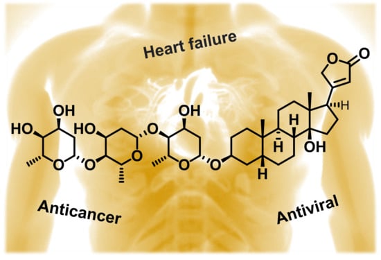
Among the most widely used secondary metabolites belong cardiac glycosides (CGs) which are among the 200 most commonly prescribed drugs in the USA (the year 2018) [5]. CGs are produced by several plants mainly as protection against pests [6,7]. Probably the best known is the production by plants of the Digitalis genus (Scrophulariaceae), from which the most important representatives of this group are mostly isolated, namely digoxin (Dg) and digitoxin (Dgt). Both substances are well-established drugs for the treatment of heart failure and cardiac arrhythmias, even though they have a relatively narrow therapeutic window that limits their use [8]. The mechanism of action of not only these representatives but also of the whole CG group is the inhibition of Na+/K+-ATPase (NKA), which causes a disruption of the ionic balance of the cell and, thus, leads to an increase in muscle contraction (Figure 1). However, Ca2+ ions, which serve as mediators of muscle contraction, are also involved in signaling pathways that ultimately lead to cell death by apoptosis. The latter fact is the reason why CGs are currently being studied as possible drugs for cancer treatment. Moreover, in recent decades (ca. 30 years), the fact that CGs can activate several signaling cascades involved in cell proliferation through interaction with NKA has come to light. It was revealed that CGs, via the interaction with NKA, can activate several signaling cascades that are involved in the regulation of cell proliferation, thereby, contributing to anticancer activity. The role of CGs in the regulation of blood pressure and, more recently, their antiviral effects were also discovered.
2. Occurrence of Cardiac Glycosides
CGs became more widely known to the medical community due to W. Withering, who used extracts from dried foxglove leaves to treat edema [9]. At that time, due to imperfect purification techniques, these extracts contained also other substances occurring in foxglove leaves, such as the already mentioned flavonoids [10], therefore, it was not possible then to determine which substances were responsible for the pharmacological effect of the extracts.
Nowadays, we know that the most important bioactive substances contained in these plants are CGs, and although many drugs are now produced by organic synthesis, CGs are still being isolated from foxglove (Digitalis), which are therefore their most important producers [11]. From Table 1, summarizing the most important producers of CGs in the plant kingdom, it is apparent that the plants always contain several structurally similar CGs. The most abundant CGs (Figure 2 and Figure 3) then usually have their name derived from the designation of the corresponding plant.
As is obvious from Table 1, CGs are produced by many different plants, some of which are either common household plants (e.g., Nerium oleander) [27,28] or can be mistaken for other herbs [29,30], thus, increasing the risk of accidental, or even purposeful CG poisoning. All the aforementioned CGs belong to the so-called cardenolides, which are, with few exceptions, produced exclusively by plants. Because of this, CGs have been primarily known as plant secondary metabolites, although information also exists on their production by animals (Table 2).
In the case of cardenolides, they are referred to as endogenous CGs, which are produced in humans by the adrenal cortex [36]. The second group of CGs comprises the so-called bufadienolides, which are also found in plants, for example, proscillaridin in the plant Drimia maritima [37]. However, bufadienolides are also present to a greater extent in animals, e.g., marinobufagenin in the toad Rhinella marina and humans [31,34] and bufotoxin in Bufo bufo gargarizans [35]. Another occurrence of bufadienolides is summarized in Steyn et al. [38]. It is also worth mentioning that the name of the whole group of bufadienolides is derived from the toads of the genus Bufo.
3. Production of Cardiac Glycosides
Although the plants containing CGs have been known to mankind for several centuries, Dgt was not isolated from Digitalis purpurea until 1875, when it was achieved by German pharmacologist O. Schmiedeberg [39]. Dg had to wait another 55 years for its first isolation, which was achieved in 1930 by the English chemist S. Smith, who isolated it from Digitalis lanata [40]. Currently, Dg is known in the United States of America primarily under the tradenames Lanoxin and Dgt as Crystodigin. Nevertheless, for commercial purposes, both compounds are still obtained by isolation from the plants Digitalis lanata and Digitalis purpurea. Therefore, the aim is to optimize the cultivation process of these two plants as much as possible to achieve higher yields of Dg and Dgt as well as other medicinally relevant CGs (Figure 4). These optimizations can be done by several approaches, either by feeding the plants with CG precursors, production elicitation, different cultivation, or genetic engineering.
3.1. Precursor Feeding and Elicitation
CG production is affected by several factors, the first and probably also the most important one is the sufficient supply of necessary nutrients. Higher accumulation of CGs in Digitalis lanata and Digitalis purpurea was detected in vitro with the supply of both: suitable carbohydrates (sucrose, glucose, and raffinose) [41] and steroids, which in part serve as precursors for CG biosynthesis (Figure 5)—cholesterol, progesterone [42,43], 21-hydroxypregnane, 5β-pregnan-3,20-dione, 3β-hydroxy-5β-pregnan-20-one, 3β,14β,21-trihydroxy-5β-pregnane-20-one [44].
Furthermore, CG production can also be increased with SiO2-based elicitors (Silioplant®), chitosan oligosaccharide (Chitoplant®), and methyl jasmonate. All mentioned elicitors significantly increase the CG in vitro production in Digitalis lanata by elevating oxidative stress due to the increase in hydrogen peroxide and malondialdehyde (an indicator of lipid peroxidation) levels [45]. It is oxidative stress which is associated with increased CG production [46].
3.2. Cultivation Techniques
Another way to increase CG production is to cultivate Digitalis purpurea using biotechnology. In such a case, the plants are cultivated in a controlled environment, thus, eliminating the issues associated with nutrient supply, weather fluctuations, seasonal changes, and pests. One of the possible biotechnological production methods is the use of the so-called temporary immersion system, in which plants are exposed to the nutrient medium for a limited time at precisely defined intervals [47]. For the production of CGs in Digitalis purpurea, this method of cultivation was first described in 2009, when it was found that the production of biomass, as well as compounds 1 and 2, are affected by the frequency of immersions [48]. Another possibility is to cultivate undifferentiated Digitalis purpurea cells, as shown in Hagimory et al. [49]; however, these undifferentiated cells produce significantly less of Dgt compared to shoot-forming cultures, which are often used for temporary immersion system cultivation. For this reason, culturing undifferentiated cells from Digitalis purpurea for the mass production of CGs is not very promising.
3.3. Genetic Engineering
Another way to increase CG production is to use genetic engineering. In general, efforts to increase the production of secondary metabolites may target metabolic enzymes, regulatory elements, or other genes. Figure 5 shows the biosynthetic pathway of digitoxin in Digitalis purpurea and Digitalis lanata. Glycosylation steps from digitoxigenin to digitoxin use glycosyltransferases with UDP-α-d-digitoxose as a donor of digitoxose. This pathway offers opportunities for intervention by genetic engineering.
Gram-negative bacteria Agrobacterium tumefaciens and Agrobacterium rhizogenes are often used for genetic modification of plants, which can introduce their genetic information into the host plant using the so-called Ti or Ri plasmids, respectively [50,51]. This approach has been used in several studies for both Digitalis purpurea and Digitalis lanata [52,53,54,55,56]. This method is subsequently used for the identification of key enzymes involved in CG biosynthesis. One of these enzymes is progesterone-5β-reductase (EC 1.3.1.3). Kairuz et al. introduced the gene for this enzyme from Arabidopsis thaliana into Digitalis purpurea [57]. In this way, the prepared strain of Digitalis purpurea, the production of compounds Dg and Dgt increased two and four times, respectively.
Moreover, high throughput sequencing of RNA from Digitalis purpurea [58] is also used to predict sequences of enzymes involved in the cardiac glycoside biosynthesis pathway and series of non-coding RNAs with regulatory roles. These findings can then be used in a synthetic biology approach to address cardiac glycoside biosynthesis. Recently, genes coding selected enzymes from Digitalis lanata, Arabidopsis thaliana, bacterium Comamonas testosteronii, mouse (Mus musculus), and cattle (Bos taurus) were cloned into two vectors, which were inserted into yeast cells (Saccharomyces cerevisiae). These cells were then able to produce 3β,21-dihydroxy-5β-pregnan-20-one [59].
3.4. Physical Factors
Last, but not least, other factors that are being studied in connection with the increased accumulation of secondary metabolites in plants are physical factors such as the effect of light and temperature. It is known that plants, whether cultivated in the field or in laboratory conditions, need light to live. Verma et al. studied the effect of different wavelengths of light on the level of accumulation of Dg and Dgt in Digitalis purpurea, in which the combination of red (maximum intensity at 650 nm, 20%) and blue (maximum intensity at 450 nm, 80%) parts of the visible spectra have proved to be the most efficient [60]. As for the temperature, Wietmarschen et al. [11] cultivated Digitalis lanata at 15–25 °C with a cold shock, which significantly increased the production of Dg and, thus, it underlines the importance of stress factors for increased CG production.
4. Structure of Cardiac Glycosides
The structure of CGs, like other steroid substances, is derived from the steroid skeleton (steran). The steroid skeleton generally consists of three six-membered carbon rings (A, B, C) and one five-membered ring (D) [Figure 6C]. Rings A and B are joined in a cis conformation, as are rings C and D. In contrast, rings B and C are joined in a trans conformation. The main substituents, typical of CGs, are then attached to this steroid skeleton, namely the unsaturated lactone ring C-17 in the β position and the saccharide in the C-3 position (Figure 6A). According to the type of lactone cycle, CGs are divided into the already mentioned cardenolides, which contain an unsaturated five-membered furanone ring (Figure 6A), and bufadienolides (Figure 6B), which contain an unsaturated six-membered pyranone ring. The attached carbohydrates are different, but most often they are d-digitoxopyranosyl, d-glucopyranosyl, d-oleandropyranosyl, l-rhamnopyranosyl, d-cymaropyranosyl, d-xylopyranosyl (Figure 2 and Figure 3). All CGs are further substituted at C-10 and C-13 at the β positions by a methyl group and C-14 also at the β position with a hydroxyl group. Substituents in other positions are individual for a given derivative. Common substitutions include those at C-12 and C-16 (Figure 2) or C-5 and C-10 (Figure 3). In most cases, these are also methyl or hydroxyl groups; however, in some cases, there is also a carbonyl group, an esterified hydroxyl group, or an epoxide. Sometimes a double bond also occurs in the structure of the steroid skeleton.
5. Na+/K+-ATPase Binding of Cardiac Glycosides
The most important molecular target of CGs is the already mentioned NKA, which is located in the cytoplasmic membrane of animal cells. The main task of NKA in cells is the transport of Na+ and K+ ions across the cytoplasmic membrane against a concentration gradient. To do this, NKA uses the energy released from the hydrolysis of adenosine triphosphate (ATP), which leads to conformational changes that affect the affinity of these ions to the protein and thus enable their transport. These conformational changes are the first factor that affects CG’s affinity for NKA. It was confirmed that NKA is in the so-called high-affinity state if it is phosphorylated and K+ binding has not yet taken place. In contrast, the change to a low-affinity state occurs after the binding of K+ ions, which are therefore agonists of CGs [61]. The affinity of CGs for NKA is also affected by another ion, Mg2+. As shown in a stability study of the ouabain-NKA complex, the complex is more stable at higher Mg2+ relative to K+ due to the competition between these two ions [62]. Besides, Mg2+ ions in NKA induce conformational changes that lead to its transition to a high-affinity state.
The second factor that affects the affinity of CGs for NKA and is closely related to the first is the amino acid composition of the binding site. The CGs binding site consists of six α-helices located in the transmembrane domain of the α-subunit of this protein and is accessible from the extracellular space [62]. This binding site can be divided into a non-polar portion composed of l-Ile315, l-Phe316, l-Gly319, l-Phe783, l-Phe786, and l-Leu793 based on the polarity of the amino acids present and a polar portion composed of l-Gln111, l-Glu117, l-Asp121, l-Asn122, and l-Thr797 [62]. The amino acids l-Gln111, l-Asn122, and l-Thr797, which form hydrogen bonds with the hydroxyl groups of the CG steroid skeleton, are particularly important for the binding of CGs to NKA. Substitution of these amino acids leads to a significant reduction in the NKA sensitivity to CGs, for example, IC50 of Dg, Dgt, and ouabain is increased by three orders of magnitude [63], and, therefore, is the reason for the resistance of some animal species such as mice, rats, and some insect species to CGs. Substitutions of l-Asp121 for l-Gly or l-Glu, l-Asn122 for l-His, or l-Asp, l-Gln111 for l-Thr or l-Arg, and l-Thr797 for l-Ala (Figure 7) were found in these organisms [7,64,65].
Other substitutions have been also described. These, however, have less impact on NKA resistance to CGs than those described above. Namely, they are the following: l-Leu330 for l-Gln, l-Ala331 for l-Gly, l-Thr338 for l-Ala, l-Thr338 for l-Asn, and l-Phe982 for l-Ser [66]. The importance of polar interactions for CG binding to NKA is underlined by the work of Magpusao et al., who blocked the hydroxyl groups of ouabain at the C-1 and C19 positions with an acetonide group (Figure 8) [67]. In this case, the resulting derivative showed a 120-fold increase in half-maximal inhibitory concentration (IC50) compared to the parent compound.
Another part of the CG molecules that significantly contributes to their binding to NKA is the lactone cycle, which in the case of cardenolides is five-membered and contains a double bond. It is its saturation or opening of the whole cycle that results in a significant reduction in the inhibitory activity of the resulting ouabain derivative [68]. The presence of a hydroxyl group at the C-21 position in Dg also leads to a decrease in the inhibitory activity of the resulting derivative due to inappropriate rotation of the steroid skeleton at the NKA binding site [69]. However, other derivatives of Dg were also prepared and exhibited higher inhibitory activity compared to Dg and mainly enhanced selectivity for some NKA isoforms, which are in the case of the α-subunit four of them. These cases involved particularly the introduction of a benzyl group at the C-21 position of Dg. The benzyl group was subsequently substituted by short non-oxygen and ether groups and depending on the type of the attached substituent, there were changes in compound selectivity (almost over 100% in case of non-oxygen derivative) for NKA isoforms α1, α2, α3 [70]. Syeda et al. went even further and decided to replace the entire furanone cycle of ouabain with a triazole, to which various hydrocarbon residues were attached at the N-1 position of the triazole (Figure 9). In the case of a bound benzyl residue, there was a reduction of the IC50 against the α4 NKA isoform by three orders of magnitude compared to the parent compound ouabain. On the contrary, in the case of the other isoforms, the derivatives of ouabain exhibited an increase in IC50 [71].
The last part of the CG molecule, which also plays an important role in their binding to NKA, and also affects the polarity of the whole molecule, is the sugar moiety. CG activity increases with decreasing degree of glycosylation; however, this rule is broken for aglycones, since aglycones generally exhibit lower inhibitory activity toward NKA than glycosylated forms [72], although exceptions exist. Petschenka et al. found that digoxigenin showed approximately three times higher IC50 on lamb NKA compared to Dg [73]. Importantly, not only the number of sugars moieties attached to the CG core but also the type of sugar significantly affects CG selectivity for the individual NKA isoforms. For example, oleandrin, which contains l-oleandrose in its molecule, was approximately three and two times more selective for the α1 and α3 NKA isoforms, respectively, compared to the α2 NKA isoform. In contrast, ouabain containing l-rhamnose exhibited no selectivity [74].
6. Biological Activity of the Most Important Cardiac Glycosides
The mechanism of action of Dg and Dgt and other CGs is the already mentioned inhibition of NKA, which disrupts the physiological concentration of Na+ and K+ ions. The Na+/Ca2+ exchanger then reacts to the augmented intracellular Na+ concentration by reversing the direction of transport, which results in an increase in the intracellular Ca2+ concentration. This in turn leads to enhanced muscle contraction [75]. However, the cell also responds to increased Ca2+ levels through other mechanisms. The endoplasmic reticulum (ER) serves the cell as the main Ca2+ reservoir. Active transport to the ER is ensured by sarco-/endoplasmic reticular Ca2+-ATPase [76,77], which contributes to the maintenance of the cytosolic Ca2+ concentration at a relatively low level (ca. 100 nM) [78]. However, in the case of a significant and especially a long-term increase in the cytosolic concentration of Ca2+, this mechanism ceases to be sufficient. In such a case, other organelles must be involved in the regulation of Ca2+, namely mitochondria, which in part also serve as a reservoir (or “buffer”) of Ca2+. Nevertheless, an excessive increase in Ca2+ concentration in the mitochondria disrupts the electrochemical potential of the mitochondrial membrane, which begins to rupture gradually, releasing cytochrome c into the cytosol and activating the apoptotic pathway [79,80,81]. Moreover, inhibition of NKA also leads to depletion of intracellular K+ which is also one of the hallmarks of apoptosis, because it leads to cell shrinkage, activation of caspases, and DNA fragmentation [82,83,84]. Disruption of ion homeostasis is considered to be one of the most important mechanisms in relation to the anticancer activity of CGs.
The second mechanism of action of Dg and Dgt, and other CGs, is also associated with NKA, but in this case, NKA lacks its transport function and, conversely, exhibits a signaling function. In this case, NKA forms a signaling complex in the caveolae of the cytoplasmic membrane together with caveolin, non-receptor tyrosine kinase (SrcK), epidermal growth factor receptor (EGFR), phospholipase C and phosphatidylinositol-3-kinase [85,86,87], see Figure 10 for summarization. In this situation, three signaling cascades are activated after the interaction of CGs with NKA:
- (i)
- SrcK/EGFR—in this case, activated SrcK transactivates EGFR, which in turn activates the Ras/Raf/MEK/MAPK pathway [88,89,90]. This activates transcription factors and generates reactive oxygen species. Reactive oxygen species subsequently interact with the NKA signalosome, which activates other SrcK molecules and, thus, amplifies the SrcK/EGFR pathway signal [91,92].
- (ii)
- SrcK/phospholipase C—activated phospholipase C hydrolyzes the ester bond of phosphatidylinositol-4,5-bisphosphate and the released inositol-1,4,5-triphosphate subsequently interacts with inositol triphosphate receptors on the ER, the opening of which causes Ca2+ oscillation [93,94]. Ca2+ oscillations subsequently induce the activation of the antiapoptotic subunit p65 of the nuclear factor kappa-light-chain-enhancer of activated B cells (NF-κB), which serves as a transcription factor and increases the production of the antiapoptotic factor Bcl-xL from the Bcl-2 family of proteins [95].
- (iii)
Effects on other cellular targets have also been described for CGs, most of which are dependent on NKA signaling. These include inhibition of focal adhesion kinase and related inhibition of new vascular bundle growth (angiogenesis), inhibition of hypoxia-induced factor 1α (HIF-1α) production, increase of p21Cip1 cell cycle inhibitor production, inhibition of topoisomerase I and II, inhibition of steroid receptor coactivators 1 and 3 (SRC-1 and 3), and the opening of volume-regulated anion channels. All of these effects are related to the anticancer activity of CGs, however, their antiviral effects have recently been discovered. A particular CG effect has been described on human immunodeficiency virus (HIV), hepatitis C virus, human papillomavirus, and cytomegalovirus (HCMV). The following chapters summarize the roles of CGs in the most important indications.
6.1. Heart Disease and Blood Pressure
If we omit the use of CG in the period before W. Withering, the first therapeutic use of CGs was the treatment of heart failure and cardiac arrhythmias. The use of CGs for this indication is based on their well-researched action as NKA inhibitors, thereby, disrupting the ionic homeostasis of the cell. As already mentioned in Chapter 5, inhibition of NKA by CGs subsequently causes an increased intracellular Ca2+ concentration, thereby, enhancing cardiac muscle contraction. This condition is called the positive inotropic effect [75]. Currently, only Dg is used in most countries to treat these heart diseases, although both derivatives, Dg and Dgt, act by the same mechanism. However, in Norway, for example, Dgt is preferred since it is eliminated from the body by non-urinary routes to a higher extent compared to Dg. As it is shown by a mathematical model which fits observed data [99] for Dgt and Dg, the rate of their elimination is 61.6 and 41%, respectively. Therefore, the excretion of Dgt is less dependent on renal function. This fact makes it a more suitable therapeutic, especially for elderly patients who may suffer from impaired kidney function. On the other hand, Dgt shows a longer elimination half-life (~8 days) compared to Dg (~36 h), which is thus more rapidly eliminated from the body upon intoxication (the observed mean of therapeutic serum concentrations of Dg are ca. 1.4 ng·mL−1, toxic concentrations are 2–3 times higher) [100], and for this reason, it is generally more preferred for medicinal use [101,102].
Apart from the role of CGs in the therapy of heart diseases, their action is also related to increased blood pressure. In the past, patients with hypertension have been found to have elevated levels of so-called endogenous ouabain, a substance structurally identical to plant ouabain [103]. Moreover, two isomers of endogenous ouabain that differ in polarity were discovered quite recently [104]. Endogenous ouabain is a substance that is produced in the human body by the adrenal cortex [36] and along with other endogenous CGs plays a role in the regulation of blood pressure. This role has been confirmed by several studies [105,106,107,108,109]. Agunanne et al. investigated the effect of commercial Digibind® against marinobufagenin, another endogenous CGs, on blood pressure in a pregnant rat model. Digibind®, which consists of antigen-binding fragments commonly used in Dg intoxication, was able to significantly reduce rat blood pressure (by 25%) after its administration (10 mg·kg−1 every day from day 10 to 20 of pregnancy; animals were continuously injected by deoxycorticosterone acetate and their drinking water was replaced with 0.9% saline) by binding marinobufagenin in plasma [106]. It has also been confirmed that the plasma levels of endogenous ouabain increase in response to central nervous system stimulation by angiotensin II (2.5 ng·min−1 for 14 days), again leading to higher blood pressure [104]. Another way, in which endogenous CGs could contribute to higher blood pressure is the effect on vascular vasoconstriction, which is caused by NKA inhibition and a consequent increase in intracellular Ca2+ levels. The mechanism of this increase of blood pressure, which is summarized in Blaustein et al. [107]) is probably also related to the fact that endogenous ouabain is synthesized in the body in response to higher sodium excretion (twice the rate of a baseline excretion) [105]. The last mechanism of blood pressure increase is related to the NKA/SrcK signalosome when after activation of the Ras/Raf/MEK/MAPK signaling cascade, cardiac hypertrophy occurs (infusion of 15 µg·kg−1 per day of ouabain into rats for 18 weeks), which is associated with higher pumping capabilities of the heart and subsequently leads to increased blood pressure [108].
6.2. Cardiac Glycosides and Cancer
The reason why are CGs nowadays so extensively studied is due to their promising anticancer potential. The cytotoxic effect of CGs was first reported in 1967 [109]. Shiratori evaluated the activity of 37 compounds isolated mainly from Digitalis purpurea in HeLa-S3 cancer cells although subsequent in vivo experiments in mice did not show any anticancer activity of the CGs (probably caused by resistant NKA isoform present in mice). However, already in that time, the authors speculated that the cytotoxic effect of CGs is probably associated with a membrane protein exhibiting ATPase activity. After more than 10 years, Stenkvist et al. [110] published a short study on the morphology of breast cancer cells, which were smaller and less prone to metastasis in patients taking CGs. This study was subsequently supported by another one showing that patients on CG therapy have a 9.6 times lower risk of cancer recurrence five years after mastectomy [111].
The cytotoxic action of CGs in a wide range of cancer cell lines is currently known; however, the exact mechanism of action has not been reliably elucidated, yet. Nevertheless, it is clear that in most cases, the cytotoxic action of CGs involves interaction with NKA. The first discovered anticancer mechanism of CG action was their ability to induce apoptosis in cancer cells in vitro, which was controlled by changes in the ionic balance after the NKA inhibition by CGs [79,80,81]. To disrupt the ionic homeostasis of cells in vitro, a relatively high concentration of CGs is needed, it is commonly in tens to hundreds of nanomolar concentrations depending on the given CG derivative [72].
Other anticancer mechanisms of CGs are also associated with NKA; however, in this case, the concentration of CGs must be significantly lower, in low nanomolar concentrations, units of nM maximum. In this case, the NKA signaling cascade described in Chapter 6 is triggered. Although these steps are often associated with the induction of cell proliferation, the opposite effects have been reported for some cancer cell lines, i.e., inhibition of cell proliferation and induction of apoptosis [112,113,114,115,116,117,118,119,120,121,122]. This is based on the fact that this signaling pathway is connected with other pathways associated with proliferation inhibition and cytotoxic effects. Activation of these pathways is likely to predominate in cancer cells, and for this reason, CGs have different effects on cancerous and noncancerous cell lines.
Several of these anticancer mechanisms were described in the past. The first is the inhibition of focal adhesion kinase, which subsequently suppresses motility of human cells from lung carcinoma (A549) and canine kidney cells (MSV-MDCK) in vitro and, conversely, promotes cell adhesion, leading to a reduction in the metastatic potential of these cells and also to a reduced ability to form new vascular bundles (angiogenesis) [112,113]. Another anticancer mechanism of CGs is cell cycle arrest caused by increasing the production of the cell cycle inhibitor p21Cip1 in estrogen receptor-negative human breast cancer cells (MDA-MB-435s) [114]. p21Cip1 was previously shown to arrest the cell cycle in the G1 and G2 phases in vitro by inhibiting various cyclin-dependent kinases [115,116,117]. Besides suppressing cell motility and arresting the cell cycle, inhibition of HIF-1α production has been also linked to NKA signaling. HIF-1α regulates the expression of proangiogenic genes, specifically vascular endothelial growth factor and the angiopoietin/Tie-2 system in vitro in primary human endothelial cells [118], thereby, improving tumor blood flow. Consequently, inhibition of HIF-1α production results in reduced angiogenesis, thereby, reducing tumor development which was observed in vitro in human hepatoblastoma cells transfected with reporter genes for hypoxia-inducible expression of firefly luciferase (Hep3B-c1), CD133 positive human glioma stem cells (GSC), and human glioma cells (U-87MG) [119,120,121].
Furthermore, the presence of volume-regulated anion channels (VRAC) has been demonstrated in membrane microdomains along with NKA signalosome. Crosstalk between NKA and VRAC is likely to be mediated by nicotinamide adenine dinucleotide phosphate oxidase and reactive oxygen species. Activated VRAC then reduces cell volume and induces anticancer activity. This mechanism was observed in vitro only in colorectal cancer cells (HT-29) and, conversely, was not observed in noncancerous primary human fibroblasts (Hs68) or human embryonic cells (HEK 293T) [123].
Besides NKA signaling, another molecular target of CGs was found. In 2006, it was proven that CGs inhibit the activity of topoisomerase I and II, the stress-reducing enzymes participating in the overwinding or underwinding of DNA [124]. This happens independently of NKA signaling. Interestingly, from three evaluated CGs (proscillaridin A, Dg, and ouabain), only proscillaridin A can inhibit topoisomerase I activity in breast cancer cells (MCF-7) at 100nM concentration, compared to Dg and ouabain, which did not exhibit inhibitory activity even at 100 µM concentration. In contrast, all these three CGs inhibited topoisomerase II activity indicating that the pyranone cycle of bufadienolides or changes in the distribution of substituents on the steroid CG skeleton is probably responsible for the specific inhibition of topoisomerase I [125]. Moreover, CGs, namely Dg and bufalin, inhibit the transcriptional activity of SRC-1 and SRC-3. These receptors are growth regulators belonging to the p160 family of nuclear receptor coactivators and are often overexpressed in human breast cancer positive on the epidermal growth factor receptor 2 [126,127,128] or recurring prostate cancer [129,130]. Wang et al. showed that Dg and bufalin inhibited both SRC-1 and SRC-3 and, thus, blocked in vitro proliferation of HeLa cells transfected with reporter genes [131]. Another anticancer mechanism of CGs, also independent of NKA, is altering the membrane fluidity. This phenomenon was observed for oleandrin in human histiocytic lymphoma cells [132]. The alterations in the plasma membrane fluidity in HL-60 cells differentiated into neutrophils led to down-regulation of interleukin-8 receptors which are commonly up-regulated in many tumors [133]. These three mechanisms underline the fact that NKA is not the only target of CGs and that maybe in the future more targets of CGs will be discovered.
6.3. Antiviral Activity of Cardiac Glycosides
Apart from potential cancer treatment, the antiviral activity of Dg and Dgt against HIV was recently discovered [134]. By activating the NKA signalosome, Dg and Dgt can inhibit the expression of the viral genome. This is independent of changes in intracellular Ca2+ concentration, indicating that CGs suppress HIV protein expression of HIV envelope proteins (Env) and group-specific antigen (Gag) polyprotein at concentrations at which NKA is not inhibited [135]. In particular, Dg was found to reduce the activity of serine/arginine-rich protein kinases (SRPK) from the cell division control protein 2-like kinases (CLK) family involved in RNA splicing, thereby affecting alternative RNA splicing and suppressing virus replication in vitro in human primary cells obtained from healthy or HIV-infected individuals [136].
Researchers also discovered the antiviral activity of CGs against HCMV, which involves two different mechanisms. The first one is associated with the production of ether-à-go-go related gene family K+ channels (ErgCh) and NF-κB. Both proteins are overproduced in HCMV-affected cells and, conversely, after culturing with Dg, Dgt, and ouabain at nanomolar concentrations, both ErgCh and NF-κB production decreased, which correlated with the ability of CGs to suppress HCMV immediate-early, early, and late protein production [137]. The second mechanism is related to adenosine monophosphate-activated protein kinase (AMPK) activation and autophagy. This activation is associated with the interaction of CGs with the α1 subunit of NKA. In human foreskin fibroblasts (HFF) infected by HCMV, unc-51 like autophagy activating kinase 1 (ULK1) is phosphorylated by the mammalian target of rapamycin (mTOR) on l-Ser757, resulting in suppression of autophagy. In contrast, in Dgt-treated cells, ULK1 is phosphorylated on l-Ser317 and, conversely, mTOR activity is suppressed, inducing autophagy, and inhibiting HCMV [138] Both mechanisms seems to be connected with NKA signaling activity.
Besides HIV and HCMV, the antiviral properties of CGs against other viruses, such as herpes simplex virus type 1 (HSV-1), influenza virus, or Middle East respiratory syndrome coronavirus (MERS-CoV) were found. In the case of HSV-1, viral gene expression and virus release are probably inhibited by changes in intracellular Na+ concentration [139], because as reported by Zhang et al. [140], HSV-1 replication in primary neuronal cultures in vitro significantly increased (5–8 times) after blocking Na+ channels by tetrodotoxin. Another virus, the replication of which can be inhibited by ouabain, is the influenza virus. The viral inhibition is mediated by a decrease in the intracellular concentration of K+, caused by in vitro NKA inhibition by ouabain in lung carcinoma cells (A549). A similar effect was observed after cultivating these cells in a medium depleted for K+. Both cases led to inhibition of protein translation and were independent of NKA signaling [141]. In the last-mentioned virus, MERS-CoV, CG antiviral activity (unlike in the influenza virus) is associated with NKA signaling, as a blockade of virus entry into the cell has been reported at low CG concentrations (50 nM of ouabain or 10–15 nM of bufalin) in vitro in hepatocellular carcinoma (Huh-7) cells [142].
7. Conclusions
CGs as plant toxins have been known to mankind for centuries and during that time, they have built up a solid place in pharmacy as drugs for the treatment of heart failure and arrhythmias due to their positive inotropic effect on the heart muscle cells. Besides, they have been shown to be cytotoxic or to suppress cancer cell proliferation [143] by interacting with NKA by mechanisms that differ from their action on healthy cells. The first part of this review article focuses on the occurrence of CGs and the optimization of their production, which is still a major challenge, as CGs are currently still isolated from plants, unlike many other drugs that are produced by organic synthesis or microbial biotechnology. The next part was devoted to the CG structure concerning their ability to interact with NKA. This research is connected with the study of CGs as anticancer drugs [144,145]. The therapeutic window of these compounds is very narrow, which complicates their use in clinical practice not only in the field of cardiology but also as potential anticancer drugs. Although the anticancer mechanism of CGs is based mainly on activation of the NKA signalosome which occurs at their non-toxic concentrations, efforts have been made to further reduce the systemic toxicity of CGs by employing sophisticated drug delivery strategies to target them into tumors. One approach is to use peptide-based targeting, which was used in the case of periplocymarin, a cardiac glycoside isolated from Periploca plants. This compound was conjugated with octreotide, a cyclic octapeptide mimicking somatostatin hormone. This conjugation widened the therapeutic window in vitro on various cancer cell lines and also increased the anticancer effect in vivo [146]. Another approach to widen the therapeutic window is to deliver CGs using nanoparticles based on chitosan and a synthetic polymer [147]. The antiviral activity of CGs described in the last part also employs a various range of different mechanisms dependent on a specific viral infection. Some of them are caused by ion disbalance driven by NKA inhibition (HSV-1, influenza virus), others, as in the case of anticancer activity, by activation of NKA signalosome (HIV, MERS-CoV, HCMV). Summarized, CGs have come a long way over the centuries from arrow poisons to life-saving drugs. Besides, new findings suggest that their journey may not be nearing the end.
Author Contributions
Conceptualization, J.B. and S.R. Writing—original draft, J.B. and S.R.; Writing—review and editing, M.J., V.S. and S.R.; Supervision, V.S. and S.R. All authors have read and agreed to the published version of the manuscript.
Funding
This work was supported by the project an internal grant from the budget for the implementation of the activities of the Institutional Plan of the UCT Prague in 2020 grant No. A1_FPBT_2020_004 and A2_FPBT_2020_015.
Institutional Review Board Statement
Not applicable.
Informed Consent Statement
Not applicable.
Data Availability Statement
Not applicable.
Conflicts of Interest
The authors declare no conflict of interest.
Abbreviations
| AMPK | Adenosine monophosphate-activated protein kinase |
| ATP | Adenosine triphosphate |
| CG | Cardiac glycoside |
| CLK | Cell division control protein 2-like kinases |
| Dg | Digoxin |
| Dgt | Digitoxin |
| DNA | Deoxyribonucleic acid |
| EGFR | Epidermal growth factor receptor |
| Env | Envelope proteins |
| ER | Endoplasmic reticulum |
| ErgCh | Ether-à-go-go related gene family K+ channels |
| Gag | Group-specific antigen |
| Hs68 | Primary human fibroblasts |
| HCMV | Human cytomegalovirus |
| HEK 293T | Human embryonic cells |
| HeLa-S3 | Human cervix carcinoma cells (subclone) |
| HIF-1α | Hypoxia-induced factor 1α |
| HIV | Human immunodeficiency virus |
| HSV-1 | Herpes simplex virus type 1 |
| HT-29 | Human cells from colorectal carcinoma |
| IC50 | Half-maximal inhibitory concentration |
| MAPK | Mitogen-activated protein kinase |
| MEK | Mitogen-activated protein kinase kinase |
| MERS-CoV | Middle East respiratory syndrome coronavirus |
| mTOR | Mammalian target of rapamycin |
| NF-κB | Nuclear factor kappa-light-chain-enhancer of activated B cells |
| NKA | Na+/K+-ATPase |
| PDK-1 | Phosphoinositide-dependent protein kinase-1 |
| Raf | Serine/threonine kinase |
| Ras | Rat sarcoma protein |
| RNA | Ribonucleic acid |
| siRNA | Short interfering ribonucleic acid |
| SRPK | Serine/arginine-rich protein kinases |
| SrcK | Non-receptor tyrosine kinase |
| SRC-1 | Steroid receptor coactivator 1 |
| SRC-3 | Steroid receptor coactivator 3 |
| ULK1 | Unc-51 like autophagy activating kinase 1 |
| VRAC | Volume-regulated anion channels |
References
- Lichman, B.R. The scaffold-forming steps of plant alkaloid biosynthesis. Nat. Prod. Rep. 2021, 38, 103–129. [Google Scholar] [CrossRef]
- Žuvela, P.; David, J.; Yang, X.; Huang, D.; Wong, M.W. Non-linear quantitative structure⁻activity relationships modelling, mechanistic study and in-silico design of flavonoids as potent antioxidants. Int. J. Mol. Sci. 2019, 20, 2328. [Google Scholar] [CrossRef]
- Son, N.; Thuy, P.T.; Trang, N.V. Antioxidative capacities of stilbenoid suaveolensone A and flavonoid suaveolensone B: A detailed analysis of structural-electronic properties and mechanisms. J. Mol. Struct. 2021, 1224, 129025. [Google Scholar] [CrossRef]
- Cui, S.; Jiang, H.; Chen, L.; Xu, J.; Sun, W.; Sun, H.; Xie, Z.; Xu, Y.; Yang, F.; Liu, W.; et al. Design, synthesis and evaluation of wound healing activity for β-sitosterols derivatives as potent Na+/K+-ATPase inhibitors. Bioorg. Chem. 2020, 98, 103150. [Google Scholar] [CrossRef] [PubMed]
- The Top 200 Drugs of 2018. Available online: https://clincalc.com/DrugStats/ (accessed on 26 April 2021).
- Zalucki, M.P.; Brower, L.P.; Alonso, M.A. Detrimental effects of latex and cardiac glycosides on survival and growth of first-instar monarch butterfly larvae Danaus plexippus feeding on the sandhill milkweed Asclepias Humistrata. Ecol. Entomol. 2001, 26, 212–224. [Google Scholar] [CrossRef]
- Dobler, S.; Petschenka, G.; Wagschal, V.; Flacht, L. Convergent adaptive evolution–how insects master the challenge of cardiac glycoside-containing host plants. Entomol. Exp. Appl. 2015, 157, 30–39. [Google Scholar] [CrossRef]
- Goldberger, Z.D.; Goldberger, A.L. Therapeutic ranges of serum digoxin concentrations in patients with heart failure. Am. J. Cardiol. 2012, 109, 1818–1821. [Google Scholar] [CrossRef]
- Withering, W. An Account of the Foxglove, and Some of Its Medical Uses: With Practical Remarks on Dropsy, and Other Diseases; Cambridge University Press: Cambridge, UK, 2014; ISBN 978-11-0770-613-2. [Google Scholar]
- Turumtay, H.; Turumtay, E.A.; Selvi, E.K.; Sahin, H.; Sandallı, C.; Yazıcı, Z.A. Three seasonal comprehensive evaluation process of Digitalis trojana Ivan’s phenolics. Ind. Crop. Prod. 2016, 94, 160–166. [Google Scholar] [CrossRef]
- Van Wietmarschena, E.H.A.; Hagels, H.; Peters, R.; Heisteke, J.; Greef, J.; Wang, M. Optimizing growth conditions for digoxin production in Digitalis lanata Ehrh. World J. Tradit. Chin. Med. 2016, 2, 24–35. [Google Scholar] [CrossRef]
- Grosa, G.; Allegrone, G.; Del Grosso, E. LC-ESI-MS/MS characterization of strophanthin-K. J. Pharm. Biomed. Anal. 2005, 38, 79–86. [Google Scholar] [CrossRef]
- Makarevich, I.F.; Kovalev, S.V. Cardiac glycosides from Strophanthus kombe. Chem. Nat. Compd. 2006, 42, 189–193. [Google Scholar] [CrossRef]
- Hammerstein, F.; Kaiser, F. Quantitative direct fluorometric determination of extracts of medicinal plants on thin-layer-chromatograms. Planta Med. 1972, 21, 5–15. [Google Scholar] [CrossRef]
- Pellati, F.; Bruni, R.; Bellardi, M.G.; Bertaccini, A.; Benvenuti, S. Optimization and validation of a high-performance liquid chromatography method for the analysis of cardiac glycosides in Digitalis lanata. J. Chromatogr. A 2009, 1216, 3260–3269. [Google Scholar] [CrossRef] [PubMed]
- Usai, M.; Atzei, A.D.; Marchetti, M. Cardenolides content in wild Sardinian Digitalis purpurea L. populations. Nat. Prod. Res. 2007, 21, 798–804. [Google Scholar] [CrossRef] [PubMed]
- Ikeda, Y.; Fujii, Y.; Nakaya, I.; Yamazaki, M. Quantitative HPLC analysis of cardiac glycosides in Digitalis purpurea leaves. J. Nat. Prod. 1995, 58, 897–901. [Google Scholar] [CrossRef]
- Fujii, Y.; Ikeda, Y.; Yamazaki, M. Separation and determination of purpurea glycosides in Digitalis purpurea leaves by micro-HPLC. J. High Resolut. Chromatogr. 1987, 10, 137–140. [Google Scholar] [CrossRef]
- Bai, L.; Zhao, M.; Toki, A.; Hasegawa, T.; Sakai, J.I.; Yang, X.Y.; Bai, Y.; Ogura, H.; Mitsui, T.; Kataoka, T.; et al. Polar cardenolide monoglycosides from stems and twigs of Nerium oleander and their biological activities. J. Wood Sci. 2011, 57, 47–55. [Google Scholar] [CrossRef]
- Turkmen, Z.; Mercan, S.; Cengiz, S. An HPTLC method for the determination of oleandrin in Nerium plant extracts and its application to forensic toxicology. J. Planar Chromatogr. 2013, 26, 279–283. [Google Scholar] [CrossRef]
- Opletal, L.; Vokac, K.; Hanus, V.; Sovova, M.; Blunden, G.; Patel, A.; Dacke, C. Simultaneous determination of cardenolides and coumarins in the seeds of Coronilla varia L. Folia Pharm. Univ. Carol. 1998, 21–22, 89–94. [Google Scholar]
- Welsh, K.J.; Huang, R.S.P.; Actor, J.K.; Dasgupta, A. Rapid detection of the active cardiac glycoside convallatoxin of lily of the valley using LOCI digoxin assay. Am. J. Clin. Pathol. 2014, 142, 307–312. [Google Scholar] [CrossRef]
- Higano, T.; Kuroda, M.; Sakagami, H.; Mimaki, Y. Convallasaponin A, a new 5β-spirostanol triglycoside from the rhizomes of Convallaria majalis. Chem. Pharm. Bull. 2007, 55, 337–339. [Google Scholar] [CrossRef] [PubMed]
- Saxena, V.K.; Chaturvedi, P.K. Novel cardenolide, canarigenin-3-O-α-l-rhamnopyranosyl-(I→5)-O-β-d-xylofuranoside, from rhizomes of Convallaria majalis. J. Nat. Prod. 1992, 55, 39–42. [Google Scholar] [CrossRef]
- Krenn, L.; Schlifelner, L.; Stimpfl, T.; Kopp, B. A new HPLC method for the quantification of cardenolides in Convallaria majalis. Pharmazie 1996, 51, 906–909. [Google Scholar]
- Fumiko, A.B.E.; Yamauchi, T. Cardenolide glycosides from the roots of Apocynum cannabinum. Chem. Pharm. Bullet. 1994, 42, 2028–2031. [Google Scholar] [CrossRef]
- Radenkova-Saeva, J.; Atanasov, P. Cardiac glycoside plants self-poisoning. Acta Med. Bulg. 2014, 41, 99–104. [Google Scholar] [CrossRef]
- Oerther, S.E. Plant poisonings: Common plants that contain cardiac glycosides. J. Emerg. Nurs. 2011, 37, 102–103. [Google Scholar] [CrossRef] [PubMed]
- Maffè, S.; Cucchi, L.; Zenone, F.; Bertoncelli, C.; Beldì, F.; Colombo, M.L.; Bielli, M.; Paino, A.M.; Parravicini, U.; Paffoni, P.; et al. Digitalis must be banished from the table: A rare case of acute accidental Digitalis intoxication of a whole family. J. Cardiovasc. Med. 2009, 10, 727–732. [Google Scholar] [CrossRef] [PubMed]
- Lin, C.C.; Yang, C.C.; Phua, D.H.; Deng, J.F.; Lu, L.H. An outbreak of foxglove leaf poisoning. J. Chin. Med. Assoc. 2010, 73, 97–100. [Google Scholar] [CrossRef]
- Keppel, M.H.; Piecha, G.; März, W.; Cadamuro, J.; Auer, S.; Felder, T.K.; Mrazek, C.; Oberkofler, H.; Trummer, C.; Grübler, M.R.; et al. The endogenous cardiotonic steroid Marinobufagenin and decline in estimated glomerular filtration rate at follow-up in patients with arterial hypertension. PLoS ONE 2019, 14, e0212973. [Google Scholar] [CrossRef] [PubMed]
- Bauer, N.; Müller-Ehmsen, J.; Krämer, U.; Hambarchian, N.; Zobel, C.; Schwinger, R.H.; Neu, H.; Kirch, U.; Grünbaum, E.G.; Schoner, W. Ouabain-like compound changes rapidly on physical exercise in humans and dogs: Effects of beta-blockade and angiotensin-converting enzyme inhibition. Hypertension 2005, 45, 1024–1028. [Google Scholar] [CrossRef] [PubMed]
- Nesher, M.; Dvela, M.; Igbokwe, V.U.; Rosen, H.; Lichtstein, D. Physiological roles of endogenous ouabain in normal rats. Am. J. Physiol. Heart Circ. Physiol. 2009, 297, H2026–H2034. [Google Scholar] [CrossRef] [PubMed]
- Lenaerts, C.; Wells, M.; Hambÿe, S.; Blankert, B. Marinobufagenin extraction from Rhinella marina toad glands: Alternative approaches for a systematized strategy. J. Sep. Sci. 2019, 42, 1384–1392. [Google Scholar] [CrossRef]
- Meng, Q.; Yau, L.F.; Lu, J.G.; Wu, Z.Z.; Zhang, B.X.; Wang, J.R.; Jiang, Z.H. Chemical profiling and cytotoxicity assay of bufadienolides in toad venom and toad skin. J. Ethnopharmacol. 2016, 187, 74–82. [Google Scholar] [CrossRef]
- El-Masri, M.A.; Clark, B.J.; Qazzaz, H.M.; Valdes, R., Jr. Human adrenal cells in culture produce both ouabain-like and dihydroouabain-like factors. Clin. Chem. 2002, 48, 1720–1730. [Google Scholar] [CrossRef]
- Bozorgi, M.; Amin, G.; Kasebzade, S.; Shekarchi, M. Development and validation of a HPLC-UV method for determination of proscillaridin A in Drimia maritima. Res. J. Pharm. 2016, 3, 1–7. [Google Scholar]
- Steyn, P.S.; van Heerden, F.R. Bufadienolides of plant and animal origin. Nat. Prod. Rep. 1998, 15, 397–413. [Google Scholar] [CrossRef]
- Schmiedeberg, O. Pharmacologically active ingredients of Digitalis purpurea L. Chem. Zent. 1875, 46, 262. [Google Scholar]
- Smith, S. LXXI1.-digoxin, a new digitalis glucoside. J. Chem. Soc. 1930, 508–510. [Google Scholar] [CrossRef]
- Hagimori, M.; Matsumoto, T.; Obi, Y. Studies on the production of Digitalis cardenolides by plant tissue culture III. Effects of nutrients on digitoxin formation by shoot-forming cultures of Digitalis purpurea L. grown in liquid media. Plant Cell Physiol. 1982, 23, 1205–1211. [Google Scholar] [CrossRef]
- Patil, J.G.; Ahire, M.L.; Nitnaware, K.M.; Panda, S.; Bhatt, V.P.; Kishor, P.B.; Nikam, T.D. In vitro propagation and production of cardiotonic glycosides in shoot cultures of Digitalis purpurea L. by elicitation and precursor feeding. Appl. Microbiol. Biotechnol. 2013, 97, 2379–2393. [Google Scholar] [CrossRef]
- Groeneveld, H.W.; van Tegelen, L.J.; Versluis, K. Cardenolide and neutral lipid biosynthesis from malonate in Digitalis lanata. Planta Med. 1992, 58, 239–244. [Google Scholar] [CrossRef]
- Haussmann, W.; Kreis, W.; Stuhlemmer, U.; Reinhard, E. Effects of various pregnanes and two 23-nor-5-cholenic acids on cardenolide accumulation in cell and organ cultures of Digitalis lanata. Planta Med. 1997, 63, 446–453. [Google Scholar] [CrossRef] [PubMed]
- Pérez-Alonso, N.; Capote, A.; Gerth, A.; Jiménez, E. Increased cardenolides production by elicitation of Digitalis lanata shoots cultured in temporary immersion systems. Plant Cell Tissue Organ Cult. 2012, 110, 153–162. [Google Scholar] [CrossRef]
- Paranhos, A.; Fernández-Tárrago, J.; Corchete, P. Relationship between active oxygen species and cardenolide production in cell cultures of Digitalis thapsi: Effect of calcium restriction. New Phytol. 1999, 141, 51–60. [Google Scholar] [CrossRef]
- Etienne, H.; Berthouly, M. Temporary immersion systems in plant micropropagation. Plant Cell Tissue Organ Cult. 2002, 69, 215–231. [Google Scholar] [CrossRef]
- Pérez-Alonso, N.; Wilken, D.; Gerth, A.; Jähn, A.; Nitzsche, H.M.; Kerns, G.; Capote-Perez, A.; Jiménez, E. Cardiotonic glycosides from biomass of Digitalis purpurea L. cultured in temporary immersion systems. Plant Cell Tissue Organ Cult. 2009, 99, 151–156. [Google Scholar] [CrossRef]
- Hagimori, M.; Matsumoto, T.; Obi, Y. Studies on the production of Digitalis cardenolides by plant tissue culture: II. Effect of light and plant growth substances on digitoxin formation by undifferentiated cells and shoot-forming cultures of Digitalis purpurea L. grown in liquid media. Plant Physiol. 1982, 69, 653–656. [Google Scholar] [CrossRef]
- Nester, E.W. Agrobacterium: Nature’s genetic engineer. Front. Plant Sci. 2015, 5, 730. [Google Scholar] [CrossRef]
- Chandra, S. Natural plant genetic engineer Agrobacterium rhizogenes: Role of T-DNA in plant secondary metabolism. Biotechnol. Lett. 2012, 34, 407–415. [Google Scholar] [CrossRef]
- Saito, K.; Yamazaki, M.; Shimomura, K.; Yoshimatsu, K.; Murakoshi, I. Genetic transformation of foxglove (Digitalis purpurea) by chimeric foreign genes and production of cardioactive glycosides. Plant Cell Rep. 1990, 9, 121–124. [Google Scholar] [CrossRef]
- Lehmann, U.; Moldenhauer, D.; Thomar, S.; Dietrich, B.; Luckner, M. Regeneration of plants from Digitalis Lanata cells transformed with Agrobacterium Tumefaciens carrying bacterial genes encoding neomycin phosphotransferase II and {j-glucuronidase. J. Plant Physiol. 1995, 147, 53–57. [Google Scholar] [CrossRef]
- Pradel, H.; Dumke-Lehmann, U.; Diettrich, B.; Luckner, M. Hairy root cultures of Digitalis lanata. Secondary metabolism and plant regeneration. J. Plant Physiol. 1997, 151, 209–215. [Google Scholar] [CrossRef]
- Koga, M.; Hirashima, K.; Nakahara, T. The transformation system in foxglove (Digitalis purpurea L.) using Agrobacterium rhizogenes and traits of the regenerants. Plant Biotechnol. 2000, 17, 99–104. [Google Scholar] [CrossRef]
- Pérez-Alonso, N.; Chong-Pérez, B.; Capote, A.; Pérez, A.; Izquierdo, Y.; Angenon, G.; Jiménez, E. Agrobacterium tumefaciens-mediated genetic transformation of Digitalis purpurea L. Plant Biotechnol. Rep. 2014, 8, 387–397. [Google Scholar] [CrossRef]
- Kairuz, E.; Pérez-Alonso, N.; Capote-Pérez, A.; Pérez-Pérez, A.; Espinosa-Antón, A.A.; Angenon, G.; Jiménez, A.; Chong-Pérez, B. Enhancement of cardenolide production in transgenic Digitalis purpurea L. by expressing a progesterone-5β-reductase from Arabidopsis thaliana L. Ind. Crop. Prod. 2020, 146, 112166. [Google Scholar] [CrossRef]
- Wu, B.; Li, Y.; Yan, H.; Ma, Y.; Luo, H.; Yuan, L.; Chen, S.; Lu, S. Comprehensive transcriptome analysis reveals novel genes involved in cardiac glycoside biosynthesis and mlncRNAs associated with secondary metabolism and stress response in Digitalis purpurea. BMC Genom. 2012, 13, 15. [Google Scholar] [CrossRef] [PubMed]
- Rieck, C.; Geiger, D.; Munkert, J.; Messerschmidt, K.; Petersen, J.; Strasser, J.; Meitinger, N.; Kreis, W. Biosynthetic approach to combine the first steps of cardenolide formation in Saccharomyces cerevisiae. Microbiol. Open 2019, 8, e925. [Google Scholar] [CrossRef] [PubMed]
- Verma, S.K.; Gantait, S.; Jeong, B.R.; Hwang, S.J. Enhanced growth and cardenolides production in Digitalis purpurea under the influence of different LED exposures in the plant factory. Sci. Rep. 2018, 8, 18009. [Google Scholar] [CrossRef] [PubMed]
- Ogawa, H.; Shinoda, T.; Cornelius, F.; Toyoshima, C. Crystal structure of the sodium-potassium pump (Na,K-ATPase) with bound potassium and ouabain. Proc. Natl. Acad. Sci. USA 2009, 106, 13742–13747. [Google Scholar] [CrossRef] [PubMed]
- Laursen, M.; Yatime, L.; Nissen, P.; Fedosova, N.U. Crystal structure of the high-affinity Na+K+-ATPase-ouabain complex with Mg2+ bound in the cation binding site. Proc. Natl. Acad. Sci. USA 2013, 110, 10958–10963. [Google Scholar] [CrossRef]
- Calderón-Montaño, J.M.; Burgos-Morón, E.; López-Lázaro, M. The in vivo antitumor activity of cardiac glycosides in mice xenografted with human cancer cells is probably an experimental artifact. Oncogene 2014, 33, 2947–2948. [Google Scholar] [CrossRef]
- O’Brien, W.J.; Wallick, E.T.; Lingrel, J.B. Amino acid residues of the Na,K-ATPase involved in ouabain sensitivity do not bind the sugar moiety of cardiac glycosides. J. Biol. Chem. 1993, 268, 7707–7712. [Google Scholar] [CrossRef]
- Dalla, S.; Swarts, H.G.; Koenderink, J.B.; Dobler, S. Amino acid substitutions of Na,K-ATPase conferring decreased sensitivity to cardenolides in insects compared to mammals. Insect Biochem. Mol. Biol. 2013, 43, 1109–1115. [Google Scholar] [CrossRef] [PubMed]
- Croyle, M.L.; Woo, A.L.; Lingrel, J.B. Extensive random mutagenesis analysis of the Na+/K+-ATPase alpha subunit identifies known and previously unidentified amino acid residues that alter ouabain sensitivity implications for ouabain binding. Eur. J. Biochem. 1997, 248, 488–495. [Google Scholar] [CrossRef]
- Magpusao, A.N.; Omolloh, G.; Johnson, J.; Gascón, J.; Peczuh, M.W.; Fenteany, G. Cardiac glycoside activities link Na+/K+ ATPase ion-transport to breast cancer cell migration via correlative SAR. ACS Chem. Biol. 2015, 10, 561–569. [Google Scholar] [CrossRef]
- Manunta, P.; Hamilton, B.P.; Hamlyn, J.M. Structure-activity relationships for the hypertensinogenic activity of ouabain role of the sugar and lactone ring. Hypertension 2001, 37, 472–477. [Google Scholar] [CrossRef]
- Ren, Y.; Ribas, H.T.; Heath, K.; Wu, S.; Ren, J.; Shriwas, P.; Chen, X. Na+/K+-ATPase-targeted cytotoxicity of (+)-digoxin and several semisynthetic derivatives. J. Nat. Prod. 2020, 83, 638–648. [Google Scholar] [CrossRef] [PubMed]
- Pessôa, M.T.C.; Alves, S.L.G.; Taranto, A.G.; Villar, J.A.F.P.; Blanco, G.; Barbosa, L.A. Selectivity analyses of γ-benzylidene digoxin derivatives to different Na,K-ATPase α isoforms: A molecular docking approach. J. Enzym. Inhib. Med. Chem. 2018, 33, 85–97. [Google Scholar] [CrossRef]
- Syeda, S.S.; Sánchez, G.; Hong, K.H.; Hawkinson, J.E.; Georg, G.I.; Blanco, G. Design, synthesis, and in vitro and in vivo evaluation of ouabain analogues as potent and selective Na,K-ATPase α4 isoform inhibitors for male contraception. J. Med. Chem. 2018, 61, 1800–1820. [Google Scholar] [CrossRef] [PubMed]
- Wang, H.Y.L.; Xin, W.; Zhou, M.; Stueckle, T.A.; Rojanasakul, Y.; O’Doherty, G.A. Stereochemical survey of digitoxin monosaccharides. ACS Med. Chem. Lett. 2011, 2, 73–78. [Google Scholar] [CrossRef] [PubMed]
- Petschenka, G.; Fei, C.S.; Araya, J.J.; Schröder, S.; Timmermann, B.N.; Agrawal, A.A. Relative selectivity of plant cardenolides for Na+/K+-ATPases from the monarch butterfly and non-resistant insects. Front. Plant Sci. 2018, 9, 1424. [Google Scholar] [CrossRef] [PubMed]
- Katz, A.; Lifshitz, Y.; Bab-Dinitz, E.; Kapri-Pardes, E.; Goldshleger, R.; Tal, D.M.; Karlish, S.J. Selectivity of digitalis glycosides for isoforms of human Na,K-ATPase. J. Biol. Chem. 2010, 285, 19582–19592. [Google Scholar] [CrossRef] [PubMed]
- Reuter, H.; Henderson, S.A.; Han, T.; Ross, R.S.; Goldhaber, J.I.; Philipson, K.D. The Na+-Ca2+ exchanger is essential for the action of cardiac glycosides. Circ. Res. 2002, 90, 305–308. [Google Scholar] [CrossRef]
- Peterková, L.; Kmoníčková, E.; Ruml, T.; Rimpelová, S. Sarco/endoplasmic reticulum calcium ATPase inhibitors: Beyond anticancer perspective. J. Med. Chem. 2020, 63, 1937–1963. [Google Scholar] [CrossRef]
- Peterková, L.; Rimpelová, S.; Kmoníčková, E.; Ruml, T. Sesquiterpene Lactones: From Weed to Remedy. Chem. Listy 2019, 113, 149–155. [Google Scholar]
- Bygrave, F.L.; Benedetti, A. What is the concentration of calcium ions in the endoplasmic reticulum? Cell Calcium 1996, 19, 547–551. [Google Scholar] [CrossRef]
- McConkey, D.J.; Lin, Y.; Nutt, L.K.; Ozel, H.Z.; Newman, R.A. Cardiac glycosides stimulate Ca2+ increases and apoptosis in androgen-independent, metastatic human prostate adenocarcinoma cells. Cancer Res. 2000, 60, 3807–3812. [Google Scholar] [PubMed]
- Pan, L.; Zhang, Y.; Zhao, W.; Zhou, X.; Wang, C.; Deng, F. The cardiac glycoside oleandrin induces apoptosis in human colon cancer cells via the mitochondrial pathway. Cancer Chemother. Pharmacol. 2017, 80, 91–100. [Google Scholar] [CrossRef] [PubMed]
- Garrido, C.; Galluzzi, L.; Brunet, M.; Puig, P.E.; Didelot, C.; Kroemer, G. Mechanisms of cytochrome c release from mitochondria. Cell Death Differ. 2006, 13, 1423–1433. [Google Scholar] [CrossRef] [PubMed]
- Hughes, F.M., Jr.; Cidlowski, J.A. Potassium is a critical regulator of apoptotic enzymes in vitro and in vivo. Adv. Enzym. Regul. 1999, 39, 157–171. [Google Scholar] [CrossRef]
- Cain, K.; Langlais, C.; Sun, X.M.; Brown, D.G.; Cohen, G.M. Physiological concentrations of K+ inhibit cytochrome c-dependent formation of the apoptosome. J. Biol. Chem. 2001, 276, 41985–41990. [Google Scholar] [CrossRef] [PubMed]
- Andersson, B.; Janson, V.; Behnam-Motlagh, P.; Henriksson, R.; Grankvist, K. Induction of apoptosis by intracellular potassium ion depletion: Using the fluorescent dye PBFI in a 96-well plate method in cultured lung cancer cells. Toxicol. In Vitro 2006, 20, 986–994. [Google Scholar] [CrossRef] [PubMed]
- Wang, H.; Haas, M.; Liang, M.; Cai, T.; Tian, J.; Li, S.; Xie, Z. Ouabain assembles signaling cascades through the caveolar Na+/K+-ATPase. J. Biol. Chem. 2004, 279, 17250–17259. [Google Scholar] [CrossRef] [PubMed]
- Liang, M.; Tian, J.; Liu, L.; Pierre, S.; Liu, J.; Shapiro, J.; Xie, Z.J. Identification of a pool of non-pumping Na/K-ATPase. J. Biol. Chem. 2007, 282, 10585–10593. [Google Scholar] [CrossRef] [PubMed]
- Nie, Y.; Bai, F.; Chaudhry, M.A.; Pratt, R.; Shapiro, J.I.; Liu, J. The Na/K-ATPase α1 and c-Src form signaling complex under native condition: A crosslinking approach. Sci. Rep. 2020, 10, 6006. [Google Scholar] [CrossRef]
- Kometiani, P.; Li, J.; Gnudi, L.; Kahn, B.B.; Askari, A.; Xie, Z. Multiple signal transduction pathways link Na+/K+-ATPase to growth-related genes in cardiac myocytes. The roles of Ras and mitogen-activated protein kinases. J. Biol. Chem. 1998, 273, 15249–15256. [Google Scholar] [CrossRef]
- Haas, M.; Askari, A.; Xie, Z. Involvement of Src and epidermal growth factor receptor in the signal-transducing function of Na+/K+-ATPase. J. Biol. Chem. 2000, 275, 7832–7837. [Google Scholar] [CrossRef]
- Haas, M.; Wang, H.; Tian, J.; Xie, Z. Src-mediated inter-receptor cross-talk between the Na+/K+-ATPase and the epidermal growth factor receptor relays the signal from ouabain to mitogen-activated protein kinases. J. Biol. Chem. 2002, 277, 18694–18702. [Google Scholar] [CrossRef]
- Xie, Z.; Kometiani, P.; Liu, J.; Li, J.; Shapiro, J.I.; Askari, A. Intracellular reactive oxygen species mediate the linkage of Na+/K+-ATPase to hypertrophy and its marker genes in cardiac myocytes. J. Biol. Chem. 1999, 274, 19323–19328. [Google Scholar] [CrossRef]
- Wang, Y.; Ye, Q.; Liu, C.; Xie, J.X.; Yan, Y.; Lai, F.; Duan, Q.; Li, X.; Tian, J.; Xie, Z. Involvement of Na/K-ATPase in hydrogen peroxide-induced activation of the Src/ERK pathway in LLC-PK1 cells. Free Radic. Biol. Med. 2014, 71, 415–426. [Google Scholar] [CrossRef]
- Miyakawa-Naito, A.; Uhlén, P.; Lal, M.; Aizman, O.; Mikoshiba, K.; Brismar, H.; Zelenin, S.; Aperia, A. Cell signaling microdomain with Na,K-ATPase and inositol 1,4,5-trisphosphate receptor generates calcium oscillations. J. Biol. Chem. 2003, 278, 50355–50361. [Google Scholar] [CrossRef]
- Yuan, Z.; Cai, T.; Tian, J.; Ivanov, A.V.; Giovannucci, D.R.; Xie, Z. Na/K-ATPase tethers phospholipase C and IP3 receptor into a calcium-regulatory complex. Mol. Biol. Cell 2005, 16, 4034–4045. [Google Scholar] [CrossRef]
- Burlaka, I.; Liu, X.L.; Rebetz, J.; Arvidsson, I.; Yang, L.; Brismar, H.; Karpman, D.; Aperia, A. Ouabain protects against Shiga toxin-triggered apoptosis by reversing the imbalance between Bax and Bcl-xL. J. Am. Soc. Nephrol. 2013, 24, 1413–1423. [Google Scholar] [CrossRef]
- Wu, J.; Akkuratov, E.E.; Bai, Y.; Gaskill, C.M.; Askari, A.; Liu, L. Cell signaling associated with Na+/K+-ATPase: Activation of phosphatidylinositide 3-kinase IA/Akt by ouabain is independent of Src. Biochemistry 2013, 52, 9059–9067. [Google Scholar] [CrossRef]
- Wick, M.J.; Dong, L.Q.; Riojas, R.A.; Ramos, F.J.; Liu, F. Mechanism of phosphorylation of protein kinase B/Akt by a constitutively active 3-phosphoinositide-dependent protein kinase-1. J. Biol. Chem. 2000, 275, 40400–40406. [Google Scholar] [CrossRef] [PubMed]
- Balendran, A.; Biondi, R.M.; Cheung, P.C.; Casamayor, A.; Deak, M.; Alessi, D.R. A 3-phosphoinositide-dependent protein kinase-1 (PDK1) docking site is required for the phosphorylation of protein kinase Czeta (PKCzeta) and PKC-related kinase 2 by PDK1. J. Biol. Chem. 2000, 275, 20806–20813. [Google Scholar] [CrossRef] [PubMed]
- Jelliffe, R.W.; Buell, J.; Kalaba, R.; Sridhar, R.; Rockwell, R. A mathematical study of the metabolic conversion of digitoxin to digoxin in man. Math. Biosci. 1970, 6, 387–403. [Google Scholar] [CrossRef]
- Lee, T.H.; Smith, T.W. Serum digoxin concentration and diagnosis of Digitalis toxicity current concepts. Clin. Pharmacokinet. 1983, 8, 279–285. [Google Scholar] [CrossRef] [PubMed]
- Ochs, H.R.; Pabst, J.; Greenblatt, D.J.; Hartlapp, J. Digitoxin accumulation. Br. J. Clin. Pharmacol. 1982, 14, 225–229. [Google Scholar] [CrossRef] [PubMed]
- Roever, C.; Ferrante, J.; Gonzalez, E.C.; Pal, N.; Roetzheim, R.G. Comparing the toxicity of digoxin and digitoxin in a geriatric population: Should an old drug be rediscovered? South Med. J. 2000, 93, 199–202. [Google Scholar] [CrossRef]
- Hamlyn, J.M.; Blaustein, M.P.; Bova, S.; DuCharme, D.W.; Harris, D.W.; Mandel, F.; Mathews, W.R.; Ludens, J.H. Identification and characterization of a ouabain-like compound from human plasma. Proc. Natl. Acad. Sci. USA 1991, 88, 6259–6263. [Google Scholar] [CrossRef] [PubMed]
- Hamlyn, J.M.; Linde, C.I.; Gao, J.; Huang, B.S.; Golovina, V.A.; Blaustein, M.P.; Leenen, F.H. Neuroendocrine humoral and vascular components in the pressor pathway for brain angiotensin II: A new axis in long term blood pressure control. PLoS ONE 2014, 9, e108916. [Google Scholar] [CrossRef]
- Manunta, P.; Messaggio, E.; Ballabeni, C.; Sciarrone, M.T.; Lanzani, C.; Ferrandi, M.; Hamlyn, J.M.; Cusi, D.; Galletti, F.; Bianchi, G. Salt Sensitivity Study Group of the Italian Society of Hypertension. Plasma ouabain-like factor during acute and chronic changes in sodium balance in essential hypertension. Hypertension 2001, 38, 198–203. [Google Scholar] [CrossRef]
- Agunanne, E.; Horvat, D.; Uddin, M.N.; Puschett, J. The treatment of preeclampsia in a rat model employing. Digibind. Am. J. Perinatol. 2010, 27, 299–305. [Google Scholar] [CrossRef] [PubMed]
- Blaustein, M.P.; Hamlyn, J.M. Signaling mechanisms that link salt retention to hypertension: Endogenous ouabain, the Na+ pump, the Na+/Ca2+ exchanger and TRPC proteins. Biochim. Biophys. Acta 2010, 1802, 1219–1229. [Google Scholar] [CrossRef]
- Ferrandi, M.; Molinari, I.; Barassi, P.; Minotti, E.; Bianchi, G.; Ferrari, P. Organ hypertrophic signaling within caveolae membrane subdomains triggered by ouabain and antagonized by PST 2238. J. Biol. Chem. 2004, 279, 33306–33314. [Google Scholar] [CrossRef]
- Shiratori, O. Growth inhibitory effect of cardiac glycosides and aglycones on neoplastic cells: In vitro and in vivo studies. GANN Jpn. J. Cancer Res. 1967, 58, 521–528. [Google Scholar]
- Stenkvist, B.; Bengtsson, E.; Eklund, G.; Eriksson, O.; Holmquist, J.; Nordin, B.; Westman-Naeser, S. Evidence of a modifying influence of heart glucosides on the development of breast cancer. Anal. Quant. Cytol. 1980, 2, 49–54. [Google Scholar]
- Stenkvist, B.; Bengtsson, E.; Dahlqvist, B.; Eriksson, O.; Jarkrans, T.; Nordin, B. Cardiac glycosides and breast cancer, revisited. N. Engl. J. Med. 1982, 306, 484. [Google Scholar] [PubMed]
- Barwe, S.P.; Anilkumar, G.; Moon, S.Y.; Zheng, Y.; Whitelegge, J.P.; Rajasekaran, S.A.; Rajasekaran, A.K. Novel role for Na,K-ATPase in phosphatidylinositol 3-kinase signaling and suppression of cell motility. Mol. Biol. Cell 2005, 16, 1082–1094. [Google Scholar] [CrossRef]
- Shin, H.K.; Ryu, B.J.; Choi, S.W.; Kim, S.H.; Lee, K. Inactivation of Src-to-ezrin pathway: A possible mechanism in the ouabain-mediated inhibition of A549 cell migration. Biomed. Res. Int. 2015, 2015, 537136. [Google Scholar] [CrossRef]
- Kometiani, P.; Liu, L.; Askari, A. Digitalis-induced signaling by Na+/K+-ATPase in human breast cancer cells. Mol. Pharmacol. 2005, 67, 929–936. [Google Scholar] [CrossRef] [PubMed]
- Luo, Y.; Hurwitz, J.; Massagué, J. Cell-cycle inhibition by independent CDK and PCNA binding domains in p21Cip1. Nature 1995, 375, 159–161. [Google Scholar] [CrossRef] [PubMed]
- Dulić, V.; Stein, G.H.; Far, D.F.; Reed, S.I. Nuclear accumulation of p21Cip1 at the onset of mitosis: A role at the G2/M-phase transition. Mol. Cell Biol. 1998, 18, 546–557. [Google Scholar] [CrossRef]
- Harper, J.W.; Elledge, S.J.; Keyomarsi, K.; Dynlacht, B.; Tsai, L.H.; Zhang, P.; Dobrowolski, S.; Bai, C.; Connell-Crowley, L.; Swindell, E. Inhibition of cyclin-dependent kinases by p21. Mol. Biol. Cell 1995, 6, 387–400. [Google Scholar] [CrossRef]
- Yamakawa, M.; Liu, L.X.; Date, T.; Belanger, A.J.; Vincent, K.A.; Akita, G.Y.; Kuriyama, T.; Cheng, S.H.; Gregory, R.J.; Jiang, C. Hypoxia-inducible factor-1 mediates activation of cultured vascular endothelial cells by inducing multiple angiogenic factors. Circ. Res. 2003, 93, 664–673. [Google Scholar] [CrossRef]
- Zhang, H.; Qian, D.Z.; Tan, Y.S.; Lee, K.; Gao, P.; Ren, Y.R.; Rey, S.; Hammers, H.; Chang, D.; Pili, R.; et al. Digoxin and other cardiac glycosides inhibit HIF-1alpha synthesis and block tumor growth. Proc. Natl. Acad. Sci. USA 2008, 105, 19579–19586. [Google Scholar] [CrossRef]
- Lee, D.H.; Oh, S.C.; Giles, A.J.; Jung, J.; Gilbert, M.R.; Park, D.M. Cardiac glycosides suppress the maintenance of stemness and malignancy via inhibiting HIF-1α in human glioma stem cells. Oncotarget 2017, 8, 40233–40245. [Google Scholar] [CrossRef]
- Yang, X.S.; Xu, Z.W.; Yi, T.L.; Xu, R.C.; Li, J.; Zhang, W.B.; Zhang, S.; Sun, H.T.; Yu, Z.Q.; Xu, H.X.; et al. Ouabain suppresses the growth and migration abilities of glioma U-87MG cells through inhibiting the Akt/mTOR signaling pathway and downregulating the expression of HIF-1α. Mol. Med. Rep. 2018, 17, 5595–5600. [Google Scholar] [CrossRef]
- Bejček, J.; Spiwok, V.; Kmoníčková, E.; Ruml, T.; Rimpelová, S. Cardiac glycosides: On their therapeutic potential for cancer treatment. Chem. Listy 2021, 115, 4–12. [Google Scholar]
- Fujii, T.; Shimizu, T.; Yamamoto, S.; Funayama, K.; Fujita, K.; Tabuchi, Y.; Ikari, A.; Takeshima, H.; Sakai, H. Crosstalk between Na+,K+-ATPase and a volume-regulated anion channel in membrane microdomains of human cancer cells. Biochim. Biophys. Acta Mol. Basis Dis. 2018, 1864, 3792–3804. [Google Scholar] [CrossRef]
- Pommier, Y.; Sun, Y.; Huang, S.N.; Nitiss, J.L. Roles of eukaryotic topoisomerases in transcription, replication and genomic stability. Nat. Rev. Mol. Cell Biol. 2016, 17, 703–721. [Google Scholar] [CrossRef] [PubMed]
- Bielawski, K.; Winnicka, K.; Bielawska, A. Inhibition of DNA topoisomerases I and II, and growth inhibition of breast cancer MCF-7 cells by ouabain, digoxin and proscillaridin A. Biol. Pharm. Bull. 2006, 29, 1493–1497. [Google Scholar] [CrossRef]
- Bouras, T.; Southey, M.C.; Venter, D.J. Overexpression of the steroid receptor coactivator AIB1 in breast cancer correlates with the absence of estrogen and progesterone receptors and positivity for p53 and HER2/neu. Cancer Res. 2001, 61, 903–907. [Google Scholar]
- Fleming, F.J.; Myers, E.; Kelly, G.; Crotty, T.B.; McDermott, E.W.; O’Higgins, N.J.; Hill, A.D.; Young, L.S. Expression of SRC-1, AIB1, and PEA3 in HER2 mediated endocrine resistant breast cancer; a predictive role for SRC-1. J. Clin. Pathol. 2004, 57, 1069–1074. [Google Scholar] [CrossRef] [PubMed]
- Qin, L.; Liu, Z.; Chen, H.; Xu, J. The steroid receptor coactivator-1 regulates twist expression and promotes breast cancer metastasis. Cancer Res. 2009, 69, 3819–3827. [Google Scholar] [CrossRef]
- Gregory, C.W.; He, B.; Johnson, R.T.; Ford, O.H.; Mohler, J.L.; French, F.S.; Wilson, E.M. A mechanism for androgen receptor-mediated prostate cancer recurrence after androgen deprivation therapy. Cancer Res. 2001, 61, 4315–4359. [Google Scholar] [PubMed]
- Zhou, H.J.; Yan, J.; Luo, W.; Ayala, G.; Lin, S.H.; Erdem, H.; Ittmann, M.; Tsai, S.Y.; Tsai, M.J. SRC-3 is required for prostate cancer cell proliferation and survival. Cancer Res. 2005, 65, 7976–7983. [Google Scholar] [CrossRef]
- Wang, Y.; Lonard, D.M.; Yu, Y.; Chow, D.C.; Palzkill, T.G.; Wang, J.; Qi, R.; Matzuk, A.J.; Song, X.; Madoux, F.; et al. Bufalin is a potent small-molecule inhibitor of the steroid receptor coactivators SRC-3 and SRC-1. Cancer Res. 2014, 74, 1506–1517. [Google Scholar] [CrossRef]
- Raghavendra, P.B.; Sreenivasan, Y.; Manna, S.K. Oleandrin induces apoptosis in human, but not in murine cells: Dephosphorylation of Akt, expression of FasL, and alteration of membrane fluidity. Mol. Immunol. 2007, 44, 2292–2302. [Google Scholar] [CrossRef]
- Manna, S.K.; Sreenivasan, Y.; Sarkar, A. Cardiac glycoside inhibits IL-8-induced biological responses by downregulating IL-8 receptors through altering membrane fluidity. J. Cell Physiol. 2006, 207, 195–207. [Google Scholar] [CrossRef]
- Laird, G.M.; Eisele, E.E.; Rabi, S.A.; Nikolaeva, D.; Siliciano, R.F. A novel cell-based high-throughput screen for inhibitors of HIV-1 gene expression and budding identifies the cardiac glycosides. J. Antimicrob. Chemother. 2014, 69, 988–994. [Google Scholar] [CrossRef] [PubMed]
- Wong, R.W.; Lingwood, C.A.; Ostrowski, M.A.; Cabral, T.; Cochrane, A. Cardiac glycoside/aglycones inhibit HIV-1 gene expression by a mechanism requiring MEK1/2-ERK1/2 signaling. Sci. Rep. 2018, 8, 850. [Google Scholar] [CrossRef]
- Wong, R.W.; Balachandran, A.; Ostrowski, M.A.; Cochrane, A. Digoxin suppresses HIV-1 replication by altering viral RNA processing. PLoS Pathog. 2013, 9, e1003241. [Google Scholar] [CrossRef]
- Kapoor, A.; Cai, H.; Forman, M.; He, R.; Shamay, M.; Arav-Boger, R. Human cytomegalovirus inhibition by cardiac glycosides: Evidence for involvement of the HERG gene. Antimicrob. Agents Chemother. 2012, 56, 4891–4899. [Google Scholar] [CrossRef] [PubMed]
- Mukhopadhyay, R.; Venkatadri, R.; Katsnelson, J.; Arav-Boger, R. Digitoxin suppresses human cytomegalovirus replication via Na+, K+/ATPase α1 subunit-dependent AMP-activated protein kinase and autophagy activation. J. Virol. 2018, 92, e01861. [Google Scholar] [CrossRef]
- Su, C.T.; Hsu, J.T.; Hsieh, H.P.; Lin, P.H.; Chen, T.C.; Kao, C.L.; Lee, C.N.; Chang, S.Y. Anti-HSV activity of digitoxin and its possible mechanisms. Antivir. Res. 2008, 79, 62–70. [Google Scholar] [CrossRef]
- Zhang, C.X.; Ofiyai, H.; He, M.; Bu, X.; Wen, Y.; Jia, W. Neuronal activity regulates viral replication of herpes simplex virus type 1 in the nervous system. J. Neurovirol. 2005, 11, 256–264. [Google Scholar] [CrossRef]
- Amarelle, L.; Katzen, J.; Shigemura, M.; Welch, L.C.; Cajigas, H.; Peteranderl, C.; Celli, D.; Herold, S.; Lecuona, E.; Sznajder, J.I. Cardiac glycosides decrease influenza virus replication by inhibiting cell protein translational machinery. Am. J. Physiol. Lung Cell Mol. Physiol. 2019, 316, L1094–L1106. [Google Scholar] [CrossRef]
- Burkard, C.; Verheije, M.H.; Haagmans, B.L.; van Kuppeveld, F.J.; Rottier, P.J.; Bosch, B.J.; de Haan, C.A. ATP1A1-mediated Src signaling inhibits coronavirus entry into host cells. J. Virol. 2015, 89, 4434–4448. [Google Scholar] [CrossRef]
- Rimpelová, S.; Zimmermann, T.; Drašar, P.B.; Dolenský, B.; Bejček, J.; Kmoníčková, E.; Cihlářová, P.; Gurská, S.; Kuklíková, L.; Hajdůch, M.; et al. Steroid glycosides hyrcanoside and deglucohyrcanoside: On isolation, structural identification, and anticancer activity. Foods 2021, 10, 136. [Google Scholar] [CrossRef] [PubMed]
- Škubník, J.; Pavlíčková, V.; Rimpelová, S. Cardiac glycosides as immune system modulators. Biomolecules 2021, 11, 659. [Google Scholar] [CrossRef]
- Bejček, J.; Spiwok, V.; Kmoníčková, E.; Rimpelová, S. Na+/K+-ATPase revisited: On its mechanism of action, role in cancer, and activity modulation. Molecules 2021, 26, 1905. [Google Scholar] [CrossRef]
- Zhang, H.Y.; Xu, W.Q.; Zheng, Y.Y.; Omari-Siaw, E.; Zhu, Y.; Cao, X.; Tong, S.S.; Yu, J.N.; Xu, X.M. Octreotide-periplocymarin conjugate prodrug for improving targetability and anti-tumor efficiency: Synthesis, in vitro and in vivo evaluation. Oncotarget 2016, 7, 86326–86338. [Google Scholar] [CrossRef] [PubMed]
- Zhu, J.J.; Zhang, X.X.; Miao, Y.Q.; He, S.F.; Tian, D.M.; Yao, X.S.; Tang, J.S.; Gan, Y. Delivery of acetylthevetin B, an antitumor cardiac glycoside, using polymeric micelles for enhanced therapeutic efficacy against lung cancer cells. Acta Pharmacol. Sin. 2017, 38, 290–300. [Google Scholar] [CrossRef] [PubMed]
Figure 1.
Cellular mechanism of cardiac glycoside action. TnC = troponin.

Figure 2.
Chemical structures of C-16 (panel A symbol “ ![Toxins 13 00344 i001]() ” highlight the configuration of linkage of sugar to C-3 hydroxyl of the steroid) and C-12 and C-16 (panel B) substituted cardiac glycosides (red dots highlight the acetylation on sugar moiety).
” highlight the configuration of linkage of sugar to C-3 hydroxyl of the steroid) and C-12 and C-16 (panel B) substituted cardiac glycosides (red dots highlight the acetylation on sugar moiety).
 ” highlight the configuration of linkage of sugar to C-3 hydroxyl of the steroid) and C-12 and C-16 (panel B) substituted cardiac glycosides (red dots highlight the acetylation on sugar moiety).
Figure 2.
Chemical structures of C-16 (panel A symbol “ ![Toxins 13 00344 i001]() ” highlight the configuration of linkage of sugar to C-3 hydroxyl of the steroid) and C-12 and C-16 (panel B) substituted cardiac glycosides (red dots highlight the acetylation on sugar moiety).
” highlight the configuration of linkage of sugar to C-3 hydroxyl of the steroid) and C-12 and C-16 (panel B) substituted cardiac glycosides (red dots highlight the acetylation on sugar moiety).
” highlight the configuration of linkage of sugar to C-3 hydroxyl of the steroid) and C-12 and C-16 (panel B) substituted cardiac glycosides (red dots highlight the acetylation on sugar moiety).
Figure 3.
Chemical structures of natural C-19 formyl cardiac glycosides and ouabain (symbol “ ![Toxins 13 00344 i002]() ” highlight the configuration of linkage of sugar to C-3 hydroxyl of the steroid).
” highlight the configuration of linkage of sugar to C-3 hydroxyl of the steroid).
 ” highlight the configuration of linkage of sugar to C-3 hydroxyl of the steroid).
Figure 3.
Chemical structures of natural C-19 formyl cardiac glycosides and ouabain (symbol “ ![Toxins 13 00344 i002]() ” highlight the configuration of linkage of sugar to C-3 hydroxyl of the steroid).
” highlight the configuration of linkage of sugar to C-3 hydroxyl of the steroid).
” highlight the configuration of linkage of sugar to C-3 hydroxyl of the steroid).
Figure 4.
Production optimization of cardenolides in Digitalis plants.

Figure 5.
Biosynthesis of digitoxin in Digitalis purpurea and Digitalis lanata (differently colored dots highlight the structural change caused via enzymatic transformation).
Figure 5.
Biosynthesis of digitoxin in Digitalis purpurea and Digitalis lanata (differently colored dots highlight the structural change caused via enzymatic transformation).

Figure 6.
Chemical structure of digitoxin with highlighted parts of the molecule (A). Chemical structure of unsaturated lactone ring in bufadienolides (B). Ring conformation of a steroid skeleton (C).
Figure 6.
Chemical structure of digitoxin with highlighted parts of the molecule (A). Chemical structure of unsaturated lactone ring in bufadienolides (B). Ring conformation of a steroid skeleton (C).

Figure 7.
The crystal structure of Na+/K+-ATPase with bound ouabain. Highlighted are amino acid residues which in the case of mutation, have a significant impact on cardiac glycoside binding.
Figure 7.
The crystal structure of Na+/K+-ATPase with bound ouabain. Highlighted are amino acid residues which in the case of mutation, have a significant impact on cardiac glycoside binding.

Figure 8.
Chemical structures of acetonides derived from ouabain (differently colored dots highlight the structural change caused by chemical transformation).
Figure 8.
Chemical structures of acetonides derived from ouabain (differently colored dots highlight the structural change caused by chemical transformation).

Figure 9.
Triazole derived from ouabain (differently colored dots highlight the structural change caused by chemical transformation).
Figure 9.
Triazole derived from ouabain (differently colored dots highlight the structural change caused by chemical transformation).

Figure 10.
A signaling complex of Na+/K+-ATPase is composed of multiple structural proteins (ankyrin—involved in organizing the Na+/K+-ATPase: IP3R complex and caveolin), receptors (IP3R—inositol 1,4,5-trisphosphate receptor, EGFR—epidermal growth factor receptor), and protein and lipid kinases (Src-kinase, PI3K). CGs binding to Na+/K+-ATPase induce conformational changes in the enzyme which affect its interactions with other intracellular proteins. CGs binding activates the Src/EGFR/Ras/Raf/MEK/ERK kinase (ERK—extracellular-signal-regulated protein kinase) cascade and the PI3K/Akt (PI3K—phosphatidylinositol 3-kinase, Akt—protein kinase B) pathway. These events promote PLC-catalyzed (PLC; phospholipase C) production of IP3 (inositol 1,4,5-trisphosphate) and DAG (diacylglycerol), which activate IP3R in the endoplasmic reticulum membrane and PKC (protein kinase C). IP3R is the Ca2+ channel that releases Ca2+ (yellow octagon) from the endoplasmic reticulum to the cytoplasm in response to an IP3 increase. NCX—Na+/Ca2+ exchanger; PIP2—phosphatidylinositol 4,5-bisphosphate; PIP3—phosphatidylinositol 3,4,5-trisphosphate; ROS—reactive oxygen species.


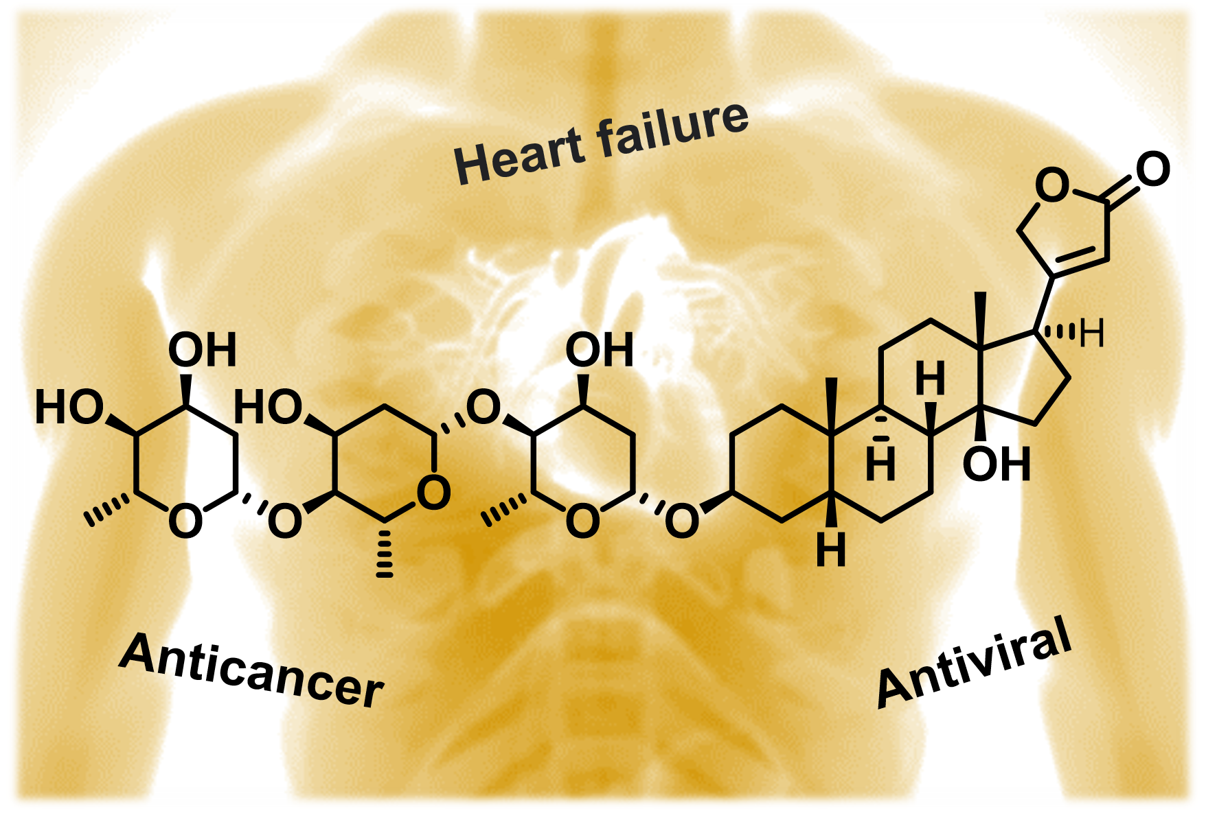
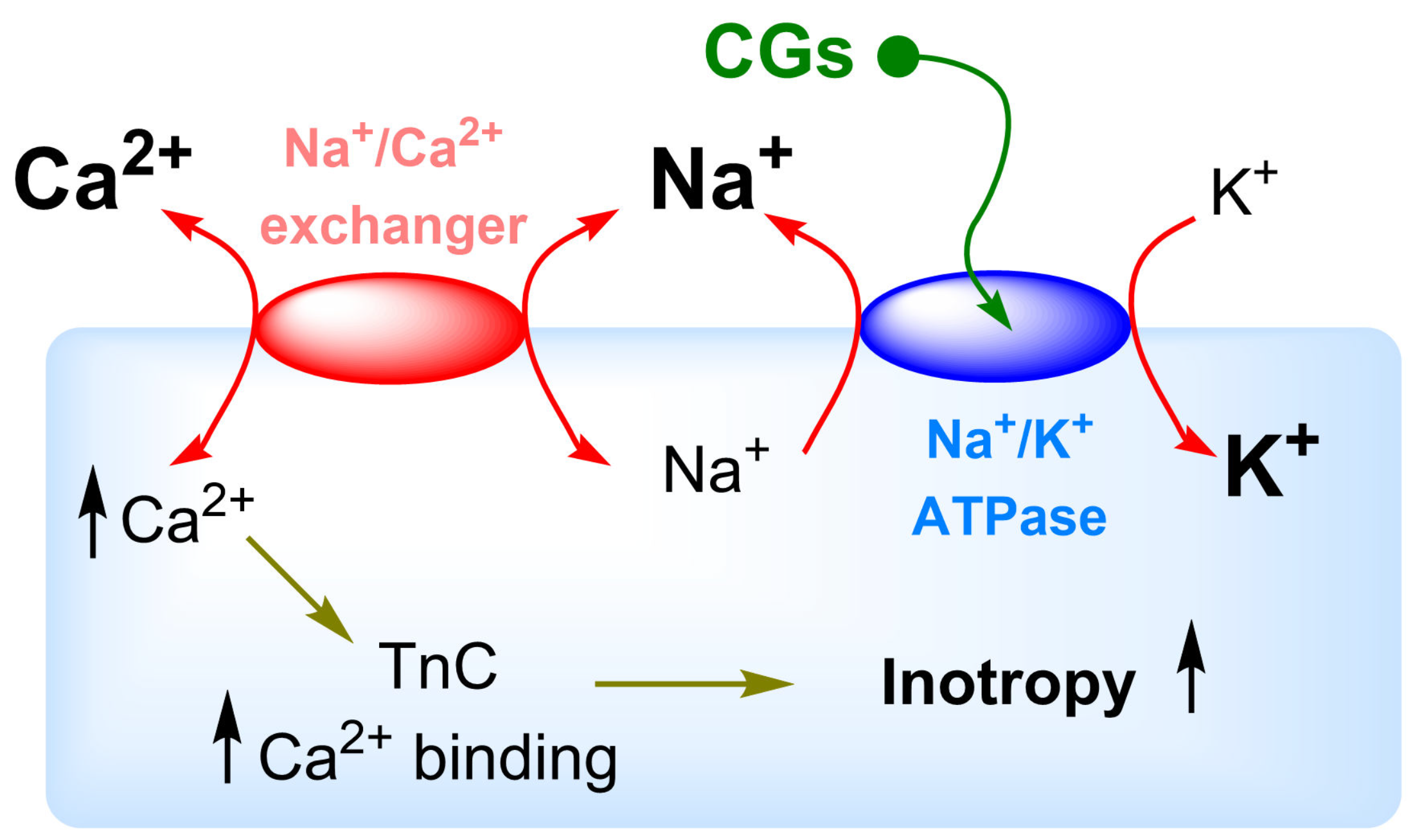
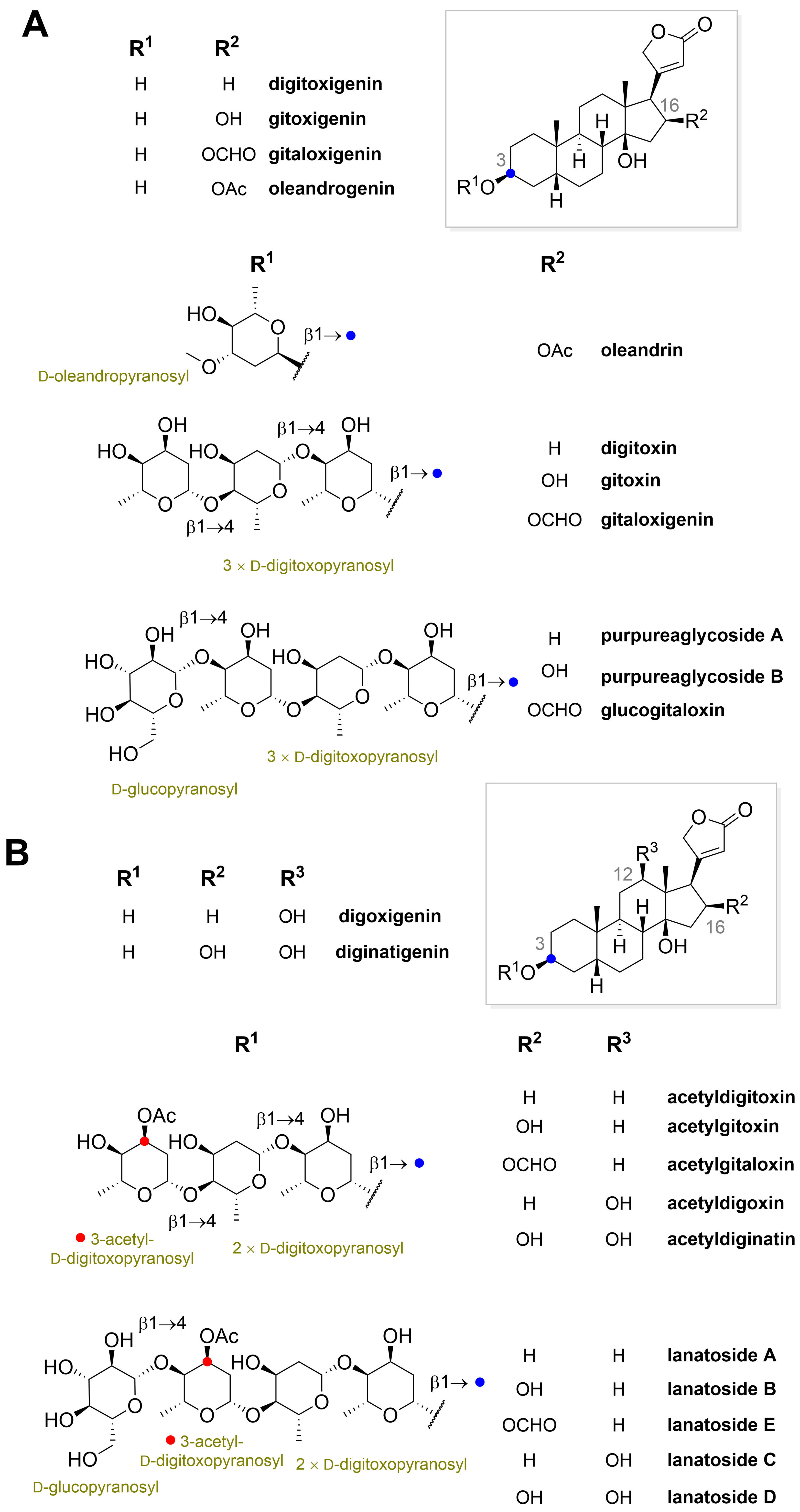
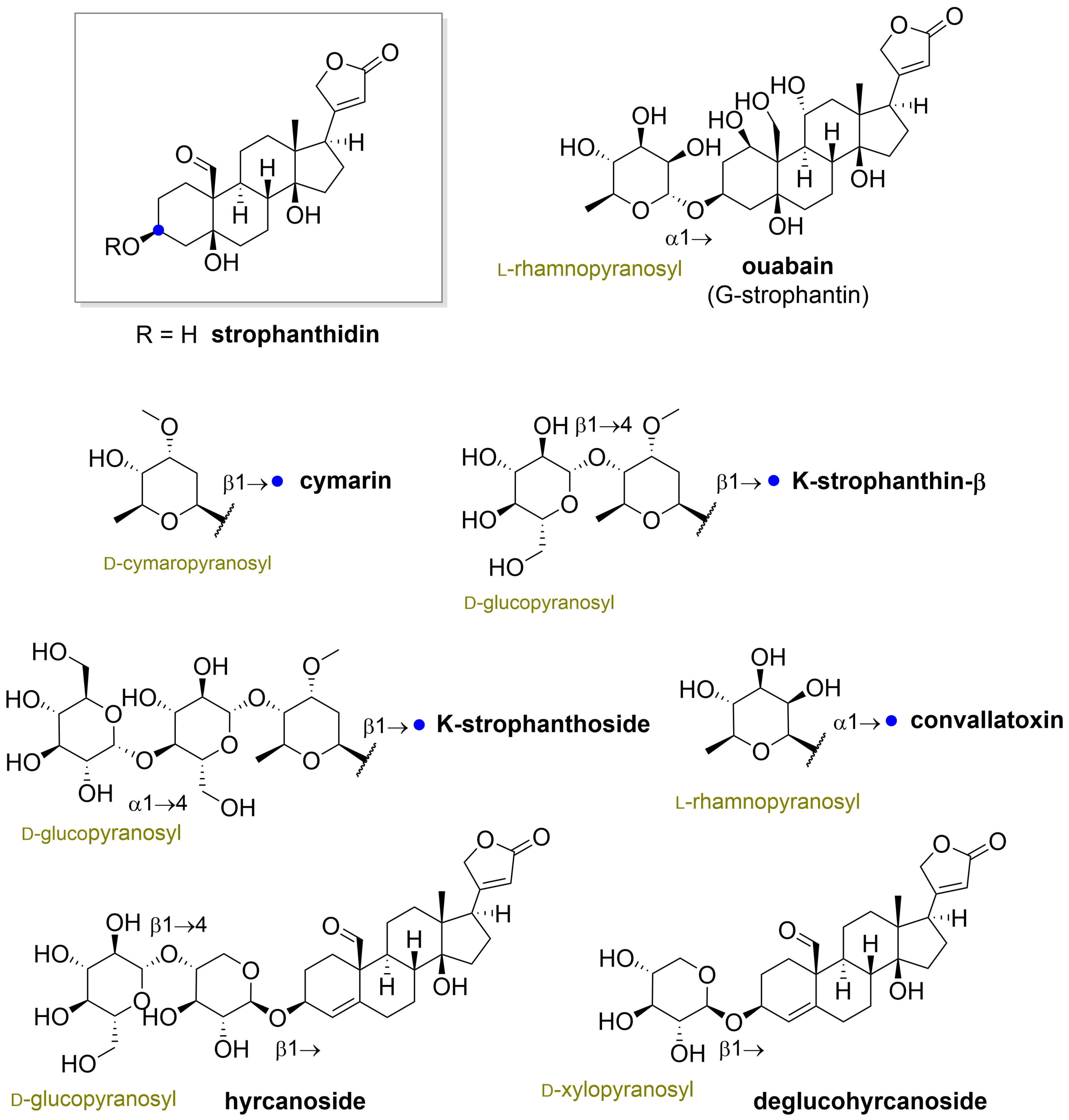
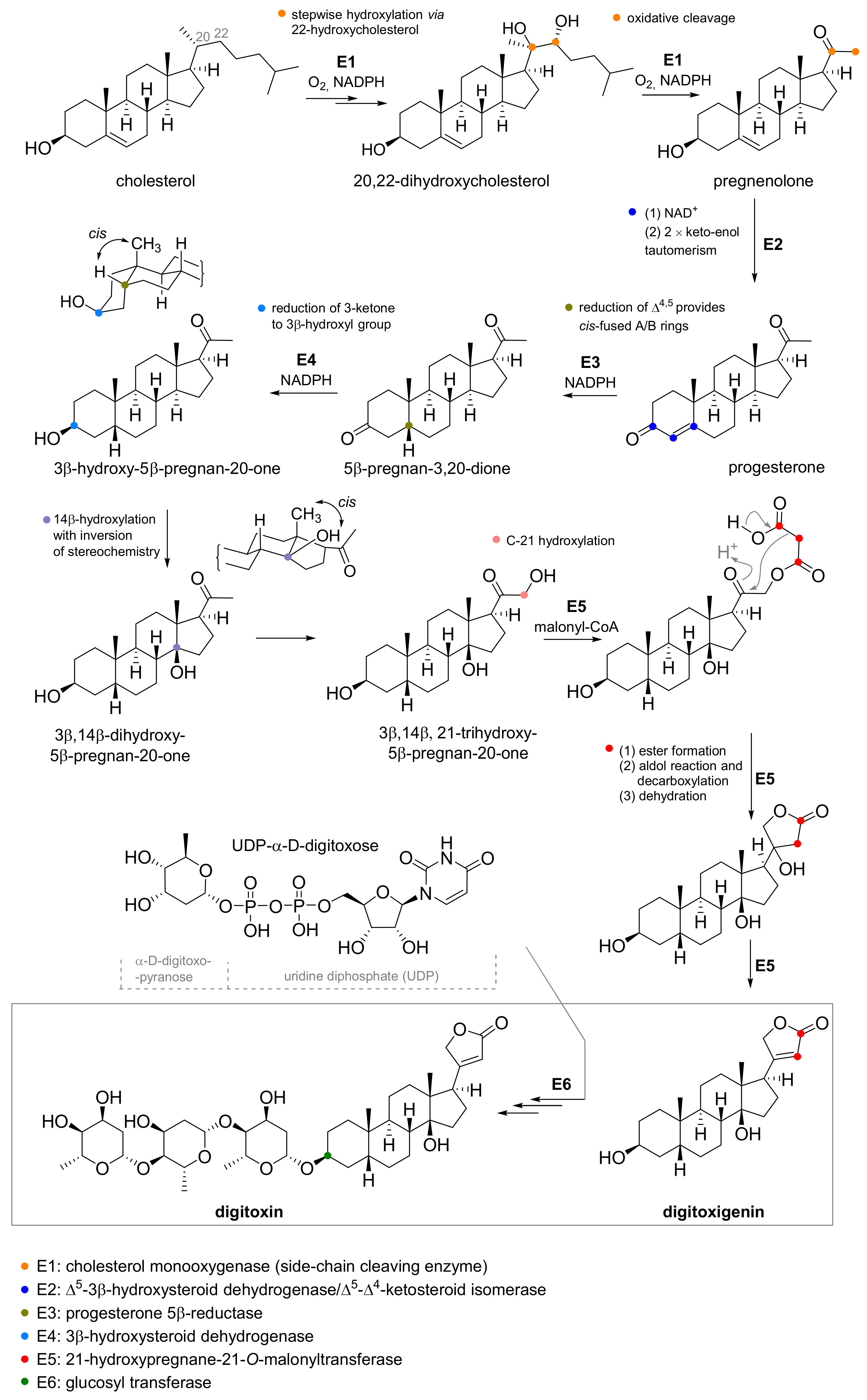
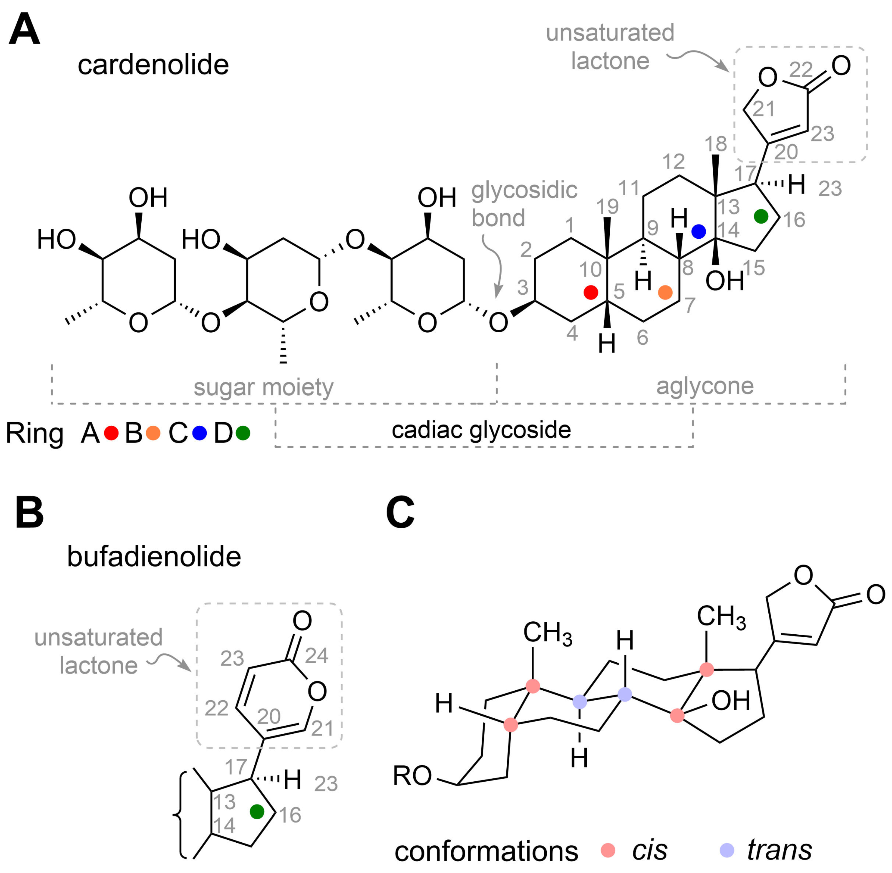
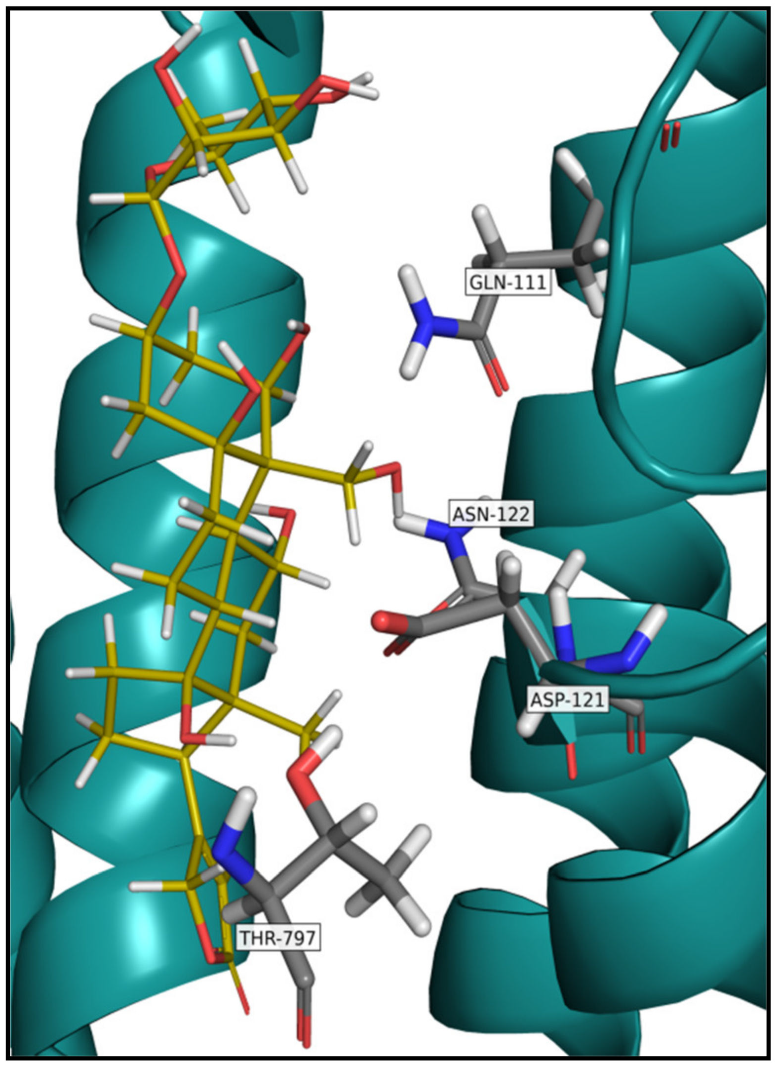
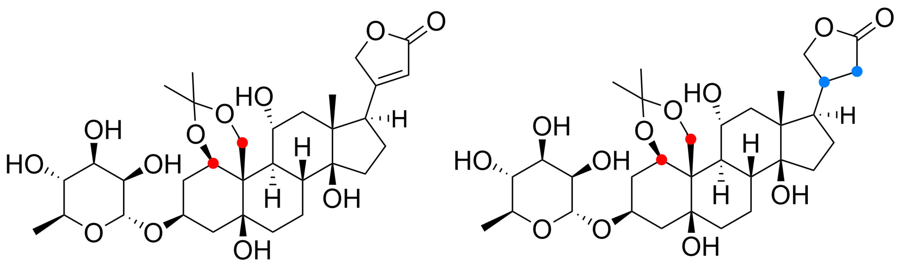
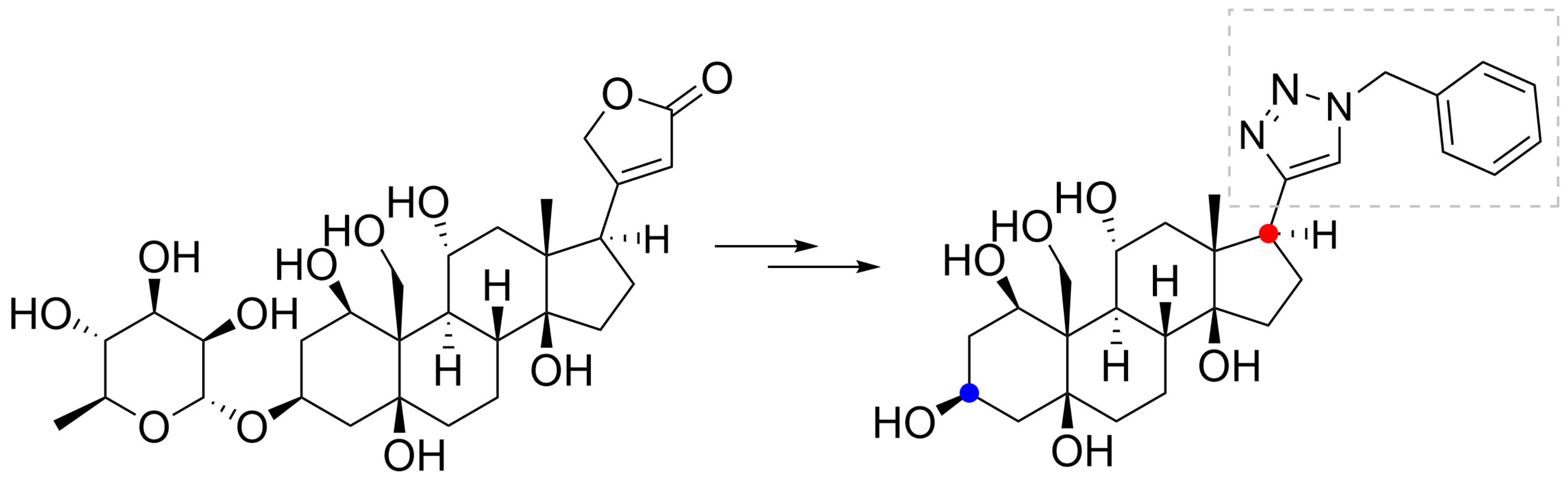
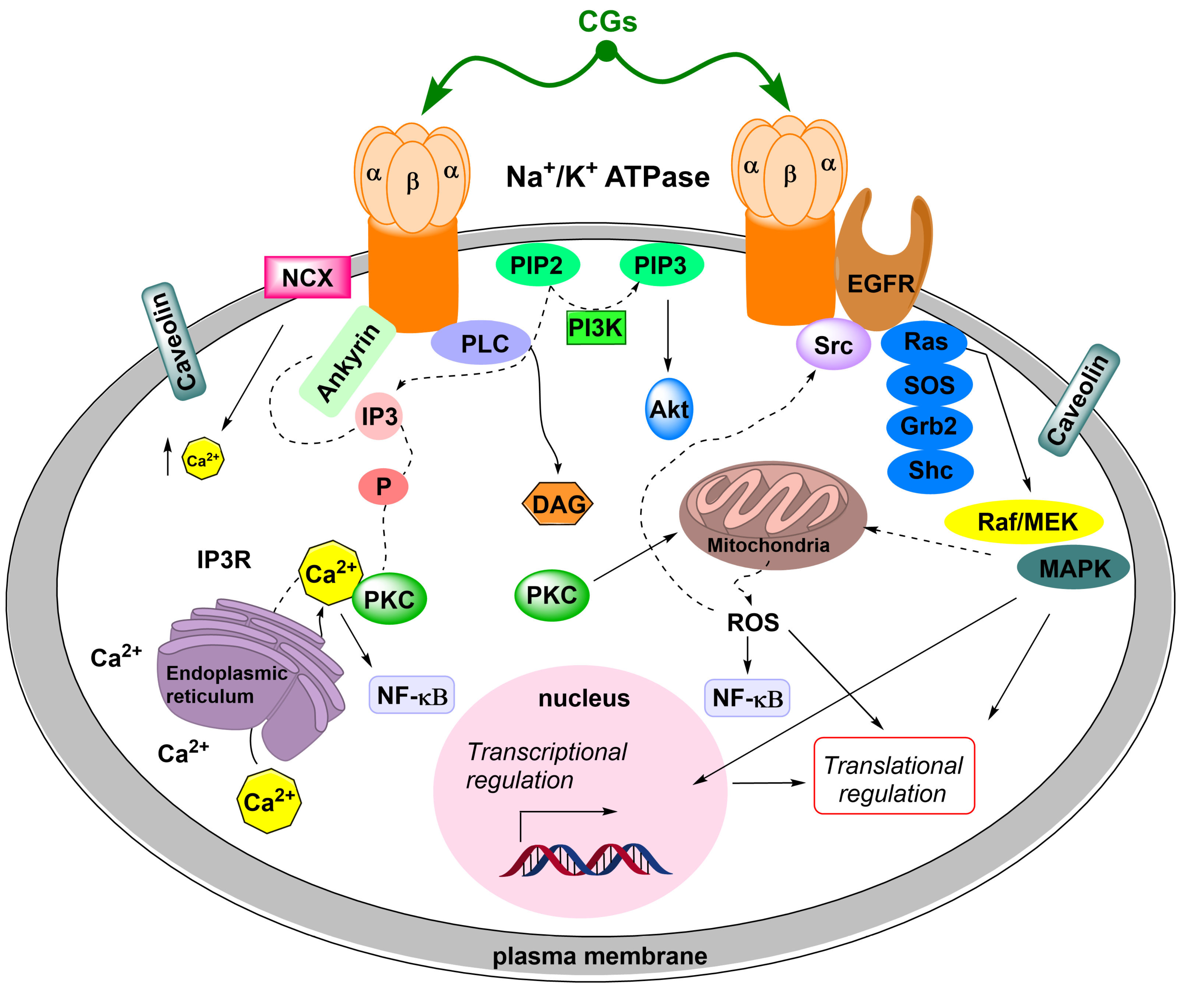
Table 1.
An overview of the most important producers of cardiac glycosides.
| Source | Cardiac Glycoside | Reference |
|---|---|---|
| Strophanthus kombe (Apocynaceae) | K-strophanthoside, cymarin, helveticoside, strophanthidin, erysimoside, k-strophanthin-β, neoglucoerysimoside | [12,13] |
| Strophanthus gratus (Apocynaceae) | G-strophanthin (ouabain) | [14] |
| Digitalis lanata (Scrophulariaceae) | Digoxigenin, deacetyllanatoside C, digoxigenin-bis-digitoxoside, gitoxigenin, digoxin, lanatoside C, digitoxigenin, α-acetyldigoxin, β-acetyldigoxin, lanatoside B, gitoxin, lanatoside A, digitoxin | [15] |
| Digitalis purpurea (Scrophulariaceae) | Digitoxin, digitoxigenin, gitoxin, gitoxigenin, gitaloxin, glucodigitoxin, glucogitoxin, glucogitaloxin | [16,17,18] |
| Nerium oleander (Apocynaceae) | Oleandrin, neritaloside, cardenolide B-1, oleagenin, odoroside H, oleaside A, neriaside | [19,20] |
| Coronilla varia (Fabaceae) | Hyrcanoside, deglucohyrcanoside | [21] |
| Convallaria majalis (Liliaceae) | Convallatoxin, perconval, canariengenin, rhodexin, periplorhamnoside, convallatoxol, peripalloside, strophalloside, strophanolloside, deglucocheirotoxin, lukondjoside, convalloside, deglucocheirotoxol, periguloside, rhodexoside | [22,23,24,25] |
| Apocynum cannabinum (Apocynaceae) | Strophanthidin, cymarin, cynocannoside, helveticoside, apobioside, apocannoside, cannogenol | [26] |
Table 2.
Examples of cardiac glycosides produced by animals.
| Source | Cardiac Glycoside | Reference |
|---|---|---|
| Homo sapiens sapiens | Ouabain (endogenous), marinobufagenin | [31] |
| Canis lupus familiaris | Ouabain (endogenous) | [32] |
| Wistar rats | Ouabain (endogenous) | [33] |
| Rhinella marina | Marinobufagenin | [34] |
| Bufo bufo gargarizans | Bufotoxin | [35] |
Publisher’s Note: MDPI stays neutral with regard to jurisdictional claims in published maps and institutional affiliations. |
© 2021 by the authors. Licensee MDPI, Basel, Switzerland. This article is an open access article distributed under the terms and conditions of the Creative Commons Attribution (CC BY) license (https://creativecommons.org/licenses/by/4.0/).
Share and Cite
MDPI and ACS Style
Bejček, J.; Jurášek, M.; Spiwok, V.; Rimpelová, S. Quo vadis Cardiac Glycoside Research? Toxins 2021, 13, 344. https://0-doi-org.brum.beds.ac.uk/10.3390/toxins13050344
AMA Style
Bejček J, Jurášek M, Spiwok V, Rimpelová S. Quo vadis Cardiac Glycoside Research? Toxins. 2021; 13(5):344. https://0-doi-org.brum.beds.ac.uk/10.3390/toxins13050344
Chicago/Turabian StyleBejček, Jiří, Michal Jurášek, Vojtěch Spiwok, and Silvie Rimpelová. 2021. "Quo vadis Cardiac Glycoside Research?" Toxins 13, no. 5: 344. https://0-doi-org.brum.beds.ac.uk/10.3390/toxins13050344
Note that from the first issue of 2016, this journal uses article numbers instead of page numbers. See further details here.