Article

Occurrence, Serotypes and Virulence Characteristics of Shiga-Toxin-Producing Escherichia coli Isolates from Goats on Communal Rangeland in South Africa

by
Mogaugedi N. Malahlela
1,
Beniamino T. Cenci-Goga
1,2,
Munyaradzi C. Marufu
3,
Thierry Y. Fonkui
1,
Luca Grispoldi
2,
Eric Etter
4,5,6,
Alan Kalake
7 and
Musafiri Karama
1,*
1
Veterinary Public Health Section, Department of Paraclinical Sciences, Faculty of Veterinary Science, University of Pretoria, Onderstepoort 0110, South Africa
2
Departiment of Veterinary Medicine, Laboratorio di Ispezione Degli Alimenti di Origine Animale, University of Perugia, 06126 Perugia, Italy
3
Department of Veterinary Tropical Diseases, Faculty of Veterinary Science, University of Pretoria, Onderstepoort 0110, South Africa
4
Department of Production Animal Studies, Faculty of Veterinary Science, University of Pretoria, Onderstepoort 0110, South Africa
5
Centre de Coopération Internationale en Recherche Agronomique Pour le Développement (CIRAD), Unité Mixte de Recherche (UMR), Animal Santé, Territoires, Risques et Ecosystèmes (ASTRE), 97170 Petit Bourg, France
6
ASTRE, University Montpellier, CIRAD, INRAE, France Baillarguet International Campus, University of Montpellier, 34070 Montpellier, France
7
Gauteng Department of Agriculture and Rural Development, Johannesburg 2001, South Africa
*
Author to whom correspondence should be addressed.
Submission received: 7 April 2022 / Revised: 6 May 2022 / Accepted: 13 May 2022 / Published: 18 May 2022

Abstract

:
Shiga-toxin-producing Escherichia coli is a foodborne pathogen commonly associated with human disease characterized by mild or bloody diarrhea hemorrhagic colitis and hemolytic uremic syndrome. This study investigated the occurrence of STEC in fecal samples of 289 goats in South Africa using microbiological culture and PCR. Furthermore, 628 goat STEC isolates were characterized by serotype (O:H) and major virulence factors by PCR. STEC was found in 80.2% (232/289) of goat fecal samples. Serotyping of 628 STEC isolates revealed 63 distinct serotypes including four of the major top seven STEC serogroups which were detected in 12.1% (35/289) of goats: O157:H7, 2.7% (8/289); O157:H8, 0.3%, (1/289); O157:H29, 0.3% (1/289); O103:H8, 7.6% (22/289); O103:H56, 0.3% (1/289); O26:H2, 0.3% (1/289); O111:H8, 0.3% (1/289) and 59 non-O157 STEC serotypes. Twenty-four of the sixty-three serotypes were previously associated with human disease. Virulence genes were distributed as follows: stx1, 60.6% (381/628); stx2, 72.7% (457/628); eaeA, 22.1% (139/628) and hlyA, 78.0% (490/628). Both stx1 and stx2 were found in 33.4% (210/628) of isolates. In conclusion, goats in South Africa are a reservoir and potential source of diverse STEC serotypes that are potentially virulent for humans. Further molecular characterization will be needed to fully assess the virulence potential of goat STEC isolates and their capacity to cause disease in humans.
Key Contribution: This research is a contribution towards STEC surveillance and improves our understanding of the epidemiology and virulence characteristics of goat STEC in South Africa.

1. Introduction

Shiga-toxin-producing E. coli (STEC) is a foodborne pathogen commonly associated with enteric disease in humans characterized by mild watery or bloody diarrhea hemorrhagic colitis (HC) and the hemolytic uremic syndrome (HUS) as a complication in 5–10% of humans. According to the Foodborne Disease Burden Epidemiology Reference Group (FERG), STEC was responsible for around 2.5 million of new cases of human disease, of which 1.2 million may have been foodborne, with 3330 HUS cases and 269 deaths, which corresponded to 27,000 disability-adjusted life years (DALYs) in 2010 [1].
Domestic ruminants including cattle, sheep, and goats are the main reservoirs of STEC [2,3,4,5]. Ingestion of contaminated meat, dairy products, vegetables, and water is a risk factor for acquiring STEC infection in humans [2,6,7]. Furthermore, contact with animals carrying STEC has also been associated with disease in humans [8,9].
More than 1000 different serotypes of Shiga-toxin-producing Escherichia coli (STEC) have been described in humans, animals and the environment [10,11,12]. STEC O157:H7 was the first serotype to be associated with a human disease outbreak and remains the most frequent strain in human illness [13]. However, numerous non-O157 STEC serotypes have also been linked to outbreaks and severe disease in humans including HUS [10,11,12,14,15]. STEC O26, O45, O103, O111, O121 and O145 are the six most frequently incriminated non-O157 serogroups in human disease [14,15]. Together with STEC O157, these “top 6” non-O157 serogroups are the major seven STEC serogroups, also colloquially termed “Big 7 or Top 7” STEC. Furthermore, according to the Centers for Disease Control and Prevention (CDC), at least 13 serotypes associated with top 6 STEC serogroups including O26:H11 or nonmotile (NM); O45:H2 or NM; O103:H2, H11, H25; or NM; O111:H8 or NM; O121:H19 or H7; and O145: NM may be responsible for up to 80% of cases of human STEC disease in the United States [14]. In South Africa, serotypes O26:H11, O111:H8, O157:H7 and O107/O117:H7 were frequently implicated in disease between 2006–2013 [16].
Bacteriophage-encoded Shiga toxins (stx1 and stx2) and a number of stx subtypes are considered the major STEC virulence factors [17,18,19,20]. Identification of STEC is based on detection of one or more Shiga-toxin-encoding genes (stx1 and stx2) [17,19,20]. STEC that carry stx2 are more frequently associated with severe disease including HUS in comparison to strains that possess stx2 alone or both stx1 and stx2 concomitantly [21,22,23,24,25].
Intimin (eaeA) is an additional important STEC virulence factor [26,27]. The gene encoding intimin is located on a 35 Kb pathogenicity island in E. coli O157:H7 termed the locus of enterocyte effacement (LEE) [28,29]. Intimin is responsible for intimate adherence of STEC to intestinal epithelial cells and formation of typical attaching and effacing (A/E) lesions in the intestine characterized by actin-rich pedestals and loss of brush border microvilli under bound bacteria [30]. Furthermore, STEC possess plasmid-encoded virulence markers including a hemolysin (hlyA) and additional virulence-associated genes which are located on pathogenicity-islands [31,32,33,34,35].
Current reports on the occurrence and characteristics of STEC in animals and humans in South Africa are scanty. Furthermore, the few studies that have reported on the occurrence of STEC in South Africa have largely investigated the presence of STEC in cattle populations [36,37,38] while studies on the prevalence of STEC in other ruminants including goats are lacking. Therefore, the main objectives of this study were (1) to determine the occurrence of STEC in goats raised on communal rangeland in South Africa and (2) characterize STEC by serotype (O:H) and major virulence factors (stx1, stx2, eaeA, and hlyA). The overall goal is to contribute to STEC monitoring and surveillance in South Africa.

2. Results

2.1. STEC Occurrence

A total of 289 fecal samples were collected from four goat herds on communal rangeland (herd A, B, C, and D) in the Gauteng province of South Africa and screened for STEC. PCR revealed that 80.2% (232/289) of goat fecal samples were positive for STEC. STEC was detected in 75.3% (116/154) of goats in herd A; 90.6% (39/43) in herd B; 78.8% (41/52) in herd C and 90% (36/40) in herd D (Table 1 and Figure 1).

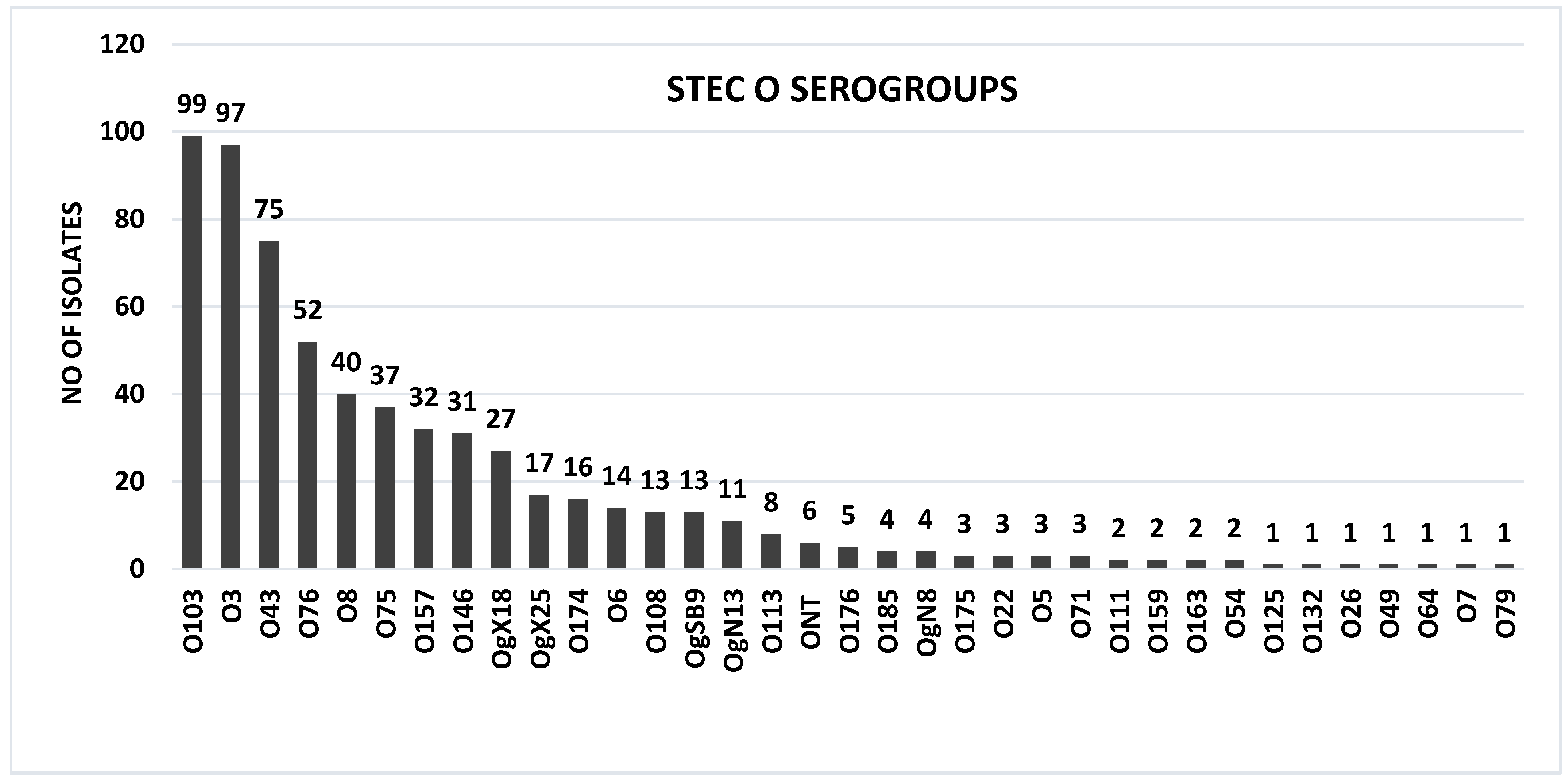
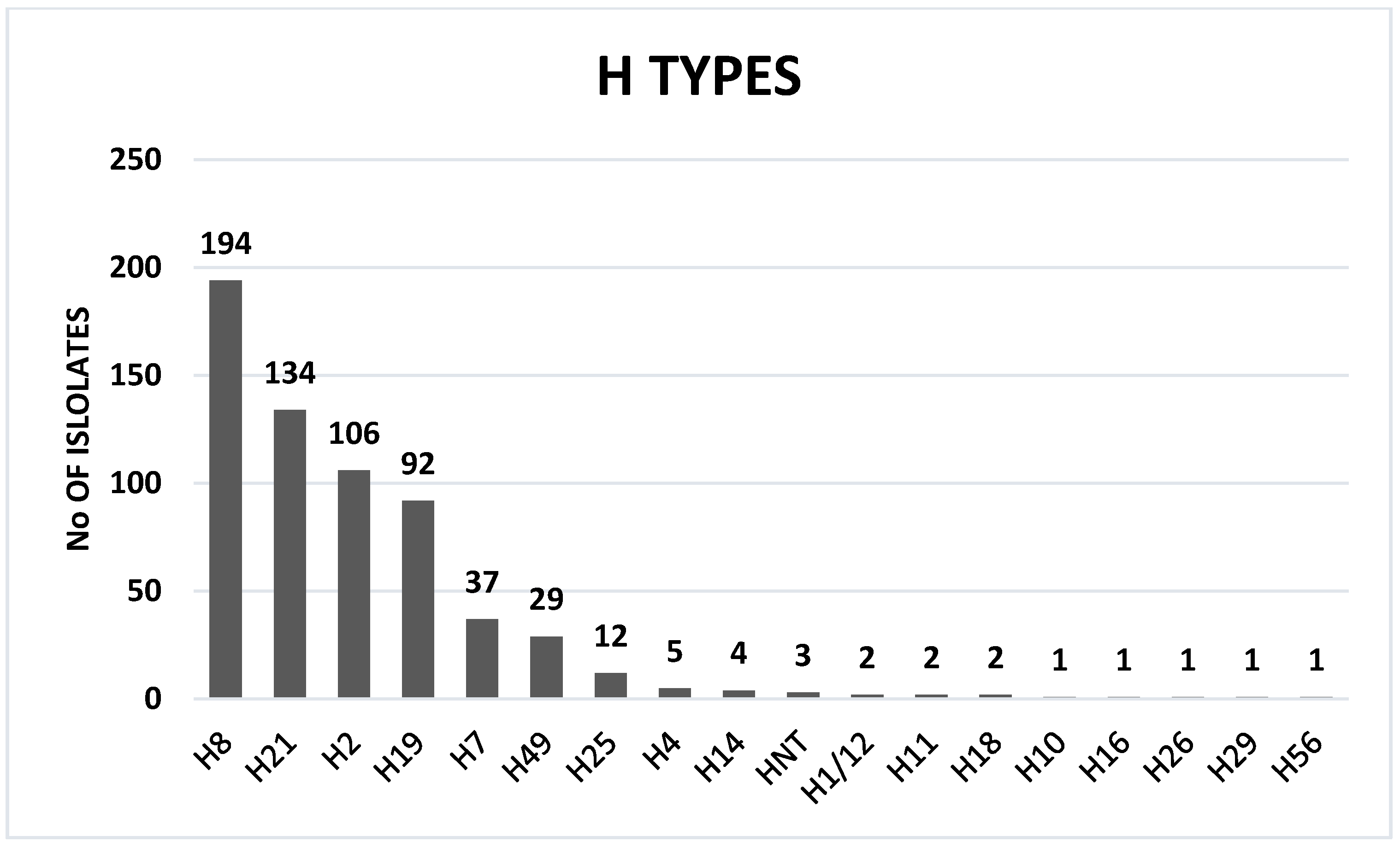
2.2. STEC O:H Serotypes

At least 99% (622/628) of isolates were O:H serotypeable by PCR. PCR serogrouping revealed 34 O groups and 17 H types with a total of 63 O:H distinct serotypes. Six isolates were O-untypable (ONT) (Figure 2 and Table 1) and three H-untypable (HNT) (Figure 3 and Table 1). The highest number of STEC serotypes was recovered from herd A—41 serotypes; followed by herd B—21 serotypes; herd C—10 serotypes; and herd D—8 serotypes. STEC O76:H19 was recovered from all herds: STEC O146:H21 and OgX18:H2 were recovered from herd A, C, and D. STEC O8:H14, OgSB9:H19 and OgX25:H8 were found in herd A and B. STEC O43:H2, O43:H8, O113:H8 and O157:H7 were recovered from herd A and C. STEC O3:H21 and O8:H19 were recovered from herd A and D.
Among the 34 O serogroups, 18 were associated with a single H type (O5:H19, O7:H7, O22:H8, O26:H2, O49:H11, O64:H18, O75:H8, O79:H8, O111:H8, O113:H8, O125:H19, O132:H8, O146:H21, O174:H8, O176:H4, O185:H8, ON8:H7, OX25:H8), and 16 O groups (O3, O6, O8, O43, O54, O71, O76, O103, O108, O157, O159, O163, O175, ON13, OSB9 and OX18) were associated with more than one H type (Supplementary Table S1). The following 17 H types were detected: H1/12, H2, H4, H7, H8, H10, H11, H14, H16, H18, H19, H21, H25, H26, H29, H49 and H56 (Figure 3). The distribution of different STEC serotypes per goat herd is shown in Table 1.
Among the 63 O:H distinct serotypes, 55.5%, (35/63) were each represented by a single isolate while the remaining 44.4% (28/63) were represented by more than one isolate (Table 1). O:H serotype combinations can be found in Supplementary Table S1.
The six most frequent goat STEC serotypes were: O3:H21, 7.9% (23/289); O103:H8, 7.6% (22/289); O43:H2, 6.5% (19/289); O76:H19, 5.8% (17/289); O75:H8, 3.1%, (9/289); O157:H7, 2.7% (8/289). Big seven STEC serotypes were recovered from 11.0% (32/289) of goats. Big seven STEC serotypes were distributed as follows among goats: O157:H7, 2.7% (8/289); O103:H8, 7.6% (22/289); O26:H2, 0.3% (1/289); O111:H8, 0.3% (1/289); O103:H56, 0.3% (1/289); O157:H8, 0.3% (1/289); O157:H29, 0.3% (1/289).

2.3. STEC Virulence Characteristics

The distribution of four STEC virulence genes among the 628 STEC isolates was as follows: stx1, 60.6% (381/628); stx2, 72.7% (457/628); eaeA, 22.1% (139/628); hlyA, 78.0% (490/628). Both stx1 and stx2 were found concomitantly in 33.4% (210/628) of isolates (Supplementary Table S2). The following major gene combinations were observed: stx1 stx2 hlyA, 26.9%, (169/628); stx2 eaeA hlyA 20.3%, (128/628) and stx1 hlyA 20.0%, (126/628). STEC characteristics are depicted in Supplementary Table S2.
The eaeA gene was observed in 22.1%, (139/628) of isolates which corresponded to 12.8% (37/289) of goats which were eaeA positive. Of the 139 isolates that were eaeA positive, 131 belonged to five of the major seven serogroups including O157:H7 (30/628 isolates, 8/289 goats), O157:H8 (1/628 isolates, 1/289 goats), O103:H8 (97/628 isolates, 22/289 goats), O26:H2 (1/628 isolate, 1/289 goats) and O111:H8 (2/628 isolates, 1/289 goats). In addition, eight isolates (1.2%) which were non-Big seven STEC serotypes possessed eaeA: O71:H14 (1/628 isolate, 1/289 goats), O108:H25 (6/628 isolates, 2/289 goats), O163:H8 (1/628 isolate, 1/289 goats) were also eaeA-positive (1.2%, 8/628). Most of the eaeA positive isolates, 92.0% (128/139) had the stx2eaeA genotype (O71:H14, O103:H8, 157:H7, O157:H8) while the remaining 7.9% (11/139) isolates (O26:H2, O163:H8, O103:H8, O108:H25 and O111:H8) were stx1eaeA positive.

3. Discussion

Previous reports from different countries have shown that goats are a reservoir of STEC [4,39,40,41,42]. Furthermore, contact with goats and food products of goat origin have been associated with STEC disease in humans [4,43]. However, published reports on the occurrence and characteristics of STEC in goats are few in comparison to cattle and sheep. Furthermore, reports on the occurrence of STEC in goats in South Africa are non-existent. This study investigated the occurrence of STEC and characterized STEC isolates in four separate goat herds in South Africa. The overall occurrence of STEC in the goat populations surveyed was 80.2% (232/289). The occurrence of STEC in this study was very high in comparison to similar studies in Germany [42,44], Brazil (57.5%) [40], Spain (47.7%) [39,45], Vietnam (31.5%) [41] and Bangladesh (11.8%) [46] which reported STEC detection rates ranging from 11.8% to 75.3% in goats. Other reports have found STEC occurrence rates ranging from 23.9% to 89.3% in different countries, but these studies were conducted on far smaller goat sample populations (≤46) to warrant a valid comparison with the present study [47,48,49,50].
The within-herd occurrence of STEC ranged from 75.3% (116/154) to 90.6% (39/43) which was significantly higher in comparison to similar studies in Brazil (46.7–73.3%) [40] and Vietnam (15–65%) [41]. Moreover, all the four goat herds were positive for STEC, in agreement with similar reports elsewhere [40,41,42]. However, the number of goat samples which were tested per herd in this study was “significantly” higher compared to the reports from Brazil (106), Vietnam (205) and Germany (93) which may explain why the within-herd STEC occurrence in this study was also higher. The higher occurrence of STEC in goats in this study may be ascribed to higher shedding of STEC in the goat population studied, variations in geographic locations, age (kids vs. adults), goat diet (grazing or browsing vs. concentrate), and management practices. Furthermore, the use of a suitable enrichment broth and two selective and sensitive STEC culture and isolation media may have increased STEC recovery [51,52,53,54,55,56].
In the present study, 99.0% of goat STEC isolates were serotypeable by PCR. A total of 63 serotypes (34 O and 17 H groups) were recovered from goats. The number of serotypes detected in this study was very high compared to previous studies [39,40,45]. The recovery of a very high number of serotypes may also be ascribed to the high shedding of STEC in the goat populations tested. Furthermore, the use of a sensitive, specific, accurate and reliable PCR protocol for O:H serotyping may have led to the identification of more serotypes than usually found with traditional serotyping [57,58,59]. Furthermore, PCR O:H serotyping has the advantage of detecting O-untypable (OUNT) and H-nontypeable and/or non-motile (HNT/NM) E. coli isolates that carry genes encoding O:H antigens but cannot be expressed. In this study, we were able to validate the Iguchi et al. [58,59] and Banjo et al. [57] E. coli PCR serotyping (O:H) protocols which were highly discriminatory and unambiguously serotyped the large number of goats STEC isolates tested in this study [57,58,59]. To our knowledge, this is the most extensive serotyping of goat STEC isolates, worldwide.
Among the 63 serotypes, only 4 serotypes belonged to the major 7 STEC serogroups. STEC O103:H8 (15.6%) was the most frequent Big seven STEC among goats, followed by STEC O157:H7 (4.7%), O111:H8 and STEC O26:H2. Overall, the major seven STEC serotypes accounted for 21.3% of all isolates which were serotyped, in contrast to most similar studies which never recovered major seven STEC from goats [39,40,43,44,45,60,61]. However, Schilling et al. [48] found a higher proportion of top seven STEC, although the recovered serotypes were those which have never been reported in human disease, in contrast to our results which showed that most of the top seven serotypes we recovered were previously incriminated in human disease outbreaks except for STEC O157:H29, O103:H8 and O103:H56.
Previously, STEC O157:H7 has been incriminated in foodborne disease after consumption of raw goat milk and home-made cheese made from raw milk [43,62,63]. Furthermore, STEC O157 and STEC O103 have been incriminated in human disease after contact with goats in the USA [8] while sources other than goats have frequently associated STEC O157:H7, O111:H8 and O26:H2 to human disease worldwide including South Africa [16]. According to the STEC seropathotype classification, STEC O157:H7 is considered a seropathotype A strain, frequently incriminated in outbreaks and severe human disease while O111:H8 and O26:H2 are moderately implicated in outbreaks and less frequent in severe human disease, in comparison to STEC O157:H7 [32]. However, in this study most major seven STEC isolates were classified as STEC O103:H8. Previously, STEC O103:H8 was isolated from healthy goats and calves in China and Argentina, respectively [64,65]. In addition, only one study has reported the recovery of STEC O103:H8 from patients and asymptomatic food handlers in Japan [66]. However, this study never specified whether the STEC O103:H8 isolate was from patients or asymptomatic food handlers [66]. Therefore, although O103:H8 is classified as a major STEC (serogroup), its importance as a human pathogen remains unclear as there are no reports until now which have unequivocally associated this STEC serotype with human disease.
The remaining 59 serotypes were non-O157, of which 24 have been previously incriminated in mild to severe human disease worldwide including South Africa, Europe, North America and Asia [10,11,12,16,67]. The recovery of STEC serotypes which have been associated with mild to severe human disease is evidence that goats are a reservoir and a potential source of these highly pathogenic STEC strains in South Africa.
Highly diverse and farm specific STEC serotypes were observed in individual goat herds except for STEC O76:H19 which was the serotype shared among the four goat herds surveyed while STEC O146:H21 and OgX18:H2 were recorded in three herds. Overall, the highly diverse and farm specific serotypes are most likely a reflection of the fact that the four herds were situated in geographically separate and distant areas from each other to allow isolate interchange between herds.
Regarding the virulence characteristics of the STEC isolates under study, stx2 was more frequent that stx1 among goat STEC in contrast to similar studies which have shown that stx1 is predominant among goat STEC isolates [39,41,42,45,50,68,69,70,71]. However, our findings agree with a study by Oliveira et al. [40] which reported that stx2 was more prevalent in goat STEC isolates. Reports on clinical STEC have suggested that stx2-positive isolates are more virulent and frequently incriminated in severe human disease including hemorrhagic colitis and hemolytic uremic syndrome in comparison to STEC isolates carrying stx1 or both stx1 and stx2 [21,22,23,24,25].
The hlyA gene was present in 78.0% (490/628) of goat STEC isolates, consistent with previous reports which have shown similar rates in goat STEC elsewhere [39,42,44]. However, lower rates of hlyA ranging from 35–60.9% have also been reported [40,41,45]. The hlyA gene encodes a pore-forming hemolysin which lyses human erythrocytes with subsequent release of iron from heme, a chemical needed for STEC growth and survival in the intestine. Previously, the presence and expression of hlyA has been associated with severe STEC disease in humans including HC and HUS [35]. However, STEC that were hlyA-negative have also been incriminated in severe disease including bloody diarrhea, HC and HUS, thereby suggesting that the pathogenic role of hlyA in STEC remains uncertain [23].
Most of the goat STEC were eaeA-negative except for the top seven STECs (22.1%) including O157:H7, O26:H2, O111:H8 and O103:H8 and a few (0.7%) non-O157/non-top seven isolates: O71:H14, O108:H25 and O163:H8, in agreement with previous studies which have shown that eaeA is not common among goat STEC [40,42,45]. The presence of eaeA in goat top seven STEC isolates is of clinical significance as eaeA is considered an important STEC adhesin and marker of high virulence and potential to cause severe disease (HC and HUS) in humans [72], especially when accompanied with stx2 [23]. However, in some cases, eaeA-negative serotypes (O91:H21 and O113:H21) STEC have also been associated with severe disease thereby suggesting that other virulence or unknown host factors may influence disease severity [72,73,74]. The absence of eaeA may indicate that goat STEC are less virulent and may also explain why goat STEC are rarely incriminated in human disease worldwide. Of particular interest were eaeA-positive goat isolates which belonged to serotypes O103:H8, O71:H14, O108:H25 and O163:H8 but have never been associated with human disease or outbreaks. These isolates will be worth monitoring closely as possession of eaeA may be indicative of higher virulence potential and likelihood to cause severe disease in humans.

4. Conclusions

Historically, studies on the presence of STEC in goats are very few compared to cattle which are considered the main STEC reservoir. This study is the first report on the presence of STEC in goats in South Africa. The findings of this study show that goats carry a diverse range of STEC serotypes, some of which have been previously incriminated in mild to severe enteric disease in humans. Collectively, these findings suggest that goats grazing on communal rangeland in South Africa are a reservoir and potential source of STEC for humans in South Africa. Further molecular characterization of goat STEC isolates will be needed in the future to fully assess the virulence potential of goat STEC and capacity to cause disease in humans. In addition, studies that compare STEC isolates from goats and humans will be necessary to fully understand the role played by goats as a source of STEC human disease in South Africa. Data from this study will be useful for understanding the epidemiology of STEC in animals and formulating policies aimed at preventing and controlling zoonotic or foodborne diseases along the food chain.

5. Materials and Methods

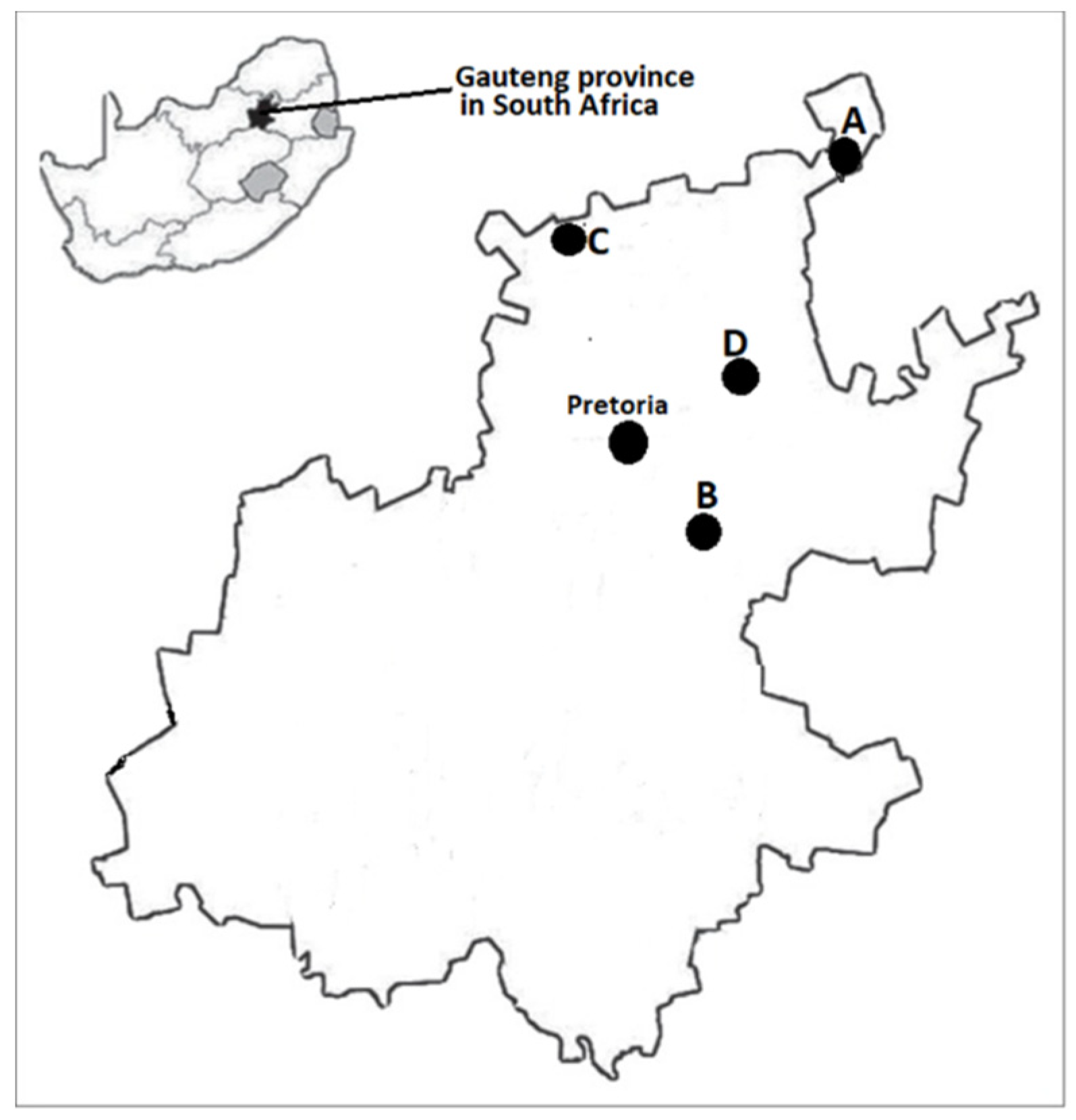
5.1. Study Population and Sample Collection

Goat fecal samples (N = 289) were obtained from four goat herds. The goat herds were located on different communal rangelands in Gauteng province, South Africa. The herds were designated using alphabetical letters: herd A (n = 154), herd B (n = 43), herd C (n = 52) and herd D (n = 40). Each herd was visited once. Refer to Figure 4 for a map of the Gauteng province, South Africa showing the locations of the different herds (A, B, C and D) from which goat fecal samples were obtained. Fresh fecal samples were collected by rectal palpation, using a new nitrile examination glove per animal. Samples were placed in sterile specimen containers and transported in a cooler box on ice to the laboratory where they were stored at 4 °C until further processing. Ethical clearance for conducting this research was obtained from the Research Ethics and Animal Ethics Committees of the Faculty of Veterinary Science, University of Pretoria, under approval number REC110-21.

5.2. STEC Culture

Each fecal sample (5 g) was enriched at a 1:10 ratio in EC broth (CM0990, Oxoid, Basingstoke, UK) supplemented with Novobiocin (N1628, Sigma-Aldrich, St. Louis, MO, USA) at 37 °C for 18–24 h. A 100 µL aliquot of the overnight enrichment was spread on Drigalski Lactose agar (CM0531, Oxoid, Basingstoke, UK) and CHROMagar STEC base ST162(B) containing supplement ST162(S) (CHROMagar, Paris, France, http://www.chromagar.com, accessed on 6 April 2022).

5.3. DNA Extraction and STEC Screening

All Drigalski Lactose and CHROMagar STEC agar Petri dishes showing bacterial growth were screened for STEC by PCR [75]. Briefly, a loopful of bacterial colony sweep was collected from each Drigalski Lactose agar and CHROMagar STEC plate showing growth and suspended in 1 mL of FA Buffer (223143, Becton Dickinson and Company, Sparks, MD, USA) [38]. The suspension was homogenised and washed by vortexing, then centrifuged for 5 min. After centrifugation, the supernatant was discarded, and the pellet was re-suspended in FA buffer. After the second wash and centrifugation rounds, the pellet was re-suspended in 500 µL of sterile water, mixed and boiled at 100 °C for 25 min. The boiled preparation was thawed on ice and stored at −20 °C for further processing [38]. A multiplex PCR (mPCR) protocol was used to screen the DNA template for stx1, stx2, eaeA and hlyA using previously described cycling parameters and primers [75]. Briefly, each 25 µL PCR reaction mixture contained 2.5 μL of 10X Thermopol reaction buffer, 2.0 μL of 2.5 mM dNTPs (deoxynucleotide triphosphates), 0.25 μL of 100 mM MgCl2, 0.6 μL of each primer (10 µM final concentration), 1 U of Taq DNA Polymerase and 5 μL of DNA template. The DNA from Escherichia coli O157:H7 strain EDL933 (ATCC 43895) and sterile water were used as positive and negative PCR controls, respectively. All PCR reagents were purchased from New England BioLabs (NEB, Ipswich, MA, USA) except for the primers which were supplied by Inqaba Biotec (Pretoria, South Africa).

5.4. STEC Isolation and Identification

For STEC isolation and identification, colony sweeps were collected from Drigalski Lactose agar and CHROMagar plates which were positive for stx1 and/or stx2 on PCR and streaked onto Drigalski Lactose agar and CHROMagar STEC to obtain single colonies. Five single colonies were purified from each plate and multiplied individually on Luria Bertani agar (REF244520, Becton and Dickinson & Company, Sparks, MD, USA). Once again, DNA was extracted from purified colonies by the boiling method [38]. DNA from each purified colony was screened for stx1, stx2, eaeA and hlyA by PCR [75] to verify and confirm the STEC status of each pure colony. Colonies which were positive for stx1 and/or stx2 were preserved at −80 °C in a bacterial freezing mixture [38] for further O:H serotyping.

5.5. STEC Serotyping

All confirmed STEC pure single colonies were serotyped (O:H) by PCR using previously described primers and cycling conditions [57,58,59]. STEC strains which were previously serotyped by traditional serotyping at the National Microbiology Laboratory, Public Health Agency of Canada, Guelph, Ontario, Canada, and the Laboratorio de Referencia de Escherichia coli (LREC), Facultad de Veterinaria, Universidad de Santiago de Compostela, Lugo, Spain and a number of E. coli O:H types in our collection (unpublished) were also used as positive controls in PCR serotyping assays. Furthermore, the following STEC isolates which were provided by the European Union Reference Laboratory for Escherichia coli, Istituto Superiore di Sanità, Rome Italy, were used as positive controls for serotyping the major seven STEC serogroups: STEC-C210-03 (O157), STEC-ED476 (STEC O111), STEC-C1178-04 (STEC O145), STEC-C125-06 (STEC O103) and STEC-ED745 (O26).

Supplementary Materials

The following supporting information can be downloaded at: https://0-www-mdpi-com.brum.beds.ac.uk/article/10.3390/toxins14050353/s1, Table S1: Association between O group and H-type(s) among goat STEC Isolates; Table S2: Goat STEC major virulence factors and gene combinations.

Author Contributions

Conceptualization, M.K., M.N.M. and A.K.; data curation, M.K, M.N.M. and T.Y.F.; formal analysis, M.K., M.N.M. and T.Y.F.; funding acquisition, M.K., B.T.C.-G., E.E., L.G. and A.K.; investigation, M.K., M.N.M., M.C.M. and T.Y.F.; methodology, B.T.C.-G., M.N.M., T.Y.F. and. L.G.; resources, M.K., B.T.C.-G. and L.G.; Project administration, M.K.; supervision, M.K. and M.C.M. writing—original draft, M.K. and M.N.M. Writing—review and editing M.K., M.N.M. and M.C.M. All authors have read and agreed to the published version of the manuscript.

Funding

This research was funded by the National Research Foundation (NRF) of South Africa (CSRP170528234222 and SARCHI COP Grant 120317) the South African Medical Research Council Self-Initiated Research (MRC/SIR 2017-2019), and UNICEF Future Africa-UP One Health for Change research grants-2021.

Institutional Review Board Statement

The animal study protocol was approved by the Research and Animal Ethics Committees of the Faculty of Veterinary Science, University of Pretoria (REC110-21 on 1 December 2021) for studies involving animals.

Data Availability Statement

Not applicable.

Acknowledgments

We would like to thank Ali Makgato for collecting the goat samples. This manuscript is part of a dissertation submitted (Mogaugedi N. Malahlela) in the Veterinary Public Health section, Department of Paraclinical Sciences, University of Pretoria, in partial fulfilment of the requirements for the degree of Master of Science (Veterinary Science).

Conflicts of Interest

The authors declare no conflict of interest.

References

  1. Kirk, M.D.; Pires, S.M.; Black, R.E.; Caipo, M.; Crump, J.A.; Devleesschauwer, B.; Döpfer, D.; Fazil, A.; Fischer-Walker, C.L.; Hald, T. World Health Organization estimates of the global and regional disease burden of 22 foodborne bacterial, protozoal, and viral diseases, 2010: A data synthesis. PLoS Med. 2015, 12, e1001921. [Google Scholar]
  2. Hussein, H.S. Prevalence and pathogenicity of Shiga toxin-producing Escherichia coli in beef cattle and their products. J. Anim. Sci. 2007, 85, E63–E72. [Google Scholar] [CrossRef] [PubMed] [Green Version]
  3. Persad, A.K.; Lejeune, J.T. Animal Reservoirs of Shiga Toxin-Producing Escherichia coli. Enterohemorrhagic Escherichia coli and Other Shiga Toxin-Producing E. coli.; American Society of Microbiology: Washington, DC, USA, 2015; pp. 211–230. [Google Scholar]
  4. La Ragione, R.M.; Best, A.; Woodward, M.J.; Wales, A.D. Escherichia coli O157:H7 colonization in small domestic ruminants. FEMS Microbiol. Rev. 2009, 33, 394–410. [Google Scholar] [CrossRef] [PubMed] [Green Version]
  5. McCarthy, S.C.; Burgess, C.M.; Fanning, S.; Duffy, G. An Overview of Shiga-toxin producing Escherichia coli carriage and prevalence in the ovine meat production chain. Foodborne Pathog. Dis. 2021, 18, 147–168. [Google Scholar] [CrossRef] [PubMed]
  6. Isaäcson, M.; Canter, P.; Effler, P.; Arntzen, L.; Bomans, P.; Heenan, R. Haemorrhagic colitis epidemic in Africa. Lancet 1993, 341, 961. [Google Scholar] [CrossRef]
  7. Lynch, M.F.; Tauxe, R.V.; Hedberg, C.W. The growing burden of foodborne outbreaks due to contaminated fresh produce: Risks and opportunities. Epidemiol. Infect. 2009, 137, 307–315. [Google Scholar] [CrossRef]
  8. Nichols, M.C.; Gacek, P.; Phan, Q.; Gambino-Shirley, K.J.; Gollarza, L.M.; Schroeder, M.N.; Mercante, A.; Mullins, J.; Blackstock, A.; Laughlin, M.E.; et al. Agritourism and kidding season: A large outbreak of human Shiga toxin-producing Escherichia coli O157 (STEC O157) infections linked to a goat dairy farm—Connecticut, 2016. Front. Vet. Sci. 2021, 8. [Google Scholar] [CrossRef]
  9. Schlager, S.; Lepuschitz, S.; Ruppitsch, W.; Ableitner, O.; Pietzka, A.; Neubauer, S.; Stoeger, A.; Lassnig, H.; Mikula, C.; Springer, B. Petting zoos as sources of Shiga toxin-producing Escherichia coli (STEC) infections. Int. J. Med. Microbiol. 2018, 308, 927–932. [Google Scholar] [CrossRef]
  10. Bettelheim, K.A.; Goldwater, P.N. A Compendium of Shigatoxigenic Escherichia coli: Origins, bacteriological and clinical data on the serogroups, serotypes and untypeable strains of E. coli reported between 1980 and 2017, excluding O157: H7. Adv. Microbiol. 2019, 9, 905–909. [Google Scholar] [CrossRef] [Green Version]
  11. Beutin, L.; Fach, P. Detection of Shiga toxin-producing Escherichia coli from nonhuman sources and strain typing. Microbiol. Spectr. 2014, 2, 263–285. [Google Scholar] [CrossRef] [Green Version]
  12. EFSA. Scientific Opinion on VTEC-seropathotype and scientific criteria regarding pathogenicity assessment. EFSA J. 2013, 11, 3138. [Google Scholar]
  13. Riley, L.W.; Remis, R.S.; Helgerson, S.D.; McGee, H.B.; Wells, J.G.; Davis, B.R.; Hebert, R.J.; Olcott, E.S.; Johnson, L.M.; Hargrett, N.T.; et al. Hemorrhagic colitis associated with a rare Escherichia coli serotype N. Engl. J. Med. 1983, 308, 681–685. [Google Scholar] [CrossRef] [PubMed]
  14. Brooks, J.T.; Sowers, E.G.; Wells, J.G.; Greene, K.D.; Griffin, P.M.; Hoekstra, R.M.; Strockbine, N.A. Non-O157 Shiga toxin–producing Escherichia coli infections in the United States, 1983–2002. J. Infect. Dis. 2005, 192, 1422–1429. [Google Scholar] [CrossRef] [PubMed] [Green Version]
  15. Hughes, J.M.; Wilson, M.E.; Johnson, K.E.; Thorpe, C.M.; Sears, C.L. The emerging clinical importance of non-O157 Shiga toxin-producing Escherichia coli. Clin. Infect. Dis. 2006, 43, 1587–1595. [Google Scholar] [CrossRef]
  16. Karama, M.; Cenci-Goga, B.T.; Malahlela, M.; Smith, A.M.; Keddy, K.H.; El-Ashram, S.; Kabiru, L.M.; Kalake, A. Virulence characteristics and antimicrobial resistance profiles of shiga toxin-producing Escherichia coli isolates from humans in South Africa: 2006–2013. Toxins 2019, 11, 424. [Google Scholar] [CrossRef] [PubMed] [Green Version]
  17. O’Brien, A.D.; Newland, J.W.; Miller, S.F.; Holmes, R.K.; Smith, H.W.; Formal, S.B. Shiga-like toxin-converting phages from Escherichia coli strains that cause hemorrhagic colitis or infantile diarrhea. Science 1984, 226, 694–696. [Google Scholar] [CrossRef]
  18. Scheutz, F.; Teel, L.D.; Beutin, L.; Piérard, D.; Buvens, G.; Karch, H.; Mellmann, A.; Caprioli, A.; Tozzoli, R.; Morabito, S.; et al. Multicenter evaluation of a sequence-based protocol for subtyping Shiga toxins and standardizing Stx nomenclature. J. Clin. Microbiol. 2012, 50, 2951–2963. [Google Scholar] [CrossRef] [Green Version]
  19. Scotland, S.; Smith, H.; Willshaw, G.; Rowe, B. Vero cytotoxin production in strain of Escherichia coli is determined by genes carried on bacteriophage. Lancet 1983, 322, 216. [Google Scholar] [CrossRef]
  20. Strockbine, N.A.; Jackson, M.; Sung, L.; Holmes, R.; O’Brien, A.D. Cloning and sequencing of the genes for Shiga toxin from Shigella dysenteriae type 1. J. Bacteriol. 1988, 170, 1116–1122. [Google Scholar] [CrossRef] [Green Version]
  21. Boerlin, P.; McEwen, S.A.; Boerlin-Petzold, F.; Wilson, J.B.; Johnson, R.P.; Gyles, C.L. Associations between virulence factors of Shiga toxin-producing Escherichia coli and disease in humans. J. Clin. Microbiol. 1999, 37, 497–503. [Google Scholar] [CrossRef] [Green Version]
  22. Donohue-Rolfe, A.; Kondova, I.; Oswald, S.; Hutto, D.; Tzipori, S. Escherichia coli 0157: H7 strains that express Shiga toxin (Stx) 2 alone are more neurotropic for gnotobiotic piglets than are isotypes producing only Stx1 or both Stx1 and Stx2. J. Infect. Dis. 2000, 181, 1825–1829. [Google Scholar] [CrossRef] [Green Version]
  23. Ethelberg, S.; Olsen, K.E.P.; Scheutz, F.; Jensen, C.; Schiellerup, P.; Engberg, J.; Petersen, A.M.; Olesen, B.; Gerner-Smidt, P.; Mølbak, K. Virulence factors for hemolytic uremic syndrome, Denmark. Emerg. Infect. Dis. 2004, 10, 842–847. [Google Scholar] [CrossRef] [PubMed]
  24. Ostroff, S.M.; Tarr, P.I.; Neill, M.A.; Lewis, J.H.; Hargrett-Bean, N.; Kobayashi, J.M. Toxin genotypes and plasmid profiles as determinants of systemic sequelae in Escherichia coli O157: H7 infections. J. Infect. Dis. 1989, 160, 994–998. [Google Scholar] [CrossRef] [PubMed]
  25. Persson, S.; Olsen, K.E.P.; Ethelberg, S.; Scheutz, F. Subtyping method for Escherichia coli Shiga toxin (Verocytotoxin) 2 variants and correlations to clinical manifestations. J. Clin. Microbiol. 2007, 45, 2020–2024. [Google Scholar] [CrossRef] [PubMed] [Green Version]
  26. Jerse, A.E.; Yu, J.; Tll, B.D.; Kaper, J.B. A genetic locus of enteropathogenic Escherichia coli necessary for the production of attaching and effacing lesions on tissue culture cells. Proc. Natl. Acad. Sci. USA 1990, 87, 7839–7843. [Google Scholar] [CrossRef] [Green Version]
  27. McKee, M.L.; O’Brien, A.D. Truncated enterohemorrhagic Escherichia coli (EHEC) O157: H7 intimin (EaeA) fusion proteins promote adherence of EHEC strains to HEp-2 cells. Infect. Immun. 1996, 64, 2225–2233. [Google Scholar] [CrossRef] [Green Version]
  28. McDaniel, T.K.; Jarvis, K.G.; Donnenberg, M.S.; Kaper, J.B. A genetic locus of enterocyte effacement conserved among diverse enterobacterial pathogens. Proc. Natl. Acad. Sci. USA 1995, 92, 1664–1668. [Google Scholar] [CrossRef] [Green Version]
  29. McDaniel, T.K.; Kaper, J.B. A cloned pathogenicity island from enteropathogenic Escherichia coli confers the attaching and effacing phenotype on E. coli K-12. Mol. Microbiol. 1997, 23, 399–407. [Google Scholar] [CrossRef] [Green Version]
  30. Pai, C.; Kelly, J.; Meyers, G. Experimental infection of infant rabbits with verotoxin-producing Escherichia coli. Infect. Immun. 1986, 51, 16–23. [Google Scholar] [CrossRef] [Green Version]
  31. Coombes, B.K.; Wickham, M.E.; Mascarenhas, M.; Gruenheid, S.; Finlay, B.B.; Karmali, M.A. Molecular analysis as an aid to assess the public health risk of non-O157 shiga toxin-producing Escherichia coli strains. Appl. Environ. Microbiol. 2008, 74, 2153–2160. [Google Scholar] [CrossRef] [Green Version]
  32. Karmali, M.A.; Mascarenhas, M.; Shen, S.; Ziebell, K.; Johnson, S.; Reid-Smith, R.; Isaac-Renton, J.; Clark, C.; Rahn, K.; Kaper, J.B. Association of genomic O island 122 of Escherichia coli EDL 933 with Verocytotoxin-producing Escherichia coli seropathotypes that are linked to epidemic and/or serious disease. J. Clin. Microbiol. 2003, 41, 4930–4940. [Google Scholar] [CrossRef] [PubMed] [Green Version]
  33. Schmidt, H.; Beutin, L.; Karch, H. Molecular analysis of the plasmid-encoded hemolysin of Escherichia coli O157: H7 strain EDL 933. Infect. Immun. 1995, 63, 1055–1061. [Google Scholar] [CrossRef] [PubMed] [Green Version]
  34. Schmidt, H.; Hensel, M. Pathogenicity islands in bacterial pathogenesis. Clin. Microbiol. Rev. 2004, 17, 14–56. [Google Scholar] [CrossRef] [PubMed] [Green Version]
  35. Schmidt, H.; Karch, H. Enterohemolytic phenotypes and genotypes of Shiga toxin-producing Escherichia coli O111 strains from patients with diarrhea and hemolytic-uremic syndrome. J. Clin. Microbiol. 1996, 34, 2364–2367. [Google Scholar] [CrossRef] [Green Version]
  36. Ateba, C.N.; Mbewe, M. Genotypic characterization of Escherichia coli O157: H7 isolates from different sources in the north-west province, South Africa, using enterobacterial repetitive intergenic consensus PCR analysis. Int. J. Mol. Sci. 2014, 15, 9735–9747. [Google Scholar] [CrossRef]
  37. Karama, M.; Mainga, A.O.; Cenci-Goga, B.T.; Malahlela, M.; El-Ashram, S.; Kalake, A. Molecular profiling and antimicrobial resistance of Shiga toxin-producing Escherichia coli O26, O45, O103, O121, O145 and O157 isolates from cattle on cow-calf operations in South Africa. Sci. Rep. 2019, 9, 1–15. [Google Scholar] [CrossRef] [Green Version]
  38. Mainga, A.O.; Cenci-Goga, B.T.; Malahlela, M.N.; Tshuma, T.; Kalake, A.; Karama, M. Occurrence and characterization of seven major Shiga toxin-producing Escherichia coli serotypes from healthy cattle on cow–calf operations in South Africa. Zoonoses Public Health. 2018, 65, 777–789. [Google Scholar] [CrossRef]
  39. Cortés, C.; De la Fuente, R.; Blanco, J.; Blanco, M.; Blanco, J.E.; Dhabi, G.; Mora, A.; Justel, P.; Contreras, A.; Sánchez, A.; et al. Serotypes, virulence genes and intimin types of verotoxin-producing Escherichia coli and enteropathogenic E. coli isolated from healthy dairy goats in Spain. Vet. Microbiol. 2005, 110, 67–76. [Google Scholar] [CrossRef]
  40. Oliveira, M.G.; Brito, J.R.; Gomes, T.A.; Guth, B.E.; Vieira, M.A.; Naves, Z.V.; Vaz, T.M.; Irino, K. Diversity of virulence profiles of Shiga toxin-producing Escherichia coli serotypes in food-producing animals in Brazil. Int. J. Food Microbiol. 2008, 127, 139–146. [Google Scholar] [CrossRef]
  41. Vu-Khac, H.; Cornick, N.A. Prevalence and genetic profiles of Shiga toxin-producing Escherichia coli strains isolated from buffaloes, cattle, and goats in central Vietnam. Vet. Microbiol. 2008, 126, 356–363. [Google Scholar] [CrossRef]
  42. Zschöck, M.; Hamann, H.P.; Kloppert, B.; Wolter, W. Shiga-toxin-producing Escherichia coli in faeces of healthy dairy cows, sheep and goats: Prevalence and virulence properties. Lett. Appl. Microbiol. 2000, 31, 203–208. [Google Scholar] [CrossRef] [PubMed]
  43. Bielaszewska, M.; Janda, J.; Bláhová, K.; Minaříková, H.; Jíková, E.; Karmali, M.A.; Laubová, J.; Šikulová, J.; Preston, M.A.; Khakhria, R.; et al. Human Escherichia coli O157:H7 infection associated with the consumption of unpasteurized goat’s milk. Epidemiol. Infect. 1997, 119, 299–305. [Google Scholar] [CrossRef] [PubMed]
  44. Beutin, L.; Geier, D.; Steinruck, H.; Zimmermann, S.; Scheutz, F. Prevalence and some properties of verotoxin (Shiga-like toxin)-producing Escherichia coli in seven different species of healthy domestic animals. J. Clin. Microbiol. 1993, 31, 2483. [Google Scholar] [CrossRef] [PubMed] [Green Version]
  45. Orden, J.; Ruiz-Santa-Quiteria, J.; Blanco, M.; Blanco, J.; Mora, A.; Cid, D.; González, E.; Blanco, J.; De la Fuente, R. Prevalence and characterization of Vero cytotoxin-producing Escherichia coli isolated from diarrhoeic and healthy sheep and goats. Epidemiol. Infect. 2003, 130, 313–321. [Google Scholar] [CrossRef]
  46. Islam, M.A.; Mondol, A.S.; De Boer, E.; Beumer, R.R.; Zwietering, M.H.; Talukder, K.A.; Heuvelink, A.E. Prevalence and genetic characterization of shiga toxin-producing Escherichia coli isolates from slaughtered animals in Bangladesh. Appl. Environ. Microbiol. 2008, 74, 5414–5421. [Google Scholar] [CrossRef] [PubMed] [Green Version]
  47. Čobeljić, M.; Dimić, B.; Opačić, D.; Lepšanović, Z.; Stojanović, V.; Lazić, S. The prevalence of Shiga toxin-producing Escherichia coli in domestic animals and food in Serbia. Epidemiol. Infect. 2005, 133, 359–366. [Google Scholar] [CrossRef]
  48. Schilling, A.-K.; Hotzel, H.; Methner, U.; Sprague, L.D.; Schmoock, G.; El-Adawy, H.; Ehricht, R.; Wöhr, A.-C.; Erhard, M.; Geue, L. Zoonotic agents in small ruminants kept on city farms in Southern Germany. Appl. Environ. Microbiol. 2012, 78, 3785–3793. [Google Scholar] [CrossRef] [Green Version]
  49. Isler, M.; Wissmann, R.; Morach, M.; Zurfluh, K.; Stephan, R.; Nüesch-Inderbinen, M. Animal petting zoos as sources of Shiga toxin-producing Escherichia coli, Salmonella and extended-spectrum β-lactamase (ESBL)-producing Enterobacteriaceae. Zoonoses Public Health 2021, 68, 79–87. [Google Scholar] [CrossRef]
  50. Novotna, R.; Alexa, P.; Hamrik, J.; Madanat, A.; Smola, J.; Cizek, A. Isolation and characterization Shiga toxin-producing Escherichia coli from sheep and goats in Jordan with evidence of multiresistant serotype O157: H7. Vet. Med. Czech 2005, 50, 111–118. [Google Scholar] [CrossRef] [Green Version]
  51. Fan, R.; Shao, K.; Yang, X.; Bai, X.; Fu, S.; Sun, H.; Xu, Y.; Wang, H.; Li, Q.; Hu, B.; et al. High prevalence of non-O157 Shiga toxin-producing Escherichia coli in beef cattle detected by combining four selective agars. BMC Microbiol. 2019, 19, 213. [Google Scholar] [CrossRef] [Green Version]
  52. Stromberg, Z.R.; Lewis, G.L.; Marx, D.B.; Moxley, R.A. Comparison of enrichment broths for supporting growth of Shiga toxin-producing Escherichia coli. Curr. Microbiol. 2015, 71, 214–219. [Google Scholar] [CrossRef] [PubMed]
  53. Gouali, M.; Ruckly, C.; Carle, I.; Lejay-Collin, M.; Weill, F.X. Evaluation of CHROMagar STEC and STEC O104 chromogenic agar media for detection of Shiga Toxin-producing Escherichia coli in stool specimens. J. Clin. Microbiol. 2013, 51, 894–900. [Google Scholar] [CrossRef] [PubMed] [Green Version]
  54. Pradel, N.; Livrelli, V.; De Champs, C.; Palcoux, J.B.; Reynaud, A.; Scheutz, F.; Sirot, J.; Joly, B.; Forestier, C. Prevalence and characterization of Shiga toxin-producing Escherichia coli isolated from cattle, food, and children during a one-year prospective study in France. J. Clin. Microbiol. 2000, 38, 1023–1031. [Google Scholar] [CrossRef] [PubMed] [Green Version]
  55. Verhaegen, B.; Van Damme, I.; Heyndrickx, M.; Botteldoorn, N.; Elhadidy, M.; Verstraete, K.; Dierick, K.; Denayer, S.; De Zutter, L.; De Reu, K. Evaluation of detection methods for non-O157 Shiga toxin-producing Escherichia coli from food. Int. J. Food Microbiol. 2016, 219, 64–70. [Google Scholar] [CrossRef]
  56. Wylie, J.L.; Van Caeseele, P.; Gilmour, M.W.; Sitter, D.; Guttek, C.; Giercke, S. Evaluation of a new chromogenic agar medium for detection of Shiga toxin-producing Escherichia coli (STEC) and relative prevalences of O157 and non-O157 STEC in Manitoba, Canada. J. Clin. Microbiol. 2013, 51, 466–471. [Google Scholar] [CrossRef] [Green Version]
  57. Banjo, M.; Iguchi, A.; Seto, K.; Kikuchi, T.; Harada, T.; Scheutz, F.; Iyoda, S. Escherichia coli H-genotyping PCR: A complete and practical platform for molecular H typing. J. Clin. Microbiol. 2018, 56, e00190-18. [Google Scholar] [CrossRef] [Green Version]
  58. Iguchi, A.; Iyoda, S.; Seto, K.; Morita-Ishihara, T.; Scheutz, F.; Ohnishi, M. Escherichia coli O-genotyping PCR: A comprehensive and practical platform for molecular O serogrouping. J. Clin. Microbiol. 2015, 53, 2427–2432. [Google Scholar] [CrossRef] [Green Version]
  59. Iguchi, A.; Nishii, H.; Seto, K.; Mitobe, J.; Lee, K.; Konishi, N.; Obata, H.; Kikuchi, T.; Iyoda, S. Additional Og-typing PCR techniques targeting Escherichia coli-novel and Shigella-unique O-antigen biosynthesis gene clusters. J. Clin. Microbiol. 2020, 58. [Google Scholar] [CrossRef]
  60. Keen, J.E.; Wittum, T.E.; Dunn, J.R.; Bono, J.L.; Durso, L.M. Shiga-toxigenic Escherichia coli O157 in agricultural fair livestock, United States. Emerg. Infect. Dis. 2006, 12, 780–786. [Google Scholar] [CrossRef]
  61. Mersha, G.; Asrat, D.; Zewde, B.M.; Kyule, M. Occurrence of Escherichia coli O157:H7 in faeces, skin and carcasses from sheep and goats in Ethiopia. Lett. Appl. Microbiol. 2010, 50, 71–76. [Google Scholar] [CrossRef]
  62. Espié, E.; Vaillant, V.; Mariani-Kurkdjian, P.; Grimont, F.; Martin-Schaller, R.; De Valk, H.; Vernozy-Rozand, C. Escherichia coli O157 outbreak associated with fresh unpasteurized goats’ cheese. Epidemiol. Infect. 2006, 134, 143–146. [Google Scholar] [CrossRef] [PubMed]
  63. McIntyre, L.; Fung, J.; Paccagnella, A.; Isaac-Renton, J.; Rockwell, F.; Emerson, B.; Preston, T. Escherichia coli O157 outbreak associated with the ingestion of unpasteurized goat’s milk in British Columbia, 2001. Can. Commun. Dis. Rep. 2002, 28, 6–8. [Google Scholar] [PubMed]
  64. Bai, X.; Hu, B.; Xu, Y.; Sun, H.; Zhao, A.; Ba, P.; Fu, S.; Fan, R.; Jin, Y.; Wang, H.; et al. Molecular and phylogenetic characterization of non-O157 Shiga toxin-producing Escherichia coli strains in China. Front. Cell. Infect. Microbiol. 2016, 6, 143. [Google Scholar] [CrossRef] [PubMed] [Green Version]
  65. Fernández, D.; Sanz, M.; Parma, A.; Padola, N. Characterization of Shiga toxin-producing Escherichia coli isolated from newborn, milk-fed, and growing calves in Argentina. J. Dairy Sci. 2012, 95, 5340–5343. [Google Scholar] [CrossRef] [PubMed]
  66. Baba, H.; Kanamori, H.; Kudo, H.; Kuroki, Y.; Higashi, S.; Oka, K.; Takahashi, M.; Yoshida, M.; Oshima, K.; Aoyagi, T.; et al. Genomic analysis of Shiga toxin-producing Escherichia coli from patients and asymptomatic food handlers in Japan. PLoS ONE 2019, 14, e0225340. [Google Scholar] [CrossRef]
  67. Hong, S.; Oh, K.-H.; Cho, S.-H.; Kim, J.-C.; Park, M.-S.; Lim, H.-S.; Lee, B.-K. Asymptomatic healthy slaughterhouse workers in South Korea carrying Shiga toxin-producing Escherichia coli. FEMS Immunol. Med. Microbiol. 2009, 56, 41–47. [Google Scholar] [CrossRef] [Green Version]
  68. Horcajo, P.; Domínguez-Bernal, G.; De La Fuente, R.; Ruiz-Santa-Quiterai, J.A.; Orden, J.A. Association of Vt1C with Verotoxin-producing Escherichia coli from goats and sheep. J. Vet. Diagn. Investig. 2010, 22, 332–334. [Google Scholar] [CrossRef] [Green Version]
  69. Jajarmi, M.; Askari Badouei, M.; Imani Fooladi, A.A.; Ghanbarpour, R.; Ahmadi, A. Pathogenic potential of Shiga toxin-producing Escherichia coli strains of caprine origin: Virulence genes, Shiga toxin subtypes, phylogenetic background and clonal relatedness. BMC Vet. Res. 2018, 14, 97. [Google Scholar] [CrossRef] [Green Version]
  70. Mahanti, A.; Samanta, I.; Bandyopadhyay, S.; Joardar, S.N. Molecular characterization and antibiotic susceptibility pattern of caprine Shiga toxin producing-Escherichia coli (STEC) isolates from India. Iran. J. Vet. Res. 2015, 16, 31–35. [Google Scholar]
  71. Wani, S.A.; Samanta, I.; Munshi, Z.H.; Bhat, M.A.; Nishikawa, Y. Shiga toxin-producing Escherichia coli and enteropathogenic Escherichia coli in healthy goats in India: Occurrence and virulence properties. J. Appl. Microbiol. 2006, 100, 108–113. [Google Scholar] [CrossRef]
  72. EFSA. Pathogenicity assessment of Shiga toxin-producing Escherichia coli (STEC) and the public health risk posed by contamination of food with STEC. EFSA J. 2020, 18, e05967. [Google Scholar]
  73. Karmali, M.A.; Petric, M.; Lim, C.; Fleming, P.C.; Arbus, G.S.; Lior, H. The association between idiopathic hemolytic uremic syndrome and infection by verotoxin-producing Escherichia coli. J. Infect. Dis. 1985, 151, 775–782. [Google Scholar] [CrossRef] [PubMed]
  74. Paton, A.W.; Woodrow, M.C.; Doyle, R.M.; Lanser, J.A.; Paton, J.C. Molecular characterization of a Shiga toxigenic Escherichia coli O113:H21 strain lacking eae responsible for a cluster of cases of hemolytic- uremic syndrome. J. Clin. Microbiol. 1999, 37, 3357–3361. [Google Scholar] [CrossRef] [Green Version]
  75. Paton, A.W.; Paton, J.C. Detection and characterization of Shiga toxigenic Escherichia coli by using multiplex PCR assays for stx1, stx2, eaeA, enterohemorrhagic E. coli hlyA, rfbO111, and rfbO157. J. Clin. Microbiol. 1998, 36, 598–602. [Google Scholar] [CrossRef] [PubMed] [Green Version]
Figure 1. STEC Occurrence within goat herds.
Toxins 14 00353 g001
Figure 2. Distribution of STEC O serogroups.
Toxins 14 00353 g002
Figure 3. Distribution of STEC H-types.
Toxins 14 00353 g003
Figure 4. A map of the Gauteng province, South Africa showing the locations of the different herds (A, B, C and D) from which goat fecal samples were obtained.
Toxins 14 00353 g004
Table 1. Occurrence of STEC serotypes in the four goat herds (A, B, C and D).
HerdsHerd OccurrenceO Serogroups (N = 34)Serotypes
(N = 63)
Isolates (N = 628) Number of
Goats
(N = 289)
A(75.3%) 116/154O3 (58)O3:H211
O3:H1111
O3:H1911
O3:H215514
O8 (29)O8:H1411
O8:H1982
O8:H2151
O8:H49153
O22 (3)O22:H831
O26 (1)O26:H211
O43 (50)O43:H24912
O43:H811
O49 (1)O49:H1111
O54 (2)O54:H1611
O54:H1911
O64 (1)O64:H1811
O71 (3)O71:H1/1221
O71:H1411
O76 (39)O76:H211
O76:H19389
O103 (99)O103:H89822
O103:H5611
O108 (13)O108:H1911
O108:H25123
O111 (2)O111:H821
O113 (7)O113:H873
O146 (7)O146:H2172
O157 (30)O157:H7297
O157:H811
O163 (1)O163:H211
O175 (2)O175:H711
O175:H1911
O185 (4)O185:H843
OgN8 (4)OgN8:H741
OgN13 (11)OgN13:H1991
OgN13:H1011
OgN13:H-11
OgSB9 (7)OgSB9:H211
OgSB9:H1962
OgX18 (2)OgX18:H221
OgX25 (15)OgX25:H8153
ONT (6)ONT:H1811
ONT:H1911
ONT:H2611
ONT:HNT32
B(90.6%) 39/43O5 (3)O5:H1931
O6 (14)O6:H811
O6:H2111
O6:H49126
O7 (1)O7:H711
O8 (3)O8:H711
O8:H822
O8:H1422
O75 (37)O75:H8379
O76 (8)O76:H1974
O76:4911
O79 (1)O79:H811
O125 (1)O125:H1911
O132 (1)O132:H811
O159 (1)O159:H4911
O163 (1)O163:H811
O176 (5)O176:H451
OgSB9 (6)OgSB9:H1952
OgSB9:H2111
OgX18 (1)OgX18:H2111
OgX25 (2)OgX25:H821
C(78.8%) 41/52O43 (25)O43:H2247
O43:H811
O76 (4)O76:H1943
O113 (1)O113:H811
O146 (11)O146:H21113
O157 (2)O157:H711
O157:H2911
O174 (12)O174:H8124
O175 (1)O175:H2111
OgX18 (9)OgX18:H292
D(90%) 36/40O3 (39)O3:H21399
O8 (6)O8:H211
O8:H1951
O76 (1)O76:H1911
O146 (13)O146:H21132
O159 (1)O159:H211
O174 (4)O174:H841
OgX18 (15)OgX18:H2154
Publisher’s Note: MDPI stays neutral with regard to jurisdictional claims in published maps and institutional affiliations.

