Article

Botulinum Neurotoxins: Qualitative and Quantitative Analysis Using the Mouse Phrenic Nerve Hemidiaphragm Assay (MPN)

Toxogen GmbH, Feodor-Lynen-Strasse 35, 30625 Hannover, Germany
*
Author to whom correspondence should be addressed.
Submission received: 1 July 2015 / Revised: 26 August 2015 / Accepted: 1 September 2015 / Published: 25 November 2015

Abstract

:
The historical method for the detection of botulinum neurotoxin (BoNT) is represented by the mouse bioassay (MBA) measuring the animal survival rate. Since the endpoint of the MBA is the death of the mice due to paralysis of the respiratory muscle, an ex vivo animal replacement method, called mouse phrenic nerve (MPN) assay, employs the isolated N. phrenicus-hemidiaphragm tissue. Here, BoNT causes a dose-dependent characteristic decrease of the contraction amplitude of the indirectly stimulated muscle. Within the EQuATox BoNT proficiency 13 test samples were analysed using the MPN assay by serial dilution to a bath concentration resulting in a paralysis time within the range of calibration curves generated with BoNT/A, B and E standards, respectively. For serotype identification the diluted samples were pre-incubated with polyclonal anti-BoNT/A, B or E antitoxin or a combination of each. All 13 samples were qualitatively correctly identified thereby delivering superior results compared to single in vitro methods like LFA, ELISA and LC-MS/MS. Having characterized the BoNT serotype, the final bath concentrations were calculated using the calibration curves and then multiplied by the respective dilution factor to obtain the sample concentration. Depending on the source of the BoNT standards used, the quantitation of ten BoNT/A containing samples delivered a mean z-score of 7 and of three BoNT/B or BoNT/E containing samples z-scores <2, respectively.

Graphical Abstract

1. Introduction

The potential misuse of botulinum neurotoxins (BoNTs) as biological weapons, the accidental intoxication through food and their increasing applications as therapeutic drugs for treatment of many neurological and non-neurological disorders require meaningful methods for detection and quantification. Because of their high potencies, the concentrations of deadly or clinically effective doses are far below the detection limit of most standard chemical detection methods. Furthermore, pure chemical methods do not differentiate between active and inactive BoNT or peptidic fragments thereof [1]. Therefore, the method of first choice is a reliable bioassay that can detect BoNT concentrations in the low nanogram or upper picogram range. For decades, the mouse bioassay (MBA) determining the median lethal dose (MLD) of BoNT in mice represented the gold standard among various biological, chemical or immunological detection systems for BoNT [1,2]. The MLD can easily be calculated from the numbers of deceased versus surviving mice after treatment with increasing doses of toxin [3,4,5,6] and has been accepted by pharmaceutical regulatory agencies worldwide as well as being implemented in official standard operating procedures like German DIN10102 [7] and US Association of Official Analytical Chemists (AOAC Official Method of Analysis 977.26) [8] for detection of BoNT in food. It comprises, however, many disadvantages. The MBA is costly, it lasts for a long period (up to 5 days) and, most important, many mice suffer from botulism and die painfully by respiratory failure due to flaccid paralysis of the diaphragm muscles which represents its major end point [9]. The death of mice, however, could also be caused by a wide range of other systemic effects, i.e., pneumonia or heart failure causing limited specificity and precision of the MBA. A shortened version of the MBA is the mouse time-to-death method employing high concentrations of BoNT which reduces the experimental time to a single day [10]. Local paralysis assays like the digital abductions score (DAS) are sublethal and reflect the pharmacokinetics of intra muscularly injected BoNT, but still constitute an animal experiment and lack precision due to subjective read out. In contrast, the isolated mouse phrenic nerve (MPN) hemidiaphragm assay, an ex vivo method examining the full physiological pharmacodynamic of BoNT by closely reproducing in vivo respiratory failure, replaces the MBA since sacrificing animals, e.g., for scientific purposes, is by definition not an animal experiment. A precursor of the MPN test performed with rat organs was first published by Bülbring in 1946 [11] and later adapted to tissue of mice [12,13]. The change of the species increased the sensitivity of the test dramatically. However, no difference in the paralytic half-time was observed, e.g., when phrenic nerve hemidiaphragm preparations from mice of outbred strain Naval Medical Research Institute (NMRI) and inbred strain C57BL/6 were compared [14]. Furthermore, the MPN assay not only replaces animal experiments, it also reduces consumption of mice. Whereas LD50 determination of a single BoNT by MBA requires at least 100 mice (10 BoNT dilutions for groups of 10 mice) [4], the MPN assay requires less than 15 hemidiaphragm preparations. In addition, although only the left phrenic nerve is nicely exposed after opening the chest an experienced operator can also successfully dissect the right phrenic nerve despite being located behind vital organs and closely attached to main blood vessels. Hereby, the use of left and right hemidiaphragms further halves the consumption of animals. After the application of BoNT to an organ bath in which the MPN preparation has been mounted, the contraction amplitude of the indirectly stimulated muscle declined after a short lag time continuously in a characteristic sigmoidale pattern until complete paralysis occurs (Figure 1). The contractions of the hemidiaphragm are recorded via a force transducer and appropriate hard- and software for analysis over time. The time period between application of BoNT into the organ bath and the time point when the contraction amplitude is halved to its original value (paralysis time or t1/2) is the read out to determine the presence of BoNT, its efficacy and potency as well as its concentration in comparison to BoNT standard material. It has been demonstrated that the paralysis time correlates with the toxicity (MLD, LD50, Units) determined by the MBA [15]. Thus, the MPN is precise, reduces consumption of animals, replaces animal experiments, and hence represents a superior substitute for the mouse bioassay [16]. Therefore, it is listed, e.g., as an alternative method for assaying pharmaceutical preparations of injectable BoNT/A in the European Pharmacopoeia [17]. In addition, the MPN assay has been used in numerous scientific publications to decipher the mechanism of action of botulinum neurotoxins, identify its cellular receptors and screen for inhibitors of BoNT [13,14,18,19,20,21,22,23,24,25,26,27,28,29,30,31,32,33,34,35,36]. Furthermore, the MPN assay is suitable to identify BoNT serotypes employing neutralizing antitoxins, characterize neutralizing antibodies as pharmaceutical countermeasure against botulism and measure the occurrence of neutralizing anti-BoNT antibodies in patients’ sera treated with BoNT pharmaceuticals [37,38,39,40,41,42,43,44,45,46,47,48,49,50,51,52]. The MPN has also been proven in the real world. The detection of BoNT/B in ham has helped physicians to find the right diagnosis (BoNT/B poisoning) and appropriate treatment in an unclear human case marked by neurological symptoms [53].
Here, the MPN assay was employed to analyze complex samples delivered within the international EQuATox BoNT proficiency test organized by the Robert Koch-Institut comprising 24 laboratories [54]. All 13 samples spiked with different amounts of in depth characterized recombinant BoNT reference materials [55] (Table 1) were qualitatively correctly identified by the MPN assay. Employing in house BoNT calibration curves the quantitation of ten BoNT/A containing samples delivered a mean z-score of 7 and of three BoNT/B or BoNT/E containing samples z-scores <2.
Figure 1. Time course of hemidiaphragm paralysis caused by BoNT/A. The amplitude of muscle contraction in mN represents the difference between recorded basal and maximal tensions. All samples were administered at t0 = 300 s. Addition of 0.1% BSA/PBS lead to spontaneous reduction of only ~35% over a period of 4 h. Upon administration of 500 pg/mL BoNT/A the amplitude remained unchanged for a dose dependent latent period and then decayed in a steep sigmoidale curve down to zero. The time period between application t0 and the inflection point when the contraction amplitude halved, the so called paralysis time t1/2, was used for construction of the calibration curves (Figure 2). Whereas 500 pg/mL BoNT/A yielded t1/2 = 51 min, thermal denaturation or neutralization by serotype-specific antiserum of BoNT/A did not cause paralysis, but only led to spontaneous partial reduction of the contraction amplitude similar to the negative control sample.
Toxins 07 04855 g001

2. Materials and Methods

2.1. Materials

Standards of pure BoNT (150 kDa) were obtained from List Biological Laboratories, USA (BoNT/A) and manufactured recombinantly by toxogen GmbH, Germany, (BoNT/B and E), respectively. Anti-BoNT/A (300 IU/mL), anti-BoNT/B (500 IU/mL) and anti-BoNT/E (estimated 100 IU/mL) antisera (rabbit) were kindly provided by Jürgen Frevert, Battelle Institute, Frankfurt, Germany. Cross reactivity was checked against BoNT/A, B and E at serotype specific neutralizing titers (anti-A 0.75 mU/mL; anti-B 1.25 mU/mL; anti-E 0.25 mU/mL). All antisera lacked cross reactivity against the other two serotypes. The 13 samples to be analyzed were received in liquid form, one mL each and stored at a temperature between 4–8 °C. Aliquots of the samples were directly diluted in Earl’s Balanced Salt Solution (EBSS) and if necessary diluted further stepwise. In case dilution was 1:40 or lower, the samples were dialyzed against EBSS before dilution to prevent effects of matrices caused by differing ion concentrations. If the course of paralysis had shown an untypical trend in this study, the samples would have been dialyzed also at lower dilutions. The equipment and software were purchased from Föhr Medical Instruments GmbH (FMI), Seeheim, Germany.
Table 1. Detection and quantification of BoNT in 13 PT samples. Paralysis times were determined based on various dilutions of the samples employing the MPN assay. In the first run, samples were diluted 1:1000 and tested for paralysing activity. Depending on the value of the paralysis time, higher or lower dilutions were tested for activity to obtain paralysis times within the range of the calibration curves (Figure 2). Upon serotype identification (Table 2) the paralysis times within the grey fields were chosen for the calculation of the concentration of BoNT except for sample 8. The individual concentration of BoNT/A and B in sample 8 was calculated employing paralysis times obtained upon complete neutralization of the other serotype and listed in Table 2.
SampleSerotypeMatrixDilution Factor of SampleBoNT Concentrationz (BoNT)Q (BoNT)(%)
10,00020001000500200100504013X1 (ng/mL)X2 (ng/mL)NX (ng/mL)Xa (ng/mL)D (ng/mL)
Measured Paralysis Time t1/2 (min)
S1BoNT/AMeat extract 142 69/78 44.027.0235.510.525.09.32.39239
S2BoNT/A0.1% BSA/PBS 120 88/83 17.421.8219.69.99.73.90.9898
S3-0.1% BSA/PBS >160 >180 >180>180 4------
S4BoNT/E0.1% BSA/PBS 154 11611892/82 12.518.3215.410.94.51.60.4242
S5BoNT/AMeat extract 65/70 27720922431081354.91.25125
S6BoNT/B0.1% BSA/PBS >180 87/88 10.09.029.59.00.50.20.065.7
S7BoNT/A0.1% BSA/PBS 65/60 27737623271002278.92.27227
S8ABoNT/A0.1% BSA/PBS 138 83/79 22.0 # §26.3 # §224.24.719164.11411
11.4 * §8.7 * §210.04.75.3
S8BBoNT/B0.1% BSA/PBS 5.60 §4.30 §24.954.50.40.40.110
S9BoNT/A0.1% BSA/PBS >180 121/120 1.081.0421.060.50.64.51.14114
S10BoNT/AMilk 139 69/85 44.019.8231.910.3228.22.09209
S11BoNT/ASerum 121 74/81 22.624.0223.39.8135.41.37137
S12BoNT/A0.1% BSA/PBS66/643635 2.617 #2.942 #22.751001−998−3.9−1−100
2617 *2942 *2278010011779
S13BoNT/AMilk 59/54 4015622482112370133.31331
X1,2: reported concentration based on single measurement; X: mean of reported participant’s results; Xa: nominal concentrations as determined by the organizing laboratory using sandwich ELISAs [54]; D: difference between X and Xa; z: z-score z =   X X a σ ; Q: Q-score Q ( % ) = 100 ( % ) · X   X a X a ; * correctly calculated X1 and X2 based on paralysis times → # reporting error; § paralysis times denoted in Table 2 were used for calculation of X1,2.
Table 2. Identification of BoNT serotypes by neutralization with antiserum. 1:100 diluted samples (except S12 1:1000) were preincubated first with 1:400 diluted anti-BoNT/A antiserum (A). In cases where paralysis time (t1/2) exceeded 180 min, the BoNT serotype was supposed to be neutralized and assigned BoNT/A. In S8, anti-BoNT/A antiserum led to a partial neutralization (~80 min vs. ~130 min), thus beside BoNT/A another serotype had to be present. 1:400 diluted anti-BoNT/B antiserum only neutralized S6 (B) and 1:400 diluted anti-BoNT/E antiserum exclusively neutralized S4 (E). Neither anti-BoNT/B nor anti-BoNT/E antiserum neutralized S8. However, combined anti-BoNT/A and anti-BoNT/B antiserum completely neutralized the agents in S8. Paralysis times used for quantification of BoNT/A and BoNT/B in S8 are in bold and grey fields, respectively.
Anti-BoNT AntiserumABEA + BA + EB + EBoNT Serotype
Sample #Paralysis Time t1/2 (min)ReportedAssigned
S1>180 AA
S2>180 AA
S48574>180 EE
S5>180 AA
S6109/103>18091 BB
S7>180 AA
S8134/1218280>18011688A + BA + B
S9>180 AA
S10>180 AA
S11>180 AA
S12>180 AA
S13>180 AA
Figure 2. Calibration curves of BoNT/A (A); BoNT/B (B) and BoNT/E (C), respectively. The bath concentration of the respective BoNT is plotted against the paralysis time. Each data point is the mean of five (BoNT/A) and three (BoNT/B and E) single measurements ±standard deviation, respectively. To these calibration curves, depending on the orders of magnitude of BoNT concentration covered, power or logarithmic functions were fitted and used to quantify the various types of BoNT detected in the samples.
Toxins 07 04855 g002

2.2. Experimental Procedures
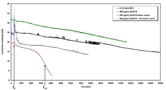
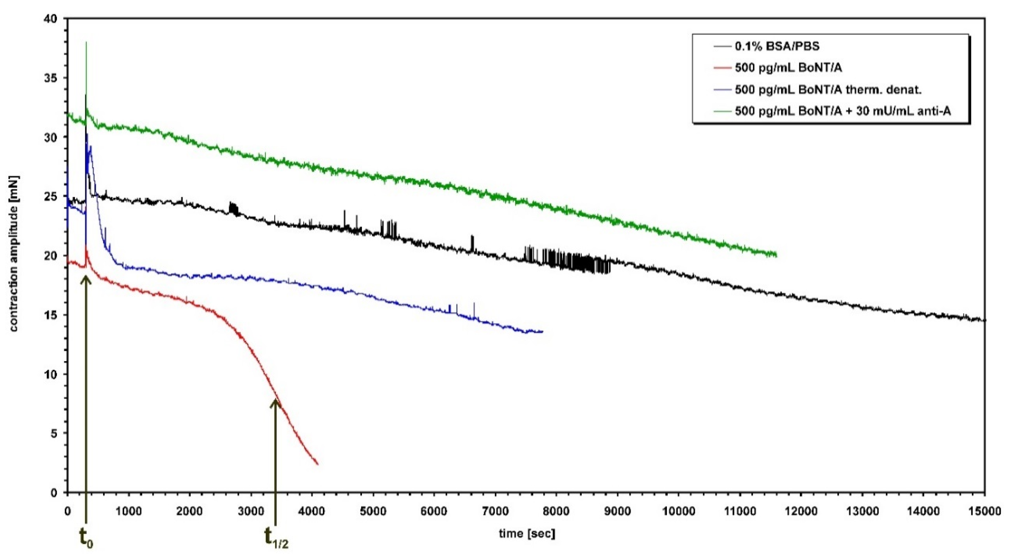
The left phrenic nerve is nicely exposed soon after opening the chest, in contrast to the right phrenic nerve located far behind vital organs and closely attached to main blood vessels as well as connective tissue including fat. To obtain high rates of successful dissection only the left phrenic nerve hemidiaphragms were excised from male mice of strain RjHan:NMRI (18–22 g, St Berthevin Cedex, Janvier, France) and placed in an organ bath [11,12] containing 4 mL of EBSS supplemented with bovine albumin (0.1%). The pH was adjusted to 7.4 and oxygen saturation was achieved by gassing with 95% O2 and 5% CO2. The phrenic nerve was continuously electro-stimulated at a frequency of 1 Hz via two ring electrodes. The pulse duration was 0.1 ms and the amperage 25 mA to achieve maximal contraction amplitudes. Isometric contractions were recorded with a force transducer (Scaime, Annemasse, France) and the software VitroDat (FMI). The resting tension of the diaphragm was approximately 10 mN. Indirectly stimulated control muscles maintained an undiminished contractile response (twitch >65%) over 4 h (Figure 1). In each experiment, the preparation was first allowed to equilibrate for 15 min under control conditions. Then, the incubation solution was replaced by the toxin-containing solution (BoNT/A, B or E). After toxin application, the amplitude remained unchanged for some time (Figure 1), then decreased gradually depending on the toxin concentration. Toxin concentrations were such as to allow reduction of the contraction amplitude by 50% between 40 and 150 min. The times required to decrease the amplitude by 50% (paralysis time t1/2 ≤ 180 min) at different BoNT concentrations were used to construct the calibration curves for BoNT/A, B and E (Figure 2). Optimal power or logarithmic functions depending on the spread of the calibration curves were fitted to the calibration curves: y (BoNT/A; 0.031, 0.125, 0.5, 2, 8 pg/mL) = 46.387x−0.2622, R2 = 0.9864, y (BoNT/B; 0.0625, 0.125, 0.25, 0.5, 1 ng/mL) = −18.899Ln(x) + 61.633, R2 = 0.9565, y (BoNT/E; 0.378, 0.131, 0.045, 1.098 ng/mL) = −25.783Ln(x) + 56.158, R2 = 0.948. Resulting paralytic half-times of the diluted samples were converted to BoNT concentrations in the 12 samples employing the above fitted functions.

3. Results

Because only 1 mL of each sample was available, in a first series of experiments each of the 13 samples (S1–S13) delivered [54] was diluted in EBSS by a factor of 1000 to save material and tested for BoNT activity. If paralysis occurred and paralysis time was below 120 min, the sample was judged positive for presence of substances that cause paralysis of a nerve muscle preparations. When paralysis time was below 55 min the sample was further diluted until paralysis time was within the range of the calibration curves, i.e., >50 min. If the paralysis time lay outside their ranges the original sample was diluted again with a lower factor (Table 1). This procedure was repeated until a factor of 40 was reached. To avoid effect of matrices, samples to be diluted 1:40 or less were dialyzed against EBSS before being added into the organ bath. Dialysis did not affect activity of toxins. Table 1 demonstrates that paralysis times of all tissue preparations except of that treated with S3 were within the range of the calibration curves and the respective samples were therefore judged positive i.e., they were contaminated with a substance that causes paralysis of a nerve muscle preparation. These numbers were used to calculate the concentration of BoNT within the bath solution. Sample 3 was diluted by a factor of 13 and the test was repeated. Again, the paralysis time exceeded the range of the calibration curves.
The results demonstrate that 12 out of 13 samples contained substances leading to muscle paralysis. In a second series of experiments, the cause of paralysis was examined with the help of specific polyclonal antibodies against BoNT/A, B and E, respectively. All positive samples were tested again after the 4 mL sample containing solution was incubated for 60 min with 10 µL rabbit anti-BoNT/A antiserum (bath concentration 0.75 U/mL) at 37 °C. In nine out of 12 samples, the paralysis time exceeded 180 min, thus the muscle paralyzing substance was neutralized (Table 2). Hence, it was concluded that these matrices were contaminated with BoNT/A. This procedure was repeated with the remaining three samples, however, instead of anti-BoNT/A, 10 µL of anti-BoNT/B (1.25 U/mL) and anti-BoNT/E (0.25 U/mL), respectively, were tested for BoNT neutralizing activity. As shown in Table 2, S6 could be neutralized by anti-BoNT/B and S4 by anti-BoNT/E, consequently S4 contained BoNT/E and S6 BoNT/B, respectively. However, the tissue paralyzing substance in S8 was not neutralized by any anti-BoNT, but anti-BoNT/A prolonged paralysis time considerably, leading to the suspicion that besides BoNT/A at least a second tissue paralyzing substance was present. Therefore, in the final series of experiments dual combinations of the three anti-BoNT antisera were tested for neutralizing activity. Only the combination of anti-BoNT/A and anti-BoNT/B could neutralize the paralyzing capability completely. Thus, the paralysis time obtained in the presence of anti-BoNT/A represents the time due to BoNT/B, and the paralysis time achieved when anti-BoNT/B was present represents the time due to BoNT/A (Table 2).
With the help of the calibration curves, the concentrations of the respective BoNT were calculated and these concentrations were then multiplied by the dilution factor. These results were compared with the nominal values (see Table 1).

4. Discussion

The potential misuse of BoNTs as biological weapons or terrorist tools [56], the occurrence of accidental intoxications caused by consumption of contaminated food [53] and the usage of BoNT as a therapeutic drug [57,58] require sensitive methods for the detection of minute amounts. Because of their extremely high potency, the concentrations of clinically effective or even deadly BoNT doses are below the limit of detection of common chemical detection systems. It is, therefore, of utmost importance to develop reliable methods that can be used to economically screen food, water or air for possible toxin contaminations in the case of potential terrorist attacks, to analyze body fluids and stool in case of potential intoxication and to measure the potency of therapeutic products for safety reasons. The demands of the various methods differ with respect to the different purposes. In some cases it might suffice to detect simply the presence of the protein in its active or inactive form [1]. However, immunological methods are especially error-prone in terms of false negative results due to the vast diversity of BoNT [59] and the occurrence of novel variants [60]. Functional methods will only detect active, and thus dangerous, BoNT independent of its primary structure. For BoNTs manufactured for therapeutic purposes, non-functional methods are not appropriate to prove activity, stability and quality of the drug, because all steps leading to blockage of vesicle fusion in nerve cells are essential for activity and have to be covered by the test. Therefore, a method used for quality control must test the function of each domain of the neurotoxin (binding, translocation, release of enzymatic domain and its catalytic activity) in one assay or in a set of assays. If a single assay is desired, this can only be performed by in vivo, ex vivo and cellular assays. The MBA still represents the most customarily employed method for detection of BoNT in complex matrices, as well as its precise quantification for pharmaceutical purposes, and has been accepted by regulatory agencies worldwide [9,17]. However, its major and multiple disadvantages have been described above and elsewhere [9,61]. Since the endpoint of the test is the paralysis of the respiratory muscle, a truncated version of the test is represented by an isolated nerve-muscle, the phrenic nerve hemidiaphragm preparation [11,12,13], which has been successfully employed by several laboratories worldwide to determine potency of BoNT preparations, decipher basic science mechanisms, identify BoNT inhibitors, characterize BoNT-neutralizing antibodies, and screen sera of BoNT-treated patients for development of BoNT neutralizing antibodies. Its usefulness in detecting BoNT in complex matrices has only been challenged in rare botulism cases so far [53].
Within the present EQuATox BoNT proficiency test, 13 blinded samples had to be analyzed with respect to presence or absence of BoNT/A, B, E and/or F and, if possible, also with respect to quantity of BoNT. Since the MPN assay detects only active BoNT, inactivated or denatured toxin escapes the detection. Furthermore, muscle paralysis caused by BoNT-derived activity has to be delimited against any other muscle-paralyzing substance. On the one hand, the time course of paralysis is very characteristic for BoNT. In contrast to peripheral muscle relaxants or channel blockers that act promptly after application, paralysis due to BoNT starts after a concentration dependent latent period of >25 min and shows a slow and progressive decline of contractions. This characteristic course may be mimicked by slow-acting cellular poisons like ricin [62]. However, on the other hand, BoNT activity can be neutralized by highly serotype-specific anti-BoNT antibodies. Thus, with the help of monovalent antiserum it should be possible to identify a BoNT serotype. For this proficiency test anti-BoNT/A, B and E antisera were available; consequently, other serotypes would have evaded detection.
The analysis of all 13 samples was performed with 63 individual MPN tests, of which 40 tests were run to quantify the amount of toxin and 23 tests to determine the serotype. Here, only the left hemidiaphragm was dissected, so, including wastage, only 66 mice were consumed. Dissecting both halves of the diaphragm would have even halved the number of mice required. Beside the absence of any animal suffering with the low amount of mice consumed, the MPN assay clearly outnumbers the MBA, which required more than 500 mice in some laboratories for a semi-quantitative analysis of the PT samples. Although this time, the PT sample analyses were performed randomly over a period of three weeks, in an emergency situation considering the technical capacity of toxogen, the same analysis, including sample preparation (dialysis), serotype identification and quantification, would have been finished within 2–3 days whereas one run of the MBA already requires 4 days.
Out of 13 samples only one sample (S3) was free of paralyzing activity and thereby presumably free of any BoNT. However, with respect to the limit of detection, it cannot be excluded that this sample might contain a BoNT at a concentration of less than 30 pg/mL in the cases of BoNT/A and 50 pg/mL of BoNT/B and E, respectively (Figure 2). All remaining samples paralyzed the muscle. BoNT/A alone was identified in 9 samples, a combination of BoNT/A and B in S8 and only BoNT/B and BoNT/E in S6 and S4, respectively. The sample matrices did not impair the analysis at any point. In summary, all 13 samples were correctly analyzed (Table 2) by the MPN assay, which had a performance superior to several immunological assays and some MBAs performed in other participating laboratories [54]. 100% qualitatively correct results were also returned by 4 out of 5 Endopep-MS assays [63], two sandwich-ELISAs, a single LFA and 4 out of 9 MBA [54].
With respect to the quantification of BoNT/A, a mean z-score of 7 ± 5.7 and a mean Q-score of 1.79 were achieved. The true mean Q-score excluding a miscalculation (S8A) and a reporting error (S12: language specific punctuation translation error) is +1.85. This two-fold higher quantification of BoNT/A reflects the fact that the BoNT/A calibration curve was established with a BoNT/A from an external supplier in the absence of certified reference material. On the other hand, quantification of BoNT/B and E yielded z-scores of 0.3 and 1.6, respectively (Table 1). Here, the calibration curves were generated employing the same reference material [55] used for spiking PT samples S4, S6 and S8 [54], which strongly supports the urgent provision of internationally available certified BoNT reference material. Furthermore, it demonstrates the high precision of the MPN assay.
In conclusion, the analysis of the EQuATox PT samples demonstrated the power of the MPN assay in detecting functionally active BoNT in complex matrices in a short time with high precision in a laboratory environment, but relies on availability of appropriate anti-BoNT antisera for serotype identification and qualified reference material for precise quantitation.

Acknowledgments

We thank Beate Laske and Beate Winter for their excellent technical assistance. This study was partly supported by the European Union’s Seventh Framework Programme (FP7/2007-361 2013) under grant agreement No. 285120.

Author Contributions

H.B. conceived and designed the experiments; B.L. and B.W. performed the experiments; H.B. analyzed the data; A.R. contributed reagents; H.B. and A.R. wrote the paper.

Conflicts of Interest

The authors declare no conflict of interest. The funding sponsor had no role in the design of the study; in the collection, analyses, or interpretation of data; in the writing of the manuscript; or in the decision to publish the results.

References

  1. Dorner, M.B.; Schulz, K.M.; Kull, S.; Dorner, B.G. Complexity of botulinum neurotoxins: Challenges for detection technology. Curr. Top. Microbiol. Immunol. 2013, 364, 219–255. [Google Scholar] [PubMed]
  2. Lindström, M.; Korkeala, H. Laboratory diagnostics of botulism. Clin. Microbiol. Rev. 2006, 19, 298–314. [Google Scholar] [CrossRef] [PubMed]
  3. Reed, L.J.; Muench, H. A simple method of estimating 50% endpoints. Am. J. Hyg. 1938, 27, 493–497. [Google Scholar]
  4. Pearce, L.B.; Borodic, G.E.; First, E.R.; MacCallum, R.D. Measurement of botulinum toxin activity: Evaluation of the lethality assay. Toxicol. Appl. Pharmacol. 1994, 128, 69–77. [Google Scholar] [CrossRef] [PubMed]
  5. Pearce, L.B.; Borodic, G.E.; Johnson, E.A.; First, E.R.; MacCallum, R. The median paralysis unit: A more pharmacologically relevant unit of biologic activity for botulinum toxin. Toxicon 1995, 33, 217–227. [Google Scholar] [CrossRef]
  6. Pearce, L.B.; First, E.R.; Borodic, G.E. Botulinum toxin potency: A mystery resolved by the median paralysis. J. R. Soc. Med. 1994, 87, 571–572. [Google Scholar] [PubMed]
  7. Deutsches Institut für Normung. Microbiological Analysis of Meat and Meat Products; Detection of Clostridium Botulinum and Botulinum Toxin; Beuth Verlag: Berlin, Germany, 1988; Volume 10102, pp. 1–5. [Google Scholar]
  8. Anon. AOAC official method. In Clostridium Botulinum and Its Toxins in Food; AOAC International: Rockville, MD, USA, 1977; Volume 977, p. 26. [Google Scholar]
  9. Adler, S.; Bicker, G.; Bigalke, H.; Bishop, C.; Blumel, J.; Dressler, D.; Fitzgerald, J.; Gessler, F.; Heuschen, H.; Kegel, B.; et al. The current scientific and legal status of alternative methods to the LD50 test for botulinum neurotoxin potency testing. The report and recommendations of a ZEBET Expert Meeting. Altern. Lab. Anim. 2010, 38, 315–330. [Google Scholar] [PubMed]
  10. Boroff, D.A.; Fleck, U. Statistical analysis of a rapid in vivo method for the titration of the toxin of Clostridium botulinum. J. Bacteriol. 1966, 92, 1580–1581. [Google Scholar] [PubMed]
  11. Bülbring, E. Observations on the isolated phrenic nerve diaphragm preparation of the rat. Br. J. Pharmacol. Chemother. 1946, 1, 38–61. [Google Scholar] [CrossRef] [PubMed]
  12. Habermann, E.; Dreyer, F.; Bigalke, H. Tetanus toxin blocks the neuromuscular transmission in vitro like botulinum A toxin. Naunyn Schmiedeberg’s Arch. Pharmacol. 1980, 311, 33–40. [Google Scholar] [CrossRef]
  13. Simpson, L.L.; Rapport, M.M. The binding of botulinum toxin to membrane lipids: Sphingolipids, steroids and fatty acids. J. Neurochem. 1971, 18, 1751–1759. [Google Scholar] [CrossRef] [PubMed]
  14. Rummel, A.; Häfner, K.; Mahrhold, S.; Darashchonak, N.; Holt, M.; Jahn, R.; Beermann, S.; Karnath, T.; Bigalke, H.; Binz, T. Botulinum neurotoxins C, E and F bind gangliosides via a conserved binding site prior to stimulation-dependent uptake with botulinum neurotoxin F utilising the three isoforms of SV2 as second receptor. J. Neurochem. 2009, 110, 1942–1954. [Google Scholar] [CrossRef] [PubMed]
  15. Wohlfarth, K.; Göschel, H.; Frevert, J.; Dengler, R.; Bigalke, H. Botulinum A toxins: Units versus units. Naunyn Schmiedeberg’s Arch. Pharmacol. 1997, 355, 335–340. [Google Scholar] [CrossRef]
  16. Rasetti-Escargueil, C.; Jones, R.G.; Liu, Y.; Sesardic, D. Measurement of botulinum types A, B and E neurotoxicity using the phrenic nerve-hemidiaphragm: Improved precision with in-bred mice. Toxicon 2009, 53, 503–511. [Google Scholar] [CrossRef] [PubMed]
  17. EDQM. Botulinum Toxin Type A for Injection. In European Pharmacopoeia, 5th ed.; Council of Europe; 2006; pp. 1117–1119. [Google Scholar]
  18. Pirazzini, M.; Henke, T.; Rossetto, O.; Mahrhold, S.; Krez, N.; Rummel, A.; Montecucco, C.; Binz, T. Neutralisation of specific surface carboxylates speeds up translocation of botulinum neurotoxin type B enzymatic domain. FEBS Lett. 2013, 587, 3831–3836. [Google Scholar] [CrossRef] [PubMed]
  19. Rummel, A.; Bade, S.; Alves, J.; Bigalke, H.; Binz, T. Two carbohydrate binding sites in the HCC-domain of tetanus neurotoxin are required for toxicity. J. Mol. Biol. 2003, 326, 835–847. [Google Scholar] [CrossRef]
  20. Rummel, A.; Eichner, T.; Weil, T.; Karnath, T.; Gutcaits, A.; Mahrhold, S.; Sandhoff, K.; Proia, R.L.; Acharya, K.R.; Bigalke, H.; et al. Identification of the protein receptor binding site of botulinum neurotoxins B and G proves the double-receptor concept. Proc. Natl. Acad. Sci. USA 2007, 104, 359–364. [Google Scholar] [CrossRef] [PubMed]
  21. Rummel, A.; Mahrhold, S.; Bigalke, H.; Binz, T. The HCC-domain of botulinum neurotoxins A and B exhibits a singular ganglioside binding site displaying serotype specific carbohydrate interaction. Mol. Microbiol. 2004, 51, 631–643. [Google Scholar] [CrossRef] [PubMed]
  22. Rummel, A.; Mahrhold, S.; Bigalke, H.; Binz, T. Exchange of the HCC domain mediating double receptor recognition improves the pharmacodynamic properties of botulinum neurotoxin. FEBS J. 2011, 278, 4506–4515. [Google Scholar] [CrossRef] [PubMed]
  23. Mahrhold, S.; Strotmeier, J.; Garcia-Rodriguez, C.; Lou, J.; Marks, J.D.; Rummel, A.; Binz, T. Identification of the SV2 protein receptor-binding site of botulinum neurotoxin type E. Biochem. J. 2013, 453, 37–47. [Google Scholar] [CrossRef] [PubMed]
  24. Strotmeier, J.; Gu, S.; Jutzi, S.; Mahrhold, S.; Zhou, J.; Pich, A.; Eichner, T.; Bigalke, H.; Rummel, A.; Jin, R.; et al. The biological activity of botulinum neurotoxin type C is dependent upon novel types of ganglioside binding sites. Mol. Microbiol. 2011, 81, 143–156. [Google Scholar] [CrossRef] [PubMed]
  25. Strotmeier, J.; Lee, K.; Volker, A.K.; Mahrhold, S.; Zong, Y.; Zeiser, J.; Zhou, J.; Pich, A.; Bigalke, H.; Binz, T.; et al. Botulinum neurotoxin serotype D attacks neurons via two carbohydrate-binding sites in a ganglioside-dependent manner. Biochem. J. 2010, 431, 207–216. [Google Scholar] [CrossRef] [PubMed]
  26. Strotmeier, J.; Mahrhold, S.; Krez, N.; Janzen, C.; Lou, J.; Marks, J.D.; Binz, T.; Rummel, A. Identification of the synaptic vesicle glycoprotein 2 receptor binding site in botulinum neurotoxin A. FEBS Lett. 2014, 588, 1087–1093. [Google Scholar] [CrossRef] [PubMed]
  27. Gu, S.; Rumpel, S.; Zhou, J.; Strotmeier, J.; Bigalke, H.; Perry, K.; Shoemaker, C.B.; Rummel, A.; Jin, R. Botulinum neurotoxin is shielded by NTNHA in an interlocked complex. Science 2012, 335, 977–981. [Google Scholar] [CrossRef] [PubMed]
  28. Sheridan, R.E.; Deshpande, S.S.; Smith, T. Comparison of in vivo and in vitro mouse bioassays for botulinum toxin antagonists. J. Appl. Toxicol. 1999, 19, S29–S33. [Google Scholar] [CrossRef]
  29. Sheridan, R.E.; Smith, T.J.; Adler, M. Primary cell culture for evaluation of botulinum neurotoxin antagonists. Toxicon 2005, 45, 377–382. [Google Scholar] [CrossRef] [PubMed]
  30. Sheridan, R.E.; Deshpande, S.S.; Nicholson, J.D.; Adler, M. Structural features of aminoquinolines necessary for antagonist activity against botulinum neurotoxin. Toxicon 1997, 35, 1439–1451. [Google Scholar] [CrossRef]
  31. Adler, M.; Scovill, J.; Parker, G.; Lebeda, F.J.; Piotrowski, J.; Deshpande, S.S. Antagonism of botulinum toxin-induced muscle weakness by 3,4-diaminopyridine in rat phrenic nerve-hemidiaphragm preparations. Toxicon 1995, 33, 527–537. [Google Scholar] [CrossRef]
  32. Eleopra, R.; Tugnoli, V.; Quatrale, R.; Gastaldo, E.; Rossetto, O.; de Grandis, D.; Montecucco, C. Botulinum neurotoxin serotypes A and C do not affect motor units survival in humans: An electrophysiological study by motor units counting. Clin. Neurophysiol. 2002, 113, 1258–1264. [Google Scholar] [CrossRef]
  33. Eleopra, R.; Montecucco, C.; Devigili, G.; Lettieri, C.; Rinaldo, S.; Verriello, L.; Pirazzini, M.; Caccin, P.; Rossetto, O. Botulinum neurotoxin serotype D is poorly effective in humans: An in vivo electrophysiological study. Clin. Neurophysiol. 2013, 124, 999–1004. [Google Scholar] [CrossRef] [PubMed]
  34. Rummel, A.; Karnath, T.; Henke, T.; Bigalke, H.; Binz, T. Synaptotagmins I and II act as nerve cell receptors for botulinum neurotoxin G. J. Boil. Chem. 2004, 279, 30865–30870. [Google Scholar] [CrossRef] [PubMed]
  35. Mahrhold, S.; Rummel, A.; Bigalke, H.; Davletov, B.; Binz, T. The synaptic vesicle protein 2C mediates the uptake of botulinum neurotoxin A into phrenic nerves. FEBS Lett. 2006, 580, 2011–2014. [Google Scholar] [CrossRef] [PubMed]
  36. Simpson, L.L. Studies on the binding of botulinum toxin type A to the rat phrenic nerve-hemidiaphragm preparation. Neuropharmacology 1974, 13, 683–691. [Google Scholar] [CrossRef]
  37. Göschel, H.; Wohlfarth, K.; Frevert, J.; Dengler, R.; Bigalke, H. Botulinum A toxin therapy: Neutralizing and nonneutralizing antibodies—Therapeutic consequences. Exp. Neurol. 1997, 147, 96–102. [Google Scholar] [CrossRef] [PubMed]
  38. Dressler, D.; Dirnberger, G.; Bhatia, K.P.; Irmer, A.; Quinn, N.P.; Bigalke, H.; Marsden, C.D. Botulinum toxin antibody testing: Comparison between the mouse protection assay and the mouse lethality assay. Mov. Disord. 2000, 15, 973–976. [Google Scholar] [CrossRef]
  39. Herrmann, J.; Mall, V.; Bigalke, H.; Geth, K.; Korinthenberg, R.; Heinen, F. Secondary non-response due to development of neutralising antibodies to botulinum toxin A during treatment of children with cerebral palsy. Neuropediatrics 2000, 31, 333–334. [Google Scholar] [CrossRef] [PubMed]
  40. Rollnik, J.D.; Wohlfarth, K.; Dengler, R.; Bigalke, H. Neutralizing botulinum toxin type a antibodies: Clinical observations in patients with cervical dystonia. Neurol. Clin. Neurophysiol. 2001, 2001, 2–4. [Google Scholar] [CrossRef] [PubMed]
  41. Dressler, D.; Bigalke, H. Botulinum toxin antibody type A titres after cessation of botulinum toxin therapy. Mov. Disord. 2002, 17, 170–173. [Google Scholar] [CrossRef] [PubMed]
  42. Dressler, D.; Bigalke, H.; Benecke, R. Botulinum toxin type B in antibody-induced botulinum toxin type A therapy failure. J. Neurol. 2003, 250, 967–969. [Google Scholar] [CrossRef] [PubMed]
  43. Dressler, D.; Bigalke, H. Antibody-induced failure of botulinum toxin type B therapy in de novo patients. Eur. Neurol. 2004, 52, 132–135. [Google Scholar] [CrossRef] [PubMed]
  44. Herrmann, J.; Geth, K.; Mall, V.; Bigalke, H.; Monting, S.J.; Linder, M.; Kirschner, J.; Berweck, S.; Korinthenberg, R.; Heinen, F.; et al. Clinical impact of antibody formation to botulinum toxin A in children. Ann. Neurol. 2004, 55, 732–735. [Google Scholar] [CrossRef] [PubMed]
  45. Dressler, D.; Lange, M.; Bigalke, H. Mouse diaphragm assay for detection of antibodies against botulinum toxin type B. Mov. Disord. 2005, 20, 1617–1619. [Google Scholar] [CrossRef] [PubMed]
  46. Lange, O.; Bigalke, H.; Dengler, R.; Wegner, F.; deGroot, M.; Wohlfarth, K. Neutralizing antibodies and secondary therapy failure after treatment with botulinum toxin type A: Much ado about nothing? Clin. Neuropharmacol. 2009, 32, 213–218. [Google Scholar] [CrossRef] [PubMed]
  47. Rasetti-Escargueil, C.; Liu, Y.; Rigsby, P.; Jones, R.G.; Sesardic, D. Phrenic nerve-hemidiaphragm as a highly sensitive replacement assay for determination of functional botulinum toxin antibodies. Toxicon 2011, 57, 1008–1016. [Google Scholar] [CrossRef] [PubMed]
  48. Miethe, S.; Rasetti-Escargueil, C.; Liu, Y.; Chahboun, S.; Pelat, T.; Avril, A.; Frenzel, A.; Schirrmann, T.; Thullier, P.; Sesardic, D.; et al. Development of neutralizing scFv-Fc against botulinum neurotoxin A light chain from a macaque immune library. mAbs 2014, 6, 446–459. [Google Scholar] [CrossRef] [PubMed]
  49. Sesardic, D.; Jones, R.G.; Leung, T.; Alsop, T.; Tierney, R. Detection of antibodies against botulinum toxins. Mov. Disord. 2004, 19, S85–S91. [Google Scholar] [CrossRef] [PubMed]
  50. Nowakowski, A.; Wang, C.; Powers, D.B.; Amersdorfer, P.; Smith, T.J.; Montgomery, V.A.; Sheridan, R.; Blake, R.; Smith, L.A.; Marks, J.D. Potent neutralization of botulinum neurotoxin by recombinant oligoclonal antibody. Proc. Natl. Acad. Sci. USA 2002, 99, 11346–11350. [Google Scholar] [CrossRef] [PubMed]
  51. Amersdorfer, P.; Wong, C.; Smith, T.; Chen, S.; Deshpande, S.; Sheridan, R.; Marks, J.D. Genetic and immunological comparison of anti-botulinum type A antibodies from immune and non-immune human phage libraries. Vaccine 2002, 20, 1640–1648. [Google Scholar] [CrossRef]
  52. Amersdorfer, P.; Wong, C.; Chen, S.; Smith, T.; Deshpande, S.; Sheridan, R.; Finnern, R.; Marks, J.D. Molecular characterization of murine humoral immune response to botulinum neurotoxin type A binding domain as assessed by using phage antibody libraries. Infect. Immun. 1997, 65, 3743–3752. [Google Scholar] [PubMed]
  53. Merz, B.; Bigalke, H.; Stoll, G.; Naumann, M. Botulism type B presenting as pure autonomic dysfunction. Clin. Auton. Res. 2003, 13, 337–338. [Google Scholar] [CrossRef] [PubMed]
  54. Worbs, S.; Fiebig, U.; Zeleny, R.; Schimmel, H.; Rummel, A.; Luginbühl, W.; Dorner, B.G. Qualitative and quantitative detection of botulinum neurotoxins from complex matrices: Results of the first international proficiency test. Toxins 2015, 7. [Google Scholar] [CrossRef] [Green Version]
  55. Weisemann, J.; Krez, N.; Fiebig, U.; Worbs, S.; Skiba, M.; Endermann, T.; Dorner, M.B.; Bergström, T.; Muñoz, A.; Zegers, I.; et al. Generation and characterisation of six recombinant botulinum neurotoxins as reference material to serve in an international proficiency. Toxins 2015, 7. [Google Scholar] [CrossRef]
  56. Bigalke, H.; Rummel, A. Medical aspects of toxin weapons. Toxicology 2005, 214, 210–220. [Google Scholar] [CrossRef] [PubMed]
  57. Bigalke, H. Botulinum toxin: Application, safety, and limitations. Curr. Top. Microbiol. Immunol. 2013, 364, 307–317. [Google Scholar] [PubMed]
  58. Pickett, A.M.; Hambleton, P. Dose standardisation of botulinum toxin. Lancet 1994, 344, 474–475. [Google Scholar] [CrossRef]
  59. Rossetto, O.; Pirazzini, M.; Montecucco, C. Botulinum neurotoxins: Genetic, structural and mechanistic insights. Nat. Rev. Microbiol. 2014, 12, 535–549. [Google Scholar] [CrossRef] [PubMed]
  60. Dover, N.; Barash, J.R.; Hill, K.K.; Xie, G.; Arnon, S.S. Molecular characterization of a novel botulinum neurotoxin type h gene. J. Infect. Dis. 2014, 209, 192–202. [Google Scholar] [CrossRef] [PubMed]
  61. Pellett, S. Progress in cell based assays for botulinum neurotoxin detection. Curr. Top. Microbiol. Immunol. 2013, 364, 257–285. [Google Scholar] [PubMed]
  62. Wiley, R.G.; Oeltmann, T.N. Anatomically selective peripheral nerve ablation using intraneural ricin injection. J. Neurosci. Methods 1986, 17, 43–53. [Google Scholar] [CrossRef]
  63. Kalb, S.R.; Baudys, J.; Wang, D.; Barr, J.R. Recommended mass spectrometry-based strategies to identify botulinum neurotoxin-containing samples. Toxins 2015, 7, 1765–1778. [Google Scholar] [CrossRef] [PubMed]

