Article

Venom of the Coral Snake Micrurus clarki: Proteomic Profile, Toxicity, Immunological Cross-Neutralization, and Characterization of a Three-Finger Toxin

1
Instituto Clodomiro Picado, Facultad de Microbiología, Universidad de Costa Rica, San José 11501, Costa Rica
2
Programa de Ofidismo y Escorpionismo, Universidad de Antioquia, Medellín 050010, Colombia
*
Author to whom correspondence should be addressed.
Submission received: 26 March 2016 / Revised: 25 April 2016 / Accepted: 2 May 2016 / Published: 5 May 2016
(This article belongs to the Special Issue Venomics, Venom Proteomics and Venom Transcriptomics)

Abstract

:
Micrurus clarki is an uncommon coral snake distributed from the Southeastern Pacific of Costa Rica to Western Colombia, for which no information on its venom could be found in the literature. Using a ‘venomics’ approach, proteins of at least nine families were identified, with a moderate predominance of three-finger toxins (3FTx; 48.2%) over phospholipase A2 (PLA2; 36.5%). Comparison of this venom profile with those of other Micrurus species suggests that it may represent a more balanced, ‘intermediate’ type within the dichotomy between 3FTx- and PLA2-predominant venoms. M. clarki venom was strongly cross-recognized and, accordingly, efficiently neutralized by an equine therapeutic antivenom against M. nigrocinctus, revealing their high antigenic similarity. Lethal activity for mice could be reproduced by a PLA2 venom fraction, but, unexpectedly, not by fractions corresponding to 3FTxs. The most abundant venom component, hereby named clarkitoxin-I, was identified as a short-chain (type I) 3FTx, devoid of lethal effect in mice, whose target remains to be defined. Its amino acid sequence of 66 residues shows high similarity with predicted sequences of venom gland transcripts described for M. fulvius, M. browni, and M. diastema.

Graphical Abstract

1. Introduction

Coral snakes (Elapidae) comprise some 71 species that are widely distributed in tropical and subtropical regions of the New World, from Southeastern USA to Central Argentina [1,2,3]. Envenomings by these snakes are far less frequent than those inflicted by viperids, but are of medical concern [4,5].
Micrurus clarki (Figure 1A) is an uncommon, slender to medially robust coral snake with a reported maximum length of 920 mm, although most adults average between 400 and 600 mm. This species has a tricolored pattern with narrow yellow bands bordering the black bands followed by single red bands, and a characteristic black head cap that extends over most of the snout and parietals. M. clarki has a discontinuous distribution from the Tárcoles basin in Central Pacific Costa Rica southward to the Pacific lowlands of Western Panamá, where it inhabits lowlands of the Canal Zone, to Pacific Darien, and Western Colombia. Along this distribution, M. clarki inhabits tropical wet forest, but also transitional tropical wet-tropical dry forest, where it is mostly found at low elevations, with some exceptions reported up to 900 m above sea level [1,6,7]. Little is known about the natural history of this coral snake. The species was named in honor of H.C. Clark, a medical doctor that directed the Gorgas Memorial Institute in Panamá, considered a pioneer in tropical medicine [8]. It is considered mainly a terrestrial and primarily nocturnal species whose natural diet is known to include the marbled swamp eel Synbranchus marmoratus, and colubrid snakes [6].
Our search of the mainstream literature databases did not find any information on the venom of M. clarki, whose general characteristics seem to be completely unknown. As part of an ongoing effort to characterize the venom proteomes of all snake species from Costa Rica [9], in the present work we report the composition, toxicity, immunological cross-recognition, and neutralization of the venom of M. clarki obtained from specimens collected in the Southeastern Pacific of this country. In addition, a three-finger toxin from this venom was isolated and characterized.

2. Results and Discussion

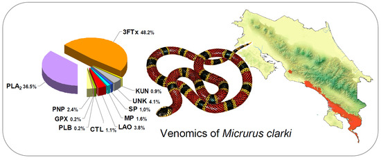
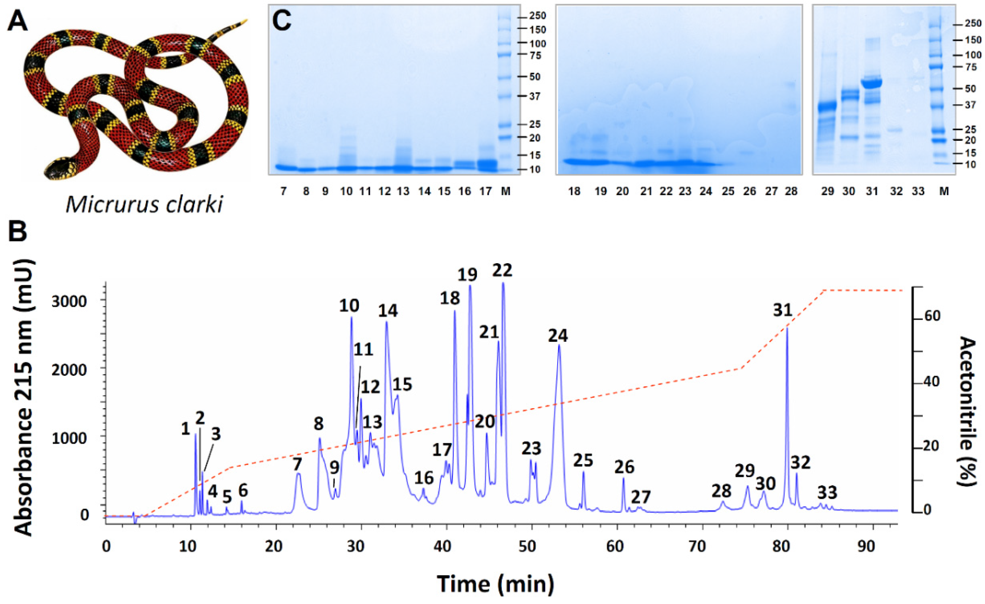
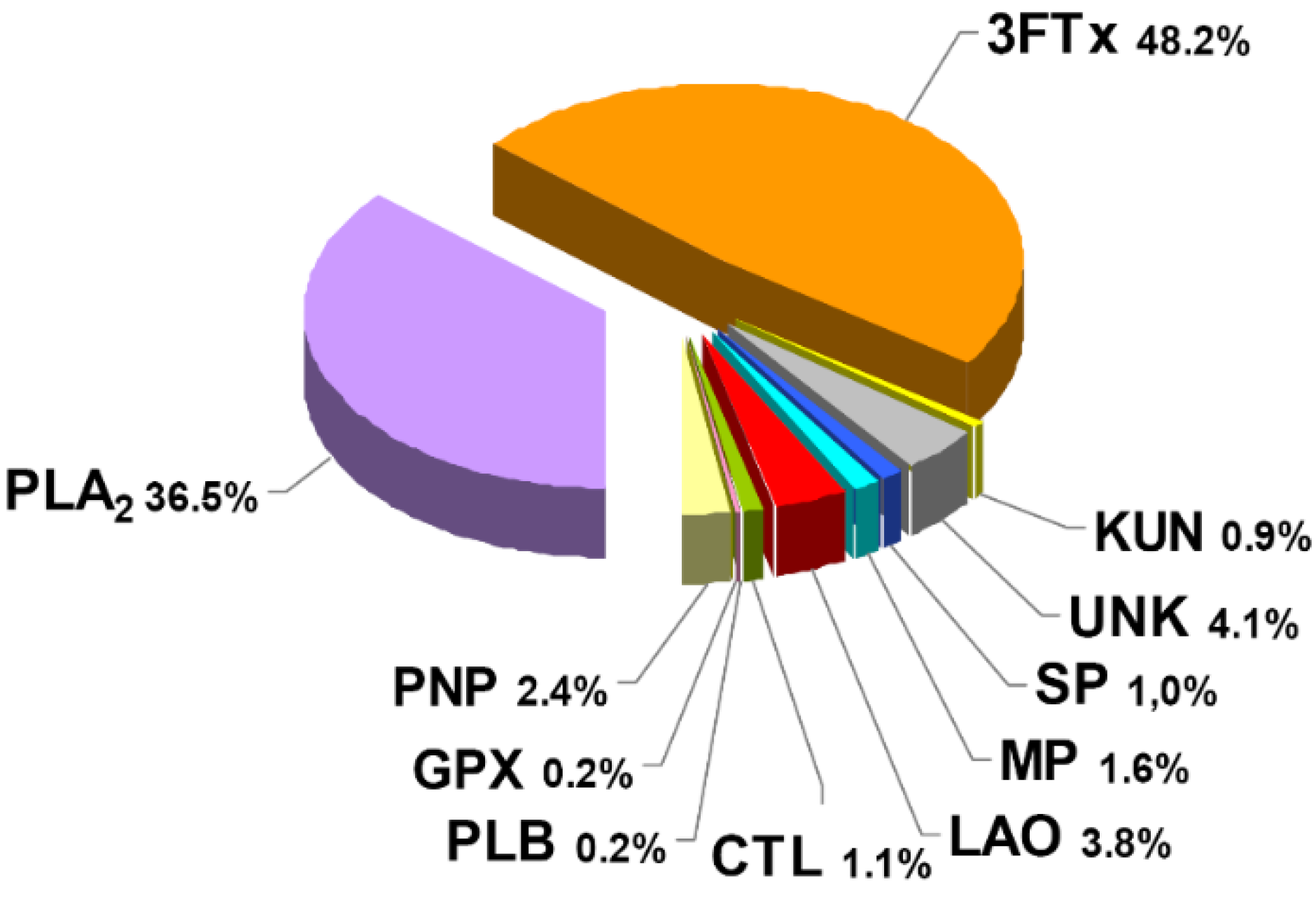
2.1. Proteomic Profile of Micrurus Clarki Venom

The venom of M. clarki (1A) was initially separated into 33 fractions by RP-HPLC (Figure 1B), which were further resolved into 42 protein bands after SDS-PAGE (Figure 1C). After in-gel tryptic digestion of these, followed by MALDI-TOF-TOF analysis, protein family assignments were obtained for 34 of the bands, estimated to represent 95.9% of the total venom proteins. The remaining eight bands for which no identifications were obtained (indicated as ‘‘unknown’’ in Table 1 and Figure 2), altogether add to the remaining 4.1% of the venom proteins.
Although not markedly predominant, the protein family with the highest abundance in M. clarki venom corresponds to three-finger toxins (3FTx), which account for almost half of the protein content (48.2%), followed by phospholipase A2 (PLA2), representing roughly one-third (36.5%).
In addition to its abundant 3FTxs and PLA2s, other proteins detected in lower proportions (within the range of 0.9%–3.8%) belong to the L-amino acid oxidase, metalloproteinase, serine proteinase, Kunitz-type serine proteinase inhibitor, and C-type lectin/lectin-like families, together with traces (~0.2%) of phospholipase B and glutathion peroxidase components (Figure 2). Altogether, proteins in this venom belong to at least nine families. As expected, the rapidly eluting peaks in the RP-HPLC separation of the venom (Figure 1B; peaks 1–6), did not show proteins by electrophoresis, and are considered to contain either small peptides or non-proteinaceous compounds (PNP; 2.4%). The most intense of these initial peaks (1–3) were analyzed by nESI-MS, and clear evidence for adenosine being present in peak 1 was obtained (Figure 3). Adenosine has been found in several venoms from Micrurus species, although not ubiquitously [10,11], and may have a contributing role in envenoming strategies [12], a hypothesis which still remains to be studied in detail.

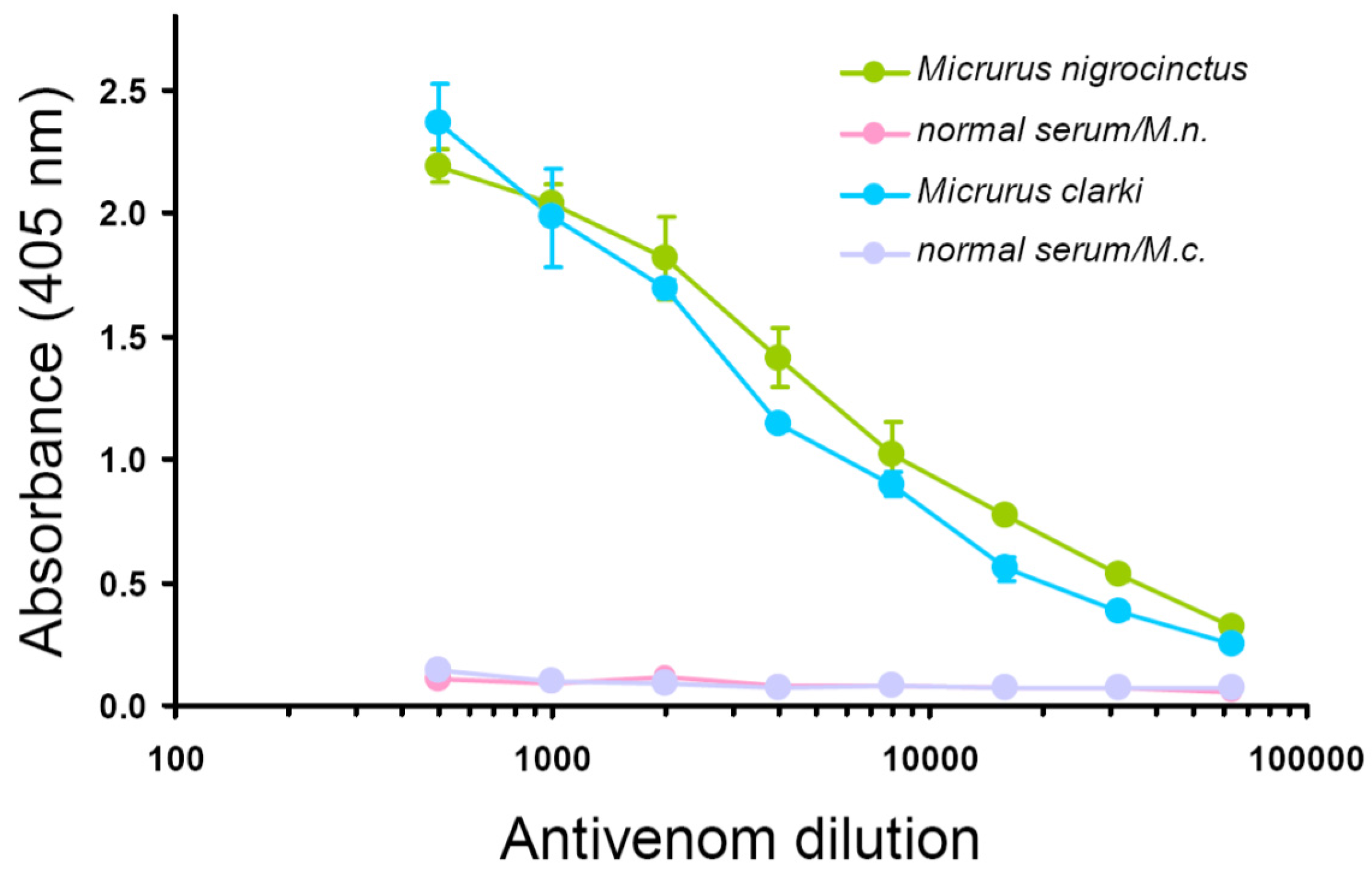
2.2. Immunological Properties of Micrurus Clarki Venom

Since the antivenom used therapeutically for coral snake envenomings in Central America (SAC-ICP) is prepared by immunization of horses with the venom of a single species, M. nigrocinctus, it was of interest to characterize its immunological cross-recognition and cross-neutralization toward the untested venom of M. clarki. As shown by ELISA titration curves, this antivenom strongly cross-recognized M. clarki whole venom, resulting in antibody binding signals virtually as high as those obtained for the homologous (M. nigrocinctus) venom (Figure 4). This finding evidences the high degree of antigenic conservation between venoms of these two coral snake species. Following this result, a more detailed cross-recognition analysis was performed using the RP-HPLC-separated venom fractions of M. clarki, revealing additional information. First, it evidenced that the antivenom contains antibodies against all of the venom fractions tested, since the binding signals recorded were, in all cases, significantly higher than those of the negative control equine immunoglobulins (Figure 5). Second, it showed that immunorecognition of smaller proteins, such as 3FTxs or Kunitz-type components, is generally weaker in comparison to recognition of the larger PLA2s or LAAOs (Figure 5). The lower titers of antibodies against low molecular mass proteins may reflect differences in their immunogenicity, and/or epitope availability in the immunoassay, and is in agreement with earlier studies performed with other Micrurus venoms [13,14] or with venoms from other elapids [15,16].

2.3. Lethal Activity of Micrurus Clarki Venom and its Neutralization by Antivenom

Immunorecognition results obtained for M. clarki venom or its main fractions predicted that the SAC-ICP antivenom should be capable of efficiently cross-neutralizing its toxicity. Since no information was available on the lethal potency of this venom, experiments were first performed to estimate this value, indicating an intravenous median lethal dose (LD50) of 12.4 μg/mouse (95% confidence limits of 7.1–23.5 μg/mouse) or 0.73 μg/g body weight (0.42–1.38 μg/g). Using 4 × LD50 of venom (50 μg) as a challenge, the SAC-ICP antivenom indeed protected all mice from intravenous lethality when preincubated with the venom at all ratios tested (100, 200, 300, and 400 μg venom/mL antivenom). Due to the low amounts of venom available, we could not test neutralization beyond the 400 μg/mL ratio in order to estimate the median Effective Dose (ED50). Nevertheless, these results indicate that the neutralization potency of SAC-ICP against the lethal action of M. clarki venom is at least as high as that against the homologous (M. nigrocinctus) venom (unpublished data from the Quality Control Lab of Instituto Clodomiro Picado), in agreement with the immunological cross-recognition results (Figure 4 and Figure 5).

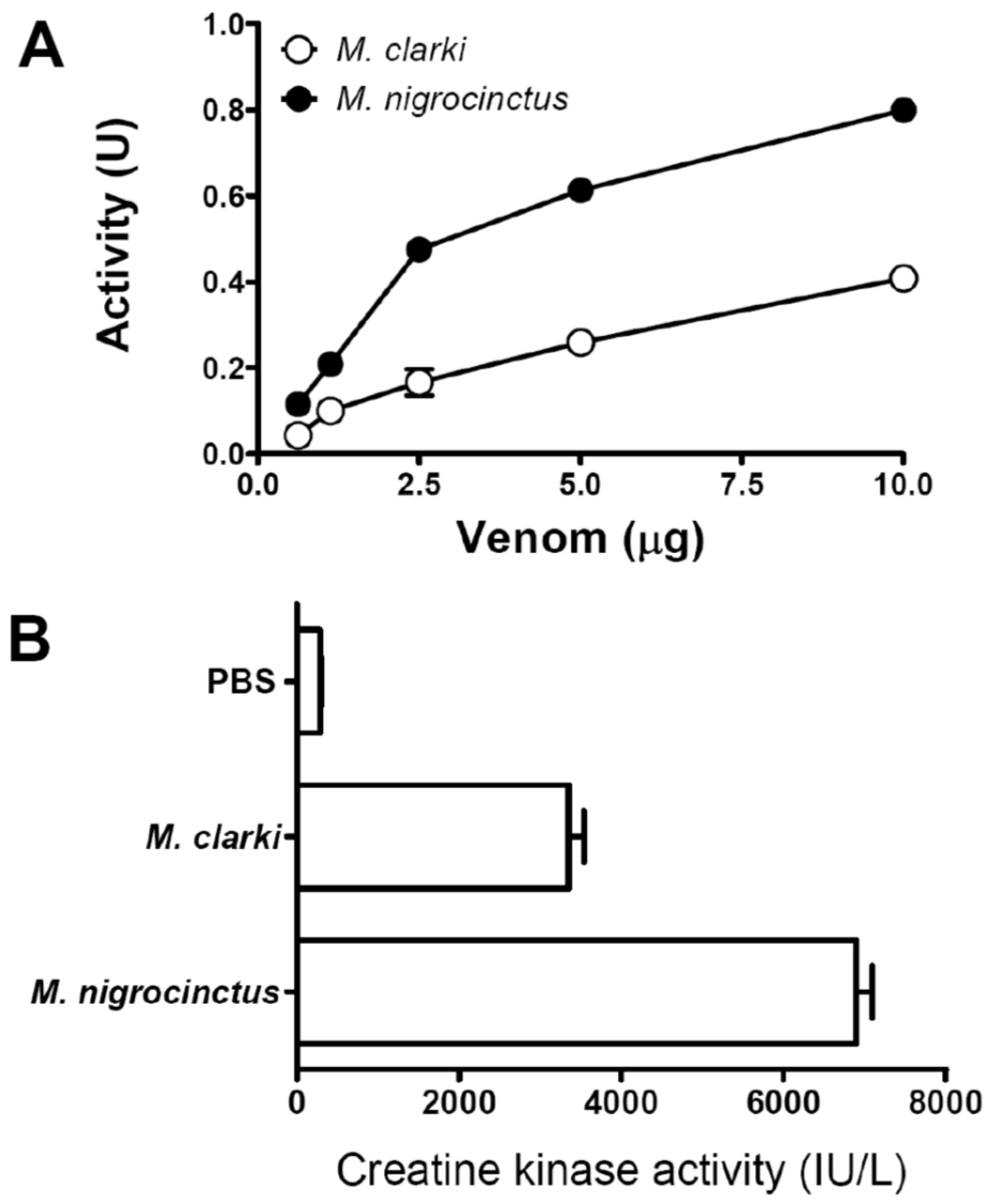
2.4. Phospholipase A2 and Myotoxic Activities of Micrurus Clarki Venom

The venom of M. clarki showed PLA2 activity upon the synthetic substrate NOBA (Figure 6A), in agreement with the presence of proteins, identified as PLA2s in its composition (Figure 2 and Table 1). In comparison to the venom of M. nigrocinctus, that of M. clarki presents a lower activity (Figure 6). This finding correlates well with the proportion of PLA2 enzymes found in the two venoms: 48.0% in M. nigrocinctus [13], and 36.5% in M. clarki (Figure 2).
On the other hand, the venom of M. clarki displayed a significant myotoxic activity in mice (Figure 6B). This effect was evidently less potent in comparison to that of M. nigrocinctus venom, under identical assay conditions (Figure 6B). This observation is also in line with the differences in PLA2 content in the venoms of these two species, since the myotoxicity induced by Micrurus venom has been related to PLA2 enzymes [13,14].

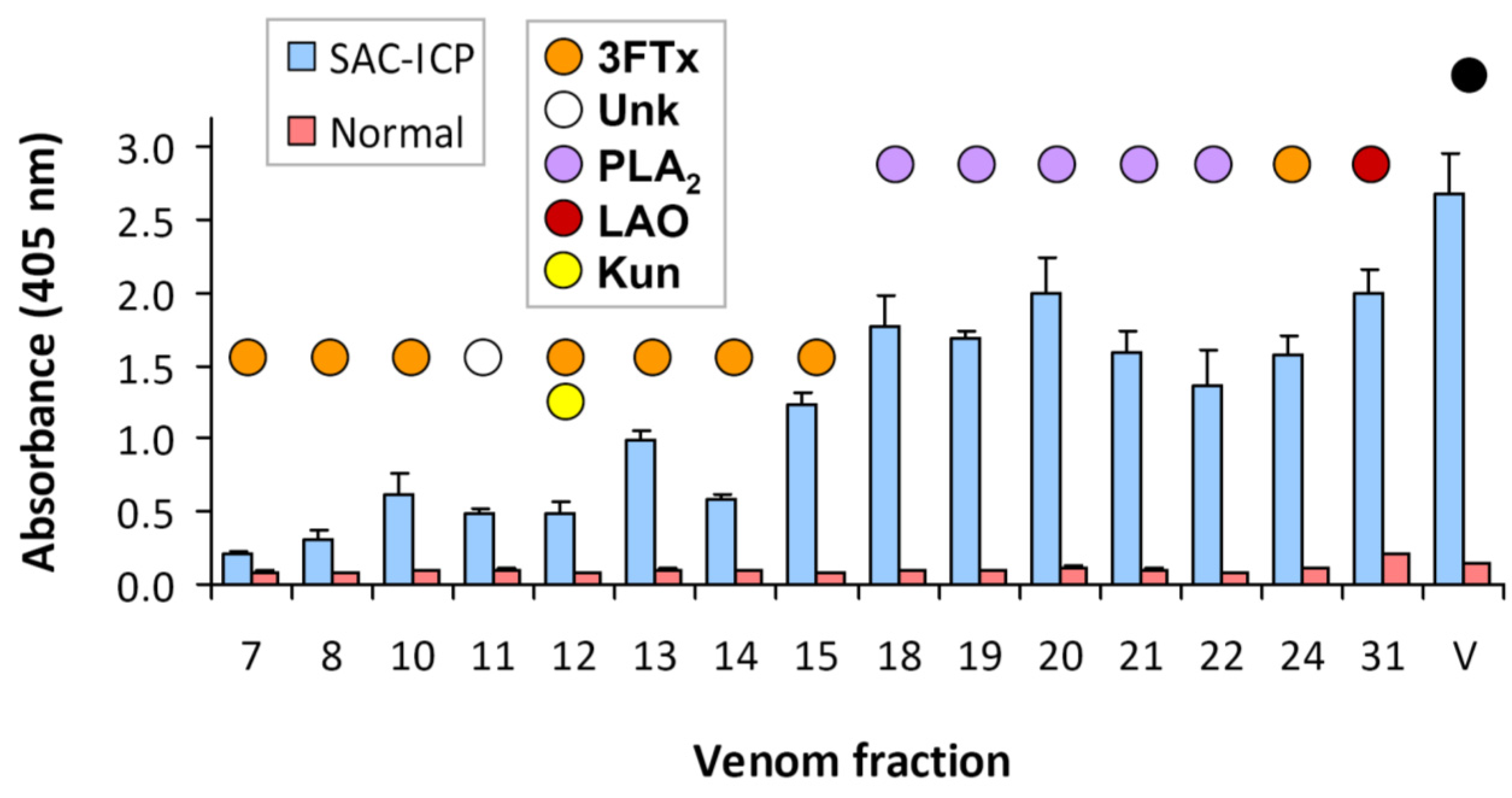
2.5. Lethality Screening of Micrurus Clarki Venom Fractions in Mice

Aiming to identify which of the venom proteins M. clarki contribute to its lethal effect in mice, and, therefore, predict their potential relevance to human envenomings, screenings were performed with its most abundant RP-HPLC fractions, using a challenge of 10 μg/mouse by the intravenous route. Intriguingly, fractions 7, 8, 10, 13, 14, 15, 24 (corresponding to 3FTxs), 12 (containing a 3FTx and a Kunitz-type protein), 18, or 19 (corresponding to PLA2s) did not induce lethality in this screening. Of all fractions tested, only fraction 21, corresponding to a PLA2, was individually lethal to mice. This finding is surprising, since many 3FTxs in snake venoms act as post-synaptic blockers of nicotinic acetylcholine receptors at the neuromuscular junction of mammals and/or birds, therefore being endowed with potent lethal effect [17,18,19]. The possible loss of activity by the compact, disulfide-rich 3FTxs, due to exposure to acidic acetonitrile gradients during their RP-HPLC separation seems unlikely, since a previous study showed that such procedure did not reduce the lethal potency of Dendroaspis polylepis (Elapidae) venom, in which 3FTxs play a predominant role [15]. Therefore, it is puzzling that lethality was observed only in a single (PLA2) fraction of the chromatographic profile of M. clarki venom, and in none of the 3FTx fractions. This finding is suggestive of possible synergisms among different venom components to induce lethality. However, the evaluation of this possibility requires more detailed studies, which could not be performed due to the scarcity of venom.

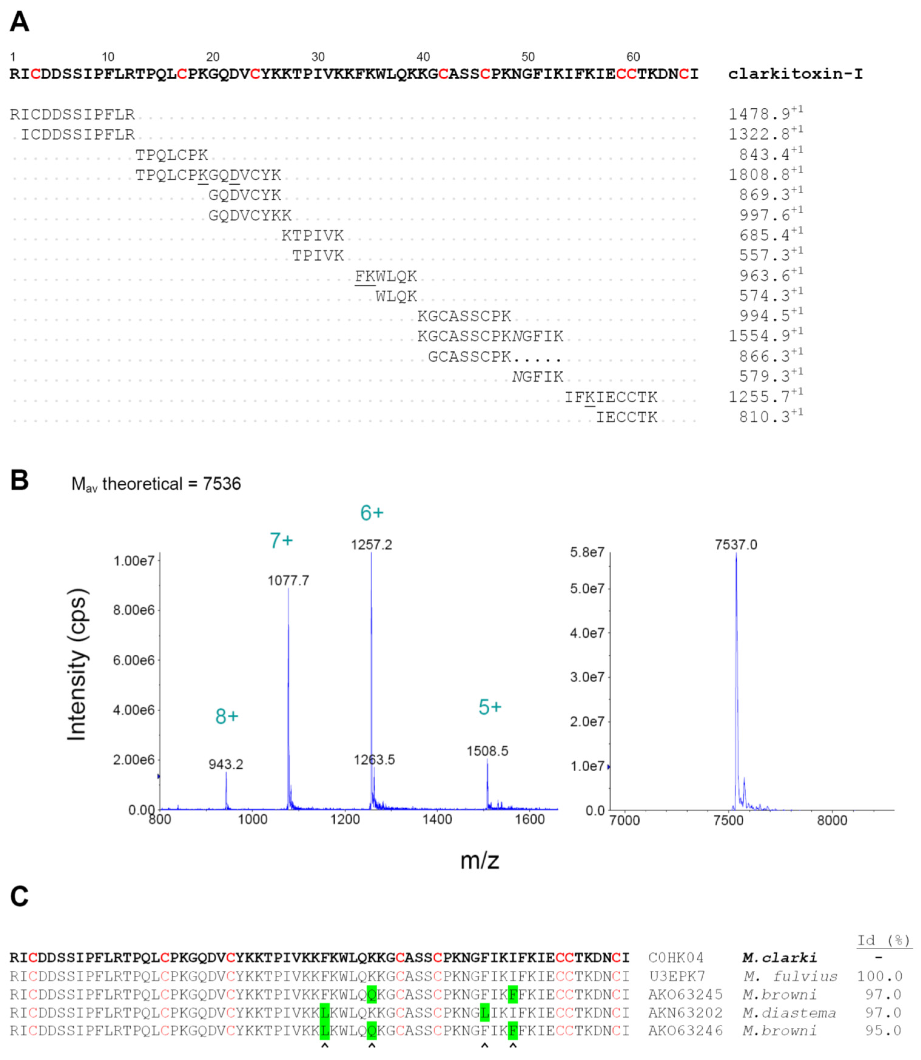
2.6. Characterization of Clarkitoxin-I

Of particular interest was the most abundant venom component, peak 24, representing 16.1% of the total proteins, which was identified as a member of the 3FTx family (Table 1) but was devoid of lethal effect in mice. By performing an in-solution trypsin digestion and MALDI-TOF-TOF de novo sequencing of RP-HPLC-separated peptides, the complete amino acid sequence of this protein, hereby named clarkitoxin-I, was determined (Figure 7A). Despite the fact that this protein is not toxic in mice, we have coined the name ‘clarkitoxin’ because it belongs to the family of the 3FTxs, and because it might be toxic for other species, particularly natural prey species [20]. Its C-terminal four residues (DNCI) were not directly observed in the analysis of tryptic peptides, but their presence is deduced on the basis of (a) matching the isotope-averaged mass (Mav) observed for the intact protein of 7537 ± 1 Da (Figure 7B) with its theoretically-expected Mav of 7536 Da, according to this sequence; and (b) the absolute conservation of the DNCI sequence in four highly similar deduced sequences of venom gland transcripts or cDNAs from M. browni (AKO63246, AKO63245), M. diastema (AKN63202), and M. fulvius (U3EPK7) (Figure 7C). The protein sequence data reported in this paper will appear in the UniProt Knowledgebase under the accession number C0HK04. Clarkitoxin-I presents 66 amino acid residues, with the conserved pattern of eight cysteine positions and spacings that identifies it as a short-chain (type I) 3FTx, and is indeed identical to the sequence predicted by transcript U3EPK7 (cluster 10) sequenced from the venom gland transcriptome of M. fulvius [21], which however has not been isolated and characterized at the protein level. Three homologous sequences from M. browni and M. diastema present identities of 95%–97% in comparison to clarkitoxin-I (Figure 7C), but the next most similar protein retrieved in a BLAST search, F5CPD3 from M. altirostris, drops abruptly to a value of 50% identity. This large gap in similarity suggests that clarkitoxin-I, together with the closely-related variants described in M. fulvius, M. diastema, and M. browni, may form a group that should be phylogenetically distant from other proteins of the elapid 3FTx family, and most likely of uncharacterized functional properties (owing to the scarcity of Micrurus venoms).
Clarkitoxin-I was not lethal when injected by the intravenous route at 10 μg/mouse, and even the intraperitoneal injection of 30 μg into a single mouse did not cause death or evident abnormal signs. Therefore, this 3FTx protein can be considered to be devoid of significant α-neurotoxicity in mice. Toxicity to other groups of organisms such as small colubrids or swamp eels, being natural prey for coral snakes [6], was not tested and, thus, remains an open possibility. In this regard, a rational explanation for the prominent abundance of this protein in M. clarki venom cannot yet be proposed. Determination of the precise target specificity of this 3FTx is a pending task, which will require an adequate source of protein, such as recombinant or synthetic, due to the difficulties of obtaining venom from this coral snake species.

2.7. Dichotomy of Venom Types in Micrurus and the Position of Micrurus Clarki

The increasing proteomic data on Micrurus venoms have revealed a dichotomy between 3FTx- and PLA2-predominant types, which shows a correlation with phylogenetic relationships among the taxa [15]. In this regard, by containing a moderately higher proportion of 3FTxs than PLA2s, the venom of M. clarki could be grouped together with 3FTx-rich venoms such as those of M. altirostris, M. corallinus, M. mipartitus, M. multifasciatus, and M. alleni [15,22,23]. However, it should be noted that in the venoms of these latter coral snakes, 3FTxs represent a considerably higher proportion of the total proteins (61.1%–83.0%), and their relative PLA2 content, consequently, is markedly lower (8.2%–29.0%). Considering that the predominance of 3FTxs over PLA2s (48.2% vs. 36.5%, respectively) is less pronounced in the venom of M. clarki, its compositional pattern appears to differ from the markedly 3FTx-predominant Micrurus venoms described to date, and seems to represent a more ‘intermediate’ pattern. In fact, some functional and immunological characteristics of M. clarki venom show similarities with those observed in the group of PLA2-rich venoms. For example, Micrurus venoms from Costa Rica, which present the 3FTx-predominant expression pattern, have been found to diverge antigenically from the (PLA2-predominant) venom of M. nigrocinctus, used for the production of therapeutic antivenom in the region. As a consequence, they are weakly neutralized (M. alleni) or not neutralized at all (M. mipartitus/M. multifasciatus) by this antivenom [15,23]. In contrast, the present study shows that the venom of M. clarki clearly departs from this trend, by presenting a strong immunological cross-reactivity with the venom of M. nigrocinctus, which leads to an excellent cross-neutralization by its antivenom. An additional difference between M. clarki venom and 3FTx-predominant venoms, e.g., M. mipartitus from Colombia, can be pointed out from the present findings. The latter contains a highly abundant (28%) 3FTx, mipartoxin-I, which is potently lethal to mice [23,24], whereas the most abundant protein in M. clarki venom, clarkitoxin-I (16%), is hereby characterized as a 3FTx that lacks a lethal effect in mice. Additionally, the venom of M. clarki showed a considerable myotoxic effect in mice, in contrast with the 3FTx-rich venoms of M. alleni [11] or M. mipartitus [23], which do not display significant myotoxicity. Moreover, the amino acid sequence of clarkitoxin-I, its most abundant component, reveals a high conservation (95%–100% identity) in at least three other Micrurus species (M. diastema, M. browni, and M. fulvius) from the North American clade, displaying a PLA2-predominant pattern [15,25,26]. Although a definitive phylogenetic hypothesis for the species included in the genus Micrurus is still pending, a preliminary analysis of the phylogenetic positions of monadal coral snakes of Middle America based on two mitochondrial genes [27] has shown a close relationship between M. clarki and the M. nigrocinctus-M. fulvius clade. Altogether, the present results portray the venom of M. clarki as being only moderately 3FTx-predominant, displaying a more intermediate pattern between the two poles of the 3FTx/PLA2 dichotomy, and sharing several functional and antigenic characteristics with the PLA2-predominant Micrurus venoms.

3. Conclusions

To the best of our knowledge, the venom of M. clarki is studied here for the first time, by presenting its proteomics-based compositional profile, toxicity, and immunological recognition/neutralization properties. Protein composition indicates that this venom contains a higher proportion of 3FTxs over PLA2s, however not with a predominance as marked as in other coral snake venoms previously described to express the ‘3FTx-rich’ pattern. To further investigate the relationship between phylogeny and venom composition in New World elapids [15] it will be of importance to increase the number of Micrurus venoms characterized by means of a venomics approach, since only a minor fraction of the nearly 70 coral snake species distributed in America have had such type of analysis performed. The present study completes the venom proteomic profiling of all five coral snake species reported in Costa Rica, namely M. nigrocinctus, M. mosquitensis, M. alleni, M. multifasciatus (also described as M. mipartitus), and M. clarki, revealing a remarkable diversity in their protein composition, toxicity, and antigenic properties.

4. Experimental Section

4.1. Venom and Antivenom

Venom was obtained from three M. clarki adult specimens, all collected in localities of the Southeastern Pacific region of Costa Rica (ICP-078 Punta Mala, Puerto Cortés, Puntarenas, Costa Rica; ICP-079 Lagunas, Aguirre, Puntarenas, Costa Rica; and ICP-806 Platanillo, Pérez Zeledón, San José, Costa Rica) and kept at the Serpentarium of Instituto Clodomiro Picado, University of Costa Rica. Venom samples were pooled, lyophilized, and stored at −20 °C until analysis. A total of ~15 mg of venom was obtained. For the purpose of studying immunological cross-recognition and neutralization of this venom, the monospecific equine anti-coral snake antivenom against M. nigrocinctus, produced at Instituto Clodomiro Picado (SAC-ICP; batch 520, expiry date March 2016) was used.

4.2. Proteomic Profiling of the Venom

The ‘‘snake venomics’’ analytical strategy [28] was followed, as previously described [29]. In brief, the venom was decomplexated by a RP-HPLC step, followed by SDS-PAGE of each of the obtained fractions. Protein bands were excised and in-gel digested with trypsin after reduction and alkylation. The obtained peptides were submitted to MALDI-TOF/TOF mass spectrometry on a Proteomics Analyzer 4800 Plus (Applied Biosystems, Foster City, CA, USA) instrument for assignment to known protein families by similarity with sequences contained in the UniProt/SwissProt database (Serpentes, January 2016), using the ProteinPilot® software (ProteinPilot Software v.4.0, 2010) and the Paragon® algorithm (ABsciex, Redwood City, CA, USA), at a confidence level of ≥95%.

4.3. Quantitative Estimation of the Protein Family Composition of the Venom

The relative abundance of each protein (% of total venom proteins; [29]) was estimated by integration of the RP-HPLC peak signals at 215 nm, using Chem Station® B.04.01 software (Agilent, Santa Clara, CA, USA, 2009). For HPLC peaks presenting more than one SDS-PAGE band, percentage distribution was assigned by densitometry, using a ChemiDoc® recorder and Image Lab® v.2.0 software (Bio-Rad, Hercules, CA, USA).

4.4. Immunological Cross-Recognition of Venom and its RP-HPLC Fractions

The ability of equine antivenom raised against M. nigrocinctus venom (SAC-ICP) to cross-recognize whole M. clarki venom, or its RP-HPLC fractions, was assessed by ELISA [14]. Crude venoms of M. clarki and M. nigrocinctus were dissolved in 0.1 M Tris, 0.15 M NaCl, pH 9.0 buffer, and coated onto microplates (Nunc) at 1 µg/100 µL/well, overnight at 4 °C. Plates were washed five times with PBS (0.12 M NaCl, 40 mM sodium phosphate, pH 7.2), blocked for 60 min with 100 µL/well of PBS containing 2% bovine serum albumin (BSA), and decanted. Then, 100 µL of antivenom (serially diluted from 1:500 to 1:64,000) were added and incubated for 2 h. An identical preparation of immunoglobulins obtained from a non-immunized horse was used as a negative control. Plates were washed five times with PBS, followed by the addition of 100 µL of anti-equine immunoglobulins-alkaline phosphatase conjugate (Sigma), diluted 1:4000 in PBS-BSA, and incubated for 2 h. After five washings with PBS, color was developed with 1 mg/mL p-nitrophenylphosphate in diethanolamine buffer, pH 9.8, and absorbances were recorded at 405 nm. All samples were assayed in triplicate wells. A similar ELISA procedure was used to assess the recognition of RP-HPLC fractions of M. clarki venom by the SAC-ICP antivenom. In this case, microplates were coated with 0.4 µg/100 µL/well of each fraction and the binding of equine antibodies was tested at an antivenom dilution of 1:1000, in PBS-BSA, as described above.

4.5. Mass Determination of Clarkitoxin-I by Electrospray Mass Spectrometry

The protein from peak 24 (2 µg) of the RP-HPLC separation of M. clarki venom was diluted in 10 µL of 50% acetonitrile containing 0.1% formic acid, and loaded into a metal-coated glass capillary (Proxeon) for direct infusion into a QTrap3200 mass spectrometer (Applied Biosystems, Foster City, CA, USA) using a nano-electrospray ionization source operating at 1200 V. The sample was analyzed in the positive enhanced multi-charge mode, and the ion series was deconvoluted with the aid of the Analyst v.1.5 software (ABsciex, Redwood City, CA, USA, 2008) to obtain the isotope-averaged molecular mass (Mav) of the protein.

4.6. Amino Acid Sequencing of Clarkitoxin-I by Tandem Mass Spectrometry

The protein from peak 24 (75 µg) of the RP-HPLC separation of M. clarki venom was dissolved in ammonium bicarbonate (50 mM), reduced with dithiothreitol (10 mM) for 30 min at 56 °C, and alkylated with iodacetamide (50 mM) for 20 min in the dark. Sequencing grade trypsin (1.5 µg) was added and incubated for 18 h at 37°C. The resulting peptide mixture was separated by RP-HPLC on a C18 column (2.1 × 150 mm, 5 µm particle diameter) eluted at 300 µL/min with a gradient from 0.1% trifluoroacetic acid (TFA) in water (solvent A) to acetonitrile/0.1% TFA (solvent B) as follows: 10 min at 0% B, 5 min to 12% B, 30 min to 30% B, 5 min to 70% B, and 10 min at 70% B. Peptides were collected manually, dried by vacuum centrifugation, and redissolved in 20 µL of 50% acetonitrile containing 0.1% TFA, followed by MALDI-TOF/TOF analysis as previously described [29].

4.7. Venom Lethality and its Neutralization by Antivenom

Venom was dissolved in PBS, and various doses were injected by the intravenous route into groups of four CD-1 mice of 16–18 g of body weight, of either sex. Deaths occurring during the next 48 h were recorded and the median lethal dose (LD50) for the venom was calculated by probits [30].
The ability of the equine antivenom against M. nigrocinctus (SAC-ICP) to neutralize the lethal activity of M. clarki venom was assessed by a preincubation-type approach. Groups of four mice (16–18 g) were injected intravenously with 100 µL of a solution that contained 50 µg of venom (equivalent to 4 × LD50), which had been previously mixed and incubated for 30 min at 37 °C with antivenom in order to attain various venom/antivenom ratios: 100, 200, 300, and 400 µg of venom/mL of antivenom. A control group of mice received the same dose of venom, incubated with PBS instead of antivenom. Deaths occurring during the following 48 h were recorded in each group. All in vivo assays followed guidelines by the Institutional Committee on the Use and Care of Laboratory Animals (CICUA) at the University of Costa Rica (CICUA 041-15, 21 October 2015).

4.8. Phospholipase A2 and Myotoxic Activities of Micrurus Clarki Venom

The PLA2 activity of M. clarki venom was tested on the monodisperse synthetic substrate 4-nitro-3-octanoyloxy-benzoic acid (4-NOBA) as described [14]. Variable amounts of venoms (in 25 μL of 10 mM Tris, 10 mM CaCl2, 100 mM NaCl, pH 8.0 buffer) were added to microplate wells (in triplicates), mixed with 25 μL of substrate (1 mg/mL in acetonitrile) and 250 μL of the same buffer. After incubation for 60 min at 37 °C, absorbances were recorded at 405 nm in a microplate reader and activity was expressed as the absorbance change in comparison to the substrate alone. For comparative purposes, the venom of M. nigrocinctus was run in parallel in this assay, under identical conditions.
The myotoxic activity of M. clarki venom was tested in mice (n = 4), which received an intramuscular injection of 5 μg in the gastrocnemius, in a volume of 50 μL of PBS. A control group of mice received only PBS. The venom of M. nigrocinctus was injected in another group of mice, under identical conditions, for comparison. Three hours after injection, all animals were bled from the tip of the tail, and the activity of creatine kinase in their plasma was determined using a UV kinetic-based enzymatic assay (CK-Nac, Biocon, Brea, CA, USA), as described [14].

4.9. Lethality Screening of Venom Fractions

The main venom fractions obtained by the RP-HPLC separation were screened for lethal activity in mice, by injecting 10 µg/100 µL by the intravenous route and recording deaths up to 48 h, using two mice per fraction. In addition, clarkitoxin-I was injected at a dose of 30 µg in one mouse, by the intraperitoneal route.

Acknowledgments

The authors specially thank Jazmín Arias, Aarón Gómez, and Danilo Chacón (ICP Serpentarium) for the careful maintenance of coral snakes and collection of venoms, as well as Nancy Vargas and Víctor Montero for excellent laboratory assistance, and Julián Fernández for fruitful discussions. Thanks are also due to Quetzal Dwyer for collecting some of the specimens used in this study, and to Theresa Wolanin for English editing of this manuscript. Financial support was partly provided by Vicerrectoría de Investigación (Universidad de Costa Rica; 741-B3-760, ‘‘Network for proteomic characterization of snake venoms of medical and biological relevance in Latin America)’’. P. Rey-Suárez acknowledges ‘‘Comité para el Desarrollo de la Investigación and Sostenibilidad’’ (Universidad de Antioquia, Colombia) and COLCIENCIAS (#617) for supporting her travel to Instituto Clodomiro Picado.

Author Contributions

B.L., M.S., P.R.-S., and J.M.G. conceived and designed the experiments; B.L., P.R-S., W.B., and J.M.G. performed the experiments; B.L., M.S., P.R.-S., W.B., and J.M.G. analyzed the data; M.S. contributed materials; B.L., M.S., P.R.-S., J.M.G. wrote the paper.

Conflicts of Interest

B.L., M.S., and J.M.G. work at Instituto Clodomiro Picado, Universidad de Costa Rica, where the anti-coral snake antivenom used in this study is produced. The funding sponsors had no role in the design of the study; in the collection, analyses, or interpretation of data; in the writing of the manuscript, and in the decision to publish the results.

References

  1. Roze, J.A. Coral Snakes of the Americas. Biology, Identification and Venoms; Krieger Publishing Company: Malabar, FL, USA, 1996. [Google Scholar]
  2. Da Silva, N.J., Jr.; Sites, J.W. Phylogeny of South American triad coral snakes (Elapidae: Micrurus) based on molecular characters. Herpetologica 2001, 57, 1–22. [Google Scholar]
  3. Campbell, J.A.; Lamar, W.W. The Venomous Reptiles of the Western Hemisphere; Cornell University Press: Ithaca, NY, USA, 2004. [Google Scholar]
  4. De Roodt, A.R.; de Titto, E.; Dolab, J.A.; Chippaux, J.P. Envenoming by coral snakes (Micrurus) in Argentina during the period between 1979–2003. Rev. Inst. Med. Trop. Sao Paulo 2013, 55, 13–18. [Google Scholar] [CrossRef] [PubMed]
  5. Bucaretchi, F.; de Capitani, E.M.; Vieira, R.J.; Rodrigues, C.K.; Zannin, M.; da Silva, N.J., Jr.; Casais-e-Silva, L.L.; Hyslop, S. Coral snake bites (Micrurus spp.) in Brazil: A review of literature reports. Clin. Toxicol. 2016, 54, 222–234. [Google Scholar] [CrossRef] [PubMed]
  6. Solórzano, A. Serpientes de Costa Rica (Snakes of Costa Rica); Instituto Nacional de Biodiversidad (INBio): Heredia, Costa Rica, 2014. [Google Scholar]
  7. Castro-Herrera, F.; Vargas-Salinas, F. Anfibios y reptiles en el departamento del Valle del Cauca, Colombia. Biota Colomb. 2013, 9, 251–277. (In Spanish) [Google Scholar]
  8. Schmidt, K.P. Notes on Central American and Mexican Coral Snakes; Field Museum Natural History: Chicago, IL, USA, 1936; pp. 205–216. [Google Scholar]
  9. Lomonte, B.; Fernández, J.; Sanz, L.; Angulo, Y.; Sasa, M.; Gutiérrez, J.M.; Calvete, J.J. Venomous snakes of Costa Rica: Biological and medical implications of their venom proteomic profiles analyzed through the strategy of ‘‘snake venomics’‘. J. Proteom. 2014, 105, 323–339. [Google Scholar] [CrossRef] [PubMed]
  10. Aird, S.D. Taxonomic distribution and quantitative analysis of free purine and pyrimidine nucleosides in snake venoms. Comp. Biochem. Physiol. 2005, 140, 109–126. [Google Scholar] [CrossRef] [PubMed]
  11. Fernández, J.; Vargas, N.; Pla, D.; Sasa, M.; Rey-Suárez, P.; Sanz, L.; Gutiérrez, J.M.; Calvete, J.J.; Lomonte, B. Snake venomics of Micrurus alleni and Micrurus mosquitensis from the Caribbean region of Costa Rica reveals two divergent compositional patterns in New World elapids. Toxicon 2015, 107, 217–233. [Google Scholar] [CrossRef] [PubMed]
  12. Aird, S.D. Ophidian envenomation strategies and the role of purines. Toxicon 2002, 40, 335–393. [Google Scholar] [CrossRef]
  13. Fernández, J.; Alape-Girón, A.; Angulo, Y.; Sanz, L.; Gutiérrez, J.M.; Calvete, J.J.; Lomonte, B. Venomic and antivenomic analyses of the Central American coral snake, Micrurus nigrocinctus (Elapidae). J. Proteome Res. 2011, 10, 1816–1827. [Google Scholar] [CrossRef] [PubMed]
  14. Rey-Suárez, P.; Núñez, V.; Fernández, J.; Lomonte, B. Integrative characterization of the venom of the coral snake Micrurus dumerilii (Elapidae) from Colombia: Proteome, toxicity, and cross-neutralization by antivenom. J. Proteom. 2016, 136, 262–273. [Google Scholar] [CrossRef] [PubMed]
  15. Lausten, A.H.; Lomonte, B.; Lohse, B.; Fernández, J.; Gutiérrez, J.M. Unveiling the nature of black mamba (Dendroaspis polylepis) venom through venomics and antivenom immunoprofiling: Identification of key toxin targets for antivenom development. J. Proteom. 2015, 119, 126–142. [Google Scholar] [CrossRef] [PubMed]
  16. Lauridsen, L.P.; Laustsen, A.H.; Lomonte, B.; Gutiérrez, J.M. Toxicovenomics and antivenom profiling of the Eastern green mamba (Dendroaspis angusticeps). J. Proteom. 2016, 136, 248–261. [Google Scholar] [CrossRef] [PubMed]
  17. Pawlak, J.; Mackessy, S.P.; Sixberry, N.M.; Stura, E.A.; Du, M.H.L.; Ménez, R.; Foo, C.S.; Ménez, A.; Nirthanan, S.; Kini, R.M. Irditoxin, a novel covalently linked heterodimeric three-finger toxin with high taxon-specific neurotoxicity. FASEB J. 2009, 23, 534–545. [Google Scholar] [CrossRef] [PubMed]
  18. Fry, B.G.; Wüster, W.; Kini, R.M.; Brusic, V.; Khan, A.; Venkataraman, D.; Rooney, A.P. Molecular evolution and phylogeny of elapid snake venom three-finger toxins. J. Mol. Evol. 2003, 57, 110–129. [Google Scholar] [CrossRef] [PubMed]
  19. Kini, R.M. Evolution of three-finger toxins—A versatile mini protein scaffold. Acta Chim. Slov. 2011, 58, 693–701. [Google Scholar] [PubMed]
  20. Hart, A.J.; Isbister, G.K.; Hodgson, W.C. In vitro neurotoxic effects of Pseudechis spp. venoms: A comparison of avian and murine skeletal muscle preparations. Toxicon 2013, 63, 112–115. [Google Scholar] [CrossRef] [PubMed]
  21. Margres, M.J.; Aronow, K.; Loyacano, J.; Rokyta, D.R. The venom-gland transcriptome of the eastern coral snake (Micrurus fulvius) reveals high venom complexity in the intragenomic evolution of venoms. BMC Genom. 2013, 14, 1. [Google Scholar] [CrossRef] [PubMed]
  22. Corrêa-Netto, C.; Junqueira-de-Azevedo, I.L.; Silva, D.A.; Ho, P.L.; Leitão-de-Araújo, M.; Alves, M.L.; Sanz, L.; Foguel, D.; Zingali, R.B.; Calvete, J.J. Snake venomics and venom gland transcriptomic analysis of Brazilian coral snakes, Micrurus altirostris and M. corallinus. J. Proteom. 2011, 74, 1795–1809. [Google Scholar]
  23. Rey-Suárez, P.; Núñez, V.; Gutiérrez, J.M.; Lomonte, B. Proteomic and biological characterization of the venom of the redtail coral snake, Micrurus mipartitus (Elapidae), from Colombia and Costa Rica. J. Proteom. 2011, 75, 655–667. [Google Scholar] [CrossRef] [PubMed]
  24. Rey-Suárez, P.; Stuani-Floriano, R.; Rostelato-Ferreira, S.; Saldarriaga, M.; Núñez, V.; Rodrigues-Simioni, L.; Lomonte, B. Mipartoxin-I, a novel three-finger toxin, is the major neurotoxic component in the venom of the redtail coral snake Micrurus mipartitus (Elapidae). Toxicon 2012, 60, 851–863. [Google Scholar] [CrossRef] [PubMed]
  25. Bénard-Valle, M.; Carbajal-Saucedo, A.; de Roodt, A.; López-Vera, E.; Alagón, A. Biochemical characterization of the venom of the coral snake Micrurus tener and comparative biological activities in the mouse and a reptile model. Toxicon 2014, 77, 6–15. [Google Scholar] [CrossRef] [PubMed]
  26. Vergara, I.; Pedraza-Escalona, M.; Paniagua, D.; Restano-Cassulini, R.; Zamudio, F.; Batista, C.V.; Possani, L.D.; Alagón, A. Eastern coral snake Micrurus fulvius venom toxicity in mice is mainly determined by neurotoxic phospholipases A2. J. Proteom. 2014, 105, 295–306. [Google Scholar] [CrossRef] [PubMed]
  27. Sasa, M.; Smith, E.N. Phylogenetic analysis of monadal coral snakes genus Micrurus from Middle America. In Proceedings of the 2001 Joint Annual Meeting of the Herpetologists League and the Society for the Study of Amphibians and Reptiles, Indianapolis, IN, USA, 27–31 July 2001; pp. 131–132.
  28. Calvete, J.J. Proteomic tools against the neglected pathology of snake bite envenoming. Expert Rev. Proteom. 2011, 8, 739–758. [Google Scholar] [CrossRef] [PubMed]
  29. Lomonte, B.; Tsai, W.C.; Ureña-Díaz, J.M.; Sanz, L.; Mora-Obando, D.; Sánchez, E.E.; Fry, B.G.; Gutiérrez, J.M.; Gibbs, H.L.; Calvete, J.J. Venomics of New World pit vipers: Genus-wide comparisons of venom proteomes across Agkistrodon. J. Proteom. 2014, 96, 103–116. [Google Scholar] [CrossRef] [PubMed]
  30. Finney, D.J. Statistical Methods in Biological Assay; Charles Griffin and Company Ltd.: London, UK, 1971. [Google Scholar]
Figure 1. (A) Micrurus clarki and (B) separation of its venom (2 mg) by RP-HPLC, followed by (C) SDS-PAGE. Venom was fractionated on a C18 RP-HPLC column and eluted with an acetonitrile gradient (dashed line) at 1 mL/min. Fractions were further separated by SDS-PAGE under reducing conditions. Molecular weight markers (Mw) are indicated in kDa. Coomassie-stained bands were excised, in-gel digested with trypsin, and subjected to MALDI-TOF/TOF analysis for assignment to protein families, as shown in Table 1.
Toxins 08 00138 g001
Figure 2. Composition of Micrurus clarki venom proteome according to protein families, expressed as percentages of the total protein content. 3FTx: three-finger toxin; PLA2: phospholipase A2; LAO: L-amino acid oxidase; CTL: C-type lectin/lectin-like; MP: metalloproteinase, SP: serine proteinase; KUN: Kunitz-type serine proteinase inhibitor; GPX: Glutathione peroxidase; PLB: phospholipase B; PNP: peptides and/or non-proteinaceous components; UNK: unknown/unidentified.
Toxins 08 00138 g002
Figure 3. Detection of adenosine in Micrurus clarki venom by nESI-MS/MS. (A) Fraction 1 from Figure 1 was directly infused into the nano-spray ion source of a QTrap 3200 mass spectrometrer, and scanned in positive enhanced MS mode; and (B) the ion labeled at m/z 268.2 (dotted red circle in A) was selected for collision-induced dissociation, showing the characteristic fragment of m/z 136.1, which corresponds to the transition of adenosine to adenine.
Toxins 08 00138 g003
Figure 4. Cross-recognition of the venom of Micrurus clarki by an equine antivenom raised against M. nigrocinctus venom (SAC-ICP), as evaluated by ELISA. Crude venom from each of the two species was coated onto microplates at 1 μg/well, as described in Methods. Serial dilutions of the antivenom were added and the binding of antibodies was detected by anti-equine immunoglobulins conjugated to alkaline phosphatase, followed by color development using p-nitrophenylphosphate substrate. A mock antivenom prepared using the plasma of a normal, non-immunized horse, was used as a negative control for background. Each point represents mean ± SD of triplicate wells.
Toxins 08 00138 g004
Figure 5. Immunorecognition of the RP-HPLC fractions of Micrurus clarki venom by an equine monospecific antivenom raised against M. nigrocinctus venom (SAC-ICP), as evaluated by ELISA. Venom fractions (0.4 μg/well) were coated onto microplates, and the binding of antibodies was detected as described in Methods, using anti-equine immunoglobulins conjugated to alkaline phosphatase, followed by color development using p-nitrophenylphosphate substrate. Mock antivenom prepared using the plasma of a normal, non-immunized horse, was used as a negative control for background. Each bar represents mean ± SD of triplicate wells. All bars corresponding to SAC-ICP have values that are significantly higher (p < 0.05; Student’s t-test) than controls. Colored circles above the bars indicate the protein family identified in each chromatographic fraction: three-finger toxin (3FTx), unknown (Unk), phospholipase A2 (PLA2), L-amino acid oxidase (LAO), and Kunitz-type serine protease inhibitor (Kun). The black circle represents the crude venom (V) control.
Toxins 08 00138 g005
Figure 6. (A) Phospholipase A2 activity of the venoms of Micrurus clarki and M. nigrocinctus upon the monodisperse synthetic substrate 4-nitro-3-octanoyloxy-benzoic acid, under conditions described in Methods. Each point represents mean ± SD of triplicate assays; and (B) myotoxic activity of the venoms of M. clarki and M. nigrocinctus in mice. Venoms (5 μg/50 μL) or phosphate-buffered saline (PBS, 50 μL) were injected by intramuscular route and the plasma creatine kinase activity was determined 3 h later. Each bar represents mean ± SD of four mice per group.
Toxins 08 00138 g006
Figure 7. Biochemical characterization of clarkitoxin-I, a three-finger toxin isolated from the venom of Micrurus clarki. (A) Amino acid sequence of clarkitoxin-I, as determined by MALDI-TOF/TOF of tryptic peptides. The protein corresponds to peak 24 of the RP-HPLC separation of venom presented in Figure 1. De novo-derived sequences from the peptides and their observed monoisotopic masses are indicated below the assembled sequence. Cysteine residues (red) were carboxamidomethylated. Few residues additionally carboxamidomethylated in the tryptic digests are underlined, and asparagine residues that showed deamidation are indicated by italics. The C-terminal four residues (DNCI) were not observed in the analysis of tryptic peptides, but their presence is deduced on the basis of matching the isotope-averaged mass observed for the intact protein (B) and the theoretically calculated mass. Moreover, these four amino acids at the C-terminal region are conserved in the alignment of clarkitoxin-I with four homologous deduced proteins (C) reported in venom gland transcripts of M. fulvius, M. browni, and M. diastema. Amino acid positions that differ in comparison to clarkitoxin are highlighted in green, and sequence identity values are shown at the right (Id%). The sequence here reported for clarkitoxin-I will appear in the UniProt Knowledgebase under the accession number C0HK04.
Toxins 08 00138 g007
Table 1. Assignment of the RP-HPLC/SDS-PAGE separated fractions of Micrurus clarki venom to protein families by MALDI-TOF-TOF of selected peptide ions from in-gel trypsin-digested protein bands.
Peak%Mass, kDa ▼ (Da)Peptide IonMS/MS-Derived Sequence *Conf (%)Sc **Protein Family; ~ Related Protein and Species ***
m/zz
11.3------PNP; adenosine
2–61.1------PNP
73.210 ▼ (6757)1707.01FSPGIQTSQTCPAGQK99.0113FTx; ~U3FVH8, 3FTx 4a Micrurus fulvius
85.99 ▼ (6638, 7276)976.51MFIRTHR76.653FTx; ~P01414 , toxin FS-2 Dendroaspis polylepis
1322.71ICDDSSIPFLR99.073FTx; ~U3EPK7, 3FTx 6 Micrurus fulvius
90.29 ▼1314.71FYFAYQCTSKmanman3FTx; ~C6JUP1, 3FTx Micrurus corallinus
108.09 ▼ (6361, 6451)2663.21DGFYSVTCTEKENLCFTMFSAR99.0113FTx; ~C6JUP4, 3FTx Micrurus corallinus
1375.61ENLCFTMFSAR99.011
110.49 ▼-----Unknown
121.89 ▼ (7045, 7140, 7198)1661.81GAYNVCCSTDLCNK99.0153FTx; ~U3FAE1, 3FTx 3a Micrurus fulvius
1374.71KCLEFIYGGCQ99.012Kunitz; ~Q6ITB9, mulgin-3 Pseudechis australis
133.69 ▼ (7071, 7201)1786.91VCYTIFLVGPSYPpyEK93.563FTx; ~AKN63197, 3FTx Micrurus diastema
146.99 ▼ (6845)1687.81GPYNVCCSTDLCNK9973FTx; ~B3EWF8, mipartoxin-I Micrurus mipartitus
1310.71AIEFGCAASCPK99123FTx; ~AKN63198, 3FTx DK, Micrurus diastema
15a0.412 ▼-----unknown
15b2.59 ▼ (6843, 7311)1314.71FYFAYQCTSKmanman3FTx; ~C6JUP1, 3FTx Micrurus corallinus
16a0.312 ▼-----unknown
16b0.210 ▼1373.61CKDFVCNCDR99.07PLA2; ~U3FYP8, PLA2 13 Micrurus fulvius
17a1.713 ▼1373.61CKDFVCNCDR99.014PLA2; ~U3FYP8, PLA2 13 Micrurus fulvius
1085.41DFVCNCDR96.86
17b1.29 ▼-----unknown
186.613 ▼ (13353)2969.31HWVSFTNYGCYCGYGGSGTPVDELDK99.012PLA2; ~P81167, nigroxin B Micrurus nigrocinctus
1373.51CKDFVCNCDR99.012PLA2; ~U3FYP8, PLA2 13 Micrurus fulvius
2983.31HWLSFTNYGCYCGYGGSGTPVDELDK98.56
1911.313 ▼ (13477)1373.61CKDFVCNCDR99.09PLA2; ~U3FYN8, PLA2 9 Micrurus fulvius
201.813 ▼2855.31SAWDFTNYGCYCGAGGSGTPVDELDR99.015PLA2; ~Q8AXW7, PLA2 Micrurus corallinus
215.312 ▼ (13331)2841.31AWDYNNdaYGCYCGQGGSGTPVDDLDR9912PLA2; ~R4G7M2, Sut-22 Suta fasciata
1335.71VHDDCYAAAEK9918PLA2; ~ U3FYP1, PLA2 3b Micrurus fulvius
1827.81CCQVHDNCYNEAEK9922PLA2; ~ A4FS04, natratoxin, Naja atra
22a2.420 ▼1278.61VHDDCYAAAEK99.07PLA2; ~U3FYP1, PLA2 3b Micrurus fulvius
22b4.513 ▼1373.51CKDFVCNCDR99.08PLA2; ~U3FYP8, PLA2 13 Micrurus fulvius
2883.21Sf°AWDFTNYGCYCGAGGSGTPVDELDR99.015PLA2; ~Q8AXW7, PLA2 Micrurus corallinus
23a0.613 ▼1278.51VHDDCYAAAEK99.08PLA2; ~U3FYP1, PLA2 3b Micrurus fulvius
23b2.111 ▼2883.31PWkyIGYVNYGCYCGAGGSGTPVDELDR99.016PLA2; ~P00606, PLA2 Bungarus multicinctus
2416.110 ▼ (7537)1478.71RICDDSSIPFLR99123FTx; ~U3EPK7, 3FTx 10a Micrurus fulvius
1322.61ICDDSSIPFLR9915
1108.61KGCASSCPKN9913
250.910 ▼1322.61ICDDSSIPFLR99.093FTx; ~U3EPK7, 3FTx 6 Micrurus fulvius
261.122 ▼1183.71NVWIGLNDPRmanmanCTL; ~ACC67944, mannose-binding 1 Pseudechis porphyriacus
270.1------unknown
28a0.238 ▼1054.51TYWHYER99.011MP; ~U3FWL3, MTP4 Micrurus fulvius
1130.61EVFDGHTIGRmanmanMP; ~P85314, MP Bothrops moojeni
28b0.229 ▼1297.61SAECPTDSFQR99.07MP: ~R4G7J1, MP-Hop-13 Hoplocephalus bungaroides
29a0.638 ▼-----unknown
29b0.228 ▼1011.61FYVVVDNR90.99MP; ~ABQ01135, stephensease-1 Hoplocephalus. stephensii
29c0.225 ▼1011.61FYVVVDNRmanmanMP; ~ABQ01135, stephensease-1 Hoplocephalus. stephensii
30a0.451 ▼995.61FdhYVVVDNRmanmanMP; ~ABQ01135, stephensease-1 Hoplocephalus. stephensii
30b0.446 ▼995.61FdhYVVVDNRmanmanMP; ~ABQ01135, stephensease-1 Hoplocephalus. stephensii
30c0.129 ▼2171.11GDSGGPLICNGQIQGIVSWGR99.07SP; ~Q5MCS0, harobin Hydrophis hardwickii
30d0.220 ▼1329.71IHDIKWNFEK99.013GPX; ~XP_013914274, glutathione peroxidase 3 Thamnophis sirtalis
31a2.261 ▼1637.81NDLEGWHVNLGPMR99.023LAO; ~U3FYQ2, LAAO 1a Micrurus fulvius
1131.51SDDIFSYER99.013-
1576.91IQDNTENVRVAYRam99.013
1484.71EADYEEFLEIAR99.019
31b0.550 ▼1963.11TSGDIVINDLSLIHQLPK99.07LAO; ~U3FYQ2, LAAO 1a Micrurus fulvius
2275.11IHFAGEYTANDHGWIDSTIK99.08-
1484.71EADYEEFLEIAR99.09-
1310.71RFDEIVGGM°xDR99.010LAO; ~A0A0A1WCY6, B variant Echis coloratus
1460.81QVVPESLFAWER95.07PLB: ~V8ND68, PLB-like 1 Ophiophagus hannah
31c1.142 ▼1484.71EADYEEFLEIAR99.013LAO; ~U3FYQ2, LAAO 1a Micrurus fulvius
1963.01TSGDIVINDLSLIHQLPK99.017
2275.01IHFAGEYTANDHGWIDSTIK99.020
1131.51SDDIFSYER99.011
1576.81IQDNTENVRVAYRam99.012
31d0.720 ▼-----unknown
31e0.217 ▼1589.81NDLEGWHVNLGPMdtR99.09LAO; ~U3FYQ2, LAAO 1a Micrurus fulvius
1466.71EpyADYEEFLEIAR99.011
1833.81EFVQEDENAWYYIK99.019
1637.81NDLEGWHVNLGPMR99.012
1484.71EADYEEFLEIAR99.017
31f0.414 ▼-----unknown
320.923 ▼1152.61LPFYADWIK99.012SP; ~U3FBP8, prostasin-like protein Micrurus fulvius
▼ reduced SDS-PAGE apparent mass, in kDa. Selected peaks were analyzed by mass spectrometry, and their masses (Da) are shown in parentheses; * Cysteine residues are carbamidomethylated. Possible, although unconfirmed/ambiguous amino acid modifications, suggested by the automated identification software are shown in superscript, with the following abbreviations: ox: oxidized; da: deamidated; py: pyroglutamic; fo: formyl; dh: dehydrated; am: amidated; ky: hydroxykynurenin; dt: dethiomethyl; ** Confidence (Conf) and score (Sc) values are calculated by the Paragon® algorithm of ProteinPilot® v.4.0. Few spectra that were manually resolved are indicated by ‘‘man’‘; *** Abbreviations: PNP: peptide or non-proteic; 3FTx: three-finger toxin; PLA2: phospholipase A2; Kunitz: Kunitz-type serine proteinase inhibitor; CTL: C-type lectin/lectin-like; MP: metalloproteinase; SP: serine proteinase; LAO: L-amino acid oxidase; PLB: phospholibase B; GPX: glutathione peroxidase.

