Review

Enteric Pathogens and Their Toxin-Induced Disruption of the Intestinal Barrier through Alteration of Tight Junctions in Chickens

1
Clinic for Poultry and Fish Medicine, Department for Farm Animals and Veterinary Public Health, University of Veterinary Medicine Vienna, 1210 Vienna, Austria
2
Department of Animal Hygiene, Poultry and Environment, Faculty of Veterinary Medicine, South Valley University, Qena 83523, Egypt
*
Author to whom correspondence should be addressed.
Submission received: 22 December 2016 / Revised: 31 January 2017 / Accepted: 6 February 2017 / Published: 10 February 2017
(This article belongs to the Collection Leading Opinions (Closed))

Abstract

:
Maintaining a healthy gut environment is a prerequisite for sustainable animal production. The gut plays a key role in the digestion and absorption of nutrients and constitutes an initial organ exposed to external factors influencing bird’s health. The intestinal epithelial barrier serves as the first line of defense between the host and the luminal environment. It consists of a continuous monolayer of intestinal epithelial cells connected by intercellular junctional complexes which shrink the space between adjacent cells. Consequently, free passing of solutes and water via the paracellular pathway is prevented. Tight junctions (TJs) are multi-protein complexes which are crucial for the integrity and function of the epithelial barrier as they not only link cells but also form channels allowing permeation between cells, resulting in epithelial surfaces of different tightness. Tight junction’s molecular composition, ultrastructure, and function are regulated differently with regard to physiological and pathological stimuli. Both in vivo and in vitro studies suggest that reduced tight junction integrity greatly results in a condition commonly known as “leaky gut”. A loss of barrier integrity allows the translocation of luminal antigens (microbes, toxins) via the mucosa to access the whole body which are normally excluded and subsequently destroys the gut mucosal homeostasis, coinciding with an increased susceptibility to systemic infection, chronic inflammation and malabsorption. There is considerable evidence that the intestinal barrier dysfunction is an important factor contributing to the pathogenicity of some enteric bacteria. It has been shown that some enteric pathogens can induce permeability defects in gut epithelia by altering tight junction proteins, mediated by their toxins. Resolving the strategies that microorganisms use to hijack the functions of tight junctions is important for our understanding of microbial pathogenesis, because some pathogens can utilize tight junction proteins as receptors for attachment and subsequent internalization, while others modify or destroy the tight junction proteins by different pathways and thereby provide a gateway to the underlying tissue. This review aims to deliver an overview of the tight junction structures and function, and its role in enteric bacterial pathogenesis with a special focus on chickens. A main conclusion will be that the molecular mechanisms used by enteric pathogens to disrupt epithelial barrier function in chickens needs a much better understanding, explicitly highlighted for Campylobacter jejuni, Salmonella enterica and Clostridium perfringens. This is a requirement in order to assist in discovering new strategies to avoid damages of the intestinal barrier or to minimize consequences from infections.

1. Introduction

Epithelial cells are tightly bound together by intercellular junctional complexes that regulate the passage of ions and molecules through the paracellular pathway. Reduced tight junction integrity greatly increases ion conductance across the paracellular route compared to the transcellular route, resulting in a phenomenon described as leaky gut [1]. This condition basically enables pathogens and endotoxins to access the whole body including vital organs.
Di Pierro [2] reported that the opening and closing of the paracellular junction is tightly regulated, under normal conditions. However, dysregulation and loss of the cellular junction integrity contributes to disease development. The degree of sealing of tight junctions varies according to external stimuli, physiological and pathological conditions.
Tight junctions are multi-protein complexes that not only hold cells of a same tissue together but also form channels which allow permeation between the cells, resulting in epithelial surfaces of different tightness. The main component of tight junctions proteins are occludin, tricellulin, and claudins. Tight junctions are regulated in their molecular composition, ultrastructure, and function by intracellular proteins and the cytoskeleton. Consequently, TJs play a crucial role in the physiological function of epithelial cells.
In general, changes in gut permeability can be induced via modulation of TJs (down or up-regulation of the TJ proteins), or relocation of TJs or/and cytokine and hydrogen peroxide-induced decrease in transepithelial tissue resistance [3]. Hecht [4] showed that enteric pathogens target the intercellular tight junctions and can disrupt them either directly by affecting specific TJ proteins or indirectly by altering the cellular cytoskeleton (through changes in the perijunctional actomyosin ring). Disruption of specific TJ proteins can result from degradation by bacterial derived proteases or by biochemical alterations such as phosphorylation or dephosphorylation.
The barrier function of TJs and intestinal permeability can be directly determined in vitro with mounted tissue in the Ussing chamber technique based upon a decrease in transepithelial electrical resistance (TEER) and an increase in the paracellular flux of macromolecules such as mannitol, reflecting a quantifiable indicator for the intestinal barrier [5,6]. The barrier function in vivo may also be assessed indirectly by characterizing TJ proteins or by serological detection of substances such as bacterial lipopolysaccharides (LPS) in the blood [7] (Table 1).
The assessment of tight-junction integrity is complex, which is reflected by the finding that not only the quantity of mRNA but also phosphorylation and folding together with localization of TJ proteins are of importance [11,12]. However, most of these features are poorly understood and deserve more detailed investigations.
In general, pathogens can disrupt the tight junctions’ barrier function by different mechanisms including direct reorganization or degradation of specific TJ proteins, reorganization of the cell cytoskeleton, and activation of host cell signaling events [13]. Additionally, it was reported that some enteric pathogens appear to influence tight junction functions by utilizing TJ proteins as receptors for internalization and breakdown of the epithelial barrier [14]. Consequently, it can be summarized that enteric pathogens can develop a broad range of mechanisms to change the host tight junction barrier function. Furthermore, it was reported that pathogen induced alterations of the actin cytoskeleton through modification of host cell pathways, such as the activation of myosin light chain kinase (MLCK), contraction of the perijunctional actomyosin via phosphorylation of MLC by MLCK, alters the activity of the Rho family of GTPase binding proteins, which are involved in the assembly and/or organization of the actin cytoskeleton [15,16,17].
Finally, TJ proteins have a dominant role in barrier formation, as resolved mainly from work with mammals. Based upon nutrient uptake, permeability studies and the way chickens react to microorganisms it appears that the constitution of the epithelial barrier in birds is somewhat different to that in mammals [18,19,20,21,22], which might help to explain differences in the clinical outcome following infection with the same kind of pathogen. Consequently, resolving the structure of TJs in the chicken gut would help to elucidate how compartmental separation and transepithelial transport takes place at different age of the animals, keeping in mind that the constitution of tight junctions can be used as a marker for gut health and integrity. A better knowledge of the composition of TJ proteins in chickens is also crucial to understand certain pathogenic pathways.
The following sections will not only outline the molecular structure and function of tight junctions, disruptions of TJ proteins by enteric food borne pathogens will also be addressed due to their importance for birds’ health. In addition, an overview will be provided about the strategies used for restoration of the impaired barrier permeability.

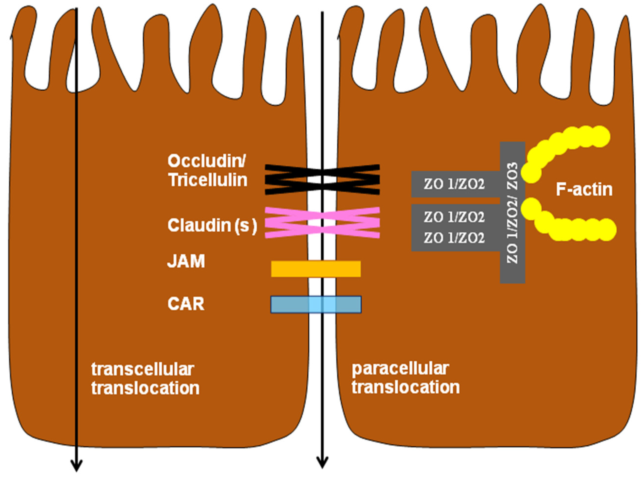
2. Molecular Structure and Function of Tight Junctions

Generally, TJs are multi protein complexes consisting of transmembrane proteins, linked to the actin cytoskeleton via cytoplasmic proteins [23]. Approximately 50 TJ proteins have been identified. Transmembrane proteins, principally claudins, occludin, junctional adhesion molecules (JAMs), the coxsackie virus and adenovirus receptor (CAR) and tricellulin (Figure 1), are contributing to the semi-permeable barrier, whereas cytosolic proteins not only link membrane components to the actin cytoskeleton they also participate in signaling between TJs and the cell nucleus.
Proteins of the claudin family are a main component of tight junctions and form a seal that modulates paracellular transport in the intestinal epithelium [25]. It was also suggested that claudins have an important role in the regulation of cellular signaling [26,27]. Claudin-1, -3, and -5, and cldn-16, ZO-1 and ZO-2 expressions were demonstrated in the chicken intestinal epithelium [26,28,29,30]. It is known that claudin-1, -3, -4, -5, -7, and -19 are pore-sealing claudins. An increased expression of these proteins leads to a very tight epithelia, coinciding with an increased transepithelial electrical resistance (TER) and decreased solute permeability (mainly sodium ions) across the epithelial monolayer [31,32,33]. Conversely, claudin-2 and -15 are considered as the pore-forming claudins, because of their ability to form paracellular anion/cation pores as well as water channels, enabling them to decrease epithelial tightness and to increase solute permeability by allowing the passage of sodium ions [34,35,36]. Taken together, claudins enable strict control over the paracellular flux of cations and anions [37].
Occludin is a TJ protein consisting of four transmembrane domains with the capability to shift to various paracellular locations and, therefore, altering epithelial permeability. Movement of occludin from the tight junction into cytoplasmic vesicles occurs frequently during barrier function loss [38] and has been shown to be triggered by multiple stimuli, such as oxidative stress and inflammation [39]. Cani et al. [11] showed that occludin expression is inversely correlated with the translocation of Fluorescein isothiocyanate (FITC) dextran from the gastrointestinal tract to the blood, emphasizing its importance in maintaining the barrier function.
Zona occludens-1 (ZO-1) was the first protein identified at tight junctions. It localizes at the cytoplasmic surface of the cell membrane, close to the TJ strands [40,41]. There are three ZO types: ZO-1, ZO-2 and ZO-3. ZO-1 plays a major role in the formation of TJs in epithelial cells compared with ZO-2 and ZO-3 [35]. Furthermore, ZO-1 serves as an important linker between the TJ and the actin cytoskeleton and is thought to be a functionally critical tight junction component. It was also found that ZO-1 is directly associated with occludin [42].
A fourth transmembrane protein, tricellulin, has also been recently discovered, a tight-junction protein forming a linkage between three adjacent cells [43]. Tricellulin is found concentrated at tricellular contacts in epithelial cellular sheets identified in epithelial cells of the kidney, intestine and stomach [43,44]. Tricellulin is a tetra-span protein with four transmembrane domains and two extracellular loops. Currently the role of tricellulin at TJs is largely unknown.
Paracellular permeability across the intestinal epithelium is regulated by tight junctions [32,45]. Over the past few years, many studies focused on identifying the mechanisms that permit a selective diffusion of ions and solutes along the paracellular pathway and much knowledge about the molecular composition of TJs has been provided [23]. Each TJ protein has a specific function which has just started to be elucidated [46]. For example, JAMs were shown to play a role in tight-junction formation, but not in barrier maintenance [47].
Tight junctions promote two functions (fence and screening functions) which are crucial for an appropriate epithelial function. The fence function is vital in maintaining apical and basolateral character, whereas the screening function acts as a gatekeeper, regulating paracellular transport of solutes between the luminal and basolateral space [48].
Finally, tight junctions are regulated by several intracellular pathways including myosin light chain kinase (MLCK), mitogen-activated protein kinases (MAPK), protein kinase C (PKC) and the Rho family of small GTPases [24]. The MLCK pathway is one of the most abundant in the gut, and is a crucial step in the regulation of tight-junctional permeability by several external stimuli, such as cytokines and pathogens [15] and the inhibition of MLCK prevents the deterioration of barrier function. To understand how tight junction proteins change in the course of barrier dysfunction, it is important to analyze such multiple proteins, in order to understand their interactions and to determine the activation status of regulatory pathways.

3. Infection and Inflammation Disrupt Barrier Function

The main entry site for pathogenic bacteria, feed contaminants such as mycotoxin and other pathogens is the digestive tract. On the contrary, the intestine forms a major physical barrier preventing pathogens and toxic compounds to cross the mucosa and to enter the body, coinciding with the activation of the immune system (innate and adaptive immune responses) against pathogens and toxic compounds. Thus, intestinal integrity is critical for maintaining a physical barrier between the intestinal lumen and the body and to protect against infection. The barrier function of TJ is of critical importance in gut physiology. TJs also play important roles in signal transduction mechanisms that regulate cell proliferation, differentiation and gene expression [49]. Under normal physiological conditions, tight junction barrier integrity remains intact and transport of toxic luminal substances together with molecules across the tight junctions is very well regulated [50].
Shen et al. [51] reported that different factors can affect the permeability of the intestinal tight junction barrier. They demonstrated that small quantities of luminal endotoxin, commensal microflora and pathogens may cross the epithelium and enter circulation through the tight junctions, when animals are under stress or suffer from an intestinal inflammation. Pathogens can also stimulate the localized secretion of pro-inflammatory cytokines from immune and intestinal epithelial cells. Consequently, these inflammatory and stress responses may induce phosphorylation of myosin light chain by myosin light chain kinase, resulting in contraction and opening of the intestinal epithelial tight junctions and an increased intestinal permeability [52,53]. In this context, it needs to be mentioned that heat stress is of high importance in poultry production and an influence on broilers physiology was demonstrated, as stressed birds suffer from multiple physiological disturbances such as damages to intestinal mucosa and higher intestinal paracellular permeability [54].
Some bacterial pathogens can impair intestinal barrier function by disruption of tight junctions and initiation of inflammatory cascades [55]. In addition, most of them attack epithelial cells either directly using effector proteins or through the elaboration of enterotoxins. Berkes et al. [56] reported that many enteropathogenic bacteria have been implicated in the disruption of tight junctions including enteropathogenic Escherichia coli (EPEC), Clostridium difficile, Clostridium perfringens, Helicobacter pylori, Campylobacter jejuni, Campylobacter concisus, and Salmonella Typhimurium. Some of these bacteria disrupt tight junctions through disorganization of specific tight junction proteins, including zonula occludens, occludin, and claudin [57]. It was also demonstrated that some of them, such as pathogenic E. coli, cause a withdrawal of ZO-1, occludin and claudins from the TJ. Furthermore, many of the barrier-disruptive mechanisms were reported such as dephosphorylation of occludin [58], decreased junctional protein expression [59] and stimulation of non-muscle myosin through myosin light chain kinase (MLCK) [60] and Rho GTPases [61].
There is evidence demonstrating a role of pro-inflammatory cytokines, such as interferon-γ (IFN-γ) and tumour necrosis factor-α (TNF-α), in endocytosis of tight junction proteins from the apical junctional complex (AJC) through MLCK and Rho-associated kinase (ROCK)-mediated manipulation of the host cell cytoskeleton [62,63,64]. In addition, pro-inflammatory stimuli trigger intestinal epithelia to express more of the relatively permeable tight junction proteins (e.g., claudin-2) and less of the relatively impermeable junction proteins (claudins-1, -3, -4, -5, and -8), resulting in a decreased barrier function [65,66]. Epithelial barrier dysfunctions occur in inflammatory bowel diseases that contribute to leaky-flux diarrhea, coinciding with a loss of solutes and water. Furthermore, down-regulation of pore-sealing claudins (4, 5, and 8), but up-regulation of pore-forming claudin-2 is observed in Crohn’s disease [66,67].
Moreover, it was shown that bacterial toxins, such as endotoxins of Gram-negative bacteria (LPS), could induce disorders in intestinal epithelial barrier function [7]. Therefore, intestinal and systemic diseases are associated with leaky epithelial barrier and consequently increased intestinal permeability to endotoxin. Additionally, Albin et al. [68] showed that endotoxins can alter the intestinal integrity and junctional organization. Finally, the disruption of gut barrier could induce a malabsorption of nutrients and translocation of enteric bacteria to various internal organs, leading to disease and reduced growth performance [5,69,70,71,72].
Generally, it can be concluded that an impaired gut barrier function is a common characteristic of many local but also systemic infections and a leaking gut is thought to contribute to the severity of clinical symptoms. Finally, enteric pathogens utilize a diverse array of strategies to alter host tight junction barrier function and such alterations can contribute to different infection outcomes. Thus, mechanisms whereby certain enteric pathogens disrupt the tight junctional complexes will be addressed below in more detail and how, in turn, these disruptions may be implicated in gastrointestinal dysfunction.

3.1. Enteropathogenic Escherichia coli (EPEC)

Enteropathogenic E. coli are a major cause of bacterial diarrhea and hemorrhagic colitis in both humans and animals [73,74,75] with consequences on intestinal epithelial barrier function. Ugalde-Silva et al. [76] reported that EPEC injects effector proteins directly from the bacterial cytoplasm to the host cell cytoplasm and thereby alters the eukaryotic cell functions through modifying or blocking cell signaling pathways. Philpott et al. [77] demonstrated in an in vitro study that EPEC induces a time and dose dependent drop in tissue resistance across intestinal epithelial cell monolayers with an increase in the paracellular permeability.
Furthermore, Muza-Moons et al. [78] showed that the tight junction proteins occludin, claudin and ZO-1 are affected by an EPEC infection in T84 intestinal epithelial cells. Roxas et al. [79] reported that E. coli induced changes in intestinal ion permeability in the colon of mice were due to alterations in tight junction architecture. Applying immunofluorescence microscopy, a redistribution of the tight junction proteins occludin and claudin-3 together with an increased expression of claudin-2 could be demonstrated. Furthermore, it was demonstrated that the acute exposure to E. coli (enteric non-pathogenic and pathogenic) reduced the epithelial ion conductance [80]. It was also found that an infection with EPEC reduced significantly the TJ proteins (occludin (phosphorylated form), ZO-1 and claudin-1), which is supported by other findings reporting a rapid and progressive dephosphorylation of occludin following EPEC infection [58].
Hofman [81] showed that bacterial toxins have the ability to dilate TJs and increase paracellular permeability. These effects may result from direct modification of TJ proteins (occludin, claudins, ZO-1, ZO-2, ZO-3) or by direct binding to a TJ component or by alteration of the peri-junctional actin filaments, keeping in mind that overlapping effects may appear. It was reported that the EPEC secreted effector protein F (EspF) is necessary for disrupting the tight junction barrier function in vitro [82] (Table 2). A study using epithelial cell lines has demonstrated that an EPEC infection leads to a decrease in TER as well as a disruption of tight junction barrier function through redistribution, dephosphorylation and dissociation of tight junction proteins [82,83]. In an in vivo study, it was mentioned that the EPEC-induced tight junction barrier disruption is EspF dependent at earlier time points of infection, while altered barrier function at the later time point was shown to coincide with increased production of the pro-inflammatory cytokine TNF-alpha [84,85]. Altogether, it can be summarized that the intestinal epithelial response to infection can be multifactorial.

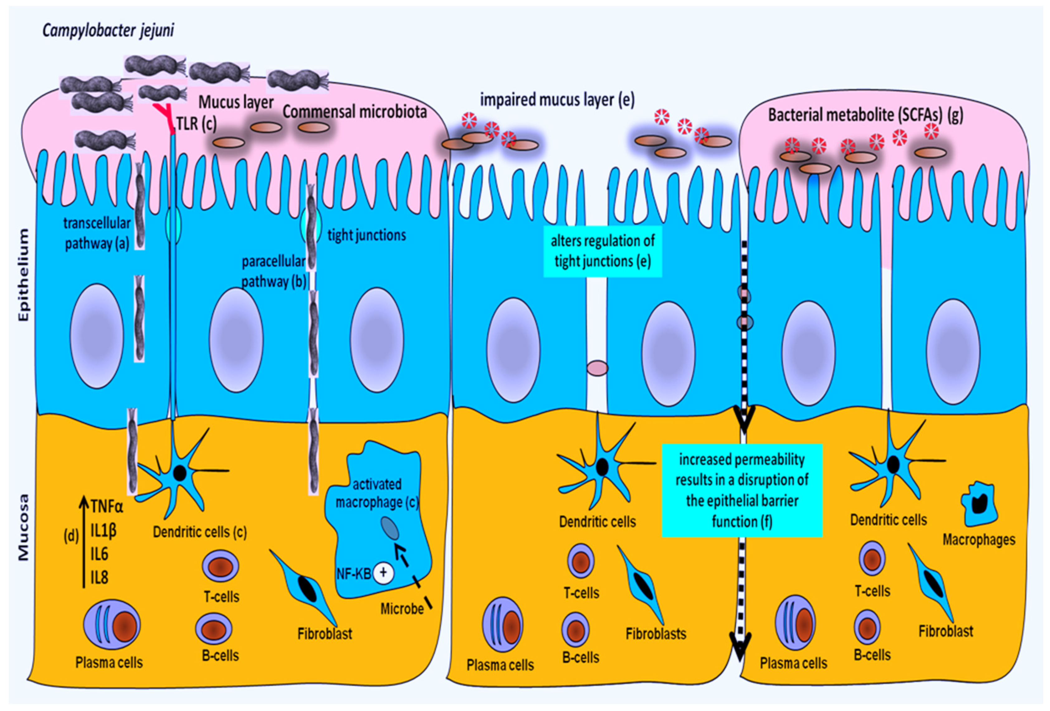
3.2. Campylobacter jejuni

Different studies demonstrated that C. jejuni can disrupt the structure of tight junction proteins, in order to facilitate their paracellular passage into the underlying tissues [86,87,88]. The mechanisms by which C. jejuni affects tight junction functions are summarized in Table 3. In one of these studies, it was demonstrated that Campylobacter can affect the intestinal integrity by disrupting occludin, an integral tight junction protein, enhancing the paracellular passage of Campylobacter in Caco-2 cell monolayers [86]. Accordingly, the host will elevate levels of pro-inflammatory cytokines, such as TNF-α and IFN-γ which have been shown to affect the structure of tight junctions, to disrupt the barrier function and to facilitate the passage of luminal antigens into the underlying tissues [87]. In addition, paracellular leakage contributes to a disturbance of selective intestinal transport (e.g., toxin absorption) and diarrhea [89].
In another in vitro study with Caco-2 cells [90] it was demonstrated that C. jejuni is capable to enter host eukaryotic cells via endocytosis. In the same study it was also revealed that C. jejuni 81116, in the presence of IFN-γ alone or with TNF-α, resulted in a focal redistribution of occludin and increased cellular damage within 24 h. Furthermore, co-infection of C. jejuni and E. coli caused a significant decrease in TEER within 6 g, with a focal redistribution of occludin, correlating with an influx of C. jejuni into the basal side of enterocytes. Dodson [90] also hypothesized that once Campylobacter colonized the gastrointestinal tract of a susceptible host, the infection causes an intestinal inflammation which would result in a rapid loss of tight junction barrier function. Similarly, other studies showed that C. jejuni-induced barrier dysfunction was associated with altered claudin-4 expression and distribution [91,92].
Recently, some studies showed that C. jejuni promotes the translocation of C. jejuni itself as well as other commensal bacteria in mammals and chickens [70,93,94]. It is supposed that the intestinal bacteria target various intracellular pathways, change the expression and distribution of TJ proteins and thereby alter gut permeability. In addition, this pathogen can affect the gut barrier functions by inducing fluid and electrolyte secretion and initiate inflammatory responses [56]. Hence, it was shown that C. jejuni infections in some broiler lines (fast-growing lines) are characterized by diarrhea, a prolonged inflammatory response and induction of lymphocyte activation in cecal tissue [95]. Moreover, C. jejuni colonization was associated with an alteration of the gut microbiota with changes in bacterial metabolic activity (short-chain fatty acids, SCFAs) [70,71]. In a recent study, we showed that Campylobacter infection strongly interferes with Ca2+ signaling [69]. It can be hypothesized that such an interaction of C. jejuni with [Ca2+] can have profound effects on cellular functions and may support cellular invasion of Campylobacter by microvillar cytoskeleton rearrangement which needs further approval.
Chickens are recognized as an imperative source of thermophilic Campylobacter, carrying this pathogen in their intestinal tract. Recently, it was revealed that C. jejuni colonization in the chicken intestine was accompanied with mucosal damage and a higher intestinal permeability which indicates that C. jejuni may translocate via the paracellular, in addition to the transcellular, pathway [5,69,95,96] (Figure 2). Although both pathways can be involved in bacteria translocation, the paracellular pathway appears to be of particular importance in dissemination towards inner organs. In chickens, it can be hypothesized that the barrier function of the intestinal epithelium is markedly altered in colonized birds and this alteration could be a part of the colonization strategy leading to persistent infection of the gut [97]. However, the mechanisms involved in C. jejuni-induced barrier dysfunction in chickens remain unclear and further studies are needed to determine how Campylobacter influences the gut barrier during infection. Additionally, elucidating the mechanisms by which C. jejuni is able to cross the gut should help in finding suitable options to decrease the level of bacteria in internal organs.

3.3. Salmonella enterica

Salmonella is another important food-borne pathogen with limited clinical signs in chickens, although intestinal inflammation and elevated cross-contamination are noticed [98,99,100]. Infection of epithelial cell monolayers by S. Typhimurium resulted in a disrupted TJ structure and function [16,17,101,102,103] (Table 4). Similarly, in an in vitro experiment it was demonstrated that the infection of T84 intestinal epithelial cells with S. Typhimurium elicited a rapid drop in tissue resistance, accompanied by an increase in the paracellular flux of fluorescence labeled markers across the infected cell monolayer [103]. Coinciding with the increased paracellular permeability, Salmonella caused a decrease in the expression of both ZO-1 and phosphorylated occludin, redistribution of claudin-1 and ZO-2 proteins, facilitation of bacterial translocation, and loss of barrier function [103].
It has also been described that S. Typhimurium invasion of intestinal epithelia is accompanied by a loss of epithelial integrity and, consequently, an impaired epithelial function in mice [57,104]. Moreover, an infection of intestinal epithelial cells (T84 or MDCK) with S. Typhimurium can cause a progressive decrease in transepithelial electrical resistance, alteration of intestinal TJ proteins, a damage of intestinal barrier function and facilitates the translocation of both pathogenic and non-pathogenic bacteria across epithelial cell monolayers, indicating a disruption of the tight junction barrier [102,103]. It was further reported that the increased paracellular permeability following a S. Typhimurium infection can be due to the contraction of the perijunctional actin ring and alteration in the Rho GTPase activity via the type three secretion system (T3SS) effector proteins, thereby altering the function of tight junctions [16,17,102].
In a similar way, Zhang et al. [100] found that a S. Typhimurium challenge decreased claudin-1 and occludin mRNA expression in the ileum of broiler chickens. Moreover, it was reported that the intestinal tight junction proteins claudin-1, claudin-4 and occludin mRNA expression in the jejunum at 14 days post infection (dpi) were significantly decreased by a S. Typhimurium challenge in broilers [105]. This down-regulation of TJ proteins resulted in an enhancement of paracellular permeability and disruption of the intestinal barrier, thereby allowing the diffusion of macromolecules, such as bacterial toxins (endotoxin) and pathogens, from the intestinal lumen into the blood circulation [106,107].
Like S. Typhimurium, Salmonella Enteritidis could also alter the tight junction function. Awad et al. [20] found that luminal S. Enteritidis affects the intestinal epithelium of chickens in the same way as its endotoxin and it decreases intestinal ion permeability of chickens directly after acute exposure. This is in contrast to findings in pigs where Salmonella endotoxin does not elicit an acute decrease in permeability [108]. This finding could explain why chickens do not experience overt secretory diarrhea when infected by this pathogen in contrast to pigs and other species, including humans, which is seen as a result of a differently regulated gut function. Finally, intestinal TJ disruption results not only in an increased permeability to luminal antigens and bacteria translocation, it also lowers the absorption of nutrients [24,56], consequently, can interfere with productivity and enhance severity of clinical signs.

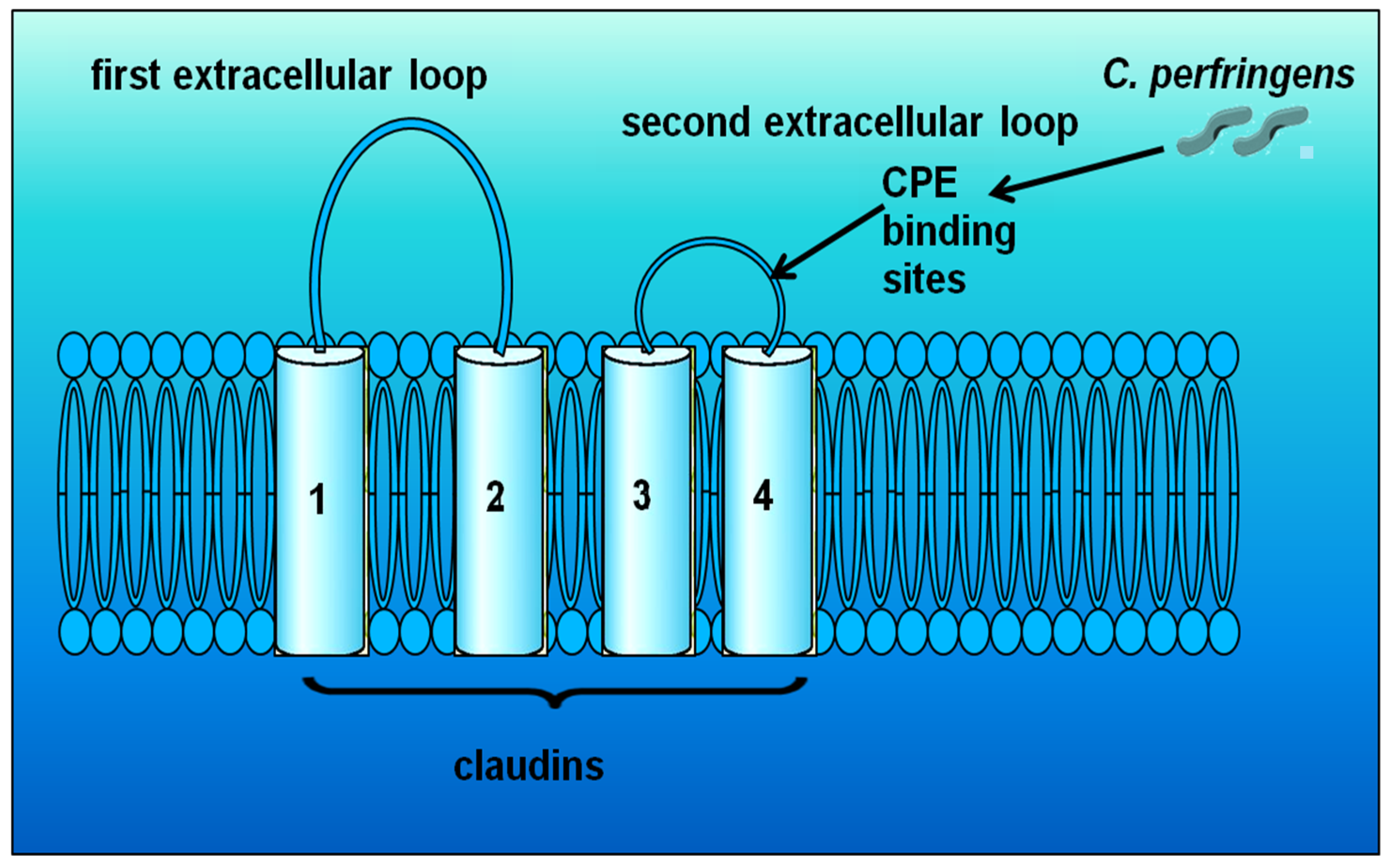
3.4. Clostridium perfringens

Necrotic enteritis in poultry is a frequently reported disease condition caused by the abundant growth of Clostridium perfringens in the intestine. C. perfringens strains are characterized by the production of major toxins (alpha, beta, epsilon and iota), many of these toxins have been demonstrated to contribute to the virulence of bacteria and to play a key role in the pathogenesis of animal infections [109]. Producing different toxins increases the flexibility of C. perfringens in causing disease under varying host conditions [110]. Recently, it was found that the majority of C. perfringens isolates from chickens with clinical signs of necrotic enteritis carry the necrotic enteritis B-like toxin (NetB). Earlier, it was believed that proliferation of Clostridium perfringens and production of alpha-toxin is the major factor for necrotic enteritis (NE) in poultry; however, NetB has recently been shown to be an essential virulence factor in clinical necrotic enteritis in broiler chickens [110,111,112,113]. NetB is a cytotoxic, haemolytic, pore-forming toxin for avian cells and it was revealed that the development of necrotic enteritis in chickens was dependent on the ability to produce NetB [111]. Beside the clinical form of C. perfringens necrotic enteritis in poultry, the subclinical form has also been described in the field, characterized by a damaged intestinal mucosa, decrease in digestion and absorption and reduced performance [114,115,116]. Subclinical infections are coinciding with a reduction in growth performance and negatively impacting productivity, without being recognized and treated [110].
Many pathogens impair junctional structures indirectly by activation of signaling cascades of host cells. However, Clostridium perfringens enterotoxin (CPE) uses TJ proteins directly as cell surface receptors to attach [117]. CPE, a cytotoxic, pore-forming toxin, uses the claudin family as cellular receptors and it has been shown that it attaches to claudin-3 and claudin-4 of MDCK cell monolayers (Figure 3) [118,119,120,121]. Similarly, Saitoh et al. [122] revealed that CPE can bind to specific claudins, resulting in the disintegration of TJs and an increase in the paracellular permeability across epithelial cell layers. Additionally, Singh et al. [123] showed that CPE can also interact with other TJs like occludin, following binding of the enterotoxin to its receptors (claudins) in Caco-2 cell monolayers. In another study, the application of CPE to basolateral membranes was found to affect the tight junction structure by inducing a fragmentation of tight junctions within 1 h [124]. After binding, CPE damages the membrane permeability and leads to calcium influx into the cell, resulting in cell damage [125].
Nava and Vidal [126] demonstrated in human intestinal cells that an infection with a C. perfringens type C strain induced a significant drop on TEER and this change was mediated by redistribution of TJs protein occludin and Claudin-3. In rats, it was shown that the phospholipase C activity of the alpha toxin impaired the intestinal mucosal barrier and increased the permeability of the intestine through activation of phospholipase (Table 5) [127]. In chickens, it was reported that mucosal addition of C. perfringens alpha toxin can impair the intestinal mucosal barrier [128,129]. Finally, Collier et al. [130] observed that the paracellular permeability was higher in tissues from chickens infected with C. perfringens.

4. Impaired Barrier Function and Growth Performance

In poultry, the effects of enteric pathogens are not always obvious, but even in those cases where chickens do not show clinical symptoms, they may have negative effects on feed consumption, growth, immune system and other health parameters. Since it was shown that the damage of the intestinal barrier may increase the passage of pathogens to access the underlying lamina propia and activate the host’s immune compartment, impaired nutrient absorption which results in the availability of the necessary growth substrate for the proliferation of pathogens [69,70].
For instance, it was reported that the exposure to bacterial endotoxin (e.g., Escherichia coli or Salmonella typhimurium) affects the birds’ performance by a reduction in body weight and a worsening of feed conversion rate [7]. Furthermore, some studies showed that Campylobacter negatively impacted poultry production by a reduction of body weight with consequences on the well-being of chickens [5,95,96]. Additionally, subclinical necrotic enteritis was estimated to result in a 12% reduction in body weight and a 10.9% increase in Feed conversion ratio (FCR) compared with healthy birds, imposing a significant economic burden on the poultry industry worldwide [131]. It was estimated that losses due to altered body weight and FCR associated with subclinical necrotic enteritis range from US$878.19 to US$1480.52 per flock (20,000 birds) to reach the market weight [131].
The mechanisms by which enteric pathogens affect growth through the impaired gut barrier may comprise: (1) interference with protein synthesis and degradation; (2) alteration of the intestinal integrity (damage of intestinal villi) and disruption of the normal activity of nutrient transporters, resulting in reduced nutrient absorption; (3) increase of nutrients available for luminal pathogen proliferation, resulting in an amplification of the severity of infection; (4) increase of the maintenance requirements of the gut (for immune function needs) and thus decrease nutrient availability for the host; and (5) increase nutrient loss (decrease digestibility) by interfering with digestive enzyme synthesis and/or activities [69,70,96,132].
Finally, enteric pathogens (subclinical infections) are an important concern to the poultry industry because of production losses, reduced welfare of birds and increased risk of contamination of poultry products for human consumption, leading to high economic losses.

5. Restoration of the Impaired Barrier Function

Intestinal leakage, as a result of increased paracellular permeability, is a dominant feature within the pathophysiology of many enteric pathogens and could ultimately lead to an increased translocation of intestinal bacteria into the body. Therefore, impaired gut barrier function needs to be restored. Many strategies have been used for restoration either via dietary management or immunotherapy (stimulates or restores the ability of the immune defense system to counteract infection). In this context, it could be demonstrated that dietary fiber exerts beneficial effects in the gut through its bacterial metabolite, the short-chain fatty acid butyrate [133]. It has also been demonstrated that a supplementation of the chicken diet by either prebiotic, probiotic or synbiotic has an impact on barrier function [134,135,136,137,138,139,140,141,142,143,144]. It was shown that these feed additives act as quantitatively available substrates for the gastrointestinal microflora within the gut of the host [145]. They also enhance the growth of beneficial bacteria (Bifidobacterium and Lactobacillus), inhibit the growth of pathogenic bacteria like Escherichia coli and Salmonella spp., and, in consequence, improve the microbial balance in the gastrointestinal tract [146,147,148]. It was also reported that functional oligosaccharides could ameliorate the adverse effects on barrier integrity caused by heat stress in chickens [54]. In addition, the gut microbiota (commensal) itself is known to modulate barrier function which could be a potential therapeutic target [149]. This is supported by the finding that certain strains of Lactobacilli could reduce the permeability by increasing the relocation of occludin and ZO-1 tight junction in duodenal epithelial cells [150].
Similarly, a particular focus has been elicited on prevention of necrotic enteritis in poultry caused by Clostridium perfringens by the use of microbes (Bacillus and Lactobacillus) or microbe-derived products (yeasts) [151]. Liu et al. [152] found that dietary supplementation of exogenous lysozyme decreased the C. perfringens colonization and improved the intestinal barrier function of chickens. Furthermore, it was demonstrated that a prebiotic product (arabinogalactan Fibregum) was effective in controlling NE [153].
Linking protein synthesis with intestinal barrier permeability reflects an important feature [154,155,156,157]. Amino acids are not only important substrates for protein synthesis they are critical in supporting gut barrier integrity and function. Thereby, amino acids supplementation can be useful for alleviating intestine injuries. Thus far, attention was drawn towards glutamine offering a beneficial effect on the intestinal mucosa and gut function, which can be explained by its capability to act as an important energy source, similar to glucose [158]. It was evidenced that glutamine deprivation causes Caco-2 cell injury [159], whereas glutamine supplementation protects Caco-2 cells from barrier dysfunction [160]. Additionally, dietary supplementations with host defense peptides (HDPs) were recently shown to enhance mucosal barrier function directly by inducing the expression of TJ proteins and indirectly by displaying an improvement in nutrient utilization and a reduction in Clostridium spp. and coliform bacteria in the intestinal tract of broiler chickens [161].
Some immune-based therapies were designed to reduce intestinal inflammation and subsequent systemic immune activation in mice. Such immune activations were associated with reduced microbial translocation and enhanced expression of gut-junction genes. Recently, several studies have been focused on anti-inflammatory therapy which could block pro-inflammatory pathways. Some studies showed that a flavonoid (Nobiletin®) exerted significant anti-inflammatory effects via downregulation of inducible nitric oxide synthase (iNOS) and cyclooxygenase 2 (COX-2) expressions in vivo in rats and cell cultures (BV-2 or Caco-2) [162,163]. Therefore, basic research has to continue to provide insight into the ultimate strategies to be favored in order to restore the barrier function and to protect against onset and progression of enteric infections, coinciding with inflammation.

6. Importance of the Chicken Intestinal Epithelial Barrier

Sustaining a healthy gut is a prerequisite for efficient performance of farm animals, especially poultry with its high growth rate. The gut plays a key role in the digestion and absorption of nutrients and it constitutes one of the main entrance gates exposed to external factors that can challenge the bird’s health. The intestinal epithelial barrier serves as the first boundary of defense between the organism and the luminal environment. It consists of a continuous monolayer of intestinal epithelial cells which are connected together by an intercellular junctional complex limiting the space between adjacent cells. This minimizes the access of pathogens and toxins to spread into the host. Substantial evidence indicates that intestinal barrier dysfunction is considered as etiological factor in the pathogenesis of some enteric diseases [164]. Furthermore, paracellular ions and nutrient permeation is restricted by the presence of tight junctions and consequently can affect the intestinal absorptive function.
Thus, tight junction proteins play a dominant role in barrier formation. However, it is still a matter of debate how the paracellular barrier of the chicken intestine is organized, horizontally and vertically, to support a strict compartmental separation on the one hand and the transepithelial transport rates on the other hand. Thus, more knowledge on the composition of tight junction proteins in chickens are fundamental for understanding pathogenic pathways, further supporting a primary role of the epithelium tight junction in the pathogenesis of intestinal enteric pathogens and emphasizing the importance to maintain a healthy and effective intestinal barrier. In addition, chickens are an important source of zoonotic enteric pathogens. Therefore, elucidating the changes of mucosal barrier during enteric pathogens is crucial and may help in providing new tools to restore the intestinal barrier functions during infection.

7. Conclusions

Tight junctions are formed at the lateral sites of the cell and regulate the paracellular passage of molecules. However, not all tight junctions, consisting of multiple proteins, are merely tight as some tight junction proteins build their own transport pathways by forming channels selective for small cations, anions, or water, resulting in epithelial surfaces of different tightness. Tight junctions are regulated in their molecular composition, ultrastructure and function by intracellular scaffolding proteins and cytoskeleton. Such a cascade of interaction is not only part of cellular physiology and various adaptation processes, it can also be impaired by different microorganisms. Chronic infections with certain enteric pathogens can compromise intestinal barrier function and activate a systemic response which consequently could reduce growth efficiency.
Although the intestinal barrier and intestinal permeability are important for health and disease, the mucosal barrier and its role in enteric disease are still poorly defined in chickens. Therefore, future studies should aim to elucidate the molecular basis of the differential responses of the chicken gut to infections with certain microorganisms, which is critical for bird’s health. The improvement of food safety would be an additional surplus. Based on existing data, it can be concluded that modulation of microbiota with probiotics for repairing the gut barrier reflects a promising approach that warrants future investigations to minimize the effects of enteropathogenic microorganims in poultry.

Acknowledgments

This work was performed within the CEPI (Centre of Excellence for Poultry Innovation) project, which was funded by the European Regional Development Fund, Cross-border Cooperation Programme Austria–Hungary 2014–2020. Grant No: ATHU19.

Author Contributions

W.A.A. conceived and designed the work; W.A.A. and C.H. analyzed the data; and W.A.A. and M.H. wrote the paper.

Conflicts of Interest

The authors declare no conflict of interest.

References

  1. Tomita, M.; Ohkubo, R.; Hayashi, M. Lipopolysaccharide transport system across colonic epithelial cells in normal and infective rat. Drug Metab. Pharmacokinet. 2004, 19, 33–40. [Google Scholar] [CrossRef] [PubMed]
  2. Di Pierro, M.; Lu, R.; Uzzau, S.; Wang, W.; Margaretten, K.; Pazzani, C.; Maimone, F.; Fasano, A. Zonula occludens toxin structure-function analysis. Identification of the fragment biologically active on tight junctions and of the zonulin receptor binding domain. J. Biol. Chem. 2001, 276, 19160–19165. [Google Scholar] [CrossRef] [PubMed]
  3. Sultana, R.; McBain, A.J.; O’Neilla, C.A. Strain-dependent augmentation of tight-junction barrier function in human primary epidermal keratinocytes by Lactobacillus and Bifidobacterium Lysates. Appl. Environ. Microbiol. 2013, 79, 4887–4894. [Google Scholar] [CrossRef] [PubMed]
  4. Hecht, G. Microbes and microbial toxins: Paradigms for microbial–mucosal interactions. VII. Enteropathogenic Escherichia coli: Physiological alterations from an extracellular position. Am. J. Physiol. Gastrointest. Liver Physiol. 2001, 281, G1–G7. [Google Scholar] [PubMed]
  5. Awad, W.A.; Molnár, A.; Aschenbach, J.R.; Ghareeb, K.; Khayal, B.; Hess, C.; Liebhart, D.; Dublecz, K.; Hess, M. Campylobacter infection in chickens modulates the intestinal epithelial barrier function. Innate Immun. 2015, 21, 151–160. [Google Scholar] [CrossRef] [PubMed]
  6. Ghareeb, K.; Awad, W.A.; Böhm, J.; Zebeli, Q. Impacts of the feed contaminant deoxynivalenol on the intestine of monogastric animals: Poultry and swine. J. Appl. Toxol. 2015, 35, 327–337. [Google Scholar] [CrossRef] [PubMed]
  7. Ghareeb, K.; Awad, W.A.; Böhm, J.; Zebeli, Q. Impact of luminal and systemic endotoxin exposure on gut function, immune response and performance of chickens. World’s Poult. Sci. J. 2016, 72, 367–380. [Google Scholar] [CrossRef]
  8. Bjarnason, I.; Macpherson, A.; Hollander, D. Intestinal permeability: An overview. Gastroenterology 1995, 108, 1566–1581. [Google Scholar] [CrossRef]
  9. Nighot, P.K.; Blikslager, A.T. ClC-2 regulates mucosal barrier function associated with structural changes to the villus and epithelial tight junction. Am. J. Physiol. Gastrointest. Liver Physiol. 2010, 299, G449–G456. [Google Scholar] [CrossRef] [PubMed]
  10. Awad, W.A.; Böhm, J.; Razzazi-Fazeli, E.; Hulan, H.W.; Zentek, J. Effects of deoxynivalenol on general performance and electrophysiological properties of intestinal mucosa of broiler chickens. Poult. Sci. 2004, 83, 1964–1972. [Google Scholar] [CrossRef] [PubMed]
  11. Cani, P.D.; Possemiers, S.; Van de Wiele, T.; Guiot, Y.; Everard, A.; Rottier, O.; Geurts, L.; Naslain, D.; Neyrinck, A.; Lambert, D.M.; et al. Changes in gut microbiota control inflammation in obese mice through a mechanism involving GLP-2-driven improvement of gut permeability. Gut 2009, 58, 1091–1103. [Google Scholar] [CrossRef] [PubMed]
  12. Shen, L. Tight junctions on the move: Molecular mechanisms for epithelial barrier regulation. Ann. N. Y. Acad. Sci. 2012, 1258, 9–18. [Google Scholar] [CrossRef] [PubMed]
  13. Fasano, A.; Nataro, J.P. Intestinal epithelial tight junctions as targets for enteric bacteria-derived toxins. Adv. Drug Deliv. Rev. 2004, 56, 795–807. [Google Scholar] [CrossRef] [PubMed]
  14. O’Hara, J.R.; Buret, A.G. Mechanisms of intestinal tight junctional disruption during infection. Front. Biosci. 2008, 13, 7008–7021. [Google Scholar] [PubMed]
  15. Scott, K.G.; Meddings, J.B.; Kirk, D.R.; Lees-Miller, S.P.; Buret, A.G. Intestinal infection with Giardia spp. reduces epithelial barrier function in a myosin light chain kinase-dependent fashion. Gastroenterology 2002, 123, 1179–1190. [Google Scholar] [CrossRef] [PubMed]
  16. Tafazoli, F.; Magnusson, K.E.; Zheng, L. Disruption of epithelial barrier integrity by Salmonella enterica serovar typhimurium requires geranylgeranylated proteins. Infect. Immun. 2003, 71, 872–881. [Google Scholar] [CrossRef] [PubMed]
  17. Boyle, E.C.; Brown, N.F.; Finlay, B.B. Salmonella enterica serovar typhimurium effectors SopB, SopE, SopE2 and SipA disrupt tight junction structure and function. Cell Microbiol. 2006, 8, 1946–1957. [Google Scholar] [CrossRef] [PubMed]
  18. Awad, W.A.; Razzazi-Fazeli, E.; Böhm, J.; Zentek, J. Influence of deoxynivalenol on the d-glucose transport across the isolated epithelium of different intestinal segments of laying hens. J. Anim. Physiol. Anim. Nutr. 2007, 91, 175–180. [Google Scholar] [CrossRef] [PubMed]
  19. Awad, W.A.; Vahjen, W.; Aschenbach, J.R.; Zentek, J. A diet naturally contaminated with the Fusarium mycotoxin deoxynivalenol down regulates gene expression of glucose transporters in the intestine of broiler chickens. Livestock Sci. 2011, 140, 72–79. [Google Scholar] [CrossRef]
  20. Awad, W.A.; Aschenbach, J.R.; Khayal, B.; Hess, C.; Hess, M. Intestinal epithelial responses to Salmonella enterica serovar enteritidis: Effects on the intestinal permeability and ion transport. Poult. Sci. 2012, 91, 2949–2957. [Google Scholar] [CrossRef] [PubMed]
  21. Awad, W.A.; Aschenbach, J.R.; Setyabudi, F.; Razzazi-Fazeli, E.; Böhm, J.; Zentek, J. In vitro effects of deoxynivalenol on small intestinal d-glucose uptake and absorption of deoxynivalenol across the isolated jejunal epithelium of laying hens. Poult. Sci. 2007, 86, 15–20. [Google Scholar] [CrossRef] [PubMed]
  22. Mitjans, M.; Barniol, G.; Ferrer, R. Mucosal surface area in chicken small intestine during development. Cell Tissue Res. 1997, 290, 71–78. [Google Scholar] [CrossRef] [PubMed]
  23. Aijaz, S.; Balda, M.S.; Matter, K. Tight Junctions: Molecular architecture and function. Int. Rev. Cytol. 2006, 248, 261–298. [Google Scholar] [PubMed]
  24. Ulluwishewa, D.; Anderson, R.C.; McNabb, W.C.; Moughan, P.J.; Wells, J.M.; Roy, N.C. Regulation of tight junction permeability by intestinal bacteria and dietary components. J. Nutr. 2011, 141, 769–776. [Google Scholar] [CrossRef] [PubMed]
  25. Krause, G.; Winkler, L.; Mueller, S.L.; Haseloff, R.F.; Piontek, J.; Blasig, I.E. Structure and function of claudins. Biochim. Biophys. Acta 2008, 1778, 631–645. [Google Scholar] [CrossRef] [PubMed]
  26. Haworth, K.E.; El-Hanfy, A.; Prayag, S.; Healy, C.; Dietrich, S.; Sharpe, P. Expression of Claudin-3 during chick development. Gene Expr. Patterns 2005, 6, 40–44. [Google Scholar] [CrossRef] [PubMed]
  27. Simard, A.; Di Pietro, E.; Young, C.R.; Plaza, S.; Ryan, A.K. Alterations in heart looping induced by overexpression of the tight junction protein Claudin-1 are dependent on its C-terminal cytoplasmic tail. Mech. Dev. 2006, 123, 210–227. [Google Scholar] [CrossRef] [PubMed]
  28. Simard, A.; Di Pietro, E.; Ryan, A.K. Gene expression pattern of Claudin-1 during chick embryogenesis. Gene Expr. Patterns 2005, 5, 553–560. [Google Scholar] [CrossRef] [PubMed]
  29. Ozden, O.; Black, B.L.; Ashwell, C.M.; Tipsmark, C.K.; Borski, R.J.; Grubb, B.J. Developmental profile of claudin-3, -5, and -16 proteins in the epithelium of chick intestine. Anat. Rec. 2010, 293, 1175–1183. [Google Scholar] [CrossRef] [PubMed]
  30. Osselaere, A.; Santos, R.; Hautekiet, V.; De Backer, P.; Chiers, K.; Ducatelle, R.; Croubels, S. Deoxynivalenol impairs hepatic and intestinal gene expression of selected oxidative stress, tight junction and inflammation proteins in broiler chickens, but addition of an adsorbing agent shifts the effects to the distal parts of the small intestine. PLoS ONE 2013, 8, e69014. [Google Scholar] [CrossRef] [PubMed] [Green Version]
  31. Van Itallie, C.; Rahner, C.; Anderson, J.M. Regulated expression of Claudin-4 decreases paracellular conductance through a selective decrease in sodium permeability. J. Clin. Investig. 2001, 107, 1319–1327. [Google Scholar] [CrossRef] [PubMed]
  32. Van Itallie, C.M.; Anderson, J.M. Claudins and Epithelial Paracellular Transport. Annu. Rev. Physiol. 2006, 68, 403–429. [Google Scholar] [CrossRef] [PubMed]
  33. Krause, G.; Winkler, L.; Piehl, C.; Blasig, I.; Piontek, J.; Müller, S.L. Structure and function of extracellular claudin domains. Ann. N. Y. Acad. Sci. 2009, 1165, 34–43. [Google Scholar] [CrossRef] [PubMed]
  34. Furuse, M.; Furuse, K.; Sasaki, H.; Tsukita, S. Conversion of zonulae occludentes from tight to leaky strand type by introducing claudin-2 into Madin-Darby canine kidney I cells. J. Cell Biol. 2001, 153, 263–272. [Google Scholar] [CrossRef] [PubMed]
  35. Tsukita, S.; Furuse, M.; Itoh, M. Multifunctional strands in tight junctions. Nat. Rev. Mol. Cell Biol. 2001, 2, 285–293. [Google Scholar] [CrossRef] [PubMed]
  36. Van Itallie, C.M.; Holmes, J.; Bridges, A.; Gookin, J.L.; Coccaro, M.R.; Proctor, W.; Colegio, O.R.; Anderson, J.M. The density of small tight junction pores varies among cell types and is increased by expression of claudin-2. J. Cell Sci. 2008, 121, 298–305. [Google Scholar] [CrossRef] [PubMed]
  37. Groschwitz, K.R.; Hogan, S.P. Intestinal barrier function: Molecular regulation and disease pathogenesis. J. Allergy Clin. Immunol. 2009, 124, 3–20. [Google Scholar] [CrossRef] [PubMed]
  38. Shen, L.; Weber, C.R.; Turner, J.R. The tight junction protein complex undergoes rapid and continuous molecular remodeling at steady state. J. Cell Biol. 2008, 181, 683–695. [Google Scholar] [CrossRef] [PubMed]
  39. John, L.J.; Fromm, M.; Schulzke, J.D. Epithelial barriers in intestinal inflammation. Antioxid. Redox Signal. 2011, 15, 1255–1270. [Google Scholar] [CrossRef] [PubMed]
  40. Stevenson, B.R.; Siliciano, J.D.; Mooseker, M.S.; Goodanough, D.A. Identification of ZO-1: A high molecular weight polypcptide associated with tight junction (zonula occludens) in a variety of epithelia. J. Cell Biol. 1986, 103, 755–766. [Google Scholar] [CrossRef] [PubMed]
  41. Hunziker, W.T.; Kiener, K.; Xu, J. Vertebrate animal models unravel physiological roles for zonula occludens tight junction adaptor proteins. Ann. N. Y. Acad. Sci. 2009, 1165, 28–33. [Google Scholar] [CrossRef] [PubMed]
  42. Furuse, M.; Itoh, M.; Hirase, T.; Nagafuchi, A.; Yonemura, S.; Tsukita, S.; Tsukita, S. Direct association of occludin with ZO-1 and its possible involvement in the localisation of occludin at tight junctions. J. Cell Biol. 1994, 127, 1617–1626. [Google Scholar] [CrossRef] [PubMed]
  43. Ikenouchi, J.; Furuse, M.; Furuse, K.; Sasaki, H.; Tsukita, S.; Tsukita, S. Tricellulin constitutes a novel barrier at tricellular contacts of epithelial cells. J. Cell Biol. 2005, 171, 939–945. [Google Scholar] [CrossRef] [PubMed]
  44. Schluter, H.; Moll, I.; Wolburg, H.; Franke, W.W. The different structures containing tight junction proteins in epidermal and other stratified epithelial cells, including squamous cell metaplasia. Eur. J. Cell Biol. 2007, 86, 645–655. [Google Scholar] [CrossRef] [PubMed]
  45. Gonzalez-Mariscal, L.; Betanzos, A.; Nava, P.; Jaramillo, B.E. Tight junction proteins. Prog. Biophys. Mol. Biol. 2003, 81, 1–44. [Google Scholar] [CrossRef]
  46. Niessen, C.M. Tight junctions/adherens junctions: Basic structure and function. J. Investig. Dermatol. 2007, 127, 2525–2532. [Google Scholar] [CrossRef] [PubMed]
  47. Assimakopoulos, S.F.; Papageorgiou, I.; Charonis, A. Enterocytes’ tight junctions: From molecules to diseases. World J. Gastrointest. Pathophysiol. 2011, 2, 123–137. [Google Scholar] [CrossRef] [PubMed]
  48. Tsukita, S.; Yamazaki, Y.; Katsuno, T.; Tamura, A.; Tsukita, S. Tight junction-based epithelial microenvironment and cell proliferation. Oncogene 2008, 27, 6930–6938. [Google Scholar] [CrossRef] [PubMed]
  49. McCrea, P.D.; Gu, D.; Balda, M.S. Junctional music that the nucleus hears: Cell-cell contact signaling and the modulation of gene activity. Cold Spring Harb. Perspect. Biol. 2009, 1, a002923. [Google Scholar]
  50. Awad, W.A.; Ghareeb, K.; Böhm, J. Evaluation of the chicory inulin efficacy on ameliorating the intestinal morphology and modulating the intestinal electrophysiological properties in broiler chickens. J. Anim. Physiol. Anim. Nutr. 2011, 95, 65–72. [Google Scholar] [CrossRef] [PubMed]
  51. Shen, L.; Weber, C.R.; Raleigh, D.R.; Yu, D.; Turner, J.R. Tight junction pore and leak pathways: A dynamic duo. Ann. Rev. Physiol. 2011, 73, 283–309. [Google Scholar] [CrossRef] [PubMed]
  52. Turner, J.R. Intestinal mucosal barrier function in health and disease. Nat. Rev. Immunol. 2009, 9, 799–809. [Google Scholar] [CrossRef] [PubMed]
  53. Turner, J.R.; Rill, B.K.; Carlson, S.L.; Carnes, D.; Kerner, R.; Mrsny, R.J.; Madara, J.L. Physiological regulation of epithelial tight junctions is associated with myosin light-chain phosphorylation. Am. J. Physiol. 1997, 273, C1378–C1385. [Google Scholar] [PubMed]
  54. Song, J.; Jiao, L.F.; Xiao, K.; Luan, Z.S.; Hu, C.H.; Shi, B.; Zhan, X.A. Cello-oligosaccharide ameliorates heat stress-induced impairment of intestinal microflora, morphology and barrier integrity in broilers. Anim. Feed Sci. Technol. 2013, 185, 175–181. [Google Scholar] [CrossRef]
  55. Guttman, J.A.; Finlay, B.B. Tight junctions as targets of infectious agents. Biochim. Biophys. Acta 2009, 1788, 832–841. [Google Scholar] [CrossRef] [PubMed]
  56. Berkes, J.; Viswanathan, V.K.; Savkovic, S.D.; Hecht, G. Intestinal epithelial responses to enteric pathogens: Effects on the tight junction barrier, ion transport, and inflammation. Gut 2003, 52, 439–451. [Google Scholar] [CrossRef] [PubMed]
  57. Sears, C.L. Molecular physiology and pathophysiology of tight junctions V. Assault of the tight junction by enteric pathogens. Am. J. Physiol. Gastrointest. Liver Physiol. 2000, 279, G1129–G1134. [Google Scholar] [PubMed]
  58. Simonovic, I.; Rosenberg, J.; Koutsouris, A.; Hecht, G. Enteropathogenic Escherichia coli dephosphorylates and dissociates occludin from intestinal epithelial tight junctions. Cell. Microbiol. 2000, 2, 305–315. [Google Scholar] [CrossRef] [PubMed]
  59. Howe, K.L.; Reardon, C.; Wang, A.; Nazli, A.; McKay, D.M. Transforming growth factor-beta regulation of epithelial tight junction proteins enhances barrier function and blocks enterohemorrhagic Escherichia coli O157:H7-induced increased permeability. Am. J. Pathol. 2005, 167, 1587–1597. [Google Scholar] [CrossRef]
  60. Philpott, D.J.; McKay, D.M.; Mak, W.; Perdue, M.H.; Sherman, P.M. Signal transduction pathways involved in enterohemorrhagic Escherichia coli-induced alterations in T84 epithelial permeability. Infect. Immun. 1998, 66, 1680–1687. [Google Scholar] [PubMed]
  61. Matsuzawa, T.; Kuwae, A.; Abe, A. Enteropathogenic Escherichia coli type III effectors EspG and EspG2 alter epithelial paracellular permeability. Infect. Immun. 2005, 73, 6283–6289. [Google Scholar] [CrossRef] [PubMed]
  62. Zolotarevsky, Y.; Hecht, G.; Koutsouris, A.; Gonzalez, D.E.; Quan, C.; Tom, J.; Mrsny, R.J.; Turner, J.R. A membrane-permeant peptide that inhibits MLC kinase restores barrier function in in vitro models of intestinal disease. Gastroenterology 2002, 123, 163–172. [Google Scholar] [CrossRef] [PubMed]
  63. Bouma, G.; Strober, W. The immunological and genetic basis of inflammatory bowel disease. Nat. Rev. Immunol. 2003, 3, 521–533. [Google Scholar] [CrossRef] [PubMed]
  64. Utech, M.; Ivanov, A.I.; Samarin, S.N.; Bruewer, M.; Turner, J.R.; Mrsny, R.J.; Parkos, C.A.; Nusrat, A. Mechanism of IFN-gamma-induced endocytosis of tight junction proteins: Myosin II-dependent vacuolarization of the apical plasma membrane. Mol. Biol. Cell 2005, 16, 5040–5052. [Google Scholar] [CrossRef] [PubMed]
  65. Prasad, S.; Mingrino, R.; Kaukinen, K.; Hayes, K.L.; Powell, R.M.; MacDonald, T.T.; Collins, J.E. Inflammatory processes have differential effects on claudins 2, 3 and 4 in colonic epithelial cells. Lab. Investig. 2005, 85, 1139–1162. [Google Scholar] [CrossRef] [PubMed]
  66. Zeissig, S.; Bürgel, N.; Günzel, D.; Richter, J.; Mankertz, J.; Wahnschaffe, U.; Kroesen, A.J.; Zeitz, M.; Fromm, M.; Schulzke, J.D. Changes in expression and distribution of claudin-2, -5 and -8 lead to discontinuous tight junctions and barrier dysfunction in active Crohn’s disease. Gut 2007, 56, 61–72. [Google Scholar] [CrossRef] [PubMed]
  67. Das, P.; Goswami, P.; Das, T.K.; Nag, T.; Sreenivas, V.; Ahuja, V.; Panda, S.K.; Gupta, S.D.; Makharia, G.K. Comparative tight junction protein expressions in colonic Crohn’s disease, ulcerative colitis, and tuberculosis: A new perspective. Virchows Arch. 2012, 460, 261–270. [Google Scholar] [CrossRef] [PubMed]
  68. Albin, D.M.; Wubben, J.E.; Rowlett, J.M.; Tappenden, K.A.; Nowak, R.A. Changes in small intestinal nutrient transport and barrier function after lipopolysaccharide exposure in two pig breeds. J. Anim. Sci. 2007, 85, 2517–2523. [Google Scholar] [CrossRef] [PubMed]
  69. Awad, W.A.; Smorodchenko, A.; Hess, C.; Aschenbach, J.R.; Molnár, A.; Dublecz, K.; Khayal, B.; Pohl, E.E.; Hess, M. Increased intracellular calcium level and impaired nutrient absorption are important pathogenicity traits in the chicken intestinal epithelium during Campylobacter jejuni colonization. Appl. Microbiol. Biotechnol. 2015, 99, 6431–6441. [Google Scholar] [CrossRef] [PubMed]
  70. Awad, W.A.; Dublecz, F.; Hess, C.; Dublecz, K.; Khayal, B.; Aschenbach, J.R.; Hess, M. Campylobacter jejuni colonization promotes the translocation of Escherichia coli to extra-intestinal organs and disturbs the short-chain fatty acids profiles in the chicken gut. Poult. Sci. 2016, 95, 2259–2265. [Google Scholar] [CrossRef] [PubMed]
  71. Awad, W.A.; Mann, E.; Dzieciol, M.; Hess, C.; Schmitz-Esser, S.; Wagner, M.; Hess, M. Age-related differences in the luminal and mucosa-associated gut microbiome of broiler chickens and shifts associated with Campylobacter jejuni colonization. Front. Cell. Infect. Microbiol. 2016, 6, 154. [Google Scholar] [CrossRef] [PubMed]
  72. Kuttappan, V.A.; Berghman, L.R.; Vicuna, E.A.; Latorre, J.D.; Menconi, A.; Wolchok, J.D.; Wolfenden, A.D.; Faulkner, O.B.; Tellez, G.I.; Hargis, B.M.; et al. Poultry enteric inflammation model with dextran sodium sulfate mediated chemical induction and feed restriction in broilers. Poult. Sci. 2015, 94, 1220–1226. [Google Scholar] [CrossRef] [PubMed]
  73. Shen, J.; Wu, X.; Hu, D.; Jiang, H. Pharmacokinetics of florfenicol in healthy and Escherichia coli-infected broiler chickens. Res. Vet. Sci. 2002, 73, 137–140. [Google Scholar] [CrossRef]
  74. Kaper, J.B.; Nataro, J.P.; Mobley, H.L. Pathogenic Escherichia coli. Nat. Rev. Microbiol. 2004, 2, 123–140. [Google Scholar] [CrossRef] [PubMed]
  75. Spitz, J.; Yuhan, R.; Koutsouris, A.; Blatt, C.; Alverdy, J.; Hecht, G. Enteropathogenic Escherichia coli adherence to intestinal epithelial monolayers diminishes barrier function. Am. J. Physiol. 1995, 268, G374–G379. [Google Scholar] [PubMed]
  76. Ugalde-Silva, P.; Gonzalez-Lugo, O.; Navarro-Garcia, F. Tight junction disruption induced by type 3 secretion system effectors injected by enteropathogenic and enterohemorrhagic Escherichia coli. Front. Cell. Infect. Microbiol. 2016, 6, 87. [Google Scholar] [CrossRef] [PubMed]
  77. Philpott, D.J.; McKay, D.M.; Sherman, P.M.; Perdue, M.H. Infection of T84 cells with enteropathogenic Escherichia coli alters barrier and transport functions. Am. J. Physiol. 1996, 270, G634–G645. [Google Scholar] [PubMed]
  78. Muza-Moons, M.M.; Schneeberger, E.E.; Hecht, G.A. Enteropathogenic Escherichia coli infection leads to appearance of aberrant tight junctions strands in the lateral membrane of intestinal epithelial cells. Cell. Microbiol. 2004, 6, 783–793. [Google Scholar] [CrossRef] [PubMed]
  79. Roxas, J.L.; Koutsouris, A.; Bellmeyer, A.; Tesfay, S.; Royan, S.; Falzari, K.; Harris, A.; Cheng, H.; Rhee, K.J.; Hecht, G. Enterohemorrhagic E. coli alters murine intestinal epithelial tight junction protein expression and barrier function in Shiga toxin independent manner. Lab. Investig. 2010, 90, 1152–1168. [Google Scholar] [CrossRef] [PubMed]
  80. Awad, W.A.; Hess, C.; Khayal, B.; Aschenbach, J.R.; Hess, M. In vitro exposure to Escherichia coli decreases ion conductance in the jejunal epithelium of broiler chickens. PLoS ONE 2014, 9, e92156. [Google Scholar] [CrossRef] [PubMed]
  81. Hofman, P. Pathological interactions of bacteria and toxins with the gastrointestinal epithelial tight junctions and/or the zonula adherens; an update. Cell. Mol. Biol. 2003, 49, 65–75. [Google Scholar] [PubMed]
  82. Zhang, Q.; Li, Q.R.; Wang, C.Y.; Liu, X.X.; Li, N.; Li, J.S. Enteropathogenic Escherichia coli changes distribution of occludin and ZO-1 in tight junction membrane microdomains in vivo. Microb. Pathog. 2010, 48, 28–34. [Google Scholar] [CrossRef] [PubMed]
  83. Li, Q.; Zhang, Q.; Wang, C.; Li, N.; Li, J. Invasion of enteropathogenic Escherichia coli into host cells through epithelial tight junctions. FEBS J. 2008, 275, 6022–6032. [Google Scholar] [CrossRef] [PubMed]
  84. Shifflett, D.E.; Clayburgh, D.R.; Koutsouris, A.; Turner, J.R.; Hecht, G.A. Enteropathogenic E. coli disrupts tight junction barrier function and structure in vivo. Lab. Investig. 2005, 85, 1308–1324. [Google Scholar] [CrossRef] [PubMed]
  85. Wang, F.; Graham, W.V.; Wang, Y.; Witkowski, E.D.; Schwarz, B.T.; Turner, J.R. Interferon-gamma and tumor necrosis factor-alpha synergize to induce intestinal epithelial barrier dysfunction by up-regulating myosin light chain kinase expression. Am. J. Pathol. 2005, 166, 409–419. [Google Scholar] [CrossRef]
  86. MacCallum, A.; Hardy, S.P.; Everest, P.H. Campylobacter jejuni inhibits the absorptive transport functions of Caco-2 cells and disrupts cellular tight junctions. Microbiology 2005, 151, 2451–2458. [Google Scholar] [CrossRef] [PubMed]
  87. Chen, M.L.; Ge, Z.; Fox, J.G.; Schauer, D.B. Disruption of tight junctions and induction of proinflammatory cytokine responses in colonic epithelial cells by Campylobacter jejuni. Infect. Immun. 2006, 74, 6581–6589. [Google Scholar] [CrossRef] [PubMed]
  88. Wine, E.; Chan, V.L.; Sherman, P.M. Campylobacter jejuni mediated disruption of polarized epithelial monolayers is cell-type specific, time dependent, and correlates with bacterial invasion. Pediatr. Res. 2008, 64, 599–604. [Google Scholar] [CrossRef] [PubMed]
  89. Troeger, H.; Loddenkemper, C.; Schneider, T.; Schreier, E.; Epple, H.J.; Zeitz, M.; Fromm, M.; Schulzke, J.D. Structural and functional changes of the duodenum in human norovirus infection. Gut 2009, 58, 1070–1077. [Google Scholar] [CrossRef] [PubMed]
  90. Dodson, A. Host Factors Affecting the Virulence of Campylobacter. Ph.D. Thesis, University of Bristol, Bristol, UK, 2010. [Google Scholar]
  91. Lamb-Rosteski, J.; Kalischuk, L.; Douglas Inglis, G.; Buret, G. Epidermal growth factor inhibits Campylobacter jejuni-induced claudin-4 disruption, loss of epithelial barrier function, and Escherichia coli translocation. Infect. Immun. 2008, 76, 3390–3398. [Google Scholar] [CrossRef] [PubMed]
  92. Konkel, M.E.; Christensen, J.E.; Keech, A.M.; Monteville, M.R.; Klena, J.D.; Garvi, S.G. Identification of a fibronectin-binding domain within the Campylobacter jejuni CadF protein. Mol. Microbiol. 2005, 57, 1022–1035. [Google Scholar] [CrossRef] [PubMed]
  93. Kalischuk, L.D.; Inglis, G.D.; Buret, A.G. Campylobacter jejuni induces transcellular translocation of commensal bacteria via lipid rafts. Gut Pathog. 2009, 1, 2. [Google Scholar] [PubMed]
  94. Kalischuk, L.D.; Leggett, F.; Inglis, G.D. Campylobacter jejuni induces transcytosis of commensal bacteria across the intestinal epithelium through M-like cells. Gut Pathog. 2010, 2, 14. [Google Scholar] [CrossRef] [PubMed]
  95. Humphrey, S.; Chaloner, G.; Kemmett, K.; Davidson, N.; Williams, N.; Kipar, A.; Humphrey, T.; Wigley, P. Campylobacter jejuni is not merely a commensal in commercial broiler chickens and affects bird welfare. MBio 2014, 5, e01364-14. [Google Scholar] [CrossRef] [PubMed]
  96. Awad, W.A.; Aschenbach, J.R.; Ghareeb, K.; Khayal, B.; Hess, C.; Hess, M. Campylobacter jejuni influences the expression of nutrient transporter genes in the intestine of chickens. Vet. Microbiol. 2014, 172, 195–201. [Google Scholar] [CrossRef] [PubMed]
  97. Van Deun, K.; Pasmansm, F.; Ducatelle, R.; Flahoum, B.; Vissenberg, K.; Martel, A.; Van den Broeck, W.; Van Immerseel, F.; Haesebrouck, F. Colonization strategy of Campylobacter jejuni results in persistent infection of the chicken gut. Vet. Microbiol. 2008, 130, 285–297. [Google Scholar] [CrossRef] [PubMed]
  98. Suzuki, S. Pathogenicity of Salmonella enteritidis in poultry. Int. J. Food Microbiol. 1994, 21, 89–105. [Google Scholar] [CrossRef]
  99. Fasina, Y.O.; Holt, P.S.; Moran, E.T.; Moore, R.W.; Conner, D.E.; McKee, S.R. Intestinal cytokine response of commercial source broiler chicks to Salmonella typhimurium infection. Poult. Sci. 2008, 87, 1335–1346. [Google Scholar] [CrossRef] [PubMed]
  100. Zhang, B.; Shao, Y.; Liu, D.; Yin, P.; Guo, Y.; Yuan, J. Zinc prevents Salmonella enterica serovar typhimurium-induced loss of intestinal mucosal barrier function in broiler chickens. Avian Pathol. 2012, 41, 361–367. [Google Scholar] [CrossRef] [PubMed]
  101. Jepson, M.A.; Collaresbuzato, C.B.; Clark, M.A.; Hirst, B.H.; Simmons, N.L. Rapid disruption of epithelial barrier function by Salmonella typhimurium is associated with structural modification of intercellular-junctions. Infect. Immun. 1995, 63, 356–359. [Google Scholar] [PubMed]
  102. Jepson, M.A.; Schlecht, H.B.; Collares-Buzato, C.B. Localization of dysfunctional tight junctions in Salmonella enterica serovar typhimurium-infected epithelial layers. Infect. Immun. 2000, 68, 7202–7208. [Google Scholar] [CrossRef] [PubMed]
  103. Koehler, H.; Sakaguchi, T.; Hurley, B.P.; Kase, B.A.; Reinecker, H.C.; McCormick, B.A. Salmonella enteric serovar typhimurium regulates intercellular junction proteins and facilitates transepithelial neutrophil and bacterial passage. Am. J. Physiol. Gastrointest. Liver Physiol. 2007, 293, G178–G187. [Google Scholar] [CrossRef] [PubMed]
  104. Clark, M.A.; Hirst, B.H.; Jepson, M.A. Inoculum composition and Salmonella pathogenicity island 1 regulate M-cell invasion and epithelial destruction by Salmonella typhimurium. Infect. Immun. 1998, 66, 724–731. [Google Scholar] [PubMed]
  105. Shao, Y.; Guo, Y.; Wang, Z. Beta-1,3/1,6-glucan alleviated intestinal mucosal barrier impairment of broiler chickens challenged with Salmonella enterica serotype typhimurium. Poult. Sci. 2013, 92, 1764–1773. [Google Scholar] [CrossRef] [PubMed]
  106. Parlesak, A.; Schafer, C.; Schutz, T.; Bode, J.C.; Bode, C. Increased intestinal permeability to macromolecules and endotoxemia in patients with chronic alcohol abuse in different stages of alcohol-induced liver disease. J. Hepatol. 2000, 32, 742–747. [Google Scholar] [CrossRef]
  107. Kucharzik, T.; Walsh, S.V.; Chen, J.; Parkos, C.A.; Nusrat, A. Neutrophil transmigration in inflammatory bowel disease is associated with differential expression of epithelial intercellular junction proteins. Am. J. Pathol. 2001, 159, 2001–2009. [Google Scholar] [CrossRef]
  108. Aschenbach, J.R.; Seidler, T.; Ahrens, F. Luminal Salmonella endotoxin affects epithelial and mast cell function in the proximal colon of pigs. Scand. J. Gastroenterol. 2003, 38, 719–726. [Google Scholar] [PubMed]
  109. Keyburn, A.L.; Boyce, J.D.; Vaz, P.; Bannam, T.L.; Ford, M.E.; Parker, D.; Di Rubbo, A.; Rood, J.I.; Moore, R.J. NetB, a new toxin that is associated with avian necrotic enteritis caused by Clostridium perfringens. PLoS Pathog. 2008, 4, e26. [Google Scholar] [CrossRef] [PubMed]
  110. Uzal, F.A.; Freedman, J.C.; Shrestha, A.; Theoret, J.R.; Garcia, J.; Awad, M.M.; Adams, V.; Moore, R.J.; Rood, J.I.; McClane, B.A. Towards an understanding of the role of Clostridium perfringens toxins in human and animal disease. Future Microbiol. 2014, 9, 361–377. [Google Scholar] [CrossRef] [PubMed]
  111. Keyburn, A.L.; Bannam, T.L.; Moore, R.J.; Rood, J.I. NetB, a pore-forming toxin from necrotic enteritis strains of Clostridium perfringens. Toxins 2010, 2, 1913–1927. [Google Scholar] [CrossRef] [PubMed]
  112. Rood, J.I.; Keyburn, A.L.; Moore, R.J. NetB and necrotic enteritis: The hole movable story. Avian Pathol. 2016, 45, 295–301. [Google Scholar] [CrossRef] [PubMed]
  113. Titball, R.W.; Naylor, C.E.; Miller, J.; Moss, D.S.; Basak, A.K. Opening of the active site of Clostridium perfringens alpha-toxin may be triggered by membrane binding. Int. J. Med. Microbiol. 2000, 290, 357–361. [Google Scholar] [CrossRef]
  114. Titball, R.W.; Naylor, C.E.; Basak, A.K. The Clostridium perfringens α toxin. Anaerobe 1999, 5, 51–64. [Google Scholar] [CrossRef] [PubMed]
  115. Naylor, C.E.; Eaton, J.T.; Howells, A.; Justin, N.; Moss, D.S.; Titball, R.W.; Basak, A.K. The structure of the key toxin in gas gangrene. Nat. Struct. Biol. 1998, 5, 738–746. [Google Scholar] [CrossRef] [PubMed]
  116. Titball, R.W. Bacterial phosopholipase C. Microbiol. Rev. 1993, 57, 347–366. [Google Scholar] [PubMed]
  117. Eichner, M.; Protze, J.; Piontek, A.; Krause, G.; Piontek, J. Targeting and alteration of tight junctions by bacteria and their virulence factors such as Clostridium perfringens enterotoxin. Pflugers Arch. 2017, 469, 77–90. [Google Scholar] [CrossRef] [PubMed]
  118. Veshnyakova, A.; Protze, J.; Rossa, J.; Blasig, I.; Krause, G.; Piontek, J. On the interaction of Clostridium perfringens enterotoxin with claudins. Toxins 2010, 2, 1336–1356. [Google Scholar] [CrossRef] [PubMed]
  119. Sonoda, N.; Furuse, M.; Sasaki, H.; Yonemura, S.; Katahira, J.; Horiguchi, Y.; Tsukita, S. Clostridium perfringens enterotoxin fragment removes specific claudin from tight junction strands: Evidence for direct involvement of claudin in tight junction barrier. J. Cell Biol. 1999, 147, 195–204. [Google Scholar] [CrossRef] [PubMed]
  120. Fujita, K.; Katahira, J.; Horiguchi, Y.; Sonoda, N.; Furuse, M.; Tsukita, S. Clostridium perfringens enterotoxin binds to the second extracellualr loop of claudin-3, a tight junction integral membrane protein. FEBS Lett. 2000, 476, 258–261. [Google Scholar] [CrossRef]
  121. Günzel, D.; Yu, A.S. Claudins and the modulation of tight junction permeability. Physiol. Rev. 2013, 93, 525–569. [Google Scholar] [CrossRef] [PubMed]
  122. Saitoh, Y.; Suzuki, H.; Tani, K.; Nishikawa, K.; Irie, K.; Ogura, Y.; Tamura, A.; Tsukita, S.; Fujiyoshi, Y. Tight junctions. Structural insight into tight junction disassembly by Clostridium perfringens enterotoxin. Science 2015, 347, 775–778. [Google Scholar] [CrossRef] [PubMed]
  123. Singh, U.; Van Itallie, C.M.; Mitic, L.L.; Anderson, J.M.; McClane, B.A. CaCo-2 cells treated with Clostridium perfringens enterotoxin form multiple large complex species, one of which contains the tight junction protein occludin. J. Biol. Chem. 2000, 275, 18407–18417. [Google Scholar] [CrossRef] [PubMed]
  124. Rahner, C.; Mitic, L.L.; McClane, B.A.; Anderson, J.M. Clostridium perfringens enterotoxin impairs bile flow in the isolated perfused rat liver and induces fragmentation of tight junction fibrils. Hepatology 1999, 30, 326A. [Google Scholar]
  125. Smedley, J.G.; McClane, B.A. Fine mapping of the N-terminal cytotoxicity region of Clostridium perfringens enterotoxin by site-directed mutagenesis. Infect. Immun. 2004, 72, 6914–6923. [Google Scholar] [CrossRef] [PubMed]
  126. Nava, P.; Vidal, J.E. The CpAL system regulates changes of the trans-epithelial resistance of human enterocytes during Clostridium perfringens type C infection. Anaerobe 2016, 39, 143–149. [Google Scholar] [CrossRef] [PubMed]
  127. Otamiri, T. Phosopholipase C-mediated intestinal mucosal damage is ameliorated by quinacrine. Food Chem. Toxicol. 1989, 27, 399–402. [Google Scholar] [CrossRef]
  128. Rehman, H.; Ijaz, A.; Specht, A.; Dill, D.; Hellweg, P.; Männer, K.; Zentek, J. In vitro effects of alpha toxin from Clostridium perfringens on the electrophysiological parameters of jejunal tissues from laying hens preincubated with inulin and N-acetyl-l-cysteine. Poult. Sci. 2009, 88, 199–204. [Google Scholar] [CrossRef] [PubMed]
  129. Rehman, H.; Awad, W.A.; Lindner, I.; Hess, M.; Zentek, J. Clostridium perfringens alpha toxin affects electrophysiological properties of isolated jejunal mucosa of laying hens. Poult. Sci. 2006, 85, 1298–1302. [Google Scholar] [CrossRef] [PubMed]
  130. Collier, C.T.; van der Klis, J.D.; Deplancke, B.; Anderson, D.B.; Gaskins, H.R. The effects of tylosin on bacterial mucolysis, Clostridium perfringens colonization, and intestinal barrier function in a chick model of necrotic enteritis. Antimicrob. Agents Chemother. 2003, 47, 3311–3317. [Google Scholar] [CrossRef] [PubMed]
  131. Skinner, J.T.; Bauer, S.; Young, V.; Pauling, G.; Wilson, J. An economic analysis of the impact of subclinical (mild) necrotic enteritis in broiler chickens. Avian Dis. 2010, 54, 1237–1240. [Google Scholar] [PubMed]
  132. Han, X.Y.; Huang, Q.C.; Li, W.F.; Jiang, J.F.; Xu, Z.R. Changes in growth performance, digestive enzyme activities and nutrient digestibility of cherry valley ducks in response to aflatoxin B1 levels. Livest. Sci. 2008, 119, 216–220. [Google Scholar] [CrossRef]
  133. Molnár, A.; Hess, C.; Pál, L.; Wágner, L.; Awad, W.A.; Husvéth, F.; Hess, M.; Dublecz, K. Composition of diet modifies colonization dynamics of Campylobacter jejuni in broiler chickens. J. Appl. Microbiol. 2015, 118, 245–254. [Google Scholar] [CrossRef] [PubMed]
  134. Awad, W.A.; Razzazi-Fazeli, E.; Böhm, J.; Ghareeb, K.; Zentek, J. Effect of addition of a probiotic microorganism to broiler diets contaminated with deoxynivalenol on performance and histological alterations of intestinal villi of broiler chickens. Poult. Sci. 2006, 85, 974–979. [Google Scholar] [CrossRef] [PubMed]
  135. Awad, W.A.; Ghareeb, K.; Nitsch, S.; Pasteiner, S.; Abdel-Raheem, S.; Böhm, J. Effects of dietary inclusion of prebiotic, probiotic and synbiotic on the intestinal glucose absorption of broiler chickens. Int. J. Poult. Sci. 2008, 7, 686–691. [Google Scholar] [CrossRef]
  136. Awad, W.A.; Ghareeb, K.; Abdel-Raheem, S.; Böhm, J. Effects of dietary inclusion of probiotic and synbiotic on growth performance, organ weights, and intestinal histomorphology of broiler chickens. Poult. Sci. 2009, 88, 49–56. [Google Scholar] [CrossRef] [PubMed]
  137. Awad, W.A.; Ghareeb, K.; Böhm, J. Animal feed additive and the effect of the Fusarium toxin deoxynivalenol on the electrophysiological measurement of transepithelial ion transport of young chickens with ussing chamber technique. Int. J. Poult. Sci. 2009, 8, 25–27. [Google Scholar] [CrossRef]
  138. Awad, W.A.; Ghareeb, K.; Böhm, J. Effect of addition of a probiotic micro-organism to broiler diet on intestinal mucosal architecture and electrophysiological parameters. J. Anim. Physiol. Anim. Nutr. 2010, 94, 486–494. [Google Scholar] [CrossRef] [PubMed]
  139. Awad, W.; Ghareeb, K.; Böhm, J. Intestinal structure and function of broiler chickens on diets supplemented with a synbiotic containing Enterococcus faecium and oligosaccharides. Int. J. Molecul. Sci. 2008, 9, 2205–2216. [Google Scholar] [CrossRef] [PubMed]
  140. Awad, W.A.; Hess, M.; Twarużek, M.; Grajewski, J.; Kosicki, R.; Böhm, J.; Zentek, J. The impact of the Fusarium mycotoxin deoxynivalenol on the health and performance of broiler chickens. Int. J. Mol. Sci. 2011, 12, 7996–8012. [Google Scholar] [CrossRef] [PubMed]
  141. Awad, W.A.; Ghareeb, K.; Paßlack, N.; Zentek, J. Dietary inulin alters the intestinal absorptive and barrier function of piglet intestine after weaning. Res. Vet. Sci. 2013, 95, 249–254. [Google Scholar] [CrossRef] [PubMed]
  142. Ghareeb, K.; Awad, W.A.; Mohnl, M.; Böhm, J.; Schatzmayr, G. Control strategies for Campylobacter infection in poultry production. World’s Poult. Sci. J. 2013, 69, 57–76. [Google Scholar] [CrossRef]
  143. Ghareeb, K.; Awad, W.A.; Mohnl, M.; Porta, R.; Biarnés, M.; Böhm, J.; Schatzmayr, G. Evaluating the efficacy of an avian-specific probiotic to reduce the colonization of Campylobacter jejuni in broiler chickens. Poult. Sci. 2012, 91, 1825–1832. [Google Scholar] [CrossRef] [PubMed]
  144. Awad, W.A.; Ghareeb, K. Some aspects of control of Salmonella infection in poultry for minimising contamination in the food chain. World’s Poult. Sci. J. 2014, 70, 519–530. [Google Scholar] [CrossRef]
  145. Roberfroid, M.B.; Vanloo, J.A.E.; Gibson, G.R. The bifidogenic nature of chicory inulin and its hydrolysis products. J. Nutr. 1998, 128, 11–19. [Google Scholar] [PubMed]
  146. Fukata, T.; Sasai, K.; Miyamoto, T.; Baba, E. Inhibitory effects of competitive exclusion and fructooligosaccharide, singly and in combination, on Salmonella colonization of chicks. J. Food Prot. 1999, 62, 229–233. [Google Scholar] [CrossRef] [PubMed]
  147. Xu, Z.R.; Hu, C.H.; Xia, M.S.; Zhan, X.A.; Wang, M.Q. Effects of dietary fructooligosaccharide on digestive enzyme activities, intestinal microflora and morphology of male broilers. Poult. Sci. 2003, 82, 1030–1036. [Google Scholar] [CrossRef]
  148. Rehman, H.; Vahjen, W.; Awad, W.A.; Zentek, J. Indigenous bacteria and bacterial metabolic products in the gastrointestinal tract of broilers. Arch. Anim. Nutr. 2007, 61, 319–335. [Google Scholar] [CrossRef] [PubMed]
  149. McCarville, J.L.; Caminero, A.; Verdu, E.F. Novel perspectives on therapeutic modulation of the gut microbiota. Ther. Adv. Gastroenterol. 2016, 9, 580–593. [Google Scholar] [CrossRef] [PubMed]
  150. Ahrne, S.; Hagslatt, M.L. Effect of Lactobacilli on paracellular permeability in the gut. Nutrients 2011, 3, 104–117. [Google Scholar] [CrossRef] [PubMed]
  151. Caly, D.L.; D’Inca, R.; Auclair, E.; Drider, D. Alternatives to antibiotics to prevent necrotic enteritis in broiler chickens: A microbiologist’s perspective. Front. Microbiol. 2015, 6, 1336. [Google Scholar] [CrossRef] [PubMed]
  152. Liu, D.; Guo, Y.; Wang, Z.; Yuan, J. Exogenous lysozyme influences Clostridium perfringens colonization and intestinal barrier function in broiler chickens. Avian Pathol. 2010, 39, 17–24. [Google Scholar] [CrossRef] [PubMed]
  153. Vidanarachchi, J.K.; Mikkelsen, L.L.; Constantinoiu, C.C.; Choct, M.; Iji, P.A. Natural plant extracts and prebiotic compounds as alternatives to antibiotics in broiler chicken diets in a necrotic enteritis challenge model. Anim. Prod. Sci. 2013, 53, 1247–1259. [Google Scholar] [CrossRef]
  154. Awad, W.A.; Zentek, J. The feed contaminant deoxynivalenol affects the intestinal barrier permeability through inhibition of protein synthesis. Arch. Toxicol. 2015, 89, 961–965. [Google Scholar] [CrossRef] [PubMed]
  155. Awad, W.A.; Ghareeb, K.; Böhm, J. The toxicity of Fusarium mycotoxin deoxynivalenol in poultry feeding. World’s Poult. Sci. J. 2012, 68, 651–668. [Google Scholar] [CrossRef]
  156. Awad, W.A.; Ghareeb, K.; Zentek, J. Mechanisms underlying the inhibitory effect of the feed contaminant deoxynivalenol on glucose absorption in broiler chickens. Vet. J. 2014, 202, 188–190. [Google Scholar] [CrossRef] [PubMed]
  157. Awad, W.A.; Aschenbach, J.R.; Zentek, J. Cytotoxicity and metabolic stress induced by deoxynivalenol in the porcine intestinal IPEC-J2 cell line. J. Anim. Physiol. Anim. Nutr. 2012, 96, 709–716. [Google Scholar] [CrossRef] [PubMed]
  158. Fleming, S.E.; Zambell, K.L.; Fitch, M.D. Glucose and glutamine provide similar proportions of energy to mucosal cells of rat small intestine. Am. J. Physiol. 1997, 273, G968–G978. [Google Scholar] [PubMed]
  159. Panigrahi, P.; Gewolb, I.H.; Bamford, P.; Horvath, K. Role of glutamine in bacterial transcytosis and epithelial cell injury. J. Parenter. Enteral. Nutr. 1997, 21, 75–80. [Google Scholar] [CrossRef]
  160. Li, N.; DeMarco, V.G.; West, C.M.; Neu, J. Glutamine supports recovery from loss of transepithelial resistance and increase of permeability induced by media change in Caco-2 cells. J. Nutr. Biochem. 2003, 14, 401–408. [Google Scholar] [CrossRef]
  161. Robinson, K.; Deng, Z.; Hou, Y.; Zhang, G. Regulation of the intestinal barrier function by host defense peptides. Front. Vet. Sci. 2015, 2, 57. [Google Scholar] [CrossRef] [PubMed]
  162. Cui, Y.; Wu, J.; Jung, S.C.; Park, D.B.; Maeng, Y.H.; Hong, J.Y.; Kim, S.J.; Lee, S.R.; Kim, S.J.; Kim, S.J.; et al. Anti-neuroinflammatory activity of nobiletin on suppression of microglial activation. Biol. Pharm. Bull. 2010, 33, 1814–1821. [Google Scholar] [CrossRef] [PubMed]
  163. Xiong, Y.; Chen, D.; Yu, C.; Lv, B.; Peng, J.; Wang, J.; Lin, Y. Citrus nobiletin ameliorates experimental colitis by reducing inflammation and restoring impaired intestinal barrier function. Mol. Nutr. Food Res. 2015, 59, 829–842. [Google Scholar] [CrossRef] [PubMed]
  164. Pastorelli, L.; De Salvo, C.; Mercado, J.R.; Vecchi, M.; Pizarro, T.T. Central role of the gut epithelial barrier in the pathogenesis of chronic intestinal inflammation: Lessons learned from animal models and human genetics. Front. Immunol. 2013, 4, 280. [Google Scholar] [CrossRef] [PubMed]
Figure 1. Schematic outline of the principle pathways (transcellular and paracellular) of translocation across the intestinal epithelium with tight junction proteins. JAM = Junctional adhesion molecule, CAR = Coxsackie virus and adenovirus receptor, ZO = Zonula occludens (adapted from Ulluwishewa et al. [24]).
Toxins 09 00060 g001
Figure 2. Pathophysiology of Campylobacter in chickens: translocation via transcellular (a) and paracellular pathways (b). Macrophages and dendritic cells (innate immune cells) recognize the pathogenic bacteria through molecular pattern-recognition receptors (Toll-like receptor, TLR) (c), change their functional status from tolerogenic to an activated phenotype. Activation of nuclear factor-κB (NF-kB) pathway stimulates gene transcription, resulting in increased production of pro-inflammatory cytokines (TNF-α, interleukins 1β, IL 6 and IL8) [95] (d). Campylobacter induces a disruption of tight junctions and the mucus film (e) with a higher permeability of the intestinal epithelium (f), resulting in an increased uptake of luminal antigens (e.g., microbes, and toxins). In addition, Campylobacter utilizes SCFAs as a source of carbon and energy in the intestine, consequently alters gut colonization dynamics and may also influence physiological processes due to altered microbial metabolite profiles [70] (g).
Toxins 09 00060 g002
Figure 3. Claudins-3 and -4 are the sites of Clostridium perfringens enterotoxin (CPE) binding (adapted from Günzel and Yu [121]).
Toxins 09 00060 g003
Table 1. In vitro and in vivo methods for measuring intestinal permeability.
ProcedureIn VivoIn VitroReference
direct measurement of intestinal permeabilityCr51-EDTA (0.34 kDa)
FITC dextran (4 kDa)
FITC dextrans (4–2000 kDa)
Fluorescein (0.38 kDa)
Horseraddish peroxidase (44 kDa)
Mannitol (0.18 kDa)
Trans-epithelial resistance
Bjarnason et al. [8]
Nighot et al. [9]
Awad et al. [10]
indirect measurement of intestinal permeabilityTJ proteins
LPS (plasma or serum)
LPS binding protein
-Bjarnason et al. [8]
Table 2. Interaction of enteropathogenic Escherichia coli with tight junctions.
Pathogen/MechanismIn Vivo/In VitroEffectsReference
EPEC dephosphorylates and dissociates occludinin vitrocontraction of the perijunctional actomyosin ring
increase in paracellular permeability and perturbing tight junction barrier function
Simonovic et al. [58]
EPEC redistributes occludinin vivodisruption of ion transport
perturbation of intestinal barrier function
Shifflet et al. [84]
EPEC induces redistribution of ZO-1 and occludinin vivoincrease in paracellular permeability
change of tight junction structure
Zhang et al. [82]
EPEC alters the distribution of the TJ protein ZO-1in vitroalteration of barrier and transport functionsPhilpott et al. [77]
Table 3. Interaction of Campylobacter with tight junctions.
Pathogen/MechanismIn Vivo/In VitroEffectsReference
C. jejuni (NCTC 12744) disrupts epithelial barrier functionin vivoperturbation of TJ by increasing intestinal permeabilityAwad et al. [5]
C. jejuni 81116 induces redistribution of occludinin vitrodecrease in transepithelial electrical resistanceDodson [90]
C. jejuni 81–176 induces translocation of commensal bacteria via a lipid raft-mediated transcellular processin vivopromotes the translocation of non-invasive bacteria across the intestinal epitheliumKalischuk et al. [93]
C. jejuni RM1221 alters the distribution of the tight junction protein claudin-4in vitroincrease in transepithelial permeabilityLamb-Rosteski et al. [91]
C. jejuni (NCTC 12744) interferes with intracellular Ca2+ signalingin vivoalteration of barrier and transport functions
facilitates the translocation of E. coli
Awad et al. [69,70]
Table 4. Interaction of Salmonella with tight junctions.
Pathogen/MechanismIn Vivo/In VitroEffectsReference
Salmonella Enteritidis compromises the intestinal epithelium barrierin vitrodecrease in the trans-epithelial ion conductanceAwad et al. [20]
Salmonella Typhimurium decreases in claudin-1, claudin-4, and occludin mRNA proteins expressionin vivodisruption of the epithelial barrier functionShao et al. [105]
Salmonella Typhimurium decreases claudin-1 and occludin mRNA expressionin vivoalteration of the intestinal mucosal barrier functionZhang et al. [100]
Salmonella Typhimurium decreases the mRNA expression of both ZO-1 and occludin, causes a redistribution of both epithelial TJ proteins claudin-1 and ZO-2in vitrodamage of the intestinal barrier function
facilitates the translocation of pathogenic and non-pathogenic bacteria
Koehler et al. [103]
Table 5. Interaction of Clostridium perfringens with tight junctions.
Pathogen/MechanismIn Vivo/In VitroEffectsReference
C. perfringens type C causes a redistribution of epithelial TJ proteins occludin and claudin-3in vitrodecreases the trans-epithelial electrical resistanceNava and Vidal [126]
C. perfringens alters epithelial TJs barrier through activation of phospholipasein vivoperturbation of TJ by an increased intestinal permeabilityOtamiri [127]
C. perfringens decreases claudin-1 and occludin mRNA expressionin vivoalteration of the intestinal barrier function by increasing intestinal permeabilityCollier et al. [130]
C. perfringens enterotoxin targets directly TJ protein claudins as receptorsin vitroimpairment of TJ barrier function
increase in paracellular permeability
Saitoh et al. [122]

