Next Issue
Volume 11, January
Previous Issue
Volume 10, November


Cancers, Volume 10, Issue 12 (December 2018) – 65 articles

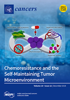
Cover Story (view full-size image): The resistance of cancer cells to commonly used therapeutics is a key clinical challenge. The behaviour of cancer cells is influenced by their environment, which evolves along with tumor progression. Chemoresistance can be stimulated by elements of the tumor microenvironment, including extracellular matrix proteins, hypervascularization, hypoxia, and paracrine factors. This environment is generated by cancer cells and other associated cells, and is notable for its self-maintenance behaviour, which exacerbates tumor development. Through a network of interactions, environmental cues promote cancer cell proliferation and the epithelial–mesenchymal transition, and affect drug availability, leading to a chemoresistant phenotype. This review discusses the self-perpetuating tumor microenvironment and its effects on chemoresistance. View this paper
  • Issues are regarded as officially published after their release is announced to the table of contents alert mailing list.
  • You may sign up for e-mail alerts to receive table of contents of newly released issues.
  • PDF is the official format for papers published in both, html and pdf forms. To view the papers in pdf format, click on the "PDF Full-text" link, and use the free Adobe Reader to open them.
Order results
Result details
Section
Select all
Export citation of selected articles as:
13 pages, 1186 KiB  
Article
Frequency of Circulating Tumor Cells (CTC) in Patients with Brain Metastases: Implications as a Risk Assessment Marker in Oligo-Metastatic Disease
by Annkathrin Hanssen, Carlotta Riebensahm, Malte Mohme, Simon A. Joosse, Janna-Lisa Velthaus, Lars Arne Berger, Christian Bernreuther, Markus Glatzel, Sonja Loges, Katrin Lamszus, Manfred Westphal, Sabine Riethdorf, Klaus Pantel and Harriet Wikman
Cancers 2018, 10(12), 527; https://0-doi-org.brum.beds.ac.uk/10.3390/cancers10120527 - 19 Dec 2018
Cited by 41 | Viewed by 5098
Abstract
Forty percent of non-small cell lung cancer (NSCLC) patients develop brain metastases, resulting in a dismal prognosis. However, patients in an oligo-metastatic brain disease setting seem to have better outcomes. Here, we investigate the possibility of using circulating tumor cells (CTCs) as biomarkers [...] Read more.
Forty percent of non-small cell lung cancer (NSCLC) patients develop brain metastases, resulting in a dismal prognosis. However, patients in an oligo-metastatic brain disease setting seem to have better outcomes. Here, we investigate the possibility of using circulating tumor cells (CTCs) as biomarkers to differentiate oligo-metastatic patients for better risk assessment. Using the CellSearch® system, few CTCs were detected among NSCLC patients with brain metastases (n = 52, 12.5% ≥ two and 8.9% ≥ five CTC/7.5 mL blood) and especially oligo-metastatic brain patients (n = 34, 5.9%, and 2.9%). Still, thresholds of both ≥ two and ≥ five CTCs were independent prognostic indicators for shorter overall survival time among all of the NSCLC patients (n = 90, two CTC ≥ HR: 1.629, p = 0.024, 95% CI: 1.137–6.465 and five CTC ≥ HR: 2.846, p = 0.0304, CI: 1.104–7.339), as well as among patients with brain metastases (two CTC ≥ HR: 4.694, p = 0.004, CI: 1.650–13.354, and five CTC ≥ HR: 4.963, p = 0.003, CI: 1.752–14.061). Also, oligo-brain NSCLC metastatic patients with CTCs had a very poor prognosis (p = 0.019). Similarly, in other tumor entities, only 9.6% of patients with brain metastases (n = 52) had detectable CTCs. Our data indicate that although patients with brain metastases more seldom harbor CTCs, they are still predictive for overall survival, and CTCs might be a useful biomarker to identify oligo-metastatic NSCLC patients who might benefit from a more intense therapy. Full article
(This article belongs to the Special Issue Circulating Tumor Cells (CTCs))
Show Figures

Figure 1

18 pages, 2709 KiB  
Article
Ponatinib Inhibits Multiple Signaling Pathways Involved in STAT3 Signaling and Attenuates Colorectal Tumor Growth
by Fiona H. Tan, Tracy L. Putoczki, Jieqiong Lou, Elizabeth Hinde, Frédéric Hollande, Julie Giraud, Stanley S. Stylli, Lucia Paradiso, Hong-Jian Zhu, Oliver M. Sieber and Rodney B. Luwor
Cancers 2018, 10(12), 526; https://0-doi-org.brum.beds.ac.uk/10.3390/cancers10120526 - 19 Dec 2018
Cited by 16 | Viewed by 4571
Abstract
Signal transducer and activator of transcription 3 (STAT3) signaling is a major driver of colorectal cancer (CRC) growth, however therapeutics, which can effectively target this pathway, have so far remained elusive. Here, we performed an extensive screen for STAT3 inhibitors among a library [...] Read more.
Signal transducer and activator of transcription 3 (STAT3) signaling is a major driver of colorectal cancer (CRC) growth, however therapeutics, which can effectively target this pathway, have so far remained elusive. Here, we performed an extensive screen for STAT3 inhibitors among a library of 1167 FDA-approved agents, identifying Ponatinib as a lead candidate. We found that Ponatinib inhibits STAT3 activity driven by EGF/EGFR, IL-6/IL-6R and IL-11/IL-11R, three major ligand/receptor systems involved in CRC development and progression. Ponatinib was able to inhibit CRC migration and tumor growth in vivo. In addition, Ponatinib displayed a greater ability to inhibit STAT3 activity and mediated superior anti-proliferative efficacy compared to five FDA approved SRC and Janus Kinase (JAK) inhibitors. Finally, long-term exposure of CRC cells to Ponatinib, Dasatinib and Bosutinib resulted in acquired resistance to Dasatinib and Bosutinib occurring within six weeks. However, acquired resistance to Ponatinib was observed after long-term exposure of >4 months. Overall, our results identify a novel anti-STAT3 property of Ponatinib and thus, Ponatinib offers a potential therapeutic strategy for CRC. Full article
Show Figures

Figure 1

19 pages, 2795 KiB  
Article
CBP/β-Catenin/FOXM1 Is a Novel Therapeutic Target in Triple Negative Breast Cancer
by Alexander Ring, Cu Nguyen, Goar Smbatyan, Debu Tripathy, Min Yu, Michael Press, Michael Kahn and Julie E. Lang
Cancers 2018, 10(12), 525; https://0-doi-org.brum.beds.ac.uk/10.3390/cancers10120525 - 19 Dec 2018
Cited by 34 | Viewed by 5830
Abstract
Background: Triple negative breast cancers (TNBCs) are an aggressive BC subtype, characterized by high rates of drug resistance and a high proportion of cancer stem cells (CSC). CSCs are thought to be responsible for tumor initiation and drug resistance. cAMP-response element-binding (CREB) binding [...] Read more.
Background: Triple negative breast cancers (TNBCs) are an aggressive BC subtype, characterized by high rates of drug resistance and a high proportion of cancer stem cells (CSC). CSCs are thought to be responsible for tumor initiation and drug resistance. cAMP-response element-binding (CREB) binding protein (CREBBP or CBP) has been implicated in CSC biology and may provide a novel therapeutic target in TNBC. Methods: RNA Seq pre- and post treatment with the CBP-binding small molecule ICG-001 was used to characterize CBP-driven gene expression in TNBC cells. In vitro and in vivo TNBC models were used to determine the therapeutic effect of CBP inhibition via ICG-001. Tissue microarrays (TMAs) were used to investigate the potential of CBP and associated proteins as biomarkers in TNBC. Results: The CBP/ß-catenin/FOXM1 transcriptional complex drives gene expression in TNBC and is associated with increased CSC numbers, drug resistance and poor survival outcome. Targeting of CBP/β-catenin/FOXM1 with ICG-001 eliminated CSCs and sensitized TNBC tumors to chemotherapy. Immunohistochemistry of TMAs demonstrated a significant correlation between FOXM1 expression and TNBC subtype. Conclusion: CBP/β-catenin/FOXM1 transcriptional activity plays an important role in TNBC drug resistance and CSC phenotype. CBP/β-catenin/FOXM1 provides a molecular target for precision therapy in triple negative breast cancer and could form a rationale for potential clinical trials. Full article
Show Figures

Graphical abstract

15 pages, 244 KiB  
Review
BRCA Mutations and Breast Cancer Prevention
by Joanne Kotsopoulos
Cancers 2018, 10(12), 524; https://0-doi-org.brum.beds.ac.uk/10.3390/cancers10120524 - 19 Dec 2018
Cited by 66 | Viewed by 12043
Abstract
Women who inherit a deleterious BRCA1 or BRCA2 mutation face substantially increased risks of developing breast cancer, which is estimated at 70%. Although annual screening with magnetic resonance imaging (MRI) and mammography promotes the earlier detection of the disease, the gold standard for [...] Read more.
Women who inherit a deleterious BRCA1 or BRCA2 mutation face substantially increased risks of developing breast cancer, which is estimated at 70%. Although annual screening with magnetic resonance imaging (MRI) and mammography promotes the earlier detection of the disease, the gold standard for the primary prevention of breast cancer remains bilateral mastectomy. In the current paper, I review the evidence regarding the management of healthy BRCA mutation carriers, including key risk factors and protective factors, and also discuss potential chemoprevention options. I also provide an overview of the key findings from the literature published to date, with a focus on data from studies that are well-powered, and preferably prospective in nature. Full article
(This article belongs to the Special Issue BRCA Mutations and Cancer)
20 pages, 959 KiB  
Review
Obesity-Linked Cancers: Current Knowledge, Challenges and Limitations in Mechanistic Studies and Rodent Models
by Yang Xin Zi Xu and Suresh Mishra
Cancers 2018, 10(12), 523; https://0-doi-org.brum.beds.ac.uk/10.3390/cancers10120523 - 18 Dec 2018
Cited by 16 | Viewed by 7454
Abstract
The worldwide prevalence of obesity has doubled during the last 50 years, and according to the World Obesity Federation, one third of the people on Earth will be obese by the year 2025. Obesity is described as a chronic, relapsing and multifactorial disease [...] Read more.
The worldwide prevalence of obesity has doubled during the last 50 years, and according to the World Obesity Federation, one third of the people on Earth will be obese by the year 2025. Obesity is described as a chronic, relapsing and multifactorial disease that causes metabolic, biomechanical, and psychosocial health consequences. Growing evidence suggests that obesity is a risk factor for multiple cancer types and rivals smoking as the leading preventable cause for cancer incidence and mortality. The epidemic of obesity will likely generate a new wave of obesity-related cancers with high aggressiveness and shortened latency. Observational studies have shown that from cancer risk to disease prognosis, an individual with obesity is consistently ranked worse compared to their lean counterpart. Mechanistic studies identified similar sets of abnormalities under obesity that may lead to cancer development, including ectopic fat storage, altered adipokine profiles, hormone fluctuations and meta-inflammation, but could not explain how these common mechanisms produce over 13 different cancer types. A major hurdle in the mechanistic underpinning of obesity-related cancer is the lack of suitable pre-clinical models that spontaneously develop obesity-linked cancers like humans. Current approaches and animal models fall short when discerning the confounders that often coexist in obesity. In this mini-review, we will briefly survey advances in the different obesity-linked cancers and discuss the challenges and limitations in the rodent models employed to study their relationship. We will also provide our perspectives on the future of obesity-linked cancer research. Full article
(This article belongs to the Special Issue Obesity as a Risk Factor for Cancer)
Show Figures

Figure 1

16 pages, 1079 KiB  
Review
NK Cell-Based Glioblastoma Immunotherapy
by Irene Golán, Laura Rodríguez de la Fuente and Jose A. Costoya
Cancers 2018, 10(12), 522; https://0-doi-org.brum.beds.ac.uk/10.3390/cancers10120522 - 18 Dec 2018
Cited by 62 | Viewed by 6135
Abstract
Glioblastoma (GB) is the most aggressive and most common malignant primary brain tumor diagnosed in adults. GB shows a poor prognosis and, unfortunately, current therapies are unable to improve its clinical outcome, imposing the need for innovative therapeutic approaches. The main reason for [...] Read more.
Glioblastoma (GB) is the most aggressive and most common malignant primary brain tumor diagnosed in adults. GB shows a poor prognosis and, unfortunately, current therapies are unable to improve its clinical outcome, imposing the need for innovative therapeutic approaches. The main reason for the poor prognosis is the great cell heterogeneity of the tumor mass and its high capacity for invading healthy tissues. Moreover, the glioblastoma microenvironment is capable of suppressing the action of the immune system through several mechanisms such as recruitment of cell modulators. Development of new therapies that avoid this immune evasion could improve the response to the current treatments for this pathology. Natural Killer (NK) cells are cellular components of the immune system more difficult to deceive by tumor cells and with greater cytotoxic activity. Their use in immunotherapy gains strength because they are a less toxic alternative to existing therapy, but the current research focuses on mimicking the NK attack strategy. Here, we summarize the most recent studies regarding molecular mechanisms involved in the GB and immune cells interaction and highlight the relevance of NK cells in the new therapeutic challenges. Full article
(This article belongs to the Special Issue Natural Killer Cells and Cancer Therapy)
Show Figures

Graphical abstract

19 pages, 6677 KiB  
Article
The Human Papillomavirus (HPV) E6 Oncoprotein Regulates CD40 Expression via the AT-Hook Transcription Factor AKNA
by Joaquin Manzo-Merino, Alfredo Lagunas-Martínez, Carla O. Contreras-Ochoa, Marcela Lizano, Leonardo J. Castro-Muñoz, Crysele Calderón-Corona, Kirvis Torres-Poveda, Alicia Román-Gonzalez, Rogelio Hernández-Pando, Margarita Bahena-Román and Vicente Madrid-Marina
Cancers 2018, 10(12), 521; https://0-doi-org.brum.beds.ac.uk/10.3390/cancers10120521 - 17 Dec 2018
Cited by 6 | Viewed by 3632
Abstract
Persistent infection with high-risk Human Papillomavirus (HR-HPV) is the main requisite for cervical cancer development. Normally, HPV is limited to the site of infection and regulates a plethora of cellular elements to avoid the immune surveillance by inducing an anti-inflammatory state, allowing the [...] Read more.
Persistent infection with high-risk Human Papillomavirus (HR-HPV) is the main requisite for cervical cancer development. Normally, HPV is limited to the site of infection and regulates a plethora of cellular elements to avoid the immune surveillance by inducing an anti-inflammatory state, allowing the progress through the viral cycle and the carcinogenic process. Recent findings suggest that the AT-hook transcriptional factor AKNA could play a role in the development of cervical cancer. AKNA is strongly related to the expression of co-stimulatory molecules such CD40/CD40L to achieve an anti-tumoral immune response. To date, there is no evidence demonstrating the effect of the HPV E6 oncoprotein on the AT-hook factor AKNA. In this work, minimal expression of AKNA in cervical carcinoma compared to normal tissue was found. We show the ability of E6 from high-risk HPVs 16 and 18 to interact with and down-regulate AKNA as well as its co-stimulatory molecule CD40 in a proteasome dependent manner. We also found that p53 interacts with AKNA and promotes AKNA expression. Our results indicate that the de-regulation of CD40 and AKNA is induced by the HPV E6 oncoprotein, and this event involves the action of p53 suggesting that the axis E6/p53A/AKNA might play an important role in the de-regulation of the immune system during the carcinogenic process induced by HR-HPV. Full article
Show Figures

Figure 1

42 pages, 5883 KiB  
Article
Oncogenic Activation of Nrf2, Though as a Master Antioxidant Transcription Factor, Liberated by Specific Knockout of the Full-Length Nrf1α that Acts as a Dominant Tumor Repressor
by Lu Qiu, Meng Wang, Shaofan Hu, Xufang Ru, Yonggang Ren, Zhengwen Zhang, Siwang Yu and Yiguo Zhang
Cancers 2018, 10(12), 520; https://0-doi-org.brum.beds.ac.uk/10.3390/cancers10120520 - 17 Dec 2018
Cited by 25 | Viewed by 6538
Abstract
Liver-specific knockout of Nrf1 in the mouse leads to spontaneous development of non- alcoholic steatohepatitis with dyslipidemia, and then its deterioration results in hepatoma, but the underlying mechanism remains elusive to date. A similar pathological model is reconstructed here by using human Nrf1α-specific [...] Read more.
Liver-specific knockout of Nrf1 in the mouse leads to spontaneous development of non- alcoholic steatohepatitis with dyslipidemia, and then its deterioration results in hepatoma, but the underlying mechanism remains elusive to date. A similar pathological model is reconstructed here by using human Nrf1α-specific knockout cell lines. Our evidence has demonstrated that a marked increase of the inflammation marker COX2 definitely occurs in Nrf1α−/ cells. Loss of Nrf1α leads to hyperactivation of Nrf2, which results from substantial decreases in Keap1, PTEN and most of 26S proteasomal subunits in Nrf1α−/ cells. Further investigation of xenograft model mice showed that malignant growth of Nrf1α−/-derived tumors is almost abolished by silencing of Nrf2, while Nrf1α+/+-tumor is markedly repressed by an inactive mutant (i.e., Nrf2−/ΔTA), but largely unaffected by a priori constitutive activator (i.e., caNrf2ΔN). Mechanistic studies, combined with transcriptomic sequencing, unraveled a panoramic view of opposing and unifying inter-regulatory cross-talks between Nrf1α and Nrf2 at different layers of the endogenous regulatory networks from multiple signaling towards differential expression profiling of target genes. Collectively, Nrf1α manifests a dominant tumor-suppressive effect by confining Nrf2 oncogenicity. Though as a tumor promoter, Nrf2 can also, in turn, directly activate the transcriptional expression of Nrf1 to form a negative feedback loop. In view of such mutual inter-regulation by between Nrf1α and Nrf2, it should thus be taken severe cautions to interpret the experimental results from loss of Nrf1α, Nrf2 or both. Full article
Show Figures

Figure 1

15 pages, 2764 KiB  
Article
EphA3 Pay-Loaded Antibody Therapeutics for the Treatment of Glioblastoma
by Carolin Offenhäuser, Fares Al-Ejeh, Simon Puttick, Kathleen S. Ensbey, Zara C. Bruce, Paul R. Jamieson, Fiona M. Smith, Brett W. Stringer, Benjamin Carrington, Adrian V. Fuchs, Craig A. Bell, Rosalind Jeffree, Stephen Rose, Kristofer J. Thurecht, Andrew W. Boyd and Bryan W. Day
Cancers 2018, 10(12), 519; https://0-doi-org.brum.beds.ac.uk/10.3390/cancers10120519 - 17 Dec 2018
Cited by 26 | Viewed by 6164
Abstract
The EphA3 receptor has recently emerged as a functional tumour-specific therapeutic target in glioblastoma (GBM). EphA3 is significantly elevated in recurrent disease, is most highly expressed on glioma stem cells (GSCs), and has a functional role in maintaining self-renewal and tumourigenesis. An unlabelled [...] Read more.
The EphA3 receptor has recently emerged as a functional tumour-specific therapeutic target in glioblastoma (GBM). EphA3 is significantly elevated in recurrent disease, is most highly expressed on glioma stem cells (GSCs), and has a functional role in maintaining self-renewal and tumourigenesis. An unlabelled EphA3-targeting therapeutic antibody is currently under clinical assessment in recurrent GBM patients. In this study, we assessed the efficacy of EphA3 antibody drug conjugate (ADC) and radioimmunotherapy (RIT) approaches using orthotopic animal xenograft models. Brain uptake studies, using positron emission tomography/computed tomography (PET/CT) imaging, show EphA3 antibodies are effectively delivered across the blood-tumour barrier and accumulate at the tumour site with no observed normal brain reactivity. A robust anti-tumour response, with no toxicity, was observed using EphA3, ADC, and RIT approaches, leading to a significant increase in overall survival. Our current research provides evidence that GBM patients may benefit from pay-loaded EphA3 antibody therapies. Full article
(This article belongs to the Special Issue Glioblastoma: State of the Art and Future Perspectives)
Show Figures

Figure 1

15 pages, 2379 KiB  
Article
RNA-Sequencing Analysis of Adrenocortical Carcinoma, Pheochromocytoma and Paraganglioma from a Pan-Cancer Perspective
by Joakim Crona, Samuel Backman, Staffan Welin, David Taïeb, Per Hellman, Peter Stålberg, Britt Skogseid and Karel Pacak
Cancers 2018, 10(12), 518; https://0-doi-org.brum.beds.ac.uk/10.3390/cancers10120518 - 15 Dec 2018
Cited by 9 | Viewed by 5150
Abstract
Adrenocortical carcinoma (ACC) and pheochromocytoma and paraganglioma (PPGL) are defined by clinicopathological criteria and can be further sub-divided based on different molecular features. Whether differences between these molecular subgroups are significant enough to re-challenge their current clinicopathological classification is currently unknown. It is [...] Read more.
Adrenocortical carcinoma (ACC) and pheochromocytoma and paraganglioma (PPGL) are defined by clinicopathological criteria and can be further sub-divided based on different molecular features. Whether differences between these molecular subgroups are significant enough to re-challenge their current clinicopathological classification is currently unknown. It is also not fully understood to which other cancers ACC and PPGL show similarity to. To address these questions, we included recent RNA-Seq data from the Cancer Genome Atlas (TCGA) and Therapeutically Applicable Research to Generate Effective Treatments (TARGET) datasets. Two bioinformatics pipelines were used for unsupervised clustering and principal components analysis. Results were validated using consensus clustering model and interpreted according to previous pan-cancer experiments. Two datasets consisting of 3319 tumors from 35 disease categories were studied. Consistent with the current classification, ACCs clustered as a homogenous group in a pan-cancer context. It also clustered close to neural crest derived tumors, including gliomas, neuroblastomas, pancreatic neuroendocrine tumors, and PPGLs. Contrary, some PPGLs mixed with pancreatic neuroendocrine tumors or neuroblastomas. Thus, our unbiased gene-expression analysis of PPGL did not overlap with their current clinicopathological classification. These results emphasize some importances of the shared embryological origin of these tumors, all either related or close to neural crest tumors, and opens for investigation of a complementary categorization based on gene-expression features. Full article
(This article belongs to the Special Issue Pheochromocytoma (PHEO) and Paraganglioma (PGL))
Show Figures

Figure 1

15 pages, 10738 KiB  
Article
In Silico Approach for Immunohistochemical Evaluation of a Cytoplasmic Marker in Breast Cancer
by Claudia Mazo, Estibaliz Orue-Etxebarria, Ignacio Zabalza, Maria d. M. Vivanco, Robert M. Kypta and Andoni Beristain
Cancers 2018, 10(12), 517; https://0-doi-org.brum.beds.ac.uk/10.3390/cancers10120517 - 15 Dec 2018
Cited by 4 | Viewed by 4741
Abstract
Breast cancer is the most frequently diagnosed cancer in women and the second most common cancer overall, with nearly 1.7 million new cases worldwide every year. Breast cancer patients need accurate tools for early diagnosis and to improve treatment. Biomarkers are increasingly used [...] Read more.
Breast cancer is the most frequently diagnosed cancer in women and the second most common cancer overall, with nearly 1.7 million new cases worldwide every year. Breast cancer patients need accurate tools for early diagnosis and to improve treatment. Biomarkers are increasingly used to describe and evaluate tumours for prognosis, to facilitate and predict response to therapy and to evaluate residual tumor, post-treatment. Here, we evaluate different methods to separate Diaminobenzidine (DAB) from Hematoxylin and Eosin (H&E) staining for Wnt-1, a potential cytoplasmic breast cancer biomarker. A method comprising clustering and Color deconvolution allowed us to recognize and quantify Wnt-1 levels accurately at pixel levels. Experimental validation was conducted using a set of 12,288 blocks of m × n pixels without overlap, extracted from a Tissue Microarray (TMA) composed of 192 tissue cores. Intraclass Correlations (ICC) among evaluators of the data of 0.634 , 0.791 , 0.551 and 0.63 for each Allred class and an average ICC of 0.752 among evaluators and automatic classification were obtained. Furthermore, this method received an average rating of 4.26 out of 5 in the Wnt-1 segmentation process from the evaluators. Full article
(This article belongs to the Special Issue Cancer Biomarkers)
Show Figures

Figure 1

12 pages, 1051 KiB  
Article
Combination Therapy after TACE for Hepatocellular Carcinoma with Macroscopic Vascular Invasion: Stereotactic Body Radiotherapy versus Sorafenib
by Lujun Shen, Mian Xi, Lei Zhao, Xuhui Zhang, Xiuchen Wang, Zhimei Huang, Qifeng Chen, Tianqi Zhang, Jingxian Shen, Mengzhong Liu and Jinhua Huang
Cancers 2018, 10(12), 516; https://0-doi-org.brum.beds.ac.uk/10.3390/cancers10120516 - 14 Dec 2018
Cited by 23 | Viewed by 3898
Abstract
Stereotactic body radiotherapy (SBRT) has shown promising results in the control of macroscopic vascular invasion in patients with hepatocellular carcinoma (HCC); however, its efficacy in comparison to sorafenib when combined with transarterial chemoembolization (TACE) remains to be determined. Between 2009 and 2017, 77 [...] Read more.
Stereotactic body radiotherapy (SBRT) has shown promising results in the control of macroscopic vascular invasion in patients with hepatocellular carcinoma (HCC); however, its efficacy in comparison to sorafenib when combined with transarterial chemoembolization (TACE) remains to be determined. Between 2009 and 2017, 77 HCC patients with macroscopic vascular invasion receiving TACE–SBRT or TACE–sorafenib combination therapies were enrolled. The best treatment responses, overall survival (OS), and progression-free survival (PFS) of the two treatment arms were compared. Of the patients enrolled, 26 patients (33.8%) received TACE–SBRT treatment, and 51 (66.2%) received TACE–sorafenib treatment. The patients in the TACE–SBRT group were more frequently classified as elder in age (p = 0.012), having recurrent disease (p = 0.026), and showing lower rates of multiple hepatic lesions (p = 0.005) than patients in TACE–sorafenib group. After propensity score matching (PSM), 26 pairs of well-matched HCC patients were selected; patients in the TACE–SBRT group showed better overall response rates in trend compared to those in the TACE–sorafenib group. The hazard ratio (HR) of OS to PFS for the TACE–SBRT approach and the TACE–sorafenib approach was 0.36 (95% CI, 0.17–0.75; p = 0.007) and 0.35 (95% CI, 0.20–0.62; p < 0.001), respectively. For HCC patients with macrovascular invasion, TACE plus SBRT could provide improved OS and PFS compared to TACE–sorafenib therapy. Full article
Show Figures

Figure 1

24 pages, 716 KiB  
Review
Radiotherapy as a New Player in Immuno-Oncology
by Shang-Jui Wang and Bruce Haffty
Cancers 2018, 10(12), 515; https://0-doi-org.brum.beds.ac.uk/10.3390/cancers10120515 - 14 Dec 2018
Cited by 21 | Viewed by 4569
Abstract
Recent development in radiation biology has revealed potent immunogenic properties of radiotherapy in cancer treatments. However, antitumor immune effects of radiotherapy are limited by the concomitant induction of radiation-dependent immunosuppressive effects. In the growing era of immunotherapy, combining radiotherapy with immunomodulating agents has [...] Read more.
Recent development in radiation biology has revealed potent immunogenic properties of radiotherapy in cancer treatments. However, antitumor immune effects of radiotherapy are limited by the concomitant induction of radiation-dependent immunosuppressive effects. In the growing era of immunotherapy, combining radiotherapy with immunomodulating agents has demonstrated enhancement of radiation-induced antitumor immune activation that correlated with improved treatment outcomes. Yet, how to optimally deliver combination therapy regarding dose-fractionation and timing of radiotherapy is largely unknown. Future prospective testing to fine-tune this promising combination of radiotherapy and immunotherapy is warranted. Full article
(This article belongs to the Special Issue New Developments in Radiotherapy)
Show Figures

Figure 1

19 pages, 1766 KiB  
Review
Racial Disparity and Triple-Negative Breast Cancer in African-American Women: A Multifaceted Affair between Obesity, Biology, and Socioeconomic Determinants
by Sumit Siddharth and Dipali Sharma
Cancers 2018, 10(12), 514; https://0-doi-org.brum.beds.ac.uk/10.3390/cancers10120514 - 14 Dec 2018
Cited by 123 | Viewed by 11984
Abstract
Triple negative breast cancer (TNBC) is a molecularly heterogeneous disease whose incidence is disproportionately higher in African American (AA) women compared to European American (EA) women. Earlier onset, more advanced stage at diagnosis, and aggressive tumor phenotype are some of the characteristic features [...] Read more.
Triple negative breast cancer (TNBC) is a molecularly heterogeneous disease whose incidence is disproportionately higher in African American (AA) women compared to European American (EA) women. Earlier onset, more advanced stage at diagnosis, and aggressive tumor phenotype are some of the characteristic features of TNBC in women with African ethnicity in comparison to EA women, denoting one of the most significant examples of racial disparity in oncology. It is still contentious whether health disparities result in aggressive behavior of TNBC in AA women or it is indeed a molecularly distinct disease. Given the “gaps-in-knowledge” surrounding racial disparity in TNBC, this review discusses various socioeconomic factors and the genetic predispositions contributing to poor prognosis of TNBC in AA women. While socioeconomic factors may contribute to poorer survival, multiple preclinical and clinical studies suggest inherent genetic risk factors and aberrant activation of oncogenic pathways in AA TNBC. Additionally, AA women are more likely to be obese and obesity is known to drive a molecular circuitry resulting in aggressive tumor progression indicating a potential obesity-TNBC axis at work in AA women. Given the multifactorial nature of AA TNBC, a transdisciplinary approach may help bridge the disparity that exists between AA and EA TNBC. Full article
Show Figures

Figure 1

16 pages, 980 KiB  
Review
BRCA1-Dependent Transcriptional Regulation: Implication in Tissue-Specific Tumor Suppression
by Xiaowen Zhang and Rong Li
Cancers 2018, 10(12), 513; https://0-doi-org.brum.beds.ac.uk/10.3390/cancers10120513 - 14 Dec 2018
Cited by 12 | Viewed by 5178
Abstract
Germ-line mutations in breast cancer susceptibility gene 1 (BRCA1) predominantly predispose women to breast and ovarian cancers. BRCA1 is best known for its functions in maintenance of genomic integrity including repairing DNA double-strand breaks through homologous recombination and suppressing DNA replication [...] Read more.
Germ-line mutations in breast cancer susceptibility gene 1 (BRCA1) predominantly predispose women to breast and ovarian cancers. BRCA1 is best known for its functions in maintenance of genomic integrity including repairing DNA double-strand breaks through homologous recombination and suppressing DNA replication stress. However, whether these universally important BRCA1 functions in maintenance of genomic stability are sufficient to account for its tissue-specific tumor-suppressing function remains unclear. Accumulating evidence indicates that there are previously underappreciated roles of BRCA1 in transcriptional regulation and chromatin remodeling. In this review, we discuss the functional significance of interactions between BRCA1 and various transcription factors, its role in epigenetic regulation and chromatin dynamics, and BRCA1-dependent crosstalk between the machineries of transcription and genome integrity. Furthermore, we propose a model of how transcriptional regulation could contribute to tissue-dependent tumor-suppressing function of BRCA1. Full article
(This article belongs to the Special Issue BRCA Mutations and Cancer)
Show Figures

Figure 1

17 pages, 2057 KiB  
Article
The STAT3/Slug Axis Enhances Radiation-Induced Tumor Invasion and Cancer Stem-like Properties in Radioresistant Glioblastoma
by Jang-Chun Lin, Jo-Ting Tsai, Tsu-Yi Chao, Hsin-I Ma and Wei-Hsiu Liu
Cancers 2018, 10(12), 512; https://0-doi-org.brum.beds.ac.uk/10.3390/cancers10120512 - 13 Dec 2018
Cited by 48 | Viewed by 4135
Abstract
Glioblastoma multiforme (GBM) requires radiotherapy (RT) as a part of definitive management strategy. RT is highly effective, destroying cancer cells that may exist around the surgical tumor bed. However, GBM still has a poor prognosis and a high local recurrence rate after RT. [...] Read more.
Glioblastoma multiforme (GBM) requires radiotherapy (RT) as a part of definitive management strategy. RT is highly effective, destroying cancer cells that may exist around the surgical tumor bed. However, GBM still has a poor prognosis and a high local recurrence rate after RT. Accumulating research indicates that GBM contains cancer stem-like cells (CSCs), which are radioresistant and result in therapeutic failure. Additionally, GBM cells can aggressively invade normal brain tissue, inducing therapeutic failure. Using clinical observations, we evaluated the effect of radiation on tumor control. We also explored the biomolecular pathways that connect radioresistance and CSC- and epithelial-mesenchymal transition (EMT)-associated phenotypes in patient-derived GBM cells. Transwell and microarray assay demonstrated that radioresistant GBM cells (GBM-R2I2) exhibit increased invasion and self-renewal abilities compared with parental GBM cells. Finally, to identify potential mechanisms underlying these observations, we used a PCR array to search for molecular markers of cell motility. Signal transducer and activator of transcription 3 (STAT3) directly bound to the Slug promoter in a chromatin immunoprecipitation assay. Reduced STAT3 decreased Slug expression and suppressed cell invasion in GBM-R2I2 cells while increasing Slug reversed these effects. In addition, STAT3 knockdown significantly inhibited CSC properties, synergistically increased the radiotherapeutic effect, and effectively increased the survival rate in vivo. We deciphered a new pathway of GBM radioresistance, invasion, and recurrence via the STAT3/Slug axis that could be a new target of GBM therapy. Full article
Show Figures

Figure 1

26 pages, 2094 KiB  
Article
Use of Germline Genetic Variability for Prediction of Chemoresistance and Prognosis of Breast Cancer Patients
by Viktor Hlavac, Maria Kovacova, Katerina Elsnerova, Veronika Brynychova, Renata Kozevnikovova, Karel Raus, Katerina Kopeckova, Sona Mestakova, David Vrana, Jiri Gatek, Pavel Ostasov, Radka Vaclavikova and Pavel Soucek
Cancers 2018, 10(12), 511; https://0-doi-org.brum.beds.ac.uk/10.3390/cancers10120511 - 12 Dec 2018
Cited by 13 | Viewed by 4113
Abstract
The aim of our study was to set up a panel for targeted sequencing of chemoresistance genes and the main transcription factors driving their expression and to evaluate their predictive and prognostic value in breast cancer patients. Coding and regulatory regions of 509 [...] Read more.
The aim of our study was to set up a panel for targeted sequencing of chemoresistance genes and the main transcription factors driving their expression and to evaluate their predictive and prognostic value in breast cancer patients. Coding and regulatory regions of 509 genes, selected from PharmGKB and Phenopedia, were sequenced using massive parallel sequencing in blood DNA from 105 breast cancer patients in the testing phase. In total, 18,245 variants were identified of which 2565 were novel variants (without rs number in dbSNP build 150) in the testing phase. Variants with major allele frequency over 0.05 were further prioritized for validation phase based on a newly developed decision tree. Using emerging in silico tools and pharmacogenomic databases for functional predictions and associations with response to cytotoxic therapy or disease-free survival of patients, 55 putative variants were identified and used for validation in 805 patients with clinical follow up using KASPTM technology. In conclusion, associations of rs2227291, rs2293194, and rs4376673 (located in ATP7A, KCNAB1, and DFFB genes, respectively) with response to neoadjuvant cytotoxic therapy and rs1801160 in DPYD with disease-free survival of patients treated with cytotoxic drugs were validated and should be further functionally characterized. Full article
(This article belongs to the Special Issue Cancer Chemoresistance)
Show Figures

Figure 1

14 pages, 248 KiB  
Review
Management of Typical and Atypical Pulmonary Carcinoids Based on Different Established Guidelines
by Rohit Gosain, Sarbajit Mukherjee, Sai S. Yendamuri and Renuka Iyer
Cancers 2018, 10(12), 510; https://0-doi-org.brum.beds.ac.uk/10.3390/cancers10120510 - 12 Dec 2018
Cited by 49 | Viewed by 4942
Abstract
Neuroendocrine tumors (NETs) are a group of malignancies that originated from neuroendocrine cells, with the most common sites being lungs and the gastrointestinal tract. Lung NETs comprise 25% of all lung malignancies. Small cell lung cancer is the most common form of lung [...] Read more.
Neuroendocrine tumors (NETs) are a group of malignancies that originated from neuroendocrine cells, with the most common sites being lungs and the gastrointestinal tract. Lung NETs comprise 25% of all lung malignancies. Small cell lung cancer is the most common form of lung NETs, and other rare forms include well-differentiated typical carcinoids (TCs) and poorly differentiated atypical carcinoids (ACs). Given the paucity of randomized studies, rational treatment is challenging. Therefore, it is recommended that these decisions be made using a multidisciplinary collaborative approach. Surgery remains the mainstay of treatment, when feasible. Following surgery, various guidelines offer different recommendations in the adjuvant setting. In this paper, we describe the adjuvant management of lung NETs, as recommended by different guidelines, and highlight their differences. In addition to that, we also discuss the management of metastatic lung NETS, including the use of peptide receptor radionucleotide therapy. Full article
16 pages, 2125 KiB  
Article
Mitochondrial Hyperactivation and Enhanced ROS Production are Involved in Toxicity Induced by Oncogenic Kinases Over-Signaling
by Monica Ceccon, Mario Mauri, Luca Massimino, Giovanni Giudici, Rocco Piazza, Carlo Gambacorti-Passerini and Luca Mologni
Cancers 2018, 10(12), 509; https://0-doi-org.brum.beds.ac.uk/10.3390/cancers10120509 - 12 Dec 2018
Cited by 3 | Viewed by 2982
Abstract
Targeted therapy is an effective, rational, and safe approach to solid and hematological tumors treatment. Unfortunately, a significant fraction of patients treated with tyrosine kinase inhibitors (TKI) relapses mainly because of gene amplification, mutations, or other bypass mechanisms. Recently a growing number of [...] Read more.
Targeted therapy is an effective, rational, and safe approach to solid and hematological tumors treatment. Unfortunately, a significant fraction of patients treated with tyrosine kinase inhibitors (TKI) relapses mainly because of gene amplification, mutations, or other bypass mechanisms. Recently a growing number of papers showed how, in some cases, resistance due to oncogene overexpression may be associated with drug addiction: cells able to proliferate in the presence of high TKI doses become also TKI dependent, undergoing cellular stress, and apoptosis/death upon drug withdrawal. Notably, if a sub-cellular population survives TKI discontinuation it is also partially re-sensitized to the same drug. Thus, it is possible that a subset of patients relapsing upon TKI treatment may benefit from a discontinuous therapeutic schedule. We focused on two different hematologic malignancies, chronic myeloid leukemia (CML) and anaplastic large cell lymphoma (ALCL), both successfully treatable with TKIs. The two models utilized (LAMA and SUP-M2) differed in having oncogene overexpression as the sole cause of drug resistance (CML), or additionally carrying kinase domain mutations (ALCL). In both cases drug withdrawal caused a sudden overload of oncogenic signal, enhanced mitochondria activity, induced the release of a high amount of reactive oxygen species (ROS), and caused genotoxic stress and massive cell death. In LAMA cells (CML) we could rescue the cells from death by partially blocking downstream oncogenic signaling or lowering ROS detrimental effect by adding reduced glutathione. Full article
Show Figures

Figure 1

1 pages, 156 KiB  
Erratum
Erratum: Shibata S.; et al. Proton Beam Therapy without Fiducial Markers Using Four-Dimensional CT Planning for Large Hepatocellular Carcinomas. Cancers 2018, 10, 71
by Satoshi Shibata, Shigeyuki Takamatsu, Kazutaka Yamamoto, Miu Mizuhata, Sayuri Bou, Yoshitaka Sato, Mariko Kawamura, Satoko Asahi, Yuji Tameshige, Yoshikazu Maeda, Makoto Sasaki, Tomoyasu Kumano, Satoshi Kobayashi, Hiroyasu Tamamura and Toshifumi Gabata
Cancers 2018, 10(12), 508; https://0-doi-org.brum.beds.ac.uk/10.3390/cancers10120508 - 11 Dec 2018
Cited by 2 | Viewed by 2009
Abstract
The authors wish to make the following corrections to this paper [...] Full article
20 pages, 4835 KiB  
Article
The Role of Arrestin Domain-Containing 3 in Regulating Endocytic Recycling and Extracellular Vesicle Sorting of Integrin β4 in Breast Cancer
by Young Hwa Soung, Shane Ford, Cecilia Yan and Jun Chung
Cancers 2018, 10(12), 507; https://0-doi-org.brum.beds.ac.uk/10.3390/cancers10120507 - 11 Dec 2018
Cited by 15 | Viewed by 3757
Abstract
Despite the established role of integrin β4 (ITG β4) in breast cancer progression, the importance of endocytic recycling of ITG β4 and its regulatory mechanism are poorly understood. Here, we found that a sub-population of ITG β4 is sorted into early endosomes, recycled [...] Read more.
Despite the established role of integrin β4 (ITG β4) in breast cancer progression, the importance of endocytic recycling of ITG β4 and its regulatory mechanism are poorly understood. Here, we found that a sub-population of ITG β4 is sorted into early endosomes, recycled back to the plasma membrane, and secreted in the form of extracellular vesicles (EVs) upon EGF treatment in triple negative breast cancer (TNBC) cells. A metastasis suppressor, ARRDC3 (arrestin domain-containing 3) prevents EGF-driven endocytic recycling of ITG β4 by inducing NEDD4-dependent ubiquitination of ITG β4 and targeting endosomal ITG β4 into lysosomes. Endocytic recycling of ITG β4 is linked to sorting of ITG β4 into EVs (ITG β4+ EVs). ITG β4+ EVs are mainly detectable from supernatants of TNBC cells and their production is inhibited by ARRDC3 expression. ARRDC3 reduces the metastatic potentials of breast cancer cell-derived EVs by reducing ITG β4 levels in EVs. Overall, current studies provide novel mechanistic insights on the regulatory mechanism of ITG β4 recycling, and its importance in invasive potentials of TNBC EVs, thus providing the basis for therapeutic targeting of the ARRDC3/ITG β4 pathway in TNBC. Full article
Show Figures

Figure 1

22 pages, 643 KiB  
Review
Targeting BRCA Deficiency in Breast Cancer: What are the Clinical Evidences and the Next Perspectives?
by Emanuel Nicolas, François Bertucci, Renaud Sabatier and Anthony Gonçalves
Cancers 2018, 10(12), 506; https://0-doi-org.brum.beds.ac.uk/10.3390/cancers10120506 - 11 Dec 2018
Cited by 36 | Viewed by 4367
Abstract
Breast cancers (BC) associated with germline mutations of BRCA1/2 represent 3–5% of cases. BRCA1/2-associated BC have biological features leading to genomic instability and potential sensitivity to DNA damaging agents, including poly(ADP-ribose) polymerase (PARP) and platinum agents. In this review, we will summarize [...] Read more.
Breast cancers (BC) associated with germline mutations of BRCA1/2 represent 3–5% of cases. BRCA1/2-associated BC have biological features leading to genomic instability and potential sensitivity to DNA damaging agents, including poly(ADP-ribose) polymerase (PARP) and platinum agents. In this review, we will summarize clinical trials of chemotherapy and PARP inhibitors (PARPi), alone or in combination, at the early or late stage of BRCA1/2-associated BC. We will also present the mechanisms of resistance to PARPi as well as the new therapeutic strategies of association with PARPi. Finally, we will discuss under which conditions the use of DNA damaging agents can be extended to the BRCA1/2-wild type population, the BRCAness concept. Full article
(This article belongs to the Special Issue BRCA Mutations and Cancer)
Show Figures

Figure 1

17 pages, 2931 KiB  
Article
Intracellular Iron Chelation by a Novel Compound, C7, Reactivates Epstein–Barr Virus (EBV) Lytic Cycle via the ERK-Autophagy Axis in EBV-Positive Epithelial Cancers
by Stephanie Pei Tung Yiu, Kwai Fung Hui, Chung King Choi, Richard Yi Tsun Kao, Chi Wang Ma, Dan Yang and Alan Kwok Shing Chiang
Cancers 2018, 10(12), 505; https://0-doi-org.brum.beds.ac.uk/10.3390/cancers10120505 - 11 Dec 2018
Cited by 21 | Viewed by 3942
Abstract
Pharmaceutical reactivation of lytic cycle of Epstein–Barr virus (EBV) represents a potential therapeutic strategy against EBV-associated epithelial malignancies, e.g., gastric carcinoma (GC) and nasopharyngeal carcinoma (NPC). A novel lytic-inducing compound, C7, which exhibits structural similarity to Di-2-Pyridyl Ketone 4, 4-Dimethyl-3-Thiosemicarbazone (Dp44mT), a known [...] Read more.
Pharmaceutical reactivation of lytic cycle of Epstein–Barr virus (EBV) represents a potential therapeutic strategy against EBV-associated epithelial malignancies, e.g., gastric carcinoma (GC) and nasopharyngeal carcinoma (NPC). A novel lytic-inducing compound, C7, which exhibits structural similarity to Di-2-Pyridyl Ketone 4, 4-Dimethyl-3-Thiosemicarbazone (Dp44mT), a known chelator of intracellular iron, is found to reactivate EBV lytic cycle in GC and NPC. This study aims to investigate the role of intracellular iron chelation by C7 and other iron chelators in lytic reactivation of EBV in GC and NPC. Testing of six structural analogs of C7 revealed only those which have high affinity towards transition metals could induce EBV lytic cycle. Precomplexing C7 and iron chelators to iron prior to treatment of the cells abolished EBV lytic reactivation. Though hypoxia signaling pathway was activated, it was not the only pathway associated with EBV reactivation. Specifically, C7 and iron chelators initiated autophagy by activating extracellular signal-regulated kinase (ERK1/2) to reactivate EBV lytic cycle since autophagy and EBV lytic reactivation were abolished in cells treated with ERK1/2 blockers whilst inhibition of autophagy by 3-Methyladenine (3-MA) and atg5 knockdown significantly abolished EBV lytic reactivation. In summary, we discovered a novel mechanism of reactivation of the EBV lytic cycle through intracellular iron chelation and induction of ERK-autophagy axis in EBV-positive epithelial malignancies, raising the question whether clinically available iron chelators can be incorporated into existing therapeutic regimens to treat these cancers. Full article
Show Figures

Figure 1

13 pages, 391 KiB  
Article
Pathological and Molecular Characteristics of Colorectal Cancer with Brain Metastases
by Pauline Roussille, Gaelle Tachon, Claire Villalva, Serge Milin, Eric Frouin, Julie Godet, Antoine Berger, Sheik Emambux, Christos Petropoulos, Michel Wager, Lucie Karayan-Tapon and David Tougeron
Cancers 2018, 10(12), 504; https://0-doi-org.brum.beds.ac.uk/10.3390/cancers10120504 - 10 Dec 2018
Cited by 18 | Viewed by 3494
Abstract
Background: Colorectal cancers (CRC) with brain metastases (BM) are scarcely described. The main objective of this study was to determine the molecular profile of CRC with BM. Methods: We included 82 CRC patients with BM. KRAS, NRAS, BRAF and mismatch repair [...] Read more.
Background: Colorectal cancers (CRC) with brain metastases (BM) are scarcely described. The main objective of this study was to determine the molecular profile of CRC with BM. Methods: We included 82 CRC patients with BM. KRAS, NRAS, BRAF and mismatch repair (MMR) status were investigated on primary tumors (n = 82) and BM (n = 38). ALK, ROS1, cMET, HER-2, PD-1, PD-L1, CD3 and CD8 status were evaluated by immunohistochemistry, and when recommended, by fluorescence in situ hybridization. Results: In primary tumors, KRAS, NRAS and BRAF mutations were observed in 56%, 6%, and 6% of cases, respectively. No ROS1, ALK and cMET rearrangement was detected. Only one tumor presented HER-2 amplification. Molecular profiles were mostly concordant between BM and paired primary tumors, except for 9% of discordances for RAS mutation. CD3, CD8, PD-1 and PD-L1 expressions presented some discordance between primary tumors and BM. In multivariate analysis, multiple BM, lung metastases and PD-L1+ tumor were predictive of poor overall survival. Conclusions: CRCs with BM are associated with high frequency of RAS mutations and significant discordance for RAS mutational status between BM and paired primary tumors. Multiple BM, lung metastases and PD-L1+ have been identified as prognostic factors and can guide therapeutic decisions for CRC patients with BM. Full article
(This article belongs to the Special Issue Application of Bioinformatics in Cancers)
Show Figures

Figure 1

27 pages, 1247 KiB  
Review
Transporter and Lysosomal Mediated (Multi)drug Resistance to Tyrosine Kinase Inhibitors and Potential Strategies to Overcome Resistance
by Daniel J. De Klerk, Richard J. Honeywell, Gerrit Jansen and Godefridus J. Peters
Cancers 2018, 10(12), 503; https://0-doi-org.brum.beds.ac.uk/10.3390/cancers10120503 - 10 Dec 2018
Cited by 41 | Viewed by 6914
Abstract
Tyrosine kinase inhibitors are a class of chemotherapeutic drugs that target specific protein kinases. These tyrosine kinase inhibitors constitute a relatively new class of drugs which target for instance Bcr-Abl, Epidermal Growth Factor Receptor (EGFR) and Vascular Endothelial Growth Factor Receptor (VEGFR). Despite [...] Read more.
Tyrosine kinase inhibitors are a class of chemotherapeutic drugs that target specific protein kinases. These tyrosine kinase inhibitors constitute a relatively new class of drugs which target for instance Bcr-Abl, Epidermal Growth Factor Receptor (EGFR) and Vascular Endothelial Growth Factor Receptor (VEGFR). Despite some initial successes, the overall therapeutic benefit of tyrosine kinase inhibitors in the clinic has been mixed. Next to mutations in the target, multidrug resistance is a major obstacle for which still no clinically effective strategies have been developed. Major mechanisms of multidrug resistance are mediated by drug efflux transporter proteins. Moreover, there is accumulating evidence that multidrug resistance can also be caused by lysosomal sequestration of drugs, effectively trapping tyrosine kinase inhibitors and preventing them from reaching their target. Lysosomal drug sequestration seems to work together with ATP-binding cassette transporters, increasing the capacity of lysosomes to mediate sequestration. Both membrane efflux transporter proteins and lysosomes present potential therapeutic targets that could reverse multidrug resistance and increase drug efficacy in combination therapy. This review describes both mechanisms and discusses a number of proposed strategies to circumvent or reverse tyrosine kinase inhibitor-related multidrug resistance. Full article
(This article belongs to the Special Issue Cancer Chemoresistance)
Show Figures

Figure 1

12 pages, 920 KiB  
Review
Treatment of Pain in Cancer: Towards Personalised Medicine
by Marieke H. J. Van den Beuken-van Everdingen, Sander M. J. Van Kuijk, Daisy J. A. Janssen and Elbert A. J. Joosten
Cancers 2018, 10(12), 502; https://0-doi-org.brum.beds.ac.uk/10.3390/cancers10120502 - 10 Dec 2018
Cited by 39 | Viewed by 6600
Abstract
Despite increased attention to cancer pain, pain prevalence in patients with cancer has not improved over the last decade and one third of cancer patients on anticancer therapy and half of patients with advanced disease still suffer from moderate to severe pain. In [...] Read more.
Despite increased attention to cancer pain, pain prevalence in patients with cancer has not improved over the last decade and one third of cancer patients on anticancer therapy and half of patients with advanced disease still suffer from moderate to severe pain. In this review, we explore the possible reasons for the ongoing high prevalence of cancer pain and discuss possible future directions for improvement in personalised pain management. Among possible reasons for the lack of improvement are: Barriers for patients to discuss pain with clinicians spontaneously; pain measurement instruments are not routinely used in daily practice; limited knowledge concerning the assessment of undertreatment; changes in patients’ characteristics, including the ageing of the population; lack of significant improvement in the treatment of neuropathic pain; limitations of pharmacological treatment and lack of evidence-based nonpharmacological treatment strategies. In order to improve cancer pain treatment, we recommend: (1) Physicians proactively ask about pain and measure pain using assessment instruments; (2) the development of an optimal tool measuring undertreatment; (3) educational interventions to improve health care workers’ skills in pain management; (4) the development of more effective and personalised pharmacological and nonpharmacological pain treatment. Full article
(This article belongs to the Special Issue Cancer Pains)
Show Figures

Figure 1

16 pages, 3357 KiB  
Article
Venous Thromboembolism in Asian Patients with Pancreatic Cancer Following Palliative Chemotherapy: Low Incidence but a Negative Prognosticator for Those with Early Onset
by Jen-Shi Chen, Chia-Yen Hung, Hung Chang, Chien-Ting Liu, Yen-Yang Chen, Chang-Hsien Lu, Pei-Hung Chang, Yu-Shin Hung and Wen-Chi Chou
Cancers 2018, 10(12), 501; https://0-doi-org.brum.beds.ac.uk/10.3390/cancers10120501 - 10 Dec 2018
Cited by 19 | Viewed by 3074
Abstract
Background: Few studies have reported the epidemiology and clinical outcome of venous thromboembolism (VTE) in Asian patients with pancreatic cancer. This study investigated the incidence, risk factors, and clinical outcome of VTE in patients with pancreatic cancer following palliative chemotherapy. Methods: The medical [...] Read more.
Background: Few studies have reported the epidemiology and clinical outcome of venous thromboembolism (VTE) in Asian patients with pancreatic cancer. This study investigated the incidence, risk factors, and clinical outcome of VTE in patients with pancreatic cancer following palliative chemotherapy. Methods: The medical records of 838 patients with newly diagnosed locally advanced or metastatic pancreatic cancer who underwent palliative chemotherapy between 2010 and 2016 at four institutes in Taiwan were retrospectively reviewed. The clinical characteristics of all patients were analyzed to identify independent predictors of VTE and their effects on survival outcome. Results: During the median follow-up period of 7.7 months (range, 0.6–55.6), VTE occurred in 67 (8.0%) of the 838 patients. Leukocyte count > 11,000/μL and presence of liver metastases were the independent predictors of VTE. Patients with VTE did not show significantly poorer survival outcomes than those without VTE. However, early-onset VTE that occurred within 1.5 months after chemotherapy initiation was an independent negative prognosticator for overall survival. Conclusion: VTE incidence was found to be lower in Asian patients with pancreatic cancer than in their Western counterparts. Early-onset VTE, but not late-onset VTE, is a negative prognosticator for survival outcomes. Full article
(This article belongs to the Special Issue Advances in Pancreatic Cancer Research)
Show Figures

Figure 1

26 pages, 2994 KiB  
Article
Mitochondrial RNA Expression and Single Nucleotide Variants in Association with Clinical Parameters in Primary Breast Cancers
by Marjolein J. A. Weerts, Marcel Smid, John A. Foekens, Stefan Sleijfer and John W. M. Martens
Cancers 2018, 10(12), 500; https://0-doi-org.brum.beds.ac.uk/10.3390/cancers10120500 - 09 Dec 2018
Cited by 7 | Viewed by 3244
Abstract
The human mitochondrial DNA (mtDNA) encodes 37 genes, including thirteen proteins essential for the respiratory chain, and RNAs functioning in the mitochondrial translation apparatus. The total number of mtDNA molecules per cell (mtDNA content) is variable between tissue types and also between tumors [...] Read more.
The human mitochondrial DNA (mtDNA) encodes 37 genes, including thirteen proteins essential for the respiratory chain, and RNAs functioning in the mitochondrial translation apparatus. The total number of mtDNA molecules per cell (mtDNA content) is variable between tissue types and also between tumors and their normal counterparts. For breast cancer, tumors tend to be depleted in their mtDNA content compared to adjacent normal mammary tissue. Various studies have shown that primary breast tumors harbor somatic mtDNA variants. A decrease in mtDNA content or the presence of somatic variants could indicate a reduced mitochondrial function within breast cancer. In this explorative study we aimed to further understand genomic changes and expression of the mitochondrial genome within breast cancer, by analyzing RNA sequencing data of primary breast tumor specimens of 344 cases. We demonstrate that somatic variants detected at the mtRNA level are representative for somatic variants in the mtDNA. Also, the number of somatic variants within the mitochondrial transcriptome is not associated with mutational processes impacting the nuclear genome, but is positively associated with age at diagnosis. Finally, we observe that mitochondrial expression is related to ER status. We conclude that there is a large heterogeneity in somatic mutations of the mitochondrial genome within primary breast tumors, and differences in mitochondrial expression among breast cancer subtypes. The exact impact on metabolic differences and clinical relevance deserves further study. Full article
Show Figures

Figure 1

26 pages, 8363 KiB  
Article
Mitochondrial VDAC1 Silencing Leads to Metabolic Rewiring and the Reprogramming of Tumour Cells into Advanced Differentiated States
by Tasleem Arif, Avijit Paul, Yakov Krelin, Anna Shteinfer-Kuzmine and Varda Shoshan-Barmatz
Cancers 2018, 10(12), 499; https://0-doi-org.brum.beds.ac.uk/10.3390/cancers10120499 - 08 Dec 2018
Cited by 37 | Viewed by 5430
Abstract
Oncogenic properties, along with the metabolic reprogramming necessary for tumour growth and motility, are acquired by cancer cells. Thus, tumour metabolism is becoming a target for cancer therapy. Here, cancer cell metabolism was tackled by silencing the expression of voltage-dependent anion channel 1 [...] Read more.
Oncogenic properties, along with the metabolic reprogramming necessary for tumour growth and motility, are acquired by cancer cells. Thus, tumour metabolism is becoming a target for cancer therapy. Here, cancer cell metabolism was tackled by silencing the expression of voltage-dependent anion channel 1 (VDAC1), a mitochondrial protein that controls cell energy, as well as metabolic and survival pathways and that is often over-expressed in many cancers. We demonstrated that silencing VDAC1 expression using human-specific siRNA (si-hVDAC1) inhibited cancer cell growth, both in vitro and in mouse xenograft models of human glioblastoma (U-87MG), lung cancer (A549), and triple negative breast cancer (MDA-MB-231). Importantly, treatment with si-hVDAC1 induced metabolic rewiring of the cancer cells, reversing their oncogenic properties and diverting them towards differentiated-like cells. The si-hVDAC1-treated residual “tumour” showed reprogrammed metabolism, decreased proliferation, inhibited stemness and altered expression of genes and proteins, leading to cell differentiation toward less malignant lineages. These VDAC1 depletion-mediated effects involved alterations in master transcription factors associated with cancer hallmarks, such as highly increased expression of p53 and decreased expression of HIF-1a and c-Myc that regulate signalling pathways (e.g., AMPK, mTOR). High expression of p53 and the pro-apoptotic proteins cytochrome c and caspases without induction of apoptosis points to functions for these proteins in promoting cell differentiation. These results clearly show that VDAC1 depletion similarly leads to a rewiring of cancer cell metabolism in breast and lung cancer and glioblastoma, regardless of origin or mutational status. This metabolic reprogramming results in cell growth arrest and inhibited tumour growth while encouraging cell differentiation, thus generating cells with decreased proliferation capacity. These results further suggest VDAC1 to be an innovative and markedly potent therapeutic target. Full article
Show Figures

Figure 1

19 pages, 7030 KiB  
Article
IL-12 Gene Electrotransfer Triggers a Change in Immune Response within Mouse Tumors
by Guilan Shi, Chelsea Edelblute, Sezgi Arpag, Cathryn Lundberg and Richard Heller
Cancers 2018, 10(12), 498; https://0-doi-org.brum.beds.ac.uk/10.3390/cancers10120498 - 08 Dec 2018
Cited by 29 | Viewed by 4764
Abstract
Metastatic melanoma is an aggressive skin cancer with a relatively low survival rate. Immune-based therapies have shown promise in the treatment of melanoma, but overall complete response rates are still low. Previous studies have demonstrated the potential of plasmid IL-12 (pIL-12) delivered by [...] Read more.
Metastatic melanoma is an aggressive skin cancer with a relatively low survival rate. Immune-based therapies have shown promise in the treatment of melanoma, but overall complete response rates are still low. Previous studies have demonstrated the potential of plasmid IL-12 (pIL-12) delivered by gene electrotransfer (GET) to be an effective immunotherapy for melanoma. However, events occurring in the tumor microenvironment following delivery have not been delineated. Therefore, utilizing a B16F10 mouse melanoma model, we evaluated changes in the tumor microenvironment following delivery of pIL-12 using different GET parameters or injection of plasmid alone. The results revealed a unique immune cell composition after intratumoral injection of pIL-12 GET. The number of immune memory cells was markedly increased in pIL-12 GET melanoma groups compared to control group. This was validated using flow cytometry to analyze peripheral blood mononuclear cells as well as delineating immune cell content using immunohistochemistry. Significant differences in multiple cell types were observed, including CD8+ T cells, regulatory T cells and myeloid cells, which were induced to mount a CD8+PD1 T cells immune response. Taken together, these findings suggest a basic understanding of the sequence of immune activity following pIL-12 GET and also illuminates that adjuvant immunotherapy can have a positive influence on the host immune response to cancer. Full article
Show Figures

Figure 1

Previous Issue
Next Issue
Back to TopTop