Next Issue
Volume 11, March
Previous Issue
Volume 11, January


Cancers, Volume 11, Issue 2 (February 2019) – 144 articles

Cover Story (view full-size image): 
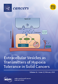
Tumour hypoxia is a feature of solid tumours that contributes to poor prognosis after treatment, due to increased resistance of hypoxic cells to radio- and chemotherapy, and increased metastasis incidence. The extent of hypoxia within a tumour is influenced by the tolerance of individual tumour cells to hypoxia, a feature that differs considerably between tumours. High numbers of hypoxic cells are a direct consequence of enhanced cellular capability to activate HIF-1α signalling, the unfolded protein response (UPR) and autophagy. Recent evidence indicates that hypoxia tolerance can be modulated by distant cells that have experienced hypoxia, and is mediated by the systemic release of factors, such as extracellular vesicles (EV) that can alter HIF-1α-, UPR-, angiogenesis- and autophagy signalling. View this paper
  • Issues are regarded as officially published after their release is announced to the table of contents alert mailing list.
  • You may sign up for e-mail alerts to receive table of contents of newly released issues.
  • PDF is the official format for papers published in both, html and pdf forms. To view the papers in pdf format, click on the "PDF Full-text" link, and use the free Adobe Reader to open them.
Order results
Result details
Section
Select all
Export citation of selected articles as:
13 pages, 1051 KiB  
Article
Developing a Prognostic Gene Panel of Epithelial Ovarian Cancer Patients by a Machine Learning Model
by Tzu-Pin Lu, Kuan-Ting Kuo, Ching-Hsuan Chen, Ming-Cheng Chang, Hsiu-Ping Lin, Yu-Hao Hu, Ying-Cheng Chiang, Wen-Fang Cheng and Chi-An Chen
Cancers 2019, 11(2), 270; https://0-doi-org.brum.beds.ac.uk/10.3390/cancers11020270 - 25 Feb 2019
Cited by 17 | Viewed by 4705
Abstract
Epithelial ovarian cancer patients usually relapse after primary management. We utilized the support vector machine algorithm to develop a model for the chemo-response using the Cancer Cell Line Encyclopedia (CCLE) and validated the model in The Cancer Genome Atlas (TCGA) and the GSE9891 [...] Read more.
Epithelial ovarian cancer patients usually relapse after primary management. We utilized the support vector machine algorithm to develop a model for the chemo-response using the Cancer Cell Line Encyclopedia (CCLE) and validated the model in The Cancer Genome Atlas (TCGA) and the GSE9891 dataset. Finally, we evaluated the feasibility of the model using ovarian cancer patients from our institute. The 10-gene predictive model demonstrated that the high response group had a longer recurrence-free survival (RFS) (log-rank test, p = 0.015 for TCGA, p = 0.013 for GSE9891 and p = 0.039 for NTUH) and overall survival (OS) (log-rank test, p = 0.002 for TCGA and p = 0.016 for NTUH). In a multivariate Cox hazard regression model, the predictive model (HR: 0.644, 95% CI: 0.436–0.952, p = 0.027) and residual tumor size < 1 cm (HR: 0.312, 95% CI: 0.170–0.573, p < 0.001) were significant factors for recurrence. The predictive model (HR: 0.511, 95% CI: 0.334–0.783, p = 0.002) and residual tumor size < 1 cm (HR: 0.252, 95% CI: 0.128–0.496, p < 0.001) were still significant factors for death. In conclusion, the patients of high response group stratified by the model had good response and favourable prognosis, whereas for the patients of medium to low response groups, introduction of other drugs or clinical trials might be beneficial. Full article
(This article belongs to the Special Issue Application of Bioinformatics in Cancers)
Show Figures

Figure 1

12 pages, 757 KiB  
Article
Selective Neck Dissection and Survival in Pathologically Node-Positive Oral Squamous Cell Carcinoma
by Shunichi Shimura, Kazuhiro Ogi, Akihiro Miyazaki, Shota Shimizu, Takeshi Kaneko, Tomoko Sonoda, Junichi Kobayashi, Tomohiro Igarashi, Akira Miyakawa, Tadashi Hasegawa and Hiroyoshi Hiratsuka
Cancers 2019, 11(2), 269; https://0-doi-org.brum.beds.ac.uk/10.3390/cancers11020269 - 25 Feb 2019
Cited by 13 | Viewed by 3275
Abstract
The most important prognostic factor in oral squamous cell carcinoma (OSCC) is neck metastasis, which is treated by neck dissection. Although selective neck dissection (SND) is a useful tool for clinically node-negative OSCC, its efficacy for neck node-positive OSCC has not been established. [...] Read more.
The most important prognostic factor in oral squamous cell carcinoma (OSCC) is neck metastasis, which is treated by neck dissection. Although selective neck dissection (SND) is a useful tool for clinically node-negative OSCC, its efficacy for neck node-positive OSCC has not been established. Sixty-eight OSCC patients with pN1–3 disease who were treated with curative surgery using SND and/or modified-radical/radical neck dissection (MRND/RND) were retrospectively reviewed. The neck control rate was 94% for pN1–3 patients who underwent SND. The five-year overall survival (OS) and disease-specific survival (DSS) in pN1-3 OSCC patients were 62% and 71%, respectively. The multivariate analysis of clinical and pathological variables identified the number of positive nodes as an independent predictor of SND outcome (OS, hazard ratio (HR) = 4.98, 95% confidence interval (CI): 1.48–16.72, p < 0.01; DSS, HR = 6.44, 95% CI: 1.76–23.50, p < 0.01). The results of this retrospective study showed that only SND for neck node-positive OSCC was appropriate for those with up to 2 lymph nodes that had a largest diameter ≤3 cm without extranodal extension (ENE) of the neck and adjuvant radiotherapy. However, the availability of postoperative therapeutic options for high-risk OSCC, including ENE and/or multiple positive lymph nodes, needs to be further investigated. Full article
Show Figures

Figure 1

12 pages, 244 KiB  
Review
Targeted Radionuclide Therapy: New Advances for Improvement of Patient Management and Response
by Javian Malcolm, Nadia Falzone, Boon Q. Lee and Katherine A. Vallis
Cancers 2019, 11(2), 268; https://0-doi-org.brum.beds.ac.uk/10.3390/cancers11020268 - 25 Feb 2019
Cited by 36 | Viewed by 5251
Abstract
Compared to external beam radiotherapy, targeted radionuclide therapy (TRT) allows for systemic radiation treatment of metastatic lesions. Published work on recent strategies to improve patient management and response to TRT through individualising patient treatment, modifying treatment pharmacokinetics and increasing anticancer potency are discussed [...] Read more.
Compared to external beam radiotherapy, targeted radionuclide therapy (TRT) allows for systemic radiation treatment of metastatic lesions. Published work on recent strategies to improve patient management and response to TRT through individualising patient treatment, modifying treatment pharmacokinetics and increasing anticancer potency are discussed in this review, with a special focus on the application of clinically evaluated radiolabelled ligands and peptides in the treatment of neuroendocrine and prostate cancers. Full article
(This article belongs to the Special Issue New Developments in Radiotherapy)
10 pages, 1764 KiB  
Article
Preoperative Imaging Evaluation after Downstaging of Pancreatic Ductal Adenocarcinoma: A Multi-Center Study
by Alessandro Beleù, Angela Calabrese, Giulio Rizzo, Paola Capelli, Nicolò Bellini, Simona Caloggero, Roberto Calbi, Paolo Tinazzi Martini, Riccardo De Robertis, Giovanni Carbognin, Giovanni Marchegiani, Aldo Scarpa, Roberto Salvia, Claudio Bassi and Mirko D’Onofrio
Cancers 2019, 11(2), 267; https://0-doi-org.brum.beds.ac.uk/10.3390/cancers11020267 - 25 Feb 2019
Cited by 18 | Viewed by 3718
Abstract
Introduction: Evaluation of pancreatic ductal adenocarcinoma (PDAC) after chemoradiotherapy downstaging is challenging due to computed tomography (CT) overestimation of tumor extension and residual vascular involvement, limiting access to surgery to some patients with potentially resectable tumors. With this study, we wanted to assess [...] Read more.
Introduction: Evaluation of pancreatic ductal adenocarcinoma (PDAC) after chemoradiotherapy downstaging is challenging due to computed tomography (CT) overestimation of tumor extension and residual vascular involvement, limiting access to surgery to some patients with potentially resectable tumors. With this study, we wanted to assess which radiological findings are most reliable at pre-operative imaging in the evaluation of PDAC after chemoradiotherapy in order to achieve complete resection. Methods: We retrospectively enrolled 71 patients with locally advanced and borderline resectable PDAC who underwent neoadjuvant chemoradiotherapy. Pre-operative CT or magnetic resonance (MR) have been evaluated by three radiologists to assess major qualitative and quantitative parameters of lesions. Accuracy, sensitivity, and specificity compared to anatomopathological results were evaluated for each parameter. Cohen’s K-coefficient has been calculated to evaluate the inter-observer agreement (IOA). Both single and consensus lecture have been tested. Different dimensional cut-offs were tested to categorize tumors according to their major axis and to compare with anatomopathological diameter, tumor persistence, and margin infiltration. Results: A 25 mm cut-off was 67% sensitive, 90% specific, and 77% accurate in assessing real tumor dimension. 25 mm cut-off reported a 64% sensitivity, 78% specificity, and 69% accuracy in assessing R0 resection. Each 5 mm increment of major axis dimension there is an odds ratio (OR) 1.79 (95% CI 1.13–2.80, p = 0.012) for R+ resection. Imaging presence of the perivascular cuff is not associated with tumor persistence and resection margin infiltration (p = 0.362). Lesion enhancement and pattern homogeneity were not accurate in determining tumor persistence. IOA was generally poor to fair, except for >25 mm cut-off classification where IOA was moderate. Diagnostic accuracy is superior in consensus lecture rather than single lecture. Conclusion: Imaging methods tend to underestimate PDAC resectability after neoadjuvant-CRT. IOA is poor to fair in evaluating most of the qualitative parameters of downstaged pancreatic adenocarcinoma. Surgery should be considered for downstaged borderline resectable PDACs, independently from perivascular cuff presence, especially for tumors smaller than 25 mm. Full article
(This article belongs to the Special Issue Advances in Pancreatic Cancer Research)
Show Figures

Figure 1

14 pages, 1591 KiB  
Article
Identification of Novel HLA Class II-Restricted Neoantigens Derived from Driver Mutations
by Susumu Iiizumi, Junya Ohtake, Naoko Murakami, Taku Kouro, Mamoru Kawahara, Fumiko Isoda, Hiroshi Hamana, Hiroyuki Kishi, Norihiro Nakamura and Tetsuro Sasada
Cancers 2019, 11(2), 266; https://0-doi-org.brum.beds.ac.uk/10.3390/cancers11020266 - 24 Feb 2019
Cited by 23 | Viewed by 7662
Abstract
Neoantigens derived from tumor-specific genetic mutations might be suitable targets for cancer immunotherapy because of their high immunogenicity. In the current study, we evaluated the immunogenicity of 10 driver mutations that are frequently expressed in various cancers using peripheral blood mononuclear cells from [...] Read more.
Neoantigens derived from tumor-specific genetic mutations might be suitable targets for cancer immunotherapy because of their high immunogenicity. In the current study, we evaluated the immunogenicity of 10 driver mutations that are frequently expressed in various cancers using peripheral blood mononuclear cells from healthy donors (n = 25). Of the 10 synthetic peptides (27-mer) derived from these mutations, the six peptides from KRAS-G12D, KRAS-G12R, KRAS-G13D, NRAS-Q61R, PIK3CA-H1047R, and C-Kit-D816V induced T cell responses, suggesting that frequent driver mutations are not always less immunogenic. In particular, immune responses to PIK3CA-H1047R, C-Kit-D816V, KRAS-G13D, and NRAS-Q61R were observed in more than 10% of the donors. All six peptides induced human leukocyte antigen (HLA) class II-restricted CD4+ T cell responses; notably, PIK3CA-H1047R contained at least two different CD4+ T cell epitopes restricted to different HLA class II alleles. In addition, PIK3CA-H1047R and C-Kit-D816V induced antigen-specific CD8+ T cells as well, indicating that they might contain both HLA class I- and class II-restricted epitopes. Since the identified neoantigens might be shared by patients with various types of cancers and are not easily lost due to immune escape, they have the potential to be promising off-the-shelf cancer immunotherapy targets in patients with the corresponding mutations. Full article
(This article belongs to the Special Issue Cancer Vaccines: Research and Applications)
Show Figures

Figure 1

41 pages, 3149 KiB  
Review
MicroRNA in Lung Cancer Metastasis
by Shang-Gin Wu, Tzu-Hua Chang, Yi-Nan Liu and Jin-Yuan Shih
Cancers 2019, 11(2), 265; https://0-doi-org.brum.beds.ac.uk/10.3390/cancers11020265 - 23 Feb 2019
Cited by 54 | Viewed by 7816
Abstract
Tumor metastasis is a hallmark of cancer, with distant metastasis frequently developing in lung cancer, even at initial diagnosis, resulting in poor prognosis and high mortality. However, available biomarkers cannot reliably predict cancer spreading sites. The metastatic cascade involves highly complicated processes including [...] Read more.
Tumor metastasis is a hallmark of cancer, with distant metastasis frequently developing in lung cancer, even at initial diagnosis, resulting in poor prognosis and high mortality. However, available biomarkers cannot reliably predict cancer spreading sites. The metastatic cascade involves highly complicated processes including invasion, migration, angiogenesis, and epithelial-to-mesenchymal transition that are tightly controlled by various genetic expression modalities along with interaction between cancer cells and the extracellular matrix. In particular, microRNAs (miRNAs), a group of small non-coding RNAs, can influence the transcriptional and post-transcriptional processes, with dysregulation of miRNA expression contributing to the regulation of cancer metastasis. Nevertheless, although miRNA-targeted therapy is widely studied in vitro and in vivo, this strategy currently affords limited feasibility and a few miRNA-targeted therapies for lung cancer have entered into clinical trials to date. Advances in understanding the molecular mechanism of metastasis will thus provide additional potential targets for lung cancer treatment. This review discusses the current research related to the role of miRNAs in lung cancer invasion and metastasis, with a particular focus on the different metastatic lesions and potential miRNA-targeted treatments for lung cancer with the expectation that further exploration of miRNA-targeted therapy may establish a new spectrum of lung cancer treatments. Full article
(This article belongs to the Special Issue MicroRNA-Associated Cancer Metastasis)
Show Figures

Figure 1

18 pages, 2138 KiB  
Article
Deciphering the Mechanism of Action Involved in Enhanced Suicide Gene Colon Cancer Cell Killer Effect Mediated by Gef and Apoptin
by Blanca Cáceres, Alberto Ramirez, Esmeralda Carrillo, Gema Jimenez, Carmen Griñán-Lisón, Elena López-Ruiz, Yaiza Jiménez-Martínez, Juan A. Marchal and Houria Boulaiz
Cancers 2019, 11(2), 264; https://0-doi-org.brum.beds.ac.uk/10.3390/cancers11020264 - 23 Feb 2019
Cited by 16 | Viewed by 4139
Abstract
Despite the great advances in cancer treatment, colorectal cancer has emerged as the second highest cause of death from cancer worldwide. For this type of tumor, the use of suicide gene therapy could represent a novel therapy. We recently demonstrated that co-expression of [...] Read more.
Despite the great advances in cancer treatment, colorectal cancer has emerged as the second highest cause of death from cancer worldwide. For this type of tumor, the use of suicide gene therapy could represent a novel therapy. We recently demonstrated that co-expression of gef and apoptin dramatically inhibits proliferation of the DLD-1 colon cell line. In the present manuscript, we try to establish the mechanism underlying the enhanced induction of apoptosis by triggering both gef and apoptin expression in colon tumor cells. Scanning microscopy reveals that simultaneous expression of gef and apoptin induces the apparition of many “pores” in the cytoplasmic membrane not detected in control cell lines. The formation of pores induced by the gef gene and accentuated by apoptin results in cell death by necrosis. Moreover, we observed the presence of apoptotic cells. Performing protein expression analysis using western blot, we revealed an activation of mitochondrial apoptosis (increased expression of Pp53, cytochrome c, Bax, and caspase 9) and also the involvement of the extrinsic pathway through caspase 8activation. In conclusion, in this manuscript we demonstrate for the first time that the extrinsic pathway of apoptosis and pore formation is also involved in the cell death caused by the co-expression of the gef and apoptin genes. Our results suggest that co-expression of gef and apoptin genes induces an increase in post-apoptotic necrotic cell death and could be a valuable tool in the design of new antitumor strategies focused on the enhancement of the immune response against cancer cell death. Full article
(This article belongs to the Special Issue Colorectal Cancers)
Show Figures

Figure 1

18 pages, 3518 KiB  
Article
Integrative Omic Profiling Reveals Unique Hypoxia Induced Signatures in Gastric Cancer Associated Myofibroblasts
by Hanna Najgebauer, Andrew F. Jarnuczak, Andrea Varro and Christopher M. Sanderson
Cancers 2019, 11(2), 263; https://0-doi-org.brum.beds.ac.uk/10.3390/cancers11020263 - 23 Feb 2019
Cited by 1 | Viewed by 3274
Abstract
Although hypoxia is known to contribute to several aspects of tumour progression, relatively little is known about the effects of hypoxia on cancer-associated myofibroblasts (CAMs), or the consequences that conditional changes in CAM function may have on tumour development and metastasis. To investigate [...] Read more.
Although hypoxia is known to contribute to several aspects of tumour progression, relatively little is known about the effects of hypoxia on cancer-associated myofibroblasts (CAMs), or the consequences that conditional changes in CAM function may have on tumour development and metastasis. To investigate this issue in the context of gastric cancer, a comparative multiomic analysis was performed on populations of patient-derived myofibroblasts, cultured under normoxic or hypoxic conditions. Data from this study reveal a novel set of CAM-specific hypoxia-induced changes in gene expression and secreted proteins. Significantly, these signatures are not observed in either patient matched adjacent tissue myofibroblasts (ATMs) or non-cancer associated normal tissue myofibroblasts (NTMs). Functional characterisation of different myofibroblast populations shows that hypoxia-induced changes in gene expression not only enhance the ability of CAMs to induce cancer cell migration, but also confer pro-tumorigenic (CAM-like) properties in NTMs. This study provides the first global mechanistic insight into the molecular changes that contribute to hypoxia-induced pro-tumorigenic changes in gastric stromal myofibroblasts. Full article
Show Figures

Figure 1

14 pages, 602 KiB  
Review
Circulating Tumor Cell Detection in Lung Cancer: But to What End?
by Véronique Hofman, Simon Heeke, Charles-Hugo Marquette, Marius Ilié and Paul Hofman
Cancers 2019, 11(2), 262; https://0-doi-org.brum.beds.ac.uk/10.3390/cancers11020262 - 23 Feb 2019
Cited by 17 | Viewed by 4460
Abstract
The understanding of the natural history and biology of lung cancer has been enhanced by studies into circulating tumor cells (CTCs). Fundamental and translational research, as well as clinical trials in the characterization and behavior of these cells, have constantly contributed to improving [...] Read more.
The understanding of the natural history and biology of lung cancer has been enhanced by studies into circulating tumor cells (CTCs). Fundamental and translational research, as well as clinical trials in the characterization and behavior of these cells, have constantly contributed to improving understanding within the domain of thoracic oncology. However, the use of these CTCs as prognostic and predictive biomarkers has not been adopted to the same extent as circulating free DNA (cf-DNA) in plasma, in the daily practice of thoracic oncologists. However, recent technological advances have firmly put the detection and characterization of CTCs in thoracic oncology back on the agenda, and have opened up perspectives for their routine clinical use. This review discusses the major advances of using CTCs in the domain of thoracic oncology, as well as the envisaged short- and long-term prospects. Full article
Show Figures

Figure 1

14 pages, 3036 KiB  
Article
Loss of Stromal Galectin-1 Enhances Multiple Myeloma Development: Emphasis on a Role in Osteoclasts
by Joséphine Muller, Elodie Duray, Margaux Lejeune, Sophie Dubois, Erwan Plougonven, Angélique Léonard, Paola Storti, Nicola Giuliani, Martine Cohen-Solal, Ute Hempel, Victor L. Thijssen, Yves Beguin, Roy Heusschen and Jo Caers
Cancers 2019, 11(2), 261; https://0-doi-org.brum.beds.ac.uk/10.3390/cancers11020261 - 23 Feb 2019
Cited by 13 | Viewed by 3667
Abstract
Multiple myeloma osteolytic disease is caused by an uncoupled bone-remodelling process with an increased osteoclast activity. Disease development relies on interactions between myeloma cells and bone marrow stromal cells. Recent findings suggest a role for glycan-binding proteins in myeloma microenvironment. Here, we investigated [...] Read more.
Multiple myeloma osteolytic disease is caused by an uncoupled bone-remodelling process with an increased osteoclast activity. Disease development relies on interactions between myeloma cells and bone marrow stromal cells. Recent findings suggest a role for glycan-binding proteins in myeloma microenvironment. Here, we investigated lectins involved in osteoclastogenesis and their role in myeloma bone disease. Microarray data analysis showed a lower expression of galectin-1 (gal-1) in mature osteoclasts compared to monocytic progenitor cells, confirmed at the RNA and protein levels in osteoclast cultures. Confocal microscopy showed that gal-1 localised predominantly in the sealing zone of mature osteoclasts. Although equal differentiated-osteoclast numbers, gal-1−/− osteoclasts showed a higher resorption activity compared to wild-type controls. Micro-computed tomography showed an aberrant bone phenotype with decreased bone densities in gal-1−/− mice. In vivo, tumour progression was faster in gal-1−/− mice and associated with a marked bone loss. Additionally, myeloma cells were found to decrease gal-1 expression in osteoclasts. Our results demonstrate that galectin-1 regulates osteoclast activity with an increased resorption by gal-1−/− osteoclasts and decreased bone densities in gal-1−/− mice. We observed an enhanced tumour development in gal-1−/− mice compared to wild-type mice, suggesting that galectin-1 has a functional role in stromal cells in myeloma microenvironment. Full article
Show Figures

Figure 1

77 pages, 3068 KiB  
Review
Emerging Therapies for Acute Myelogenus Leukemia Patients Targeting Apoptosis and Mitochondrial Metabolism
by Germana Castelli, Elvira Pelosi and Ugo Testa
Cancers 2019, 11(2), 260; https://0-doi-org.brum.beds.ac.uk/10.3390/cancers11020260 - 22 Feb 2019
Cited by 25 | Viewed by 7020
Abstract
Acute Myelogenous Leukemia (AML) is a malignant disease of the hematopoietic cells, characterized by impaired differentiation and uncontrolled clonal expansion of myeloid progenitors/precursors, resulting in bone marrow failure and impaired normal hematopoiesis. AML comprises a heterogeneous group of malignancies, characterized by a combination [...] Read more.
Acute Myelogenous Leukemia (AML) is a malignant disease of the hematopoietic cells, characterized by impaired differentiation and uncontrolled clonal expansion of myeloid progenitors/precursors, resulting in bone marrow failure and impaired normal hematopoiesis. AML comprises a heterogeneous group of malignancies, characterized by a combination of different somatic genetic abnormalities, some of which act as events driving leukemic development. Studies carried out in the last years have shown that AML cells invariably have abnormalities in one or more apoptotic pathways and have identified some components of the apoptotic pathway that can be targeted by specific drugs. Clinical results deriving from studies using B-cell lymphoma 2 (BCL-2) inhibitors in combination with standard AML agents, such as azacytidine, decitabine, low-dose cytarabine, provided promising results and strongly support the use of these agents in the treatment of AML patients, particularly of elderly patients. TNF-related apoptosis-inducing ligand (TRAIL) and its receptors are frequently deregulated in AML patients and their targeting may represent a promising strategy for development of new treatments. Altered mitochondrial metabolism is a common feature of AML cells, as supported through the discovery of mutations in the isocitrate dehydrogenase gene and in mitochondrial electron transport chain and of numerous abnormalities of oxidative metabolism existing in AML subgroups. Overall, these observations strongly support the view that the targeting of mitochondrial apoptotic or metabolic machinery is an appealing new therapeutic perspective in AML. Full article
(This article belongs to the Special Issue TRAIL Signaling in Cancer Cells)
Show Figures

Figure 1

17 pages, 2186 KiB  
Article
A Retrospective Dosimetric Study of Radiotherapy Patients with Left-Sided Breast Cancer; Patient Selection Criteria for Deep Inspiration Breath Hold Technique
by Mikaela Dell’Oro, Eileen Giles, Amy Sharkey, Martin Borg, Caroline Connell and Eva Bezak
Cancers 2019, 11(2), 259; https://0-doi-org.brum.beds.ac.uk/10.3390/cancers11020259 - 22 Feb 2019
Cited by 24 | Viewed by 5538
Abstract
Background: Several studies have investigated cardiac dose reduction when utilizing the deep inspiration breath hold (DIBH) technique in patients undergoing radiotherapy for left-sided breast cancer. This paper aims to recommend potential selection criteria based on a retrospective single institute study of free breathing [...] Read more.
Background: Several studies have investigated cardiac dose reduction when utilizing the deep inspiration breath hold (DIBH) technique in patients undergoing radiotherapy for left-sided breast cancer. This paper aims to recommend potential selection criteria based on a retrospective single institute study of free breathing (FB) and DIBH computed tomography (CT) simulation planning scans. Methods: Dosimetric comparisons were performed retrospectively for 20 patients correlating the dose reduction and patient anatomical factors (anatomical variation of chest shape, chest wall separation, total lung volume (TLV) and others). Results: Paired t-tests demonstrated significant cardiac dose reduction for most patients but not all. Minimal cardiac dose reduction was observed for three patients using their DIBH plan, with one patient receiving a higher dose. Linear regression analysis identified a positive correlation between the patient’s TLV (on the FB CT simulation scan) and the magnitude of dosimetric benefit received (0.4045 R2). Conclusion: The TLV measured on a FB plan could potentially be utilised to predict cardiac exposure and assist with patient selection for DIBH. This is important in resource allocation, as DIBH may be unnecessarily recommended for some patients with little dosimetric benefit. Full article
Show Figures

Figure 1

20 pages, 6254 KiB  
Article
Regulation of KIF2A by Antitumor miR-451a Inhibits Cancer Cell Aggressiveness Features in Lung Squamous Cell Carcinoma
by Akifumi Uchida, Naohiko Seki, Keiko Mizuno, Yasutaka Yamada, Shunsuke Misono, Hiroki Sanada, Naoko Kikkawa, Tomohiro Kumamoto, Takayuki Suetsugu and Hiromasa Inoue
Cancers 2019, 11(2), 258; https://0-doi-org.brum.beds.ac.uk/10.3390/cancers11020258 - 22 Feb 2019
Cited by 23 | Viewed by 3309
Abstract
In the human genome, miR-451a is encoded close to the miR-144 on chromosome region 17q11.2. Our previous study showed that both strands of pre-miR-144 acted as antitumor miRNAs and were involved in lung squamous cell carcinoma (LUSQ) pathogenesis. Here, we aimed to [...] Read more.
In the human genome, miR-451a is encoded close to the miR-144 on chromosome region 17q11.2. Our previous study showed that both strands of pre-miR-144 acted as antitumor miRNAs and were involved in lung squamous cell carcinoma (LUSQ) pathogenesis. Here, we aimed to investigate the functional significance of miR-451a and to identify its targeting of oncogenic genes in LUSQ cells. Downregulation of miR-451a was confirmed in LUSQ clinical specimens, and low expression of miR-451a was significantly associated with poor prognosis of LUSQ patients (overall survival: p = 0.035, disease-free survival: p = 0.029). Additionally, we showed that ectopic expression of miR-451a significantly blocked cancer cell aggressiveness. In total, 15 putative oncogenic genes were shown to be regulated by miR-451a in LUSQ cells. Among these targets, high kinesin family member 2A (KIF2A) expression was significantly associated with poor prognosis (overall survival: p = 0.043, disease-free survival: p = 0.028). Multivariate analysis showed that KIF2A expression was an independent prognostic factor in patients with LUSQ (hazard ratio = 1.493, p = 0.034). Aberrant KIF2A expression promoted the malignant transformation of this disease. Analytic strategies based on antitumor miRNAs and their target oncogenes are effective tools for identification of novel molecular pathogenesis of LUSQ. Full article
(This article belongs to the Special Issue MicroRNA-Associated Cancer Metastasis)
Show Figures

Figure 1

18 pages, 2714 KiB  
Article
The Role of Lactate Metabolism in Prostate Cancer Progression and Metastases Revealed by Dual-Agent Hyperpolarized 13C MRSI
by Robert Bok, Jessie Lee, Renuka Sriram, Kayvan Keshari, Subramaniam Sukumar, Saeed Daneshmandi, David E. Korenchan, Robert R. Flavell, Daniel B. Vigneron, John Kurhanewicz and Pankaj Seth
Cancers 2019, 11(2), 257; https://0-doi-org.brum.beds.ac.uk/10.3390/cancers11020257 - 22 Feb 2019
Cited by 38 | Viewed by 4644
Abstract
This study applied a dual-agent, 13C-pyruvate and 13C-urea, hyperpolarized 13C magnetic resonance spectroscopic imaging (MRSI) and multi-parametric (mp) 1H magnetic resonance imaging (MRI) approach in the transgenic adenocarcinoma of mouse prostate (TRAMP) model to investigate changes in tumor perfusion [...] Read more.
This study applied a dual-agent, 13C-pyruvate and 13C-urea, hyperpolarized 13C magnetic resonance spectroscopic imaging (MRSI) and multi-parametric (mp) 1H magnetic resonance imaging (MRI) approach in the transgenic adenocarcinoma of mouse prostate (TRAMP) model to investigate changes in tumor perfusion and lactate metabolism during prostate cancer development, progression and metastases, and after lactate dehydrogenase-A (LDHA) knock-out. An increased Warburg effect, as measured by an elevated hyperpolarized (HP) Lactate/Pyruvate (Lac/Pyr) ratio, and associated Ldha expression and LDH activity were significantly higher in high- versus low-grade TRAMP tumors and normal prostates. The hypoxic tumor microenvironment in high-grade tumors, as measured by significantly decreased HP 13C-urea perfusion and increased PIM staining, played a key role in increasing lactate production through increased Hif1α and then Ldha expression. Increased lactate induced Mct4 expression and an acidic tumor microenvironment that provided a potential mechanism for the observed high rate of lymph node (86%) and liver (33%) metastases. The Ldha knockdown in the triple-transgenic mouse model of prostate cancer resulted in a significant reduction in HP Lac/Pyr, which preceded a reduction in tumor volume or apparent water diffusion coefficient (ADC). The Ldha gene knockdown significantly reduced primary tumor growth and reduced lymph node and visceral metastases. These data suggested a metabolic transformation from low- to high-grade prostate cancer including an increased Warburg effect, decreased perfusion, and increased metastatic potential. Moreover, these data suggested that LDH activity and lactate are required for tumor progression. The lactate metabolism changes during prostate cancer provided the motivation for applying hyperpolarized 13C MRSI to detect aggressive disease at diagnosis and predict early therapeutic response. Full article
Show Figures

Figure 1

11 pages, 1372 KiB  
Article
Reverse Engineering Cancer: Inferring Transcriptional Gene Signatures from Copy Number Aberrations with ICAro
by Davide Angeli, Maurizio Fanciulli and Matteo Pallocca
Cancers 2019, 11(2), 256; https://0-doi-org.brum.beds.ac.uk/10.3390/cancers11020256 - 22 Feb 2019
Cited by 1 | Viewed by 3112
Abstract
The characterization of a gene product function is a process that involves multiple laboratory techniques in order to silence the gene itself and to understand the resulting cellular phenotype via several omics profiling. When it comes to tumor cells, usually the translation process [...] Read more.
The characterization of a gene product function is a process that involves multiple laboratory techniques in order to silence the gene itself and to understand the resulting cellular phenotype via several omics profiling. When it comes to tumor cells, usually the translation process from in vitro characterization results to human validation is a difficult journey. Here, we present a simple algorithm to extract mRNA signatures from cancer datasets, where a particular gene has been deleted at the genomic level, ICAro. The process is implemented as a two-step workflow. The first one employs several filters in order to select the two patient subsets: the inactivated one, where the target gene is deleted, and the control one, where large genomic rearrangements should be absent. The second step performs a signature extraction via a Differential Expression analysis and a complementary Random Forest approach to provide an additional gene ranking in terms of information loss. We benchmarked the system robustness on a panel of genes frequently deleted in cancers, where we validated the downregulation of target genes and found a correlation with signatures extracted with the L1000 tool, outperforming random sampling for two out of six L1000 classes. Furthermore, we present a use case correlation with a published transcriptomic experiment. In conclusion, deciphering the complex interactions of the tumor environment is a challenge that requires the integration of several experimental techniques in order to create reproducible results. We implemented a tool which could be of use when trying to find mRNA signatures related to a gene loss event to better understand its function or for a gene-loss associated biomarker research. Full article
(This article belongs to the Special Issue Application of Bioinformatics in Cancers)
Show Figures

Figure 1

17 pages, 4845 KiB  
Article
A Cell-Autonomous Oncosuppressive Role of Human RNASET2 Affecting ECM-Mediated Oncogenic Signaling
by Francesca Roggiani, Cristina Riva, Francesco Raspagliesi, Giovanni Porta, Roberto Valli, Roberto Taramelli, Francesco Acquati, Delia Mezzanzanica and Antonella Tomassetti
Cancers 2019, 11(2), 255; https://0-doi-org.brum.beds.ac.uk/10.3390/cancers11020255 - 22 Feb 2019
Cited by 9 | Viewed by 3377
Abstract
RNASET2 is an extracellular ribonuclease endowed with a marked antitumorigenic role in several carcinomas, independent from its catalytic activity. Besides its antitumorigenic role by the recruitment to the tumor mass of immune cells from the monocyte/macrophage lineage, RNASET2 is induced by cellular stress [...] Read more.
RNASET2 is an extracellular ribonuclease endowed with a marked antitumorigenic role in several carcinomas, independent from its catalytic activity. Besides its antitumorigenic role by the recruitment to the tumor mass of immune cells from the monocyte/macrophage lineage, RNASET2 is induced by cellular stress and involved in actin cytoskeleton remodeling affecting cell interactions with the extracellular matrix (ECM). Here, we aimed to investigate the effects of RNASET2 expression modulation on cell phenotype and behavior in epithelial ovarian cancer (EOC) cellular models. In silico analysis on two publicly available datasets of gene expression from EOC patients (n = 392) indicated that increased RNASET2 transcript levels are associated with longer overall survival. In EOC biopsies (n = 101), analyzed by immunohistochemistry, RNASET2 was found heterogeneously expressed among tumors with different clinical–pathological characteristics and, in some cases, its expression localized to tumor-associated ECM. By characterizing in vitro two models of EOC cells in which RNASET2 was silenced or overexpressed, we report that RNASET2 expression negatively affects growth capability by conferring a peculiar cell phenotype upon the interaction of EOC cells with the ECM, resulting in decreased src activation. Altogether, these data suggest that drugs targeting activated src might represent a therapeutic approach for RNASET2-expressing EOCs. Full article
Show Figures

Figure 1

14 pages, 2391 KiB  
Article
Casticin-Induced Inhibition of Cell Growth and Survival Are Mediated through the Dual Modulation of Akt/mTOR Signaling Cascade
by Jong Hyun Lee, Chulwon Kim, Jae-Young Um, Gautam Sethi and Kwang Seok Ahn
Cancers 2019, 11(2), 254; https://0-doi-org.brum.beds.ac.uk/10.3390/cancers11020254 - 22 Feb 2019
Cited by 46 | Viewed by 3906
Abstract
The Akt/mTOR signaling cascade is a critical pathway involved in various physiological and pathological conditions, including regulation of cell proliferation, survival, invasion, and angiogenesis. In the present study, we investigated the anti-neoplastic effects of casticin (CTC), identified from the plant Vitex rotundifolia L., [...] Read more.
The Akt/mTOR signaling cascade is a critical pathway involved in various physiological and pathological conditions, including regulation of cell proliferation, survival, invasion, and angiogenesis. In the present study, we investigated the anti-neoplastic effects of casticin (CTC), identified from the plant Vitex rotundifolia L., alone and/or in combination with BEZ-235, a dual Akt/mTOR inhibitor in human tumor cells. We found that CTC exerted a significant dose-dependent cytotoxicity and reduced cell proliferation in a variety of human tumor cells. Also, CTC effectively blocked the phosphorylation levels of Akt (Ser473) and mTOR (Ser2448) proteins as well as induced substantial apoptosis. Additionally treatment with CTC and BEZ-235 in conjunction resulted in a greater apoptotic effect than caused by either agent alone thus implicating the anti-neoplastic effects of this novel combination. Overall, the findings suggest that CTC can interfere with Akt/mTOR signaling cascade involved in tumorigenesis and can be used together with pharmacological agents targeting Akt/mTOR pathway. Full article
(This article belongs to the Special Issue Apoptosis in Cancer)
Show Figures

Figure 1

17 pages, 508 KiB  
Article
Trending Towards Safer Breast Cancer Surgeries? Examining Acute Complication Rates from A 13-Year NSQIP Analysis
by Michael M. Jonczyk, Jolie Jean, Roger Graham and Abhishek Chatterjee
Cancers 2019, 11(2), 253; https://0-doi-org.brum.beds.ac.uk/10.3390/cancers11020253 - 21 Feb 2019
Cited by 31 | Viewed by 5021
Abstract
As breast cancer surgery continues to evolve, this study highlights the acute complication rates and predisposing risks following partial mastectomy (PM), mastectomy(M), mastectomy with muscular flap reconstruction (M + MF), mastectomy with implant reconstruction (M + I), and oncoplastic surgery (OPS). Data was [...] Read more.
As breast cancer surgery continues to evolve, this study highlights the acute complication rates and predisposing risks following partial mastectomy (PM), mastectomy(M), mastectomy with muscular flap reconstruction (M + MF), mastectomy with implant reconstruction (M + I), and oncoplastic surgery (OPS). Data was collected from the American College of Surgeons NSQIP database (2005–2017). Complication rate and trend analyses were performed along with an assessment of odds ratios for predisposing risk factors using adjusted linear regression. 226,899 patients met the inclusion criteria. Complication rates have steadily increased in all mastectomy groups (p < 0.05). Cumulative complication rates between surgical categories were significantly different in each complication cluster (all p < 0.0001). Overall complication rates were: PM: 2.25%, OPS: 3.2%, M: 6.56%, M + MF: 13.04% and M + I: 5.68%. The most common predictive risk factors were mastectomy, increasing operative time, ASA class, BMI, smoking, recent weight loss, history of CHF, COPD and bleeding disorders (all p < 0.001). Patients who were non-diabetic, younger (age < 60) and treated as an outpatient all had protective OR for an acute complication (p < 0.0001). This study provides data comparing nationwide acute complication rates following different breast cancer surgeries. These can be used to inform patients during surgical decision making. Full article
Show Figures

Figure 1

32 pages, 1334 KiB  
Review
Next Generation Sequencing in AML—On the Way to Becoming a New Standard for Treatment Initiation and/or Modulation?
by Michael Leisch, Bettina Jansko, Nadja Zaborsky, Richard Greil and Lisa Pleyer
Cancers 2019, 11(2), 252; https://0-doi-org.brum.beds.ac.uk/10.3390/cancers11020252 - 21 Feb 2019
Cited by 36 | Viewed by 9105
Abstract
Acute myeloid leukemia (AML) is a clonal disease caused by genetic abberations occurring predominantly in the elderly. Next generation sequencing (NGS) analysis has led to a deeper genetic understanding of the pathogenesis and the role of recently discovered genetic precursor lesions (clonal hematopoiesis [...] Read more.
Acute myeloid leukemia (AML) is a clonal disease caused by genetic abberations occurring predominantly in the elderly. Next generation sequencing (NGS) analysis has led to a deeper genetic understanding of the pathogenesis and the role of recently discovered genetic precursor lesions (clonal hematopoiesis of indeterminate/oncogenic potential (CHIP/CHOP)) in the evolution of AML. These advances are reflected by the inclusion of certain mutations in the updated World Health Organization (WHO) 2016 classification and current treatment guidelines by the European Leukemia Net (ELN) and National Comprehensive Cancer Network (NCCN) and results of mutational testing are already influencing the choice and timing of (targeted) treatment. Genetic profiling and stratification of patients into molecularly defined subgroups are expected to gain ever more weight in daily clinical practice. Our aim is to provide a concise summary of current evidence regarding the relevance of NGS for the diagnosis, risk stratification, treatment planning and response assessment in AML, including minimal residual disease (MRD) guided approaches. We also summarize recently approved drugs targeting genetically defined patient populations with risk adapted- and individualized treatment strategies. Full article
Show Figures

Figure 1

22 pages, 2251 KiB  
Article
Pan-Cancer Analyses Reveal Genomic Features of FOXM1 Overexpression in Cancer
by Carter J Barger, Connor Branick, Linda Chee and Adam R. Karpf
Cancers 2019, 11(2), 251; https://0-doi-org.brum.beds.ac.uk/10.3390/cancers11020251 - 21 Feb 2019
Cited by 117 | Viewed by 12876
Abstract
FOXM1 is frequently overexpressed in cancer, but this has not been studied in a comprehensive manner. We utilized genotype-tissue expression (GTEx) normal and The Cancer Genome Atlas (TCGA) tumor data to define FOXM1 expression, including its isoforms, and to determine the genetic alterations [...] Read more.
FOXM1 is frequently overexpressed in cancer, but this has not been studied in a comprehensive manner. We utilized genotype-tissue expression (GTEx) normal and The Cancer Genome Atlas (TCGA) tumor data to define FOXM1 expression, including its isoforms, and to determine the genetic alterations that promote FOXM1 expression in cancer. Additionally, we used human fallopian tube epithelial (FTE) cells to dissect the role of Retinoblastoma (Rb)-E2F and Cyclin E1 in FOXM1 regulation, and a novel human embryonic kidney cell (HEK293T) CRISPR FOXM1 knockout model to define isoform-specific transcriptional programs. FOXM1 expression, at the mRNA and protein level, was significantly elevated in tumors with FOXM1 amplification, p53 inactivation, and Rb-E2F deregulation. FOXM1 expression was remarkably high in testicular germ cell tumors (TGCT), high-grade serous ovarian cancer (HGSC), and basal breast cancer (BBC). FOXM1 expression in cancer was associated with genomic instability, as measured using aneuploidy signatures. FTE models confirmed a role for Rb-E2F signaling in FOXM1 regulation and in particular identified Cyclin E1 as a novel inducer of FOXM1 expression. Among the three FOXM1 isoforms, FOXM1c showed the highest expression in normal and tumor tissues and cancer cell lines. The CRISPR knockout model demonstrated that FOXM1b and FOXM1c are transcriptionally active, while FOXM1a is not. Finally, we were unable to confirm the existence of a FOXM1 auto-regulatory loop. This study provides significant and novel information regarding the frequency, causes, and consequences of elevated FOXM1 expression in human cancer. Full article
(This article belongs to the Special Issue Fox Proteins and Cancers: Old Proteins with Emerging New Tales)
Show Figures

Figure 1

11 pages, 1470 KiB  
Perspective
The OncoAge Consortium: Linking Aging and Oncology from Bench to Bedside and Back Again
by Paul Hofman, Nicholas Ayache, Pascal Barbry, Michel Barlaud, Audrey Bel, Philippe Blancou, Frédéric Checler, Sylvie Chevillard, Gael Cristofari, Mathilde Demory, Vincent Esnault, Claire Falandry, Eric Gilson, Olivier Guérin, Nicolas Glaichenhaus, Joel Guigay, Marius Ilié, Bernard Mari, Charles-Hugo Marquette, Véronique Paquis-Flucklinger, Frédéric Prate, Pierre Saintigny, Barbara Seitz-Polsky, Taycir Skhiri, Ellen Van Obberghen-Schilling, Emmanuel Van Obberghen and Laurent Yvan-Charvetadd Show full author list remove Hide full author list
Cancers 2019, 11(2), 250; https://0-doi-org.brum.beds.ac.uk/10.3390/cancers11020250 - 21 Feb 2019
Cited by 2 | Viewed by 4095
Abstract
It is generally accepted that carcinogenesis and aging are two biological processes, which are known to be associated. Notably, the frequency of certain cancers (including lung cancer), increases significantly with the age of patients and there is now a wealth of data showing [...] Read more.
It is generally accepted that carcinogenesis and aging are two biological processes, which are known to be associated. Notably, the frequency of certain cancers (including lung cancer), increases significantly with the age of patients and there is now a wealth of data showing that multiple mechanisms leading to malignant transformation and to aging are interconnected, defining the so-called common biology of aging and cancer. OncoAge, a consortium launched in 2015, brings together the multidisciplinary expertise of leading public hospital services and academic laboratories to foster the transfer of scientific knowledge rapidly acquired in the fields of cancer biology and aging into innovative medical practice and silver economy development. This is achieved through the development of shared technical platforms (for research on genome stability, (epi)genetics, biobanking, immunology, metabolism, and artificial intelligence), clinical research projects, clinical trials, and education. OncoAge focuses mainly on two pilot pathologies, which benefit from the expertise of several members, namely lung and head and neck cancers. This review outlines the broad strategic directions and key advances of OncoAge and summarizes some of the issues faced by this consortium, as well as the short- and long-term perspectives. Full article
Show Figures

Figure 1

11 pages, 1981 KiB  
Article
Exosome Analysis in Tumor-Draining Pulmonary Vein Identifies NSCLC Patients with Higher Risk of Relapse after Curative Surgery
by Alfons Navarro, Laureano Molins, Ramon M. Marrades, Jorge Moises, Nuria Viñolas, Sara Morales, Jordi Canals, Joan J. Castellano, José Ramírez and Mariano Monzo
Cancers 2019, 11(2), 249; https://0-doi-org.brum.beds.ac.uk/10.3390/cancers11020249 - 21 Feb 2019
Cited by 30 | Viewed by 3993
Abstract
Since tumor-draining pulmonary vein blood (PV) is enriched in tumor-secreted products, we hypothesized that it would also be enriched in tumor-derived exosomes, which would be important in the metastasis process. We characterized exosomes from PV of 61 resected non-small cell lung cancer (NSCLC) [...] Read more.
Since tumor-draining pulmonary vein blood (PV) is enriched in tumor-secreted products, we hypothesized that it would also be enriched in tumor-derived exosomes, which would be important in the metastasis process. We characterized exosomes from PV of 61 resected non-small cell lung cancer (NSCLC) patients to evaluate its potential as relapse biomarkers. Exosomes were characterized using transmission electron microscopy, western blot and nanoparticle tracking analysis and we examined time to relapse (TTR) and overall survival (OS). Differences between PV and peripheral vein were found. PV was enriched in smaller exosomes than the paired peripheral vein (p = 0.01). Moreover, PV exosome size mode was able to identify relapsed patients (Area under the curve [AUC] = 0.781; 95%CI: 0.6641–0.8978), in whom exosome size was smaller (<112 nm; p < 0.001). The combination of PV exosome size and N (lymph node involvement) showed an AUC of 0.89 (95%CI: 0.80–0.97). Moreover, smaller PV exosome size was associated with shorter TTR (28.3 months vs. not reached, p < 0.001) and OS (43.9 months vs. not reached, p = 0.009). Multivariate analyses identified PV exosome size and stage as independent prognostic markers for TTR and OS. PV exosome size is a promising relapse biomarker after surgery that can add valuable information to clinical variables. Full article
(This article belongs to the Special Issue Liquid Biopsy for Cancer)
Show Figures

Figure 1

14 pages, 2570 KiB  
Article
Decoration of Anti-CD38 on Nanoparticles Carrying a STAT3 Inhibitor Can Improve the Therapeutic Efficacy Against Myeloma
by Yung-Hsing Huang, Mohammad Reza Vakili, Ommoleila Molavi, Yuen Morrissey, Chengsheng Wu, Igor Paiva, Amir Hasan Soleimani, Forugh Sanaee, Afsaneh Lavasanifar and Raymond Lai
Cancers 2019, 11(2), 248; https://0-doi-org.brum.beds.ac.uk/10.3390/cancers11020248 - 20 Feb 2019
Cited by 27 | Viewed by 4941
Abstract
STAT3 is an oncoprotein which has been shown to contribute to drug resistance in multiple myeloma (MM). Nonetheless, the clinical utility of STAT3 inhibitors in treating MM has been limited, partly related to some of their pharmacologic properties. To overcome these challenges, our [...] Read more.
STAT3 is an oncoprotein which has been shown to contribute to drug resistance in multiple myeloma (MM). Nonetheless, the clinical utility of STAT3 inhibitors in treating MM has been limited, partly related to some of their pharmacologic properties. To overcome these challenges, our group had previously packaged STAT3 inhibitors using a novel formulation of nanoparticles (NP) and found encouraging results. In this study, we aimed to further improve the pharmacologic properties of these NP by decorating them with monoclonal anti-CD38 antibodies. NP loaded with S3I-1757 (a STAT3 inhibitor), labeled as S3I-NP, were generated. S3I-NP decorated with anti-CD38 (labeled as CD38-S3I-NP) were found to have a similar nanoparticular size, drug encapsulation, and loading as S3I-NP. The release of S3I-1757 at 24 h was also similar between the two formulations. Using Cy5.5 labeling of the NP, we found that the decoration of anti-CD38 on these NP significantly increased the cellular uptake by two MM cell lines (p < 0.001). Accordingly, CD38-S3I-NP showed a significantly lower inhibitory concentration at 50% (IC50) compared to S3I-NP in two IL6-stimulated MM cell lines (p < 0.001). In a xenograft mouse model, CD38-S3I-NP significantly reduced the tumor size by 4-fold compared to S3I-NP on day 12 after drug administration (p = 0.006). The efficacy of CD38-S3I-NP in suppressing STAT3 phosphorylation in the xenografts was confirmed by using immunocytochemistry and Western blot analysis. In conclusion, our study suggests that the decoration of anti-CD38 on NP loaded with STAT3 inhibitors can further improve their therapeutic effects against MM. Full article
(This article belongs to the Special Issue Targeting STAT3 and STAT5 in Cancer)
Show Figures

Figure 1

23 pages, 15676 KiB  
Review
State-of-the-Art of Profiling Immune Contexture in the Era of Multiplexed Staining and Digital Analysis to Study Paraffin Tumor Tissues
by Edwin Roger Parra, Alejandro Francisco-Cruz and Ignacio Ivan Wistuba
Cancers 2019, 11(2), 247; https://0-doi-org.brum.beds.ac.uk/10.3390/cancers11020247 - 20 Feb 2019
Cited by 83 | Viewed by 9801
Abstract
Multiplexed platforms for multiple epitope detection have emerged in the last years as very powerful tools to study tumor tissues. These revolutionary technologies provide important visual techniques for tumor examination in formalin-fixed paraffin-embedded specimens to improve the understanding of the tumor microenvironment, promote [...] Read more.
Multiplexed platforms for multiple epitope detection have emerged in the last years as very powerful tools to study tumor tissues. These revolutionary technologies provide important visual techniques for tumor examination in formalin-fixed paraffin-embedded specimens to improve the understanding of the tumor microenvironment, promote new treatment discoveries, aid in cancer prevention, as well as allowing translational studies to be carried out. The aim of this review is to highlight the more recent methodologies that use multiplexed staining to study simultaneous protein identification in formalin-fixed paraffin-embedded tumor tissues for immune profiling, clinical research, and potential translational analysis. New multiplexed methodologies, which permit the identification of several proteins at the same time in one single tissue section, have been developed in recent years with the ability to study different cell populations, cells by cells, and their spatial distribution in different tumor specimens including whole sections, core needle biopsies, and tissue microarrays. Multiplexed technologies associated with image analysis software can be performed with a high-quality throughput assay to study cancer specimens and are important tools for new discoveries. The different multiplexed technologies described in this review have shown their utility in the study of cancer tissues and their advantages for translational research studies and application in cancer prevention and treatments. Full article
Show Figures

Figure 1

31 pages, 1348 KiB  
Review
FBXW7 in Cancer: What Has Been Unraveled Thus Far?
by Bethsebie Lalduhsaki Sailo, Kishore Banik, Sosmitha Girisa, Devivasha Bordoloi, Lu Fan, Clarissa Esmeralda Halim, Hong Wang, Alan Prem Kumar, Dali Zheng, Xinliang Mao, Gautam Sethi and Ajaikumar Bahulayan Kunnumakkara
Cancers 2019, 11(2), 246; https://0-doi-org.brum.beds.ac.uk/10.3390/cancers11020246 - 19 Feb 2019
Cited by 115 | Viewed by 11571
Abstract
The FBXW7 (F-box with 7 tandem WD40) protein encoded by the gene FBXW7 is one of the crucial components of ubiquitin ligase called Skp1-Cullin1-F-box (SCF) complex that aids in the degradation of many oncoproteins via the ubiquitin-proteasome system (UPS) thus regulating cellular growth. [...] Read more.
The FBXW7 (F-box with 7 tandem WD40) protein encoded by the gene FBXW7 is one of the crucial components of ubiquitin ligase called Skp1-Cullin1-F-box (SCF) complex that aids in the degradation of many oncoproteins via the ubiquitin-proteasome system (UPS) thus regulating cellular growth. FBXW7 is considered as a potent tumor suppressor as most of its target substrates can function as potential growth promoters, including c-Myc, Notch, cyclin E, c-JUN, and KLF5. Its regulators include p53, C/EBP-δ, Numb, microRNAs, Pin 1, Hes-5, BMI1, Ebp2. Mounting evidence has indicated the involvement of aberrant expression of FBXW7 for tumorigenesis. Moreover, numerous studies have also shown its role in cancer cell chemosensitization, thereby demonstrating the importance of FBXW7 in the development of curative cancer therapy. This comprehensive review emphasizes on the targets, functions, regulators and expression of FBXW7 in different cancers and its involvement in sensitizing cancer cells to chemotherapeutic drugs. Full article
Show Figures

Figure 1

13 pages, 3566 KiB  
Article
KCNMA1 Expression Is Downregulated in Colorectal Cancer via Epigenetic Mechanisms
by Maria Sofia Basile, Paolo Fagone, Katia Mangano, Santa Mammana, Gaetano Magro, Lucia Salvatorelli, Giovanni Li Destri, Gaetano La Greca, Ferdinando Nicoletti, Stefano Puleo and Antonio Pesce
Cancers 2019, 11(2), 245; https://0-doi-org.brum.beds.ac.uk/10.3390/cancers11020245 - 19 Feb 2019
Cited by 22 | Viewed by 4052
Abstract
KCNMA1 is a gene located at 10q22 that encodes the pore-forming α-subunit of the large-conductance Ca2+-activated K+ channel. KCNMA1 is down-regulated in gastric carcinoma tumors, through hypermethylation of its promoter. In the present study, we have evaluated the expression levels [...] Read more.
KCNMA1 is a gene located at 10q22 that encodes the pore-forming α-subunit of the large-conductance Ca2+-activated K+ channel. KCNMA1 is down-regulated in gastric carcinoma tumors, through hypermethylation of its promoter. In the present study, we have evaluated the expression levels of KCNMA1 both in a mouse model of Colorectal Cancer (CRC) and in human CRC samples. Additionally, epigenetic mechanisms of KCNMA1 gene regulation were investigated. We observed a significant down-regulation of KCNMA1 both in a human and mouse model of CRC. No differences in KCNMA1 levels were, however, observed at different TNM stages. We also wanted to determine whether the modulation in KCNMA1 was dependent on epigenetic mechanisms. A statistically significant inverse correlation between KCNMA1 expression and mir-17-5p levels was observed in patients with CRC. Furthermore, in the tumor samples, we found a significant hypermethylation of the promoter, in the loci cg24113782 and cg25655799, compared to healthy tissue. Overall, our data suggest the possible use of KCNMA1 as a therapeutic target in the early stages of CRC. Full article
Show Figures

Figure 1

27 pages, 5413 KiB  
Article
Mouse-Derived Isograft (MDI) In Vivo Tumor Models I. Spontaneous sMDI Models: Characterization and Cancer Therapeutic Approaches
by Peter Jantscheff, Janette Beshay, Thomas Lemarchand, Cynthia Obodozie, Christoph Schächtele and Holger Weber
Cancers 2019, 11(2), 244; https://0-doi-org.brum.beds.ac.uk/10.3390/cancers11020244 - 19 Feb 2019
Cited by 4 | Viewed by 6604
Abstract
Syngeneic in vivo tumor models are valuable for the development and investigation of immune-modulating anti-cancer drugs. In the present study, we established a novel syngeneic in vivo model type named mouse-derived isografts (MDIs). Spontaneous MDIs (sMDIs) were obtained during a long-term observation period [...] Read more.
Syngeneic in vivo tumor models are valuable for the development and investigation of immune-modulating anti-cancer drugs. In the present study, we established a novel syngeneic in vivo model type named mouse-derived isografts (MDIs). Spontaneous MDIs (sMDIs) were obtained during a long-term observation period (more than one to two years) of naïve and untreated animals of various mouse strains (C3H/HeJ, CBA/J, DBA/2N, BALB/c, and C57BL/6N). Primary tumors or suspicious tissues were assessed macroscopically and re-transplanted in a PDX-like manner as small tumor pieces into sex-matched syngeneic animals. Nine outgrowing primary tumors were histologically characterized either as adenocarcinomas, histiocytic carcinomas, or lymphomas. Growth of the tumor pieces after re-transplantation displayed model heterogeneity. The adenocarcinoma sMDI model JA-0009 was further characterized by flow cytometry, RNA-sequencing, and efficacy studies. M2 macrophages were found to be the main tumor infiltrating leukocyte population, whereas only a few T cells were observed. JA-0009 showed limited sensitivity when treated with antibodies against inhibitory checkpoint molecules (anti-mPD-1 and anti-mCTLA-4), but high sensitivity to gemcitabine treatment. The generated sMDI are spontaneously occurring tumors of low passage number, propagated as tissue pieces in mice without any tissue culturing, and thus conserving the original tumor characteristics and intratumoral immune cell populations. Full article
Show Figures

Figure 1

24 pages, 7926 KiB  
Article
Paclitaxel-Induced Src Activation Is Inhibited by Dasatinib Treatment, Independently of Cancer Stem Cell Properties, in a Mouse Model of Ovarian Cancer
by Elif Kadife, Emily Chan, Rodney Luwor, George Kannourakis, Jock Findlay and Nuzhat Ahmed
Cancers 2019, 11(2), 243; https://0-doi-org.brum.beds.ac.uk/10.3390/cancers11020243 - 19 Feb 2019
Cited by 11 | Viewed by 4114
Abstract
Approximately seventy percent of ovarian cancer patients succumb to the disease within the first 5 years of diagnosis, even after successful surgery and effective chemotherapy treatment. A small subset of chemotherapy resistant cancer stem cells (CSCs) cause relapse of ovarian cancers. This study [...] Read more.
Approximately seventy percent of ovarian cancer patients succumb to the disease within the first 5 years of diagnosis, even after successful surgery and effective chemotherapy treatment. A small subset of chemotherapy resistant cancer stem cells (CSCs) cause relapse of ovarian cancers. This study investigated the association between paclitaxel-mediated Src activation (p-Src) and CSC populations in driving ovarian cancer progression. We demonstrate that patients with high-stage serous ovarian carcinomas have significantly elevated levels of p-Src, compared to patient with low-stage and benign ovarian tumours. Additionally, p-Src was significantly enhanced in ascites-derived tumour cells obtained from recurrent patients, compared to chemonaïve patients. Paclitaxel treatment increased Src activation in ovarian cancer cells, causing enrichment of CSC marker expression in the surviving cells in vitro and in xenografts of nude mice. Dasatinib in combination with paclitaxel significantly suppressed p-Src in ovarian cancer cell lines and xenografts but had no effect on the expression of CSC markers. However, combination of paclitaxel and Dasatinib showed lower trend in invasion in liver and pancreas, compared to paclitaxel-only treatment. The tumours treated with combination therapy also had significantly lower infiltration of mononuclear cells. Robust recurrent tumour growth was observed in all mice groups after termination of treatments. The above results suggest that Dasatinib-mediated inhibition of p-Src may not be crucial for paclitaxel-induced CSC-mediated recurrence in ovarian cancer. Full article
(This article belongs to the Special Issue Ovarian Cancer Metastasis)
Show Figures

Figure 1

20 pages, 2827 KiB  
Article
Mouse-Derived Isograft (MDI) In Vivo Tumor Models II. Carcinogen-Induced cMDI Models: Characterization and Cancer Therapeutic Approaches
by Janette Beshay, Peter Jantscheff, Thomas Lemarchand, Cynthia Obodozie, Christoph Schächtele and Holger Weber
Cancers 2019, 11(2), 242; https://0-doi-org.brum.beds.ac.uk/10.3390/cancers11020242 - 19 Feb 2019
Cited by 2 | Viewed by 4245
Abstract
In this second study, we established syngeneic in vivo models named carcinogen-induced mouse-derived isografts (cMDIs). Carcinogen-induced tumors were obtained during short-term observation (3–9 months) of CBA/J mice treated with various administration routes with 3-methylcholanthrene (MCA) or N-methyl-N-nitrosourea (MNU) as carcinogens. [...] Read more.
In this second study, we established syngeneic in vivo models named carcinogen-induced mouse-derived isografts (cMDIs). Carcinogen-induced tumors were obtained during short-term observation (3–9 months) of CBA/J mice treated with various administration routes with 3-methylcholanthrene (MCA) or N-methyl-N-nitrosourea (MNU) as carcinogens. During necropsy, primary tumors and suspicious tissues were assessed macroscopically and re-transplanted (in PDX-like manner) into sex-matched syngeneic animals. Outgrowing tumors were histologically characterized as either spinocellular carcinoma (1/8) or various differentiated sarcomas (7/8). Growth curves of four sarcomas showed striking heterogeneity. These cMDIs were further characterized by flow cytometry, RNA sequencing, or efficacy studies. A variable invasion of immune cells into the tumors, as well as varying expression of tyrosine kinase receptor, IFN-γ signature, or immune cell population marker genes could be observed. Immune checkpoint inhibitor treatment (anti-mPD-1, anti-mCTLA-4, or a combination thereof) showed different responses in the various cMDI models. In general, cMDI models are carcinogen-induced tumors of low passage number that were propagated as tissue pieces in mice without any tissue culturing. Therefore, the tumors contained conserved tumor characteristics and intratumoral immune cell populations. In contrast to the previously described spontaneous MDI, carcinogen induction resulted in a greater number of individual but histologically related tumors, which were preferentially sarcomas. Full article
Show Figures

Figure 1

25 pages, 1482 KiB  
Review
Protein Phosphatases—A Touchy Enemy in the Battle Against Glioblastomas: A Review
by Arata Tomiyama, Tatsuya Kobayashi, Kentaro Mori and Koichi Ichimura
Cancers 2019, 11(2), 241; https://0-doi-org.brum.beds.ac.uk/10.3390/cancers11020241 - 19 Feb 2019
Cited by 14 | Viewed by 4496
Abstract
Glioblastoma (GBM) is the most common malignant tumor arising from brain parenchyma. Although many efforts have been made to develop therapies for GBM, the prognosis still remains poor, mainly because of the difficulty in total resection of the tumor mass from brain tissue [...] Read more.
Glioblastoma (GBM) is the most common malignant tumor arising from brain parenchyma. Although many efforts have been made to develop therapies for GBM, the prognosis still remains poor, mainly because of the difficulty in total resection of the tumor mass from brain tissue and the resistance of the residual tumor against standard chemoradiotherapy. Therefore, novel adjuvant therapies are urgently needed. Recent genome-wide analyses of GBM cases have clarified molecular signaling mechanisms underlying GBM biology. However, results of clinical trials targeting phosphorylation-mediated signaling have been unsatisfactory to date. Protein phosphatases are enzymes that antagonize phosphorylation signaling by dephosphorylating phosphorylated signaling molecules. Recently, the critical roles of phosphatases in the regulation of oncogenic signaling in malignant tumor cells have been reported, and tumorigenic roles of deregulated phosphatases have been demonstrated in GBM. However, a detailed mechanism underlying phosphatase-mediated signaling transduction in the regulation of GBM has not been elucidated, and such information is necessary to apply phosphatases as a therapeutic target for GBM. This review highlights and summarizes the phosphatases that have crucial roles in the regulation of oncogenic signaling in GBM cells. Full article
(This article belongs to the Special Issue Glioblastoma: State of the Art and Future Perspectives)
Show Figures

Figure 1

Previous Issue
Next Issue
Back to TopTop