Inhibition of BET Proteins and Histone Deacetylase (HDACs): Crossing Roads in Cancer Therapy


Laboratory of Translational Research, Azienda Unità Sanitaria Locale-IRCCS di Reggio Emilia, 42122 Reggio Emilia, Italy
*
Author to whom correspondence should be addressed.
Cancers 2019, 11(3), 304; https://0-doi-org.brum.beds.ac.uk/10.3390/cancers11030304
Submission received: 24 January 2019
/
Revised: 18 February 2019
/
Accepted: 26 February 2019
/
Published: 5 March 2019
(This article belongs to the Special Issue Histone Modification in Cancer)
Abstract
:Histone DeACetylases (HDACs) are enzymes that remove acetyl groups from histones and other proteins, regulating the expression of target genes. Pharmacological inhibition of these enzymes re-shapes chromatin acetylation status, confusing boundaries between transcriptionally active and quiescent chromatin. This results in reinducing expression of silent genes while repressing highly transcribed genes. Bromodomain and Extraterminal domain (BET) proteins are readers of acetylated chromatin status and accumulate on transcriptionally active regulatory elements where they serve as scaffold for the building of transcription-promoting complexes. The expression of many well-known oncogenes relies on BET proteins function, indicating BET inhibition as a strategy to counteract their activity. BETi and HDACi share many common targets and affect similar cellular processes to the point that combined inhibition of both these classes of proteins is regarded as a strategy to improve the effectiveness of these drugs in cancer. In this work, we aim to discuss the molecular basis of the interplay between HDAC and BET proteins, pointing at chromatin acetylation as a crucial node of their functional interaction. We will also describe the state of the art of their dual inhibition in cancer therapy. Finally, starting from their mechanism of action we will provide a speculative perspective on how these drugs may be employed in combination with standard therapies to improve effectiveness and/or overcome resistance.
1. Introduction
Histone acetylation changes chromatin organization and largely affects gene expression regulation. The chromatin acetylation status is regulated by three main protein families. The “Writers” are Histone Acetyl Transferase (HATs). They add acetyl groups to the lysine residues on target proteins including histones, leading to loosening of chromatin conformation and promoting transcription. The “Erasers” are Histone DeACetylase (HDACs). They remove acetyl groups from histone and non-histone proteins returning chromatin to a less accessible conformation and restraining transcription. Besides, high-resolution mass spectrometry studies identified 3600 lysine acetylation sites on over 1700 non-histone targets, which are preferential components of large macromolecular complexes, such as chromatin remodeling, cell cycle, and splicing [1]. The “Readers” are proteins that bind acetylated histones. The Bromodomain and Extraterminal domain (BET) family is the best characterized class of acetylation Readers. By accumulating on hyper-acetylated chromatin regions as active promoters or enhancers, these proteins serve as scaffolds for the recruitment of transcription factors and multi-protein complexes that promote transcription of target genes [2].
Cancer development and progression heavily rely on gene expression reprogramming [3,4]. Since they have a crucial role in transcription regulation, both HDAC and BET proteins have been considered as potential targets for anticancer strategies. Several HDAC and BET protein inhibitors (HDACi, BETi) have been developed and tested into preclinical and clinical models, entering into clinical application in many cancer settings. Here we aim to summarize the employment of these drugs in the cure of cancer, discussing also their potential interplay with other drugs.
2. HDAC Proteins
The HDAC superfamily comprises eleven components divided in classes (I, IIa, IIb, and IV) and seven sirtuins (class III) [5,6,7]. Historically, HDACs activity has been linked to gene repression. These enzymes accumulate on repressed genes [8], reverting HATs effects, leading to histone deacetylation, chromatin condensation, and gene repression. They are also part of complexes involved in transcription silencing, such as the HDAC/mSin3/N-CoR/SMRT repressor complex [9]. However, several studies showed that inhibition of HDACs causes the direct repression of many genes [10,11,12] and genome-wide studies [13] demonstrated that HDACs are preferentially bound to active genes. In particular, HDAC1 and HDAC3 are mainly recruited on promoters while HDAC2 and HDAC6 are present both on promoters and gene bodies. These observations highlight the fact that HDACs do not always repress transcription. For example, HDAC3 functions as a co-repressor when targeted on promoters [14], but it is necessary for the transcriptional activation of at least one class of retinoic acid response elements [14]. Also, gene expression profiling studies, conducted on colon cancer cell lines, showed that the number of up and down-regulated genes, after HDACi-treatment, is comparable [15]. This dual behavior can have multiple and non-mutually exclusive explanations. On one hand, HDACs may down-regulate transcription of transcriptional repressors, leading to the indirect induction of gene expression [16]. On the other hand, HDACs may post-translationally modify the acetylation status of non-histone transcriptional regulators, affecting their activity [17,18]. More generally, as HDACs are required to maintain the homeostasis of the histone acetylation landscape throughout the genome, they are highly present also along gene bodies and intergenic regions, cooperating to functionally differentiate these regions from enhancers and promoters [13]. If HDACs function is impaired, by the use of HDACi, the acetylation enrichment that usually characterizes active promoters and enhancers no longer differentiate these regions from the rest of the genome. As a consequence of the global increase of acetylation, there is a redistribution of histone readers leading to changes in the binding of also other proteins. In particular, Greer et al [19] demonstrated that HDACi block the transition of RNA PolII into productive elongation and this pausing is dependent on HSP90 and NELF activity.
As previously mentioned, the overall effect of HDACs on gene expression regulation requires both histone and non-histone acetylation [20]. A large number of transcription factors (TFs) and chromatin regulating proteins have been shown to be acetylated and controlled by HDACs. Acetylation can affect TFs function at multiple levels, by changing their protein–protein interactions, by affecting protein turnover, localization, and DNA-binding ability [21]. Also, depending on the protein and acetylation site, acetyl group may exert different, often opposite, effects on TF transcriptional activity [22,23,24].
Besides, HDACs affect many non-transcription related pathways. Analysis of acetylome highlighted a relevant role for acetylation homeostasis maintaining in many crucial processes, including DNA repair, nuclear transport cytoskeleton organization, ribosome, and chaperone activity [1,25].
Furthermore, recent evidence identifies a crucial role for acetylation and HDAC in metabolism and mitochondrial function [26].
HDACs have been linked to oncogenesis through multiple mechanisms, to date it is known that there is more than one mechanism through which HDACs play their role in carcinogenesis. Many studies demonstrated that both through over-expression [27,28,29,30,31,32] or aberrant interaction with transcriptional regulators [33,34,35,36] HDACs mediate the transcriptional repression of oncosuppressor genes [27,28,29,30,31,33,34,35,36]. Furthermore, different HDACs have shown to have cell context specific roles and to play different pro or anti-cancer activity depending on tumor type. The differential role of HDACs and the implication of HDAC targeting depending on the context have been extensively reviewed by Wawruszak et al. [36].
HDAC inhibitors (HDACi) can be classified according to their chemical structure into four groups: pan-inhibitors hydroxamic acids (i.e., TSA, SAHA, panobinostat), class I and IIa inhibitors aliphatic acids (i.e., VPA, butyric acids), class I and IV inhibitors benzamides (i.e., mocetinostat), and class I and IIb inhibitors cyclic tetrapeptides (i.e., romidepsin) (Figure 1) [37,38,39].
3. BET Proteins
The BET family comprises four members (BRD2, BRD3, BRD4, and BRDT), which share two N-terminal tandem bromodomains and a C-terminal Extraterminal motif. These proteins bind acetylated histones [47] and recruit other proteins to form complexes that stimulate transcription initiation and elongation [2,48].
BRD4 is the most characterized member of this family and heavily implicated in transcriptional regulation and tumorigenesis [49]. BRD4 localizes on both gene promoters and enhancers and has been shown to accumulate specifically on regulatory regions termed “super-enhancers” [50].
Many oncogenes regulating cancer cell proliferation, resistance to apoptosis, and aggressiveness are under the control of BRD4. c-MYC is the first oncogene that has been described to be regulated by BRD4, both in solid tumors and hematological malignancies [51,52], providing a rationale for the development of pharmacological inhibitors of BET proteins. Besides c-MYC, several other oncogenes have been described to be under the control of BRD4, including FOSL1 (FRA-1), BCL-2, RUNX2, and c-KIT [53,54,55,56].
BRD4 is an important player in multiple steps of the transcription hierarchy: scaffolding platform for several TF (variable based on the cellular model), recruiter of the transcriptional machinery, active modifier through its phosphorylation and acetylation activity. P-TEFb is CDK/Cyc complex consisting of CDK9 and Cyclin T1 or T2 or K, its function is essentially to induce the release of the promoter-proximal paused RNA PolII thus triggering the synthesis of full-length mRNA [57,58,59]. The majority of P-TEFb is sequestrated in the inactive complex 7SK formed by 7SKsnRNA, HEXIM1, MePCE and LARP7 [60]. BRD4 interacts with P-TEFb, releasing it from 7SK inhibitory complex, and recruits P-TEFb on gene promoters, where CDK9 phosphorylates RNA-Pol II and promotes elongation of mRNAs [61].
In addition, BRD4 has been shown to regulate molecular mechanisms related to the repair of damaged DNA and to be implicated in aberrant telomere regulation in cancer, highlighting the diversity of functions of this protein in carcinogenesis [62,63].
Supporting the role of BET proteins in tumorigenesis, a translocation between NUTM1 gene and BRD3 or BRD4 has been reported in 75% of cases of NUT-midline carcinoma, leading to a formation of a fusion protein that is thought to promote transcription of oncogenes [64].
The first BET inhibitors (BETi) to be synthetized have been the benzodiazepine I-BET762 (also known as GSK525762) and the thienodiazepine JQ1, followed by others, including I-BET 151 (GSK1210151A), OTX-015, TEN-010, ZEN003694, GS-5829, and CPI-0610 (Figure 1) [2,65,66].
In 2015, the results of phase I clinical trial for the treatment with OTX-015 of patients affected by acute leukemia, lymphoma or multiple myeloma (MM) have been published [67,68], reporting tolerable toxicity and encouraging efficacy. Other trials evaluating different BETi in different cancer settings are currently undergoing, with still uncertain results.
4. Molecular Interplay Between HDAC and BET Proteins and Dual Targeting Inhibition Strategies
HDACs control the levels of acetylation of lysines residues on histone tails, thus shaping the acetylation profile of the genome [1], BET proteins read the histone acetylation code facilitating the activation of specific target genes [2]. Thus, it is not surprising that inhibition of these two different classes of proteins may converge on the same molecular pathways and targets. Direct and indirect interplay between HDAC and BET proteins have been described.
First of all, HDAC inhibition induces profound changes in the chromatin acetylation profile increasing the overall acetylation status also in generally deprived or low-density acetylated regions like intergenic and gene body regions [10,11,12,13]. The redistribution of acetylation throughout the genome affects recruitment of BET proteins, delocalizing them from regulatory elements to new acetylated sites and restraining their positive effect on gene transcription.
Secondarily, it is established that BETi affects preferentially transcription of oncogenes to which cancer cells are addicted. This is possible because of super-enhancers, which are central for cancer cells survival and are specifically dependent on BRD4 activity [49]. Also, HDACi preferentially affect highly expressed oncogenic amplicons, through induction of RNA PolII elongation-pause at genome-wide level. This leads to transcriptional repression, affecting especially transcripts which require efficient transitioning from initiation to elongation phase and this event is highly related to acetylation of histones and other transcription regulators [11]. Finally, BRD4 has histone-chaperone activity. Exploiting its interaction with acetylated histones, it assists the passage of RNA PolII through hyperacetylated nucleosomes [69] (Figure 2).
Additional evidence of an indirect functional interaction between HDACs and BET proteins has been recently provided. Hu et al. [70] demonstrated that, upon stress stimuli, PP1a dephosphorylates H3S10ph, thus enabling the nucleosome to be deacetylated by HDAC1/2/3. Decrease in histone acetylation leads to release of the chromatin-associated BRD4. In the meantime, PP2B cooperates with PP1a to dephosphorylate CDK9, leading to the liberation of P-TEFb from the inactive complex [70].
Another possible mechanism of interplay between HDAC and BRD4 is at the level of the N-CoR/HDAC3 complex. Fu et al. [71] demonstrated that HEXIM1 binds to N-CoR enabling HDAC3 to deacetylate CDK9. This modification restrains CDK9-dependent phosphorylation of the RNA Pol II CTD domain, counteracting the BRD4-mediated activation effect on RNA-PolII pausing release at the level of TSS.
Further indirect evidence of a functional interplay between HDACs and BET proteins are provided by Pinz et al. [72] and are linked to STAT proteins. These authors demonstrated that HDACi block STAT5 transcriptional activity by interfering with the function of BRD2, likely preventing it from recruiting and stabilizing the transcriptional machinery. Their data indicate that HDACi-mediated inhibition of STAT5 target genes expression is independent of the STAT5 acetylated status and is not even associated to disruption of STAT5-containing protein complexes. Indeed, the effect is due to the delocalization of BRD2 from the soluble to the insoluble chromatin fraction thus limiting its availability to support STAT5-mediated transcription [72].
Several studies have reported the efficacy of dual inhibition of HDAC and BET proteins to induce growth arrest and apoptosis in different types of tumor cell models [73,74,75,76,77]. In murine c-Myc-induced lymphoma, Bhadury and collegues [73], found that BETi effects are mediated by transcriptional changes genetically and functionally linked to that of HDACi. Moreover, comparing their gene expression signatures (GES) with those publicly available, they noticed that about 25 to 30% of genes induced by HDACi are also induced by BETi, this was not observed among genes suppressed. In neuroblastoma cell lines [74] and acute myeloid leukemia (AML) cells (both cell lines and patients-derived blood peripheral cells) [75], co-treatment with panobinostat and JQ1 is able to induce, respectively, apoptosis and growth inhibition more effectively than the two treatments alone. Moreover, treatment had no significant effects on normal cells and blocks tumor progression, improving survival of mice engrafted with cancer cells. Again JQ1, in combination with SAHA (a pan-HDACi) causes a potent and sustained antitumoral response, in both in vitro and in vivo models of pancreatic ductal adenocarcinoma (PDAC). In particular, Mazur et al [76] identified the activation of p57 as a key mediator of cell death, both via transcriptional induction and decreased phosphorylation (via AKT). In urothelial carcinoma cell line, JQ1 combined with romidepsin demonstrated a synergistic effect on apoptosis induction [77].
A combination of panobinostat and I-BET151 causes a strong induction of apoptosis and cell cycle arrest of melanoma cell lines and cells from melanoma patients resistant to BRAFi. In particular, this effect is achieved through the mitochondrial, caspase-dependent pathway and involves the down-regulation of Hippo/Yap and AKT pathways [78]. Similarly, panobinostat and OTX-015 reduce proliferation and enhance caspase-mediated apoptosis in glioblastoma cells, in particular phosphorylation and glycolysis are suppressed leading cells to an energy crisis and a profound stress response [79].
Different combination of HDACi and BETi have been reported to target STAT5 transcriptional activity at a step subsequent to its binding to DNA, but the molecular mechanism remains unclear [72,80,81,82].
These and other works provide evidence to support the idea of combined therapy, but some researchers went further and tried to produce a single molecule able to inhibit both HDAC and BET protein [83,84,85,86]. Basically, the idea is to combine the active group of BET and HDAC inhibitors into one molecule in order to develop new and more efficient drugs.
Zhang et al. [83] have synthetized a series of novel 3,5-dimethylisoxazole derivatives as BRD4/HDAC dual inhibitors, demonstrating their antiproliferative effects on chronic myeloid leukemia (CML) and AML cell lines. Shao et al. [84] reported a 3,4,5-trihydroxy-transstilbene with a hydroxamate group to be more effective than combination of BETi and HDACi in killing c-Myc-induced murine cell lymphoma.
Atkinson et al. [85] reported a dual active BRD/HDAC inhibitor (DUAL946): a BET active tetrahydroquinoline (THQ) core with a hydroxamic acid HDAC inhibitor motif. They showed inhibitory activity both in in vitro biochemical assays and in cell cultures. Amemiya et al. [86] synthetized a N6-benzoyladenine derivative able to inhibit BRD4 and HDAC activity in vitro. They demonstrated its ability to inhibit growth and induce differentiation of AML cell lines.
5. Combining HDAC Inhibitors and BET Protein Inhibitors with Other Anti-Cancer Drugs
As previously discussed, HDACi and BETi demonstrated clinical efficacy in hematological malignancies, while efficacy in solid tumors as monotherapy seems to be limited. The strategy to combine together these two classes of epigenetic drugs holds great potential, as shown in different cancer models and already discussed in Section 4. Unfortunately, to date no clinical trials have been started to investigate the possibility to combine these two classes of anti-cancer drugs in patients. However, the ability of HDAC and BET proteins to interfere with DNA repair mechanisms and to deeply impact on chromatin organization and gene expression, modulating the levels of crucial factors for cell cycle progression, apoptosis and cell differentiation, provided a rationale to combine these drugs with other currently available anticancer therapies. Here we provide a schematic summary of the most promising combination strategies employing either HDACi of BETi in the oncological setting (see also Table 1; Table 2).
5.1. Combination of HDACi with DNA Damaging Agents and PARP Inhibitors
Induction of DNA damage is a common mechanism of action of radiotherapy and of several classes of conventional chemotherapeutic agents, including platinum-based compounds, topoisomerase inhibitors, alkylating agents and pyrimidine analogues. DNA damage leads to activation of DNA repair pathways through non-homologous-end joining (NHEJ) and/or homologous recombination (HR). Failure to repair DNA damage induces cell death through mitochondrial apoptosis activation [87]. Furthermore, inhibitors of the DNA repair protein poly ADP ribose polymerase (PARPi) have proven efficacy in the treatment of cancer types already deficient for DNA repair (e.g., BRCA-mutated ovarian cancer) [88].
HDACi have been shown to synergize with PARPi and DNA-damaging agents through multiple mechanisms, including the induction of DNA damage [89], attenuation of HR protein ATM activity [90], inhibition of both NHEJ and HR systems [91], induction of p53 acetylation and activity [92], downregulation of anti-apoptotic proteins, and upregulation of pro-apoptotic proteins [93,94].
A number of papers reported pre-clinical evidence of synergism between DNA-damaging agents and HDACi in both hematological and solid cancer models [95,96,97,98]. Additionally, several papers reported that the ability of HDACi to impair the DNA repair systems has the effect to sensitize cancer cells to PARPi treatment [99,100]. HDACi can also sensitize cancer cells to radiation treatment in different solid tumors, including glioblastoma multiforme, melanoma, head and neck squamous cell carcinoma (HNSCC), prostate carcinoma, colorectal carcinoma, and non-small cell lung cancer (NSCLC) [101,102,103,104].
Phase I and phase II clinical trials demonstrated enhanced efficacy of vorinostat in combination with carboplatin-paclitaxel compared to placebo-carboplatin-paclitaxel in solid tumors but also increased toxicity [105,106]. A modest clinical benefit was also showed by combining topoisomerase inhibitors doxorubicin with vorinostat or epirubicin with valproic acid [107,108]. Notably, in the case of doxorubicin-vorinostat combination, HDAC2 expression was identified as a predictive biomarker of response [107,108]. There are several ongoing and completed clinical trials combining HDACi with radiotherapy or with radiotherapy and chemotherapy. The results of Phase I trials have been published for the combination of vorinostat with short-term palliative radiotherapy for gastrointestinal carcinoma [109], for the combination of panobinostat with radiotherapy in high grade glioma [110], for the combination of valproic acid with radiotherapy and capecitabine in colorectal cancer and pancreatic cancer [111,112]. In all these studies the combination of chemo/radiotherapy with HDACi has been well tolerated and showed encouraging efficacy.
5.2. Combination of HDACi with RTK Pathway Inhibitors
Receptor tyrosine kinases (RTK) are a family of cell surface receptors that activate intracellular signaling pathways in response to extracellular ligands. In cancer, aberrant and/or constitutive activation of these receptors results in uncontrolled proliferation and resistance to apoptosis. Small molecule inhibitors and monoclonal antibodies against various member of this family have proven efficacy in different cancer settings, such as EGFR inhibitors in KRAS-wild type lung cancer and HER2 antibodies in HER2 positive breast cancer [125].
Starting from membrane RTKs, two main signaling cascades transduce to the nucleus pro-proliferative and pro-survival signals: the RAS/RAF/MAPK/ERK pathway and the PI3K/AKT/mTOR pathway. These pathways are frequently aberrantly activated in cancer due to activating mutations in RTK or in downstream members, like the small GTPase RAS proteins, the RAF kinases or due to inactivating mutations in oncosuppressive regulators such as PTEN. Growth signals are transmitted to the nucleus where the transcription of pro-oncogenic proteins is activated, including c-MYC and cyclin D1. Specific inhibitors of several members of these pathways have been developed and implemented as anti-cancer compounds. However, the insurgence of resistance through various mechanisms often occurs, requiring the development of more effective therapeutic strategies [126].
HDACi have been shown to have a synergistic or additive effect with multiple different inhibitors of the RTK pathway, through their ability to downregulate expression at transcriptional and/or post-translational level of RTKs, including EGFR, HER2, and HER3 [127,128]. These drugs can restore sensitivity to RTKi also by downregulating the expression of downstream effectors of RTK pathway like c-MYC and cyclin D1 [129,130] or downregulating the expression of key proteins in alternative mitogenic pathways, including MET and AKT [131]. HDACi can also sensitize cancer cells to RTKi treatment by upregulating pro-apoptotic proteins (e.g., BIM) [132] and/or downregulating anti-apoptotic proteins (e.g., survivin and XIAP) [133].
The ability of HDACi to attenuate MAPK and AKT pathways [134] and to suppress pro-survival while inducing pro-apoptotic signals [135] provides a rationale for combination of these drugs also with mTORi. Additional mechanisms have been described to underline synergism between these two classes of anti-cancer drugs, including inhibition of pro-angiogenic signals through HIF1-α protein repression [136], induction of catastrophic oxidative stress [137], and inhibition of c-MYC/E2F activity [138].
Phase I clinical studies evaluating HDACi in combination with inhibitors of RTK signaling have shown good tolerability and phase II studies are ongoing [119,121,122]. The results of phase I clinical trials for the treatment of advanced solid and hematological malignancies with a combination of vorinostat with mTORi ridaforolimus and sirolimus have been published. These studies reported good tolerability of the combination treatment and encouraging efficacy in advanced renal carcinoma, refractory Hodgkin lymphoma, perivascular epithelioid tumor, and hepatocellular carcinoma [116,117].
5.3. Combination of HDACi with Proteasome Inhibitors
Cancer cells show enhanced proliferation rate compared to normal cells and thus they require also a fully efficient system to warrant protein degradation and turn-over. Accordingly, it has been shown that cancer cells are particular sensitive to inhibition of protein-degradation proteasome complex [139]. Bortezomib and carfilzomib are two small molecule proteasome inhibitors approved by FDA for treatment of MM.
The combination of HDACi with proteasome inhibitors has been investigated in many pre-clinical studies involving different tumor models. In 2004, Pei et al. [140] showed that combination of bortezomib with HDACi induced mitochondrial dysfunction and ROS production in MM cells. The combination of these two drugs showed synergistic activity both in vitro and in vivo through a ROS-dependent mechanism. Induction of apoptosis through generation of ROS and endoplasmic reticulum (ER) stress has been reported also in other cancer types treated with combinations of bortezomib and different HDACi [141,142].
Treatment of cancer cells with proteasome inhibitors stimulates the retention of ubiquitin-conjugated proteins into structures termed aggresomes that participate in mechanisms of survival in response to proteasome inhibition. Combination of bortezomib with HDACi has been shown to reduce aggresome formation, to induce ER stress and enhance apoptosis in MM and pancreatic cancer cells [143,144].
Additionally, class I HDAC inhibitor, MGCD0103 has been shown to have an antiproliferative effect in Hodgkin lymphoma cell lines and to activate Nuclear factor-kappa B (NF-κB). Proteasome inhibitors-mediated NF-κB downregulation enhances MGCD0103-induced cell death, highlighting the presence of a further mechanism of synergy [145].
Phase I clinical trials of HDACi with proteasome inhibitors for the treatment of MM and various solid tumors demonstrated that this combination is well tolerated [113,114,124]. However, a phase II trial of vorinostat in combination with bortezomib in glioblastoma was terminated due lack of efficacy [115].
5.4. Combination of HDACi with TRAIL
TNF-related apoptosis-inducing ligand (TRAIL) induces cell death in a wide range of cancer cells but not in normal cells, through binding to death receptors TRAIL-R1 (DR4) and TRAIL-R2 (DR5/TRICK2) and triggering apoptosis by the extrinsic pathway [146,147]. TRAIL-induced apoptosis required the formation of a death-inducing signaling complex (DISC [148]) and can be attenuated by inhibitory proteins, such as FLIP [149]. TRAIL and agonistic anti-TRAIL receptor (TRAIL-R) antibodies activity in inducing apoptosis in preclinical settings has candidate these agents as promising anticancer drugs.
HDACi have been shown to synergize with TRAIL in inducing apoptosis in a number of different cancer types, through downregulation of anti-apoptotic proteins such as FLIP [150], upregulation of TRAIL-R [151,152], and enhancing the formation of the DISC complex [153].
A monoclonal antibody directed against the extracellular domain of human TRAIL (tumor necrosis factor-related apoptosis-inducing ligand) receptor 2 (TR-2), that mimics TRAIL-mediated activation of the receptor and induces cell death, has been developed and is currently in a phase I clinical trial in combination with bortezomib or vorinostat for the treatment of various types of lymphoma.
5.5. Combination of HDACi with Hormone Therapy
Therapeutic strategies inhibiting estrogen and androgen signaling have proven efficacy in breast and prostate cancer respectively and have entered clinical practice [154].
In preclinical breast cancer models, HDACi have been shown to induce estrogen receptor-alpha (ERα) and aromatase expression in ERα-negative cells, leading to increased sensitivity to tamoxifen and aromatase inhibitor letrozole [155,156,157]. In addition, treatment with HDACi entinostat has been shown to counteract acquired resistance to letrozole through modulation of HER2 expression and activity [158].
In 2011, the results of a phase II clinical trial evaluating vorinostat in combination with tamoxifen for the treatment of hormone therapy-resistant breast cancer have been published [118]. This study showed that vorinostat-tamoxifen combination is well tolerated and exhibited encouraging activity in reversing hormone resistance. In addition, HDAC2 expression has been identified as a predictive marker of response [118].
In prostate carcinoma models, HDACi have been shown to have an opposite effect of downregulating androgen receptor (AR) expression and AR-dependent signaling. This activity has been shown to sensitize prostate cancer cells to androgen deprivation and to synergistically inhibit proliferation, in particular in ERG-positive prostate cancer [159,160].
Recently, the results of a phase I/II trial for the treatment with panobinostat in combination with anti-androgen bicalutamide in castration-resistant prostate carcinoma have been published [120]. Panobinostat induced tolerable toxicity and increased sensitivity to bicalutamide with clinical benefit [120].
5.6. Combination of HDACi with Immune Checkpoint Inhibitors
Immune checkpoints are receptor/ligand connections that restrain cytotoxic T lymphocytes activation against cancer cells (e.g., PD1/PDL1 and CTLA4/B7-1/B7-2). These connections can be targeted by monoclonal antibodies that, inhibiting the immune checkpoints, stimulate the cytotoxic activity of T lymphocytes against cancer cells. This strategy is becoming part of therapeutic options for several cancer types, including NSCLC and melanoma [161].
HDACi have been shown to have immunomodulatory activity, providing a rationale for the use in combination with immune checkpoint inhibitors [162]. HDACi promote tumor cell-specific apoptosis and the presence of dead cancer cells is essential for antigen presenting cells (APC)-mediated activation of cytotoxic T cells, leading to enhanced response of immune-stimulatory therapies [163]. On the contrary, HDACi can inhibit apoptosis in T cells, preventing activation-induced cell death and enhancing CD4+ T cells infiltration and anti-cancer activity [164].
Two papers reported that HDACi treatment enhanced the effect of anti-PD1 immunotherapy in melanoma through diverging molecular mechanisms: Woods and colleagues [165] found that inhibition of class I HDAC increases the expression of PD-L1 and PD-L2 ligands, while Booth and colleagues [166] observed a downregulation of PD-L1 and PD-L2 and an upregulation of class I MHC protein MHCA, following treatment with pan-HDAC inhibitors.
In addition, it has been shown that HDACi can impair myeloid-derived suppressor cells (MDSC), a population of immune cells involved in resistance to immune checkpoint blockade. Inhibiting the function of MDSC, HDACi improve the efficacy of both anti-PD1 and anti-CTLA4 anticancer therapy [167,168].
There are several ongoing clinical trials for combinations of HDACi with immunotherapic agents. Recently, the results of a phase I/II study for the combination of entinostat with interleukin-2 in metastatic renal carcinoma have been published. This study revealed the promising clinical activity of this combination [123].
5.7. Combination of BETi with DNA-Damaging Agents and PARPi
As already mentioned, DNA-damaging agents, including cisplatin and carboplatin, are the basis of therapeutic regimens for a number of oncologic pathologies [87].
Klingbeil at al. [169] demonstrated that JQ1 treatment sensitize NSCLC cells to pro-apoptotic agents, including cisplatin, by downregulating anti-apoptotic proteins XIAP and FLIP. They also demonstrated that JQ1 has synergistic effect with cisplatin both in vitro and in vivo.
In 2017, a screen on a large panel of small cell lung cancer (SCLC) cell lines aimed to identify drugs that can be used in combination with standard-of-care etoposide/carboplatin therapy. This study showed that BET inhibitor MK-8628 can enhance cell killing in 20–25% of the considered cell lines [170].
An additive or synergistic effect of JQ1 in combination with cisplatin has been observed also in malignant pleural mesothelioma cell lines, associated with downregulation of c-MYC and FRA-1 (FOSL1) [171].
In ovarian cancer, aldehyde dehydrogenase (ALDH) activity due to ALDH1A1 gene expression has been associated with stemness and drug resistance [172]. Yokoyama et al demonstrated that ALDH1A1 gene is under the control of BRD4 and that treatment with BETi reduces ALDH activity and improves efficacy of cisplatin treatment in an orthotopic mouse model of ovarian cancer [172].
A phase I/II clinical trial for the treatment of acute myeloid leukemia with a combination of OTX-015 with azacytidine is currently ongoing.
BETi have been shown also to impair the HR pathway by directly downregulating BRCA1 and RAD51 expression, thus mimicking the presence of BRCA mutations. This finding has provided a rationale to use BETi to sensitize HR proficient cancer cells to treatment with PARPi, showing pre-clinical efficacy in breast and ovarian cancer models [173,174,175].
5.8. Combination of BETi with RTK Pathway Inhibitors
Due to the ability of BETi to downregulate the expression of different oncogenes belonging to the pathways activates by RTK, many works investigated the possibility to combine BETi with compounds targeting RTK pathway members to increase efficacy and overcome mechanisms of resistance.
It has been shown that in HNSCC resistance to cetuximab, a monoclonal antibody against EGFR, is supported by the expression of alternative RTKs, including HER3, MET, and AXL [176]. In this setting, BETi have been shown to abrogate cell viability of cetuximab resistant cells by downregulating the expression of alternative RTKs. The combined treatment has been shown to delay the insurgence of resistance to cetuximab in the patients-derived xenografts [176]. A similar mechanism has been reported in ERBB2-positive breast cancer. In this setting, BETi re-sensitize breast cancer cells become resistant to ERBB2 inhibitor lapatinib, by suppressing the expression of alternative RTKs, including ERBB3, IGF1R, DDR1, MET, and FGFR [177].
HER2 receptor aberrations, including mutations and amplifications, have been shown as a mechanism of resistance to EGFR inhibitors in NSCLC that can be targeted by third generation RTKi osimertinib. Treatment with BETi enhances osimertinib anti-tumor activity against HER-mutated NSCLC [178]. In the NSCLC setting, BETi can also enhance the activity of DDR2 inhibitor dasatinib in a model of DDR2-induced tumorigenesis [179].
BETi can synergize with BRAF inhibitors (BRAFi) in melanoma and colorectal cancer models and can re-sensitize BRAF-mutant cancer cells become resistant to BRAFi [180,181,182]. Ma et al showed that a combination of BETi and MEKi reduced c-MYC protein levels and induced significant tumor regression in either KRAS-mutated or BRAF-mutated colorectal cancer in vivo models [183]. A similar synergistic effect has been shown also in ovarian cancer model [184]. Wyce et al have shown that the presence of KRAS mutations is a biomarker of resistance to BETi in a panel of cancer cell lines and that combined treatment with MEKi resulted in a synergistic effect of the two drugs [185]. In line with these results, Echevarria-Vargas et al demonstrated that BETi and MEKi synergistically inhibit the growth of N-RAS mutant melanoma by inducing cell cycle perturbations and apoptosis through downregulation of TCF19 transcription factor [186].
Treatment with BETi CPI203 has been shown to increase the antitumoral activity of rapamycin through c-MYC downregulation both in vitro and in vivo in a model of pancreatic neuroendocrine tumor [187]. A similar effect was observed with JQ1 and rapamycin in a model of osteosarcoma, but the effect of JQ1 was mediated by the downregulation of RUNX2 [188]. In hematological malignancies, OTX-015 has been shown to target, in addition to c-MYC, also the NFKB/TLR/JAK/STAT signaling and to synergize with everolimus in B-cells lymphoma in vitro and in vivo [189,190]. An addictive effect of OTX-015 with everolimus has been reported also in cell models of triple negative breast cancer (TNBC) [191].
Finally, BETi have shown also a cooperative activity with multi-kinase inhibitor ponatinib in a panel of solid tumors cell lines (colon, breast, and ovary) [192].
A phase I/II clinical trial for the treatment of SCLC and other RAS-mutated solid tumors with I-BET762 in combination with MEKi trametinib is currently ongoing.
5.9. Combination of BETi with Hormone Therapy
BRD4 has been shown to directly interact with AR to promote AR-mediated transcriptional program. Treatment with JQ1 or I-BET762 is able to impair AR signaling and demonstrated pre-clinical efficacy in castration-resistant prostate carcinoma [193].
BETi can also downregulate ERα expression, disrupting ERα signaling and impairing the growth of tamoxifen-resistant breast cancer [194,195].
Several clinical trials are currently ongoing, testing the safety and the efficacy of combining different BETi with hormone therapy in both prostate and breast cancer settings.
5.10. Combination of BETi with CDK Inhibitors
Cyclin-dependent kinases (CDK) are a family of serine/threonine kinases regulating cell cycle progression and other cellular processes, including transcription and mRNA processing. Several inhibitors of these proteins with different specificity have been developed as anti-cancer agents, including palbociclib (CDK4/6 inhibitor), flavopiridol, and dinaciclib (CDK9 inhibitors), milciclib (CDK2 inhibitor), SNS032 (CDK2/7/9 inhibitor), and THZ1 (CDK7 inhibitor) [196].
CDK9 inhibitors have been shown to synergize with BETi in rhabdoid tumors and osteosarcoma through a c-MYC-dependent and a c-MYC-independent mechanism respectively [197,198]. CDK9-mediated phosphorylation can reactivate AR signaling in castration-resistant prostate cancer. In this setting, a combination of BETi and CDK9 inhibitors has shown efficacy [199].
In brain tumors, CDK1 and CDK2 promote oncogenesis by phosphorylating and stabilizing MYCN. In line with these findings, in a model of medulloblastoma, Bolin et al. showed that CDK2 inhibitor milciclib strongly synergized with BETi in downregulating c-MYC, leading to cell cycle arrest and apoptosis and to prolonged survival of mice orthotopically injected into the cerebellum with medulloblastoma cells [200].
In MYCN-dependent neuroblastoma, Durbin et al. demonstrated that a small number of transcription factors are required to maintain MYCN-amplified neuroblastoma and that this transcription circuitry can be inhibited by a combination of BETi and THZ1, being the effect of these drugs synergistic both in vitro and in vivo [201].
In a recent paper, Tomska et al systematically tested combinations of 32 drugs in a panel of blood cancer cell lines. They found that OTX-015 has synergistic effects with 41% of the tested compounds and the strongest interaction was obtained by combining OTX-015 with SNS032 in Burkitt Lymphoma cells [202].
5.11. Combination of BETi with BCL-2 Inhibitors
BCL-2 family proteins regulate intrinsic apoptosis, being the founder BCL-2 a potent anti-apoptotic and pro-survival factor. Abrogation of activity of anti-apoptotic BCL-2 family members through BH3 mimetics has been proposed as anti-cancer therapeutic strategy in both hematologic and solid tumors. Notably, in 2016 BH3 mimetic venetoclax has been approved by FDA for chronic lymphocytic leukemia and many clinical trials are undergoing as monotherapy or in combination with other drugs for different hematological and solid neoplasias [203].
A drug screening conducted on T-cell acute lymphoblastic leukemia primary samples revealed a synergistic activity between venetoclax and JQ1. This activity has been confirmed in xenograft models and has been ascribed to the induction of the pro-apoptotic factor BCL2L11 and concomitant reduction of BCL-2 upon JQ1 treatment [204]. The modulatory effect on the intrinsic apoptotic pathway has been described also for BETi ABBV-075. This drug has been shown to synergize with venetoclax in pre-clinical models of MM and AML [205]. In the setting of solid tumors, a similar synergistic activity of BETi and BH3 mimetics has been reported also in malignant glioma and SCLC models [206,207].
Phase I clinical trials are ongoing to test the combination of TEN-010 and ABBV-075 with venetoclax in selected hematological and solid tumors.
5.12. Combination of BETi with Immune Checkpoint Inhibitors
BETi have been reported as immune-modulatory agents, having a general anti-inflammatory effect and a specific repressive effect on interferon-stimulated genes. Two recent papers showed that BETi can increase the efficacy of both anti-PD1 and anti-CTLA4 monoclonal antibodies. In particular, in a KRAS-mutated NSCLC model, it has been shown that BETi remodel the cancer immune microenvironment, by reducing tumor-infiltrating Treg and inducing T-helper type 1 lymphocytes [208,209].
Two clinical trials are currently ongoing to test the combination of TEN-010 and BMS-986158 with checkpoint inhibitors atezolizumab and nivolumab, respectively, for the treatment of solid malignancies.
6. Conclusions
The knowledge of epigenetic mechanisms occurring during cancer development has expanded the number of possible targets and pharmacological strategies. In the last years, HDACi have proven efficacy in hematological malignancies and entered clinical practice, while efficacy of BETi is still under evaluation. However, the great potential of these classes of compounds is their capacity to interfere with basically any key pathway in cancer, through a number of different and context-dependent mechanisms. This property can be exploited by rationally combining these drugs with each other or with other classes of anti-cancer drugs, with the aim to increase their efficacy and overcome innate or secondary resistance.
Author Contributions
Writing—Review & Editing, G.M., A.C. and V.S.
Funding
This research received no external funding.
Conflicts of Interest
The authors declare no conflict of interest.
References
- Choudhary, C.; Kumar, C.; Gnad, F.; Nielsen, M.L.; Rehman, M.; Walther, T.C.; Olsen, J.V.; Mann, M. Lysine Acetylation Targets Protein Complexes and Co-Regulates Major Cellular Functions. Science 2009, 325, 834–840. [Google Scholar] [CrossRef] [PubMed]
- Shi, J.W.; Vakoc, C.R. The Mechanisms behind the Therapeutic Activity of BET Bromodomain Inhibition. Mol. Cell 2014, 54, 728–736. [Google Scholar] [CrossRef] [PubMed]
- Sarkar, S.; Horn, G.; Moulton, K.; Oza, A.; Byler, S.; Kokolus, S.; Longacre, M. Cancer development, progression, and therapy: An epigenetic overview. Int. J. Mol. Sci. 2013, 14, 21087–21113. [Google Scholar] [CrossRef] [PubMed]
- Sanchez-Vega, F.; Mina, M.; Armenia, J.; Chatila, W.K.; Luna, A.; La, K.C.; Dimitriadoy, S.; Liu, D.L.; Kantheti, H.S.; Saghafinia, S.; et al. Oncogenic Signaling Pathways in The Cancer Genome Atlas. Cell 2018, 173, 321–337. [Google Scholar] [CrossRef] [PubMed]
- Dokmanovic, M.; Clarke, C.; Marks, P.A. Histone deacetylase inhibitors: Overview and perspectives. Mol. Cancer Res. 2007, 5, 981–989. [Google Scholar] [CrossRef] [PubMed]
- Haberland, M.; Montgomery, R.L.; Olson, E.N. The many roles of histone deacetylases in development and physiology: implications for disease and therapy. Nat. Rev. Genet. 2009, 10, 32–42. [Google Scholar] [CrossRef] [PubMed] [Green Version]
- Chen, H.P.; Zhao, Y.T.; Zhao, T.C. Histone deacetylases and mechanisms of regulation of gene expression. Crit. Rev. Oncog. 2015, 20, 35–47. [Google Scholar] [CrossRef] [PubMed]
- Xu, L.; Glass, C.K.; Rosenfeld, M.G. Coactivator and corepressor complexes in nuclear receptor function. Curr. Opin. Genet. Dev. 1999, 9, 140–147. [Google Scholar] [CrossRef]
- Glass, C.K.; Rosenfeld, M.G. The coregulator exchange in transcriptional functions of nuclear receptors. Gene Dev. 2000, 14, 121–141. [Google Scholar] [PubMed]
- Chou, C.W.; Wu, M.S.; Huang, W.C.; Chen, C.C. HDAC Inhibition Decreases the Expression of EGFR in Colorectal Cancer Cells. PLoS ONE 2011, 6. [Google Scholar] [CrossRef] [PubMed]
- Kim, Y.J.; Greer, C.B.; Cecchini, K.R.; Harris, L.N.; Tuck, D.P.; Kim, T.H. HDAC inhibitors induce transcriptional repression of high copy number genes in breast cancer through elongation blockade. Oncogene 2013, 32, 2828–2835. [Google Scholar] [CrossRef] [PubMed] [Green Version]
- Sancisi, V.; Gandolfi, G.; Ambrosetti, D.C.; Ciarrocchi, A. Histone Deacetylase Inhibitors Repress Tumoral Expression of the Proinvasive Factor RUNX2. Cancer Res. 2015, 75, 1868–1882. [Google Scholar] [CrossRef] [PubMed] [Green Version]
- Wang, Z.B.; Zang, C.Z.; Cui, K.R.; Schones, D.E.; Barski, A.; Peng, W.Q.; Zhao, K.J. Genome-wide Mapping of HATs and HDACs Reveals Distinct Functions in Active and Inactive Genes. Cell 2009, 138, 1019–1031. [Google Scholar] [CrossRef] [PubMed] [Green Version]
- Yang, W.M.; Yao, Y.L.; Sun, J.M.; Davie, J.R.; Seto, E. Isolation and characterization of cDNAs corresponding to an additional member of the human histone deacetylase gene family. J. Biol. Chem. 1997, 272, 28001–28007. [Google Scholar] [CrossRef] [PubMed]
- LaBonte, M.J.; Wilson, P.M.; Fazzone, W.; Groshen, S.; Lenz, H.J.; Ladner, R.D. DNA microarray profiling of genes differentially regulated by the histone deacetylase inhibitors vorinostat and LBH589 in colon cancer cell lines. BMC Med. Genom. 2009, 2. [Google Scholar] [CrossRef] [PubMed]
- Doetzlhofer, A.; Rotheneder, H.; Lagger, G.; Koranda, M.; Kurtev, V.; Brosch, G.; Wintersberger, E.; Seiser, C. Histone deacetylase 1 can repress transcription by binding to Sp1. Mol. Cell Biol. 1999, 19, 5504–5511. [Google Scholar] [CrossRef] [PubMed]
- Luo, J.; Su, F.; Chen, D.; Shiloh, A.; Gu, W. Deacetylation of p53 modulates its effect on cell growth and apoptosis. Nature 2000, 408, 377–381. [Google Scholar] [CrossRef] [PubMed]
- Chen, L.; Fischle, W.; Verdin, E.; Greene, W.C. Duration of nuclear NF-kappaB action regulated by reversible acetylation. Science 2001, 293, 1653–1657. [Google Scholar] [CrossRef] [PubMed]
- Greer, C.B.; Tanaka, Y.; Kim, Y.J.; Xie, P.; Zhang, M.Q.; Park, I.H.; Kim, T.H. Histone Deacetylases Positively Regulate Transcription through the Elongation Machinery. Cell Rep. 2015, 13, 1444–1455. [Google Scholar] [CrossRef] [PubMed] [Green Version]
- Singh, B.N.; Zhang, G.; Hwa, Y.L.; Li, J.; Dowdy, S.C.; Jiang, S.W. Nonhistone protein acetylation as cancer therapy targets. Expert Rev. Anticancer Ther. 2010, 10, 935–954. [Google Scholar] [CrossRef] [PubMed] [Green Version]
- Sadoul, K.; Boyault, C.; Pabion, M.; Khochbin, S. Regulation of protein turnover by acetyltransferases and deacetylases. Biochimie 2008, 90, 306–312. [Google Scholar] [CrossRef] [PubMed]
- Bode, A.M.; Dong, Z. Post-translational modification of p53 in tumorigenesis. Nat. Rev. Cancer 2004, 4, 793–805. [Google Scholar] [CrossRef] [PubMed]
- Martinez-Balbas, M.A.; Bauer, U.M.; Nielsen, S.J.; Brehm, A.; Kouzarides, T. Regulation of E2F1 activity by acetylation. EMBO J. 2000, 19, 662–671. [Google Scholar] [CrossRef] [PubMed] [Green Version]
- Glozak, M.A.; Sengupta, N.; Zhang, X.; Seto, E. Acetylation and deacetylation of non-histone proteins. Gene 2005, 363, 15–23. [Google Scholar] [CrossRef] [PubMed]
- Kim, S.C.; Sprung, R.; Chen, Y.; Xu, Y.; Ball, H.; Pei, J.; Cheng, T.; Kho, Y.; Xiao, H.; Xiao, L.; et al. Substrate and functional diversity of lysine acetylation revealed by a proteomics survey. Mol. Cell 2006, 23, 607–618. [Google Scholar] [CrossRef] [PubMed]
- Menzies, K.J.; Zhang, H.; Katsyuba, E.; Auwerx, J. Protein acetylation in metabolism–– metabolites and cofactors. Nat. Rev. Endocrinol. 2016, 12, 43–60. [Google Scholar] [CrossRef] [PubMed]
- Choi, J.H.; Kwon, H.J.; Yoon, B.I.; Kim, J.H.; Han, S.U.; Joo, H.J.; Kim, D.Y. Expression profile of histone deacetylase 1 in gastric cancer tissues. JPN J. Cancer. Res. 2001, 92, 1300–1304. [Google Scholar] [CrossRef] [PubMed]
- Wilson, A.J.; Byun, D.S.; Popova, N.; Murray, L.B.; L’Italien, K.; Sowa, Y.; Arango, D.; Velcich, A.; Augenlicht, L.H.; Mariadason, J.M. Histone deacetylase 3 (HDAC3) and other class IHDACs regulate colon cell maturation and p21 expression and are deregulated in human colon cancer. J. Biol. Chem. 2006, 281, 13548–13558. [Google Scholar] [CrossRef] [PubMed]
- Huang, B.H.; Laban, M.; Leung, C.H.W.; Lee, L.; Lee, C.K.; Salto-Tellez, M.; Raju, G.C.; Hooi, S.C. Inhibition of histone deacetylase 2 increases apoptosis and p21(Cip1/WAF1) expression, independent of histone deacetylase 1. Cell Death Differ. 2005, 12, 395–404. [Google Scholar] [CrossRef] [PubMed]
- Zhu, P.; Martin, E.; Mengwasser, J.; Schlag, P.; Janssen, K.P.; Gottlicher, M. Induction of HDAC2 expression upon loss of APC in colorectal tumorigenesis. Cancer Cell 2004, 5, 455–463. [Google Scholar] [CrossRef] [Green Version]
- Zhang, Z.H.; Yamashita, H.; Toyama, T.; Sugiura, H.; Omoto, Y.; Ando, Y.; Mita, K.; Hamaguchi, M.; Hayashi, S.; Iwase, H. HDAC6 expression is correlated with better survival in breast cancer. Clin. Cancer Res. 2004, 10, 6962–6968. [Google Scholar] [CrossRef] [PubMed]
- Niegisch, G.; Knievel, J.; Koch, A.; Hader, C.; Fischer, U.; Albers, P.; Schulz, W.A. Changes in histone deacetylase (HDAC) expression patterns and activity of HDAC inhibitors in urothelial cancers. Urol. Oncol. 2013, 31, 1770–1779. [Google Scholar] [CrossRef] [PubMed]
- Wang, J.X.; Hoshino, T.; Redner, R.L.; Kajigaya, S.; Liu, J.M. ETO, fusion partner in t(8;21) acute myeloid leukemia, represses transcription by interaction with the human N-CoR/mSin3/HDAC1 complex. Proc. Natl. Acad. Sci. USA 1998, 95, 10860–10865. [Google Scholar] [CrossRef] [PubMed] [Green Version]
- Lin, R.J.; Sternsdorf, T.; Tini, M.; Evans, R.M. Transcriptional regulation in acute promyelocytic leukemia. Oncogene 2001, 20, 7204–7215. [Google Scholar] [CrossRef] [PubMed] [Green Version]
- Gui, C.Y.; Ngo, L.; Xu, W.S.; Richon, V.M.; Marks, P.A. Histone deacetylase (HDAC) inhibitor activation of p21(WAF1) involves changes in promoter-associated proteins, including HDAC1. Proc. Natl. Acad. Sci. USA 2004, 101, 1241–1246. [Google Scholar] [CrossRef] [PubMed]
- Wawruszak, A.; Kalafut, J.; Okon, E.; Czapinski, J.; Halasa, M.; Przybyszewska, A.; Miziak, P.; Okla, K.; Rivero-Muller, A.; Stepulak, A. Histone Deacetylase Inhibitors and Phenotypical Transformation of Cancer Cells. Cancers (Basel) 2019, 11, 148. [Google Scholar] [CrossRef] [PubMed]
- Suraweera, A.; O’Byrne, K.J.; Richard, D.J. Combination Therapy With Histone Deacetylase Inhibitors (HDACi) for the Treatment of Cancer: Achieving the Full Therapeutic Potential of HDACi. Front. Oncol. 2018, 8, 92. [Google Scholar] [CrossRef] [PubMed]
- Eckschlager, T.; Plch, J.; Stiborova, M.; Hrabeta, J. Histone Deacetylase Inhibitors as Anticancer Drugs. Int. J. Mol. Sci. 2017, 18, 1414. [Google Scholar] [CrossRef] [PubMed]
- Kaletsch, A.; Pinkerneil, M.; Hoffmann, M.J.; Jaguva Vasudevan, A.A.; Wang, C.; Hansen, F.K.; Wiek, C.; Hanenberg, H.; Gertzen, C.; Gohlke, H.; et al. Effects of novel HDAC inhibitors on urothelial carcinoma cells. Clin. Epigenetics 2018, 10, 100. [Google Scholar] [CrossRef] [PubMed]
- Lane, A.A.; Chabner, B.A. Histone Deacetylase Inhibitors in Cancer Therapy. J. Clin. Oncol. 2009, 27, 5459–5468. [Google Scholar] [CrossRef] [PubMed]
- Libby, E.N.; Becker, P.S.; Burwick, N.; Green, D.J.; Holmberg, L.; Bensinger, W.I. Panobinostat: A review of trial results and future prospects in multiple myeloma. Expert Rev. Hematol. 2015, 8, 9–18. [Google Scholar] [CrossRef] [PubMed]
- Rajkumar, S.V.; Dimopoulos, M.A.; Palumbo, A.; Blade, J.; Merlini, G.; Mateos, M.V.; Kumar, S.; Hillengass, J.; Kastritis, E.; Richardson, P.; et al. International Myeloma Working Group updated criteria for the diagnosis of multiple myeloma. Lancet Oncol. 2014, 15, E538–E548. [Google Scholar] [CrossRef]
- Slingerland, M.; Guchelaar, H.J.; Rosing, H.; Scheulen, M.E.; van Warmerdam, L.J.C.; Beijnen, J.H.; Gelderblom, H. Bioequivalence of Liposome-Entrapped Paclitaxel Easy-To-Use (LEP-ETU) Formulation and Paclitaxel in Polyethoxylated Castor Oil: A Randomized, Two-Period Crossover Study in Patients With Advanced Cancer. Clin. Ther. 2013, 35, 1946–1954. [Google Scholar] [CrossRef] [PubMed]
- Molife, L.R.; Attard, G.; Fong, P.C.; Karavasilis, V.; Reid, A.H.M.; Patterson, S.; Riggs, C.E.; Higano, C.; Stadler, W.M.; McCulloch, W.; et al. Phase II, two-stage, single-arm trial of the histone deacetylase inhibitor (HDACi) romidepsin in metastatic castration-resistant prostate cancer (CRPC). Ann. Oncol. 2010, 21, 109–113. [Google Scholar] [CrossRef] [PubMed]
- Haigentz, M.; Kim, M.; Sarta, C.; Lin, J.; Keresztes, R.S.; Culliney, B.; Gaba, A.G.; Smith, R.V.; Shapiro, G.I.; Chirieac, L.R.; et al. Phase II trial of the histone deacetylase inhibitor romidepsin in patients with recurrent/metastatic head and neck cancer. Oral. Oncol. 2012, 48, 1281–1288. [Google Scholar] [CrossRef] [PubMed] [Green Version]
- Luu, T.H.; Morgan, R.J.; Leong, L.; Lim, D.; McNamara, M.; Portnow, J.; Frankel, P.; Smith, D.D.; Doroshow, J.H.; Gandara, D.R.; et al. A Phase II Trial of Vorinostat (Suberoylanilide Hydroxamic Acid) in Metastatic Breast Cancer: A California Cancer Consortium Study. Clin. Cancer Res. 2008, 14, 7138–7142. [Google Scholar] [CrossRef] [PubMed] [Green Version]
- Filippakopoulos, P.; Picaud, S.; Mangos, M.; Keates, T.; Lambert, J.P.; Barsyte-Lovejoy, D.; Felletar, I.; Volkmer, R.; Muller, S.; Pawson, T.; et al. Histone Recognition and Large-Scale Structural Analysis of the Human Bromodomain Family. Cell 2012, 149, 214–231. [Google Scholar] [CrossRef] [PubMed] [Green Version]
- Moriniere, J.; Rousseaux, S.; Steuerwald, U.; Soler-Lopez, M.; Curtet, S.; Vitte, A.L.; Govin, J.; Gaucher, J.; Sadoul, K.; Hart, D.J.; et al. Cooperative binding of two acetylation marks on a histone tail by a single bromodomain. Nature 2009, 461, 664. [Google Scholar] [CrossRef] [PubMed]
- Donati, B.; Lorenzini, E.; Ciarrocchi, A. BRD4 and Cancer: going beyond transcriptional regulation. Mol. Cancer 2018, 17. [Google Scholar] [CrossRef] [PubMed]
- Loven, J.; Hoke, H.A.; Lin, C.Y.; Lau, A.; Orlando, D.A.; Vakoc, C.R.; Bradner, J.E.; Lee, T.I.; Young, R.A. Selective Inhibition of Tumor Oncogenes by Disruption of Super-Enhancers. Cell 2013, 153, 320–334. [Google Scholar] [CrossRef] [PubMed] [Green Version]
- Delmore, J.E.; Issa, G.C.; Lemieux, M.E.; Rahl, P.B.; Shi, J.W.; Jacobs, H.M.; Kastritis, E.; Gilpatrick, T.; Paranal, R.M.; Qi, J.; et al. BET Bromodomain Inhibition as a Therapeutic Strategy to Target c-Myc. Cell 2011, 146, 903–916. [Google Scholar] [CrossRef] [PubMed]
- McCleland, M.L.; Mesh, K.; Lorenzana, E.; Chopra, V.S.; Segal, E.; Watanabe, C.; Haley, B.; Maybe, O.; Vaylaoglu, M.; Gnad, F.; et al. CCAT1 is an enhancer-templated RNA that predicts BET sensitivity in colorectal cancer. J. Clin. Invest. 2016, 126, 639–652. [Google Scholar] [CrossRef] [PubMed] [Green Version]
- Lockwood, W.W.; Zejnullahu, K.; Bradner, J.E.; Varmus, H. Sensitivity of human lung adenocarcinoma cell lines to targeted inhibition of BET epigenetic signaling proteins. Proc. Natl. Acad. Sci. USA 2012, 109, 19408–19413. [Google Scholar] [CrossRef] [PubMed] [Green Version]
- Wyce, A.; Ganji, G.; Smitheman, K.N.; Chung, C.W.; Korenchuk, S.; Bai, Y.C.; Barbash, O.; Le, B.C.; Craggs, P.D.; McCabe, M.T.; et al. BET Inhibition Silences Expression of MYCN and BCL2 and Induces Cytotoxicity in Neuroblastoma Tumor Models. PLoS ONE 2013, 8. [Google Scholar] [CrossRef] [PubMed]
- Sancisi, V.; Manzotti, G.; Gugnoni, M.; Rossi, T.; Gandolfi, G.; Gobbi, G.; Torricelli, F.; Catellani, F.; do Valle, T.F.; Remondini, D.; et al. RUNX2 expression in thyroid and breast cancer requires the cooperation of three non-redundant enhancers under the control of BRD4 and c-JUN. Nucleic Acids Res. 2017, 45, 11249–11267. [Google Scholar] [CrossRef] [PubMed] [Green Version]
- Zhao, Y.; Liu, Q.; Acharya, P.; Stengel, K.R.; Sheng, Q.H.; Zhou, X.F.; Kwak, H.; Fischer, M.A.; Bradner, J.E.; Strickland, S.A.; et al. High-Resolution Mapping of RNA Polymerases Identifies Mechanisms of Sensitivity and Resistance to BET Inhibitors in t(8;21) AML. Cell Rep. 2016, 16, 2003–2016. [Google Scholar] [CrossRef] [PubMed] [Green Version]
- Peterlin, B.M. Transcription elongation takes central stage: The P-TEFb connection. Cell Cycle 2010, 9, 2933–2934. [Google Scholar] [CrossRef] [PubMed]
- Lenasi, T.; Barboric, M. P-TEFb stimulates transcription elongation and pre-mRNA splicing through multilateral mechanisms. RNA Biol. 2010, 7, 145–150. [Google Scholar] [CrossRef] [PubMed] [Green Version]
- Yang, Z.Y.; He, N.H.; Zhou, Q. Brd4 recruits P-TER to chromosomes at late mitosis to promote G(1) gene expression and cell cycle progression. Mol. Cell Biol. 2008, 28, 967–976. [Google Scholar] [CrossRef] [PubMed]
- Diribarne, G.; Bensaude, O. 7SK RNA, a non-coding RNA regulating P-TEFb, a general transcription factor. RNA Biol. 2009, 6, 122–128. [Google Scholar] [CrossRef] [PubMed] [Green Version]
- Yang, Z.Y.; Yik, J.H.N.; Chen, R.C.; He, N.H.; Jang, M.K.; Ozato, K.; Zhou, Q. Recruitment of P-TEFb for stimulation of transcriptional elongation by the bromodomain protein brd4. Mol. Cell 2005, 19, 535–545. [Google Scholar] [CrossRef] [PubMed]
- Li, X.Y.; Baek, G.; Ramanand, S.G.; Sharp, A.; Gao, Y.P.; Yuan, W.; Welti, J.; Rodrigues, D.N.; Dolling, D.; Figueiredo, I.; et al. BRD4 Promotes DNA Repair and Mediates the Formation of TMPRSS2-ERG Gene Rearrangements in Prostate Cancer. Cell Rep. 2018, 22, 796–808. [Google Scholar] [CrossRef] [PubMed]
- Wang, S.; Pike, A.M.; Lee, S.S.; Strong, M.A.; Connelly, C.J.; Greider, C.W. BRD4 inhibitors block telomere elongation. Nucleic Acids Res. 2017, 45, 8403–8410. [Google Scholar] [CrossRef] [PubMed] [Green Version]
- French, C.A.; Miyoshi, I.; Kubonishi, I.; Grier, H.E.; Perez-Atayde, A.R.; Fletcher, J.A. BRD4-NUT fusion oncogene: A novel mechanism in aggressive carcinoma. Cancer Res. 2003, 63, 304–307. [Google Scholar] [PubMed]
- Filippakopoulos, P.; Qi, J.; Picaud, S.; Shen, Y.; Smith, W.B.; Fedorov, O.; Morse, E.M.; Keates, T.; Hickman, T.T.; Felletar, I.; et al. Selective inhibition of BET bromodomains. Nature 2010, 468, 1067–1073. [Google Scholar] [CrossRef] [PubMed] [Green Version]
- Dawson, M.A.; Kouzarides, T.; Huntly, B.J. Targeting epigenetic readers in cancer. N. Engl. J. Med. 2012, 367, 647–657. [Google Scholar] [CrossRef] [PubMed]
- Amorim, S.; Stathis, A.; Gleeson, M.; Iyengar, S.; Magarotto, V.; Leleu, X.; Morschhauser, F.; Karlin, L.; Broussais, F.; Rezai, K.; et al. Bromodomain inhibitor OTX015 in patients with lymphoma or multiple myeloma: a dose-escalation, open-label, pharmacokinetic, phase 1 study. Lancet Haematol. 2016, 3, E196–E204. [Google Scholar] [CrossRef]
- Berthon, C.; Raffoux, E.; Thomas, X.; Vey, N.; Gomez-Roca, C.; Yee, K.; Taussig, D.C.; Rezai, K.; Roumier, C.; Herait, P.; et al. Bromodomain inhibitor OTX015 in patients with acute leukaemia: A dose-escalation, phase 1 study. Lancet Haematol. 2016, 3, E186–E195. [Google Scholar] [CrossRef]
- Kanno, T.; Kanno, Y.; LeRoy, G.; Campos, E.; Sun, H.W.; Brooks, S.R.; Vahedi, G.; Heightman, T.D.; Garcia, B.A.; Reinberg, D.; et al. BRD4 assists elongation of both coding and enhancer RNAs by interacting with acetylated histones. Nat. Struct. Mol. Biol. 2014, 21, 1047–1057. [Google Scholar] [CrossRef] [PubMed] [Green Version]
- Hu, X.M.; Lu, X.D.; Liu, R.Z.; Ai, N.P.; Cao, Z.H.; Li, Y.N.; Liu, J.F.; Yu, B.; Liu, K.; Wang, H.P.; et al. Histone Cross-talk Connects Protein Phosphatase 1 alpha (PP1 alpha) and Histone Deacetylase (HDAC) Pathways to Regulate the Functional Transition of Bromodomain-containing 4 (BRD4) for Inducible Gene Expression. J. Biol. Chem. 2014, 289, 23154–23167. [Google Scholar] [CrossRef] [PubMed]
- Fu, J.H.J.; Yoon, H.G.; Qin, J.; Wong, J.M. Regulation of P-TEFb elongation complex activity by CDK9 acetylation. Mol. Cell Biol. 2007, 27, 4641–4651. [Google Scholar] [CrossRef] [PubMed]
- Pinz, S.; Unser, S.; Buob, D.; Fischer, P.; Jobst, B.; Rascle, A. Deacetylase inhibitors repress STAT5-mediated transcription by interfering with bromodomain and extra-terminal (BET) protein function. Nucleic Acids Res. 2015, 43, 3524–3545. [Google Scholar] [CrossRef] [PubMed] [Green Version]
- Bhadury, J.; Nilsson, L.M.; Muralidharan, S.V.; Green, L.C.; Li, Z.L.; Gesner, E.M.; Hansen, H.C.; Keller, U.B.; McLure, K.G.; Nilsson, J.A. BET and HDAC inhibitors induce similar genes and biological effects and synergize to kill in Myc-induced murine lymphoma. Proc. Natl. Acad. Sci. USA 2014, 111, E2721–E2730. [Google Scholar] [CrossRef] [PubMed] [Green Version]
- Shahbazi, J.; Liu, P.Y.; Atmadibrata, B.; Bradner, J.E.; Marshall, G.M.; Lock, R.B.; Liu, T. The Bromodomain Inhibitor JQ1 and the Histone Deacetylase Inhibitor Panobinostat Synergistically Reduce N-Myc Expression and Induce Anticancer Effects. Clin. Cancer Res. 2016, 22, 2534–2544. [Google Scholar] [CrossRef] [PubMed]
- Fiskus, W.; Sharma, S.; Qi, J.; Valenta, J.A.; Schaub, L.J.; Shah, B.; Peth, K.; Portier, B.P.; Rodriguez, M.; Devaraj, S.G.T.; et al. Highly Active Combination of BRD4 Antagonist and Histone Deacetylase Inhibitor against Human Acute Myelogenous Leukemia Cells. Mol. Cancer Ther. 2014, 13, 1142–1154. [Google Scholar] [CrossRef] [PubMed] [Green Version]
- Mazur, P.K.; Herner, A.; Mello, S.S.; Wirth, M.; Hausmann, S.; Sanchez-Rivera, F.J.; Lofgren, S.M.; Kuschma, T.; Hahn, S.A.; Vangala, D.; et al. Combined inhibition of BET family proteins and histone deacetylases as a potential epigenetics-based therapy for pancreatic ductal adenocarcinoma. Nat. Med. 2015, 21. [Google Scholar] [CrossRef] [PubMed]
- Holscher, A.S.; Schulz, W.A.; Pinkerneil, M.; Niegisch, G.; Hoffmann, M.J. Combined inhibition of BET proteins and class I HDACs synergistically induces apoptosis in urothelial carcinoma cell lines. Clin. Epigenetics 2018, 10. [Google Scholar] [CrossRef] [PubMed]
- Heinemann, A.; Cullinane, C.; De Paoli-Iseppi, R.; Wilmott, J.S.; Gunatilake, D.; Madore, J.; Strbenac, D.; Yang, J.Y.; Gowrishankar, K.; Tiffen, J.C.; et al. Combining BET and HDAC inhibitors synergistically induces apoptosis of melanoma and suppresses AKT and YAP signaling. Oncotarget 2015, 6, 21507–21521. [Google Scholar] [CrossRef] [PubMed] [Green Version]
- Zhang, Y.R.; Ishida, C.T.; Ishida, W.; Lo, S.F.L.; Zhao, J.F.; Shu, C.; Bianchetti, E.; Kleiner, G.; Sanchez-Quintero, M.J.; Quinzii, C.M.; et al. Combined HDAC and Bromodomain Protein Inhibition Reprograms Tumor Cell Metabolism and Elicits Synthetic Lethality in Glioblastoma. Clin. Cancer Res. 2018, 24, 3941–3954. [Google Scholar] [CrossRef] [PubMed]
- Rascle, A.; Johnston, J.A.; Amati, B. Deacetylase activity is required for recruitment of the basal transcription machinery and transactivation by STAT5. Mol. Cell Biol. 2003, 23, 4162–4173. [Google Scholar] [CrossRef] [PubMed]
- Rascle, A.; Lees, E. Chromatin acetylation and remodeling at the Cis promoter during STAT5-induced transcription. Nucleic Acids Res. 2003, 31, 6882–6890. [Google Scholar] [CrossRef] [PubMed]
- Liu, S.H.; Walker, S.R.; Nelson, E.A.; Cerulli, R.; Xiang, M.; Toniolo, P.A.; Qi, J.; Stone, R.M.; Wadleigh, M.; Bradner, J.E.; et al. Targeting STAT5 in Hematologic Malignancies through Inhibition of the Bromodomain and Extra-Terminal (BET) Bromodomain Protein BRD2. Mol. Cancer Ther. 2014, 13, 1194–1205. [Google Scholar] [CrossRef] [PubMed] [Green Version]
- Zhang, Z.M.; Hou, S.H.; Chen, H.L.; Ran, T.; Jiang, F.; Bian, Y.Y.; Zhang, D.W.; Zhi, Y.L.; Wang, L.; Zhang, L.; et al. Targeting epigenetic reader and eraser: Rational design, synthesis and in vitro evaluation of dimethylisoxazoles derivatives as BRD4/HDAC dual inhibitors. Bioorg. Med. Chem. Lett. 2016, 26, 2931–2935. [Google Scholar] [CrossRef] [PubMed]
- Shao, M.F.; He, L.H.; Zheng, L.; Huang, L.X.; Zhou, Y.Y.; Wang, T.J.; Chen, Y.; Shen, M.S.; Wang, F.; Yang, Z.; et al. Structure-based design, synthesis and in vitro antiproliferative effects studies of novel dual BRD4/HDAC inhibitors. Bioorg. Med. Chem. Lett. 2017, 27, 4051–4055. [Google Scholar] [CrossRef] [PubMed]
- Atkinson, S.J.; Soden, P.E.; Angell, D.C.; Bantscheff, M.; Chung, C.W.; Giblin, K.A.; Smithers, N.; Furze, R.C.; Gordon, L.; Drewes, G.; et al. The structure based design of dual HDAC/BET inhibitors as novel epigenetic probes. Medchemcomm 2014, 5, 342–351. [Google Scholar] [CrossRef]
- Amemiya, S.; Yamaguchi, T.; Hashimoto, Y.; Noguchi-Yachide, T. Synthesis and evaluation of novel dual BRD4/HDAC inhibitors. Bioorgan Med. Chem. 2017, 25, 3677–3684. [Google Scholar] [CrossRef] [PubMed]
- Khanna, K.K.; Jackson, S.P. DNA double-strand breaks: Signaling, repair and the cancer connection. Nat. Genet. 2001, 27, 247–254. [Google Scholar] [CrossRef] [PubMed]
- Sistigu, A.; Manic, G.; Obrist, F.; Vitale, I. Trial watch––inhibiting PARP enzymes for anticancer therapy. Mol. Cell Oncol. 2016, 3. [Google Scholar] [CrossRef] [PubMed]
- Lee, J.H.; Choy, M.L.; Ngo, L.; Foster, S.S.; Marks, P.A. Histone deacetylase inhibitor induces DNA damage, which normal but not transformed cells can repair. Proc. Natl. Acad. Sci. USA 2010, 107, 14639–14644. [Google Scholar] [CrossRef] [PubMed] [Green Version]
- Thurn, K.T.; Thomas, S.; Raha, P.; Qureshi, I.; Munster, P.N. Histone Deacetylase Regulation of ATM-Mediated DNA Damage Signaling. Mol. Cancer Ther. 2013, 12, 2078–2087. [Google Scholar] [CrossRef] [PubMed] [Green Version]
- Groselj, B.; Sharma, N.L.; Hamdy, F.C.; Kerr, M.; Kiltie, A.E. Histone deacetylase inhibitors as radiosensitisers: Effects on DNA damage signalling and repair. Br. J. Cancer 2013, 108, 748–754. [Google Scholar] [CrossRef] [PubMed]
- Harms, K.L.; Chen, X.B. Histone deacetylase 2 modulates p53 transcriptional activities through regulation of p53-DNA binding activity. Cancer Res. 2007, 67, 3145–3152. [Google Scholar] [CrossRef] [PubMed]
- Maggio, S.C.; Rosato, R.R.; Kramer, L.B.; Dai, Y.; Rahmani, M.; Paik, D.S.; Czarnik, A.C.; Payne, S.G.; Spiegel, S.; Grant, S. The histone deacetylase inhibitor MS-275 interacts synergistically with fludarabine to induce apoptosis in human leukemia cells. Cancer Res. 2004, 64, 2590–2600. [Google Scholar] [CrossRef] [PubMed]
- Hajji, N.; Wallenborg, K.; Vlachos, P.; Nyman, U.; Hermanson, O.; Joseph, B. Combinatorial action of the HDAC inhibitor trichostatin A and etoposide induces caspase-mediated AIF-dependent apoptotic cell death in non-small cell lung carcinoma cells. Oncogene 2008, 27, 3134–3144. [Google Scholar] [CrossRef] [PubMed]
- Maiso, P.; Colado, E.; Ocio, E.M.; Garayoa, M.; Martin, J.; Atadja, P.; Pandiella, A.; San-Miguel, J.F. The synergy of panobinostat plus doxorubicin in acute myeloid leukemia suggests a role for HDAC inhibitors in the control of DNA repair. Leukemia 2009, 23, 2265–2274. [Google Scholar] [CrossRef] [PubMed] [Green Version]
- Lopez, G.; Liu, J.; Ren, W.; Wei, W.; Wang, S.; Lahat, G.; Zhu, Q.S.; Bornmann, W.G.; McConkey, D.J.; Pollock, R.E.; et al. Combining PCI-24781, a Novel Histone Deacetylase Inhibitor, with Chemotherapy for the Treatment of Soft Tissue Sarcoma. Clin. Cancer Res. 2009, 15, 3472–3483. [Google Scholar] [CrossRef] [PubMed] [Green Version]
- Namdar, M.; Perez, G.; Ngo, L.; Marks, P.A. Selective inhibition of histone deacetylase 6 (HDAC6) induces DNA damage and sensitizes transformed cells to anticancer agents. Proc. Natl. Acad. Sci. USA 2010, 107, 20003–20008. [Google Scholar] [CrossRef] [PubMed] [Green Version]
- Long, J.; Fang, W.Y.; Chang, L.; Gao, W.H.; Shen, Y.; Jia, M.Y.; Zhang, Y.X.; Wang, Y.; Dou, H.B.; Zhang, W.J.; et al. Targeting HDAC3, a new partner protein of AKT in the reversal of chemoresistance in acute myeloid leukemia via DNA damage response. Leukemia 2017, 31, 2761–2770. [Google Scholar] [CrossRef] [PubMed]
- Chao, O.S.; Goodman, O.B. Synergistic Loss of Prostate Cancer Cell Viability by Coinhibition of HDAC and PARP. Mol. Cancer Res. 2014, 12, 1755–1766. [Google Scholar] [CrossRef] [PubMed] [Green Version]
- Min, A.; Im, S.A.; Kim, D.K.; Song, S.H.; Kim, H.J.; Lee, K.H.; Kim, T.Y.; Han, S.W.; Oh, D.Y.; Kim, T.Y.; et al. Histone deacetylase inhibitor, suberoylanilide hydroxamic acid (SAHA), enhances anti-tumor effects of the poly (ADP-ribose) polymerase (PARP) inhibitor olaparib in triple-negative breast cancer cells. Breast Cancer Res. 2015, 17. [Google Scholar] [CrossRef] [PubMed]
- Shabason, J.E.; Tofilon, P.J.; Camphausen, K. Grand rounds at the National Institutes of Health: HDAC inhibitors as radiation modifiers, from bench to clinic. J. Cell Mol. Med. 2011, 15, 2735–2744. [Google Scholar] [CrossRef] [PubMed] [Green Version]
- Geng, L.; Cuneo, K.C.; Fu, A.; Tu, T.X.; Atadja, P.W.; Hallahan, D.E. Histone deacetylase (HDAC) inhibitor LBH589 increases duration of gamma-H2AX foci and confines HDAC4 to the cytoplasm in irradiated non-small cell lung cancer. Cancer Res. 2006, 66, 11298–11304. [Google Scholar] [CrossRef] [PubMed]
- Chen, X.F.; Wong, P.; Radany, E.; Wong, J.Y.C. HDAC Inhibitor, Valproic Acid, Induces p53-Dependent Radiosensitization of Colon Cancer Cells. Cancer Biother. Radio. 2009, 24, 689–699. [Google Scholar] [CrossRef] [PubMed] [Green Version]
- Munshi, A.; Kurland, J.F.; Nishikawa, T.; Tanaka, T.; Hobbs, M.L.; Tucker, S.L.; Ismail, S.; Stevens, C.; Meyn, R.E. Histone deacetylase inhibitors radiosensitize human melanoma cells by suppressing DNA repair activity. Clin Cancer Res. 2005, 11, 4912–4922. [Google Scholar] [CrossRef] [PubMed]
- Ramalingam, S.S.; Parise, R.A.; Ramananthan, R.K.; Lagattuta, T.F.; Musguire, L.A.; Stoller, R.G.; Potter, D.M.; Argiris, A.E.; Zwiebel, J.A.; Egorin, M.J.; et al. Phase I and pharmacokinetic study of vorinostat, A histone deacetylase inhibitor, in combination with carboplatin and paclitaxel for advanced solid malignancies. Clin Cancer Res. 2007, 13, 3605–3610. [Google Scholar] [CrossRef] [PubMed]
- Ramalingam, S.S.; Maitland, M.L.; Frankel, P.; Argiris, A.E.; Koczywas, M.; Gitlitz, B.; Thomas, S.; Espinoza-Delgado, I.; Vokes, E.E.; Gandara, D.R.; et al. Carboplatin and Paclitaxel in Combination With Either Vorinostat or Placebo for First-Line Therapy of Advanced Non-Small-Cell Lung Cancer. J. Clin. Oncol. 2010, 28, 56–62. [Google Scholar] [CrossRef] [PubMed]
- Munster, P.; Marchion, D.; Bicaku, E.; Schmitt, M.; Lee, J.H.; DeConti, R.; Simon, G.; Fishman, M.; Minton, S.; Garrett, C.; et al. Phase I trial of histone deacetylase inhibition by valproic acid followed by the topoisomerase II inhibitor epirubicin in advanced solid tumors: A clinical and translational study. J. Clin. Oncol. 2007, 25, 1979–1985. [Google Scholar] [CrossRef] [PubMed]
- Munster, P.N.; Marchion, D.; Thomas, S.; Egorin, M.; Minton, S.; Springett, G.; Lee, J.H.; Simon, G.; Chiappori, A.; Sullivan, D.; et al. Phase I trial of vorinostat and doxorubicin in solid tumours: histone deacetylase 2 expression as a predictive marker. Br. J. Cancer 2009, 101, 1044–1050. [Google Scholar] [CrossRef] [PubMed] [Green Version]
- Ree, A.H.; Dueland, S.; Folkvord, S.; Hole, K.H.; Seierstad, T.; Johansen, M.; Abrahamsen, T.W.; Flatmark, K. Vorinostat, a histone deacetylase inhibitor, combined with pelvic palliative radiotherapy for gastrointestinal carcinoma: the Pelvic Radiation and Vorinostat (PRAVO) phase 1 study. Lancet Oncol. 2010, 11, 459–464. [Google Scholar] [CrossRef]
- Shi, W.Y.; Palmer, J.D.; Werner-Wasik, M.; Andrews, D.W.; Evans, J.J.; Glass, J.; Kim, L.; Bar-Ad, V.; Judy, K.; Farrell, C.; et al. Phase I trial of panobinostat and fractionated stereotactic re-irradiation therapy for recurrent high grade gliomas. J. Neuro-Oncol. 2016, 127, 535–539. [Google Scholar] [CrossRef] [PubMed]
- Avallone, A.; Piccirillo, M.C.; Delrio, P.; Pecori, B.; Di Gennaro, E.; Aloj, L.; Tatangelo, F.; D’Angelo, V.; Granata, C.; Cavalcanti, E.; et al. Phase 1/2 study of valproic acid and short-course radiotherapy plus capecitabine as preoperative treatment in low-moderate risk rectal cancer-V-shoRT-R3 (Valproic acid - short RadioTherapy––rectum 3rd trial). BMC Cancer 2014, 14. [Google Scholar] [CrossRef] [PubMed]
- Chan, E.; Arlinghaus, L.R.; Cardin, D.B.; Goff, L.; Berlin, J.D.; Parikh, A.; Abramson, R.G.; Yankeelov, T.E.; Hiebert, S.; Merchant, N.; et al. Phase I trial of vorinostat added to chemoradiation with capecitabine in pancreatic cancer. Radiother. Oncol. 2016, 119, 312–318. [Google Scholar] [CrossRef] [PubMed] [Green Version]
- Millward, M.; Price, T.; Townsend, A.; Sweeney, C.; Spencer, A.; Sukumaran, S.; Longenecker, A.; Lee, L.; Lay, A.; Sharma, G.; et al. Phase 1 clinical trial of the novel proteasome inhibitor marizomib with the histone deacetylase inhibitor vorinostat in patients with melanoma, pancreatic and lung cancer based on in vitro assessments of the combination. Invest. New Drug 2012, 30, 2303–2317. [Google Scholar] [CrossRef] [PubMed]
- Ogawa, Y.; Ogura, M.; Tobinai, K.; Ando, K.; Suzuki, T.; Watanabe, T.; Ohmachi, K.; Uchida, T.; Hanson, M.E.; Tanaka, Y.; et al. A phase I study of vorinostat combined with bortezomib in Japanese patients with relapsed or refractory multiple myeloma. Int. J. Hematol. 2016, 103, 25–33. [Google Scholar] [CrossRef] [PubMed]
- Friday, B.B.; Anderson, S.K.; Buckner, J.; Yu, C.R.; Giannini, C.; Geoffroy, F.; Schwerkoske, J.; Mazurczak, M.; Gross, H.; Pajon, E.; et al. Phase II trial of vorinostat in combination with bortezomib in recurrent glioblastoma: A north central cancer treatment group study. Neuro-Oncology 2012, 14, 215–221. [Google Scholar] [CrossRef] [PubMed]
- Zibelman, M.; Wong, Y.N.; Devarajan, K.; Malizzia, L.; Corrigan, A.; Olszanski, A.J.; Denlinger, C.S.; Roethke, S.K.; Tetzlaff, C.H.; Plimack, E.R. Phase I study of the mTOR inhibitor ridaforolimus and the HDAC inhibitor vorinostat in advanced renal cell carcinoma and other solid tumors. Invest. New Drug 2015, 33, 1040–1047. [Google Scholar] [CrossRef] [PubMed] [Green Version]
- Park, H.; Garrido-Laguna, I.; Naing, A.; Fu, S.Q.; Falchook, G.S.; Piha-Paul, S.A.; Wheler, J.J.; Hong, D.S.; Tsimberidou, A.M.; Subbiah, V.; et al. Phase I dose-escalation study of the mTOR inhibitor sirolimus and the HDAC inhibitor vorinostat in patients with advanced malignancy. Oncotarget 2016, 7, 67521–67531. [Google Scholar] [CrossRef] [PubMed] [Green Version]
- Munster, P.N.; Thurn, K.T.; Thomas, S.; Raha, P.; Lacevic, M.; Miller, A.; Melisko, M.; Ismail-Khan, R.; Rugo, H.; Moasser, M.; et al. A phase II study of the histone deacetylase inhibitor vorinostat combined with tamoxifen for the treatment of patients with hormone therapy-resistant breast cancer. Br. J. Cancer 2011, 104, 1828–1835. [Google Scholar] [CrossRef] [PubMed] [Green Version]
- Gray, J.E.; Haura, E.; Chiappori, A.; Tanvetyanon, T.; Williams, C.C.; Pinder-Schenck, M.; Kish, J.A.; Kreahling, J.; Lush, R.; Neuger, A.; et al. A Phase I, Pharmacokinetic, and Pharmacodynamic Study of Panobinostat, an HDAC Inhibitor, Combined with Erlotinib in Patients with Advanced Aerodigestive Tract Tumors. Clin. Cancer Res. 2014, 20, 1644–1655. [Google Scholar] [CrossRef] [PubMed]
- Ferrari, A.C.; Alumkal, J.J.; Stein, M.N.; Taplin, M.E.; Babb, J.; Barnett, E.S.; Gomez-Pinillos, A.; Liu, X.M.; Moore, D.; DiPaola, R.; et al. Epigenetic Therapy with Panobinostat Combined with Bicalutamide Rechallenge in Castration-Resistant Prostate Cancer. Clin. Cancer Res. 2019, 25, 52–63. [Google Scholar] [CrossRef] [PubMed]
- Gerber, D.E.; Boothman, D.A.; Fattah, F.J.; Dong, Y.; Zhu, H.; Skelton, R.A.; Priddy, L.L.; Vo, P.; Dowell, J.E.; Sarode, V.; et al. Phase 1 study of romidepsin plus erlotinib in advanced non-small cell lung cancer. Lung Cancer 2015, 90, 534–541. [Google Scholar] [CrossRef] [PubMed] [Green Version]
- Ngamphaiboon, N.; Dy, G.K.; Ma, W.W.; Zhao, Y.J.; Reungwetwattana, T.; DePaolo, D.; Ding, Y.; Brady, W.; Fetterly, G.; Adjei, A.A. A phase I study of the histone deacetylase (HDAC) inhibitor entinostat, in combination with sorafenib in patients with advanced solid tumors. Invest. New Drug 2015, 33, 225–232. [Google Scholar] [CrossRef] [PubMed]
- Pili, R.; Quinn, D.I.; Hammers, H.J.; Monk, P.; George, S.; Dorff, T.B.; Olencki, T.; Shen, L.; Orillion, A.; Lamonica, D.; et al. Immunomodulation by Entinostat in Renal Cell Carcinoma Patients Receiving High-Dose Interleukin 2: A Multicenter, Single-Arm, Phase I/II Trial (NCI-CTEP#7870). Clin. Cancer Res. 2017, 23, 7199–7208. [Google Scholar] [CrossRef] [PubMed]
- Vogl, D.T.; Raje, N.; Jagannath, S.; Richardson, P.; Hari, P.; Orlowski, R.; Supko, J.G.; Tamang, D.; Yang, M.; Jones, S.S.; et al. Ricolinostat, the First Selective Histone Deacetylase 6 Inhibitor, in Combination with Bortezomib and Dexamethasone for Relapsed or Refractory Multiple Myeloma. Clin. Cancer Res. 2017, 23, 3307–3315. [Google Scholar] [CrossRef] [PubMed] [Green Version]
- Yamaoka, T.; Kusumoto, S.; Ando, K.; Ohba, M.; Ohmori, T. Receptor Tyrosine Kinase-Targeted Cancer Therapy. Int. J. Mol. Sci. 2018, 19, 3491. [Google Scholar] [CrossRef] [PubMed]
- Rauch, N.; Rukhlenko, O.S.; Kolch, W.; Kholodenko, B.N. MAPK kinase signalling dynamics regulate cell fate decisions and drug resistance. Curr. Opin. Struct. Biol. 2016, 41, 151–158. [Google Scholar] [CrossRef] [PubMed] [Green Version]
- LaBonte, M.J.; Wilson, P.M.; Fazzone, W.; Russell, J.; Louie, S.G.; El-Khoueiry, A.; Lenz, H.J.; Ladner, R.D. The Dual EGFR/HER2 Inhibitor Lapatinib Synergistically Enhances the Antitumor Activity of the Histone Deacetylase Inhibitor Panobinostat in Colorectal Cancer Models. Cancer Res. 2011, 71, 3635–3648. [Google Scholar] [CrossRef] [PubMed] [Green Version]
- Park, S.E.; Kim, D.E.; Kim, M.J.; Lee, J.S.; Rho, J.K.; Jeong, S.Y.; Choi, E.K.; Kim, C.S.; Hwang, J.J. Vorinostat enhances gefitinib-induced cell death through reactive oxygen species-dependent cleavage of HSP90 and its clients in non-small cell lung cancer with the EGFR mutation. Oncol. Rep. 2019, 41, 525–533. [Google Scholar] [CrossRef] [PubMed]
- Bianchi, L.; Bruzzese, F.; Leone, A.; Gagliardi, A.; Puglia, M.; Di Gennaro, E.; Rocco, M.; Gimigliano, A.; Pucci, B.; Armini, A.; et al. Proteomic analysis identifies differentially expressed proteins after HDAC vorinostat and EGFR inhibitor gefitinib treatments in Hep-2 cancer cells. Proteomics 2011, 11, 3725–3742. [Google Scholar] [CrossRef] [PubMed]
- Hu, J.; Colburn, N.H. Histone deacetylase inhibition down-regulates cyclin D1 transcription by inhibiting nuclear factor-kappa B/p65 DNA binding. Mol. Cancer Res. 2005, 3, 100–109. [Google Scholar] [CrossRef] [PubMed]
- Chen, M.C.; Chen, C.H.; Wang, J.C.; Tsai, A.C.; Liou, J.P.; Pan, S.L.; Teng, C.M. The HDAC inhibitor, MPT0E028, enhances erlotinib-induced cell death in EGFR-TKI-resistant NSCLC cells. Cell Death Dis. 2013, 4. [Google Scholar] [CrossRef] [PubMed]
- Locatelli, S.L.; Cleris, L.; Stirparo, G.G.; Tartari, S.; Saba, E.; Pierdominici, M.; Malorni, W.; Carbone, A.; Anichini, A.; Carlo-Stella, C. BIM upregulation and ROS-dependent necroptosis mediate the antitumor effects of the HDACi Givinostat and Sorafenib in Hodgkin lymphoma cell line xenografts. Leukemia 2014, 28, 1861–1871. [Google Scholar] [CrossRef] [PubMed]
- Yamada, T.; Horinaka, M.; Shinnoh, M.; Yoshioka, T.; Miki, T.; Sakai, T. A novel HDAC inhibitor OBP-801 and a PI3K inhibitor LY294002 synergistically induce apoptosis via the suppression of survivin and XIAP in renal cell carcinoma. Int. J. Oncol. 2013, 43, 1080–1086. [Google Scholar] [CrossRef] [PubMed] [Green Version]
- Wedel, S.; Hudak, L.; Seibel, J.M.; Juengel, E.; Tsaur, I.; Wiesner, C.; Haferkamp, A.; Blaheta, R.A. Inhibitory effects of the HDAC inhibitor valproic acid on prostate cancer growth are enhanced by simultaneous application of the mTOR inhibitor RAD001. Life Sci. 2011, 88, 418–424. [Google Scholar] [CrossRef] [PubMed]
- Mahalingam, D.; Medina, E.C.; Esquivel, J.A.; Espitia, C.M.; Smith, S.; Oberheu, K.; Swords, R.; Kelly, K.R.; Mita, M.M.; Mita, A.C.; et al. Vorinostat Enhances the Activity of Temsirolimus in Renal Cell Carcinoma Through Suppression of Survivin Levels. Clin. Cancer Res. 2010, 16, 141–153. [Google Scholar] [CrossRef] [PubMed]
- Verheul, H.M.W.; Salumbides, B.; Van Erp, K.; Hammers, H.; Qian, D.Z.; Sanni, T.; Atadja, P.; Pili, R. Combination strategy targeting the hypoxia inducible factor-1 alpha with mammalian target of rapamycin and histone deacetylase inhibitors. Clin. Cancer Res. 2008, 14, 3589–3597. [Google Scholar] [CrossRef] [PubMed]
- Malone, C.F.; Emerson, C.; Ingraham, R.; Barbosa, W.; Guerra, S.; Yoon, H.; Liu, L.L.; Michor, F.; Haigis, M.; Macleod, K.F.; et al. mTOR and HDAC Inhibitors Converge on the TXNIP/Thioredoxin Pathway to Cause Catastrophic Oxidative Stress and Regression of RAS-Driven Tumors. Cancer Discov. 2017, 7, 1450–1463. [Google Scholar] [CrossRef] [PubMed] [Green Version]
- Simmons, J.K.; Michalowski, A.M.; Gamache, B.J.; DuBois, W.; Patel, J.; Zhang, K.; Gary, J.; Zhang, S.L.; Gaikwad, S.; Connors, D.; et al. Cooperative Targets of Combined mTOR/HDAC Inhibition Promote MYC Degradation. Mol. Cancer Ther. 2017, 16, 2008–2021. [Google Scholar] [CrossRef] [PubMed]
- LeBlanc, R.; Catley, L.P.; Hideshima, T.; Lentzsch, S.; Mitsiades, C.S.; Mitsiades, N.; Neuberg, D.; Goloubeva, O.; Pien, C.S.; Adams, J.; et al. Proteasome inhibitor PS-341 inhibits human myeloma cell growth in vivo and prolongs survival in a murine model. Cancer Res. 2002, 62, 4996–5000. [Google Scholar] [PubMed]
- Pei, X.Y.; Dai, Y.; Grant, S. Synergistic induction of oxidative injury and apoptosis in human multiple myeloma cells by the proteasome inhibitor bortezomib and histone deacetylase inhibitors. Clin. Cancer Res. 2004, 10, 3839–3852. [Google Scholar] [CrossRef] [PubMed]
- Premkumar, D.R.; Jane, E.P.; Agostino, N.R.; DiDomenico, J.D.; Pollack, I.F. Bortezomib-induced sensitization of malignant human glioma cells to vorinostat-induced apoptosis depends on reactive oxygen species production, mitochondrial dysfunction, Noxa upregulation, Mcl-1 cleavage, and DNA damage. Mol. Carcinog. 2013, 52, 118–133. [Google Scholar] [CrossRef] [PubMed]
- Hui, K.F.; Chiang, A.K.S. Combination of proteasome and class I HDAC inhibitors induces apoptosis of NPC cells through an HDAC6-independent ER stress-induced mechanism. Int. J. Cancer 2014, 135, 2950–2961. [Google Scholar] [CrossRef] [PubMed] [Green Version]
- Catley, L.; Weisberg, E.; Kiziltepe, T.; Tai, Y.T.; Hideshima, T.; Neri, P.; Tassone, P.; Atadja, P.; Chauhan, D.; Munshi, N.C.; et al. Aggresome induction by proteasome inhibitor bortezomib and alpha-tubulin hyperacetylation by tubulin deacetylase (TDAC) inhibitor LBH589 are synergistic in myeloma cells. Blood 2006, 108, 3441–3449. [Google Scholar] [CrossRef] [PubMed]
- Nawrocki, S.T.; Carew, J.S.; Pino, M.S.; Highshaw, R.A.; Andtbacka, R.H.I.; Dunner, K.; Pal, A.; Bornmann, W.G.; Chiao, P.J.; Huang, P.; et al. Aggresome disruption: A novel strategy to enhance bortezomib-induced apoptosis in pancreatic cancer cells. Cancer Res. 2006, 66, 3773–3781. [Google Scholar] [CrossRef] [PubMed]
- Buglio, D.; Mamidipudi, V.; Khaskhely, N.M.; Brady, H.; Heise, C.; Besterman, J.; Martell, R.E.; MacBeth, K.; Younes, A. The class-I HDAC inhibitor MGCD0103 induces apoptosis in Hodgkin lymphoma cell lines and synergizes with proteasome inhibitors by an HDAC6-independent mechanism. Br. J. Haematol. 2010, 151, 387–396. [Google Scholar] [CrossRef] [PubMed]
- Wiley, S.R.; Schooley, K.; Smolak, P.J.; Din, W.S.; Huang, C.P.; Nicholl, J.K.; Sutherland, G.R.; Smith, T.D.; Rauch, C.; Smith, C.A.; et al. Identification and characterization of a new member of the TNF family that induces apoptosis. Immunity 1995, 3, 673–682. [Google Scholar] [CrossRef] [Green Version]
- Ashkenazi, A.; Dixit, V.M. Death receptors: Signaling and modulation. Science 1998, 281, 1305–1308. [Google Scholar] [CrossRef] [PubMed]
- Bodmer, J.L.; Holler, N.; Reynard, S.; Vinciguerra, P.; Schneider, P.; Juo, P.; Blenis, J.; Tschopp, J. TRAIL receptor-2 signals apoptosis through FADD and caspase-8. Nat. Cell Biol. 2000, 2, 241–243. [Google Scholar] [CrossRef] [PubMed]
- Irmler, M.; Thome, M.; Hahne, M.; Schneider, P.; Hofmann, K.; Steiner, V.; Bodmer, J.L.; Schroter, M.; Burns, K.; Mattmann, C.; et al. Inhibition of death receptor signals by cellular FLIP. Nature 1997, 388, 190–195. [Google Scholar] [CrossRef] [PubMed] [Green Version]
- Watanabe, K.; Okamoto, K.; Yonehara, S. Sensitization of osteosarcoma cells to death receptor-mediated apoptosis by HDAC inhibitors through downregulation of cellular FLIP. Cell Death Differ. 2005, 12, 10–18. [Google Scholar] [CrossRef] [PubMed]
- Insinga, A.; Monestiroli, S.; Ronzoni, S.; Gelmetti, V.; Marchesi, F.; Viale, A.; Altucci, L.; Nervi, C.; Minucci, S.; Pelicci, P.G. Inhibitors of histone deacetylases induce tumor-selective apoptosis through activation of the death receptor pathway (vol 11, pg 71, 2005). Nat. Med. 2005, 11, 71. [Google Scholar] [CrossRef]
- Nebbioso, A.; Clarke, N.; Voltz, E.; Germain, E.; Ambrosino, C.; Bontempo, P.; Alvarez, R.; Schiavone, E.M.; Ferrara, F.; Bresciani, F.; et al. Tumor-selective action of HDAC inhibitors involves TRAIL induction in acute myeloid leukemia cells. Nat. Med. 2005, 11, 77–84. [Google Scholar] [CrossRef] [PubMed]
- Inoue, S.; MacFarlane, M.; Harper, N.; Wheat, L.M.C.; Dyer, M.J.S.; Cohen, G.M. Histone deacetylase inhibitors potentiate TNF-related apoptosis-inducing ligand (TRAIL)-induced apoptosis in lymphoid malignancies. Cell Death Differ. 2004, 11, S193–S206. [Google Scholar] [CrossRef] [PubMed] [Green Version]
- Ulm, M.; Ramesh, A.V.; McNamara, K.M.; Ponnusamy, S.; Sasano, H.; Narayanan, R. Therapeutic advances in hormone-dependent cancers: focus on prostate, breast and ovarian cancers. Endocr. Connect 2019, 8, R10–R26. [Google Scholar] [CrossRef] [PubMed]
- Yang, X.W.; Phillips, D.L.; Ferguson, A.T.; Nelson, W.G.; Herman, J.G.; Davidson, N.E. Synergistic activation of functional estrogen receptor (ER)-alpha by DNA methyltransferase and histone deacetylase inhibition in human ER-alpha-negative breast cancer cells. Cancer Res. 2001, 61, 7025–7029. [Google Scholar] [PubMed]
- Restall, C.; Doherty, J.; Bin Liu, H.; Genovese, R.; Paiman, L.; Byron, K.A.; Anderson, R.L.; Dear, A.E. A novel histone deacetylase inhibitor augments tamoxifen-mediated attenuation of breast carcinoma growth. Int. J. Cancer 2009, 125, 483–487. [Google Scholar] [CrossRef] [PubMed] [Green Version]
- Sabnis, G.J.; Goloubeva, O.; Chumsri, S.; Nguyen, N.; Sukumar, S.; Brodie, A.M.H. Functional Activation of the Estrogen Receptor-alpha and Aromatase by the HDAC Inhibitor Entinostat Sensitizes ER-Negative Tumors to Letrozole. Cancer Res. 2011, 71, 1893–1903. [Google Scholar] [CrossRef] [PubMed]
- Sabnis, G.J.; Goloubeva, O.G.; Kazi, A.A.; Shah, P.; Brodie, A.H. HDAC Inhibitor Entinostat Restores Responsiveness of Letrozole-Resistant MCF-7Ca Xenografts to Aromatase Inhibitors through Modulation of Her-2. Mol. Cancer Ther. 2013, 12, 2804–2816. [Google Scholar] [CrossRef] [PubMed] [Green Version]
- Marrocco, D.L.; Tilley, W.D.; Bianco-Miotto, T.; Evdokiou, A.; Scher, H.I.; Rifkind, R.A.; Marks, P.A.; Richon, V.M.; Butler, L.M. Suberoylanilide hydroxamic acid (vorinostat) represses androgen receptor expression and acts synergistically with an androgen receptor antagonist to inhibit prostate cancer cell proliferation. Mol. Cancer Ther. 2007, 6, 51–60. [Google Scholar] [CrossRef] [PubMed] [Green Version]
- Bjorkman, M.; Iljin, K.; Halonen, P.; Sara, H.; Kaivanto, E.; Nees, M.; Kallioniemi, O.P. Defining the molecular action of HDAC inhibitors and synergism with androgen deprivation in ERG-positive prostate cancer. Int. J. Cancer 2008, 123, 2774–2781. [Google Scholar] [CrossRef] [PubMed] [Green Version]
- Wang, M.; Liu, Y.; Cheng, Y.; Wei, Y.; Wei, X. Immune checkpoint blockade and its combination therapy with small-molecule inhibitors for cancer treatment. Biochim. Biophys. Acta Rev. Cancer 2018, 1871, 199–224. [Google Scholar] [CrossRef] [PubMed]
- Shen, L.; Orillion, A.; Pili, R. Histone deacetylase inhibitors as immunomodulators in cancer therapeutics. Epigenomics-Uk 2016, 8, 415–428. [Google Scholar] [CrossRef] [PubMed]
- Christiansen, A.J.; West, A.; Banks, K.M.; Haynes, N.M.; Teng, M.W.; Smyth, M.J.; Johnstone, R.W. Eradication of solid tumors using histone deacetylase inhibitors combined with immune-stimulating antibodies. Proc. Natl. Acad. Sci. USA 2011, 108, 4141–4146. [Google Scholar] [CrossRef] [PubMed] [Green Version]
- Cao, K.; Wang, G.; Li, W.; Zhang, L.; Wang, R.; Huang, Y.; Du, L.; Jiang, J.; Wu, C.; He, X.; et al. Histone deacetylase inhibitors prevent activation-induced cell death and promote anti-tumor immunity. Oncogene 2015, 34, 5960–5970. [Google Scholar] [CrossRef] [PubMed] [Green Version]
- Woods, D.M.; Sodre, A.L.; Villagra, A.; Sarnaik, A.; Sotomayor, E.M.; Weber, J. HDAC Inhibition Upregulates PD-1 Ligands in Melanoma and Augments Immunotherapy with PD-1 Blockade. Cancer Immunol Res. 2015, 3, 1375–1385. [Google Scholar] [CrossRef] [PubMed] [Green Version]
- Booth, L.; Roberts, J.L.; Poklepovic, A.; Kirkwood, J.; Dent, P. HDAC inhibitors enhance the immunotherapy response of melanoma cells. Oncotarget 2017, 8, 83155–83170. [Google Scholar] [CrossRef] [PubMed]
- Kim, K.; Skora, A.D.; Li, Z.B.; Liu, Q.; Tam, A.J.; Blosser, R.L.; Diaz, L.A.; Papadopoulos, N.; Kinzler, K.W.; Vogelstein, B.; et al. Eradication of metastatic mouse cancers resistant to immune checkpoint blockade by suppression of myeloid-derived cells. Proc. Natl. Acad. Sci. USA 2014, 111, 11774–11779. [Google Scholar] [CrossRef] [PubMed] [Green Version]
- Orillion, A.; Hashimoto, A.; Damayanti, N.; Shen, L.; Adelaiye-Ogala, R.; Arisa, S.; Chintala, S.; Ordentlich, P.; Kao, C.A.; Elzey, B.; et al. Entinostat Neutralizes Myeloid-Derived Suppressor Cells and Enhances the Antitumor Effect of PD-1 Inhibition in Murine Models of Lung and Renal Cell Carcinoma. Clin. Cancer Res. 2017, 23, 5187–5201. [Google Scholar] [CrossRef] [PubMed]
- Klingbeil, O.; Lesche, R.; Gelato, K.A.; Haendler, B.; Lejeune, P. Inhibition of BET bromodomain-dependent XIAP and FLIP expression sensitizes KRAS-mutated NSCLC to pro-apoptotic agents. Cell Death Dis. 2016, 7. [Google Scholar] [CrossRef] [PubMed] [Green Version]
- Teicher, B.A.; Silvers, T.; Selby, M.; Delosh, R.; Laudeman, J.; Ogle, C.; Reinhart, R.; Parchment, R.; Krushkal, J.; Sonkin, D.; et al. Small cell lung carcinoma cell line screen of etoposide/carboplatin plus a third agent. Cancer Med.-US 2017, 6, 1952–1964. [Google Scholar] [CrossRef] [PubMed] [Green Version]
- Zanellato, I.; Colangelo, D.; Osella, D. JQ1, a BET Inhibitor, Synergizes with Cisplatin and Induces Apoptosis in Highly Chemoresistant Malignant Pleural Mesothelioma Cells. Curr. Cancer Drug Tar. 2018, 18, 816–828. [Google Scholar] [CrossRef] [PubMed]
- Yokoyama, Y.; Zhu, H.R.; Lee, J.H.; Kossenkov, A.V.; Wu, S.Y.; Wickramasinghe, J.M.; Yin, X.F.; Palozola, K.C.; Gardini, A.; Showe, L.C.; et al. BET Inhibitors Suppress ALDH Activity by Targeting ALDH1A1 Super-Enhancer in Ovarian Cancer. Cancer Res. 2016, 76, 6320–6330. [Google Scholar] [CrossRef] [PubMed] [Green Version]
- Yang, L.; Zhang, Y.Y.; Shan, W.W.; Hu, Z.Y.; Yuan, J.; Pi, J.J.; Wang, Y.Y.; Fan, L.L.; Tang, Z.Q.; Li, C.S.; et al. Repression of BET activity sensitizes homologous recombination-proficient cancers to PARP inhibition. Sci. Transl. Med. 2017, 9. [Google Scholar] [CrossRef] [PubMed]
- Sun, C.Y.; Yin, J.; Fang, Y.; Chen, J.; Jeong, K.J.; Chen, X.H.; Vellano, C.P.; Ju, Z.L.; Zhao, W.; Zhang, D.; et al. BRD4 Inhibition Is Synthetic Lethal with PARP Inhibitors through the Induction of Homologous Recombination Deficiency. Cancer Cell 2018, 33, 401. [Google Scholar] [CrossRef] [PubMed]
- Mio, C.; Gerratana, L.; Bolis, M.; Caponnetto, F.; Zanello, A.; Barbina, M.; Di Loreto, C.; Garattini, E.; Damante, G.; Puglisi, F. BET proteins regulate homologous recombination-mediated DNA repair: BRCAness and implications for cancer therapy. Int. J. Cancer 2019, 144, 755–766. [Google Scholar] [CrossRef] [PubMed]
- Leonard, B.; Brand, T.M.; O’Keefe, R.A.; Lee, E.D.; Zeng, Y.; Kemmer, J.D.; Li, H.; Grandis, J.R.; Bhola, N.E. BET Inhibition Overcomes Receptor Tyrosine Kinase-Mediated Cetuximab Resistance in HNSCC. Cancer Res. 2018, 78, 4331–4343. [Google Scholar] [CrossRef] [PubMed]
- Stuhlmiller, T.J.; Miller, S.M.; Zawistowski, J.S.; Nakamura, K.; Beltran, A.S.; Duncan, J.S.; Angus, S.P.; Collins, K.A.L.; Granger, D.A.; Reuther, R.A.; et al. Inhibition of Lapatinib-Induced Kinome Reprogramming in ERBB2-Positive Breast Cancer by Targeting BET Family Bromodomains. Cell Rep. 2015, 11, 390–404. [Google Scholar] [CrossRef] [PubMed] [Green Version]
- Liu, S.W.; Li, S.; Hai, J.; Wang, X.; Chen, T.; Quinn, M.M.; Gao, P.; Zhang, Y.X.; Ji, H.B.; Cross, D.A.E.; et al. Targeting HER2 Aberrations in Non-Small Cell Lung Cancer with Osimertinib. Clin. Cancer Res. 2018, 24, 2594–2604. [Google Scholar] [CrossRef] [PubMed]
- Xu, C.X.; Buczkowski, K.A.; Zhang, Y.X.; Asahina, H.; Beauchamp, E.M.; Terai, H.; Li, Y.Y.; Meyerson, M.; Wong, K.K.; Hammerman, P.S. NSCLC Driven by DDR2 Mutation Is Sensitive to Dasatinib and JQ1 Combination Therapy. Mol. Cancer Ther. 2015, 14, 2382–2389. [Google Scholar] [CrossRef] [PubMed] [Green Version]
- Singleton, K.R.; Crawford, L.; Tsui, E.; Manchester, H.E.; Maertens, O.; Liu, X.; Liberti, M.V.; Magpusao, A.N.; Stein, E.M.; Tingley, J.P.; et al. Melanoma Therapeutic Strategies that Select against Resistance by Exploiting MYC-Driven Evolutionary Convergence. Cell Rep. 2017, 21, 2796–2812. [Google Scholar] [CrossRef] [PubMed]
- Nakamura, Y.; Hattori, N.; Iida, N.; Yamashita, S.; Mori, A.; Kimura, K.; Yoshino, T.; Ushijima, T. Targeting of super-enhancers and mutant BRAF can suppress growth of BRAF-mutant colon cancer cells via repression of MAPK signaling pathway. Cancer Lett. 2017, 402, 100–109. [Google Scholar] [CrossRef] [PubMed]
- Paoluzzi, L.; Hanniford, D.; Sokolova, E.; Osman, I.; Darvishian, F.; Wang, J.; Bradner, J.E.; Hernando, E. BET and BRAF inhibitors act synergistically against BRAF-mutant melanoma. Cancer Med. 2016, 5, 1183–1193. [Google Scholar] [CrossRef] [PubMed]
- Ma, Y.F.; Wang, L.H.; Neitzel, L.R.; Loganathan, S.N.; Tang, N.; Qin, L.L.; Crispi, E.E.; Guo, Y.; Knapp, S.; Beauchamp, R.D.; et al. The MAPK Pathway Regulates Intrinsic Resistance to BET Inhibitors in Colorectal Cancer. Clin. Cancer Res. 2017, 23, 2027–2037. [Google Scholar] [CrossRef] [PubMed]
- Jing, Y.; Zhang, Z.F.; Ma, P.F.; An, S.M.; Shen, Y.; Zhu, L.; Zhuang, G.L. Concomitant BET and MAPK blockade for effective treatment of ovarian cancer. Oncotarget 2016, 7, 2545–2554. [Google Scholar] [CrossRef] [PubMed]
- Wyce, A.; Matteo, J.J.; Foley, S.W.; Felitsky, D.J.; Rajapurkar, S.R.; Zhang, X.P.; Musso, M.C.; Korenchuk, S.; Karpinich, N.O.; Keenan, K.M.; et al. MEK inhibitors overcome resistance to BET inhibition across a number of solid and hematologic cancers. Oncogenesis 2018, 7. [Google Scholar] [CrossRef] [PubMed]
- Echevarria-Vargas, I.M.; Reyes-Uribe, P.I.; Guterres, A.N.; Yin, X.F.; Kossenkov, A.V.; Liu, Q.; Zhang, G.; Krepler, C.; Cheng, C.; Wei, Z.; et al. Co-targeting BET and MEK as salvage therapy for MAPK and checkpoint inhibitor-resistant melanoma. EMBO Mol. Med. 2018, 10. [Google Scholar] [CrossRef] [PubMed]
- Wong, C.; Laddha, S.V.; Tang, L.; Vosburgh, E.; Levine, A.J.; Normant, E.; Sandy, P.; Harris, C.R.; Chan, C.S.; Xu, E.Y. The bromodomain and extra-terminal inhibitor CPI203 enhances the antiproliferative effects of rapamycin on human neuroendocrine tumors. Cell Death Dis. 2014, 5. [Google Scholar] [CrossRef] [PubMed]
- Lee, D.H.; Qi, J.; Bradner, J.E.; Said, J.W.; Doan, N.B.; Forscher, C.; Yang, H.; Koeffler, H.P. Synergistic effect of JQ1 and rapamycin for treatment of human osteosarcoma. Int. J. Cancer 2015, 136, 2055–2064. [Google Scholar] [CrossRef] [PubMed]
- Boi, M.; Gaudio, E.; Bonetti, P.; Kwee, I.; Bernasconi, E.; Tarantelli, C.; Rinaldi, A.; Testoni, M.; Cascione, L.; Ponzoni, M.; et al. The BET Bromodomain Inhibitor OTX015 Affects Pathogenetic Pathways in Preclinical B-cell Tumor Models and Synergizes with Targeted Drugs. Clin. Cancer Res. 2015, 21, 1628–1638. [Google Scholar] [CrossRef] [PubMed] [Green Version]
- Gaudio, E.; Tarantelli, C.; Ponzoni, M.; Odore, E.; Rezai, K.; Bernasconi, E.; Cascione, L.; Rinaldi, A.; Stathis, A.; Riveiro, E.; et al. Bromodomain inhibitor OTX015 (MK-8628) combined with targeted agents shows strong in vivo antitumor activity in lymphoma. Oncotarget 2016, 7, 58142–58147. [Google Scholar] [CrossRef] [PubMed] [Green Version]
- Vazquez, R.; Riveiro, M.E.; Astorgues-Xerri, L.; Odore, E.; Rezai, K.; Erba, E.; Panini, N.; Rinaldi, A.; Kwee, I.; Beltrame, L.; et al. The bromodomain inhibitor OTX015 (MK-8628) exerts anti-tumor activity in triple-negative breast cancer models as single agent and in combination with everolimus. Oncotarget 2017, 8, 7598–7613. [Google Scholar] [CrossRef] [PubMed]
- Bauer, K.; Berger, D.; Zielinski, C.C.; Valent, P.; Grunt, T.W. Hitting two oncogenic machineries in cancer cells: cooperative effects of the multi-kinase inhibitor ponatinib and the BET bromodomain blockers JQ1 or dBET1 on human carcinoma cells. Oncotarget 2018, 9, 26491–26506. [Google Scholar] [CrossRef] [PubMed]
- Asangani, I.A.; Dommeti, V.L.; Wang, X.J.; Malik, R.; Cieslik, M.; Yang, R.D.; Escara-Wilke, J.; Wilder-Romans, K.; Dhanireddy, S.; Engelke, C.; et al. Therapeutic targeting of BET bromodomain proteins in castration-resistant prostate cancer. Nature 2014, 510, 278. [Google Scholar] [CrossRef] [PubMed]
- Feng, Q.; Zhang, Z.; Shea, M.J.; Creighton, C.J.; Coarfa, C.; Hilsenbeck, S.G.; Lanz, R.; He, B.; Wang, L.; Fu, X.Y.; et al. An epigenomic approach to therapy for tamoxifen-resistant breast cancer. Cell Res. 2014, 24, 809–819. [Google Scholar] [CrossRef] [PubMed] [Green Version]
- Sengupta, S.; Biarnes, M.C.; Clarke, R.; Jordan, V.C. Inhibition of BET proteins impairs estrogen-mediated growth and transcription in breast cancers by pausing RNA polymerase advancement. Breast Cancer Res. 2015, 150, 265–278. [Google Scholar] [CrossRef] [PubMed] [Green Version]
- Whittaker, S.R.; Mallinger, A.; Workman, P.; Clarke, P.A. Inhibitors of cyclin-dependent kinases as cancer therapeutics. Pharmacol Ther. 2017, 173, 83–105. [Google Scholar] [CrossRef] [PubMed]
- Baker, E.K.; Taylor, S.; Gupte, A.; Sharp, P.P.; Walia, M.; Walsh, N.C.; Zannettino, A.C.W.; Chalk, A.M.; Burns, C.J.; Walkley, C.R. BET inhibitors induce apoptosis through a MYC independent mechanism and synergise with CDK inhibitors to kill osteosarcoma cells. Sci. Rep. 2015, 5. [Google Scholar] [CrossRef] [PubMed]
- Moreno, N.; Holsten, T.; Mertins, J.; Zhogbi, A.; Johann, P.; Kool, M.; Meisterernst, M.; Kerl, K. Combined BRD4 and CDK9 inhibition as a new therapeutic approach in malignant rhabdoid tumors. Oncotarget 2017, 8, 84986–84995. [Google Scholar] [CrossRef] [PubMed]
- Pawar, A.; Gollavilli, P.N.; Wang, S.M.; Asangani, I.A. Resistance to BET Inhibitor Leads to Alternative Therapeutic Vulnerabilities in Castration-Resistant Prostate Cancer. Cell Rep. 2018, 22, 2236–2245. [Google Scholar] [CrossRef] [PubMed]
- Bolin, S.; Borgenvik, A.; Persson, C.U.; Sundstrom, A.; Qi, J.; Bradner, J.E.; Weiss, W.A.; Cho, Y.J.; Weishaupt, H.; Swartling, F.J. Combined BET bromodomain and CDK2 inhibition in MYC-driven medulloblastoma. Oncogene 2018, 37, 2850–2862. [Google Scholar] [CrossRef] [PubMed] [Green Version]
- Durbin, A.D.; Zimmerman, M.W.; Dharia, N.V.; Abraham, B.J.; Iniguez, A.B.; Weichert-Leahey, N.; He, S.N.; Krill-Burger, J.M.; Root, D.E.; Vazquez, F.; et al. Selective gene dependencies in MYCN-amplified neuroblastoma include the core transcriptional regulatory circuitry. Nat. Genet. 2018, 50, 1240. [Google Scholar] [CrossRef] [PubMed]
- Tomska, K.; Kurilov, R.; Lee, K.S.; Hullein, J.; Lukas, M.; Sellner, L.; Walther, T.; Wagner, L.; Oles, M.; Brors, B.; et al. Drug-based perturbation screen uncovers synergistic drug combinations in Burkitt lymphoma. Sci. Rep. 2018, 8, 12046. [Google Scholar] [CrossRef] [PubMed]
- Knight, T.; Luedtke, D.; Edwards, H.; Taub, J.W.; Ge, Y. A delicate balance––The BCL-2 family and its role in apoptosis, oncogenesis, and cancer therapeutics. Biochem. Pharmacol. 2019. [Google Scholar] [CrossRef] [PubMed]
- Peirs, S.; Frismantas, V.; Matthijssens, F.; Van Loocke, W.; Pieters, T.; Vandamme, N.; Lintermans, B.; Dobay, M.P.; Berx, G.; Poppe, B.; et al. Targeting BET proteins improves the therapeutic efficacy of BCL-2 inhibition in T-cell acute lymphoblastic leukemia. Leukemia 2017, 31, 2037–2047. [Google Scholar] [CrossRef] [PubMed] [Green Version]
- Bui, M.H.; Lin, X.Y.; Albert, D.H.; Li, L.M.; Lam, L.T.; Faivre, E.J.; Warder, S.E.; Huang, X.L.; Wilcox, D.; Donawho, C.K.; et al. Preclinical Characterization of BET Family Bromodomain Inhibitor ABBV-075 Suggests Combination Therapeutic Strategies. Cancer Res. 2017, 77, 2976–2989. [Google Scholar] [CrossRef] [PubMed]
- Ishida, C.T.; Bianchetti, E.; Shu, C.; Halatsch, M.E.; Westhoff, M.A.; Karpel-Massler, G.; Siegelin, M.D. BH3-mimetics and BET-inhibitors elicit enhanced lethality in malignant glioma. Oncotarget 2017, 8, 29558–29573. [Google Scholar] [CrossRef] [PubMed]
- Lam, L.T.; Lin, X.Y.; Faivre, E.J.; Yang, Z.P.; Huang, X.L.; Wilcox, D.M.; Bellin, R.J.; Jin, S.; Tahir, S.K.; Mitten, M.; et al. Vulnerability of Small-Cell Lung Cancer to Apoptosis Induced by the Combination of BET Bromodomain Proteins and BCL2 Inhibitors. Mol. Cancer Ther. 2017, 16, 1511–1520. [Google Scholar] [CrossRef] [PubMed]
- Adeegbe, D.O.; Liu, S.W.; Hattersley, M.M.; Bowden, M.; Zhou, C.S.W.; Li, S.; Vlahos, R.; Grondine, M.; Dolgalev, I.; Ivanova, E.V.; et al. BET Bromodomain Inhibition Cooperates with PD-1 Blockade to Facilitate Antitumor Response in Kras-Mutant Non-Small Cell Lung Cancer. Cancer Immunol. Res. 2018, 6, 1234–1245. [Google Scholar] [CrossRef] [PubMed]
- Lai, X.L.; Stiff, A.; Duggan, M.; Wesolowski, R.; Carson, W.E.; Friedman, A. Modeling combination therapy for breast cancer with BET and immune checkpoint inhibitors. Proc. Natl. Acad. Sci. USA 2018, 115, 5534–5539. [Google Scholar] [CrossRef] [PubMed]
Figure 1.
Specificity of various Histone DeACetylase (HDAC) inhibitors for members of the four classes of HDACs. Compounds inhibiting Bromodomain and Extraterminal domain (BET) proteins and dual inhibitors are also showed.
Figure 1.
Specificity of various Histone DeACetylase (HDAC) inhibitors for members of the four classes of HDACs. Compounds inhibiting Bromodomain and Extraterminal domain (BET) proteins and dual inhibitors are also showed.

Figure 2.
Model of molecular interplay between HDAC and BET proteins. BRD4 is a reader of acetylated histones and can bind also to other acetylated proteins such as transcription factor (TF) or co-factors, thus functioning as a scaffold unit. Moreover, BRD4 interacts with P-TEFb, releasing it from 7SK inhibitory complex and recruiting P-TEFb to promote transcriptional pause release. BRD4 has also histone-chaperone activity: it assists the passage of RNA PolII through acetylated nucleosomes. HDACs can impair BRD4 functions by removing acetylated residues from histones and other proteins. This leads to a change in the BRD4 presence on nucleosomes and on chromatin-associated protein complexes.


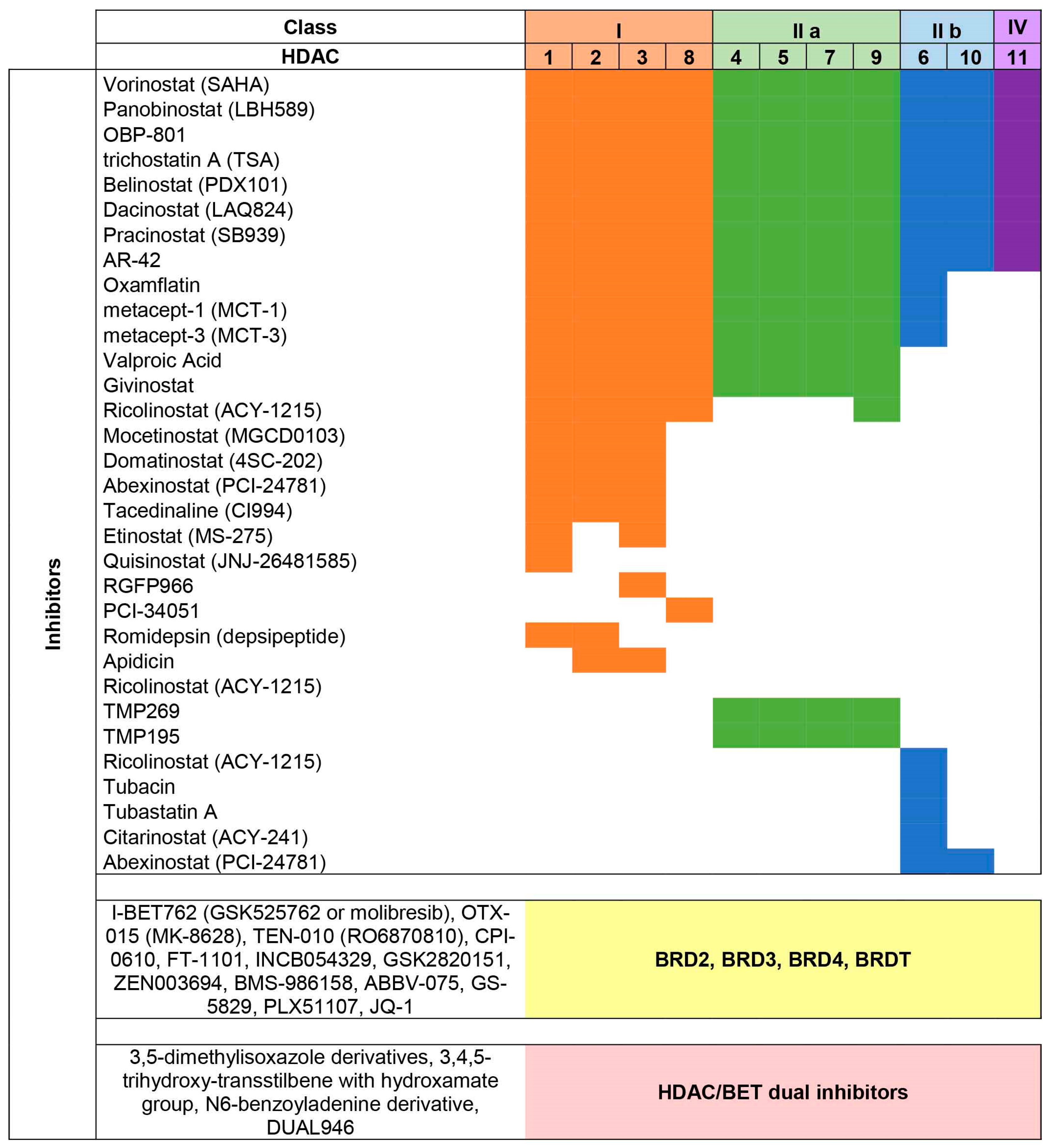
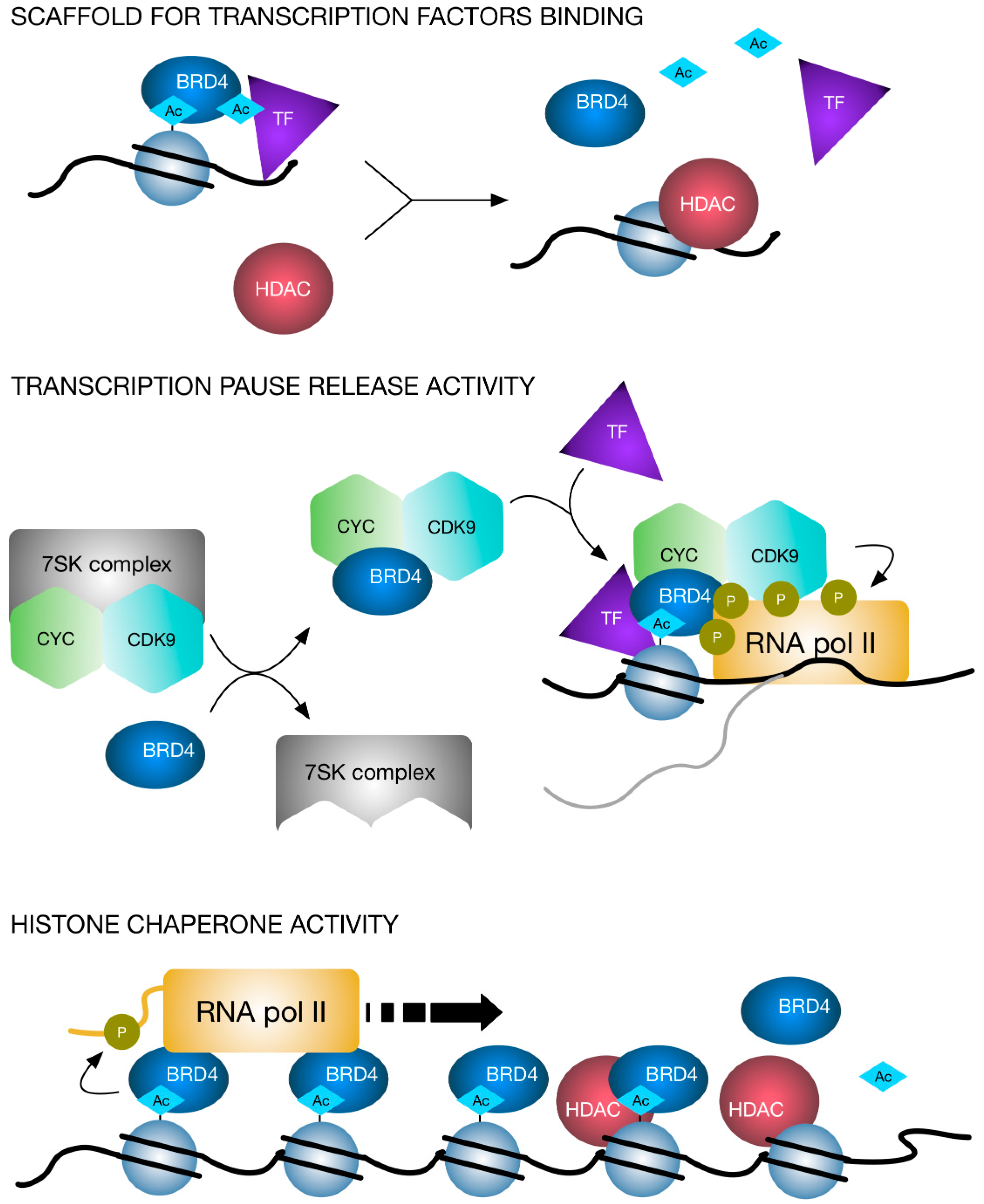
Table 1.
Summary of published clinical trials evaluating HDAC inhibitors (HDACi) in combination with other anti-cancer compounds.
Table 1.
Summary of published clinical trials evaluating HDAC inhibitors (HDACi) in combination with other anti-cancer compounds.
| HDAC Inhibitor | Class of Combination Drug | Intervention | Condition | Phase | NCT Number | Reference |
|---|---|---|---|---|---|---|
| Vorinostat | Chemotherapy | Vorinostat, paclitaxel, carboplatin | NSCLC | II | NCT00481078 | [106] |
| Chemotherapy | Vorinostat, paclitaxel, carboplatin | Adult Solid Tumor | I | NCT00287937 | [105] | |
| Chemotherapy | Vorinostat, doxorubicin | Adult Solid Tumor | I | NCT00331955 | [108] | |
| Radiotherapy | Vorinostat, radiotherapy | Pelvic Cancer | I | NCT00455351 | [109] | |
| Chemotherapy, radiotherapy | Vorinostat, capecitabine, radiotherapy | Pancreatic Cancer, Periampullary Adenocarcinoma | I | NCT00983268 | [112] | |
| Proteosome inhibitor | Vorinostat, marizomib | NSCLC, Pancreatic Cancer, Melanoma, Lymphoma, MM | I | NCT00667082 | [113] | |
| Proteosome inhibitor | Vorinostat, bortezomib | MM | I | NCT00858234 | [114] | |
| Proteosome inhibitor | Vorinostat, bortezomib | Glioblastoma, Gliosarcoma, Recurrent Adult Brain Tumor | II | NCT00641706 | [115] | |
| RTKi | Vorinostat, ridaforolimus | Lymphoma, Unspecified Adult Solid Tumor | I | NCT01169532 | [116] | |
| RTKi | Vorinostat, sirolimus, everolimus, temsirolimus | Advanced Cancer | I | NCT01087554 | [117] | |
| Hormone therapy | Vorinostat, tamoxifen | Breast Cancer | II | NCT00365599 | [118] | |
| Valproic Acid | Chemotherapy | Valproic acid, epirubicin,5-fluorouracil, cyclophosphamide | Advanced neoplasms | I | NCT00246103 | [107] |
| Chemotherapy, radiotherapy | Valproic Acid, temozolomide, radiation therapy, adjuvant therapy | High Grade Gliomas, Brain Tumors | II | NCT00302159 | [101] | |
| Chemotherapy, radiotherapy | Valproic acid, capecitabine, radiotherapy | Colorectal Cancer | I/II | NCT01898104 | [111] | |
| Panobinostat | Radiotherapy | Panobinostat, radiotherapy | Recurrent Glioma, High-grade Meningioma, Brain Metastasis | I | NCT01324635 | [110] |
| RTKi | Panobinostat, erlotinib | Lung Cancer, Head and Neck Cancer | I | NCT00738751 | [119] | |
| Hormone therapy | Panobinostat, bicalutamide | Prostate Cancer | I/II | NCT00878436 | [120] | |
| Romidepsin | RTKi | Erlotinib, romidepsin | Lung Cancer, Metastatic Cancer | I/II | NCT01302808 | [121] |
| Etinostat | RTKi | Etinostat, sorafenib | Advanced or Metastatic Solid Tumors, refractory or relapsed AML | I | NCT01159301 | [122] |
| Immunotherapy | Etinostat, aldesleukin (IL-2), radiotherapy | Clear Cell Renal Cell Carcinoma | I/II | NCT01038778 | [123] | |
| Ricolinostat | Proteosome inhibitor | Ricolinostat, bortezomib, dexamethasone | MM | I/II | NCT01323751 | [124] |
Table 2.
Summary of published and ongoing clinical trials evaluating BETi as monotherapy or in combination with other anti-cancer compounds.
Table 2.
Summary of published and ongoing clinical trials evaluating BETi as monotherapy or in combination with other anti-cancer compounds.
| BET Inhibitor | Class of Combination Drug | Intervention | Condition | Phase | NCT number | Reference |
|---|---|---|---|---|---|---|
| I-BET762 (GSK525762 or molibresib) | Monotherapy | GSK525762 | Relapsed refractory hematological malignancies | I/II | NCT01943851 | |
| Monotherapy | GSK525762 | NUT Midline Carcinoma and other Solid Cancers | I/II | NCT01587703 | ||
| Monotherapy | Molibresib | Compassionate use in NUT Midline Carcinoma | NCT03702036 | |||
| Hormone therapy | GSK525762; fulvestrant | HR+/HER2− advanced or metastatic breast cancer | I/II | NCT02964507 | ||
| Hormone therapy | GSK525762; abiraterone; enzalutamide; prednisone | Castrate-resistant prostate cancer | I | NCT03150056 | ||
| MEK inhibitors | GSK525762; trametinib | SCLC and RAS-mutated solid tumors | I/II | NCT03266159 | ||
| OTX-015 (MK-8628) | Monotherapy | OTX-015 | Hematological malignancies | I | NCT01713582 | [67,68] |
| Monotherapy | MK-8628 | Glioblastoma multiforme | II | NCT02296476 | ||
| Monotherapy | MK-8628 | Advanced solid tumors | I | NCT02698176 | ||
| Monotherapy | MK-8628 | Advanced solid tumors | I | NCT02259114 | ||
| Monotherapy | MK-8628 | Hematological malignancies | I | NCT02698189 | ||
| Chemotherapy | OTX-015; azacitidine | AML | I/II | NCT02303782 | ||
| TEN-010 (RO6870810) | Monotherapy | RO6870810 | Advanced solid tumors | I | NCT01987362 | |
| Monotherapy | RO6870810 | AML and myelodysplastic syndrome (MDS) | I | NCT02308761 | ||
| Immune checkpoint inhibitors | RO6870810; atezolizumab | TNBC and/or ovarian cancer | I | NCT03292172 | ||
| Anti CD38 | RO6870810; daratumumab | Advanced MM | I | NCT03068351 | ||
| BCL2 inhibitor | RO6870810; venetoclax; rituximab | Diffuse large B-cell lymphoma and/or high-grade B-cell lymphoma | I | NCT03255096 | ||
| CPI-0610 | Monotherapy | CPI-0610 | Malignant Peripheral Nerve Sheath Tumors | II | NCT02986919 | |
| Monotherapy | CPI-0610 | MM | I | NCT02157636 | ||
| Monotherapy | CPI-0610 | Progressive lymphoma | I | NCT01949883 | ||
| JAK inhibitor | CPI-0610; ruxolitinib | Acute Leukemia, Myelodysplastic Syndrome, Myelodysplastic/Myeloproliferative Neoplasms, and Myelofibrosis | I/II | NCT02158858 | ||
| FT-1101 | Chemotherapy | FT-1101; azacitidine | AML or non-Hodgkin Lymphoma | I | NCT02543879 | |
| INCB054329 | Monotherapy | INCB054329 | Advanced Solid Tumors and Hematologic Malignancy | I/II | NCT02431260 | |
| GSK2820151 | Monotherapy | GSK2820151 | Advanced or recurrent solid tumors | I | NCT02630251 | |
| ZEN003694 | Monotherapy | ZEN003694 | Metastatic Castration-Resistant Prostate Cancer | I | NCT02705469 | |
| Hormone therapy | ZEN003694; Enzalutamide | Metastatic Castration-Resistant Prostate Cancer | I/II | NCT02711956 | ||
| BMS-986158 | Immune checkpoint inhibitors | BMS-986158; Nivolumab | Selected advanced cancers | I/II | NCT02419417 | |
| ABBV-075 | BCL2 inhibitor | ABBV-075; Venetoclax | Selected hematological and solid cancers | I | NCT02391480 | |
| GS-5829 | Hormone therapy | GS-5829; Exemestane; Fulvestrant | Advanced Estrogen Receptor Positive, HER2 Negative-Breast Cancer | I/II | NCT02983604 | |
| Hormone therapy | GS-5829; Exemestane; Fulvestrant | Advanced solid tumors and lymphomas | I | NCT02392611 | ||
| Hormone therapy | GS-5829; Enzatulamide | Metastatic Castrate-Resistant Prostate Cancer | I/II | NCT02607228 | ||
| PLX51107 | Monotherapy | PLX51107 | Advanced solid and hematologic malignancies | I/II | NCT02683395 |
© 2019 by the authors. Licensee MDPI, Basel, Switzerland. This article is an open access article distributed under the terms and conditions of the Creative Commons Attribution (CC BY) license (http://creativecommons.org/licenses/by/4.0/).
Share and Cite
MDPI and ACS Style
Manzotti, G.; Ciarrocchi, A.; Sancisi, V. Inhibition of BET Proteins and Histone Deacetylase (HDACs): Crossing Roads in Cancer Therapy. Cancers 2019, 11, 304. https://0-doi-org.brum.beds.ac.uk/10.3390/cancers11030304
AMA Style
Manzotti G, Ciarrocchi A, Sancisi V. Inhibition of BET Proteins and Histone Deacetylase (HDACs): Crossing Roads in Cancer Therapy. Cancers. 2019; 11(3):304. https://0-doi-org.brum.beds.ac.uk/10.3390/cancers11030304
Chicago/Turabian StyleManzotti, Gloria, Alessia Ciarrocchi, and Valentina Sancisi. 2019. "Inhibition of BET Proteins and Histone Deacetylase (HDACs): Crossing Roads in Cancer Therapy" Cancers 11, no. 3: 304. https://0-doi-org.brum.beds.ac.uk/10.3390/cancers11030304
Note that from the first issue of 2016, this journal uses article numbers instead of page numbers. See further details here.