Next Issue
Volume 12, March
Previous Issue
Volume 12, January


Cancers, Volume 12, Issue 2 (February 2020) – 270 articles

Cover Story (view full-size image): 
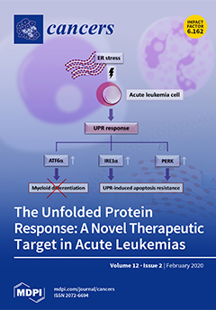
The unfolded protein response (UPR) is an adaptive response triggered by the endoplasmic reticulum stress due to altered cell protein homeostasis. The UPR can be activated by three main mediators: PERK, ATF6α, and IRE1α. Proteostasis is frequently deregulated in cancer, and the UPR is emerging as a crucial signaling network in controlling the survival of several neoplasias, including acute leukemias. The activation of the ATF6 pathway in AML cells upregulates the calreticulin that suppresses the translation of C/EBPα, contributing to the block in myeloid differentiation. XBP1, IRE1α, and GRP78 are upregulated in B-ALL patients, leading to UPR-induced apoptosis resistance, while c-Myc regulates the UPR response in T-ALL cells via increased PERK activation (prosurvival signaling). Therefore, targeting UPR-driven prosurvival pathways could represent a novel therapeutic strategy in acute leukemias. View [...] Read more.
  • Issues are regarded as officially published after their release is announced to the table of contents alert mailing list.
  • You may sign up for e-mail alerts to receive table of contents of newly released issues.
  • PDF is the official format for papers published in both, html and pdf forms. To view the papers in pdf format, click on the "PDF Full-text" link, and use the free Adobe Reader to open them.
Order results
Result details
Section
Select all
Export citation of selected articles as:
26 pages, 1163 KiB  
Review
Clinico-Biological Implications of Modified Levels of Cytokines in Chronic Lymphocytic Leukemia: A Possible Therapeutic Role
by Alessandro Allegra, Caterina Musolino, Alessandro Tonacci, Giovanni Pioggia, Marco Casciaro and Sebastiano Gangemi
Cancers 2020, 12(2), 524; https://0-doi-org.brum.beds.ac.uk/10.3390/cancers12020524 - 24 Feb 2020
Cited by 20 | Viewed by 3702
Abstract
B-cell chronic lymphocytic leukemia (B-CLL) is the main cause of mortality among hematologic diseases in Western nations. B-CLL is correlated with an intense alteration of the immune system. The altered functions of innate immune elements and adaptive immune factors are interconnected in B-CLL [...] Read more.
B-cell chronic lymphocytic leukemia (B-CLL) is the main cause of mortality among hematologic diseases in Western nations. B-CLL is correlated with an intense alteration of the immune system. The altered functions of innate immune elements and adaptive immune factors are interconnected in B-CLL and are decisive for its onset, evolution, and therapeutic response. Modifications in the cytokine balance could support the growth of the leukemic clone via a modulation of cellular proliferation and apoptosis, as some cytokines have been reported to be able to affect the life of B-CLL cells in vivo. In this review, we will examine the role played by cytokines in the cellular dynamics of B-CLL patients, interpret the contradictions sometimes present in the literature regarding their action, and evaluate the possibility of manipulating their production in order to intervene in the natural history of the disease. Full article
(This article belongs to the Special Issue Interleukin in Cancer Pathogenesis and Treatment)
Show Figures

Figure 1

22 pages, 4862 KiB  
Article
Antiparkinson Drug Benztropine Suppresses Tumor Growth, Circulating Tumor Cells, and Metastasis by Acting on SLC6A3/DAT and Reducing STAT3
by Chiharu Sogawa, Takanori Eguchi, Manh Tien Tran, Masayuki Ishige, Kilian Trin, Yuka Okusha, Eman Ahmed Taha, Yanyin Lu, Hotaka Kawai, Norio Sogawa, Masaharu Takigawa, Stuart K. Calderwood, Kuniaki Okamoto and Ken-ichi Kozaki
Cancers 2020, 12(2), 523; https://0-doi-org.brum.beds.ac.uk/10.3390/cancers12020523 - 24 Feb 2020
Cited by 30 | Viewed by 8368
Abstract
Tumor growth, progression, and therapy resistance are crucial factors in the prognosis of cancer. The properties of three-dimensional (3D) tumor-like organoids (tumoroids) more closely resemble in vivo tumors compared to two-dimensionally cultured cells and are therefore effectively used for assays and drug screening. [...] Read more.
Tumor growth, progression, and therapy resistance are crucial factors in the prognosis of cancer. The properties of three-dimensional (3D) tumor-like organoids (tumoroids) more closely resemble in vivo tumors compared to two-dimensionally cultured cells and are therefore effectively used for assays and drug screening. We here established a repurposed drug for novel anticancer research and therapeutics using a 3D tumoroid-based screening system. We screened six pharmacologically active compounds by using an original tumoroid-based multiplex phenotypic screening system with a matrix metalloproteinase 9 (MMP9) promoter-driven fluorescence reporter for the evaluation of both tumoroid formation and progression. The antiparkinson drug benztropine was the most effective compound uncovered by the screen. Benztropine significantly inhibited in vitro tumoroid formation, cancer cell survival, and MMP9 promoter activity. Benztropine also reduced the activity of oncogenic signaling transducers and trans-activators for MMP9, including STAT3, NF-κB, and β-catenin, and the properties of cancer stem cells/cancer-initiating cells. Benztropine and GBR-12935 directly targeted the dopamine transporter DAT/SLC6A3, whose genetic alterations such as amplification were correlated with poor prognosis for cancer patients. Benztropine also inhibited the tumor growth, circulating tumor cell (CTC) number, and rate of metastasis in a tumor allograft model in mice. In conclusion, we propose the repurposing of benztropine for anticancer research and therapeutics that can suppress tumor progression, CTC, and metastasis of aggressive cancers by reducing key pro-tumorigenic factors. Full article
(This article belongs to the Special Issue 3D Cell Culture Cancer Models: Development and Applications)
Show Figures

Graphical abstract

22 pages, 2429 KiB  
Review
Nanotechnology-Based Biopolymeric Oral Delivery Platforms for Advanced Cancer Treatment
by Vanessa T. Chivere, Pierre P. D. Kondiah, Yahya E. Choonara and Viness Pillay
Cancers 2020, 12(2), 522; https://0-doi-org.brum.beds.ac.uk/10.3390/cancers12020522 - 24 Feb 2020
Cited by 54 | Viewed by 5609
Abstract
Routes of drug administration and their corresponding physiochemical characteristics play major roles in drug therapeutic efficiency and biological effects. Each route of delivery has favourable aspects and limitations. The oral route of delivery is the most convenient, widely accepted and safe route. However, [...] Read more.
Routes of drug administration and their corresponding physiochemical characteristics play major roles in drug therapeutic efficiency and biological effects. Each route of delivery has favourable aspects and limitations. The oral route of delivery is the most convenient, widely accepted and safe route. However, the oral route of chemotherapeutics to date have displayed high gastric degradation, low aqueous solubility, poor formulation stability and minimum intestinal absorption. Thus, mainstream anti-cancer drugs in current formulations are not suitable as oral chemotherapeutic formulations. The use of biopolymers such as chitosan, gelatin, hyaluronic acid and polyglutamic acid, for the synthesis of oral delivery platforms, have potential to help overcome problems associated with oral delivery of chemotherapeutics. Biopolymers have favourable stimuli-responsive properties, and thus can be used to improve oral bioavailability of anti-cancer drugs. These biopolymeric formulations can protect gastric-sensitive drugs from pH degradation, target specific binding sites for targeted absorption and consequently control drug release. In this review, the use of various biopolymers as oral drug delivery systems for chemotherapeutics will be discussed. Full article
Show Figures

Graphical abstract

6 pages, 242 KiB  
Editorial
The Labyrinth of Renal Cell Carcinoma
by Claudia Manini and José I. López
Cancers 2020, 12(2), 521; https://0-doi-org.brum.beds.ac.uk/10.3390/cancers12020521 - 24 Feb 2020
Cited by 10 | Viewed by 2334
Abstract
Renal cell carcinoma (RCC) ranks in the top-ten list of malignancies both in males and females [...] Full article
(This article belongs to the Special Issue Renal Cell Carcinoma)
19 pages, 5460 KiB  
Article
Intratumoral Heterogeneity and Longitudinal Changes in Gene Expression Predict Differential Drug Sensitivity in Newly Diagnosed and Recurrent Glioblastoma
by Ella L. Kim, Maxim Sorokin, Sven Rainer Kantelhardt, Darius Kalasauskas, Bettina Sprang, Julian Fauss, Florian Ringel, Andrew Garazha, Eugene Albert, Nurshat Gaifullin, Christian Hartmann, Nicole Naumann, Sven-Ernö Bikar, Alf Giese and Anton Buzdin
Cancers 2020, 12(2), 520; https://0-doi-org.brum.beds.ac.uk/10.3390/cancers12020520 - 24 Feb 2020
Cited by 35 | Viewed by 5449
Abstract
Background: Inevitable recurrence after radiochemotherapy is the major problem in the treatment of glioblastoma, the most prevalent type of adult brain malignancy. Glioblastomas are notorious for a high degree of intratumor heterogeneity manifest through a diversity of cell types and molecular patterns. The [...] Read more.
Background: Inevitable recurrence after radiochemotherapy is the major problem in the treatment of glioblastoma, the most prevalent type of adult brain malignancy. Glioblastomas are notorious for a high degree of intratumor heterogeneity manifest through a diversity of cell types and molecular patterns. The current paradigm of understanding glioblastoma recurrence is that cytotoxic therapy fails to target effectively glioma stem cells. Recent advances indicate that therapy-driven molecular evolution is a fundamental trait associated with glioblastoma recurrence. There is a growing body of evidence indicating that intratumor heterogeneity, longitudinal changes in molecular biomarkers and specific impacts of glioma stem cells need to be taken into consideration in order to increase the accuracy of molecular diagnostics still relying on readouts obtained from a single tumor specimen. Methods: This study integrates a multisampling strategy, longitudinal approach and complementary transcriptomic investigations in order to identify transcriptomic traits of recurrent glioblastoma in whole-tissue specimens of glioblastoma or glioblastoma stem cells. In this study, 128 tissue samples of 44 tumors including 23 first diagnosed, 19 recurrent and 2 secondary recurrent glioblastomas were analyzed along with 27 primary cultures of glioblastoma stem cells by RNA sequencing. A novel algorithm was used to quantify longitudinal changes in pathway activities and model efficacy of anti-cancer drugs based on gene expression data. Results: Our study reveals that intratumor heterogeneity of gene expression patterns is a fundamental characteristic of not only newly diagnosed but also recurrent glioblastomas. Evidence is provided that glioblastoma stem cells recapitulate intratumor heterogeneity, longitudinal transcriptomic changes and drug sensitivity patterns associated with the state of recurrence. Conclusions: Our results provide a transcriptional rationale for the lack of significant therapeutic benefit from temozolomide in patients with recurrent glioblastoma. Our findings imply that the spectrum of potentially effective drugs is likely to differ between newly diagnosed and recurrent glioblastomas and underscore the merits of glioblastoma stem cells as prognostic models for identifying alternative drugs and predicting drug response in recurrent glioblastoma. With the majority of recurrent glioblastomas being inoperable, glioblastoma stem cell models provide the means of compensating for the limited availability of recurrent glioblastoma specimens. Full article
(This article belongs to the Special Issue Brain Tumors)
Show Figures

Figure 1

20 pages, 637 KiB  
Review
Monitoring Immune Responses in Neuroblastoma Patients during Therapy
by Celina L. Szanto, Annelisa M. Cornel, Saskia V. Vijver and Stefan Nierkens
Cancers 2020, 12(2), 519; https://0-doi-org.brum.beds.ac.uk/10.3390/cancers12020519 - 24 Feb 2020
Cited by 31 | Viewed by 4363
Abstract
Neuroblastoma (NBL) is the most common extracranial solid tumor in childhood. Despite intense treatment, children with this high-risk disease have a poor prognosis. Immunotherapy showed a significant improvement in event-free survival in high-risk NBL patients receiving chimeric anti-GD2 in combination with cytokines and [...] Read more.
Neuroblastoma (NBL) is the most common extracranial solid tumor in childhood. Despite intense treatment, children with this high-risk disease have a poor prognosis. Immunotherapy showed a significant improvement in event-free survival in high-risk NBL patients receiving chimeric anti-GD2 in combination with cytokines and isotretinoin after myeloablative consolidation therapy. However, response to immunotherapy varies widely, and often therapy is stopped due to severe toxicities. Objective markers that help to predict which patients will respond or develop toxicity to a certain treatment are lacking. Immunotherapy guided via immune monitoring protocols will help to identify responders as early as possible, to decipher the immune response at play, and to adjust or develop new treatment strategies. In this review, we summarize recent studies investigating frequency and phenotype of immune cells in NBL patients prior and during current treatment protocols and highlight how these findings are related to clinical outcome. In addition, we discuss potential targets to improve immunogenicity and strategies that may help to improve therapy efficacy. We conclude that immune monitoring during therapy of NBL patients is essential to identify predictive biomarkers to guide patients towards effective treatment, with limited toxicities and optimal quality of life. Full article
(This article belongs to the Special Issue Cancer Biomarkers)
Show Figures

Figure 1

25 pages, 5593 KiB  
Article
The Impact of Normalization Approaches to Automatically Detect Radiogenomic Phenotypes Characterizing Breast Cancer Receptors Status
by Rossana Castaldo, Katia Pane, Emanuele Nicolai, Marco Salvatore and Monica Franzese
Cancers 2020, 12(2), 518; https://0-doi-org.brum.beds.ac.uk/10.3390/cancers12020518 - 24 Feb 2020
Cited by 37 | Viewed by 4922
Abstract
In breast cancer studies, combining quantitative radiomic with genomic signatures can help identifying and characterizing radiogenomic phenotypes, in function of molecular receptor status. Biomedical imaging processing lacks standards in radiomic feature normalization methods and neglecting feature normalization can highly bias the overall analysis. [...] Read more.
In breast cancer studies, combining quantitative radiomic with genomic signatures can help identifying and characterizing radiogenomic phenotypes, in function of molecular receptor status. Biomedical imaging processing lacks standards in radiomic feature normalization methods and neglecting feature normalization can highly bias the overall analysis. This study evaluates the effect of several normalization techniques to predict four clinical phenotypes such as estrogen receptor (ER), progesterone receptor (PR), human epidermal growth factor receptor 2 (HER2), and triple negative (TN) status, by quantitative features. The Cancer Imaging Archive (TCIA) radiomic features from 91 T1-weighted Dynamic Contrast Enhancement MRI of invasive breast cancers were investigated in association with breast invasive carcinoma miRNA expression profiling from the Cancer Genome Atlas (TCGA). Three advanced machine learning techniques (Support Vector Machine, Random Forest, and Naïve Bayesian) were investigated to distinguish between molecular prognostic indicators and achieved an area under the ROC curve (AUC) values of 86%, 93%, 91%, and 91% for the prediction of ER+ versus ER−, PR+ versus PR−, HER2+ versus HER2−, and triple-negative, respectively. In conclusion, radiomic features enable to discriminate major breast cancer molecular subtypes and may yield a potential imaging biomarker for advancing precision medicine. Full article
(This article belongs to the Special Issue Role of Medical Imaging in Cancers)
Show Figures

Figure 1

18 pages, 2327 KiB  
Article
Hypoxia-Induced Centrosome Amplification Underlies Aggressive Disease Course in HPV-Negative Oropharyngeal Squamous Cell Carcinomas
by Karuna Mittal, Da Hoon Choi, Guanhao Wei, Jaspreet Kaur, Sergey Klimov, Komal Arora, Christopher C. Griffith, Mukesh Kumar, Precious Imhansi-Jacob, Brian D. Melton, Sonal Bhimji-Pattni, Remus M. Osan, Padmashree Rida, Paweł Golusinski and Ritu Aneja
Cancers 2020, 12(2), 517; https://0-doi-org.brum.beds.ac.uk/10.3390/cancers12020517 - 24 Feb 2020
Cited by 7 | Viewed by 3698
Abstract
Human papillomavirus-negative (HPV-neg) oropharyngeal squamous cell carcinomas (OPSCCs) are associated with poorer overall survival (OS) compared with HPV-positive (HPV-pos) OPSCCs. The major obstacle in improving outcomes of HPV-neg patients is the lack of robust biomarkers and therapeutic targets. Herein, we investigated the role [...] Read more.
Human papillomavirus-negative (HPV-neg) oropharyngeal squamous cell carcinomas (OPSCCs) are associated with poorer overall survival (OS) compared with HPV-positive (HPV-pos) OPSCCs. The major obstacle in improving outcomes of HPV-neg patients is the lack of robust biomarkers and therapeutic targets. Herein, we investigated the role of centrosome amplification (CA) as a prognostic biomarker in HPV-neg OPSCCs. A quantitative evaluation of CA in clinical specimens of OPSCC revealed that (a) HPV-neg OPSCCs exhibit higher CA compared with HPV-pos OPSCCs, and (b) CA was associated with poor OS, even after adjusting for potentially confounding clinicopathologic variables. Contrastingly, CA was higher in HPV-pos cultured cell lines compared to HPV-neg ones. This divergence in CA phenotypes between clinical specimens and cultured cells can therefore be attributed to an inaccurate recapitulation of the in vivo tumor microenvironment in the cultured cell lines, namely a hypoxic environment. The exposure of HPV-neg OPSCC cultured cells to hypoxia or stabilizing HIF-1α genetically increased CA. Both the 26-gene hypoxia signature as well as the overexpression of HIF-1α positively correlated with increased CA in HPV-neg OPSCCs. In addition, we showed that HIF-1α upregulation is associated with the downregulation of miR-34a, increase in CA and expression of cyclin- D1. Our findings demonstrate that the evaluation of CA may aid in therapeutic decision-making, and CA can serve as a promising therapeutic target for HPV-neg OPSCC patients. Full article
(This article belongs to the Special Issue Human Papillomavirus and Cancers)
Show Figures

Figure 1

13 pages, 1680 KiB  
Review
Bone Morphogenetic Protein 4 Targeting Glioma Stem-Like Cells for Malignant Glioma Treatment: Latest Advances and Implications for Clinical Application
by Sonali Nayak, Ashorne Mahenthiran, Yongyong Yang, Mark McClendon, Barbara Mania-Farnell, Charles David James, John A. Kessler, Tadanori Tomita, Shi-Yuan Cheng, Samuel I. Stupp and Guifa Xi
Cancers 2020, 12(2), 516; https://0-doi-org.brum.beds.ac.uk/10.3390/cancers12020516 - 24 Feb 2020
Cited by 13 | Viewed by 4745
Abstract
Malignant gliomas are heterogeneous neoplasms. Glioma stem-like cells (GSCs) are undifferentiated and self-renewing cells that develop and maintain these tumors. These cells are the main population that resist current therapies. Genomic and epigenomic analyses has identified various molecular subtypes. Bone morphogenetic protein 4 [...] Read more.
Malignant gliomas are heterogeneous neoplasms. Glioma stem-like cells (GSCs) are undifferentiated and self-renewing cells that develop and maintain these tumors. These cells are the main population that resist current therapies. Genomic and epigenomic analyses has identified various molecular subtypes. Bone morphogenetic protein 4 (BMP4) reduces the number of GSCs through differentiation and induction of apoptosis, thus increasing therapeutic sensitivity. However, the short half-life of BMP4 impedes its clinical application. We previously reviewed BMP4 signaling in central nervous system development and glioma tumorigenesis and its potential as a treatment target in human gliomas. Recent advances in understanding both adult and pediatric malignant gliomas highlight critical roles of BMP4 signaling pathways in the regulation of tumor biology, and indicates its potential as a therapeutic molecule. Furthermore, significant progress has been made on synthesizing BMP4 biocompatible delivery materials, which can bind to and markedly extend BMP4 half-life. Here, we review current research associated with BMP4 in brain tumors, with an emphasis on pediatric malignant gliomas. We also summarize BMP4 delivery strategies, highlighting biocompatible BMP4 binding peptide amphiphile nanostructures as promising novel delivery platforms for treatment of these devastating tumors. Full article
(This article belongs to the Special Issue Pediatric Brain Tumor)
Show Figures

Figure 1

17 pages, 19039 KiB  
Article
New Deployable Expandable Electrodes in the Electroporation Treatment in a Pig Model: A Feasibility and Usability Preliminary Study
by Francesco Izzo, Franco Ionna, Vincenza Granata, Vittorio Albino, Renato Patrone, Francesco Longo, Agostino Guida, Paolo Delrio, Daniela Rega, Dario Scala, Roberto Pezzuto, Roberta Fusco, Elio Di Bernardo, Valeria D’Alessio, Roberto Grassi, Deyanira Contartese and Raffaele Palaia
Cancers 2020, 12(2), 515; https://0-doi-org.brum.beds.ac.uk/10.3390/cancers12020515 - 23 Feb 2020
Cited by 11 | Viewed by 3417
Abstract
The aim of the study is to evaluate the usability aspects of new deployable, expandable, electrode prototypes, in terms of suitability solutions for laparoscopic applications on the liver, endoscopic trans-oral and trans-anal procedures, electroporation segmentation in several steps, mechanical functionality (flexibility, penetrability), visibility [...] Read more.
The aim of the study is to evaluate the usability aspects of new deployable, expandable, electrode prototypes, in terms of suitability solutions for laparoscopic applications on the liver, endoscopic trans-oral and trans-anal procedures, electroporation segmentation in several steps, mechanical functionality (flexibility, penetrability), visibility of the electrode under instrumental guidance, compatibility of the electrode with laparoscopic/endoscopic accesses, surgical instruments, and procedural room and safety compatibility. The electroporation was performed on an animal model (Sus Scrofa Large White 60 kg) both in laparoscopy and endoscopy, under ultrasound guidance, and in open surgery. Electrodes without divergence, with needles coming out straight, parallel to each other, and electrodes with peripheral needles (four needles), diverging from the electrode shaft axis (electrode with non-zero divergence) have been tested. To cause an evaluable necrosis effect, the number of electrical pulses was increased to induce immediate liver cell death. Histological samples were analyzed by staining with Haematoxylin/Eosin or by immunohistochemical staining to confirm complete necrosis. The prototypes of expandable electrodes, tested in laparoscopy and endoscopy and in open surgery, respectively, are suitable in terms of usability, electroporation segmentation in several steps, mechanical functionality (flexibility, penetrability), visibility under instrumental guidance, compatibility with laparoscopic/endoscopic accesses, surgical instruments and procedural room safety, patient safety (no bleeding and/or perforation), and treatment efficacy (adequate ablated volume). Electroporation treatment using new deployable expandable electrode prototypes is safe and feasible. Moreover, electrode configurations allow for a gradual increase in the ablated area in consecutive steps, as confirmed by histology and immunohistochemistry. Full article
Show Figures

Figure 1

17 pages, 512 KiB  
Review
How to Classify Pituitary Neuroendocrine Tumors (PitNET)s in 2020
by Jacqueline Trouillas, Marie-Lise Jaffrain-Rea, Alexandre Vasiljevic, Gérald Raverot, Federico Roncaroli and Chiara Villa
Cancers 2020, 12(2), 514; https://0-doi-org.brum.beds.ac.uk/10.3390/cancers12020514 - 22 Feb 2020
Cited by 120 | Viewed by 12717
Abstract
Adenohypophyseal tumors, which were recently renamed pituitary neuroendocrine tumors (PitNET), are mostly benign, but may present various behaviors: invasive, “aggressive” and malignant with metastases. They are classified into seven morphofunctional types and three lineages: lactotroph, somatotroph and thyrotroph (PIT1 lineage), corticotroph (TPIT lineage) [...] Read more.
Adenohypophyseal tumors, which were recently renamed pituitary neuroendocrine tumors (PitNET), are mostly benign, but may present various behaviors: invasive, “aggressive” and malignant with metastases. They are classified into seven morphofunctional types and three lineages: lactotroph, somatotroph and thyrotroph (PIT1 lineage), corticotroph (TPIT lineage) or gonadotroph (SF1 lineage), null cell or immunonegative tumor and plurihormonal tumors. The WHO 2017 classification suggested that subtypes, such as male lactotroph, silent corticotroph and Crooke cell, sparsely granulated somatotroph, and silent plurihormonal PIT1 positive tumors, should be considered as “high risk” tumors. However, the prognostic impact of these subtypes and of each morphologic type remains controversial. In contrast, the French five-tiered classification, taking into account the invasion, the immuno-histochemical (IHC) type, and the proliferative markers (Ki-67 index, mitotic count, p53 positivity), has a prognostic value validated by statistical analysis in 4 independent cohorts. A standardized report for the diagnosis of pituitary tumors, integrating all these parameters, has been proposed by the European Pituitary Pathology Group (EPPG). In 2020, the pituitary pathologist must be considered as a member of the multidisciplinary pituitary team. The pathological diagnosis may help the clinician to adapt the post-operative management, including appropriate follow-up and early recognition and treatment of potentially aggressive forms. Full article
Show Figures

Figure 1

14 pages, 1866 KiB  
Article
The Urinary Transcriptome as a Source of Biomarkers for Prostate Cancer
by Carla Solé, Ibai Goicoechea, Alai Goñi, Maike Schramm, María Armesto, María Arestin, Lorea Manterola, Maitena Tellaetxe, Aitor Alberdi, Leonor Nogueira, Mathieu Roumiguie, Jose Ignacio López, Juan Pablo Sanz Jaka, Ander Urruticoechea, Itziar Vergara, Ana Loizaga-Iriarte, Miguel Unda, Arkaitz Carracedo, Bernard Malavaud and Charles H. Lawrie
Cancers 2020, 12(2), 513; https://0-doi-org.brum.beds.ac.uk/10.3390/cancers12020513 - 22 Feb 2020
Cited by 14 | Viewed by 3612
Abstract
Prostate cancer (PCa) is the second most common cancer of men and is typically slow-growing and asymptomatic. The use of blood PSA as a screening method has greatly improved PCa diagnosis, but high levels of false positives has raised much interest in alternative [...] Read more.
Prostate cancer (PCa) is the second most common cancer of men and is typically slow-growing and asymptomatic. The use of blood PSA as a screening method has greatly improved PCa diagnosis, but high levels of false positives has raised much interest in alternative biomarkers. We used next-generation sequencing (NGS) to elucidate the urinary transcriptome of whole urine collected from high-stage and low-stage PCa patients as well as from patients with the confounding diagnosis of benign hyperplasia (BPH). We identified and validated five differentially expressed protein-coding genes (FTH1 BRPF1, OSBP, PHC3, and UACA) in an independent validation cohort of small-volume (1 mL) centrifuged urine (n = 94) and non-centrifuged urine (n = 84) by droplet digital (dd)PCR. These biomarkers were able to discriminate between BPH and PCa patients and healthy controls using either centrifuged or non-centrifuged whole urine samples, suggesting that the urinary transcriptome is a valuable source of non-invasive biomarkers for PCa that warrants further investigation. Full article
(This article belongs to the Special Issue Liquid Biopsy for Cancer)
Show Figures

Figure 1

21 pages, 9060 KiB  
Article
Kinome Profiling to Predict Sensitivity to MAPK Inhibition in Melanoma and to Provide New Insights into Intrinsic and Acquired Mechanism of Resistance
by Mohamad Krayem, Philippe Aftimos, Ahmad Najem, Tim van den Hooven, Adriënne van den Berg, Liesbeth Hovestad-Bijl, Rik de Wijn, Riet Hilhorst, Rob Ruijtenbeek, Malak Sabbah, Joseph Kerger, Ahmad Awada, Fabrice Journe and Ghanem E. Ghanem
Cancers 2020, 12(2), 512; https://0-doi-org.brum.beds.ac.uk/10.3390/cancers12020512 - 22 Feb 2020
Cited by 16 | Viewed by 4190
Abstract
Mitogen-activated protein kinase (MAPK) inhibition with the combination of BRAF (Rapidly Accelerated Fibrosarcoma) and MEK (Mitogen-activated protein kinase kinase) inhibitors has become the standard of first-line therapy of metastatic melanoma harbouring BRAF V600 mutations. However, about half of the patients present with primary [...] Read more.
Mitogen-activated protein kinase (MAPK) inhibition with the combination of BRAF (Rapidly Accelerated Fibrosarcoma) and MEK (Mitogen-activated protein kinase kinase) inhibitors has become the standard of first-line therapy of metastatic melanoma harbouring BRAF V600 mutations. However, about half of the patients present with primary resistance while the remaining develop secondary resistance under prolonged treatment. Thus, there is a need for predictive biomarkers for sensitivity and/or resistance to further refine the patient population likely to benefit from MAPK inhibitors. In this study, we explored a top-down approach using a multiplex kinase assay, first, to discover a kinome signature predicting sensitivity, intrinsic and acquired resistance to MAPK inhibitors in melanoma, and second, to understand the mechanism of resistance using cell lines. Pre-dose tissues from patients (four responders and three non-responders to BRAFi monotherapy) were profiled for phosphotyrosine kinase (PTK) and serine-threonine kinase (STK) activities on a PamChip® peptide microarray in the presence and absence of ex vivo BRAFi. In addition, molecular studies were conducted on four sensitive parental lines, their offspring with acquired resistance to BRAFi and two lines with intrinsic resistance. PTK and STK activities in cell lysates were measured in the presence and absence of ex vivo BRAFi and/or MEKi. In tissue lysates, concentration-dependent ex vivo inhibition of STK and PTK activities with dabrafenib was stronger in responders than in non-responders. This difference was confirmed in cell lines comparing sensitive and resistant ones. Interestingly, common features of resistance were increased activity of receptor tyrosine kinases, Proto-oncogene tyrosine-protein kinase Src (Src) family kinases and protein kinase B (PKB, AKT) signalling. These latter results were confirmed by Western blots. While dabrafenib alone showed an inhibition of STK and PTK activities in both tissues and cell lines, the combination of dabrafenib and trametinib showed an antagonism on the STK activities and a synergism on PTK activities, resulting in stronger inhibitions of overall tyrosine kinase activities. Altogether; these data reveal that resistance of tumours and cell lines to MAPK inhibitors can be predicted using a multiplex kinase assay and is associated with an increase in specific tyrosine kinase activities and globally to AKT signalling in the patient’s tissue. Thus, such a predictive kinome signature would help to identify patients with innate resistance to MAPK double inhibition in order to propose other therapies. Full article
(This article belongs to the Special Issue Targeted Therapies for Melanoma)
Show Figures

Figure 1

16 pages, 2547 KiB  
Article
Usefulness of Hybrid PET/MRI in Clinical Evaluation of Head and Neck Cancer Patients
by Natalia Samolyk-Kogaczewska, Ewa Sierko, Dorota Dziemianczyk-Pakiela, Klaudia Beata Nowaszewska, Malgorzata Lukasik and Joanna Reszec
Cancers 2020, 12(2), 511; https://0-doi-org.brum.beds.ac.uk/10.3390/cancers12020511 - 22 Feb 2020
Cited by 31 | Viewed by 3490
Abstract
(1) Background: The novel hybrid of positron emission tomography/magnetic resonance (PET/MR) examination has been introduced to clinical practice. The aim of our study was to evaluate PET/MR usefulness in preoperative staging of head and neck cancer (HNC) patients (pts); (2) Methods: Thirty eight [...] Read more.
(1) Background: The novel hybrid of positron emission tomography/magnetic resonance (PET/MR) examination has been introduced to clinical practice. The aim of our study was to evaluate PET/MR usefulness in preoperative staging of head and neck cancer (HNC) patients (pts); (2) Methods: Thirty eight pts underwent both computed tomography (CT) and PET/MR examination, of whom 21 pts underwent surgical treatment as first-line therapy and were further included in the present study. Postsurgical tissue material was subjected to routine histopathological (HP) examination with additional evaluation of p16, human papillomavirus (HPV), Epstein-Barr virus (EBV) and Ki67 status. Agreement of clinical and pathological T staging, sensitivity, specificity, positive predictive value (PPV), negative predictive value (NPV) of CT and PET/MR in metastatic lymph nodes detection were defined. The verification of dependences between standardized uptake value (SUV value), tumor geometrical parameters, number of metastatic lymph nodes in PET/MR and CT, biochemical parameters, Ki67 index, p16, HPV and EBV status was made with statistical analysis of obtained results; (3) Results: PET/MR is characterized by better agreement in T staging, higher specificity, sensitivity, PPV and NPV of lymph nodes evaluation than CT imaging. Significant correlations were observed between SUVmax and maximal tumor diameter from PET/MR, between SUVmean and CT tumor volume, PET/MR tumor volume, maximal tumor diameter assessed in PET/MR. Other correlations were weak and insignificant; (4) Conclusions: Hybrid PET/MR imaging is useful in preoperative staging of HNC. Further studies are needed. Full article
(This article belongs to the Special Issue Role of Medical Imaging in Cancers)
Show Figures

Figure 1

14 pages, 2314 KiB  
Article
Genomic Characterization of Non-Invasive Differentiated-Type Gastric Cancer in the Japanese Population
by Koki Nakamura, Yuji Urabe, Kenichi Kagemoto, Ryo Yuge, Ryohei Hayashi, Atsushi Ono, C. Nelson Hayes, Shiro Oka, Masanori Ito, Takashi Nishisaka, Kazuaki Tanabe, Koji Arihiro, Hideki Ohdan, Shinji Tanaka and Kazuaki Chayama
Cancers 2020, 12(2), 510; https://0-doi-org.brum.beds.ac.uk/10.3390/cancers12020510 - 22 Feb 2020
Cited by 7 | Viewed by 3254
Abstract
Background and aims: Recent genomic characterization of gastric cancer (GC) by sequencing has revealed a large number of cancer-related genes. Research to characterize the genomic landscape of cancer has focused on established invasive cancer to develop biomarkers for therapeutic or diagnostic targets, and [...] Read more.
Background and aims: Recent genomic characterization of gastric cancer (GC) by sequencing has revealed a large number of cancer-related genes. Research to characterize the genomic landscape of cancer has focused on established invasive cancer to develop biomarkers for therapeutic or diagnostic targets, and nearly all GC reports have been about advanced GC. The aim of this study is to identify recurrently mutated genes in non-invasive GC and, in particular, the driver mutations that are associated with the development of GC. Methods and results: We performed whole-exome sequencing of 19 fresh frozen specimens of differentiated-type non-invasive GC and targeted sequencing for 168 genes of 30 formalin-fixed paraffin-embedded archival specimens of differentiated-type non-invasive GC. We found that TP53 and LRP1 are significantly associated with non-invasive GC. It has been reported that LPR1 is associated with CagA autophagy in gastric mucosa. Therefore, we downloaded RNA sequence data for gastric cancer from the The Cancer Genome Atlas (TCGA) Genomic Data Commons Data Portal and examined the differences in LRP1 gene expression levels. The expression level was significantly lower in cases without LRP1 mutation than in cases with LRP1 mutation. Based on these results, fluorescent immunostaining for CagA was performed for 49 of the above samples to evaluate CagA accumulation within the cancerous tissue. Accumulation of CagA was significantly greater when an LRP1 mutation was present than without a mutation. Conclusion: These data suggest that LRP1 mutation is an important change promoting the transformation of gastric mucosa to GC early in the carcinogenesis of cancer. Full article
Show Figures

Figure 1

12 pages, 1887 KiB  
Article
Small Molecule Binds with Lymphocyte Antigen 6K to Induce Cancer Cell Death
by Senyi Benti, Purushottam B. Tiwari, Dustin W. Goodlett, Leily Daneshian, Grant B. Kern, Mark D. Smith, Aykut Uren, Maksymilian Chruszcz, Linda S. Shimizu and Geeta Upadhyay
Cancers 2020, 12(2), 509; https://0-doi-org.brum.beds.ac.uk/10.3390/cancers12020509 - 22 Feb 2020
Cited by 9 | Viewed by 4886
Abstract
Elevated gene expression of Lymphocyte antigen 6K (LY6K) in cancer cells is associated with poor survival outcomes in multiple different cancer types including cervical, breast, ovarian, lung, and head and neck cancer. Since inhibition of LY6K expression inhibits cancer cell growth, we set [...] Read more.
Elevated gene expression of Lymphocyte antigen 6K (LY6K) in cancer cells is associated with poor survival outcomes in multiple different cancer types including cervical, breast, ovarian, lung, and head and neck cancer. Since inhibition of LY6K expression inhibits cancer cell growth, we set out to explore whether pharmacological inhibition of LY6K could produce the same effect. We screened small molecule libraries for direct binding to recombinant LY6K protein in a surface plasmon resonance assay. We found that NSC243928 directly binds to the full-length and mature forms of LY6K and inhibits growth of HeLa cells that express LY6K. NSC243928 did not display binding with LY6D or LY6E. Our data demonstrate a first-time proof of principle study that pharmacological inhibition of LY6K using small molecules in cancer cells is a valid approach to developing targeted therapies against LY6K. This approach will be specifically relevant in hard-to-treat cancers where LY6K is highly expressed, such as cervical, pancreatic, ovarian, head and neck, lung, gastric, and triple-negative breast cancers. Full article
Show Figures

Figure 1

12 pages, 423 KiB  
Review
Adjuvant Therapy in Adrenocortical Carcinoma: Reflections and Future Directions
by Sara Bedrose, Marilyne Daher, Lina Altameemi and Mouhammed Amir Habra
Cancers 2020, 12(2), 508; https://0-doi-org.brum.beds.ac.uk/10.3390/cancers12020508 - 22 Feb 2020
Cited by 33 | Viewed by 5189
Abstract
Adrenocortical carcinoma (ACC) is a rare and aggressive malignancy with high risk of recurrence despite macroscopically complete surgical resection. The main predictors of ACC recurrence include advanced disease stage, incomplete surgical resection, cortisol production, certain genetic alterations, and high proliferation rate (Ki-67 proliferation [...] Read more.
Adrenocortical carcinoma (ACC) is a rare and aggressive malignancy with high risk of recurrence despite macroscopically complete surgical resection. The main predictors of ACC recurrence include advanced disease stage, incomplete surgical resection, cortisol production, certain genetic alterations, and high proliferation rate (Ki-67 proliferation index). Mitotane has been the mainstay adjuvant therapy of ACC. However, the use of mitotane is based on retrospective and occasionally conflicting evidence. As mitotane levels can take a few months before reaching therapeutic levels, there is an emerging practice of combining platinum-based chemotherapy with mitotane in the adjuvant setting. Retrospective data indicate that radiotherapy is an option for select patients, particularly those with positive resection margins. There are multiple knowledge gaps in selecting patients for adjuvant therapy. It is of great importance to establish risk calculators to predict recurrence and to implement molecular profiling of ACC to guide adjuvant therapy. The role of immunotherapy in metastatic ACC is emerging and if deemed efficacious, then future studies will be needed to ascertain the role of adjuvant immunotherapy in ACC. Full article
(This article belongs to the Special Issue Adrenocortical Carcinoma)
Show Figures

Figure 1

11 pages, 4926 KiB  
Article
Successful Identification of Nasopharyngeal Carcinoma in Nasopharyngeal Biopsies Using Deep Learning
by Wen-Yu Chuang, Shang-Hung Chang, Wei-Hsiang Yu, Cheng-Kun Yang, Chi-Ju Yeh, Shir-Hwa Ueng, Yu-Jen Liu, Tai-Di Chen, Kuang-Hua Chen, Yi-Yin Hsieh, Yi Hsia, Tong-Hong Wang, Chuen Hsueh, Chang-Fu Kuo and Chao-Yuan Yeh
Cancers 2020, 12(2), 507; https://0-doi-org.brum.beds.ac.uk/10.3390/cancers12020507 - 22 Feb 2020
Cited by 28 | Viewed by 5814
Abstract
Pathologic diagnosis of nasopharyngeal carcinoma (NPC) can be challenging since most cases are nonkeratinizing carcinoma with little differentiation and many admixed lymphocytes. Our aim was to evaluate the possibility to identify NPC in nasopharyngeal biopsies using deep learning. A total of 726 nasopharyngeal [...] Read more.
Pathologic diagnosis of nasopharyngeal carcinoma (NPC) can be challenging since most cases are nonkeratinizing carcinoma with little differentiation and many admixed lymphocytes. Our aim was to evaluate the possibility to identify NPC in nasopharyngeal biopsies using deep learning. A total of 726 nasopharyngeal biopsies were included. Among them, 100 cases were randomly selected as the testing set, 20 cases as the validation set, and all other 606 cases as the training set. All three datasets had equal numbers of NPC cases and benign cases. Manual annotation was performed. Cropped square image patches of 256 × 256 pixels were used for patch-level training, validation, and testing. The final patch-level algorithm effectively identified NPC patches, with an area under the receiver operator characteristic curve (AUC) of 0.9900. Using gradient-weighted class activation mapping, we demonstrated that the identification of NPC patches was based on morphologic features of tumor cells. At the second stage, whole-slide images were sequentially cropped into patches, inferred with the patch-level algorithm, and reconstructed into images with a smaller size for training, validation, and testing. Finally, the AUC was 0.9848 for slide-level identification of NPC. Our result shows for the first time that deep learning algorithms can identify NPC. Full article
Show Figures

Figure 1

11 pages, 1378 KiB  
Article
Germline Variants in Phosphodiesterase Genes and Genetic Predisposition to Pediatric Adrenocortical Tumors
by Emilia Modolo Pinto, Fabio R. Faucz, Luana Z. Paza, Gang Wu, Elizabeth S. Fernandes, Jerome Bertherat, Constantine A. Stratakis, Enzo Lalli, Raul C. Ribeiro, Carlos Rodriguez-Galindo, Bonald C. Figueiredo and Gerard P. Zambetti
Cancers 2020, 12(2), 506; https://0-doi-org.brum.beds.ac.uk/10.3390/cancers12020506 - 22 Feb 2020
Cited by 17 | Viewed by 4639
Abstract
Phosphodiesterases (PDEs) form a superfamily of enzymes that catalyze the hydrolysis of cyclic nucleotides adenosine 3′5′-cyclic monophosphate (cAMP) and guanosine 3′5′-cyclic monophosphate (cGMP) to their inactive 5′ monophosphates. cAMP plays a critical role as a second messenger in endocrine tissues, and activation of [...] Read more.
Phosphodiesterases (PDEs) form a superfamily of enzymes that catalyze the hydrolysis of cyclic nucleotides adenosine 3′5′-cyclic monophosphate (cAMP) and guanosine 3′5′-cyclic monophosphate (cGMP) to their inactive 5′ monophosphates. cAMP plays a critical role as a second messenger in endocrine tissues, and activation of cAMP signaling has been reported in endocrine tumors. Germline variants in PDEs have been associated with benign cortisol-secreting adrenocortical adenomas and testicular germ cell cancer but not adrenocortical carcinoma. We performed whole genome sequencing (WGS) and whole exome sequencing (WES) of paired blood and tumor samples from 37 pediatric adrenocortical tumors (ACTs). Germline inactivating variants in PDEs were observed in 9 of 37 (24%) patients. Tumor DNA analysis revealed loss of heterozygosity, with maintenance of the mutated allele in all cases. Our results suggest that germline variants in PDEs and other regulators of the cAMP-signaling pathway may contribute to pediatric adrenocortical tumorigenesis, perhaps by cooperating with germline hypomorphic mutant TP53 alleles and uniparental disomy of chromosome 11p15 (Beckwith–Wiedemann syndrome). Full article
(This article belongs to the Special Issue Adrenocortical Carcinoma)
Show Figures

Figure 1

19 pages, 5282 KiB  
Article
Neutrophil Extracellular Trap Formation Correlates with Favorable Overall Survival in High Grade Ovarian Cancer
by Besnik Muqaku, Dietmar Pils, Johanna C. Mader, Stefanie Aust, Andreas Mangold, Liridon Muqaku, Astrid Slany, Giorgia Del Favero and Christopher Gerner
Cancers 2020, 12(2), 505; https://0-doi-org.brum.beds.ac.uk/10.3390/cancers12020505 - 21 Feb 2020
Cited by 32 | Viewed by 4641
Abstract
It is still a question of debate whether neutrophils, often found in the tumor microenvironment, mediate tumor-promoting or rather tumor-inhibiting activities. The present study focuses on the involvement of neutrophils in high grade serous ovarian cancer (HGSOC). Macroscopic features classify two types of [...] Read more.
It is still a question of debate whether neutrophils, often found in the tumor microenvironment, mediate tumor-promoting or rather tumor-inhibiting activities. The present study focuses on the involvement of neutrophils in high grade serous ovarian cancer (HGSOC). Macroscopic features classify two types of peritoneal tumor spread in HGSOC. Widespread and millet sized lesions characterize the miliary type, while non-miliary metastases are larger and associated with better prognosis. Multi-omics and FACS data were generated from ascites samples. Integrated data analysis demonstrates a significant increase of neutrophil extracellular trap (NET)-associated molecules in non-miliary ascites samples. A co-association network analysis performed with the ascites data further revealed a striking correlation between NETosis-associated metabolites and several eicosanoids. The congruence of data generated from primary neutrophils with ascites analyses indicates the predominance of NADPH oxidase 2 (NOX)-independent NETosis. NETosis is associated with protein S100A8/A9 release. An increase of the S100A8/CRP abundance ratio was found to correlate with favorable survival of HGSOC patients. The analysis of additional five independent proteome studies with regard to S100A8/CRP ratios confirmed this observation. In conclusion, NET formation seems to relate with better cancer patient outcome. Full article
Show Figures

Graphical abstract

11 pages, 291 KiB  
Article
Blood-Based SOX2-Promoter Methylation in Relation to Exercise and PM2.5 Exposure among Taiwanese Adults
by Chun-Lang Su, Disline Manli Tantoh, Ying-Hsiang Chou, Lee Wang, Chien-Chang Ho, Pei-Hsin Chen, Kuan-Jung Lee, Oswald Ndi Nfor, Shu-Yi Hsu, Wen-Miin Liang and Yung-Po Liaw
Cancers 2020, 12(2), 504; https://0-doi-org.brum.beds.ac.uk/10.3390/cancers12020504 - 21 Feb 2020
Cited by 7 | Viewed by 2881
Abstract
Increased ventilation during exercise in polluted areas could trigger airway inflammation. We evaluated blood DNA methylation of the SOX2-promoter region in relation to exercise and PM2.5 in Taiwanese adults. Data of 948 participants aged 30–70 years were retrieved from the Taiwan Biobank [...] Read more.
Increased ventilation during exercise in polluted areas could trigger airway inflammation. We evaluated blood DNA methylation of the SOX2-promoter region in relation to exercise and PM2.5 in Taiwanese adults. Data of 948 participants aged 30–70 years were retrieved from the Taiwan Biobank Database (2008–2015) and the Air Quality Monitoring Database (2006–2011). PM2.5 was positively associated with SOX2-promoter methylation (β = 0.000216; p < 0.0001). The interaction between PM2.5 and exercise on SOX2-promoter methylation was significant (p = 0.0146). After stratification by exercise habits, PM2.5 was positively associated with SOX2 methylation in only individuals who did regular exercise (β = 0.0003490; p < 0.0001). After stratification by exercise habits and residential areas, SOX2-promoter methylation levels in those who lived in the southern area were higher for both the regular exercise (β = 0.00272; p = 0.0172) and no regular exercise groups (β = 0.002610 and p = 0.0162). SOX2-promoter methylation levels in those who lived in the northern area and did regular exercise were lower; β = -0.00314 (p = 0.0036). In conclusion, PM2.5 was positively associated with SOX2-promoter methylation in participants who did regular exercise. Living in the southern area was positively associated with SOX2-promoter methylation regardless of exercise habits. Full article
23 pages, 3531 KiB  
Article
A Novel pH-Tunable Secondary Conformation Containing Mixed Micellar System in Anticancer Treatment
by Fu-Ying Shih, Wen-Ping Jiang, Xiaojie Lin, Sheng-Chu Kuo, Guan-Jhong Huang, Yu-Chi Hou, Chih-Shiang Chang, Yang Liu and Yi-Ting Chiang
Cancers 2020, 12(2), 503; https://0-doi-org.brum.beds.ac.uk/10.3390/cancers12020503 - 21 Feb 2020
Cited by 7 | Viewed by 2693
Abstract
In this study, for the first time, we precisely assembled the poly-γ-benzyl-l-glutamate and an amphiphilic copolymer d-α-tocopherol polyethylene glycol succinate into a mixed micellar system for the embedment of the anticancer drug doxorubicin. Importantly, the intracellular drug-releasing behaviors could be controlled by changing [...] Read more.
In this study, for the first time, we precisely assembled the poly-γ-benzyl-l-glutamate and an amphiphilic copolymer d-α-tocopherol polyethylene glycol succinate into a mixed micellar system for the embedment of the anticancer drug doxorubicin. Importantly, the intracellular drug-releasing behaviors could be controlled by changing the secondary structures of poly-γ-benzyl-l-glutamate via the precise regulation of the buffer’s pH value. Under neutral conditions, the micellar architectures were stabilized by both α-helix secondary structures and the microcrystalline structures. Under acidic conditions (pH 4.0), the interior structures transformed into a coil state with a disordered alignment, inducing the release of the loaded drug. A remarkable cytotoxicity of the Dox-loaded mixed micelles was exhibited toward human lung cancer cells in vitro. The internalizing capability into the cancer cells, as well as the intracellular drug-releasing behaviors, were also identified and observed. The secondary structures containing Dox-loaded mixed micelles had an outstanding antitumor efficacy in human lung cancer A549 cells-bearing nude mice, while little toxicities occurred or interfered with the hepatic or renal functions after the treatments. Thus, these pH-tunable α-helix-containing mixed micelles are innovative and promising for controlled intracellular anticancer drug delivery. Full article
(This article belongs to the Special Issue Cancer Nanomedicine)
Show Figures

Graphical abstract

18 pages, 6503 KiB  
Article
Obesity Promotes Cooperation of Cancer Stem-Like Cells and Macrophages to Enhance Mammary Tumor Angiogenesis
by Lauren E. Hillers-Ziemer, Rachel Q. McMahon, Margaret Hietpas, Gretchen Paderta, Jennelle LeBeau, Jessica McCready and Lisa M. Arendt
Cancers 2020, 12(2), 502; https://0-doi-org.brum.beds.ac.uk/10.3390/cancers12020502 - 21 Feb 2020
Cited by 22 | Viewed by 3699
Abstract
Obesity is correlated with worsened prognosis and treatment resistance in breast cancer. Macrophage-targeted therapies are currently in clinical trials, however, little is known about how obesity may impact treatment efficacy. Within breast adipose tissue, obesity leads to chronic, macrophage-driven inflammation, suggesting that obese [...] Read more.
Obesity is correlated with worsened prognosis and treatment resistance in breast cancer. Macrophage-targeted therapies are currently in clinical trials, however, little is known about how obesity may impact treatment efficacy. Within breast adipose tissue, obesity leads to chronic, macrophage-driven inflammation, suggesting that obese breast cancer patients may benefit from these therapies. Using a high fat diet model of obesity, we orthotopically transplanted cancer cell lines into the mammary glands of obese and lean mice. We quantified changes in tumor invasiveness, angiogenesis and metastasis, and examined the efficacy of macrophage depletion to diminish tumor progression in obese and lean mice. Mammary tumors from obese mice grew significantly faster, were enriched for cancer stem-like cells (CSCs) and were more locally invasive and metastatic. Tumor cells isolated from obese mice demonstrated enhanced expression of stem cell-related pathways including Sox2 and Notch2. Despite more rapid growth, mammary tumors from obese mice had reduced necrosis, higher blood vessel density, and greater macrophage recruitment. Depletion of macrophages in obese tumor-bearing mice resulted in increased tumor necrosis, reduced endothelial cells, and enhanced recruitment of CD8+ T cells compared to IgG-treated controls. Macrophages may be an important clinical target to improve treatment options for obese breast cancer patients. Full article
Show Figures

Figure 1

21 pages, 4528 KiB  
Article
The c-Myc/AKT1/TBX3 Axis Is Important to Target in the Treatment of Embryonal Rhabdomyosarcoma
by Danica Sims, Hapiloe Mabaruti Maranyane, Victoria Damerell, Dhirendra Govender, Ashwin W. Isaacs, Jade Peres and Sharon Prince
Cancers 2020, 12(2), 501; https://doi.org/10.3390/cancers12020501 - 21 Feb 2020
Cited by 8 | Viewed by 3857
Abstract
Rhabdomyosarcoma is a highly aggressive malignant cancer that arises from skeletal muscle progenitor cells and is the third most common solid tumour in children. Despite significant advances, rhabdomyosarcoma still presents a therapeutic challenge, and while targeted therapy has shown promise, there are limited [...] Read more.
Rhabdomyosarcoma is a highly aggressive malignant cancer that arises from skeletal muscle progenitor cells and is the third most common solid tumour in children. Despite significant advances, rhabdomyosarcoma still presents a therapeutic challenge, and while targeted therapy has shown promise, there are limited options because the molecular drivers of rhabdomyosarcoma are poorly understood. We previously reported that the T-box transcription factor 3 (TBX3), which has been identified as a druggable target in many cancers, is overexpressed in rhabdomyosarcoma patient samples and cell lines. To identify new molecular therapeutic targets to treat rhabdomyosarcoma, this study investigates the potential oncogenic role(s) for TBX3 and the factors responsible for upregulating it in this cancer. To this end, rhabdomyosarcoma cell culture models in which TBX3 was either stably knocked down or overexpressed were established and the impact on key hallmarks of cancer were examined using growth curves, soft agar and scratch motility assays, as well as tumour-forming ability in nude mice. Our data show that TBX3 promotes substrate-dependent and -independent proliferation, migration and tumour formation. We further reveal that TBX3 is upregulated by c-Myc transcriptionally and AKT1 post-translationally. This study identifies c-Myc/AKT1/TBX3 as an important axis that could be targeted for the treatment of rhabdomyosarcoma. Full article
Show Figures

Figure 1

17 pages, 4434 KiB  
Article
Protein Tyrosine Phosphatase-1B Inhibition Disrupts IL13Rα2-Promoted Invasion and Metastasis in Cancer Cells
by Rubén A. Bartolomé, Ángela Martín-Regalado, Marta Jaén, Markella Zannikou, Peng Zhang, Vivian de los Ríos, Irina V. Balyasnikova and J. Ignacio Casal
Cancers 2020, 12(2), 500; https://0-doi-org.brum.beds.ac.uk/10.3390/cancers12020500 - 21 Feb 2020
Cited by 28 | Viewed by 4319
Abstract
Background: Interleukin 13 receptor alpha 2 subunit (IL13Rα2) is overexpressed in glioblastoma (GBM), metastatic colorectal cancer (CRC) and ovarian cancer (OC). Here, we investigated the IL13Rα2 interactome searching for novel targets in cancer invasion and metastasis. Methods: The interactome of IL13Rα2 was determined [...] Read more.
Background: Interleukin 13 receptor alpha 2 subunit (IL13Rα2) is overexpressed in glioblastoma (GBM), metastatic colorectal cancer (CRC) and ovarian cancer (OC). Here, we investigated the IL13Rα2 interactome searching for novel targets in cancer invasion and metastasis. Methods: The interactome of IL13Rα2 was determined in GBM by using a proteomic analysis and then validated in CRC and OC. Cell signaling was investigated using siRNA interference, protein tyrosine phosphatase-1B (PTP1B) inhibitors and Western blot analysis. Animal models of GBM and metastatic CRC were used for testing PTP1B inhibitors. Results: PTP1B was identified and validated as a mediator of IL13Rα2 signaling. An in silico analysis revealed that PTP1B overexpression is associated with lower overall survival of patients in the three types of cancer. PTP1B silencing or treatment with Claramine, a PTP1B inhibitor, caused a significant decrease in IL-13-mediated adhesion, migration and invasion of IL13Rα2-expressing cancer cells by inhibiting the dephosphorylation of Src Tyr530 and consequently, the phosphorylation of Src Tyr419, AKT and ERK1/2. In addition, Claramine inhibited EGF-mediated activation of EGFR Tyr1068. In vivo treatment with Claramine caused a total inhibition of liver metastasis in mice inoculated with CRC cells and a significant increase in the survival of mice bearing intracranial GBM patient-derived xenografts. Conclusions: We have uncovered that IL13 signaling through IL13Rα2 requires PTP1B activity and therefore, PTP1B inhibition represents a promising therapeutic strategy in multiple types of cancer, including glioblastoma. Full article
Show Figures

Graphical abstract

13 pages, 2944 KiB  
Communication
Mesothelial-to-Mesenchymal Transition Contributes to the Generation of Carcinoma-Associated Fibroblasts in Locally Advanced Primary Colorectal Carcinomas
by Carlos H. Gordillo, Pilar Sandoval, Patricia Muñoz-Hernández, Lucía Pascual-Antón, Manuel López-Cabrera and José A. Jiménez-Heffernan
Cancers 2020, 12(2), 499; https://0-doi-org.brum.beds.ac.uk/10.3390/cancers12020499 - 21 Feb 2020
Cited by 23 | Viewed by 3439
Abstract
During peritoneal metastasis, cancer cells spread from abdominal solid tumors, disseminate through the peritoneal fluid and attach to and invade through mesothelial cells (MCs) that line the peritoneum. Intestinal adenocarcinomas originating in the mucosa infiltrate the submucosa, muscle layer, and serosa in order [...] Read more.
During peritoneal metastasis, cancer cells spread from abdominal solid tumors, disseminate through the peritoneal fluid and attach to and invade through mesothelial cells (MCs) that line the peritoneum. Intestinal adenocarcinomas originating in the mucosa infiltrate the submucosa, muscle layer, and serosa in order to finally colonize the peritoneal cavity. However, the mechanism by which metastatic cells leave the primary tumor and reach the peritoneal cavity has not been previously described. Hence, we investigate whether MCs lining visceral peritoneum, through a mesothelial-to-mesenchymal transition (MMT), are a source of carcinoma-associated fibroblasts (CAFs), which could contribute to cancer progression toward the peritoneal cavity. CAFs detected in biopsies from patients with superficially invasive colorectal cancer differed from locally advanced tumors. An aberrant accumulation of myofibroblasts expressing mesothelial markers was found in the stroma of deeply infiltrative tumors located in the neighborhood of a frequently activated mesothelium. We suggest that MMT is a key event in the early stages of peritoneal dissemination. Full article
(This article belongs to the Special Issue A Special Issue on Tumor Stroma )
Show Figures

Figure 1

17 pages, 5103 KiB  
Article
Lipid–Saporin Nanoparticles for the Intracellular Delivery of Cytotoxic Protein to Overcome ABC Transporter-Mediated Multidrug Resistance In Vitro and In Vivo
by Guan-Nan Zhang, Pranav Gupta, Ming Wang, Anna Maria Barbuti, Charles R. Ashby, Jr., Yun-Kai Zhang, Leli Zeng, Qiaobing Xu, Ying-Fang Fan and Zhe-Sheng Chen
Cancers 2020, 12(2), 498; https://0-doi-org.brum.beds.ac.uk/10.3390/cancers12020498 - 21 Feb 2020
Cited by 7 | Viewed by 2960
Abstract
Although the judicious use of anticancer drugs that target one or more receptor tyrosine kinases constitutes an effective strategy to attenuate tumor growth, drug resistance is commonly encountered in cancer patients. The ATP-binding cassette transporters are one of the major contributors to the [...] Read more.
Although the judicious use of anticancer drugs that target one or more receptor tyrosine kinases constitutes an effective strategy to attenuate tumor growth, drug resistance is commonly encountered in cancer patients. The ATP-binding cassette transporters are one of the major contributors to the development of multidrug resistance as their overexpression significantly decreases the intracellular concentration and thus, the efficacy of certain anticancer drugs. Therefore, the development of treatment strategies that would not be susceptible to efflux or excretion by specific ABC transporters could overcome resistance to treatment. Here, we investigated the anticancer efficacy of saporin, a ribosome-inactivating protein. Since saporin has poor permeability across the cell membrane, it was encapsulated in a lipid-based nanoparticle system (EC16-1) that effectively delivered the formulation (EC16-1/saporin) intracellularly and produced anti-cancer efficacy. EC16-1/saporin, at nanomolar concentrations, significantly inhibited the cellular proliferation of parental and ABCB1- and ABCG2-overexpressing cancer cells. EC16-1/saporin did not significantly alter the subcellular localization of ABCB1 and ABCG2. In addition, EC16-1/saporin induced apoptosis in parental and ABCB1- and ABCG2-overexpressing cancer cells. In a murine model system, EC16-1/saporin significantly inhibited the tumor growth in mice xenografted with parental and ABCB1- and ABCG2-overexpressing cancer cells. Our findings suggest that the EC16-1/saporin combination could potentially be a novel therapeutic treatment in patients with parental or ABCB1- and ABCG2-positive drug-resistant cancers. Full article
Show Figures

Figure 1

19 pages, 2142 KiB  
Article
Genomic Signature of the Standardized Uptake Value in 18F-Fluorodeoxyglucose Positron Emission Tomography in Breast Cancer
by Seon-Kyu Kim, Sung Gwe Ahn, Jeong-Yeon Mun, Mi-So Jeong, Soong June Bae, Ju-Seog Lee, Joon Jeong, Sun-Hee Leem and In-Sun Chu
Cancers 2020, 12(2), 497; https://0-doi-org.brum.beds.ac.uk/10.3390/cancers12020497 - 20 Feb 2020
Cited by 13 | Viewed by 3284
Abstract
The standardized uptake value (SUV), an indicator of the degree of glucose uptake in 18F-fluorodeoxyglucose positron emission tomography (FDG-PET), has been used for predicting the clinical behavior of malignant tumors. However, its characteristics have been insufficiently explored at the genomics level. Here, [...] Read more.
The standardized uptake value (SUV), an indicator of the degree of glucose uptake in 18F-fluorodeoxyglucose positron emission tomography (FDG-PET), has been used for predicting the clinical behavior of malignant tumors. However, its characteristics have been insufficiently explored at the genomics level. Here, we aim to identify genomic signatures reflecting prognostic SUV characteristics in breast cancer (BRC). Through integrative genomic profiling of 3710 BRC patients, including 254 patients who underwent preoperative FDG-PET, we identified an SUV signature, which showed independent clinical utility for predicting BRC prognosis (hazard ratio [HR] 1.27, 95% confidence interval [CI] = 1.12 to 1.45, p = 2.23 × 10⁻⁴). The risk subgroups classified by the signature exhibited mutually exclusive mutation patterns of TP53 and PIK3CA and showed significantly different responsiveness to immunotherapy. Experimental assays revealed that a signaling axis defined by TP53FOXM1 and its downstream effectors in glycolysis–gluconeogenesis, including LDHA, might be important mediators in the FDG-PET process. Our molecular characterizations support an understanding of glucose metabolism and poor prognosis in BRC with a high SUV, utilizable in clinical practice to assist other diagnostic tools. Full article
Show Figures

Figure 1

19 pages, 3300 KiB  
Article
Increased Expression of Micro-RNA-23a Mediates Chemoresistance to Cytarabine in Acute Myeloid Leukemia
by Stefan Hatzl, Bianca Perfler, Sonja Wurm, Barbara Uhl, Franz Quehenberger, Susanne Ebner, Jakob Troppmair, Andreas Reinisch, Albert Wölfler, Heinz Sill and Armin Zebisch
Cancers 2020, 12(2), 496; https://0-doi-org.brum.beds.ac.uk/10.3390/cancers12020496 - 20 Feb 2020
Cited by 13 | Viewed by 3592
Abstract
Resistance to chemotherapy is one of the primary obstacles in acute myeloid leukemia (AML) therapy. Micro-RNA-23a (miR-23a) is frequently deregulated in AML and has been linked to chemoresistance in solid cancers. We, therefore, studied its role in chemoresistance to cytarabine (AraC), which forms [...] Read more.
Resistance to chemotherapy is one of the primary obstacles in acute myeloid leukemia (AML) therapy. Micro-RNA-23a (miR-23a) is frequently deregulated in AML and has been linked to chemoresistance in solid cancers. We, therefore, studied its role in chemoresistance to cytarabine (AraC), which forms the backbone of all cytostatic AML treatments. Initially, we assessed AraC sensitivity in three AML cell lines following miR-23a overexpression/knockdown using MTT-cell viability and soft-agar colony-formation assays. Overexpression of miR-23a decreased the sensitivity to AraC, whereas its knockdown had the opposite effect. Analysis of clinical data revealed that high miR-23a expression correlated with relapsed/refractory (R/R) AML disease stages, the leukemic stem cell compartment, as well as with inferior overall survival (OS) and event-free survival (EFS) in AraC-treated patients. Mechanistically, we demonstrate that miR-23a targets and downregulates topoisomerase-2-beta (TOP2B), and that TOP2B knockdown mediates AraC chemoresistance as well. Likewise, low TOP2B expression also correlated with R/R-AML disease stages and inferior EFS/OS. In conclusion, we show that increased expression of miR-23a mediates chemoresistance to AraC in AML and that it correlates with an inferior outcome in AraC-treated AML patients. We further demonstrate that miR-23a causes the downregulation of TOP2B, which is likely to mediate its effects on AraC sensitivity. Full article
Show Figures

Figure 1

20 pages, 3758 KiB  
Article
A SOX2 Reporter System Identifies Gastric Cancer Stem-Like Cells Sensitive to Monensin
by Diana Pádua, Rita Barros, Ana Luísa Amaral, Patrícia Mesquita, Ana Filipa Freire, Mafalda Sousa, André Filipe Maia, Inês Caiado, Hugo Fernandes, António Pombinho, Carlos Filipe Pereira and Raquel Almeida
Cancers 2020, 12(2), 495; https://0-doi-org.brum.beds.ac.uk/10.3390/cancers12020495 - 20 Feb 2020
Cited by 29 | Viewed by 5805
Abstract
Gastric cancer remains a serious health burden with few therapeutic options. Therefore, the recognition of cancer stem cells (CSCs) as seeds of the tumorigenic process makes them a prime therapeutic target. Knowing that the transcription factors SOX2 and OCT4 promote stemness, our approach [...] Read more.
Gastric cancer remains a serious health burden with few therapeutic options. Therefore, the recognition of cancer stem cells (CSCs) as seeds of the tumorigenic process makes them a prime therapeutic target. Knowing that the transcription factors SOX2 and OCT4 promote stemness, our approach was to isolate stem-like cells in human gastric cancer cell lines using a traceable reporter system based on SOX2/OCT4 activity (SORE6-GFP). Cells transduced with the SORE6-GFP reporter system were sorted into SORE6+ and SORE6– cell populations, and their biological behavior characterized. SORE6+ cells were enriched for SOX2 and exhibited CSC features, including a greater ability to proliferate and form gastrospheres in non-adherent conditions, a larger in vivo tumor initiating capability, and increased resistance to 5-fluorouracil (5-FU) treatment. The overexpression and knockdown of SOX2 revealed a crucial role of SOX2 in cell proliferation and drug resistance. By combining the reporter system with a high-throughput screening of pharmacologically active small molecules we identified monensin, an ionophore antibiotic, displaying selective toxicity to SORE6+ cells. The ability of SORE6-GFP reporter system to recognize cancer stem-like cells facilitates our understanding of gastric CSC biology and serves as a platform for the identification of powerful therapeutics for targeting gastric CSCs. Full article
Show Figures

Figure 1

Previous Issue
Next Issue
Back to TopTop