Next Issue
Volume 12, June
Previous Issue
Volume 12, April


Cancers, Volume 12, Issue 5 (May 2020) – 290 articles

Cover Story (view full-size image): 
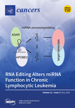
RNA editing is a post-transcriptional modification of RNA that leads to specific adenosine-to-inosine (a guanosine analog) or cytosine-to-uracil changes. Adenosine-to-inosine editing is mediated by ADARs (adenosine deaminases that act on RNA), and cytosine-to-uracil editing is catalyzed by apolipoprotein B mRNA editing, catalytic polypeptide-like (APOBEC) enzymes. In this study, we investigated RNA editing using next-generation miRNA sequencing data from chronic lymphocytic leukemia samples and found leukemia-specific editing of some miRNAs. Many of these editing events affect the seeding regions of the respective miRNA, which changes the specificity for particular mRNA targets. Hence, our study shows that in addition to deregulated miRNA expression, aberrant miRNA editing should also be considered in the pathogenesis of cancer. View this paper.
  • Issues are regarded as officially published after their release is announced to the table of contents alert mailing list.
  • You may sign up for e-mail alerts to receive table of contents of newly released issues.
  • PDF is the official format for papers published in both, html and pdf forms. To view the papers in pdf format, click on the "PDF Full-text" link, and use the free Adobe Reader to open them.
Order results
Result details
Section
Select all
Export citation of selected articles as:
16 pages, 3969 KiB  
Article
Impact of the Injection Site on Growth Characteristics, Phenotype and Sensitivity towards Cytarabine of Twenty Acute Leukaemia Patient-Derived Xenograft Models
by Julia Schueler, Gabriele Greve, Dorothée Lenhard, Milena Pantic, Anna Edinger, Eva Oswald and Michael Lübbert
Cancers 2020, 12(5), 1349; https://0-doi-org.brum.beds.ac.uk/10.3390/cancers12051349 - 25 May 2020
Cited by 5 | Viewed by 3385
Abstract
Rodent models have contributed significantly to the understanding of haematological malignancies. One important model system in this context are patient-derived xenografts (PDX). In the current study, we examined 20 acute leukaemia PDX models for growth behaviour, infiltration in haemopoietic organs and sensitivity towards [...] Read more.
Rodent models have contributed significantly to the understanding of haematological malignancies. One important model system in this context are patient-derived xenografts (PDX). In the current study, we examined 20 acute leukaemia PDX models for growth behaviour, infiltration in haemopoietic organs and sensitivity towards cytarabine. PDX were injected intratibially (i.t.), intrasplenicaly (i.s.) or subcutaneously (s.c.) into immune compromised mice. For 18/20 models the engraftment capacity was independent of the implantation site. Two models could exclusively be propagated in one or two specific settings. The implantation site did influence tumour growth kinetics as median overall survival differed within one model depending on the injection route. The infiltration pattern was similar in i.t. and i.s. models. In contrast to the s.c. implantation, only one model displayed circulating leukaemic cells outside of the locally growing tumour mass. Cytarabine was active in all four tested models. Nevertheless, the degree of sensitivity was specific for an individual model and implantation site. In summary, all three application routes turned out to be feasible for the propagation of PDX. Nevertheless, the distinct differences between the settings highlight the need for well characterized platforms to ensure the meaningful interpretation of data generated using those powerful tools. Full article
(This article belongs to the Special Issue Patient-Derived Xenograft-Models in Cancer Research)
Show Figures

Figure 1

16 pages, 3631 KiB  
Article
DFIQ, a Novel Quinoline Derivative, Shows Anticancer Potential by Inducing Apoptosis and Autophagy in NSCLC Cell and In Vivo Zebrafish Xenograft Models
by Hurng-Wern Huang, Yung-Ding Bow, Chia-Yih Wang, Yen-Chun Chen, Pei-Rong Fu, Kuo-Feng Chang, Tso-Wen Wang, Chih-Hua Tseng, Yeh-Long Chen and Chien-Chih Chiu
Cancers 2020, 12(5), 1348; https://0-doi-org.brum.beds.ac.uk/10.3390/cancers12051348 - 25 May 2020
Cited by 17 | Viewed by 3060
Abstract
Lung cancer is one of the deadliest cancers worldwide due to chemoresistance in patients with late-stage disease. Quinoline derivatives show biological activity against HIV, malaria, bacteriuria, and cancer. DFIQ is a novel synthetic quinoline derivative that induces cell death in both in vitro [...] Read more.
Lung cancer is one of the deadliest cancers worldwide due to chemoresistance in patients with late-stage disease. Quinoline derivatives show biological activity against HIV, malaria, bacteriuria, and cancer. DFIQ is a novel synthetic quinoline derivative that induces cell death in both in vitro and in vivo zebrafish xenograft models. DFIQ induced cell death, including apoptosis, and the IC50 values were 4.16 and 2.31 μM at 24 and 48 h, respectively. DFIQ was also found to induce apoptotic protein cleavage and DNA damage, reduce cell cycle-associated protein expression, and disrupt reactive oxygen species (ROS) reduction, thus resulting in the accumulation of superoxide radicals. Autophagy is also a necessary process associated with chemotherapy-induced cell death. Lysosome accumulation and lysosome-associated membrane protein-2 (LAMP2) depletion were observed after DFIQ treatment, and cell death induction was restored upon treatment with the autophagy inhibitor 3-methyladenine (3-MA). Nevertheless, ROS production was found to be involved in DFIQ-induced autophagy activation and LAMP2 depletion. Our data provide the first evidence for developing DFIQ for clinical usage and show the regulatory mechanism by which DFIQ affects ROS, autophagy, and apoptosis. Full article
Show Figures

Figure 1

18 pages, 1731 KiB  
Review
Pancreatic Cancer Associated Fibroblasts (CAF): Under-Explored Target for Pancreatic Cancer Treatment
by Jeffrey Norton, Deshka Foster, Malini Chinta, Ashley Titan and Michael Longaker
Cancers 2020, 12(5), 1347; https://0-doi-org.brum.beds.ac.uk/10.3390/cancers12051347 - 25 May 2020
Cited by 77 | Viewed by 7782
Abstract
Pancreatic cancer is the 4th leading cause of cancer deaths in the United States. The pancreatic cancer phenotype is primarily a consequence of oncogenes disturbing the resident pancreas parenchymal cell repair program. Many solid tumor types including pancreatic cancer have severe tumor fibrosis [...] Read more.
Pancreatic cancer is the 4th leading cause of cancer deaths in the United States. The pancreatic cancer phenotype is primarily a consequence of oncogenes disturbing the resident pancreas parenchymal cell repair program. Many solid tumor types including pancreatic cancer have severe tumor fibrosis called desmoplasia. Desmoplastic stroma is coopted by the tumor as a support structure and CAFs aid in tumor growth, invasion, and metastases. This stroma is caused by cancer associated fibroblasts (CAFs), which lay down extensive connective tissue in and around the tumor cells. CAFs represent a heterogeneous population of cells that produce various paracrine molecules such as transforming growth factor-beta (TGF-beta) and platelet derived growth factors (PDGFs) that aid tumor growth, local invasion, and development of metastases. The hard, fibrotic shell of desmoplasia serves as a barrier to the infiltration of both chemo- and immunotherapy drugs and host immune cells to the tumor. Although there have been recent improvements in chemotherapy and surgical techniques for management of pancreatic cancer, the majority of patients will die from this disease. Therefore, new treatment strategies are clearly needed. CAFs represent an under-explored potential therapeutic target. This paper discusses what we know about the role of CAFs in pancreatic cancer cell growth, invasion, and metastases. Additionally, we present different strategies that are being and could be explored as anti-CAF treatments for pancreatic cancer. Full article
(This article belongs to the Special Issue A Special Issue on Tumor Stroma )
Show Figures

Figure 1

9 pages, 1018 KiB  
Article
Genetic Analysis Reveals a Significant Contribution of CES1 to Prostate Cancer Progression in Taiwanese Men
by Chien-Chih Ke, Lih-Chyang Chen, Chia-Cheng Yu, Wei-Chung Cheng, Chao-Yuan Huang, Victor C. Lin, Te-Ling Lu, Shu-Pin Huang and Bo-Ying Bao
Cancers 2020, 12(5), 1346; https://0-doi-org.brum.beds.ac.uk/10.3390/cancers12051346 - 25 May 2020
Cited by 11 | Viewed by 2874
Abstract
The genes that influence prostate cancer progression remain largely unknown. Since the carboxylesterase gene family plays a crucial role in xenobiotic metabolism and lipid/cholesterol homeostasis, we hypothesize that genetic variants in carboxylesterase genes may influence clinical outcomes for prostate cancer patients. A total [...] Read more.
The genes that influence prostate cancer progression remain largely unknown. Since the carboxylesterase gene family plays a crucial role in xenobiotic metabolism and lipid/cholesterol homeostasis, we hypothesize that genetic variants in carboxylesterase genes may influence clinical outcomes for prostate cancer patients. A total of 478 (36 genotyped and 442 imputed) single nucleotide polymorphisms (SNPs) in five genes of the carboxylesterase family were assessed in terms of their associations with biochemical recurrence (BCR)-free survival in 643 Taiwanese patients with prostate cancer who underwent radical prostatectomy. The strongest association signal was shown in CES1 (P = 9.64 × 10⁻⁴ for genotyped SNP rs8192935 and P = 8.96 × 10⁻⁵ for imputed SNP rs8192950). After multiple test correction and adjustment for clinical covariates, CES1 rs8192935 (P = 9.67 × 10⁻⁴) and rs8192950 (P = 9.34 × 10⁻⁵) remained significant. These SNPs were correlated with CES1 expression levels, which in turn were associated with prostate cancer aggressiveness. Furthermore, our meta-analysis, including eight studies, indicated that a high CES1 expression predicted better outcomes among prostate cancer patients (hazard ratio 0.82, 95% confidence interval 0.70–0.97, P = 0.02). In conclusion, our findings suggest that CES1 rs8192935 and rs8192950 are associated with BCR and that CES1 plays a tumor suppressive role in prostate cancer. Full article
(This article belongs to the Special Issue The Study of Cancer Susceptibility Genes)
Show Figures

Figure 1

15 pages, 1843 KiB  
Article
Genomic and Transcriptomic Characteristics According to Size of Papillary Thyroid Microcarcinoma
by Young Shin Song, Byung-Hee Kang, Seungbok Lee, Seong-Keun Yoo, Young Sik Choi, Jungsun Park, Dong Yoon Park, Kyu Eun Lee, Jeong-Sun Seo and Young Joo Park
Cancers 2020, 12(5), 1345; https://0-doi-org.brum.beds.ac.uk/10.3390/cancers12051345 - 25 May 2020
Cited by 11 | Viewed by 3411
Abstract
It is controversial as to whether papillary thyroid microcarcinoma (PTMC) has some genomic and transcriptomic characteristics that differentiate between an early-stage lesion that would eventually evolve into the larger papillary thyroid cancer (PTC), and an occult indolent cancer in itself. To investigate this, [...] Read more.
It is controversial as to whether papillary thyroid microcarcinoma (PTMC) has some genomic and transcriptomic characteristics that differentiate between an early-stage lesion that would eventually evolve into the larger papillary thyroid cancer (PTC), and an occult indolent cancer in itself. To investigate this, we comprehensively elucidated the genomic and transcriptomic landscapes of PTMCs of different sizes, using a large-scaled database. This study included 3435 PTCs, 1985 of which were PTMCs. We performed targeted next-generation sequencing for 221 PTCs and integrated these data with the data including The Cancer Genome Atlas (TCGA) project. The frequency of v-raf murine sarcoma viral oncogene homolog B (BRAF)V600E mutation was higher in PTMCs >0.5 cm than that in very small PTMCs (≤0.5 cm) and decreased again in PTCs >2 cm. Among PTMCs, the prevalence of mutations in rat sarcoma (RAS) and telomerase reverse transcriptase (TERT) promoter was not significantly different according to their size, but lower than in large PTCs. There was no change in the tumor mutational burden, the number of driver mutations, and transcriptomic profiles with tumor size, among PTMCs and all PTCs. Although a few genes with differential expression and TERT promoter mutations were found in a few PTMCs, our findings showed that there were no useful genomic or transcriptomic characteristics for the prediction of the future progression of PTMC. Full article
(This article belongs to the Special Issue Biomarkers of Thyroid Cancer)
Show Figures

Figure 1

11 pages, 2721 KiB  
Article
A Machine-learning Approach for the Assessment of the Proliferative Compartment of Solid Tumors on Hematoxylin-Eosin-Stained Sections
by Francesco Martino, Silvia Varricchio, Daniela Russo, Francesco Merolla, Gennaro Ilardi, Massimo Mascolo, Giovanni Orabona dell’Aversana, Luigi Califano, Guglielmo Toscano, Giuseppe De Pietro, Maria Frucci, Nadia Brancati, Filippo Fraggetta and Stefania Staibano
Cancers 2020, 12(5), 1344; https://0-doi-org.brum.beds.ac.uk/10.3390/cancers12051344 - 25 May 2020
Cited by 20 | Viewed by 4666
Abstract
We introduce a machine learning-based analysis to predict the immunohistochemical (IHC) labeling index for the cell proliferation marker Ki67/MIB1 on cancer tissues based on morphometrical features extracted from hematoxylin and eosin (H&E)-stained formalin-fixed, paraffin-embedded (FFPE) tumor tissue samples. We provided a proof-of-concept prediction [...] Read more.
We introduce a machine learning-based analysis to predict the immunohistochemical (IHC) labeling index for the cell proliferation marker Ki67/MIB1 on cancer tissues based on morphometrical features extracted from hematoxylin and eosin (H&E)-stained formalin-fixed, paraffin-embedded (FFPE) tumor tissue samples. We provided a proof-of-concept prediction of the Ki67/MIB1 IHC positivity of cancer cells through the definition and quantitation of single nuclear features. In the first instance, we set our digital framework on Ki67/MIB1-stained OSCC (oral squamous cell carcinoma) tissue sample whole slide images, using QuPath as a working platform and its integrated algorithms, and we built a classifier in order to distinguish tumor and stroma classes and, within them, Ki67-positive and Ki67-negative cells; then, we sorted the morphometric features of tumor cells related to their Ki67 IHC status. Among the evaluated features, nuclear hematoxylin mean optical density (NHMOD) presented as the best one to distinguish Ki67/MIB1 positive from negative cells. We confirmed our findings in a single-cell level analysis of H&E staining on Ki67-immunostained/H&E-decolored tissue samples. Finally, we tested our digital framework on a case series of oral squamous cell carcinomas (OSCC), arranged in tissue microarrays; we selected two consecutive sections of each OSCC FFPE TMA (tissue microarray) block, respectively stained with H&E and immuno-stained for Ki67/MIB1. We automatically detected tumor cells in H&E slides and generated a “false color map” (FCM) based on NHMOD through the QuPath measurements map tool. FCM nearly coincided with the actual immunohistochemical result, allowing the prediction of Ki67/MIB1 positive cells in a direct visual fashion. Our proposed approach provides the pathologist with a fast method of identifying the proliferating compartment of the tumor through a quantitative assessment of the nuclear features on H&E slides, readily appreciable by visual inspection. Although this technique needs to be fine-tuned and tested on larger series of tumors, the digital analysis approach appears to be a promising tool to quickly forecast the tumor’s proliferation fraction directly on routinely H&E-stained digital sections. Full article
(This article belongs to the Special Issue Surgical Pathology in the Digital Era)
Show Figures

Figure 1

10 pages, 888 KiB  
Article
Cigarette Smoking Is Associated with Increased Risk of Malignant Gliomas: A Nationwide Population-Based Cohort Study
by Stephen Ahn, Kyung-Do Han, Yong-Moon Park, Jung Min Bae, Sang Uk Kim, Sin-Soo Jeun and Seung Ho Yang
Cancers 2020, 12(5), 1343; https://doi.org/10.3390/cancers12051343 - 25 May 2020
Cited by 10 | Viewed by 2859
Abstract
The association between cigarette smoking and the risk of developing malignant glioma (MG) remains unclear. We aimed to evaluate this potential association in a large general population, using a well-established and validated longitudinal nationwide database. Using data from the Korean National Health Insurance [...] Read more.
The association between cigarette smoking and the risk of developing malignant glioma (MG) remains unclear. We aimed to evaluate this potential association in a large general population, using a well-established and validated longitudinal nationwide database. Using data from the Korean National Health Insurance System cohort, 9,811,768 people over 20 years old without any cancer history in 2009 were followed until the end of 2017. We documented 6100 MG cases (ICD-10 code C71) during the median follow-up period of 7.31 years. Current smokers had a higher risk of developing MG (HR = 1.22, CI: 1.13–1.32) compared with never-smokers, after adjusting for confounders. This association was stronger for those who smoked ≥ 20 cigarettes daily (HR = 1.50, CI: 1.36–1.64). Furthermore, having 30 or more pack-years of smoking over the course of one’s lifetime was associated with an increased risk of developing MG in a dose-dependent manner, compared with never-smokers (HR = 1.31, CI: 1.16–1.48 for 30–39 pack-years of smoking; HR = 1.36, CI: 1.17–1.59 for 40–49 pack-years of smoking; HR = 1.68; CI: 1.44–1.95 for ≥ 50 pack-years of smoking). These results suggest that cigarette smoking may be associated with developing MG. Further prospective studies could help elucidate this association. Full article
(This article belongs to the Section Cancer Epidemiology and Prevention)
Show Figures

Figure 1

20 pages, 3410 KiB  
Article
MAT2A as Key Regulator and Therapeutic Target in MLLr Leukemogenesis
by Kathy-Ann Secker, Bianca Bloechl, Hildegard Keppeler, Silke Duerr-Stoerzer, Hannes Schmid, Dominik Schneidawind, Johan Jeong, Thomas Hentrich, Julia M. Schulze-Hentrich and Corina Schneidawind
Cancers 2020, 12(5), 1342; https://0-doi-org.brum.beds.ac.uk/10.3390/cancers12051342 - 24 May 2020
Cited by 14 | Viewed by 5310
Abstract
Epigenetic dysregulation plays a pivotal role in mixed-lineage leukemia (MLL) pathogenesis, therefore serving as a suitable therapeutic target. S-adenosylmethionine (SAM) is the universal methyl donor in human cells and is synthesized by methionine adenosyltransferase 2A (MAT2A), which is deregulated in different cancer [...] Read more.
Epigenetic dysregulation plays a pivotal role in mixed-lineage leukemia (MLL) pathogenesis, therefore serving as a suitable therapeutic target. S-adenosylmethionine (SAM) is the universal methyl donor in human cells and is synthesized by methionine adenosyltransferase 2A (MAT2A), which is deregulated in different cancer types. Here, we used our human CRISPR/Cas9-MLL-rearranged (CRISPR/Cas9-MLLr) leukemia model, faithfully mimicking MLLr patients’ pathology with indefinite growth potential in vitro, to evaluate the unknown role of MAT2A. Comparable to publicly available patient data, we detected MAT2A to be significantly overexpressed in our CRISPR/Cas9-MLLr model compared to healthy controls. By using non-MLLr and MLLr cell lines and our model, we detected an MLLr-specific enhanced response to PF-9366, a new MAT2A inhibitor, and small interfering (si) RNA-mediated knockdown of MAT2A, by alteration of the proliferation, viability, differentiation, apoptosis, cell cycling, and histone methylation. Moreover, the combinational treatment of PF-9366 with chemotherapy or targeted therapies against the SAM-dependent methyltransferases, disruptor of telomeric silencing 1 like (DOT1L) and protein arginine methyltransferase 5 (PRMT5), revealed even more pronounced effects. In summary, we uncovered MAT2A as a key regulator in MLL leukemogenesis and its inhibition led to significant anti-leukemic effects. Therefore, our study paves the avenue for clinical application of PF-9366 to improve the treatment of poor prognosis MLLr leukemia. Full article
Show Figures

Figure 1

9 pages, 224 KiB  
Perspective
Small Molecule KRAS Inhibitors: The Future for Targeted Pancreatic Cancer Therapy?
by Josef Gillson, Yogambha Ramaswamy, Gurvinder Singh, Alemayehu A. Gorfe, Nick Pavlakis, Jaswinder Samra, Anubhav Mittal and Sumit Sahni
Cancers 2020, 12(5), 1341; https://0-doi-org.brum.beds.ac.uk/10.3390/cancers12051341 - 24 May 2020
Cited by 32 | Viewed by 5195
Abstract
Pancreatic ductal adenocarcinoma (PDAC) is one of the deadliest solid tumors in the world. Currently, there are no approved targeted therapies for PDAC. Mutations in Kirsten rat sarcoma viral oncogene homologue (KRAS) are known to be a major driver of PDAC [...] Read more.
Pancreatic ductal adenocarcinoma (PDAC) is one of the deadliest solid tumors in the world. Currently, there are no approved targeted therapies for PDAC. Mutations in Kirsten rat sarcoma viral oncogene homologue (KRAS) are known to be a major driver of PDAC progression, but it was considered an undruggable target until recently. Moreover, PDAC also suffers from drug delivery issues due to the highly fibrotic tumor microenvironment. In this perspective, we provide an overview of recent developments in targeting mutant KRAS and strategies to overcome drug delivery issues (e.g., nanoparticle delivery). Overall, we propose that the antitumor effects from novel KRAS inhibitors along with strategies to overcome drug delivery issues could be a new therapeutic way forward in PDAC. Full article
(This article belongs to the Special Issue Recent Advances in Pancreatic Ductal Adenocarcinoma)
21 pages, 3208 KiB  
Article
Hakin-1, a New Specific Small-Molecule Inhibitor for the E3 Ubiquitin-Ligase Hakai, Inhibits Carcinoma Growth and Progression
by Olaia Martinez-Iglesias, Alba Casas-Pais, Raquel Castosa, Andrea Díaz-Díaz, Daniel Roca-Lema, Ángel Concha, Álvaro Cortés, Federico Gago and Angélica Figueroa
Cancers 2020, 12(5), 1340; https://0-doi-org.brum.beds.ac.uk/10.3390/cancers12051340 - 23 May 2020
Cited by 15 | Viewed by 5443
Abstract
The requirement of the E3 ubiquitin-ligase Hakai for the ubiquitination and subsequent degradation of E-cadherin has been associated with enhanced epithelial-to-mesenchymal transition (EMT), tumour progression and carcinoma metastasis. To date, most of the reported EMT-related inhibitors were not developed for anti-EMT purposes, but [...] Read more.
The requirement of the E3 ubiquitin-ligase Hakai for the ubiquitination and subsequent degradation of E-cadherin has been associated with enhanced epithelial-to-mesenchymal transition (EMT), tumour progression and carcinoma metastasis. To date, most of the reported EMT-related inhibitors were not developed for anti-EMT purposes, but indirectly affect EMT. On the other hand, E3 ubiquitin-ligase enzymes have recently emerged as promising therapeutic targets, as their specific inhibition would prevent wider side effects. Given this background, a virtual screening was performed to identify novel specific inhibitors of Hakai, targeted against its phosphotyrosine-binding pocket, where phosphorylated-E-cadherin specifically binds. We selected a candidate inhibitor, Hakin-1, which showed an important effect on Hakai-induced ubiquitination. Hakin-1 also inhibited carcinoma growth and tumour progression both in vitro, in colorectal cancer cell lines, and in vivo, in a tumour xenograft mouse model, without apparent systemic toxicity in mice. Our results show for the first time that a small molecule putatively targeting the E3 ubiquitin-ligase Hakai inhibits Hakai-dependent ubiquitination of E-cadherin, having an impact on the EMT process. This represents an important step forward in a future development of an effective therapeutic drug to prevent or inhibit carcinoma tumour progression. Full article
(This article belongs to the Special Issue Targeted Cancer Therapy)
Show Figures

Figure 1

26 pages, 1210 KiB  
Review
Regulation of Src Family Kinases during Colorectal Cancer Development and Its Clinical Implications
by Wook Jin
Cancers 2020, 12(5), 1339; https://0-doi-org.brum.beds.ac.uk/10.3390/cancers12051339 - 23 May 2020
Cited by 50 | Viewed by 8205
Abstract
Src family kinases (SFKs) are non-receptor kinases that play a critical role in the pathogenesis of colorectal cancer (CRC). The expression and activity of SFKs are upregulated in patients with CRC. Activation of SFKs promotes CRC cell proliferation, metastases to other organs and [...] Read more.
Src family kinases (SFKs) are non-receptor kinases that play a critical role in the pathogenesis of colorectal cancer (CRC). The expression and activity of SFKs are upregulated in patients with CRC. Activation of SFKs promotes CRC cell proliferation, metastases to other organs and chemoresistance, as well as the formation of cancer stem cells (CSCs). The enhanced expression level of Src is associated with decreased survival in patients with CRC. Src-mediated regulation of CRC progression involves various membrane receptors, modulators, and suppressors, which regulate Src activation and its downstream targets through various mechanisms. This review provides an overview of the current understanding of the correlations between Src and CRC progression, with a special focus on cancer cell proliferation, invasion, metastasis and chemoresistance, and formation of CSCs. Additionally, this review discusses preclinical and clinical strategies to improve the therapeutic efficacy of drugs targeting Src for treating patients with CRC. Full article
(This article belongs to the Special Issue The Role of Src Kinase Family in Cancer)
Show Figures

Figure 1

13 pages, 6132 KiB  
Commentary
Anatomical Theories of the Pathophysiology of Cancer-Related Lymphoedema
by Hiroo Suami
Cancers 2020, 12(5), 1338; https://0-doi-org.brum.beds.ac.uk/10.3390/cancers12051338 - 23 May 2020
Cited by 26 | Viewed by 8176
Abstract
Lymphoedema is a well-known concern for cancer survivors. A crucial issue in lymphoedema is that we cannot predict who will be affected, and onset can occur many years after initial cancer treatment. The variability of time between cancer treatment and lymphoedema onset is [...] Read more.
Lymphoedema is a well-known concern for cancer survivors. A crucial issue in lymphoedema is that we cannot predict who will be affected, and onset can occur many years after initial cancer treatment. The variability of time between cancer treatment and lymphoedema onset is an unexplained mystery. Retrospective cohort studies have investigated the risk factors for lymphoedema development, with extensive surgery and the combination of radiation and surgery identified as common high-risk factors. However, these studies could not predict lymphoedema risk in each individual patient in the early stages, nor could they explain the timing of onset. The study of anatomy is one promising tool to help shed light on the pathophysiology of lymphoedema. While the lymphatic system is the area least investigated in the field of anatomical science, some studies have described anatomical changes in the lymphatic system after lymph node dissection. Clinical imaging studies in lymphangiography, lymphoscintigraphy and indocyanine green (ICG) fluorescent lymphography have reported post-operative anatomical changes in the lymphatic system, including dermal backflow, lymphangiogenesis and creation of alternative pathways via the deep and torso lymphatics, demonstrating that such dynamic anatomical changes contribute to the maintenance of lymphatic drainage pathways. This article presents a descriptive review of the anatomical and imaging studies of the lymphatic system in the normal and post-operative conditions and attempts to answer the questions of why some people develop lymphoedema after cancer and some do not, and what causes the variability in lymphoedema onset timing. Full article
(This article belongs to the Special Issue Cancer Related Lymphedema)
Show Figures

Figure 1

27 pages, 1092 KiB  
Review
Targeting Mononuclear Phagocyte Receptors in Cancer Immunotherapy: New Perspectives of the Triggering Receptor Expressed on Myeloid Cells (TREM-1)
by Federica Raggi and Maria Carla Bosco
Cancers 2020, 12(5), 1337; https://0-doi-org.brum.beds.ac.uk/10.3390/cancers12051337 - 23 May 2020
Cited by 14 | Viewed by 4859
Abstract
Inflammatory cells are major players in the onset of cancer. The degree of inflammation and type of inflammatory cells in the tumor microenvironment (TME) are responsible for tilting the balance between tumor progression and regression. Cancer-related inflammation has also been shown to influence [...] Read more.
Inflammatory cells are major players in the onset of cancer. The degree of inflammation and type of inflammatory cells in the tumor microenvironment (TME) are responsible for tilting the balance between tumor progression and regression. Cancer-related inflammation has also been shown to influence the efficacy of conventional therapy. Mononuclear phagocytes (MPs) represent a major component of the inflammatory circuit that promotes tumor progression. Despite their potential to activate immunosurveillance and exert anti-tumor responses, MPs are subverted by the tumor to support its growth, immune evasion, and spread. MP responses in the TME are dictated by a network of stimuli integrated through the cross-talk between activatory and inhibitory receptors. Alterations in receptor expression/signaling can create excessive inflammation and, when chronic, promote tumorigenesis. Research advances have led to the development of new therapeutic strategies aimed at receptor targeting to induce a tumor-infiltrating MP switch from a cancer-supportive toward an anti-tumor phenotype, demonstrating efficacy in different human cancers. This review provides an overview of the role of MP receptors in inflammation-mediated carcinogenesis and discusses the most recent updates regarding their targeting for immunotherapeutic purposes. We focus in particular on the TREM-1 receptor, a major amplifier of MP inflammatory responses, highlighting its relevance in the development and progression of several types of inflammation-associated malignancies and the promises of its inhibition for cancer immunotherapy. Full article
Show Figures

Figure 1

13 pages, 244 KiB  
Review
Therapeutic Approach to Low-Grade Serous Ovarian Carcinoma: State of Art and Perspectives of Clinical Research
by Angiolo Gadducci and Stefania Cosio
Cancers 2020, 12(5), 1336; https://0-doi-org.brum.beds.ac.uk/10.3390/cancers12051336 - 23 May 2020
Cited by 23 | Viewed by 5137
Abstract
Low-grade serous ovarian carcinoma (LGSOC) is a distinct pathologic and clinical entity, characterized by less aggressive biological behavior, lower sensitivity to chemotherapy and longer survival compared with high-grade serous ovarian carcinoma. LGSOC often harbors activating mutations of genes involved in mitogen activated protein [...] Read more.
Low-grade serous ovarian carcinoma (LGSOC) is a distinct pathologic and clinical entity, characterized by less aggressive biological behavior, lower sensitivity to chemotherapy and longer survival compared with high-grade serous ovarian carcinoma. LGSOC often harbors activating mutations of genes involved in mitogen activated protein kinase (MAPK) pathway. Patients with disease confined to the gonad(s) should undergo bilateral salpingo-oophorectomy, total hysterectomy and comprehensive surgical staging, although fertility-sparing surgery can be considered in selected cases. Women with stage IA-IB disease should undergo observation alone after surgery, whereas observation, chemotherapy or endocrine therapy are all possible options for those with stage IC-IIA disease. Patients with advanced disease should undergo primary debulking surgery with the aim of removing all macroscopically detectable disease, whereas neoadjuvant chemotherapy followed by interval debuking surgery. After surgery, the patients can receive either carboplatin plus paclitaxel followed by endocrine therapy or endocrine therapy alone. Molecularly targeted agents, and especially MEK inhibitors and Cyclin-dependent kinase (CDK) inhibitors, are currently under evaluation. Additional research on the genomics of LGSOC and clinical trials on the combination of MEK inhibitors with hormonal agents, other molecularly targeted agents or metformin, are strongly warranted to improve the prognosis of patients with this malignancy. Full article
(This article belongs to the Special Issue Preclinical and Clinical Advances in Ovarian Cancer)
14 pages, 2112 KiB  
Article
Can 18F-NaF PET/CT before Autologous Stem Cell Transplantation Predict Survival in Multiple Myeloma?
by Christos Sachpekidis, Annette Kopp-Schneider, Maximilian Merz, Anna Jauch, Marc-Steffen Raab, Hartmut Goldschmidt and Antonia Dimitrakopoulou-Strauss
Cancers 2020, 12(5), 1335; https://0-doi-org.brum.beds.ac.uk/10.3390/cancers12051335 - 23 May 2020
Cited by 5 | Viewed by 6099
Abstract
There is an unmet need for positron emission tomography (PET) radiotracers that can image bone disease in multiple myeloma (MM) in a more sensitive and specific way than the widely used 18F-fluorodeoxyglucose (18F-FDG). Sodium fluoride (18F-NaF) is a [...] Read more.
There is an unmet need for positron emission tomography (PET) radiotracers that can image bone disease in multiple myeloma (MM) in a more sensitive and specific way than the widely used 18F-fluorodeoxyglucose (18F-FDG). Sodium fluoride (18F-NaF) is a highly sensitive tracer of bone reconstruction, evolving as an important imaging agent for the assessment of malignant bone diseases. We attempted to investigate for the first time the prognostic significance of 18F-NaF PET/CT in newly diagnosed, symptomatic MM patients planned for autologous stem cell transplantation (ASCT). Forty-seven patients underwent dynamic and static PET/CT with 18F-NaF before treatment. After correlation with the respective findings on CT and 18F-FDG PET/CT that served as reference, the 18F-NaF PET findings were compared with established factors of high-risk disease, like cytogenetic abnormalities as well as bone marrow plasma cell infiltration rate. Furthermore, the impact of 18F-NaF PET/CT on progression-free survival (PFS) was analyzed. Correlation analysis revealed a moderate, significant correlation of the 18F-NaF parameters SUVaverage and K1 in reference tissue with bone marrow plasma cell infiltration rate. However, no significant correlation was observed regarding all other 18F-NaF PET parameters. Survival analysis revealed that patients with a pathologic 18F-NaF PET/CT have a shorter PFS (median = 36.2 months) than those with a physiologic scan (median = 55.6 months) (p = 0.02). Nevertheless, no quantitative 18F-NaF parameter could be shown to adversely affect PFS. In contrast, the respective analysis for quantitative dynamic 18F-FDG PET/CT revealed that the parameters SUVmax, fractional blood volume (VB), k3 and influx from reference tissue as well as SUVaverage from MM lesions had a significant negative impact on patient survival. The herein presented findings highlight the rather limited role of 18F-NaF PET/CT as a single PET approach in MM. Full article
(This article belongs to the Special Issue PET/CT in Multiple Myeloma Patients)
Show Figures

Figure 1

16 pages, 1198 KiB  
Review
Boron Neutron Capture Therapy and Photodynamic Therapy for High-Grade Meningiomas
by Yukiko Nakahara, Hiroshi Ito, Jun Masuoka and Tatsuya Abe
Cancers 2020, 12(5), 1334; https://0-doi-org.brum.beds.ac.uk/10.3390/cancers12051334 - 23 May 2020
Cited by 12 | Viewed by 3217
Abstract
Meningiomas are the most common type of intracranial brain tumors in adults. The majority of meningiomas are benign with a low risk of recurrence after resection. However, meningiomas defined as grades II or III, according to the 2016 World Health Organization (WHO) classification, [...] Read more.
Meningiomas are the most common type of intracranial brain tumors in adults. The majority of meningiomas are benign with a low risk of recurrence after resection. However, meningiomas defined as grades II or III, according to the 2016 World Health Organization (WHO) classification, termed high-grade meningiomas, frequently recur, even after gross total resection with or without adjuvant radiotherapy. Boron neutron capture therapy (BNCT) and photodynamic therapy (PDT) are novel treatment modalities for malignant brain tumors, represented by glioblastomas. Although BNCT is based on a nuclear reaction and PDT uses a photochemical reaction, both of these therapies result in cellular damage to only the tumor cells. The aim of this literature review is to investigate the possibility and efficacy of BNCT and PDT as novel treatment modalities for high-grade meningiomas. The present review was conducted by searching PubMed and Scopus databases. The search was conducted in December 2019. Early clinical studies of BNCT have demonstrated activity for high-grade meningiomas, and a phase II clinical trial is in progress in Japan. As for PDT, studies have investigated the effect of PDT in malignant meningioma cell lines to establish PDT as a treatment for malignant meningiomas. Further laboratory research combined with proper controlled trials investigating the effects of these therapies is warranted. Full article
(This article belongs to the Special Issue Meningioma: Genomic Discoveries and Recent Therapeutic Advances)
Show Figures

Figure 1

23 pages, 964 KiB  
Review
A Bird’s-Eye View of Cell Sources for Cell-Based Therapies in Blood Cancers
by Benjamin Motais, Sandra Charvátová, Matouš Hrdinka, Michal Šimíček, Tomáš Jelínek, Tereza Ševčíková, Zdeněk Kořístek, Roman Hájek and Juli R. Bagó
Cancers 2020, 12(5), 1333; https://0-doi-org.brum.beds.ac.uk/10.3390/cancers12051333 - 23 May 2020
Cited by 9 | Viewed by 4443
Abstract
Hematological malignancies comprise over a hundred different types of cancers and account for around 6.5% of all cancers. Despite the significant improvements in diagnosis and treatment, many of those cancers remain incurable. In recent years, cancer cell-based therapy has become a promising approach [...] Read more.
Hematological malignancies comprise over a hundred different types of cancers and account for around 6.5% of all cancers. Despite the significant improvements in diagnosis and treatment, many of those cancers remain incurable. In recent years, cancer cell-based therapy has become a promising approach to treat those incurable hematological malignancies with striking results in different clinical trials. The most investigated, and the one that has advanced the most, is the cell-based therapy with T lymphocytes modified with chimeric antigen receptors. Those promising initial results prepared the ground to explore other cell-based therapies to treat patients with blood cancer. In this review, we want to provide an overview of the different types of cell-based therapies in blood cancer, describing them according to the cell source. Full article
(This article belongs to the Special Issue Hematologic Malignancy)
Show Figures

Figure 1

12 pages, 7219 KiB  
Article
Application of Next-Generation Sequencing for the Genomic Characterization of Patients with Smoldering Myeloma
by Martina Manzoni, Valentina Marchica, Paola Storti, Bachisio Ziccheddu, Gabriella Sammarelli, Giannalisa Todaro, Francesca Pelizzoni, Simone Salerio, Laura Notarfranchi, Alessandra Pompa, Luca Baldini, Niccolò Bolli, Antonino Neri, Nicola Giuliani and Marta Lionetti
Cancers 2020, 12(5), 1332; https://0-doi-org.brum.beds.ac.uk/10.3390/cancers12051332 - 23 May 2020
Cited by 7 | Viewed by 2621
Abstract
Genomic analysis could contribute to a better understanding of the biological determinants of the evolution of multiple myeloma (MM) precursor disease and an improved definition of high-risk patients. To assess the feasibility and value of next-generation sequencing approaches in an asymptomatic setting, we [...] Read more.
Genomic analysis could contribute to a better understanding of the biological determinants of the evolution of multiple myeloma (MM) precursor disease and an improved definition of high-risk patients. To assess the feasibility and value of next-generation sequencing approaches in an asymptomatic setting, we performed a targeted gene mutation analysis and a genome-wide assessment of copy number alterations (CNAs) by ultra-low-pass whole genome sequencing (ULP-WGS) in six patients with monoclonal gammopathy of undetermined significance and 25 patients with smoldering MM (SMM). Our comprehensive genomic characterization highlighted heterogeneous but substantial values of the tumor fraction, especially in SMM; a rather high degree of genomic complexity, in terms of both mutations and CNAs, and inter-patient variability; a higher incidence of gene mutations and CNAs in SMM, confirming ongoing evolution; intraclonal heterogeneity; and instances of convergent evolution. ULP-WGS of these patients proved effective in revealing the marked genome-wide level of their CNAs, most of which are not routinely investigated. Finally, the analysis of our small SMM cohort suggested that chr(8p) deletions, the DNA tumor fraction, and the number of alterations may have clinical relevance in the progression to overt MM. Although validation in larger series is mandatory, these findings highlight the promising impact of genomic approaches in the clinical management of SMM. Full article
(This article belongs to the Special Issue The Asymptomatic Version of Myeloma: MGUS and Smoldering Myeloma)
Show Figures

Figure 1

17 pages, 5213 KiB  
Article
Gold Nanoparticle Mediated Multi-Modal CT Imaging of Hsp70 Membrane-Positive Tumors
by Melanie A. Kimm, Maxim Shevtsov, Caroline Werner, Wolfgang Sievert, Wu Zhiyuan, Oliver Schoppe, Bjoern H. Menze, Ernst J. Rummeny, Roland Proksa, Olga Bystrova, Marina Martynova, Gabriele Multhoff and Stefan Stangl
Cancers 2020, 12(5), 1331; https://0-doi-org.brum.beds.ac.uk/10.3390/cancers12051331 - 22 May 2020
Cited by 23 | Viewed by 3656
Abstract
Imaging techniques such as computed tomographies (CT) play a major role in clinical imaging and diagnosis of malignant lesions. In recent years, metal nanoparticle platforms enabled effective payload delivery for several imaging techniques. Due to the possibility of surface modification, metal nanoparticles are [...] Read more.
Imaging techniques such as computed tomographies (CT) play a major role in clinical imaging and diagnosis of malignant lesions. In recent years, metal nanoparticle platforms enabled effective payload delivery for several imaging techniques. Due to the possibility of surface modification, metal nanoparticles are predestined to facilitate molecular tumor targeting. In this work, we demonstrate the feasibility of anti-plasma membrane Heat shock protein 70 (Hsp70) antibody functionalized gold nanoparticles (cmHsp70.1-AuNPs) for tumor-specific multimodal imaging. Membrane-associated Hsp70 is exclusively presented on the plasma membrane of malignant cells of multiple tumor entities but not on corresponding normal cells, predestining this target for a tumor-selective in vivo imaging. In vitro microscopic analysis revealed the presence of cmHsp70.1-AuNPs in the cytosol of tumor cell lines after internalization via the endo-lysosomal pathway. In preclinical models, the biodistribution as well as the intratumoral enrichment of AuNPs were examined 24 h after i.v. injection in tumor-bearing mice. In parallel to spectral CT analysis, histological analysis confirmed the presence of AuNPs within tumor cells. In contrast to control AuNPs, a significant enrichment of cmHsp70.1-AuNPs has been detected selectively inside tumor cells in different tumor mouse models. Furthermore, a machine-learning approach was developed to analyze AuNP accumulations in tumor tissues and organs. In summary, utilizing mHsp70 on tumor cells as a target for the guidance of cmHsp70.1-AuNPs facilitates an enrichment and uniform distribution of nanoparticles in mHsp70-expressing tumor cells that enables various microscopic imaging techniques and spectral-CT-based tumor delineation in vivo. Full article
(This article belongs to the Special Issue Role of Medical Imaging in Cancers)
Show Figures

Figure 1

16 pages, 1833 KiB  
Article
From CENTRAL to SENTRAL (SErum aNgiogenesis cenTRAL): Circulating Predictive Biomarkers to Anti-VEGFR Therapy
by Riccardo Giampieri, Pina Ziranu, Bruno Daniele, Antonio Zizzi, Daris Ferrari, Sara Lonardi, Alberto Zaniboni, Luigi Cavanna, Gerardo Rosati, Mariaelena Casagrande, Nicoletta Pella, Laura Demurtas, Maria Giulia Zampino, Pietro Sozzi, Valeria Pusceddu, Domenico Germano, Eleonora Lai, Vittorina Zagonel, Carla Codecà, Michela Libertini, Marco Puzzoni, Roberto Labianca, Stefano Cascinu and Mario Scartozziadd Show full author list remove Hide full author list
Cancers 2020, 12(5), 1330; https://0-doi-org.brum.beds.ac.uk/10.3390/cancers12051330 - 22 May 2020
Cited by 8 | Viewed by 3004
Abstract
Background: In the last decade, a series of analyses failed to identify predictive biomarkers of resistance/susceptibility for anti-angiogenic drugs in metastatic colorectal cancer (mCRC). We conducted an exploratory preplanned analysis of serum pro-angiogenic factors (SErum aNgiogenesis-cenTRAL) in 72 mCRC patients enrolled in the [...] Read more.
Background: In the last decade, a series of analyses failed to identify predictive biomarkers of resistance/susceptibility for anti-angiogenic drugs in metastatic colorectal cancer (mCRC). We conducted an exploratory preplanned analysis of serum pro-angiogenic factors (SErum aNgiogenesis-cenTRAL) in 72 mCRC patients enrolled in the phase II CENTRAL (ColorEctalavastiNTRiAlLdh) trial, with the aim to identify potential predictive factors for sensitivity/resistance to first line folinic acid-fluorouracil-irinotecan regimen (FOLFIRI) plus bevacizumab. Methods: First-line FOLFIRI/bevacizumab patients were prospectively assessed for the following circulating pro-angiogenic factors, evaluated with ELISA (enzyme-linked immunosorbent assay)-based technique at baseline and at every cycle: Vascular endothelial growth factor A (VEGF-A), hepatocyte growth factor (HGF), stromal derived factor-1 (SDF-1), placental derived growth factor (PlGF), fibroblast growth factor-2 (FGF-2), monocyte chemotactic protein-3 (MCP-3), interleukin-8 (IL-8). Results: Changes in circulating FGF-2 levels among different blood samples seemed to correlate with clinical outcome. Patients who experienced an increase in FGF-2 levels at the second cycle of chemotherapy compared to baseline, had a median Progression Free Survival (mPFS) of 12.85 vs. 7.57 months (Hazard Ratio—HR: 0.73, 95% Confidence Interval—CI: 0.43-1.27, p = 0.23). Similar results were seen when comparing FGF-2 concentrations between baseline and eight-week time point (mPFS 12.98 vs. 8.00 months, HR: 0.78, 95% CI: 0.46–1.33, p = 0.35). Conclusions: Our pre-planned, prospective analysis suggests that circulating FGF-2 levels’ early increase could be used as a marker to identify patients who are more likely to gain benefit from FOLFIRI/bevacizumab first-line therapy. Full article
(This article belongs to the Special Issue Recent Research on Gastrointestinal Carcinoma)
Show Figures

Figure 1

26 pages, 8415 KiB  
Article
TOM40 Inhibits Ovarian Cancer Cell Growth by Modulating Mitochondrial Function Including Intracellular ATP and ROS Levels
by Wookyeom Yang, Ha-Yeon Shin, Hanbyoul Cho, Joon-Yong Chung, Eun-ju Lee, Jae-Hoon Kim and Eun-Suk Kang
Cancers 2020, 12(5), 1329; https://0-doi-org.brum.beds.ac.uk/10.3390/cancers12051329 - 22 May 2020
Cited by 18 | Viewed by 3432
Abstract
TOM40 is a channel-forming subunit of translocase, which is essential for the movement of proteins into the mitochondria. We found that TOM40 was highly expressed in epithelial ovarian cancer (EOC) cells at both the transcriptional and translational levels; its expression increased significantly during [...] Read more.
TOM40 is a channel-forming subunit of translocase, which is essential for the movement of proteins into the mitochondria. We found that TOM40 was highly expressed in epithelial ovarian cancer (EOC) cells at both the transcriptional and translational levels; its expression increased significantly during the transformation from normal ovarian epithelial cells to EOC (p < 0.001), and TOM40 expression negatively correlated with disease-free survival (Hazard ratio = 1.79, 95% Confidence inerval 1.16–2.78, p = 0.009). TOM40 knockdown decreased proliferation in several EOC cell lines and reduced tumor burden in an in vivo xenograft mouse model. TOM40 expression positively correlated with intracellular adenosine triphosphate (ATP) levels. The low ATP and high reactive oxygen species (ROS) levels increased the activity of AMP-activated protein kinase (AMPK) in TOM40 knockdown EOC cells. However, AMPK activity did not correlate with declined cell growth in TOM40 knockdown EOC cells. We found that metformin, first-line therapy for type 2 diabetes, effectively inhibited the growth of EOC cell lines in an AMPK-independent manner by inhibiting mitochondria complex I. In conclusion, TOM40 positively correlated with mitochondrial activities, and its association enhances the proliferation of ovarian cancer. Also, metformin is an effective therapeutic option in TOM40 overexpressed ovarian cancer than normal ovarian epithelium. Full article
Show Figures

Figure 1

31 pages, 2219 KiB  
Review
Ibrutinib Resistance Mechanisms and Treatment Strategies for B-Cell Lymphomas
by Bhawana George, Sayan Mullick Chowdhury, Amber Hart, Anuvrat Sircar, Satish Kumar Singh, Uttam Kumar Nath, Mukesh Mamgain, Naveen Kumar Singhal, Lalit Sehgal and Neeraj Jain
Cancers 2020, 12(5), 1328; https://0-doi-org.brum.beds.ac.uk/10.3390/cancers12051328 - 22 May 2020
Cited by 60 | Viewed by 10016
Abstract
Chronic activation of B-cell receptor (BCR) signaling via Bruton tyrosine kinase (BTK) is largely considered to be one of the primary mechanisms driving disease progression in B–Cell lymphomas. Although the BTK-targeting agent ibrutinib has shown promising clinical responses, the presence of primary or [...] Read more.
Chronic activation of B-cell receptor (BCR) signaling via Bruton tyrosine kinase (BTK) is largely considered to be one of the primary mechanisms driving disease progression in B–Cell lymphomas. Although the BTK-targeting agent ibrutinib has shown promising clinical responses, the presence of primary or acquired resistance is common and often leads to dismal clinical outcomes. Resistance to ibrutinib therapy can be mediated through genetic mutations, up-regulation of alternative survival pathways, or other unknown factors that are not targeted by ibrutinib therapy. Understanding the key determinants, including tumor heterogeneity and rewiring of the molecular networks during disease progression and therapy, will assist exploration of alternative therapeutic strategies. Towards the goal of overcoming ibrutinib resistance, multiple alternative therapeutic agents, including second- and third-generation BTK inhibitors and immunomodulatory drugs, have been discovered and tested in both pre-clinical and clinical settings. Although these agents have shown high response rates alone or in combination with ibrutinib in ibrutinib-treated relapsed/refractory(R/R) lymphoma patients, overall clinical outcomes have not been satisfactory due to drug-associated toxicities and incomplete remission. In this review, we discuss the mechanisms of ibrutinib resistance development in B-cell lymphoma including complexities associated with genomic alterations, non-genetic acquired resistance, cancer stem cells, and the tumor microenvironment. Furthermore, we focus our discussion on more comprehensive views of recent developments in therapeutic strategies to overcome ibrutinib resistance, including novel BTK inhibitors, clinical therapeutic agents, proteolysis-targeting chimeras and immunotherapy regimens. Full article
Show Figures

Figure 1

21 pages, 637 KiB  
Review
Patient-Derived Xenograft Models of Pancreatic Cancer: Overview and Comparison with Other Types of Models
by Patrick L. Garcia, Aubrey L. Miller and Karina J. Yoon
Cancers 2020, 12(5), 1327; https://0-doi-org.brum.beds.ac.uk/10.3390/cancers12051327 - 22 May 2020
Cited by 40 | Viewed by 5571
Abstract
Pancreatic cancer (PC) is anticipated to be second only to lung cancer as the leading cause of cancer-related deaths in the United States by 2030. Surgery remains the only potentially curative treatment for patients with pancreatic ductal adenocarcinoma (PDAC), the most common form [...] Read more.
Pancreatic cancer (PC) is anticipated to be second only to lung cancer as the leading cause of cancer-related deaths in the United States by 2030. Surgery remains the only potentially curative treatment for patients with pancreatic ductal adenocarcinoma (PDAC), the most common form of PC. Multiple recent preclinical studies focus on identifying effective treatments for PDAC, but the models available for these studies often fail to reproduce the heterogeneity of this tumor type. Data generated with such models are of unknown clinical relevance. Patient-derived xenograft (PDX) models offer several advantages over human cell line-based in vitro and in vivo models and models of non-human origin. PDX models retain genetic characteristics of the human tumor specimens from which they were derived, have intact stromal components, and are more predictive of patient response than traditional models. This review briefly describes the advantages and disadvantages of 2D cultures, organoids and genetically engineered mouse (GEM) models of PDAC, and focuses on the applications, characteristics, advantages, limitations, and the future potential of PDX models for improving the management of PDAC. Full article
(This article belongs to the Special Issue Patient-Derived Xenograft-Models in Cancer Research)
Show Figures

Figure 1

13 pages, 942 KiB  
Review
Fungal Gut Microbiota Dysbiosis and Its Role in Colorectal, Oral, and Pancreatic Carcinogenesis
by Karolina Kaźmierczak-Siedlecka, Aleš Dvořák, Marcin Folwarski, Agnieszka Daca, Katarzyna Przewłócka and Wojciech Makarewicz
Cancers 2020, 12(5), 1326; https://0-doi-org.brum.beds.ac.uk/10.3390/cancers12051326 - 22 May 2020
Cited by 59 | Viewed by 5340
Abstract
The association between bacterial as well as viral gut microbiota imbalance and carcinogenesis has been intensively analysed in many studies; nevertheless, the role of fungal gut microbiota (mycobiota) in colorectal, oral, and pancreatic cancer development is relatively new and undiscovered field due to [...] Read more.
The association between bacterial as well as viral gut microbiota imbalance and carcinogenesis has been intensively analysed in many studies; nevertheless, the role of fungal gut microbiota (mycobiota) in colorectal, oral, and pancreatic cancer development is relatively new and undiscovered field due to low abundance of intestinal fungi as well as lack of well-characterized reference genomes. Several specific fungi amounts are increased in colorectal cancer patients; moreover, it was observed that the disease stage is strongly related to the fungal microbiota profile; thus, it may be used as a potential diagnostic biomarker for adenomas. Candida albicans, which is the major microbe contributing to oral cancer development, may promote carcinogenesis via several mechanisms, mainly triggering inflammation. Early detection of pancreatic cancer provides the opportunity to improve survival rate, therefore, there is a need to conduct further studies regarding the role of fungal microbiota as a potential prognostic tool to diagnose this cancer at early stage. Additionally, growing attention towards the characterization of mycobiota may contribute to improve the efficiency of therapeutic methods used to alter the composition and activity of gut microbiota. The administration of Saccharomyces boulardii in oncology, mainly in immunocompromised and/or critically ill patients, is still controversial. Full article
Show Figures

Figure 1

23 pages, 5731 KiB  
Article
Tumor Cell Associated Hyaluronan-CD44 Signaling Promotes Pro-Tumor Inflammation in Breast Cancer
by Patrice M. Witschen, Thomas S. Chaffee, Nicholas J. Brady, Danielle N. Huggins, Todd P. Knutson, Rebecca S. LaRue, Sarah A. Munro, Lyubov Tiegs, James B. McCarthy, Andrew C. Nelson and Kathryn L. Schwertfeger
Cancers 2020, 12(5), 1325; https://0-doi-org.brum.beds.ac.uk/10.3390/cancers12051325 - 22 May 2020
Cited by 19 | Viewed by 4789
Abstract
Cancer has been conceptualized as a chronic wound with a predominance of tumor promoting inflammation. Given the accumulating evidence that the microenvironment supports tumor growth, we investigated hyaluronan (HA)-CD44 interactions within breast cancer cells, to determine whether this axis directly impacts the formation [...] Read more.
Cancer has been conceptualized as a chronic wound with a predominance of tumor promoting inflammation. Given the accumulating evidence that the microenvironment supports tumor growth, we investigated hyaluronan (HA)-CD44 interactions within breast cancer cells, to determine whether this axis directly impacts the formation of an inflammatory microenvironment. Our results demonstrate that breast cancer cells synthesize and fragment HA and express CD44 on the cell surface. Using RNA sequencing approaches, we found that loss of CD44 in breast cancer cells altered the expression of cytokine-related genes. Specifically, we found that production of the chemokine CCL2 by breast cancer cells was significantly decreased after depletion of either CD44 or HA. In vivo, we found that CD44 deletion in breast cancer cells resulted in a delay in tumor formation and localized progression. This finding was accompanied by a decrease in infiltrating CD206+ macrophages, which are typically associated with tumor promoting functions. Importantly, our laboratory results were supported by human breast cancer patient data, where increased HAS2 expression was significantly associated with a tumor promoting inflammatory gene signature. Because high levels of HA deposition within many tumor types yields a poorer prognosis, our results emphasize that HA-CD44 interactions potentially have broad implications across multiple cancers. Full article
(This article belongs to the Special Issue A Special Issue on Tumor Stroma )
Show Figures

Figure 1

23 pages, 7165 KiB  
Article
An Integrated Genomic Strategy to Identify CHRNB4 as a Diagnostic/Prognostic Biomarker for Targeted Therapy in Head and Neck Cancer
by Yi-Hsuan Chuang, Chia-Hwa Lee, Chun-Yu Lin, Chia-Lin Liu, Sing-Han Huang, Jung-Yu Lee, Yi-Yuan Chiu, Jih-Chin Lee and Jinn-Moon Yang
Cancers 2020, 12(5), 1324; https://0-doi-org.brum.beds.ac.uk/10.3390/cancers12051324 - 22 May 2020
Cited by 3 | Viewed by 3094
Abstract
Although many studies have shown the association between smoking and the increased incidence and adverse prognosis of head and neck squamous cell carcinoma (HNSCC), the mechanisms and pharmaceutical targets involved remain unclear. Here, we integrated gene expression signatures, genetic alterations, and survival analyses [...] Read more.
Although many studies have shown the association between smoking and the increased incidence and adverse prognosis of head and neck squamous cell carcinoma (HNSCC), the mechanisms and pharmaceutical targets involved remain unclear. Here, we integrated gene expression signatures, genetic alterations, and survival analyses to identify prognostic indicators and therapeutic targets for smoking HNSCC patients, and we discovered that the FDA-approved drug varenicline inhibits the target for cancer cell migration/invasion. We first identified 18 smoking-related and prognostic genes for HNSCC by using RNA-Seq and clinical follow-up data. One of these genes, CHRNB4 (neuronal acetylcholine receptor subunit beta-4), increased the risk of death by approximately threefold in CHRNB4-high expression smokers compared to CHRNB4-low expression smokers (log rank, p = 0.00042; hazard ratio, 2.82; 95% CI, 1.55–5.14), former smokers, and non-smokers. Furthermore, we examined the functional enrichment of co-regulated genes of CHRNB4 and its 246 frequently occurring copy number alterations (CNAs). We found that these genes were involved in promoting angiogenesis, resisting cell death, and sustaining proliferation, and contributed to much worse outcomes for CHRNB4-high patients. Finally, we performed CHRNB4 gene editing and drug inhibition assays, and the results validate these observations. In summary, our study suggests that CHRNB4 is a prognostic indicator for smoking HNSCC patients and provides a potential new therapeutic drug to prevent recurrence or distant metastasis. Full article
Show Figures

Figure 1

17 pages, 4257 KiB  
Article
Metabolic Reprogramming in Metastatic Melanoma with Acquired Resistance to Targeted Therapies: Integrative Metabolomic and Proteomic Analysis
by Laura Soumoy, Corentin Schepkens, Mohammad Krayem, Ahmad Najem, Vanessa Tagliatti, Ghanem E. Ghanem, Sven Saussez, Jean-Marie Colet and Fabrice Journe
Cancers 2020, 12(5), 1323; https://0-doi-org.brum.beds.ac.uk/10.3390/cancers12051323 - 22 May 2020
Cited by 12 | Viewed by 3602
Abstract
Treatments of metastatic melanoma underwent an impressive development over the past few years, with the emergence of small molecule inhibitors targeting mutated proteins, such as BRAF, NRAS, or cKIT. However, since a significant proportion of patients acquire resistance to these therapies, new strategies [...] Read more.
Treatments of metastatic melanoma underwent an impressive development over the past few years, with the emergence of small molecule inhibitors targeting mutated proteins, such as BRAF, NRAS, or cKIT. However, since a significant proportion of patients acquire resistance to these therapies, new strategies are currently being considered to overcome this issue. For this purpose, melanoma cell lines with mutant BRAF, NRAS, or cKIT and with acquired resistances to BRAF, MEK, or cKIT inhibitors, respectively, were investigated using both 1H-NMR-based metabonomic and protein microarrays. The 1H-NMR profiles highlighted a similar go and return pattern in the metabolism of the BRAF, NRAS, and cKIT mutated cell lines. Indeed, melanoma cells exposed to mutation-specific inhibitors underwent metabolic disruptions following acute exposure but partially recovered their basal metabolism in long-term exposure, most likely acquiring resistance skills. The protein microarrays inquired about the potential cellular mechanisms used by the resistant cells to escape drug treatment, by showing decreased levels of proteins linked to the drug efficacy, especially in the downstream part of the MAPK signaling pathway. Integrating metabonomic and proteomic findings revealed some metabolic pathways (i.e., glutaminolysis, choline metabolism, glutathione production, glycolysis, oxidative phosphorylation) and key proteins (i.e., EPHA2, DUSP4, and HIF-1A) as potential targets to discard drug resistance. Full article
Show Figures

Graphical abstract

16 pages, 3391 KiB  
Review
Hypoxia PET Imaging with [18F]-HX4—A Promising Next-Generation Tracer
by Sebastian Sanduleanu, Alexander M.A. van der Wiel, Relinde I.Y. Lieverse, Damiënne Marcus, Abdalla Ibrahim, Sergey Primakov, Guangyao Wu, Jan Theys, Ala Yaromina, Ludwig J. Dubois and Philippe Lambin
Cancers 2020, 12(5), 1322; https://0-doi-org.brum.beds.ac.uk/10.3390/cancers12051322 - 22 May 2020
Cited by 31 | Viewed by 4245
Abstract
Hypoxia—a common feature of the majority of solid tumors—is a negative prognostic factor, as it is associated with invasion, metastasis and therapy resistance. To date, a variety of methods are available for the assessment of tumor hypoxia, including the use of positron emission [...] Read more.
Hypoxia—a common feature of the majority of solid tumors—is a negative prognostic factor, as it is associated with invasion, metastasis and therapy resistance. To date, a variety of methods are available for the assessment of tumor hypoxia, including the use of positron emission tomography (PET). A plethora of hypoxia PET tracers, each with its own strengths and limitations, has been developed and successfully validated, thereby providing useful prognostic or predictive information. The current review focusses on [18F]-HX4, a promising next-generation hypoxia PET tracer. After a brief history of its development, we discuss and compare its characteristics with other hypoxia PET tracers and provide an update on its progression into the clinic. Lastly, we address the potential applications of assessing tumor hypoxia using [18F]-HX4, with a focus on improving patient-tailored therapies. Full article
(This article belongs to the Special Issue Cancer Molecular Imaging)
Show Figures

Figure 1

20 pages, 309 KiB  
Review
Insights into the Molecular Mechanisms Behind Intralesional Immunotherapies for Advanced Melanoma
by Dejan Vidovic and Carman Giacomantonio
Cancers 2020, 12(5), 1321; https://0-doi-org.brum.beds.ac.uk/10.3390/cancers12051321 - 22 May 2020
Cited by 11 | Viewed by 2660
Abstract
The incidence of cutaneous melanoma, a highly malignant skin cancer, is increasing yearly. While surgical removal of the tumor is the mainstay of treatment for patients with locally confined disease, those with metastases face uncertainty when it comes to their treatment. As melanoma [...] Read more.
The incidence of cutaneous melanoma, a highly malignant skin cancer, is increasing yearly. While surgical removal of the tumor is the mainstay of treatment for patients with locally confined disease, those with metastases face uncertainty when it comes to their treatment. As melanoma is a relatively immunogenic cancer, current guidelines suggest using immunotherapies that can rewire the host immune response to target melanoma tumor cells. Intralesional therapy, where immunomodulatory agents are injected directly into the tumor, are an emerging aspect of treatment for in-transit melanoma because of their ability to mitigate severe off-target immune-related adverse events. However, their immunomodulatory mechanisms are poorly understood. In this review, we will summarize and discuss the different intralesional therapies for metastatic melanoma with respect to their clinical outcomes and immune molecular mechanisms. Full article
(This article belongs to the Collection Mechanism of Immunotherapy in Cancers)
1 pages, 147 KiB  
Erratum
Erratum: Boland, P.M., et al. Immunotherapy for Colorectal Cancer, Cancers 2017, 9, 50
by Patrick M. Boland and Wen Wee Ma
Cancers 2020, 12(5), 1320; https://0-doi-org.brum.beds.ac.uk/10.3390/cancers12051320 - 22 May 2020
Cited by 12 | Viewed by 1781
Abstract
The authors wish to make the following corrections to this paper [...] Full article
(This article belongs to the Special Issue Cancer Immunotherapies)
Previous Issue
Next Issue
Back to TopTop