Next Issue
Volume 12, August
Previous Issue
Volume 12, June


Cancers, Volume 12, Issue 7 (July 2020) – 293 articles

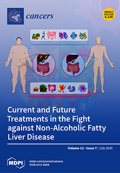
Cover Story (view full-size image): Obesity is recognised as a major contributor to the development of non-alcoholic fatty liver disease (NAFLD). With a quarter of the global population estimated to suffer from NAFLD, the quest for effective treatments is crucial. To date, few therapies aimed at reducing NAFLD are available, and have failed to deliver the desired outcomes when being evaluated in clinical trials. Lifestyle modifications have poor adherence, whilst pharmacological agents mainly alleviate only one of many mechanisms underlying the condition. Fortunately, the NAFLD treatment landscape is far from static. As such, the recently discovered intricate crosstalk between liver and gut might present an appealing avenue for drug development. While great strides have been made towards the development of NAFLD treatments, their further success hinges on a deeper understanding of the condition’s underlying mechanisms. View this paper.
  • Issues are regarded as officially published after their release is announced to the table of contents alert mailing list.
  • You may sign up for e-mail alerts to receive table of contents of newly released issues.
  • PDF is the official format for papers published in both, html and pdf forms. To view the papers in pdf format, click on the "PDF Full-text" link, and use the free Adobe Reader to open them.
Order results
Result details
Section
Select all
Export citation of selected articles as:
24 pages, 5921 KiB  
Article
Natural Killer Cell Activation Receptor NKp30 Oligomerization Depends on Its N-Glycosylation
by Ondřej Skořepa, Samuel Pazicky, Barbora Kalousková, Jan Bláha, Celeste Abreu, Tomáš Ječmen, Michal Rosůlek, Alexander Fish, Arthur Sedivy, Karl Harlos, Jan Dohnálek, Tereza Skálová and Ondřej Vaněk
Cancers 2020, 12(7), 1998; https://0-doi-org.brum.beds.ac.uk/10.3390/cancers12071998 - 21 Jul 2020
Cited by 12 | Viewed by 4216
Abstract
NKp30 is one of the main human natural killer (NK) cell activating receptors used in directed immunotherapy. The oligomerization of the NKp30 ligand binding domain depends on the length of the C-terminal stalk region, but our structural knowledge of NKp30 oligomerization and its [...] Read more.
NKp30 is one of the main human natural killer (NK) cell activating receptors used in directed immunotherapy. The oligomerization of the NKp30 ligand binding domain depends on the length of the C-terminal stalk region, but our structural knowledge of NKp30 oligomerization and its role in signal transduction remains limited. Moreover, ligand binding of NKp30 is affected by the presence and type of N-glycosylation. In this study, we assessed whether NKp30 oligomerization depends on its N-glycosylation. Our results show that NKp30 forms oligomers when expressed in HEK293S GnTI cell lines with simple N-glycans. However, NKp30 was detected only as monomers after enzymatic deglycosylation. Furthermore, we characterized the interaction between NKp30 and its best-studied cognate ligand, B7-H6, with respect to glycosylation and oligomerization, and we solved the crystal structure of this complex with glycosylated NKp30, revealing a new glycosylation-induced mode of NKp30 dimerization. Overall, this study provides new insights into the structural basis of NKp30 oligomerization and explains how the stalk region and glycosylation of NKp30 affect its ligand affinity. This furthers our understanding of the molecular mechanisms involved in NK cell activation, which is crucial for the successful design of novel NK cell-based targeted immunotherapeutics. Full article
Show Figures

Figure 1

15 pages, 2798 KiB  
Article
Immune Suppressive Effects of Plasma-Derived Exosome Populations in Head and Neck Cancer
by Inga J. Beccard, Linda Hofmann, Jan C. Schroeder, Sonja Ludwig, Simon Laban, Cornelia Brunner, Ramin Lotfi, Thomas K. Hoffmann, Edwin K. Jackson, Patrick J. Schuler and Marie-Nicole Theodoraki
Cancers 2020, 12(7), 1997; https://0-doi-org.brum.beds.ac.uk/10.3390/cancers12071997 - 21 Jul 2020
Cited by 28 | Viewed by 3513
Abstract
Plasma-derived exosomes of head and neck squamous cell carcinoma (HNSCC) patients carry inhibitory factors mediating immune suppression. Separation of tumor-derived exosomes (TEX) and non-TEX may assist in a better understanding of their respective parental cells. Here, we evaluate the impact of TEX or [...] Read more.
Plasma-derived exosomes of head and neck squamous cell carcinoma (HNSCC) patients carry inhibitory factors mediating immune suppression. Separation of tumor-derived exosomes (TEX) and non-TEX may assist in a better understanding of their respective parental cells. Here, we evaluate the impact of TEX or hematopoietic-derived exosomes on immune suppression. We evaluated apoptosis in CD8+ T cells, conversion of CD4+ T cells to regulatory T cells (Treg), and adenosine production by TEX, non-TEX, or total exosomes. Exosome protein cargo was significantly higher in total and CD45(−) exosomes from high stage compared to low stage patients. Furthermore, total and CD45(−) exosomes of high stage patients induced more apoptosis in CD8+ T cells than their low stage counterparts. CD69 suppression, a marker of reduced CD8+ T cell activation, was only mediated by CD45(−) exosomes. All fractions induced Treg differentiation, defined by CD39 expression, but only CD45(−) exosomes showed a stage-dependent conversion. CD45(−) exosomes produced higher adenosine concentrations than CD45(+) exosomes, concluding that adenosine production measured in total exosomes mainly derives from TEX. The presented results show significant induction of immune suppression by TEX in HNSCC. This immunosuppressive effect supports the potential role of exosomes as liquid biomarkers for disease stage and level of immune suppression. Full article
(This article belongs to the Special Issue Advances in Head and Neck Squamous Cell Carcinoma (HNSCC))
Show Figures

Figure 1

18 pages, 1106 KiB  
Review
SRC-Family Kinases in Acute Myeloid Leukaemia and Mastocytosis
by Edwige Voisset, Fabienne Brenet, Sophie Lopez and Paulo de Sepulveda
Cancers 2020, 12(7), 1996; https://0-doi-org.brum.beds.ac.uk/10.3390/cancers12071996 - 21 Jul 2020
Cited by 9 | Viewed by 3208
Abstract
Protein tyrosine kinases have been recognized as important actors of cell transformation and cancer progression, since their discovery as products of viral oncogenes. SRC-family kinases (SFKs) play crucial roles in normal hematopoiesis. Not surprisingly, they are hyperactivated and are essential for membrane receptor [...] Read more.
Protein tyrosine kinases have been recognized as important actors of cell transformation and cancer progression, since their discovery as products of viral oncogenes. SRC-family kinases (SFKs) play crucial roles in normal hematopoiesis. Not surprisingly, they are hyperactivated and are essential for membrane receptor downstream signaling in hematological malignancies such as acute myeloid leukemia (AML) and mastocytosis. The precise roles of SFKs are difficult to delineate due to the number of substrates, the functional redundancy among members, and the use of tools that are not selective. Yet, a large num ber of studies have accumulated evidence to support that SFKs are rational therapeutic targets in AML and mastocytosis. These two pathologies are regulated by two related receptor tyrosine kinases, which are well known in the field of hematology: FLT3 and KIT. FLT3 is one of the most frequently mutated genes in AML, while KIT oncogenic mutations occur in 80–90% of mastocytosis. Studies on oncogenic FLT3 and KIT signaling have shed light on specific roles for members of the SFK family. This review highlights the central roles of SFKs in AML and mastocytosis, and their interconnection with FLT3 and KIT oncoproteins. Full article
(This article belongs to the Special Issue The Role of Src Kinase Family in Cancer)
Show Figures

Figure 1

12 pages, 847 KiB  
Review
Do Elderly Lung Cancer Patients Aged ≥75 Years Benefit from Immune Checkpoint Inhibitors?
by Nagio Takigawa, Nobuaki Ochi, Nozomu Nakagawa, Yasunari Nagasaki, Masataka Taoka, Naruhiko Ichiyama, Ayaka Mimura, Hidekazu Nakanishi, Hiroyuki Kohara and Hiromichi Yamane
Cancers 2020, 12(7), 1995; https://0-doi-org.brum.beds.ac.uk/10.3390/cancers12071995 - 21 Jul 2020
Cited by 8 | Viewed by 2621
Abstract
Lung cancer patients ≥75 years represent nearly 40% of all lung cancer patients and continue to increase. If elderly patients have a good performance status and adequate organ function, they can be treated the same as non-elderly patients. However, few comparative studies limited [...] Read more.
Lung cancer patients ≥75 years represent nearly 40% of all lung cancer patients and continue to increase. If elderly patients have a good performance status and adequate organ function, they can be treated the same as non-elderly patients. However, few comparative studies limited to elderly patients (≥75 years) have been conducted. We review the evidence on using immune check inhibitors for the treatment of elderly patients (≥75 years old) with advanced non-small cell lung cancer. Prospective randomized or non-randomized, retrospective, registrational, insurance-based, and community-based studies have shown that elderly (≥75 years) and non-elderly patients are similarly treated with immune check inhibitors effectively and safely. However, such analyses have not shown that immune check inhibitors are significantly more effective than chemotherapy alone. In addition, patient selection might be critically performed to administer immune check inhibitors in the elderly because they are more likely to have a poor performance status with comorbidities, which lead to little benefit, even in non-elderly patients. There is a need for more evidence showing the benefit of immune check inhibitors in non-small cell lung cancer patients ≥75 years. Full article
Show Figures

Figure 1

14 pages, 279 KiB  
Review
Adjuvant Therapy for Melanoma: Past, Current, and Future Developments
by Alessandro A. E. Testori, Silvia Chiellino and Alexander C.J. van Akkooi
Cancers 2020, 12(7), 1994; https://0-doi-org.brum.beds.ac.uk/10.3390/cancers12071994 - 21 Jul 2020
Cited by 24 | Viewed by 4299
Abstract
This review describes the progress that the concept of adjuvant therapies has undergone in the last 50 years and focuses on the most recent development where an adjuvant approach has been scientifically evaluated in melanoma clinical trials. Over the past decade the development [...] Read more.
This review describes the progress that the concept of adjuvant therapies has undergone in the last 50 years and focuses on the most recent development where an adjuvant approach has been scientifically evaluated in melanoma clinical trials. Over the past decade the development of immunotherapies and targeted therapies has drastically changed the treatment of stage IV melanoma patients. These successes led to trials studying the same therapies in the adjuvant setting, in high risk resected stage III and IV melanoma patients. Adjuvant immune checkpoint blockade with anti-CTLA-4 antibody ipilimumab was the first drug to show an improvement in recurrence-free and overall survival but this was accompanied by high severe toxicity rates. Therefore, these results were bypassed by adjuvant treatment with anti-PD-1 agents nivolumab and pembrolizumab and BRAF-directed target therapy, which showed even better recurrence-free survival rates with more favorable toxicity rates. The whole concept of adjuvant therapy may be integrated with the new neoadjuvant approaches that are under investigation through several clinical trials. However, there is still no data available on whether the effective adjuvant therapy that patients finally have at their disposal could be offered to them while waiting for recurrence, sparing at least 50% of them a potentially long-term toxic side effect but with the same rate of overall survival (OS). Adjuvant therapy for melanoma has radically changed over the past few years—anti-PD-1 or BRAF-directed therapy is the new standard of care. Full article
(This article belongs to the Special Issue Advanced Melanoma)
19 pages, 3895 KiB  
Article
Plasma Treatment Limits Cutaneous Squamous Cell Carcinoma Development In Vitro and In Vivo
by Gabriella Pasqual-Melo, Thiago Nascimento, Larissa Juliani Sanches, Fernanda Paschoal Blegniski, Julya Karen Bianchi, Sanjeev Kumar Sagwal, Julia Berner, Anke Schmidt, Steffen Emmert, Klaus-Dieter Weltmann, Thomas von Woedtke, Rajesh Kumar Gandhirajan, Alessandra Lourenço Cecchini and Sander Bekeschus
Cancers 2020, 12(7), 1993; https://0-doi-org.brum.beds.ac.uk/10.3390/cancers12071993 - 21 Jul 2020
Cited by 26 | Viewed by 4211
Abstract
Cutaneous squamous cell carcinoma (SCC) is the most prevalent cancer worldwide, increasing the cost of healthcare services and with a high rate of morbidity. Its etiology is linked to chronic ultraviolet (UV) exposure that leads to malignant transformation of keratinocytes. Invasive growth and [...] Read more.
Cutaneous squamous cell carcinoma (SCC) is the most prevalent cancer worldwide, increasing the cost of healthcare services and with a high rate of morbidity. Its etiology is linked to chronic ultraviolet (UV) exposure that leads to malignant transformation of keratinocytes. Invasive growth and metastasis are severe consequences of this process. Therapy-resistant and highly aggressive SCC is frequently fatal, exemplifying the need for novel treatment strategies. Cold physical plasma is a partially ionized gas, expelling therapeutic doses of reactive oxygen and nitrogen species that were investigated for their anticancer capacity against SCC in vitro and SCC-like lesions in vivo. Using the kINPen argon plasma jet, a selective growth-reducing action of plasma treatment was identified in two SCC cell lines in 2D and 3D cultures. In vivo, plasma treatment limited the progression of UVB-induced SSC-like skin lesions and dermal degeneration without compromising lesional or non-lesional skin. In lesional tissue, this was associated with a decrease in cell proliferation and the antioxidant transcription factor Nrf2 following plasma treatment, while catalase expression was increased. Analysis of skin adjacent to the lesions and determination of global antioxidant parameters confirmed the local but not systemic action of the plasma anticancer therapy in vivo. Full article
(This article belongs to the Special Issue Advances in Plasma Oncology toward Clinical Translation)
Show Figures

Figure 1

11 pages, 2455 KiB  
Article
Clinico-Biological Features and Clonal Hematopoiesis in Patients with Severe COVID-19
by Nicolas Duployez, Jordane Demonchy, Céline Berthon, Julien Goutay, Morgan Caplan, Anne-Sophie Moreau, Anne Bignon, Alice Marceau-Renaut, Delphine Garrigue, Imelda Raczkiewicz, Sandrine Geffroy, Maxime Bucci, Kazali Alidjinou, Julie Demaret, Myriam Labalette, Thierry Brousseau, Annabelle Dupont, Antoine Rauch, Julien Poissy, Sophie Susen, Claude Preudhomme and Bruno Quesneladd Show full author list remove Hide full author list
Cancers 2020, 12(7), 1992; https://0-doi-org.brum.beds.ac.uk/10.3390/cancers12071992 - 21 Jul 2020
Cited by 21 | Viewed by 6890
Abstract
Advanced age or preexisting comorbidities have been characterized as risk factors for severe coronavirus disease 2019 (COVID-19) cases requiring hospitalization and intensive care. In recent years, clonal hematopoiesis (CH) of indeterminate potential (CHIP) has emerged as a risk factor for chronic inflammatory background [...] Read more.
Advanced age or preexisting comorbidities have been characterized as risk factors for severe coronavirus disease 2019 (COVID-19) cases requiring hospitalization and intensive care. In recent years, clonal hematopoiesis (CH) of indeterminate potential (CHIP) has emerged as a risk factor for chronic inflammatory background and subsequent aging-associated diseases. The purpose of this study was to identify biological factors (particularly leukocyte subtypes and inflammatory markers) associated with a risk of clinical deterioration (i.e., orotracheal intubation (OTI)) and to determine whether CH was likely to influence clinical and biological behavior in patients with severe COVID-19 requiring hospitalization. Here, we describe clinical and biological features, including the screening of CHIP mutants in a well-annotated cohort of 122 hospitalized patients with a laboratory-confirmed diagnosis of COVID-19 (55% requiring OTI). We showed that elevated white blood cell counts, especially neutrophils and high C-reactive protein (CRP) levels at admission, were associated with an increased requirement of OTI. We noticed a high prevalence of CH (25%, 38%, 56%, and 82% of patients aged <60 years, 60–70 years, 70–80 years, and >80 years) compared to a retrospective cohort of patients free of hematological malignancy explored with the same pipelines (10%, 21%, 37%, and 44%). However, the existence of CH did not significantly impact clinical outcome, including OTI or death, and did not correlate with other laboratory findings. Full article
(This article belongs to the Collection The Impact of COVID-19 Infection in Cancer)
Show Figures

Graphical abstract

17 pages, 3570 KiB  
Article
Ralaniten Sensitizes Enzalutamide-Resistant Prostate Cancer to Ionizing Radiation in Prostate Cancer Cells that Express Androgen Receptor Splice Variants
by Carmen A. Banuelos, Yusuke Ito, Jon K. Obst, Nasrin R. Mawji, Jun Wang, Yukiyoshi Hirayama, Jacky K. Leung, Teresa Tam, Amy H. Tien, Raymond J. Andersen and Marianne D. Sadar
Cancers 2020, 12(7), 1991; https://0-doi-org.brum.beds.ac.uk/10.3390/cancers12071991 - 21 Jul 2020
Cited by 19 | Viewed by 3537
Abstract
Blocking androgen receptor (AR) transcriptional activity by androgen deprivation therapy (ADT) improves the response to radiotherapy for intermediate and high risk prostate cancer. Unfortunately, ADT, antiandrogens, and abiraterone increase expression of constitutively active splice variants of AR (AR-Vs) which regulate DNA damage repair [...] Read more.
Blocking androgen receptor (AR) transcriptional activity by androgen deprivation therapy (ADT) improves the response to radiotherapy for intermediate and high risk prostate cancer. Unfortunately, ADT, antiandrogens, and abiraterone increase expression of constitutively active splice variants of AR (AR-Vs) which regulate DNA damage repair leading to resistance to radiotherapy. Here we investigate whether blocking the transcriptional activities of full-length AR and AR-Vs with ralaniten leads to enhanced sensitivity to radiotherapy. Combination therapies using ralaniten with ionizing radiation were evaluated for effects on proliferation, colony formation, cell cycle, DNA damage, and Western blot analyses in human prostate cancer cells that express both full-length AR and AR-Vs. Ralaniten and a potent next-generation analog (EPI-7170) decreased expression of DNA repair genes whereas enzalutamide had no effect. FACS analysis revealed a dose-dependent decrease of BrdU incorporation with increased accumulation of γH2AX with a combination of ionizing radiation with ralaniten. An additive inhibitory effect on proliferation of enzalutamide-resistant cells was achieved with a combination of ralaniten compounds with ionizing radiation. Ralaniten and EPI-7170 sensitized prostate cancer cells that express full-length AR and AR-Vs to radiotherapy whereas enzalutamide had no added benefit. Full article
(This article belongs to the Section Cancer Therapy)
Show Figures

Figure 1

14 pages, 321 KiB  
Review
Immunotherapy for Early Stage Colorectal Cancer: A Glance into the Future
by Romain Cohen, Qian Shi and Thierry André
Cancers 2020, 12(7), 1990; https://0-doi-org.brum.beds.ac.uk/10.3390/cancers12071990 - 21 Jul 2020
Cited by 9 | Viewed by 3610
Abstract
Immune checkpoint inhibitors (ICI) have reshaped therapeutic strategies for cancer patients. The development of ICI for early stage colorectal cancer is accompanied by specific challenges: (i) the selection of patients who are likely to benefit from these treatments, i.e., patients with tumors harboring [...] Read more.
Immune checkpoint inhibitors (ICI) have reshaped therapeutic strategies for cancer patients. The development of ICI for early stage colorectal cancer is accompanied by specific challenges: (i) the selection of patients who are likely to benefit from these treatments, i.e., patients with tumors harboring predictive factors of efficacy of ICI, such as microsatellite instability and/or mismatch repair deficiency (MSI/dMMR), or other potential parameters (increased T cell infiltration using Immunoscore® or others, high tumor mutational burden, POLE mutation), (ii) the selection of patients at risk of disease recurrence (poor prognostic features), and (iii) the choice of an accurate clinical trial methodological framework. In this review, we will discuss the ins and outs of clinical research of ICI for early stage MSI/dMMR CC patients in adjuvant and neoadjuvant settings. We will then summarize data that might support the development of ICI in localized colorectal cancer beyond MSI/dMMR. Full article
(This article belongs to the Special Issue Adjuvant Chemotherapy for Colorectal Cancer)
20 pages, 4254 KiB  
Article
Merkel Cell Polyomavirus T Antigens Induce Merkel Cell-Like Differentiation in GLI1-Expressing Epithelial Cells
by Thibault Kervarrec, Mahtab Samimi, Sonja Hesbacher, Patricia Berthon, Marion Wobser, Aurélie Sallot, Bhavishya Sarma, Sophie Schweinitzer, Théo Gandon, Christophe Destrieux, Côme Pasqualin, Serge Guyétant, Antoine Touzé, Roland Houben and David Schrama
Cancers 2020, 12(7), 1989; https://0-doi-org.brum.beds.ac.uk/10.3390/cancers12071989 - 21 Jul 2020
Cited by 24 | Viewed by 3036
Abstract
Merkel cell carcinoma (MCC) is an aggressive skin cancer frequently caused by the Merkel cell polyomavirus (MCPyV). It is still under discussion, in which cells viral integration and MCC development occurs. Recently, we demonstrated that a virus-positive MCC derived from a trichoblastoma, an [...] Read more.
Merkel cell carcinoma (MCC) is an aggressive skin cancer frequently caused by the Merkel cell polyomavirus (MCPyV). It is still under discussion, in which cells viral integration and MCC development occurs. Recently, we demonstrated that a virus-positive MCC derived from a trichoblastoma, an epithelial neoplasia bearing Merkel cell (MC) differentiation potential. Accordingly, we hypothesized that MC progenitors may represent an origin of MCPyV-positive MCC. To sustain this hypothesis, phenotypic comparison of trichoblastomas and physiologic human MC progenitors was conducted revealing GLI family zinc finger 1 (GLI1), Keratin 17 (KRT 17), and SRY-box transcription factor 9 (SOX9) expressions in both subsets. Furthermore, GLI1 expression in keratinocytes induced transcription of the MC marker SOX2 supporting a role of GLI1 in human MC differentiation. To assess a possible contribution of the MCPyV T antigens (TA) to the development of an MC-like phenotype, human keratinocytes were transduced with TA. While this led only to induction of KRT8, an early MC marker, combined GLI1 and TA expression gave rise to a more advanced MC phenotype with SOX2, KRT8, and KRT20 expression. Finally, we demonstrated MCPyV-large T antigens’ capacity to inhibit the degradation of the MC master regulator Atonal bHLH transcription factor 1 (ATOH1). In conclusion, our report suggests that MCPyV TA contribute to the acquisition of an MC-like phenotype in epithelial cells. Full article
(This article belongs to the Special Issue The Biological and Clinical Aspects of Merkel Cell Carcinoma)
Show Figures

Figure 1

29 pages, 2812 KiB  
Review
Systemic Treatment Selection for Patients with Advanced Pancreatic Neuroendocrine Tumours (PanNETs)
by Vera G. Megdanova-Chipeva, Angela Lamarca, Alison Backen, Mairéad G. McNamara, Jorge Barriuso, Sonia Sergieva, Lilia Gocheva, Was Mansoor, Prakash Manoharan and Juan W. Valle
Cancers 2020, 12(7), 1988; https://0-doi-org.brum.beds.ac.uk/10.3390/cancers12071988 - 21 Jul 2020
Cited by 11 | Viewed by 3755
Abstract
Pancreatic neuroendocrine tumours (PanNETs) are rare diseases and a good example of how research is not only feasible, but also of crucial importance in the scenario of rare tumours. Many clinical trials have been performed over the past two decades expanding therapeutic options [...] Read more.
Pancreatic neuroendocrine tumours (PanNETs) are rare diseases and a good example of how research is not only feasible, but also of crucial importance in the scenario of rare tumours. Many clinical trials have been performed over the past two decades expanding therapeutic options for patients with advanced PanNETs. Adequate management relies on optimal selection of treatment, which may be challenging for clinicians due to the fact that multiple options of therapy are currently available. A number of therapies already exist, which are supported by data from phase III studies, including somatostatin analogues and targeted therapies (sunitinib and everolimus). In addition, chemotherapy remains an option, with temozolomide and capecitabine being one of the most popular doublets to use. Peptide receptor radionuclide therapy was successfully implemented in patients with well-differentiated gastro-entero-pancreatic neuroendocrine tumours, but with certain questions waiting to be solved for the management of PanNETs. Finally, the role of immunotherapy is still poorly understood. In this review, the data supporting current systemic treatment options for locally advanced or metastatic PanNETs are summarized. Strategies for treatment selection in patients with PanNETs based on patient, disease, or drug characteristics is provided, as well as a summary of current evidence on prognostic and predictive biomarkers. Future perspectives are discussed, focusing on current and forthcoming challenges and unmet needs of patients with these rare tumours. Full article
(This article belongs to the Special Issue New Therapeutic Advances in Rare Tumors)
Show Figures

Figure 1

25 pages, 675 KiB  
Review
Tumor-Associated Macrophage Status in Cancer Treatment
by Anna Maria Malfitano, Simona Pisanti, Fabiana Napolitano, Sarah Di Somma, Rosanna Martinelli and Giuseppe Portella
Cancers 2020, 12(7), 1987; https://0-doi-org.brum.beds.ac.uk/10.3390/cancers12071987 - 21 Jul 2020
Cited by 100 | Viewed by 7006
Abstract
Tumor-associated macrophages (TAMs) represent the most abundant innate immune cells in tumors. TAMs, exhibiting anti-inflammatory phenotype, are key players in cancer progression, metastasis and resistance to therapy. A high TAM infiltration is generally associated with poor prognosis, but macrophages are highly plastic cells [...] Read more.
Tumor-associated macrophages (TAMs) represent the most abundant innate immune cells in tumors. TAMs, exhibiting anti-inflammatory phenotype, are key players in cancer progression, metastasis and resistance to therapy. A high TAM infiltration is generally associated with poor prognosis, but macrophages are highly plastic cells that can adopt either proinflammatory/antitumor or anti-inflammatory/protumor features in response to tumor microenvironment stimuli. In the context of cancer therapy, many anticancer therapeutics, apart from their direct effect on tumor cells, display different effects on TAM activation status and density. In this review, we aim to evaluate the indirect effects of anticancer therapies in the modulation of TAM phenotypes and pro/antitumor activity. Full article
(This article belongs to the Special Issue Targeting Innate Immunity Cells in Cancer)
Show Figures

Figure 1

14 pages, 1604 KiB  
Article
Hypoalbuminemia Reflects Nutritional Risk, Body Composition and Systemic Inflammation and Is Independently Associated with Survival in Patients with Colorectal Cancer
by Arwa S. Almasaudi, Ross D. Dolan, Christine A. Edwards and Donald C. McMillan
Cancers 2020, 12(7), 1986; https://0-doi-org.brum.beds.ac.uk/10.3390/cancers12071986 - 21 Jul 2020
Cited by 68 | Viewed by 4233
Abstract
It has long been recognized that albumin has prognostic value in patients with cancer. However, although the Global Leadership Initiative on Malnutrition GLIM criteria (based on five diagnostic criteria, three phenotypic criteria and two etiologic criteria) recognize inflammation as an important etiologic factor [...] Read more.
It has long been recognized that albumin has prognostic value in patients with cancer. However, although the Global Leadership Initiative on Malnutrition GLIM criteria (based on five diagnostic criteria, three phenotypic criteria and two etiologic criteria) recognize inflammation as an important etiologic factor in malnutrition, there are limited data regarding the association between albumin, nutritional risk, body composition and systemic inflammation, and whether albumin is associated with mortality independent of these parameters. The aim of this study was to examine the relationship between albumin, nutritional risk, body composition, systemic inflammation, and outcomes in patients with colorectal cancer (CRC). A retrospective cohort study (n = 795) was carried out in which patients were divided into normal and hypoalbuminaemic groups (albumin  < 35 g/L) in the presence and absence of a systemic inflammatory response C-reactive protein (CRP > 10 and <10 mg/L, respectively). Post-operative complications, severity of complications and mortality were considered as outcome measures. Categorical variables were analyzed using Chi-square test χ2 or linear-by-linear association. Survival data were analyzed using univariate and multivariate Cox regression. In the presence of a systemic inflammatory response, hypoalbuminemia was directly associated with Malnutrition Universal Screening Tool MUST (p < 0.001) and inversely associated with Body Mass Index BMI (p < 0.001), subcutaneous adiposity (p < 0.01), visceral obesity (p < 0.01), skeletal muscle index (p < 0.001) and skeletal muscle density (p < 0.001). There was no significant association between hypoalbuminemia and either the presence of complications or their severity. In the absence of a systemic inflammatory response (n = 589), hypoalbuminemia was directly associated with MUST (p < 0.05) and inversely associated with BMI (p < 0.01), subcutaneous adiposity (p < 0.05), visceral adiposity (p < 0.05), skeletal muscle index (p < 0.01) and skeletal muscle density (p < 0.001). Hypoalbuminemia was, independently of inflammatory markers, associated with poorer cancer-specific and overall survival (both p < 0.001). The results suggest that hypoalbuminemia in patients with CRC reflects both increased nutritional risk and greater systemic inflammatory response and was independently associated with poorer survival in patients with CRC. Full article
Show Figures

Figure 1

81 pages, 1333 KiB  
Review
Anti-Cancer Potential of Cannabinoids, Terpenes, and Flavonoids Present in Cannabis
by Andrea M. Tomko, Erin G. Whynot, Lee D. Ellis and Denis J. Dupré
Cancers 2020, 12(7), 1985; https://0-doi-org.brum.beds.ac.uk/10.3390/cancers12071985 - 21 Jul 2020
Cited by 103 | Viewed by 26455
Abstract
In recent years, and even more since its legalization in several jurisdictions, cannabis and the endocannabinoid system have received an increasing amount of interest related to their potential exploitation in clinical settings. Cannabinoids have been suggested and shown to be effective in the [...] Read more.
In recent years, and even more since its legalization in several jurisdictions, cannabis and the endocannabinoid system have received an increasing amount of interest related to their potential exploitation in clinical settings. Cannabinoids have been suggested and shown to be effective in the treatment of various conditions. In cancer, the endocannabinoid system is altered in numerous types of tumours and can relate to cancer prognosis and disease outcome. Additionally, cannabinoids display anticancer effects in several models by suppressing the proliferation, migration and/or invasion of cancer cells, as well as tumour angiogenesis. However, the therapeutic use of cannabinoids is currently limited to the treatment of symptoms and pain associated with chemotherapy, while their potential use as cytotoxic drugs in chemotherapy still requires validation in patients. Along with cannabinoids, cannabis contains several other compounds that have also been shown to exert anti-tumorigenic actions. The potential anti-cancer effects of cannabinoids, terpenes and flavonoids, present in cannabis, are explored in this literature review. Full article
(This article belongs to the Special Issue Medicinal Plants and Their Active Ingredients in Cancer)
Show Figures

Figure 1

19 pages, 1963 KiB  
Review
Impact of Eukaryotic Translation Initiation Factors on Breast Cancer: Still Much to Investigate
by Qin Chen, Bo Yang, Norbert Nass, Christoph Schatz and Johannes Haybaeck
Cancers 2020, 12(7), 1984; https://0-doi-org.brum.beds.ac.uk/10.3390/cancers12071984 - 21 Jul 2020
Cited by 7 | Viewed by 3440
Abstract
Breast carcinoma (BC) remains one of the most serious health problems. It is a heterogeneous entity, and mainly classified according to receptor status for estrogen (ER), progesterone (PR) and egf (HER2/Neu), as well as the proliferation marker ki67. Gene expression in eukaryotes is [...] Read more.
Breast carcinoma (BC) remains one of the most serious health problems. It is a heterogeneous entity, and mainly classified according to receptor status for estrogen (ER), progesterone (PR) and egf (HER2/Neu), as well as the proliferation marker ki67. Gene expression in eukaryotes is regulated at the level of both gene transcription and translation, where eukaryotic initiation factors (eIFs) are key regulators of protein biosynthesis. Aberrant translation results in an altered cellular proteome, and this clearly effects cell growth supporting tumorigenesis. The relationship between various eIFs and BC entities, as well as the related regulatory mechanisms, has meanwhile become a focus of scientific interest. Here, we give an overview on the current research state of eIF function, focusing on BC. Full article
Show Figures

Figure 1

15 pages, 1859 KiB  
Article
Integration of Serum Metabolomics into Clinical Assessment to Improve Outcome Prediction of Metastatic Soft Tissue Sarcoma Patients Treated with Trabectedin
by Gianmaria Miolo, Emanuela Di Gregorio, Asia Saorin, Davide Lombardi, Simona Scalone, Angela Buonadonna, Agostino Steffan and Giuseppe Corona
Cancers 2020, 12(7), 1983; https://0-doi-org.brum.beds.ac.uk/10.3390/cancers12071983 - 21 Jul 2020
Cited by 14 | Viewed by 2720
Abstract
Soft tissue sarcomas (STS) are a group of rare and heterogeneous cancers with few diagnostic or prognostic biomarkers. This metabolomics study aimed to identify new serum prognostic biomarkers to improve the prediction of overall survival in patients with metastatic STS. The study enrolled [...] Read more.
Soft tissue sarcomas (STS) are a group of rare and heterogeneous cancers with few diagnostic or prognostic biomarkers. This metabolomics study aimed to identify new serum prognostic biomarkers to improve the prediction of overall survival in patients with metastatic STS. The study enrolled 24 patients treated with the same trabectedin regimen. The baseline serum metabolomics profile, targeted to 68 metabolites encompassing amino acids and bile acids pathways, was quantified by liquid chromatography-tandem mass spectrometry. Correlations between individual metabolomics profiles and overall survival were examined and a risk model to predict survival was built by Cox multivariate regression. The median overall survival of the studied patients was 13.0 months (95% CI, 5.6–23.5). Among all the metabolites investigated, only citrulline and histidine correlated significantly with overall survival. The best Cox risk prediction model obtained integrating metabolomics and clinical data, included citrulline, hemoglobin and patients’ performance status score. It allowed to distinguish patients into a high-risk group with a low median overall survival of 2.1 months and a low- to moderate-risk group with a median overall survival of 19.1 months (p < 0.0001). The results of this metabolomics translation study indicate that citrulline, an amino acid belonging to the arginine metabolism, represents an important metabolic signature that may contribute to explain the high inter-patients overall survival variability of STS patients. The risk prediction model based on baseline serum citrulline, hemoglobin and performance status may represent a new prognostic tool for the early classification of patients with metastatic STS, according to their overall survival expectancy. Full article
(This article belongs to the Special Issue Sarcomas: New Biomarkers and Therapeutic Strategies)
Show Figures

Graphical abstract

18 pages, 1889 KiB  
Article
The Biological Role of Sponge Circular RNAs in Gastric Cancer: Main Players or Coadjuvants?
by Adenilson Leão Pereira, Leandro Magalhães, Rafael Pompeu Pantoja, Gilderlanio Araújo, Ândrea Ribeiro-dos-Santos and Amanda Ferreira Vidal
Cancers 2020, 12(7), 1982; https://0-doi-org.brum.beds.ac.uk/10.3390/cancers12071982 - 21 Jul 2020
Cited by 17 | Viewed by 3414
Abstract
Circular RNAs (circRNAs) are a new class of long noncoding RNAs able to perform multiple functions, including sponging microRNAs (miRNAs) and RNA-Binding Proteins (RBPs). They play an important role in gastric carcinogenesis, but its involvement during gastric cancer (GC) development and progression are [...] Read more.
Circular RNAs (circRNAs) are a new class of long noncoding RNAs able to perform multiple functions, including sponging microRNAs (miRNAs) and RNA-Binding Proteins (RBPs). They play an important role in gastric carcinogenesis, but its involvement during gastric cancer (GC) development and progression are not well understood. We gathered miRNA and/or RBPs sponge circRNAs present in GC, and accessed their biological roles through functional enrichment of their target genes or ligand RBPs. We identified 54 sponge circRNAs in GC that are able to sponge 51 miRNAs and 103 RBPs. Then, we evaluated their host gene expression using The Cancer Genome Atlas (TCGA) database and observed that COL1A2 is the most overexpressed gene, which may be due to circHIPK3/miR-29b-c/COL1A2 axis dysregulation. We identified 27 GC-related pathways that may be affected mainly by circPVT1, circHIPK3 and circNF1. Our results indicate that circHIPK3/miR-107/BDNF/LIN28 axis may mediate chemoresistance in GC, and that circPVT1, circHIPK3, circNF1, ciRS-7 and circ_0000096 appear to be involved in gastrointestinal cancer development. Lastly, circHIPK3, circNRIP1 and circSMARCA5 were identified in different ethnic populations and may be ubiquitous modulators of gastric carcinogenesis. Overall, the studied sponge circRNAs are part of a complex RBP-circRNA-miRNA-mRNA interaction network, and are involved in the establishment, chemoresistance and progression of GC. Full article
(This article belongs to the Special Issue Circular RNAs in Cancer)
Show Figures

Figure 1

14 pages, 853 KiB  
Article
High-Throughput Sequencing of Gastric Cancer Patients: Unravelling Genetic Predispositions Towards an Early-Onset Subtype
by Julita Machlowska, Przemysław Kapusta, Jacek Baj, Folkert H. M. Morsink, Paweł Wołkow, Ryszard Maciejewski, G. Johan A. Offerhaus and Robert Sitarz
Cancers 2020, 12(7), 1981; https://0-doi-org.brum.beds.ac.uk/10.3390/cancers12071981 - 21 Jul 2020
Cited by 20 | Viewed by 3894
Abstract
Background: Gastric cancer is the fourth most common cause of cancer-related death. Currently, it is broadly accepted that the molecular complexity and heterogeneity of gastric cancer, both inter- and intra-tumor, display important barriers for finding specific biomarkers for the early detection and diagnosis [...] Read more.
Background: Gastric cancer is the fourth most common cause of cancer-related death. Currently, it is broadly accepted that the molecular complexity and heterogeneity of gastric cancer, both inter- and intra-tumor, display important barriers for finding specific biomarkers for the early detection and diagnosis of this malignancy. Early-onset gastric cancer is not as prevalent as conventional gastric carcinoma, but it is a preferable model for studying the genetic background, as young patients are less exposed to environmental factors, which influence cancer development. Aim: The main objective of this study was to reveal age-dependent genotypic characteristics of gastric cancer subtypes, as well as conduct mutation profiling for the most frequent alterations in gastric cancer development, using targeted next-generation sequencing technology. Patients and methods: The study group included 53 patients, consisting of 18 patients with conventional gastric cancer and 35 with an early-onset subtype. The DNA of all index cases was used for next-generation sequencing, employing a panel of 94 genes and 284 single nucleotide polymorphisms (SNPs) (TruSight Cancer Panel, Illumina), which is characteristic for common and rare types of cancer. Results: From among the 53 samples processed for sequencing, we were able to identify seven candidate genes (STK11, RET, FANCM, SLX4, WRN, MEN1, and KIT) and nine variants among them: one splice_acceptor, four synonymous, and four missense variants. These were selected for the age-dependent differentiation of gastric cancer subtypes. We found four variants with C-Score ≥ 10, as 10% of the most deleterious substitutions: rs1800862 (RET), rs10138997 (FANCM), rs2230009 (WRN), and rs2959656 (MEN1). We identified 36 different variants, among 24 different genes, which were the most frequent genetic alterations among study subjects. We found 16 different variants among the genes that were present in 100% of the total cohort: SDHB (rs2746462), ALK (rs1670283), XPC (rs2958057), RECQL4 (rs4925828; rs11342077, rs398010167; rs2721190), DDB2 (rs326212), MEN1 (rs540012), AIP (rs4930199), ATM (rs659243), HNF1A (rs1169305), BRCA2 (rs206075; rs169547), ERCC5 (rs9514066; rs9514067), and FANCI (rs7183618). Conclusions: The technology of next-generation sequencing is a useful tool for studying the development and progression of gastric carcinoma in a high-throughput way. Our study revealed that early-onset gastric cancer has a different mutation frequency profile in certain genes compared to conventional subtype. Full article
(This article belongs to the Special Issue Recent Research on Gastrointestinal Carcinoma)
Show Figures

Figure 1

11 pages, 923 KiB  
Brief Report
Meta-Analysis Reveals Significant Sex Differences in Chronic Lymphocytic Leukemia Progression in the Eµ-TCL1 Transgenic Mouse Model
by Maximilian Koch, Sebastian Reinartz, Julia Saggau, Gero Knittel, Natascha Rosen, Oleg Fedorchenko, Lisa Thelen, Romy Barthel, Nina Reinart, Tamina Seeger-Nukpezah, Hans Christian Reinhardt, Michael Hallek and Phuong-Hien Nguyen
Cancers 2020, 12(7), 1980; https://0-doi-org.brum.beds.ac.uk/10.3390/cancers12071980 - 20 Jul 2020
Cited by 5 | Viewed by 4046
Abstract
The Eµ-TCL1 transgenic mouse model represents the most widely and extensively used animal model for chronic lymphocytic leukemia (CLL). In this report, we performed a meta-analysis of leukemia progression in over 300 individual Eµ-TCL1 transgenic mice and discovered a significantly accelerated disease progression [...] Read more.
The Eµ-TCL1 transgenic mouse model represents the most widely and extensively used animal model for chronic lymphocytic leukemia (CLL). In this report, we performed a meta-analysis of leukemia progression in over 300 individual Eµ-TCL1 transgenic mice and discovered a significantly accelerated disease progression in females compared to males. This difference is also reflected in an aggressive CLL mouse model with additional deletion of Tp53 besides the TCL1 transgene. Moreover, after serial adoptive transplantation of murine CLL cells, female recipients also succumbed to CLL earlier than male recipients. This sex-related disparity in the murine models is markedly contradictory to the human CLL condition. Thus, due to our observation we urge both careful consideration in the experimental design and accurate description of the Eµ-TCL1 transgenic cohorts in future studies. Full article
(This article belongs to the Special Issue Chronic Lymphocytic Leukemia)
Show Figures

Figure 1

14 pages, 5124 KiB  
Review
DCE-MRI of Tumor Hypoxia and Hypoxia-Associated Aggressiveness
by Jon-Vidar Gaustad, Anette Hauge, Catherine S. Wegner, Trude G. Simonsen, Kjersti V. Lund, Lise Mari K. Hansem and Einar K. Rofstad
Cancers 2020, 12(7), 1979; https://0-doi-org.brum.beds.ac.uk/10.3390/cancers12071979 - 20 Jul 2020
Cited by 22 | Viewed by 5964
Abstract
Tumor hypoxia is associated with resistance to treatment, aggressive growth, metastatic dissemination, and poor clinical outcome in many cancer types. The potential of dynamic contrast-enhanced magnetic resonance imaging (DCE-MRI) to assess the extent of hypoxia in tumors has been investigated in several studies [...] Read more.
Tumor hypoxia is associated with resistance to treatment, aggressive growth, metastatic dissemination, and poor clinical outcome in many cancer types. The potential of dynamic contrast-enhanced magnetic resonance imaging (DCE-MRI) to assess the extent of hypoxia in tumors has been investigated in several studies in our laboratory. Cervical carcinoma, melanoma, and pancreatic ductal adenocarcinoma (PDAC) xenografts have been used as models of human cancer, and the transfer rate constant (Ktrans) and the extravascular extracellular volume fraction (ve) have been derived from DCE-MRI data by using Tofts standard pharmacokinetic model and a population-based arterial input function. Ktrans was found to reflect naturally occurring and treatment-induced hypoxia when hypoxia was caused by low blood perfusion, radiation responsiveness when radiation resistance was due to hypoxia, and metastatic potential when metastasis was hypoxia-induced. Ktrans was also associated with outcome for patients with locally-advanced cervical carcinoma treated with cisplatin-based chemoradiotherapy. Together, the studies imply that DCE-MRI can provide valuable information on the hypoxic status of cervical carcinoma, melanoma, and PDAC. In this communication, we review and discuss the studies and provide some recommendations as to how DCE-MRI data can be analyzed and interpreted to assess tumor hypoxia. Full article
Show Figures

Figure 1

13 pages, 2999 KiB  
Communication
Depletion of Macrophages Improves Therapeutic Response to Gemcitabine in Murine Pancreas Cancer
by Soeren M. Buchholz, Robert G. Goetze, Shiv K. Singh, Christoph Ammer-Herrmenau, Frances M. Richards, Duncan I. Jodrell, Malte Buchholz, Patrick Michl, Volker Ellenrieder, Elisabeth Hessmann and Albrecht Neesse
Cancers 2020, 12(7), 1978; https://0-doi-org.brum.beds.ac.uk/10.3390/cancers12071978 - 20 Jul 2020
Cited by 27 | Viewed by 3936
Abstract
Background: The tumor microenvironment (TME) is composed of fibro-inflammatory cells and extracellular matrix (ECM) components. However, the exact contribution of the various TME compartments towards therapeutic response is unknown. Here, we aim to dissect the specific contribution of tumor-associated macrophages (TAMs) towards drug [...] Read more.
Background: The tumor microenvironment (TME) is composed of fibro-inflammatory cells and extracellular matrix (ECM) components. However, the exact contribution of the various TME compartments towards therapeutic response is unknown. Here, we aim to dissect the specific contribution of tumor-associated macrophages (TAMs) towards drug delivery and response in pancreatic ductal adenocarcinoma (PDAC). Methods: The effect of gemcitabine was assessed in human and murine macrophages, human pancreatic stellate cells (hPSCs), and tumor cells (L3.6pl, BxPC3 and KPC) in vitro. The drug metabolism of gemcitabine was analyzed by liquid chromatography–tandem mass spectrometry (LC–MS/MS). Preclinical studies were conducted using KrasG12D;p48-Cre and KrasG12D;p53172H;Pdx-Cre mice to investigate gemcitabine delivery at different stages of tumor progression and upon pharmacological TAM depletion. Results: Gemcitabine accumulation was significantly increased in murine PDAC tissue compared to pancreatic intraepithelial neoplasia (PanIN) lesions and healthy control pancreas tissue. In vitro, macrophages accumulated and rapidly metabolized gemcitabine resulting in a significant drug scavenging effect for gemcitabine. Finally, pharmacological TAM depletion enhanced therapeutic response to gemcitabine in tumor-bearing KPC mice. Conclusion: Macrophages rapidly metabolize gemcitabine in vitro, and pharmacological depletion improves the therapeutic response to gemcitabine in vivo. Our study supports the notion that TAMs might be a promising therapeutic target in PDAC. Full article
(This article belongs to the Section Cancer Therapy)
Show Figures

Figure 1

12 pages, 2063 KiB  
Article
Glucose Loading Enhances the Value of 18F-FDG PET/CT for the Characterization and Delineation of Cerebral Gliomas
by Dongwoo Kim, Hae Young Ko, Sangwon Lee, Yong-ho Lee, Sujin Ryu, Seon Yoo Kim, Jee-in Chung, Misu Lee, Ju Hyung Moon, Jong Hee Chang and Mijin Yun
Cancers 2020, 12(7), 1977; https://0-doi-org.brum.beds.ac.uk/10.3390/cancers12071977 - 20 Jul 2020
Cited by 4 | Viewed by 2615
Abstract
This study aimed to assess how to enhance the value of 18F-Fluorodeoxyglucose (FDG) PET/CTs for glioma grading and better delineation of the tumor boundary by glucose loading. In mouse models of brain tumor using U87MG cells, 18F-FDG-PET images were obtained after [...] Read more.
This study aimed to assess how to enhance the value of 18F-Fluorodeoxyglucose (FDG) PET/CTs for glioma grading and better delineation of the tumor boundary by glucose loading. In mouse models of brain tumor using U87MG cells, 18F-FDG-PET images were obtained after fasting and after glucose loading. There was a significant difference in the tumor-to-normal cortex-uptake ratio (TNR) between the fasting and glucose-loading scans. 14C-2-Deoxy-D-glucose (14C-DG) uptake was measured in vitro using U87MG, U373MG and primary neurons cultured with different concentrations of glucose. The tumor-to-neuron ratio of 14C-DG uptake increased with up to 10 mM of glucose. Finally, 10 low-grade and 17 high-grade glioma patients underwent fasting and glucose loading 18F-FDG PET/CT and the TNR was compared between scans. The effect of glucose loading was significant in high-grade but not in low-grade gliomas. The receiver operating characteristic curve analyses with a cut-off TNR of 0.81 showed a higher area under the curve after glucose loading than fasting for differentiating low-grade versus high-grade gliomas. In addition, the glucose loading PET/CT was more useful than the fasting PET/CT for the discrimination of oligodendrogliomas from IDH-wildtype glioblastomas. Glucose loading resulted in a greater reduction in 18F-FDG uptake in the normal cortex than in tumors, which increases the usefulness of 18F-FDG PET/CT for grading. Full article
(This article belongs to the Section Methods and Technologies Development)
Show Figures

Figure 1

21 pages, 5322 KiB  
Review
The Impact on Survival and Morbidity of Portal–Mesenteric Resection During Pancreaticoduodenectomy for Pancreatic Head Adenocarcinoma: A Systematic Review and Meta-Analysis of Comparative Studies
by Alessandro Fancellu, Niccolò Petrucciani, Alberto Porcu, Giulia Deiana, Valeria Sanna, Chiara Ninniri, Teresa Perra, Valentina Celoria and Giuseppe Nigri
Cancers 2020, 12(7), 1976; https://0-doi-org.brum.beds.ac.uk/10.3390/cancers12071976 - 20 Jul 2020
Cited by 22 | Viewed by 2415
Abstract
Background: The literature is conflicting regarding oncological outcome and morbidity associated to portal–mesenteric resection during pancreaticoduodenectomy (PD) in patients with pancreatic head adenocarcinoma (PHAC). Methods: A meta-analysis of studies comparing PD plus venous resection (PD+VR) and standard PD exclusively in patients with adenocarcinoma [...] Read more.
Background: The literature is conflicting regarding oncological outcome and morbidity associated to portal–mesenteric resection during pancreaticoduodenectomy (PD) in patients with pancreatic head adenocarcinoma (PHAC). Methods: A meta-analysis of studies comparing PD plus venous resection (PD+VR) and standard PD exclusively in patients with adenocarcinoma of the pancreatic head was conducted. Results: Twenty-three cohort studies were identified, which included 6037 patients, of which 28.6% underwent PD+VR and 71.4% underwent standard PD. Patients who received PD+VR had lower 1-year overall survival (OS) (odds radio OR 0.79, 95% CI 0.67–0.92, p = 0.003), 3-year OS (OR 0.72, 95% CI 0.59–0.87, p = 0.0006), and 5-year OS (OR 0.57, 95% CI 0.39–0.83, p = 0.003). Patients in the PD+VR group were more likely to have a larger tumor size (MD 3.87, 95% CI 1.75 to 5.99, p = 0.0003), positive lymph nodes (OR 1.24, 95% CI 1.06–1.45, p = 0.007), and R1 resection (OR 1.74, 95% CI 1.37–2.20, p < 0.0001). Thirty-day mortality was higher in the PD+VR group (OR 1.93, 95% CI 1.28–2.91, p = 0.002), while no differences between groups were observed in rates of total complications (OR 1.07, 95% CI, 0.81–1.41, p = 0.65). Conclusions: Although PD+VR has significantly increased the resection rate in patients with PHAC, it has inferior survival outcomes and higher 30-day mortality when compared with standard PD, whereas postoperative morbidity rates are similar. Further research is needed to evaluate the role of PD+VR in the context of multimodality treatment of PHAC. Full article
(This article belongs to the Section Cancer Therapy)
Show Figures

Figure 1

10 pages, 1209 KiB  
Article
Treatment of Patients with Advanced Biliary Tract Cancer with Either Oxaliplatin, Gemcitabine, and Capecitabine or Cisplatin and Gemcitabine—A Randomized Phase II Trial
by Alice Markussen, Lars Henrik Jensen, Laura Vittrup Diness and Finn Ole Larsen
Cancers 2020, 12(7), 1975; https://0-doi-org.brum.beds.ac.uk/10.3390/cancers12071975 - 20 Jul 2020
Cited by 16 | Viewed by 2294
Abstract
This study is an investigator-initiated randomized phase II trial focusing on the treatment of advanced biliary tract cancer with either oxaliplatin 50 mg/m2 and gemcitabine 1000 mg/m2 on day 1 in a two-week cycle with capecitabine 650 mg/m2 twice-daily continuously [...] Read more.
This study is an investigator-initiated randomized phase II trial focusing on the treatment of advanced biliary tract cancer with either oxaliplatin 50 mg/m2 and gemcitabine 1000 mg/m2 on day 1 in a two-week cycle with capecitabine 650 mg/m2 twice-daily continuously or cisplatin 25 mg/m2 and gemcitabine 1000 mg/m2 on day 1 and day 8 in a three-week cycle. One-hundred patients were included. Forty-seven patients received oxaliplatin, gemcitabine, and capecitabine with a median progression-free survival (mPFS) of 5.7 months (95% CI 3.0–7.8) and a median overall survival (mOS) of 8.7 months (95% CI 6.5–11.2). Forty-nine patients received cisplatin and gemcitabine with a mPFS of 7.3 months (95% CI 6.0–8.7) and a mOS of 12.0 months (95% CI 8.3–16.7). This trial confirms a mOS of 12 months with cisplatin and gemcitabine, as found in earlier trials. With a superior tumor control rate of 79% vs. 60% (p = 0.045), a difference in the mPFS of 1.6 months (HR = 0.721, p = 0.1), and a difference in the mOS of 3.3 months (HR = 0.731, p = 0.1), cisplatin and gemcitabine should still be considered the standard first-line treatment for advanced biliary tract cancer. Full article
(This article belongs to the Special Issue Research Progress of Biliary Tract Cancers)
Show Figures

Figure 1

21 pages, 1127 KiB  
Review
Reconstitution of T Cell Subsets Following Allogeneic Hematopoietic Cell Transplantation
by Linde Dekker, Coco de Koning, Caroline Lindemans and Stefan Nierkens
Cancers 2020, 12(7), 1974; https://0-doi-org.brum.beds.ac.uk/10.3390/cancers12071974 - 20 Jul 2020
Cited by 31 | Viewed by 4428
Abstract
Allogeneic (allo) hematopoietic cell transplantation (HCT) is the only curative treatment option for patients suffering from chemotherapy-refractory or relapsed hematological malignancies. The occurrence of morbidity and mortality after allo-HCT is still high. This is partly correlated with the immunological recovery of the T [...] Read more.
Allogeneic (allo) hematopoietic cell transplantation (HCT) is the only curative treatment option for patients suffering from chemotherapy-refractory or relapsed hematological malignancies. The occurrence of morbidity and mortality after allo-HCT is still high. This is partly correlated with the immunological recovery of the T cell subsets, of which the dynamics and relations to complications are still poorly understood. Detailed information on T cell subset recovery is crucial to provide tools for better prediction and modulation of adverse events. Here, we review the current knowledge regarding CD4+ and CD8+ T cells, γδ T cells, iNKT cells, Treg cells, MAIT cells and naive and memory T cell reconstitution, as well as their relations to outcome, considering different cell sources and immunosuppressive therapies. We conclude that the T cell subsets reconstitute in different ways and are associated with distinct adverse and beneficial events; however, adequate reconstitution of all the subsets is associated with better overall survival. Although the exact mechanisms involved in the reconstitution of each T cell subset and their associations with allo-HCT outcome need to be further elucidated, the data and suggestions presented here point towards the development of individualized approaches to improve their reconstitution. This includes the modulation of immunotherapeutic interventions based on more detailed immune monitoring, aiming to improve overall survival changes. Full article
Show Figures

Figure 1

13 pages, 1893 KiB  
Article
Characterization of Driver Mutations in Anaplastic Thyroid Carcinoma Identifies RAS and PIK3CA Mutations as Negative Survival Predictors
by Wei-An Lai, Chih-Yi Liu, Shih-Yao Lin, Chien-Chin Chen and Jen-Fan Hang
Cancers 2020, 12(7), 1973; https://0-doi-org.brum.beds.ac.uk/10.3390/cancers12071973 - 20 Jul 2020
Cited by 16 | Viewed by 3644
Abstract
Anaplastic thyroid carcinoma (ATC) is rare but highly aggressive. We investigated the association of selected driver mutations, including BRAF, RAS, PIK3CA, TERT promoter, TP53, POLE, and mismatch repair deficiency (MMR-D) with the clinicopathological features of ATC to identify prognostic [...] Read more.
Anaplastic thyroid carcinoma (ATC) is rare but highly aggressive. We investigated the association of selected driver mutations, including BRAF, RAS, PIK3CA, TERT promoter, TP53, POLE, and mismatch repair deficiency (MMR-D) with the clinicopathological features of ATC to identify prognostic and predictive biomarkers. Thirty-nine retrospective cases from pathology archives were enrolled for clinicopathology analysis and immunohistochemistry, and 27 cases had sufficient specimens for further molecular testing using targeted next-generation sequencing and mass spectrometry. BRAFV600E and RAS mutations were identified in 25.9% and 40.7% of ATC, respectively. BRAFV600E mutation was significantly associated with coexisting papillary thyroid carcinoma (p = 0.009) and RAS mutations with female gender (p = 0.012). In univariant analysis, the non-BRAF/RAS tumors were significantly associated with the presence of a sarcomatoid pattern (p = 0.045). PIK3CA, TERT promoter, and TP53 mutations were identified in 14.8%, 81.5%, and 70.4% of cases, respectively. No MMR-D or POLE mutations were detected. In survival analyses, RAS and PIK3CA mutations were significantly associated with inferior outcomes (p = 0.03 and p = 0.006, respectively). In conclusion, driver mutations in ATC are associated with distinct clinicopathological features. RAS and PIK3CA mutations were negative predictors for patient survival. Emerging therapeutic agents targeting BRAF, RAS, and PI3 kinase may benefit a substantial proportion of ATC patients. Full article
(This article belongs to the Special Issue Biomarkers of Thyroid Cancer)
Show Figures

Figure 1

18 pages, 4535 KiB  
Article
Inhibition of Methyltransferase DOT1L Sensitizes to Sorafenib Treatment AML Cells Irrespective of MLL-Rearrangements: A Novel Therapeutic Strategy for Pediatric AML
by Annalisa Lonetti, Valentina Indio, Maria Antonella Laginestra, Giuseppe Tarantino, Francesca Chiarini, Annalisa Astolfi, Salvatore N. Bertuccio, Alberto M. Martelli, Franco Locatelli, Andrea Pession and Riccardo Masetti
Cancers 2020, 12(7), 1972; https://0-doi-org.brum.beds.ac.uk/10.3390/cancers12071972 - 20 Jul 2020
Cited by 19 | Viewed by 3237
Abstract
Pediatric acute myeloid leukemia (AML) is an aggressive malignancy with poor prognosis for which there are few effective targeted approaches, despite the numerous genetic alterations, including MLL gene rearrangements (MLL-r). The histone methyltransferase DOT1L is involved in supporting the proliferation of [...] Read more.
Pediatric acute myeloid leukemia (AML) is an aggressive malignancy with poor prognosis for which there are few effective targeted approaches, despite the numerous genetic alterations, including MLL gene rearrangements (MLL-r). The histone methyltransferase DOT1L is involved in supporting the proliferation of MLL-r cells, for which a target inhibitor, Pinometostat, has been evaluated in a clinical trial recruiting pediatric MLL-r leukemic patients. However, modest clinical effects have been observed. Recent studies have reported that additional leukemia subtypes lacking MLL-r are sensitive to DOT1L inhibition. Here, we report that targeting DOT1L with Pinometostat sensitizes pediatric AML cells to further treatment with the multi-kinase inhibitor Sorafenib, irrespectively of MLL-r. DOT1L pharmacologic inhibition induces AML cell differentiation and modulates the expression of genes with relevant roles in cancer development. Such modifications in the transcriptional program increase the apoptosis and growth suppression of both AML cell lines and primary pediatric AML cells with diverse genotypes. Through ChIP-seq analysis, we identified the genes regulated by DOT1L irrespective of MLL-r, including the Sorafenib target BRAF, providing mechanistic insights into the drug combination activity. Our results highlight a novel therapeutic strategy for pediatric AML patients. Full article
(This article belongs to the Special Issue Childhood Leukemia)
Show Figures

Figure 1

3 pages, 158 KiB  
Editorial
JAK-STAT Signalling Pathway in Cancer
by Andrew J. Brooks and Tracy Putoczki
Cancers 2020, 12(7), 1971; https://0-doi-org.brum.beds.ac.uk/10.3390/cancers12071971 - 20 Jul 2020
Cited by 66 | Viewed by 5040
(This article belongs to the Special Issue JAK-STAT Signalling Pathway in Cancer)
12 pages, 2336 KiB  
Article
Accurate In-Vivo Quantification of CD19 CAR-T Cells after Treatment with Axicabtagene Ciloleucel (Axi-Cel) and Tisagenlecleucel (Tisa-Cel) Using Digital PCR
by Anita Badbaran, Carolina Berger, Kristoffer Riecken, Anne Kruchen, Maria Geffken, Ingo Müller, Nicolaus Kröger, Francis A. Ayuk and Boris Fehse
Cancers 2020, 12(7), 1970; https://0-doi-org.brum.beds.ac.uk/10.3390/cancers12071970 - 20 Jul 2020
Cited by 22 | Viewed by 6400
Abstract
Immunotherapy with CD19-specific chimeric antigen receptor (CAR-) T cells has shown excellent efficacy in relapsed/refractory B-cell cancers. The in vivo expansion and persistence of CAR-T cells after infusion are important response- and toxicity-determining variables, but diagnostic tools are largely missing. We showed previously [...] Read more.
Immunotherapy with CD19-specific chimeric antigen receptor (CAR-) T cells has shown excellent efficacy in relapsed/refractory B-cell cancers. The in vivo expansion and persistence of CAR-T cells after infusion are important response- and toxicity-determining variables, but diagnostic tools are largely missing. We showed previously for axi-cel that digital PCR (dPCR) is excellently suited to monitoring CAR-T cells in vivo. Here, we aimed to develop an analogous dPCR assay for tisa-cel. To do so, we cloned and sequenced the CAR construct from the lentiviral tisa-cel vector and designed primers and Black hole quencher (BHQ) probes complimentary to sequences present in the FMC63 scFv part of axi-cel (assay A), tisa-cel (T), and both constructs (U = “universal”). In conjunction with excellent specificity, all assays have a detection limit of one single CAR copy, corresponding to a sensitivity of approximately 1 in 5000 cells (0.02%) for 100 ng genomic DNA (for one vector copy per transduced cell). The new universal assay was first validated using patient samples previously quantified with the axi-cel-specific dPCR and thereafter applied to quantify and monitor adoptively transferred axi-cel and tisa-cel T cells in post-infusion samples (peripheral blood, bone marrow, liquor, and ascites). Actual CAR-T counts per µl were calculated, taking into account vector copy and peripheral blood mononuclear cell (PBMC) numbers, and showed very good correlation with flow cytometry results. We conclude that our novel dPCR assay is optimally suited to monitoring tisa-cel and axi-cel CAR-T cells in real-time in various body fluids. Full article
Show Figures

Figure 1

17 pages, 4181 KiB  
Article
Co-Expression of IL-7 Improves NKG2D-Based CAR T Cell Therapy on Prostate Cancer by Enhancing the Expansion and Inhibiting the Apoptosis and Exhaustion
by Cong He, Ying Zhou, Zhenlong Li, Muhammad Asad Farooq, Iqra Ajmal, Hongmei Zhang, Li Zhang, Lei Tao, Jie Yao, Bing Du, Mingyao Liu and Wenzheng Jiang
Cancers 2020, 12(7), 1969; https://0-doi-org.brum.beds.ac.uk/10.3390/cancers12071969 - 20 Jul 2020
Cited by 41 | Viewed by 4770
Abstract
Chimeric antigen receptor (CAR) T-cell therapy is a promising approach in treating solid tumors but the therapeutic effect is limited. Prostate cancer is a typical solid malignancy with invasive property and a highly immunosuppressive microenvironment. Ligands for the NKG2D receptor are primarily expressed [...] Read more.
Chimeric antigen receptor (CAR) T-cell therapy is a promising approach in treating solid tumors but the therapeutic effect is limited. Prostate cancer is a typical solid malignancy with invasive property and a highly immunosuppressive microenvironment. Ligands for the NKG2D receptor are primarily expressed on many cancer cells, including prostate cancer. In this study, we utilized NKG2D-based CAR to treat prostate cancer, and improved the therapeutic effect by co-expression of IL-7. The results showed that NKG2D-CAR T cells performed significantly increased cytotoxicity against prostate cancer compared to non-transduced T cells in vitro and in vivo. Moreover, the introduction of the IL-7 gene into the NKG2D-CAR backbone enhanced the production of IL-7 in an antigen-dependent manner. NKG2DIL7-CAR T cells exhibited better antitumor efficacy at 16 h and 72 h in vitro, and inhibited tumor growth in xenograft models more effectively. In mechanism, enhanced proliferation and Bcl-2 expression in CD8+ T cells, decreased apoptosis and exhaustion, and increased less-differentiated cell phenotype may be the reasons for the improved persistence and survival of NKG2DIL7-CAR T cells. In conclusion, these findings demonstrated that NKG2D is a promising option for CAR T-cell therapy on prostate cancer, and IL-7 has enhanced effect on NKG2D-based CAR T-cell immunotherapy, providing a novel adoptive cell therapy for prostate cancer either alone or in combination with IL-7. Full article
(This article belongs to the Special Issue Cancer Immunology)
Show Figures

Figure 1

Previous Issue
Next Issue
Back to TopTop