Development and Internal Validation of a Multivariable Prediction Model for Adrenocortical-Carcinoma-Specific Mortality

 ,
on behalf of the Dutch Adrenal Network
,
on behalf of the Dutch Adrenal Network
Abstract
:Simple Summary
Abstract
1. Introduction
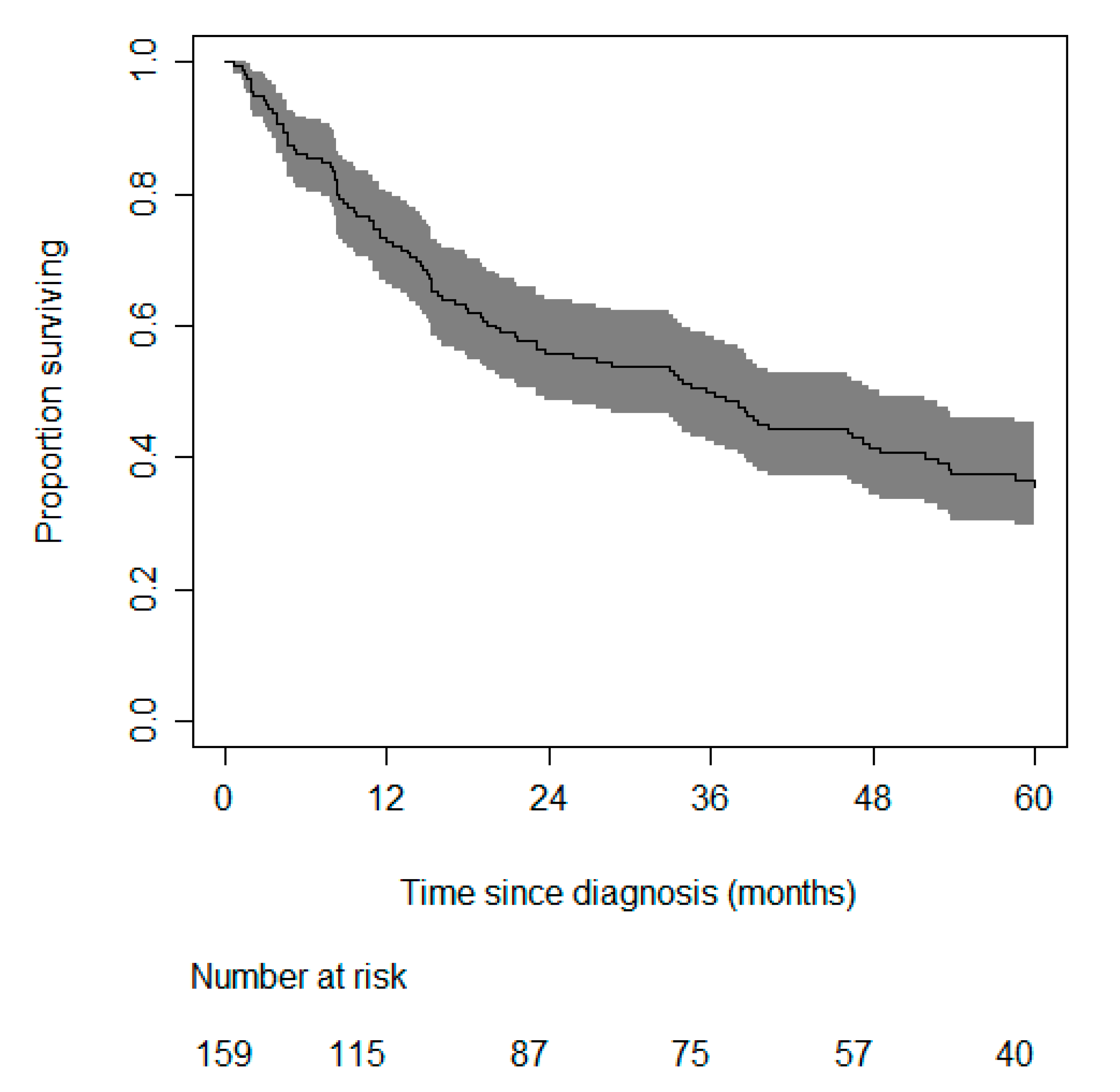
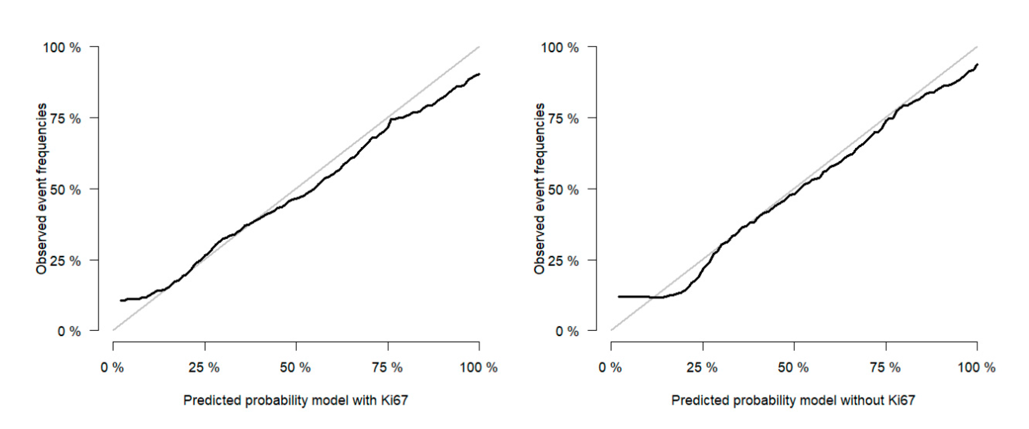
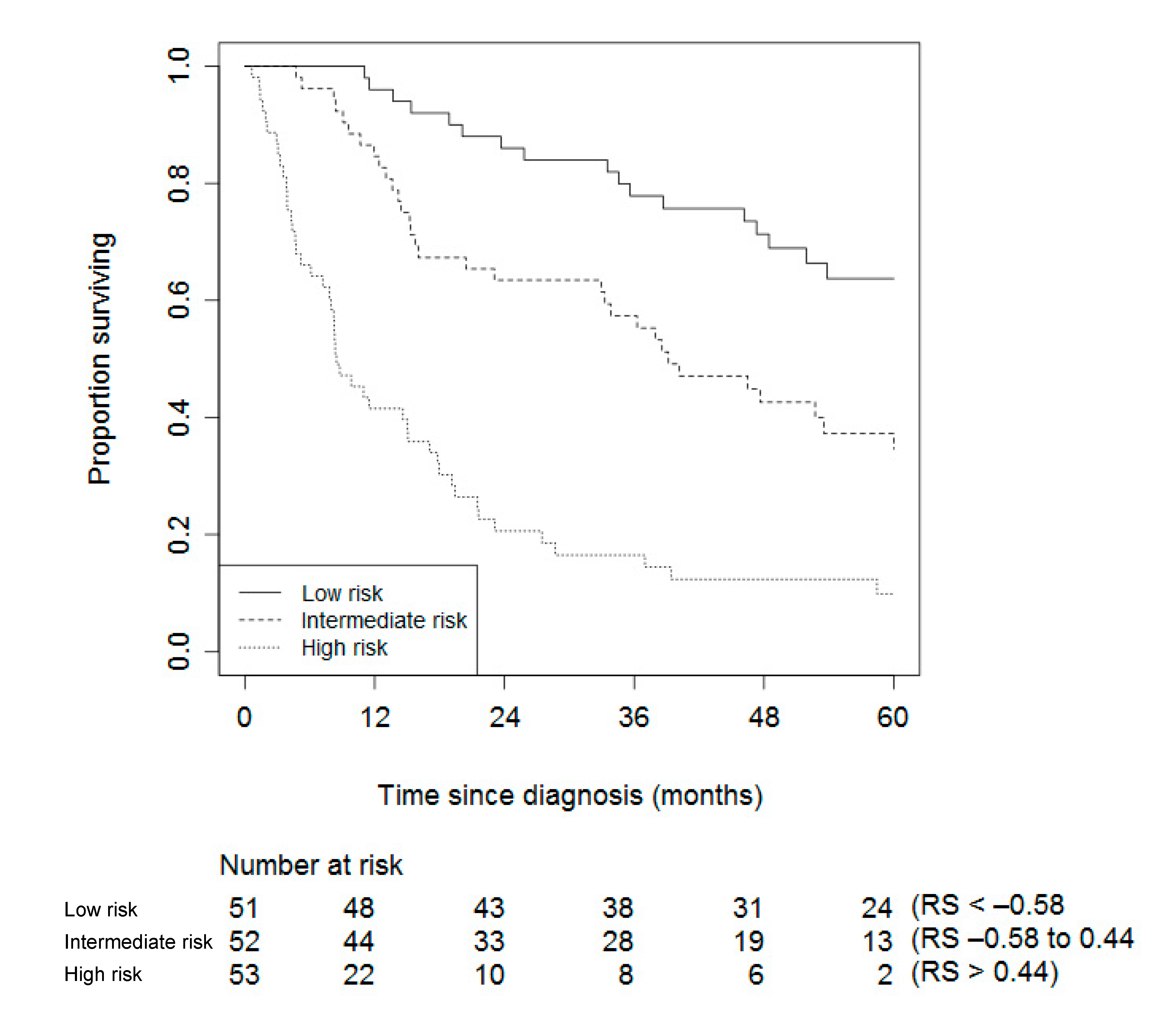
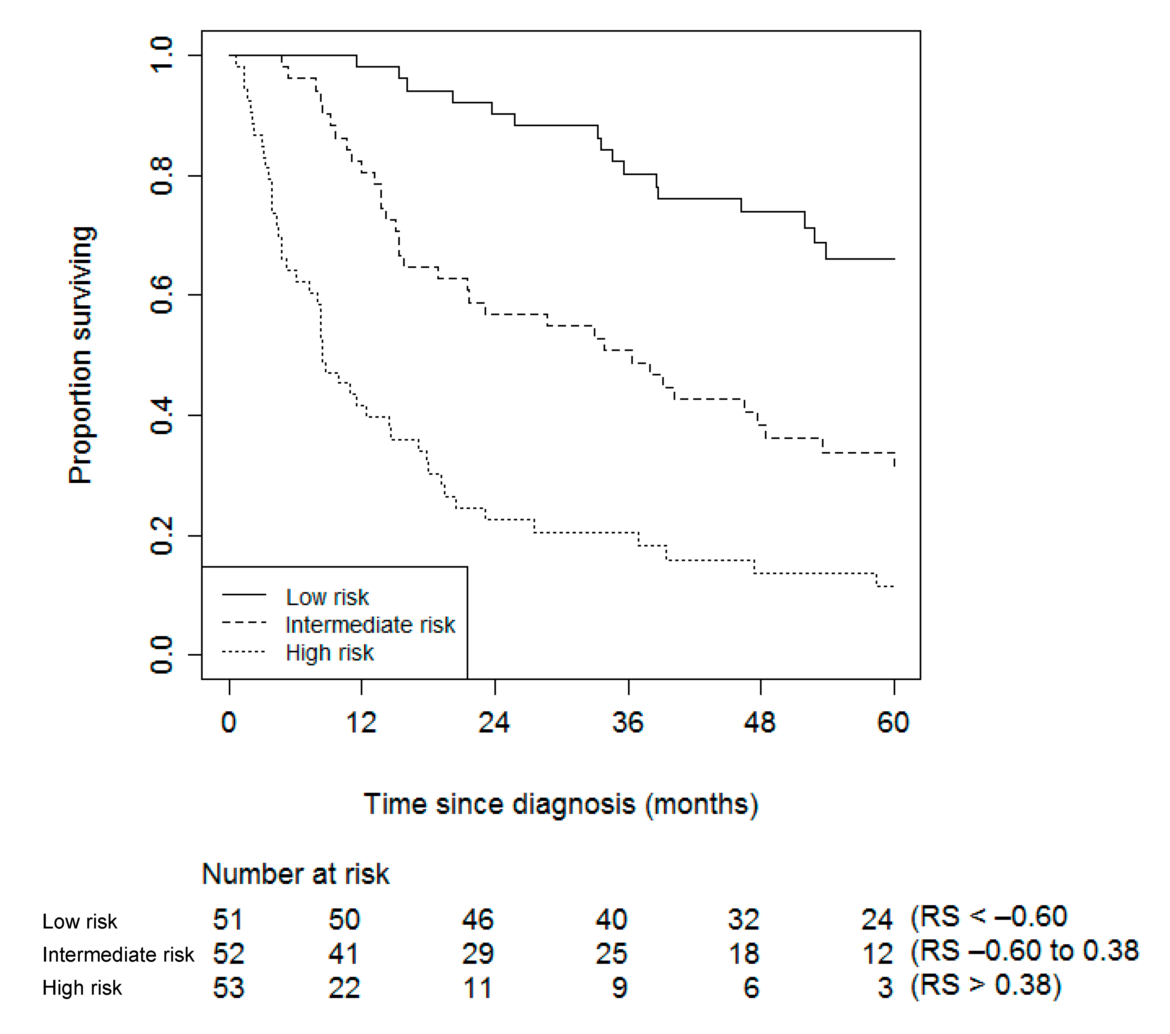
2. Results
Prediction for Future Patients
3. Discussion
4. Materials and Methods
4.1. Potential Predictor Variables
4.2. Immunohistochemistry Revision Ki67
4.3. Model Development
4.4. Internal Validation of the Model
5. Conclusions
Author Contributions
Funding
Acknowledgments
Conflicts of Interest
References
- Kerkhofs, T.M.; Verhoeven, R.H.; Van der Zwan, J.M.; Dieleman, J.; Kerstens, M.N.; Links, T.P.; Van de Poll-Franse, L.V.; Haak, H.R. Adrenocortical Carcinoma: A Population-Based Study on Incidence and Survival in the Netherlands since 1993. Eur. J. Cancer 2013, 49, 2579–2586. [Google Scholar] [CrossRef] [PubMed]
- Hermsen, I.G.; Gelderblom, H.; Kievit, J.; Romijn, J.A.; Haak, H.R. Extremely Long Survival in Six Patients Despite Recurrent and Metastatic Adrenal Carcinoma. Eur. J. Endocrinol. 2008, 158, 911–919. [Google Scholar] [CrossRef] [PubMed] [Green Version]
- Lau, S.K.; Weiss, L.M. The Weiss System for Evaluating Adrenocortical Neoplasms: 25 Years Later. Hum. Pathol. 2009, 40, 757–768. [Google Scholar] [CrossRef] [PubMed]
- Weiss, L.M. Comparative Histologic Study of 43 Metastasizing and Nonmetastasizing Adrenocortical Tumors. Am. J. Surg. Pathol. 1984, 8, 163–169. [Google Scholar] [CrossRef] [PubMed]
- Libe, R.; Borget, I.; Ronchi, C.L.; Zaggia, B.; Kroiss, M.; Kerkhofs, T.; Bertherat, J.; Volante, M.; Quinkler, M.; Chabre, O.; et al. Prognostic Factors in Stage III-IV Adrenocortical Carcinomas (ACC): An European Network for the Study of Adrenal Tumor (ENSAT) Study. Ann. Oncol. 2015, 26, 2119–2125. [Google Scholar] [CrossRef]
- van Slooten, H.; Schaberg, A.; Smeenk, D.; Moolenaar, A.J. Morphologic Characteristics of Benign and Malignant Adrenocortical Tumors. Cancer 1985, 55, 766–773. [Google Scholar] [CrossRef]
- Wachenfeld, C.; Beuschlein, F.; Zwermann, O.; Mora, P.; Fassnacht, M.; Allolio, B.; Reincke, M. Discerning Malignancy in Adrenocortical Tumors: Are Molecular Markers Useful? Eur. J. Endocrinol. 2001, 145, 335–341. [Google Scholar] [CrossRef] [Green Version]
- Berruti, A.; Fassnacht, M.; Baudin, E.; Hammer, G.; Haak, H.; Leboulleux, S.; Skogseid, B.; Allolio, B.; Terzolo, M. Adjuvant Therapy in Patients with Adrenocortical Carcinoma: A Position of an International Panel. J. Clin. Oncol. 2010, 28, e401–e402, (author’s reply e403). [Google Scholar] [CrossRef]
- Papathomas, T.G.; Pucci, E.; Giordano, T.J.; Lu, H.; Duregon, E.; Volante, M.; Papotti, M.; Lloyd, R.V.; Tischler, A.S.; van Nederveen, F.H.; et al. An International Ki67 Reproducibility Study in Adrenal Cortical Carcinoma. Am. J. Surg. Pathol. 2016, 40, 569–576. [Google Scholar] [CrossRef]
- Assie, G.; Letouze, E.; Fassnacht, M.; Jouinot, A.; Luscap, W.; Barreau, O.; Omeiri, H.; Rodriguez, S.; Perlemoine, K.; Rene-Corail, F.; et al. Integrated Genomic Characterization of Adrenocortical Carcinoma. Nat. Genet. 2014, 46, 607–612. [Google Scholar] [CrossRef]
- Ettaieb, M.; Kerkhofs, T.; van Engeland, M.; Haak, H. Past, Present and Future of Epigenetics in Adrenocortical Carcinoma. Cancers 2020, 12, 1218. [Google Scholar] [CrossRef]
- DeLellis, R.A. Pathology and Genetics of Tumours of Endocrine Organs; IARC: Lyon, France, 2004. [Google Scholar]
- Fassnacht, M.; Johanssen, S.; Quinkler, M.; Bucsky, P.; Willenberg, H.S.; Beuschlein, F.; Terzolo, M.; Mueller, H.H.; Hahner, S.; Allolio, B. Limited Prognostic Value of the 2004 International Union Against Cancer Staging Classification for Adrenocortical Carcinoma: Proposal for a Revised TNM Classification. Cancer 2009, 115, 243–250. [Google Scholar] [CrossRef] [PubMed]
- Libé, R. Adrenocortical Carcinoma (ACC): Diagnosis, Prognosis, and Treatment. Front. Cell Dev. Biol. 2015, 3, 45. [Google Scholar] [CrossRef] [PubMed] [Green Version]
- Koch, A.; Joosten, S.C.; Feng, Z.; de Ruijter, T.C.; Draht, M.X.; Melotte, V.; Smits, K.M.; Veeck, J.; Herman, J.G.; Van Neste, L. Analysis of DNA Methylation in Cancer: Location Revisited. Nat. Rev. Clin. Oncol. 2018, 15, 459–466. [Google Scholar] [CrossRef] [PubMed]
- Kong, J.; Zheng, J.; Cai, J.; Wu, S.; Diao, X.; Xie, W.; Chen, X.; Liao, C.; Yu, H.; Fan, X. A Nomogram for Individualized Estimation of Survival among Adult Patients with Adrenocortical Carcinoma After Surgery: A Retrospective Analysis and Multicenter Validation Study. Cancer Commun. 2019, 39, 80. [Google Scholar] [CrossRef] [Green Version]
- Li, Y.; Bian, X.; Ouyang, J.; Wei, S.; He, M.; Luo, Z. Nomograms to Predict overall Survival and Cancer-Specific Survival in Patients with Adrenocortical Carcinoma. Cancer. Manag. Res. 2018, 10, 6949–6959. [Google Scholar] [CrossRef] [Green Version]
- Freire, D.S.; Siqueira, S.A.C.; Zerbini, M.C.N.; Wajchenberg, B.L.; Corrêa-Giannella, M.L.; Lucon, A.M.; Pereira, M.A.A. Development and Internal Validation of an Adrenal Cortical Carcinoma Prognostic Score for Predicting the Risk of Metastasis and Local Recurrence. Clin. Endocrinol. 2013, 79, 468–475. [Google Scholar] [CrossRef]
- Zini, L.; Capitanio, U.; Jeldres, C.; Lughezzani, G.; Sun, M.; Shariat, S.F.; Isbarn, H.; Arjane, P.; Widmer, H.; Perrotte, P. External Validation of a Nomogram Predicting Mortality in Patients with Adrenocortical Carcinoma. BJU Int. 2009, 104, 1661–1667. [Google Scholar] [CrossRef]
- Beuschlein, F.; Weigel, J.; Saeger, W.; Kroiss, M.; Wild, V.; Daffara, F.; Libe, R.; Ardito, A.; Al Ghuzlan, A.; Quinkler, M.; et al. Major Prognostic Role of Ki67 in Localized Adrenocortical Carcinoma After Complete Resection. J. Clin. Endocrinol. Metab. 2015, 100, 841–849. [Google Scholar] [CrossRef]
- Assie, G.; Antoni, G.; Tissier, F.; Caillou, B.; Abiven, G.; Gicquel, C.; Leboulleux, S.; Travagli, J.P.; Dromain, C.; Bertagna, X.; et al. Prognostic Parameters of Metastatic Adrenocortical Carcinoma. J. Clin. Endocrinol. Metab. 2007, 92, 148–154. [Google Scholar] [CrossRef]
- Volante, M.; Buttigliero, C.; Greco, E.; Berruti, A.; Papotti, M. Pathological and Molecular Features of Adrenocortical Carcinoma: An Update. J. Clin. Pathol. 2008, 61, 787–793. [Google Scholar] [CrossRef]
- Ettaieb, M.H.; Duker, J.C.; Feelders, R.A.; Corssmit, E.P.; Menke-van der Houven van Oordt, C.W.; Timmers, H.J.; Kerstens, M.N.; Wilmink, J.W.; Zelissen, P.M.; Havekes, B.; et al. Synchronous vs. Metachronous Metastases in Adrenocortical Carcinoma: An Analysis of the Dutch Adrenal Network. Horm. Cancer. 2016, 7, 336–344. [Google Scholar] [CrossRef] [PubMed]
- Kim, Y.; Margonis, G.A.; Prescott, J.D.; Tran, T.B.; Postlewait, L.M.; Maithel, S.K.; Wang, T.S.; Evans, D.B.; Hatzaras, I.; Shenoy, R. Nomograms to Predict Recurrence-Free and overall Survival After Curative Resection of Adrenocortical Carcinoma. JAMA Surg. 2016, 151, 365–373. [Google Scholar] [CrossRef] [PubMed]
- Lughezzani, G.; Sun, M.; Perrotte, P.; Jeldres, C.; Alasker, A.; Isbarn, H.; Budaus, L.; Shariat, S.F.; Guazzoni, G.; Montorsi, F.; et al. The European Network for the Study of Adrenal Tumors Staging System is Prognostically Superior to the International Union Against Cancer-Staging System: A North American Validation. Eur. J. Cancer 2010, 46, 713–719. [Google Scholar] [CrossRef] [PubMed]
- Bilimoria, K.Y.; Shen, W.T.; Elaraj, D.; Bentrem, D.J.; Winchester, D.J.; Kebebew, E.; Sturgeon, C. Adrenocortical Carcinoma in the United States. Cancer 2008, 113, 3130–3136. [Google Scholar] [CrossRef]
- Do, C.C.; Leboulleux, S.; Borget, I.; Bertherat, J.; De la Fouchardiere, C.; Hecart, A.; Caron, P.; Tabarin, A.; Niccoli, P.; Rohmer, V. First Prognostic Analysis of Stage III Adrenocartical Carcinoma Patients After Complete Resection: A Retrospective French Multicentric Study from the INCA-COMETE Network. In Proceedings of the 13th European Congress of Endocrinology, Rotterdam, The Netherlands, 30 April–4 May 2011. [Google Scholar]
- Dy, B.M.; Strajina, V.; Cayo, A.K.; Richards, M.L.; Farley, D.R.; Grant, C.S.; Harmsen, W.S.; Evans, D.B.; Grubbs, E.G.; Bible, K.C.; et al. Surgical Resection of Synchronously Metastatic Adrenocortical Cancer. Ann. Surg. Oncol. 2015, 22, 146–151. [Google Scholar] [CrossRef] [PubMed]
- Kebebew, E.; Reiff, E.; Duh, Q.Y.; Clark, O.H.; McMillan, A. Extent of Disease at Presentation and Outcome for Adrenocortical Carcinoma: Have we made Progress? World J. Surg. 2006, 30, 872–878. [Google Scholar] [CrossRef]
- Stojadinovic, A.; Brennan, M.F.; Hoos, A.; Omeroglu, A.; Leung, D.H.; Dudas, M.E.; Nissan, A.; Cordon-Cardo, C.; Ghossein, R.A. Adrenocortical Adenoma and Carcinoma: Histopathological and Molecular Comparative Analysis. Mod. Pathol. 2003, 16, 742–751. [Google Scholar] [CrossRef]
- Morimoto, R.; Satoh, F.; Murakami, O.; Suzuki, T.; Abe, T.; Tanemoto, M.; Abe, M.; Uruno, A.; Ishidoya, S.; Arai, Y.; et al. Immunohistochemistry of a Proliferation Marker Ki67/MIB1 in Adrenocortical Carcinomas: Ki67/MIB1 Labeling Index is a Predictor for Recurrence of Adrenocortical Carcinomas. Endocr. J. 2008, 55, 49–55. [Google Scholar] [CrossRef] [Green Version]
- Jouinot, A.; Assié, G.; Libé, R.; Fassnacht, M.; Papathomas, T.; Barreau, O.; De La Villeon, B.; Faillot, S.; Hamzaoui, N.; Neou, M.; et al. DNA methylation is an independent prognostic marker of survival in adrenocortical cancer. J. Clin. Endocrinol. Metab. 2016, 102, 923–932. [Google Scholar] [CrossRef]
- Zheng, S.; Cherniack, A.D.; Dewal, N.; Moffitt, R.A.; Danilova, L.; Murray, B.A.; Lerario, A.M.; Else, T.; Knijnenburg, T.A.; Ciriello, G. Comprehensive Pan-Genomic Characterization of Adrenocortical Carcinoma. Cancer Cell 2016, 29, 723–736. [Google Scholar] [CrossRef] [PubMed] [Green Version]
- Mohan, D.R.; Lerario, A.M.; Else, T.; Mukherjee, B.; Almeida, M.Q.; Vinco, M.; Rege, J.; Mariani, B.M.; Zerbini, M.C.N.; Mendonca, B.B. Targeted Assessment of G0S2 Methylation Identifies a Rapidly Recurrent, Routinely Fatal Molecular Subtype of Adrenocortical Carcinoma. Clin. Cancer Res. 2019, 25, 3276–3288. [Google Scholar] [CrossRef] [Green Version]
- Barreau, O.; Assie, G.; Wilmot-Roussel, H.; Ragazzon, B.; Baudry, C.; Perlemoine, K.; Rene-Corail, F.; Bertagna, X.; Dousset, B.; Hamzaoui, N.; et al. Identification of a CpG Island Methylator Phenotype in Adrenocortical Carcinomas. J. Clin. Endocrinol. Metab. 2013, 98, E174–E184. [Google Scholar] [CrossRef] [PubMed] [Green Version]
- De Reynies, A.; Assie, G.; Rickman, D.S.; Tissier, F.; Groussin, L.; Rene-Corail, F.; Dousset, B.; Bertagna, X.; Clauser, E.; Bertherat, J. Gene Expression Profiling Reveals a New Classification of Adrenocortical Tumors and Identifies Molecular Predictors of Malignancy and Survival. J. Clin. Oncol. 2009, 27, 1108–1115. [Google Scholar] [CrossRef] [PubMed]
- Fragoso, M.C.B.; Almeida, M.Q.; Mazzuco, T.L.; Mariani, B.M.; Brito, L.P.; Gonçalves, T.C.; Alencar, G.A.; Lima, L.d.O.; Faria, A.M.; Bourdeau, I. Combined Expression of BUB1B, DLGAP5, and PINK1 as Predictors of Poor Outcome in Adrenocortical Tumors: Validation in a Brazilian Cohort of Adult and Pediatric Patients. Eur. J. Endocrinol. 2012, 166, 61. [Google Scholar] [CrossRef] [PubMed]
- Ayala-Ramirez, M.; Jasim, S.; Feng, L.; Ejaz, S.; Deniz, F.; Busaidy, N.; Waguespack, S.G.; Naing, A.; Sircar, K.; Wood, C.G.; et al. Adrenocortical Carcinoma: Clinical Outcomes and Prognosis of 330 Patients at a Tertiary Care Center. Eur. J. Endocrinol. 2013, 169, 891–899. [Google Scholar] [CrossRef]
- van Kuijk, S.M.; Viechtbauer, W.; Peeters, L.L.; Smits, L. Bias in Regression Coefficient Estimates when Assumptions for Handling Missing Data are Violated: A Simulation Study. Epidemiol. Biostat. Public Health 2016, 13. [Google Scholar] [CrossRef]
- Steyerberg, E. Clinical Prediction Models: A Practical Approach to Development, Validation, and Updating; Springer Science & Business Media: Berlin, Germany, 2008. [Google Scholar]
- Harrell, F. Regression Modeling Strategies: With Applications to Linear Models, Logistic and Ordinal Regression, and Survival Analysis; Springer: Berlin, Germany, 2015. [Google Scholar]


| Original Data | Imputed Data | |
|---|---|---|
| N (% of total) (Range) | N (% of total) (Range) | |
| Male | 61 (38) | 61 (38) |
| Female | 99 (62) | 99 (62) |
| Age at diagnosis | 55 (19–89) | 55 (19–89) |
| ENSAT Stage | ||
| I | 9 (6) | 9 (6) |
| II | 58 (36) | 58 (36) |
| III | 42 (26) | 42 (26) |
| IV | 51 (32) | 51 (32) |
| mENSAT Stage | ||
| I | 9 (6) | 9 (6) |
| II | 58 (36) | 58 (36) |
| III | 30 (19) | 30 (19) |
| IVa | 36 (22) | 36 (22) |
| IVb | 17 (11) | 17 (11) |
| IVc | 10 (6) | 10 (6) |
| Resection status | ||
| R0 | 74 (46,25) | 74 (46) |
| R1/R2/Rx | 61 (38,1) | 68 (43) |
| No surgery | 18 (11,25) | 18 (11) |
| Missing | 7 (4,4) | - |
| Ki67 | ||
| ≤5 | 27 (17) | 47 (29) |
| 6–10 | 19 (12) | 28 (18) |
| 11–15 | 8 (5) | 16 (10) |
| >15 | 30 (18,5) | 69 (43) |
| Missing | 76 (47,5) | - |
| Capsular and/or vascular invasion | ||
| Yes | 90 (56) | 125 (78) |
| No | 24 (15) | 35 (22) |
| Missing | 46 (29) | - |
| Hypercortisolism | ||
| Yes | 88 (55) | 88 (55) |
| No | 35 (22) | 72 (45) |
| Missing | 37 (23) | - |
| Complaints due to tumor mass | ||
| Yes | 78 | 78 |
| No | 13 | 82 |
| Missing | 69 | - |
| Predictor | Model with Ki67 | Model without Ki67 | ||||
|---|---|---|---|---|---|---|
| Coefficient | HR (95% CI) | Shrunk Coefficient * | Coefficient | HR (95% CI) | Shrunk Coefficient * | |
| Age at diagnosis (year) | 0.02 | 1.02 (1.01, 1.03) | 0.02 | 0.02 | 1.02 (1.00, 1.03) | 0.02 |
| Pathology positive (yes) | 0.52 | 1.68 (0.98, 2.89) | 0.47 | |||
| mENSAT (stage) | 0.61 | 1.84 (1.55, 2.18) | 0.65 | 0.66 | 1.93 (1.64, 2.28) | 0.63 |
| Ki67 (%) | 0.01 | 1.01 (1.00, 1.03) | 0.01 | |||
| Radical resection of tumor (yes) | −0.37 | 0.69 (0.46, 1.04) | −0.34 | −0.46 | 0.63 (0.42, 0.94) | −0.44 |
| Study | N | Outcome | Predictors | C Statistics | ||
|---|---|---|---|---|---|---|
| Model development | Internal validation | External validation | ||||
| Zini et al., 2009 [19] | 205 | - | 207 | CSM and ACM in patients managed with either surgery or no surgery for ACC. | CSM/ACM: age, stage (localized, regional, and distant), and surgical status (surgery or no surgery). | - |
| Kim et al., 2016 [24] | 148 | Bootstrap validation with 200 resamplings. | - | RFS and OS after curative surgical resection of ACC. | RFS: tumor size (<12 or ≥12 cm), nodal status (N0, N1, or Nx), T stage (I/II or III/IV), cortisol-secreting tumor, and capsular invasion. | RFS: 0.74 |
| OS: tumor size (<12 or ≥12 cm), nodal status (N0, N1, or Nx), and resection margin (R0 or R1). | OS: 0.70 | |||||
| Li et al., 2018 [17] | 751 | Bootstrap validation with 200 resamplings. | - | OS and CSS in patients with ACC. | OS: age at diagnosis, year of diagnosis (1973–1987, 1988–2001, 2002–2015), histologic grade (I + II, III + IV, unknown), historic stage (localized, regional, distant, unknown), and chemotherapy (no/unknown, yes). | OS: 0.677 |
| CSS: age at diagnosis, year of diagnosis (1973–1987, 1988–2001, 2002–2015), historic stage (localized, regional, distant, and unknown), and chemotherapy (no/unknown, yes). | CSS: 0.672 | |||||
| Kong et al., 2019 [16] | 404 | 318, and bootstrap validation with 1000 resamplings. | 82 + 82 * = 164 | OS in patients with ACC after surgery. | OS: age, T stage (T1-T4), N stage (N0, N1), M stage (M0, M1). | OS: 0.715 |
© 2020 by the authors. Licensee MDPI, Basel, Switzerland. This article is an open access article distributed under the terms and conditions of the Creative Commons Attribution (CC BY) license (http://creativecommons.org/licenses/by/4.0/).
Share and Cite
Ettaieb, M.H.T.; van Kuijk, S.M.J.; de Wit-Pastoors, A.; Feelders, R.A.; Corssmit, E.P.M.; Eekhoff, E.M.W.; van der Valk, P.; Timmers, H.J.L.M.; Kerstens, M.N.; Klümpen, H.-J.; et al. Development and Internal Validation of a Multivariable Prediction Model for Adrenocortical-Carcinoma-Specific Mortality. Cancers 2020, 12, 2720. https://0-doi-org.brum.beds.ac.uk/10.3390/cancers12092720
Ettaieb MHT, van Kuijk SMJ, de Wit-Pastoors A, Feelders RA, Corssmit EPM, Eekhoff EMW, van der Valk P, Timmers HJLM, Kerstens MN, Klümpen H-J, et al. Development and Internal Validation of a Multivariable Prediction Model for Adrenocortical-Carcinoma-Specific Mortality. Cancers. 2020; 12(9):2720. https://0-doi-org.brum.beds.ac.uk/10.3390/cancers12092720
Chicago/Turabian StyleEttaieb, Madeleine H. T., Sander M. J. van Kuijk, Annelies de Wit-Pastoors, Richard A. Feelders, Eleonora P. M. Corssmit, Elisabeth M. W. Eekhoff, Paul van der Valk, Henri J. L. M. Timmers, Michiel N. Kerstens, Heinz-Josef Klümpen, and et al. 2020. "Development and Internal Validation of a Multivariable Prediction Model for Adrenocortical-Carcinoma-Specific Mortality" Cancers 12, no. 9: 2720. https://0-doi-org.brum.beds.ac.uk/10.3390/cancers12092720