Next Issue
Volume 13, July-1
Previous Issue
Volume 13, June-1


Cancers, Volume 13, Issue 12 (June-2 2021) – 250 articles

Cover Story (view full-size image): 
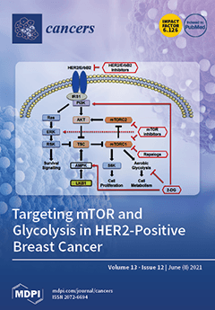
Up to one third of all breast cancers are classified as the aggressive HER2-positive subtype, which is associated with a higher risk of recurrence compared to HER2-negative breast cancers. The HER2 hyperactivity associated with this subtype drives tumor growth by up-regulation of mTOR pathways and metabolic adaptation. Combination therapies that simultaneously target HER2 and mTOR improve clinical outcomes compared with HER2 inhibition alone. Drugs that mimic glucose deprivation in HER2-positive breast cancer patients have not been evaluated; however, preclinical studies have shown that the growth of HER2-positive breast tumors is reduced in response to combining the glycolytic inhibitor 2-DG with mTOR inhibitors. Here, we review the role of mTOR, glycolysis, and signaling mechanisms in HER2-positive breast cancer. View this paper
  • Issues are regarded as officially published after their release is announced to the table of contents alert mailing list.
  • You may sign up for e-mail alerts to receive table of contents of newly released issues.
  • PDF is the official format for papers published in both, html and pdf forms. To view the papers in pdf format, click on the "PDF Full-text" link, and use the free Adobe Reader to open them.
Order results
Result details
Section
Select all
Export citation of selected articles as:
17 pages, 13230 KiB  
Article
T-Cell Lymphoblastic Lymphoma Arising in the Setting of Myeloid/Lymphoid Neoplasms with Eosinophilia: LMO2 Immunohistochemistry as a Potentially Useful Diagnostic Marker
by Magda Zanelli, Giuseppe G. Loscocco, Elena Sabattini, Maurizio Zizzo, Francesca Sanguedolce, Luigi Panico, Daniela Fanni, Raffaella Santi, Cecilia Caprera, Cristiana Rossi, Alessandra Soriano, Alberto Cavazza, Alessandro Giunta, Cristina Mecucci, Alessandro M. Vannucchi, Stefano A. Pileri and Stefano Ascani
Cancers 2021, 13(12), 3102; https://0-doi-org.brum.beds.ac.uk/10.3390/cancers13123102 - 21 Jun 2021
Cited by 7 | Viewed by 2110
Abstract
Background: Rarely, T-lymphoblastic lymphoma (T-LBL) may develop in the setting of myeloid/lymphoid neoplasms with eosinophilia (M/LNs-Eo), a group of diseases with gene fusion resulting in overexpression of an aberrant tyrosine kinase or cytokine receptor. The correct identification of this category has relevant therapeutic [...] Read more.
Background: Rarely, T-lymphoblastic lymphoma (T-LBL) may develop in the setting of myeloid/lymphoid neoplasms with eosinophilia (M/LNs-Eo), a group of diseases with gene fusion resulting in overexpression of an aberrant tyrosine kinase or cytokine receptor. The correct identification of this category has relevant therapeutic implications. LIM domain only 2 (LMO2) is overexpressed in most T-LBL, but not in immature TdT-positive T-cells in the thymus and in indolent T-lymphoblastic proliferations (iT-LBP). Methods and Results: We retrospectively evaluated 11 cases of T-LBL occurring in the context of M/LNs-Eo. Clinical, histological, immunohistochemical and molecular features were collected and LMO2 immunohistochemical staining was performed. The critical re-evaluation of these cases confirmed the diagnosis of T-LBL with morphological, immunohistochemical and molecular features consistent with T-LBL occurring in M/LNs-Eo. Interestingly, LMO2 immunohistochemical analysis was negative in 9/11 cases, whereas only 2 cases revealed a partial LMO2 expression with a moderate and low degree of intensity, respectively. Conclusions: LMO2 may represent a potentially useful marker to identify T-LBL developing in the context of M/LNs-Eo. In this setting, T-LBL shows LMO2 immunohistochemical profile overlapping with cortical thymocytes and iT-LBP, possibly reflecting different molecular patterns involved in the pathogenesis of T-LBL arising in the setting of M/LNs-Eo. Full article
(This article belongs to the Special Issue Pathogenesis and Natural History of Myeloproliferative Neoplasms)
Show Figures

Figure 1

15 pages, 3446 KiB  
Article
Circulating Tumor DNA as a Marker for Treatment Response in Metastatic Melanoma Patients Using Next-Generation Sequencing—A Prospective Feasibility Study
by Marina Berger, Andrea Thueringer, Doritt Franz, Nadia Dandachi, Emina Talakić, Georg Richtig, Erika Richtig, Peter Michael Rohrer, Lukas Koch, Ingrid Hildegard Wolf, Catharina Koch, Barbara Margaretha Rainer, Maximilian Koeller, Martin Pichler, Hanno Gerritsmann, Karl Kashofer and Ariane Aigelsreiter
Cancers 2021, 13(12), 3101; https://0-doi-org.brum.beds.ac.uk/10.3390/cancers13123101 - 21 Jun 2021
Cited by 5 | Viewed by 2345
Abstract
We prospectively performed a longitudinal analysis of circulating tumor DNA (ctDNA) from 149 plasma samples and CT scans in Stage III and IV metastatic melanoma patients (n = 20) treated with targeted agents or immunotherapy using two custom next-generation sequencing (NGS) Ion [...] Read more.
We prospectively performed a longitudinal analysis of circulating tumor DNA (ctDNA) from 149 plasma samples and CT scans in Stage III and IV metastatic melanoma patients (n = 20) treated with targeted agents or immunotherapy using two custom next-generation sequencing (NGS) Ion AmpliSeq™ HD panels including 60 and 81 amplicons in 18 genes, respectively. Concordance of matching cancer-associated mutations in tissue and plasma was 73.3%. Mutant allele frequency (MAF) levels showed a range from 0.04% to 28.7%, well detectable with NGS technologies utilizing single molecule tagging like the AmpliSeq™ HD workflow. Median followup time of the tissue and/or plasma positive cohort (n = 15) was 24.6 months and median progression-free survival (PFS) was 7.8 months. Higher MAF ≥ 1% at baseline was not significantly associated with a risk of progression (Odds Ratio = 0.15; p = 0.155). Although a trend could be seen, MAF levels did not differ significantly over time between patients with and without a PFS event (p = 0.745). Depending on the cell-free DNA amount, NGS achieved a sensitivity down to 0.1% MAF and allowed for parallel analysis of multiple mutations and previously unknown mutations. Our study indicates that NGS gene panels could be useful for monitoring disease burden during therapy with ctDNA in melanoma patients. Full article
Show Figures

Graphical abstract

27 pages, 1664 KiB  
Review
An Updated Understanding of the Role of YAP in Driving Oncogenic Responses
by Giampaolo Morciano, Bianca Vezzani, Sonia Missiroli, Caterina Boncompagni, Paolo Pinton and Carlotta Giorgi
Cancers 2021, 13(12), 3100; https://0-doi-org.brum.beds.ac.uk/10.3390/cancers13123100 - 21 Jun 2021
Cited by 16 | Viewed by 3632
Abstract
Yes-associated protein (YAP) has emerged as a key component in cancer signaling and is considered a potent oncogene. As such, nuclear YAP participates in complex and only partially understood molecular cascades that are responsible for the oncogenic response by regulating multiple processes, including [...] Read more.
Yes-associated protein (YAP) has emerged as a key component in cancer signaling and is considered a potent oncogene. As such, nuclear YAP participates in complex and only partially understood molecular cascades that are responsible for the oncogenic response by regulating multiple processes, including cell transformation, tumor growth, migration, and metastasis, and by acting as an important mediator of immune and cancer cell interactions. YAP is finely regulated at multiple levels, and its localization in cells in terms of cytoplasm–nucleus shuttling (and vice versa) sheds light on interesting novel anticancer treatment opportunities and putative unconventional functions of the protein when retained in the cytosol. This review aims to summarize and present the state of the art knowledge about the role of YAP in cancer signaling, first focusing on how YAP differs from WW domain-containing transcription regulator 1 (WWTR1, also named as TAZ) and which upstream factors regulate it; then, this review focuses on the role of YAP in different cancer stages and in the crosstalk between immune and cancer cells as well as growing translational strategies derived from its inhibitory and synergistic effects with existing chemo-, immuno- and radiotherapies. Full article
(This article belongs to the Special Issue YAP (Yes-Associated Protein) in Cancer)
Show Figures

Figure 1

15 pages, 2383 KiB  
Article
MicroRNAs Targeting HIF-2α, VEGFR1 and/or VEGFR2 as Potential Predictive Biomarkers for VEGFR Tyrosine Kinase and HIF-2α Inhibitors in Metastatic Clear-Cell Renal Cell Carcinoma
by Lisa Kinget, Eduard Roussel, Annelies Verbiest, Maarten Albersen, Cristina Rodríguez-Antona, Osvaldo Graña-Castro, Lucía Inglada-Pérez, Jessica Zucman-Rossi, Gabrielle Couchy, Sylvie Job, Aurélien de Reyniès, Annouschka Laenen, Marcella Baldewijns and Benoit Beuselinck
Cancers 2021, 13(12), 3099; https://0-doi-org.brum.beds.ac.uk/10.3390/cancers13123099 - 21 Jun 2021
Cited by 16 | Viewed by 2786
Abstract
Metastatic clear-cell renal cell carcinoma (m-ccRCC) is characterized by increased hypoxia-induced factor (HIF)-2α and vascular endothelial growth factor receptor (VEGFR)-dependent angiogenesis through loss of function of the von Hippel–Lindau protein. VEGFR tyrosine kinase inhibitors (VEGFR-TKIs) are a cornerstone of m-ccRCC treatment, and new [...] Read more.
Metastatic clear-cell renal cell carcinoma (m-ccRCC) is characterized by increased hypoxia-induced factor (HIF)-2α and vascular endothelial growth factor receptor (VEGFR)-dependent angiogenesis through loss of function of the von Hippel–Lindau protein. VEGFR tyrosine kinase inhibitors (VEGFR-TKIs) are a cornerstone of m-ccRCC treatment, and new treatments targeting HIF-2α are currently under investigation. However, predictive biomarkers for these treatments are lacking. In this retrospective cohort study including 109 patients treated with VEGFR-targeted therapies as first-line treatment, we aimed to study the possible predictive function of microRNAs (miRNAs) targeting HIF-2α, VEGFR1 and VEGFR2. We selected miRNAs inversely correlated with HIF-2α, VEGFR1 and/or VEGFR2 expression and with predicted target sites in the respective genes and subsequently studied their impact on therapeutic outcomes. We identified four miRNAs (miR-34c-5p, miR-221-3p, miR-222-3p and miR-3529-3p) inversely correlated with VEGFR1 and/or VEGFR2 expression and associated with tumor shrinkage and progression-free survival (PFS) upon treatment with VEGFR-TKIs, highlighting the potential predictive value of these miRNAs. Moreover, we identified three miRNAs (miR-185-5p, miR-223-3p and miR-3529-3p) inversely correlated with HIF-2α expression and associated with tumor shrinkage and PFS upon treatment with VEGFR-TKIs. These three miRNAs can have a predictive value not only upon treatment with VEGFR-TKIs but possibly also upon treatment with the upcoming HIF-2α inhibitor belzutifan. Full article
Show Figures

Figure 1

12 pages, 1624 KiB  
Article
Deep Learning with Quantitative Features of Magnetic Resonance Images to Predict Biochemical Recurrence of Radical Prostatectomy: A Multi-Center Study
by Ye Yan, Lizhi Shao, Zhenyu Liu, Wei He, Guanyu Yang, Jiangang Liu, Haizhui Xia, Yuting Zhang, Huiying Chen, Cheng Liu, Min Lu, Lulin Ma, Kai Sun, Xuezhi Zhou, Xiongjun Ye, Lei Wang, Jie Tian and Jian Lu
Cancers 2021, 13(12), 3098; https://0-doi-org.brum.beds.ac.uk/10.3390/cancers13123098 - 21 Jun 2021
Cited by 20 | Viewed by 3629
Abstract
Biochemical recurrence (BCR) occurs in up to 27% of patients after radical prostatectomy (RP) and often compromises oncologic survival. To determine whether imaging signatures on clinical prostate magnetic resonance imaging (MRI) could noninvasively characterize biochemical recurrence and optimize treatment. We retrospectively enrolled 485 [...] Read more.
Biochemical recurrence (BCR) occurs in up to 27% of patients after radical prostatectomy (RP) and often compromises oncologic survival. To determine whether imaging signatures on clinical prostate magnetic resonance imaging (MRI) could noninvasively characterize biochemical recurrence and optimize treatment. We retrospectively enrolled 485 patients underwent RP from 2010 to 2017 in three institutions. Quantitative and interpretable features were extracted from T2 delineated tumors. Deep learning-based survival analysis was then applied to develop the deep-radiomic signature (DRS-BCR). The model’s performance was further evaluated, in comparison with conventional clinical models. The model achieved C-index of 0.802 in both primary and validating cohorts, outweighed the CAPRA-S score (0.677), NCCN model (0.586) and Gleason grade group systems (0.583). With application analysis, DRS-BCR model can significantly reduce false-positive predictions, so that nearly one-third of patients could benefit from the model by avoiding overtreatments. The deep learning-based survival analysis assisted quantitative image features from MRI performed well in prediction for BCR and has significant potential in optimizing systemic neoadjuvant or adjuvant therapies for prostate cancer patients. Full article
(This article belongs to the Collection Artificial Intelligence in Oncology)
Show Figures

Figure 1

19 pages, 4998 KiB  
Article
Gender-Specific Efficacy Revealed by Head-to-Head Comparison of Pasireotide and Octreotide in a Representative In Vivo Model of Nonfunctioning Pituitary Tumors
by Sebastian Gulde, Tobias Wiedemann, Mathias Schillmaier, Isabel Valença, Amelie Lupp, Katja Steiger, Hsi-Yu Yen, Stephen Bäuerle, Johannes Notni, Raul Luque, Herbert Schmid, Stefan Schulz, Donna P. Ankerst, Franz Schilling and Natalia S. Pellegata
Cancers 2021, 13(12), 3097; https://0-doi-org.brum.beds.ac.uk/10.3390/cancers13123097 - 21 Jun 2021
Cited by 8 | Viewed by 2833
Abstract
Invasive nonfunctioning pituitary tumors (NFPTs) are non-resectable neoplasms associated with frequent relapse and significant comorbidities. Current treatments, including somatostatin receptor 2 (SSTR2)-directed somatostatin analogs (SSAs), often fail against NFPTs. Thus, identifying effective therapies is clinically relevant. As NFPTs express SSTR3 at high levels, [...] Read more.
Invasive nonfunctioning pituitary tumors (NFPTs) are non-resectable neoplasms associated with frequent relapse and significant comorbidities. Current treatments, including somatostatin receptor 2 (SSTR2)-directed somatostatin analogs (SSAs), often fail against NFPTs. Thus, identifying effective therapies is clinically relevant. As NFPTs express SSTR3 at high levels, pasireotide, a multireceptor-targeted SSA, might be beneficial. Here we evaluated pasireotide in the only representative model of spontaneous NFPTs (MENX rats) in vivo. Octreotide long-acting release (LAR), pasireotide LAR, or placebo, were administered to age-matched, tumor-bearing MENX rats of both sexes for 28 d or 56 d. Longitudinal high-resolution magnetic resonance imaging monitored tumor growth. While tumors in placebo-treated rats increased in volume over time, PTs in drug-treated rats displayed significant growth suppression, and occasional tumor shrinkage. Pasireotide elicited stronger growth inhibition. Radiological responses correlated with tumors’ proliferation rates. Both SSAs, but especially pasireotide, were more effective in female vs. male rats. Basal Sstr3 expression was significantly higher in the former group. It is noteworthy that female human NFPTs patients also have a trend towards higher SSTR3 expression. Altogether, our studies provide the rationale for testing pasireotide in patients with residual/recurrent NFPTs. If confirmed, the sex-related SSTR3 expression might be used as criteria to stratify NFPTs patients for treatment with pasireotide. Full article
Show Figures

Figure 1

14 pages, 1238 KiB  
Article
Distinct Characteristics and Clinical Outcomes to Predict the Emergence of MET Amplification in Patients with Non-Small Cell Lung Cancer Who Developed Resistance after Treatment with Epidermal Growth Factor Receptor Tyrosine Kinase Inhibitors
by Beung-Chul Ahn, Ji Hyun Lee, Min Hwan Kim, Kyoung-Ho Pyo, Choong-kun Lee, Sun Min Lim, Hye Ryun Kim, Byoung Chul Cho and Min Hee Hong
Cancers 2021, 13(12), 3096; https://0-doi-org.brum.beds.ac.uk/10.3390/cancers13123096 - 21 Jun 2021
Cited by 7 | Viewed by 2809
Abstract
Objectives: Patients with epidermal growth factor receptor (EGFR) mutant non-small cell lung cancer (NSCLC) ultimately acquire resistance to EGFR tyrosine kinase inhibitors (TKIs) during treatment. In 5–22% of these patients, resistance is mediated by aberrant mesenchymal epithelial transition factor (MET [...] Read more.
Objectives: Patients with epidermal growth factor receptor (EGFR) mutant non-small cell lung cancer (NSCLC) ultimately acquire resistance to EGFR tyrosine kinase inhibitors (TKIs) during treatment. In 5–22% of these patients, resistance is mediated by aberrant mesenchymal epithelial transition factor (MET) gene amplification. Here, we evaluated the emergence of MET amplification after EGFR-TKI treatment failure based on clinical parameters. Materials and Methods: We retrospectively analyzed 186 patients with advanced EGFR-mutant NSCLC for MET amplification status by in situ hybridization (ISH) assay after EGFR-TKI failure. We collected information including baseline patient characteristics, metastatic locations and generation, line, and progression-free survival (PFS) of EGFR-TKI used before MET evaluation. Multivariate logistic regression analysis was conducted to evaluate associations between MET amplification status and clinical variables. Results: Regarding baseline EGFR mutations, exon 19 deletion was predominant (57.5%), followed by L858R mutation (37.1%). The proportions of MET ISH assays performed after first/second-generation and third-generation TKI failure were 66.7% and 33.1%, respectively. The median PFS for the most recent EGFR-TKI treatment was shorter in MET amplification-positive patients than in MET amplification-negative patients (median PFS 7.0 vs. 10.4 months, p = 0.004). Multivariate logistic regression demonstrated that a history of smoking, short PFS on the most recent TKI, and less intracranial progression were associated with a high probability of MET amplification (all p < 0.05). Conclusions: Our results demonstrated the distinct clinical characteristics of patients with MET amplification-positive NSCLC after EGFR-TKI therapy. Our clinical prediction can aid physicians in selecting patients eligible for MET amplification screening and therapeutic targeting. Full article
Show Figures

Figure 1

10 pages, 1840 KiB  
Article
Renal Safety of [177Lu]Lu-PSMA-617 Radioligand Therapy in Patients with Compromised Baseline Kidney Function
by Florian Rosar, Niklas Kochems, Mark Bartholomä, Stephan Maus, Tobias Stemler, Johannes Linxweiler, Fadi Khreish and Samer Ezziddin
Cancers 2021, 13(12), 3095; https://0-doi-org.brum.beds.ac.uk/10.3390/cancers13123095 - 21 Jun 2021
Cited by 27 | Viewed by 3120
Abstract
Background: Radioligand therapy (RLT) targeting prostate-specific membrane antigen (PSMA) is an effective antitumor-treatment in metastatic castration-resistant prostate carcinoma (mCRPC). Concerns of potential nephrotoxicity are based on renal tubular PSMA expression and the resulting radiopharmaceutical retention during RLT, but data confirming clinically significant renal [...] Read more.
Background: Radioligand therapy (RLT) targeting prostate-specific membrane antigen (PSMA) is an effective antitumor-treatment in metastatic castration-resistant prostate carcinoma (mCRPC). Concerns of potential nephrotoxicity are based on renal tubular PSMA expression and the resulting radiopharmaceutical retention during RLT, but data confirming clinically significant renal toxicity are still lacking. In this study, patients with significantly impaired baseline kidney function before initiation of therapy were investigated for treatment-associated nephrotoxicity and the potential relationship with administered activities of [177Lu]Lu-PSMA-617. Methods: Twenty-two mCRPC patients with impaired renal function (glomerular filtration rate (GFR) ≤ 60 mL/min) who received more than two cycles of [177Lu]Lu-PSMA-617 RLT (median 5 cycles and median 6-week time interval between consecutive cycles) were analyzed in this study. Patients were treated within a prospective patient registry (REALITY Study, NCT04833517). Cumulative administered activities ranged from 17.1 to 85.6 GBq with a median activity of 6.5 GBq per cycle. Renal function was closely monitored during and after PSMA-RLT. Results: Mean pre-treatment GFR was 45.0 ± 10.7 mL/min. After two (22/22 patients), four (20/22 patients), and six cycles (10/22 patients) of RLT, a significant increase of GFR was noted (each p < 0.05). End-of-treatment GFR (54.1 ± 16.7 mL/min) was significantly higher than baseline GFR (p = 0.016). Only one patient experienced deterioration of renal function (change of CTCAE grade 2 to 3). The remaining patients showed no significant reduction of GFR, including follow-up assessments (6, 9, and 12 months), and even showed improved (10/22 patients) or unchanged (11/22 patients) CTCAE-based renal impairment grades during and after the end of PSMA-RLT. No significant correlation between the change in GFR and per-cycle (p = 0.605) or cumulative (p = 0.132) administered activities were found. Conclusions: As pre-treatment chronic kidney failure did not lead to detectable RLT-induced deterioration of renal function in our study, the nephrotoxic potential of [177Lu]Lu-PSMA-617 RLT may be overestimated and not of clinical priority in the setting of palliative treatment in mCRPC. We suggest not to categorically exclude patients from enrolment to PSMA-RLT due to renal impairment. Full article
(This article belongs to the Special Issue Molecular Imaging and Radio-Nuclide Therapy in Cancers)
Show Figures

Figure 1

13 pages, 1016 KiB  
Review
Transforming Growth Factor-β and Oxidative Stress in Cancer: A Crosstalk in Driving Tumor Transformation
by Valeria Ramundo, Giuliana Giribaldi and Elisabetta Aldieri
Cancers 2021, 13(12), 3093; https://0-doi-org.brum.beds.ac.uk/10.3390/cancers13123093 - 21 Jun 2021
Cited by 21 | Viewed by 3199
Abstract
Cancer metabolism involves different changes at a cellular level, and altered metabolic pathways have been demonstrated to be heavily involved in tumorigenesis and invasiveness. A crucial role for oxidative stress in cancer initiation and progression has been demonstrated; redox imbalance, due to aberrant [...] Read more.
Cancer metabolism involves different changes at a cellular level, and altered metabolic pathways have been demonstrated to be heavily involved in tumorigenesis and invasiveness. A crucial role for oxidative stress in cancer initiation and progression has been demonstrated; redox imbalance, due to aberrant reactive oxygen species (ROS) production or deregulated efficacy of antioxidant systems (superoxide dismutase, catalase, GSH), contributes to tumor initiation and progression of several types of cancer. ROS may modulate cancer cell metabolism by acting as secondary messengers in the signaling pathways (NF-kB, HIF-1α) involved in cellular proliferation and metastasis. It is known that ROS mediate many of the effects of transforming growth factor β (TGF-β), a key cytokine central in tumorigenesis and cancer progression, which in turn can modulate ROS production and the related antioxidant system activity. Thus, ROS synergize with TGF-β in cancer cell metabolism by increasing the redox imbalance in cancer cells and by inducing the epithelial mesenchymal transition (EMT), a crucial event associated with tumor invasiveness and metastases. Taken as a whole, this review is addressed to better understanding this crosstalk between TGF-β and oxidative stress in cancer cell metabolism, in the attempt to improve the pharmacological and therapeutic approach against cancer. Full article
(This article belongs to the Special Issue Effects of TGF-Beta on Cancer Cell Metabolism)
Show Figures

Figure 1

15 pages, 4814 KiB  
Article
B-Helper Neutrophils in Regional Lymph Nodes Correlate with Improved Prognosis in Patients with Head and Neck Cancer
by Ekaterina Pylaeva, Irem Ozel, Anthony Squire, Ilona Spyra, Charlotte Wallner, Magdalena Korek, Georg Korschunow, Maksim Domnich, Elena Siakaeva, Moritz Goetz, Agnes Bankfalvi, Stephan Lang, Benjamin Kansy and Jadwiga Jablonska
Cancers 2021, 13(12), 3092; https://0-doi-org.brum.beds.ac.uk/10.3390/cancers13123092 - 21 Jun 2021
Cited by 6 | Viewed by 2464
Abstract
The role of neutrophils during cancer formation and elimination is diverse. Here, for the first time, we investigate neutrophil helper cells (NBH), their influence on B cell activity in the regional lymph nodes (RLN) of head-and-neck cancer patients and the effect [...] Read more.
The role of neutrophils during cancer formation and elimination is diverse. Here, for the first time, we investigate neutrophil helper cells (NBH), their influence on B cell activity in the regional lymph nodes (RLN) of head-and-neck cancer patients and the effect of this neutrophil/B cell interaction on patient prognosis. Circulating and RLN neutrophils of patients with stage I–IV head-and-neck squamous cell carcinoma were investigated with flow cytometry and qPCR. In addition, neutrophil/B cell co-localization in RLNs was evaluated using immunohistochemistry. B cell proliferation was assessed and correlated with the distance to neutrophils. Patient survival was evaluated. Neutrophils with the helper cell phenotype were identified in the RLN of HNC patients. B cells in close proximity to such NBH showed significantly higher proliferation rates, together with elevated activation-induced cytidine deaminase (AID) expression. Notably, patient survival was significantly higher in individuals with high NBH frequencies in the B follicles of RLNs. Neutrophils in RLN can support T cell-independent activation of the adaptive immune system through B cell stimulation, capturing helper cell phenotype character. The presence of such helper neutrophils in the RLNs of HNC patients positively correlates with patient prognosis. Full article
(This article belongs to the Special Issue New Approaches in the Management of Head and Neck Cancer)
Show Figures

Figure 1

23 pages, 2216 KiB  
Review
Hepatic Arterial Infusion of Chemotherapy for Advanced Hepatobiliary Cancers: State of the Art
by Carmelo Laface, Mariarita Laforgia, Pasquale Molinari, Ippazio Ugenti, Cosmo Damiano Gadaleta, Camillo Porta and Girolamo Ranieri
Cancers 2021, 13(12), 3091; https://0-doi-org.brum.beds.ac.uk/10.3390/cancers13123091 - 21 Jun 2021
Cited by 15 | Viewed by 3844
Abstract
Liver functional failure is one of the leading causes of cancer-related death. Primary liver tumors grow up mainly in the liver, and thus happens for liver metastases deriving from other organs having a lower burden of disease at the primary site. Systemic chemotherapy [...] Read more.
Liver functional failure is one of the leading causes of cancer-related death. Primary liver tumors grow up mainly in the liver, and thus happens for liver metastases deriving from other organs having a lower burden of disease at the primary site. Systemic chemotherapy usually offers a modest benefit in terms of disease control rate, progression-free survival, and overall survival at the cost of a significant percentage of adverse events. Liver malignancies are mostly perfused by the hepatic artery while the normal liver parenchyma by the portal vein network. On these bases, the therapeutic strategy consisting of hepatic arterial infusion (HAI) of chemotherapy takes place. In literature, HAI chemotherapy was applied for the treatment of advanced hepatobiliary cancers with encouraging results. Different chemotherapeutic agents were used such as Oxaliplatin, Cisplatin, Gemcitabine, Floxuridine, 5-Fluorouracil, Epirubicin, individually or in combination. However, the efficacy of this treatment strategy remains controversial. Therefore, this review aims to summarize the current knowledge on this approach from different points of view, such as techniques, drugs pharmacology and pharmacokinetics, and clinical outcomes for advanced hepatobiliary cancers. Full article
Show Figures

Figure 1

16 pages, 603 KiB  
Article
Hematopoietic Stem Cell Transplantation-Associated Neurological Complications and Their Brain MR Imaging Findings in a Pediatric Population
by Hyewon Shin, Mi-Sun Yum, Min-Jee Kim, Jin Kyung Suh, Ho Joon Im, Hyery Kim, Kyung-Nam Koh and Tae-Sung Ko
Cancers 2021, 13(12), 3090; https://0-doi-org.brum.beds.ac.uk/10.3390/cancers13123090 - 21 Jun 2021
Cited by 1 | Viewed by 1754
Abstract
Purpose: To determine the prognostic indicators for hematopoietic stem cell transplantation (HSCT)-associated neurological complications, the clinical characteristics and brain magnetic resonance imaging (MRI) lesions in pediatric HSCT recipients were reviewed. Methods: This retrospective study included 51 patients who had underwent a brain MRI [...] Read more.
Purpose: To determine the prognostic indicators for hematopoietic stem cell transplantation (HSCT)-associated neurological complications, the clinical characteristics and brain magnetic resonance imaging (MRI) lesions in pediatric HSCT recipients were reviewed. Methods: This retrospective study included 51 patients who had underwent a brain MRI due to newly developed neurological symptoms or infection signs during chemotherapy or HSCT. We reviewed the demographics, received treatments, treatment-related morbidities, laboratory findings and brain MRI findings, which were compared between good and poor neurologic outcome groups. Results: Thirty-seven patients (72.5%) fully recovered from the neurologic deficits and fourteen (27.5%) persisted or aggravated. The children with an underlying malignant disease had significantly poorer neurological outcomes (p = 0.015). The neurologic complications associated with infection were more frequent in the poor outcome group (p = 0.038). In the neuroimaging findings, the extent of the white matter lesions was significantly higher in the poor outcome group, as was that of abnormal enhancement, ventriculomegaly, cortical change, deep gray matter abnormalities and cerebellar abnormalities. Conclusion: Most children with neurologic complications and neuroimaging abnormalities during HSCT had recovered. However, children with neurologic complications associated with infectious causes, malignant disease or severe brain MRI abnormalities should be more carefully monitored during HSCT. Full article
Show Figures

Figure 1

33 pages, 8333 KiB  
Article
A Comprehensive Bioinformatics Analysis of Notch Pathways in Bladder Cancer
by Chuan Zhang, Mandy Berndt-Paetz and Jochen Neuhaus
Cancers 2021, 13(12), 3089; https://0-doi-org.brum.beds.ac.uk/10.3390/cancers13123089 - 21 Jun 2021
Cited by 15 | Viewed by 3872
Abstract
Background: A hallmark of Notch signaling is its variable role in tumor biology, ranging from tumor-suppressive to oncogenic effects. Until now, the mechanisms and functions of Notch pathways in bladder cancer (BCa) are still unclear. Methods: We used publicly available data from the [...] Read more.
Background: A hallmark of Notch signaling is its variable role in tumor biology, ranging from tumor-suppressive to oncogenic effects. Until now, the mechanisms and functions of Notch pathways in bladder cancer (BCa) are still unclear. Methods: We used publicly available data from the GTEx and TCGA-BLCA databases to explore the role of the canonical Notch pathways in BCa on the basis of the RNA expression levels of Notch receptors, ligands, and downstream genes. For statistical analyses of cancer and non-cancerous samples, we used R software packages and public databases/webservers. Results: We found differential expression between control and BCa samples for all Notch receptors (NOTCH1, 2, 3, 4), the delta-like Notch ligands (DLL1, 3, 4), and the typical downstream gene hairy and enhancer of split 1 (HES1). NOTCH2/3 and DLL4 can significantly differentiate non-cancerous samples from cancers and were broadly altered in subgroups. High expression levels of NOTCH2/3 receptors correlated with worse overall survival (OS) and shorter disease-free survival (DFS). However, at long-term (>8 years) follow-up, NOTCH2 expression was associated with a better OS and DFS. Furthermore, the cases with the high levels of DLL4 were associated with worse OS but improved DFS. Pathway network analysis revealed that NOTCH2/3 in particular correlated with cell cycle, epithelial–mesenchymal transition (EMT), numbers of lymphocyte subtypes, and modulation of the immune system. Conclusions: NOTCH2/3 and DLL4 are potential drivers of Notch signaling in BCa, indicating that Notch and associated pathways play an essential role in the progression and prognosis of BCa through directly modulating immune cells or through interaction with cell cycle and EMT. Full article
(This article belongs to the Special Issue Bioinformatics, Big Data and Cancer)
Show Figures

Graphical abstract

17 pages, 2028 KiB  
Article
The Challenge of Choosing the Best Classification Method in Radiomic Analyses: Recommendations and Applications to Lung Cancer CT Images
by Federica Corso, Giulia Tini, Giuliana Lo Presti, Noemi Garau, Simone Pietro De Angelis, Federica Bellerba, Lisa Rinaldi, Francesca Botta, Stefania Rizzo, Daniela Origgi, Chiara Paganelli, Marta Cremonesi, Cristiano Rampinelli, Massimo Bellomi, Luca Mazzarella, Pier Giuseppe Pelicci, Sara Gandini and Sara Raimondi
Cancers 2021, 13(12), 3088; https://0-doi-org.brum.beds.ac.uk/10.3390/cancers13123088 - 21 Jun 2021
Cited by 8 | Viewed by 2613
Abstract
Radiomics uses high-dimensional sets of imaging features to predict biological characteristics of tumors and clinical outcomes. The choice of the algorithm used to analyze radiomic features and perform predictions has a high impact on the results, thus the identification of adequate machine learning [...] Read more.
Radiomics uses high-dimensional sets of imaging features to predict biological characteristics of tumors and clinical outcomes. The choice of the algorithm used to analyze radiomic features and perform predictions has a high impact on the results, thus the identification of adequate machine learning methods for radiomic applications is crucial. In this study we aim to identify suitable approaches of analysis for radiomic-based binary predictions, according to sample size, outcome balancing and the features–outcome association strength. Simulated data were obtained reproducing the correlation structure among 168 radiomic features extracted from Computed Tomography images of 270 Non-Small-Cell Lung Cancer (NSCLC) patients and the associated to lymph node status. Performances of six classifiers combined with six feature selection (FS) methods were assessed on the simulated data using AUC (Area Under the Receiver Operating Characteristics Curves), sensitivity, and specificity. For all the FS methods and regardless of the association strength, the tree-based classifiers Random Forest and Extreme Gradient Boosting obtained good performances (AUC ≥ 0.73), showing the best trade-off between sensitivity and specificity. On small samples, performances were generally lower than in large–medium samples and with larger variations. FS methods generally did not improve performances. Thus, in radiomic studies, we suggest evaluating the choice of FS and classifiers, considering specific sample size, balancing, and association strength. Full article
(This article belongs to the Special Issue New Challenges in Cancer Imaging)
Show Figures

Figure 1

18 pages, 2112 KiB  
Review
Review of PP2A Tumor Biology and Antitumor Effects of PP2A Inhibitor LB100 in the Nervous System
by Jean-Paul Bryant, Adam Levy, John Heiss and Yeshavanth Kumar Banasavadi-Siddegowda
Cancers 2021, 13(12), 3087; https://0-doi-org.brum.beds.ac.uk/10.3390/cancers13123087 - 21 Jun 2021
Cited by 6 | Viewed by 5971
Abstract
Protein phosphatase 2A (PP2A) is a ubiquitous serine/threonine phosphatase implicated in a wide variety of regulatory cellular functions. PP2A is abundant in the mammalian nervous system, and dysregulation of its cellular functions is associated with myriad neurodegenerative disorders. Additionally, PP2A has oncologic implications, [...] Read more.
Protein phosphatase 2A (PP2A) is a ubiquitous serine/threonine phosphatase implicated in a wide variety of regulatory cellular functions. PP2A is abundant in the mammalian nervous system, and dysregulation of its cellular functions is associated with myriad neurodegenerative disorders. Additionally, PP2A has oncologic implications, recently garnering attention and emerging as a therapeutic target because of the antitumor effects of a potent PP2A inhibitor, LB100. LB100 abrogation of PP2A is believed to exert its inhibitory effects on tumor progression through cellular chemo- and radiosensitization to adjuvant agents. An updated and unifying review of PP2A biology and inhibition with LB100 as a therapeutic strategy for targeting cancers of the nervous system is needed, as other reviews have mainly covered broader applications of LB100. In this review, we discuss the role of PP2A in normal cells and tumor cells of the nervous system. Furthermore, we summarize current evidence regarding the therapeutic potential of LB100 for treating solid tumors of the nervous system. Full article
Show Figures

Figure 1

15 pages, 3822 KiB  
Article
Electrochemotherapy with Bleomycin Enhances Radiosensitivity of Uveal Melanomas: First In Vitro Results in 3D Cultures of Primary Uveal Melanoma Cell Lines
by Miltiadis Fiorentzis, Ekaterina A. Sokolenko, Nikolaos E. Bechrakis, Saskia Ting, Kurt W. Schmid, Ali Sak, Martin Stuschke, Berthold Seitz and Utta Berchner-Pfannschmidt
Cancers 2021, 13(12), 3086; https://0-doi-org.brum.beds.ac.uk/10.3390/cancers13123086 - 21 Jun 2021
Cited by 10 | Viewed by 2076
Abstract
Electrochemotherapy (ECT) is emerging as a complementary treatment modality for local tumor control in various cancer entities. Irradiation is an established therapeutic option for oncologic patients, which is commonly combined with chemotherapy due to its insufficient targeting ability. The efficiency of radiotherapy for [...] Read more.
Electrochemotherapy (ECT) is emerging as a complementary treatment modality for local tumor control in various cancer entities. Irradiation is an established therapeutic option for oncologic patients, which is commonly combined with chemotherapy due to its insufficient targeting ability. The efficiency of radiotherapy for tumors can be enhanced with different radiosensitizers. ECT can potentiate the radiosensitizing effect of chemotherapeutic agents such as bleomycin. The present study aims to evaluate the radiosensitizing effect of concomitant ECT with bleomycin on 3D tumor spheroids with primary and radioresistant uveal melanoma cell lines (UPMD2, UPMM3, UM92.1, Mel270) and irradiation. The changes in the spheroid growth and the cell viability as well the cytotoxic long-term effect of the combination treatment were evaluated with various combinations of electroporation settings and bleomycin concentrations as well as radiotherapy doses. A broad range of radiosensitivity was documented among the spheroids from different uveal melanoma cell lines. The primary cell lines showed a higher radiosensitivity and required lower irradiation and bleomycin doses. The maximal tumor control with a reduction of cell survival <10% was achieved with a 5 Gy irradiation only in the primary uveal melanoma cell lines and in combination with all tested ECT settings, whereas the same result could be obtained in UM92.1 spheroids only after ECT with 20 Gy irradiation. Based on the spheroid growth and the measurement of the cross-sectional area, the Mel270 spheroids, originating from a previously irradiated recurrent uveal melanoma, required higher doses of bleomycin and ECT settings after irradiation with 5 Gy in order to achieve a significant growth reduction. No significant difference could be demonstrated for the reduction of cell viability in the combination therapy with 20 Gy and 1000 V/cm between 1 and 2.5 µg/mL bleomycin even in Mel270 spheroids, underlying the importance of a drug delivery system to potentiate the radiosensitizing effect of agents in lower doses. ECT should be further assessed for its applicability in clinical settings as a therapeutic radiosensitizing option for radioresistant tumors and a sufficient local tumor control with lower chemotherapy and irradiation doses. Full article
(This article belongs to the Special Issue Nuclear Architecture in Cancer)
Show Figures

Figure 1

20 pages, 2061 KiB  
Review
Ca2+ Signaling and Its Potential Targeting in Pancreatic Ductal Carcinoma
by Louay Bettaieb, Maxime Brulé, Axel Chomy, Mel Diedro, Malory Fruit, Eloise Happernegg, Leila Heni, Anaïs Horochowska, Mahya Housseini, Kekely Klouyovo, Agathe Laratte, Alice Leroy, Paul Lewandowski, Joséphine Louvieaux, Amélie Moitié, Rémi Tellier, Sofia Titah, Dimitri Vanauberg, Flavie Woesteland, Natalia Prevarskaya and V’yacheslav Lehen’kyiadd Show full author list remove Hide full author list
Cancers 2021, 13(12), 3085; https://0-doi-org.brum.beds.ac.uk/10.3390/cancers13123085 - 21 Jun 2021
Cited by 11 | Viewed by 3893
Abstract
Pancreatic cancer (PC) is a major cause of cancer-associated mortality in Western countries (and estimated to be the second cause of cancer deaths by 2030). The main form of PC is pancreatic adenocarcinoma, which is the fourth most common cause of cancer-related death, [...] Read more.
Pancreatic cancer (PC) is a major cause of cancer-associated mortality in Western countries (and estimated to be the second cause of cancer deaths by 2030). The main form of PC is pancreatic adenocarcinoma, which is the fourth most common cause of cancer-related death, and this situation has remained virtually unchanged for several decades. Pancreatic ductal adenocarcinoma (PDAC) is inherently linked to the unique physiology and microenvironment of the exocrine pancreas, such as pH, mechanical stress, and hypoxia. Of them, calcium (Ca2+) signals, being pivotal molecular devices in sensing and integrating signals from the microenvironment, are emerging to be particularly relevant in cancer. Mutations or aberrant expression of key proteins that control Ca2+ levels can cause deregulation of Ca2+-dependent effectors that control signaling pathways determining the cells’ behavior in a way that promotes pathophysiological cancer hallmarks, such as enhanced proliferation, survival and invasion. So far, it is essentially unknown how the cancer-associated Ca2+ signaling is regulated within the characteristic landscape of PDAC. This work provides a complete overview of the Ca2+ signaling and its main players in PDAC. Special consideration is given to the Ca2+ signaling as a potential target in PDAC treatment and its role in drug resistance. Full article
Show Figures

Figure 1

15 pages, 1626 KiB  
Article
Co-Stimulatory versus Cell Death Aspects of Agonistic CD40 Monoclonal Antibody Selicrelumab in Chronic Lymphocytic Leukemia
by Raquel Delgado, Karoline Kielbassa, Johanna ter Burg, Christian Klein, Christine Trumpfheller, Koen de Heer, Arnon P. Kater and Eric Eldering
Cancers 2021, 13(12), 3084; https://0-doi-org.brum.beds.ac.uk/10.3390/cancers13123084 - 21 Jun 2021
Cited by 5 | Viewed by 3476
Abstract
Objectives: Chronic lymphocytic leukemia (CLL) is a common form of leukemia with a heterogeneous clinical course that remains incurable due to the development of therapy resistance. In lymph node proliferation centers, signals from the microenvironment such as CD40 ligation through interaction with follicular [...] Read more.
Objectives: Chronic lymphocytic leukemia (CLL) is a common form of leukemia with a heterogeneous clinical course that remains incurable due to the development of therapy resistance. In lymph node proliferation centers, signals from the microenvironment such as CD40 ligation through interaction with follicular T helper cells shield CLL cells from apoptosis. Previous observations have shown that, despite CD40-induced changes in apoptotic mediators resulting in cell survival, CD40 activation also increases sensitivity to cell death by CD20 mAbs rituximab and obinutuzumab. To further investigate these observations, we here studied the activity of the fully human agonistic CD40 mAb selicrelumab in primary CLL cells in relation to cell activation, induced pro-survival profile, and sensitization for cell death by aCD20 mAbs, in vitro. Methods: CLL cells from peripheral blood were isolated by the Ficoll density method. The expression of activation markers and cytokine production following CD40 stimulation was quantified by flow cytometry and ELISA. The anti-apoptotic profile of CLL induced by stimulation was evaluated by the expression of BCL-2 proteins with Western blot, and resistance to venetoclax with flow cytometry. Cell death induced by the combination of selicrelumab and aCD20 mAbs was quantified by flow cytometry. Results: CLL cells treated with selicrelumab upregulated co-stimulatory molecules such as CD86, TNF-α and death receptor CD95/Fas. In contrast to the CD40 ligand-transfected NIH3T3 cells, induction of resistance to venetoclax by selicrelumab was very moderate. Importantly, selicrelumab stimulation positively sensitized CLL cells to CD20-induced cell death, comparable to CD40 ligand-transfected NIH3T3 cells. Conclusions: Taken together, these novel insights into selicrelumab-stimulatory effects in CLL may be considered for developing new therapeutic strategies, particularly in combination with obinutuzumab. Full article
(This article belongs to the Special Issue Immunotherapy in B-cell Non-Hodgkin Lymphomas)
Show Figures

Figure 1

16 pages, 1267 KiB  
Review
Response Prediction and Evaluation Using PET in Patients with Solid Tumors Treated with Immunotherapy
by Frank J. Borm, Jasper Smit, Daniela E. Oprea-Lager, Maurits Wondergem, John B. A. G. Haanen, Egbert F. Smit and Adrianus J. de Langen
Cancers 2021, 13(12), 3083; https://0-doi-org.brum.beds.ac.uk/10.3390/cancers13123083 - 21 Jun 2021
Cited by 9 | Viewed by 2434
Abstract
In multiple malignancies, checkpoint inhibitor therapy has an established role in the first-line treatment setting. However, only a subset of patients benefit from checkpoint inhibition, and as a result, the field of biomarker research is active. Molecular imaging with the use of positron [...] Read more.
In multiple malignancies, checkpoint inhibitor therapy has an established role in the first-line treatment setting. However, only a subset of patients benefit from checkpoint inhibition, and as a result, the field of biomarker research is active. Molecular imaging with the use of positron emission tomography (PET) is one of the biomarkers that is being studied. PET tracers such as conventional 18F-FDG but also PD-(L)1 directed tracers are being evaluated for their predictive power. Furthermore, the use of artificial intelligence is under evaluation for the purpose of response prediction. Response evaluation during checkpoint inhibitor therapy can be challenging due to the different response patterns that can be observed compared to traditional chemotherapy. The additional information provided by PET can potentially be of value to evaluate a response early after the start of treatment and provide the clinician with important information about the efficacy of immunotherapy. Furthermore, the use of PET to stratify between patients with a complete response and those with a residual disease can potentially guide clinicians to identify patients for which immunotherapy can be discontinued and patients for whom the treatment needs to be escalated. This review provides an overview of the use of positron emission tomography (PET) to predict and evaluate treatment response to immunotherapy. Full article
Show Figures

Figure 1

12 pages, 2353 KiB  
Article
High Serum Elafin Prediction of Poor Prognosis of Locoregional Esophageal Squamous Cell Carcinoma
by I-Chen Wu, Yao-Kuang Wang, Yi-Hsun Chen, Chun-Chieh Wu, Meng-Chieh Wu, Wei-Chung Chen, Wen-Lun Wang, Hung-Shun Lin, Chou-Cheng Chen, Shah-Hwa Chou, Yu-Peng Liu and Ming-Tsang Wu
Cancers 2021, 13(12), 3082; https://0-doi-org.brum.beds.ac.uk/10.3390/cancers13123082 - 21 Jun 2021
Cited by 1 | Viewed by 1865
Abstract
Esophageal squamous cell carcinoma (ESCC) is a highly aggressive tumor known to have locally advanced and metastatic features which cause a dismal prognosis. We sought to determine whether elafin, a non-invasive and secretory small-molecule marker, could be used to predict prognosis in locoregional [...] Read more.
Esophageal squamous cell carcinoma (ESCC) is a highly aggressive tumor known to have locally advanced and metastatic features which cause a dismal prognosis. We sought to determine whether elafin, a non-invasive and secretory small-molecule marker, could be used to predict prognosis in locoregional ESCC patients in human and in vitro studies. In our human study, 119 subjects were identified as having incident and pathologically-proved ESCC with stage I-IIIA tumors from southern Taiwan between 2000 and 2016. We measured their serum elafin levels at baseline and followed them until the date of cancer death or until January 2020, the end of this study. Those with high serum elafin levels were found to have a 1.99-fold risk (95% confidence interval: 1.17–3.38) shorter survival than those who did not. In our in vitro experiments, elevated elafin levels were found to drive ESCC cell proliferation, migration and invasion, while attenuation of elafin level by shRNA abrogated those effects. We concluded that elafin promotes ESCC motility and invasion and leads to a worse clinical prognosis in ESCC patients without distant metastasis. Full article
(This article belongs to the Collection Cancer Biomarkers)
Show Figures

Figure 1

14 pages, 1587 KiB  
Article
Are Borrmann’s Types of Advanced Gastric Cancer Distinct Clinicopathological and Molecular Entities? A Western Study
by Cristina Díaz del Arco, Luis Ortega Medina, Lourdes Estrada Muñoz, Elena Molina Roldán, M. Ángeles Cerón Nieto, Soledad García Gómez de las Heras and M. Jesús Fernández Aceñero
Cancers 2021, 13(12), 3081; https://0-doi-org.brum.beds.ac.uk/10.3390/cancers13123081 - 21 Jun 2021
Cited by 10 | Viewed by 11609
Abstract
Most studies on the clinicopathological impact of Borrmann classification for gastric cancer (GC) have been performed in Asian patients with type IV tumors, and immunohistochemical features of Borrmann types have scarcely been analyzed. We assessed the clinicopathological, molecular features and prognostic value of [...] Read more.
Most studies on the clinicopathological impact of Borrmann classification for gastric cancer (GC) have been performed in Asian patients with type IV tumors, and immunohistochemical features of Borrmann types have scarcely been analyzed. We assessed the clinicopathological, molecular features and prognostic value of Borrmann types in all patients with advanced GC resected in a Western institution (n = 260). We observed a significant relationship between Borrmann types and age, systemic symptoms, tumor size, Laurén subtype, presence of signet-ring cells, infiltrative growth, high grade, tumor necrosis, HERCEPTEST positivity, microsatellite instability (MSI) and molecular subtypes. Polypoid GC showed systemic symptoms, intestinal-type histology, low grade, expansive growth and HERCEPTEST positivity. Fungating GC occurred in symptomatic older patients. It presented intestinal-type histology, infiltrative growth and necrosis. Ulcerated GC showed smaller size, intestinal-type histology, high grade and infiltrative growth. Most polypoid and ulcerated tumors were stable-p53-not overexpressed or microsatellite unstable. Flat lesions were high-grade diffuse tumors with no MSI, and occurred in younger and less symptomatic patients. No association was found between Borrmann classification and prognosis. According to our results, Borrmann types may represent distinct clinicopathological and biological entities. Further research should be conducted to confirm the role of Borrmann classification in the stratification of patients with advanced GC. Full article
(This article belongs to the Special Issue Advanced Gastric Cancer)
Show Figures

Figure 1

5 pages, 552 KiB  
Reply
Reply to Orlhac, F.; Buvat, I. Comment on “Ibrahim et al. The Effects of In-Plane Spatial Resolution on CT-Based Radiomic Features’ Stability with and without ComBat Harmonization. Cancers 2021, 13, 1848”
by Abdalla Ibrahim, Turkey Refaee, Sergey Primakov, Bruno Barufaldi, Raymond J. Acciavatti, Renée W. Y. Granzier, Roland Hustinx, Felix M. Mottaghy, Henry C. Woodruff, Joachim E. Wildberger, Philippe Lambin and Andrew D. A. Maidment
Cancers 2021, 13(12), 3080; https://0-doi-org.brum.beds.ac.uk/10.3390/cancers13123080 - 21 Jun 2021
Cited by 7 | Viewed by 2129
Abstract
We would like to thank Orlhac and Buvat [...] Full article
(This article belongs to the Collection Artificial Intelligence in Oncology)
Show Figures

Figure 1

43 pages, 1734 KiB  
Review
Targeting the Ubiquitin-Proteasome System for Cancer Therapeutics by Small-Molecule Inhibitors
by Gabriel LaPlante and Wei Zhang
Cancers 2021, 13(12), 3079; https://0-doi-org.brum.beds.ac.uk/10.3390/cancers13123079 - 20 Jun 2021
Cited by 38 | Viewed by 8782
Abstract
The ubiquitin-proteasome system (UPS) is a critical regulator of cellular protein levels and activity. It is, therefore, not surprising that its dysregulation is implicated in numerous human diseases, including many types of cancer. Moreover, since cancer cells exhibit increased rates of protein turnover, [...] Read more.
The ubiquitin-proteasome system (UPS) is a critical regulator of cellular protein levels and activity. It is, therefore, not surprising that its dysregulation is implicated in numerous human diseases, including many types of cancer. Moreover, since cancer cells exhibit increased rates of protein turnover, their heightened dependence on the UPS makes it an attractive target for inhibition via targeted therapeutics. Indeed, the clinical application of proteasome inhibitors in treatment of multiple myeloma has been very successful, stimulating the development of small-molecule inhibitors targeting other UPS components. On the other hand, while the discovery of potent and selective chemical compounds can be both challenging and time consuming, the area of targeted protein degradation through utilization of the UPS machinery has seen promising developments in recent years. The repertoire of proteolysis-targeting chimeras (PROTACs), which employ E3 ligases for the degradation of cancer-related proteins via the proteasome, continues to grow. In this review, we will provide a thorough overview of small-molecule UPS inhibitors and highlight advancements in the development of targeted protein degradation strategies for cancer therapeutics. Full article
Show Figures

Figure 1

20 pages, 1771 KiB  
Systematic Review
The Prevalence of TET2 Gene Mutations in Patients with BCR-ABL-Negative Myeloproliferative Neoplasms (MPN): A Systematic Review and Meta-Analysis
by Yuh Cai Chia, Md Asiful Islam, Phil Hider, Peng Yeong Woon, Muhammad Farid Johan, Rosline Hassan and Marini Ramli
Cancers 2021, 13(12), 3078; https://0-doi-org.brum.beds.ac.uk/10.3390/cancers13123078 - 20 Jun 2021
Cited by 10 | Viewed by 3343
Abstract
Multiple recurrent somatic mutations have recently been identified in association with myeloproliferative neoplasms (MPN). This meta-analysis aims to assess the pooled prevalence of TET2 gene mutations among patients with MPN. Six databases (PubMed, Scopus, ScienceDirect, Google Scholar, Web of Science and Embase) were [...] Read more.
Multiple recurrent somatic mutations have recently been identified in association with myeloproliferative neoplasms (MPN). This meta-analysis aims to assess the pooled prevalence of TET2 gene mutations among patients with MPN. Six databases (PubMed, Scopus, ScienceDirect, Google Scholar, Web of Science and Embase) were searched for relevant studies from inception till September 2020, without language restrictions. The eligibility criteria included BCR-ABL-negative MPN adults with TET2 gene mutations. A random-effects model was used to estimate the pooled prevalence with 95% confidence intervals (CIs). Subgroup analyses explored results among different continents and countries, WHO diagnostic criteria, screening methods and types of MF. Quality assessment was undertaken using the Joanna Briggs Institute critical appraisal tool. The study was registered with PROSPERO (CRD42020212223). Thirty-five studies were included (n = 5121, 47.1% female). Overall, the pooled prevalence of TET2 gene mutations in MPN patients was 15.5% (95% CI: 12.1–19.0%, I2 = 94%). Regional differences explained a substantial amount of heterogeneity. The prevalence of TET2 gene mutations among the three subtypes PV, ET and MF were 16.8%, 9.8% and 15.7%, respectively. The quality of the included studies was determined to be moderate–high among 83% of the included studies. Among patients with BCR-ABL-negative MPN, the overall prevalence of TET2 gene mutations was 15.5%. Full article
(This article belongs to the Special Issue Pathogenesis and Natural History of Myeloproliferative Neoplasms)
Show Figures

Figure 1

18 pages, 1692 KiB  
Article
Virtual Biopsy for Diagnosis of Chemotherapy-Associated Liver Injuries and Steatohepatitis: A Combined Radiomic and Clinical Model in Patients with Colorectal Liver Metastases
by Guido Costa, Lara Cavinato, Chiara Masci, Francesco Fiz, Martina Sollini, Letterio Salvatore Politi, Arturo Chiti, Luca Balzarini, Alessio Aghemo, Luca di Tommaso, Francesca Ieva, Guido Torzilli and Luca Viganò
Cancers 2021, 13(12), 3077; https://0-doi-org.brum.beds.ac.uk/10.3390/cancers13123077 - 20 Jun 2021
Cited by 15 | Viewed by 2617
Abstract
Non-invasive diagnosis of chemotherapy-associated liver injuries (CALI) is still an unmet need. The present study aims to elucidate the contribution of radiomics to the diagnosis of sinusoidal dilatation (SinDil), nodular regenerative hyperplasia (NRH), and non-alcoholic steatohepatitis (NASH). Patients undergoing hepatectomy for colorectal metastases [...] Read more.
Non-invasive diagnosis of chemotherapy-associated liver injuries (CALI) is still an unmet need. The present study aims to elucidate the contribution of radiomics to the diagnosis of sinusoidal dilatation (SinDil), nodular regenerative hyperplasia (NRH), and non-alcoholic steatohepatitis (NASH). Patients undergoing hepatectomy for colorectal metastases after chemotherapy (January 2018-February 2020) were retrospectively analyzed. Radiomic features were extracted from a standardized volume of non-tumoral liver parenchyma outlined in the portal phase of preoperative post-chemotherapy computed tomography. Seventy-eight patients were analyzed: 25 had grade 2–3 SinDil, 27 NRH, and 14 NASH. Three radiomic fingerprints independently predicted SinDil: GLRLM_f3 (OR = 12.25), NGLDM_f1 (OR = 7.77), and GLZLM_f2 (OR = 0.53). Combining clinical, laboratory, and radiomic data, the predictive model had accuracy = 82%, sensitivity = 64%, and specificity = 91% (AUC = 0.87 vs. AUC = 0.77 of the model without radiomics). Three radiomic parameters predicted NRH: conventional_HUQ2 (OR = 0.76), GLZLM_f2 (OR = 0.05), and GLZLM_f3 (OR = 7.97). The combined clinical/laboratory/radiomic model had accuracy = 85%, sensitivity = 81%, and specificity = 86% (AUC = 0.91 vs. AUC = 0.85 without radiomics). NASH was predicted by conventional_HUQ2 (OR = 0.79) with accuracy = 91%, sensitivity = 86%, and specificity = 92% (AUC = 0.93 vs. AUC = 0.83 without radiomics). In the validation set, accuracy was 72%, 71%, and 91% for SinDil, NRH, and NASH. Radiomic analysis of liver parenchyma may provide a signature that, in combination with clinical and laboratory data, improves the diagnosis of CALI. Full article
(This article belongs to the Special Issue New Challenges in Cancer Imaging)
Show Figures

Graphical abstract

22 pages, 1507 KiB  
Review
The Role of Extracellular Vesicles in Disease Progression and Detection of Hepatocellular Carcinoma
by Yi-Te Lee, Benjamin V. Tran, Jasmine J. Wang, Icy Y. Liang, Sungyong You, Yazhen Zhu, Vatche G. Agopian, Hsian-Rong Tseng and Ju Dong Yang
Cancers 2021, 13(12), 3076; https://0-doi-org.brum.beds.ac.uk/10.3390/cancers13123076 - 20 Jun 2021
Cited by 28 | Viewed by 4291
Abstract
Hepatocellular carcinoma (HCC) is the most common primary liver malignancy and one of the leading causes of cancer-related death worldwide. Despite the improvements in surveillance and treatment, the prognosis of HCC remains poor. Extracellular vesicles (EVs) are a heterogeneous group of phospholipid bilayer-enclosed [...] Read more.
Hepatocellular carcinoma (HCC) is the most common primary liver malignancy and one of the leading causes of cancer-related death worldwide. Despite the improvements in surveillance and treatment, the prognosis of HCC remains poor. Extracellular vesicles (EVs) are a heterogeneous group of phospholipid bilayer-enclosed particles circulating in the bloodstream and mediating intercellular communication. Emerging studies have shown that EVs play a crucial role in regulating the proliferation, immune escape, and metastasis of HCC. In addition, because EVs are present in the circulation at relatively early stages of disease, they are getting attention as an attractive biomarker for HCC detection. Over the past decade, dedicated efforts have been made to isolate EVs more efficiently and make them useful tools in different clinical settings. In this review article, we provide an overview of the EVs isolation methods and highlight the role of EVs as mediators in the pathogenesis and progression of HCC. Lastly, we summarize the potential applications of EVs in early-stage HCC detection. Full article
Show Figures

Figure 1

26 pages, 2077 KiB  
Review
Engineered EV-Mimetic Nanoparticles as Therapeutic Delivery Vehicles for High-Grade Serous Ovarian Cancer
by Amal A. Al-Dossary, Essam A. Tawfik, Adaugo C. Isichei, Xin Sun, Jiahe Li, Abdullah A. Alshehri, Munther Alomari, Fahad A. Almughem, Ahmad M. Aldossary, Hussein Sabit and Abdulaziz M. Almalik
Cancers 2021, 13(12), 3075; https://0-doi-org.brum.beds.ac.uk/10.3390/cancers13123075 - 20 Jun 2021
Cited by 12 | Viewed by 4799
Abstract
High-grade serous ovarian cancer (HGSOC) is the most lethal gynecological malignancy among women. Several obstacles impede the early diagnosis and effective treatment options for ovarian cancer (OC) patients, which most importantly include the development of platinum-drug-resistant strains. Currently, extensive efforts are being put [...] Read more.
High-grade serous ovarian cancer (HGSOC) is the most lethal gynecological malignancy among women. Several obstacles impede the early diagnosis and effective treatment options for ovarian cancer (OC) patients, which most importantly include the development of platinum-drug-resistant strains. Currently, extensive efforts are being put into the development of strategies capable of effectively circumventing the physical and biological barriers present in the peritoneal cavity of metastatic OC patients, representing a late stage of gastrointestinal and gynecological cancer with an extremely poor prognosis. Naturally occurring extracellular vesicles (EVs) have been shown to play a pivotal role in progression of OC and are now being harnessed as a delivery vehicle for cancer chemotherapeutics. However, there are limitations to their clinical application due to current challenges in their preparation techniques. Intriguingly, there is a recent drive towards the use of engineered synthetic EVs for the delivery of chemotherapeutics and RNA interference therapy (RNAi), as they show the promise of overcoming the obstacles in the treatment of OC patients. This review discusses the therapeutic application of EVs in OC and elucidates the potential use of engineered EV-mimetic nanoparticles as a delivery vehicle for RNAi therapy and other chemotherapeutics, which would potentially improve clinical outcomes of OC patients. Full article
(This article belongs to the Special Issue Ovarian Cancer Biomarkers, Diagnostic and Therapeutic Technologies)
Show Figures

Graphical abstract

14 pages, 2982 KiB  
Article
Immunotherapy Discontinuation in Metastatic Melanoma: Lessons from Real-Life Clinical Experience
by Nethanel Asher, Noa Israeli-Weller, Ronnie Shapira-Frommer, Guy Ben-Betzalel, Jacob Schachter, Tomer Meirson and Gal Markel
Cancers 2021, 13(12), 3074; https://0-doi-org.brum.beds.ac.uk/10.3390/cancers13123074 - 20 Jun 2021
Cited by 19 | Viewed by 2307
Abstract
Background: Immunotherapy has revolutionized outcomes for melanoma patients, by significantly prolonging survival and probably even curing a fraction of metastatic patients. In daily practice, treatment for responding patients is often discontinued due to treatment-limiting toxicity, or electively, following a major tumor response. To [...] Read more.
Background: Immunotherapy has revolutionized outcomes for melanoma patients, by significantly prolonging survival and probably even curing a fraction of metastatic patients. In daily practice, treatment for responding patients is often discontinued due to treatment-limiting toxicity, or electively, following a major tumor response. To date, the criteria for a safe stop and the optimal duration of treatment remain unclear. Patients and methods: This is a real-world single-site cohort of 106 advanced melanoma patients who were treated with immunotherapy and who discontinued treatments in the absence of disease progression. Here, we describe their long-term outcomes, and analyze the differential characteristics between patients who ultimately experienced progression and those who remained in unmaintained durable response. Results: Patients were treated with anti-PD-1 monotherapy (81%) or in combination with ipilimumab (19%) for a median of 15.2 m (range, 0.7–42.3 m). Upon discontinuation, 75.5% had achieved a complete response (CR). After a median follow-up of 20.8 m (range, 6–58) from discontinuation, 32% experienced disease progression. Median time to progression was 8.5 m (range, 1.5–37). Response to re-induction with anti-PD-1 was observed in 47%. On multivariate analysis, achieving a non-CR response, immunotherapy given in advanced line, and shorter treatment duration were significantly associated with lesser progression-free survival. Conclusions: This is one of the few reports on real-world melanoma patients who discontinued immunotherapy while responding to treatment. This study reveals the key factors to bear in mind when considering an elective treatment cessation. Specifically, patients with non-CR as best response and patients treated in an advanced-line setting should be treated for longer periods, and elective discontinuation should not take place prior to 18 m. Full article
(This article belongs to the Section Cancer Immunology and Immunotherapy)
Show Figures

Figure 1

13 pages, 507 KiB  
Review
Targeted Therapies and Immune Checkpoint Inhibitors in Primary CNS Lymphoma
by Hans-Georg Wirsching, Michael Weller, Stefan Balabanov and Patrick Roth
Cancers 2021, 13(12), 3073; https://0-doi-org.brum.beds.ac.uk/10.3390/cancers13123073 - 20 Jun 2021
Cited by 15 | Viewed by 3429
Abstract
This review article outlines the current development of emerging treatment strategies for primary central nervous system lymphoma, a rare brain tumor with, thus far, limited therapeutic options. Small molecule targeted tyrosine kinase inhibitors, immunomodulatory agents, and immune checkpoint inhibitors will be discussed. The [...] Read more.
This review article outlines the current development of emerging treatment strategies for primary central nervous system lymphoma, a rare brain tumor with, thus far, limited therapeutic options. Small molecule targeted tyrosine kinase inhibitors, immunomodulatory agents, and immune checkpoint inhibitors will be discussed. The mechanisms of action, results of completed clinical studies, ongoing clinical trials, and future perspectives are summarized. Among the most promising clinical developments in the field of CNS lymphomas is ibrutinib, an inhibitor of Bruton’s tyrosine kinase, which relays activation of nuclear factor kappa B upon integration of constitutive B cell receptor and Toll-like receptor signals. Down-stream of nuclear factor kappa B, the thalidomide analogs lenalidomide and pomalidomide exert immunomodulatory functions and are currently explored against CNS lymphomas. Finally, immune checkpoint inhibitors, such as drugs targeting the PD-1 pathway, may become novel therapeutic options to unleash anti-tumor immunity in patients with primary CNS lymphoma. Full article
(This article belongs to the Special Issue Primary CNS Lymphomas: Diagnosis and Treatment)
Show Figures

Figure 1

20 pages, 4464 KiB  
Article
Balancing the CD38 Expression on Effector and Target Cells in Daratumumab-Mediated NK Cell ADCC against Multiple Myeloma
by Margaux Lejeune, Elodie Duray, Matthias Peipp, Béatrice Clémenceau, Frédéric Baron, Yves Beguin and Jo Caers
Cancers 2021, 13(12), 3072; https://0-doi-org.brum.beds.ac.uk/10.3390/cancers13123072 - 20 Jun 2021
Cited by 6 | Viewed by 3454
Abstract
Multiple myeloma (MM) is an incurable cancer characterized by the proliferation and accumulation of monoclonal plasma cells in the bone marrow. The monoclonal anti-CD38 daratumumab has taken a central place in the different treatment regimens for newly diagnosed and relapsed, refractory myeloma. In [...] Read more.
Multiple myeloma (MM) is an incurable cancer characterized by the proliferation and accumulation of monoclonal plasma cells in the bone marrow. The monoclonal anti-CD38 daratumumab has taken a central place in the different treatment regimens for newly diagnosed and relapsed, refractory myeloma. In this study, we correlated the NK cell-mediated antibody-dependent cellular cytotoxicity (ADCC) and potential fratricide induced by daratumumab with CD38-expression levels on both effector and target cells. We show that CD38 expression can be modulated by adding all-trans retinoic acid (ATRA) or interferon-α to MM cells to further fine-tune these effects. In addition, we observed that ADCC becomes inefficient when fratricide occurs and both ADCC and fratricide depend on the balance between CD38 expression on effector and target cells. However, the addition of adjuvants (retinoic acid or interferon-α) to myeloma cells or the inhibition of fratricide using a CD38-blocking nanobody on NK-cells can reverse this balance towards ADCC and thus promote lysis of target cells by ADCC. ATRA and interferon-α increased the CD38 expression at the surface of MM cells about three-fold and two-fold, respectively. This increase was of interest for MM cells with low CD38 expression, that became susceptible to daratumumab-mediated ADCC after preincubation. A CD38-blocking nanobody prevented the binding of daratumumab to these NK-cells and blunted the fratricidal effect on effector NK cells. In conclusion, our study highlights the importance of a balanced CD38 expression on target and effector cells and attempts to alter this balance will affect the susceptibility of MM cells towards daratumumab-mediated ADCC. Full article
(This article belongs to the Special Issue Emerging Treatment Strategies for Multiple Myeloma)
Show Figures

Figure 1

Previous Issue
Next Issue
Back to TopTop