Article

Blood-Based Multi-Cancer Detection Using a Novel Variant Calling Assay (DEEPGENTM): Early Clinical Results

by
Frederic Ris
1,*,
Minia Hellan
2,
Jonathan Douissard
1,
Jorge J. Nieva
3,
Frederic Triponez
4,
Yanghee Woo
5,
David Geller
6,
Nicolas C. Buchs
1,
Leo Buehler
7,
Stefan Moenig
1,
Christophe E. Iselin
8,
Wolfram Karenovics
4,
Patrick Petignat
9,
Giang Thanh Lam
9,
Manuela Undurraga Malinervo
9,
Rebecca Tuttle
2,
James Ouellette
2,
Debashish Bose
10,
Nael Ismail
1 and
Christian Toso
1
1
Division of Visceral Surgery, Department of Surgery, University Hospital Geneva and Medical School, 1211 Geneva, Switzerland
2
Surgical Oncology, Wright State University, Dayton, OH 45435, USA
3
Norris Cancer Center, Keck School of Medicine of USC, University of Southern California, Los Angeles, CA 90033, USA
4
Division of Thoracic Surgery, Department of Surgery, University Hospital Geneva and Medical School, 1211 Geneva, Switzerland
5
Division of Surgical Oncology, Department of Surgery and Cancer Immunotherapeutics Program, City of Hope, Duarte, CA 91010, USA
6
Department of Surgery, University of Pittsburgh, Pittsburg, PA 15260, USA
7
Division of Visceral Surgery, Department of Surgery, University Hospital Fribourg, 1700 Fribourg, Switzerland
8
Division of Urology, Department of Surgery, University Hospital Geneva and Medical School, 1211 Geneva, Switzerland
9
Divison of Gynecology, University Hospital Geneva and Medical School, 1211 Geneva, Switzerland
10
The Center for Hepatobiliary Disease, Mercy, Baltimore, MD 21202, USA
*
Author to whom correspondence should be addressed.
Submission received: 26 June 2021 / Revised: 2 August 2021 / Accepted: 12 August 2021 / Published: 15 August 2021

Abstract

:

Simple Summary

Cancer remains a worldwide concern with significant burdens on the population and healthcare systems. Studies have shown that early detection is paramount in positive patient outcomes, although the standard of care screening is limited to a few cancers. When a tumor cell dies, it releases DNA into the bloodstream. This cell-free DNA can be extracted, and specific mutations identified. Technologies based on this principle are gaining popularity amongst physicians for cancer patients to inform personalized treatment. Additionally, if platforms are sensitive enough, blood-based multi-cancer screening can be performed. DEEPGENTM is a next-generation sequencing platform that has been optimized for early cancer detection. This study is a preliminary analysis of cancer detection rates across seven cancers using the DEEPGENTM platform.

Abstract

This is an early clinical analysis of the DEEPGENTM platform for cancer detection. Newly diagnosed cancer patients and individuals with no known malignancy were included in a prospective open-label case-controlled study (NCT03517332). Plasma cfDNA that was extracted from peripheral blood was sequenced and data were processed using machine-learning algorithms to derive cancer prediction scores. A total of 260 cancer patients and 415 controls were included in the study. Overall, sensitivity for all cancers was 57% (95% CI: 52, 64) at 95% specificity, and 43% (95% CI: 37, 49) at 99% specificity. With 51% sensitivity and 95% specificity for all stage 1 cancers, the stage-specific sensitivities trended to improve with higher stages. Early results from this preliminary clinical, prospective evaluation of the DEEPGENTM liquid biopsy platform suggests the platform offers a clinically relevant ability to differentiate individuals with and without known cancer, even at early stages of cancer.

Graphical Abstract

1. Introduction

Cancer is a major cause of mortality worldwide and a significant burden on patients as well as the healthcare system [1]. Major challenges include the absence of early diagnosis at a large scale, especially for average risk individuals because the standard of care screening only exists for a limited number of cancers for this population [2]. Thus, the current diagnostic pipeline is mostly empirical and primarily driven by an examination of the patient’s symptoms [2,3]. Due to delays in detection, most of the patients diagnosed with cancer miss the therapeutic window of opportunity to reset the disease’s trajectory [4,5]. Therefore, early detection using rapid, cost-effective, high-precision, and minimally invasive tools holds the promise to materially decrease morbidity and mortality in cancer [6,7].
Advances in sequencing technologies and associated genomic research have proven crucial to better understand the mutational patterns that accompany cancer [8,9]. Extensive molecular profiling of tumors has successfully led to the identification of key somatic mutations that drive transformation, evolution, and the continually adaptive behavior of cancer. Considering that dying cancer cells shed cell-free DNA (cfDNA) into the bloodstream, the ability to accurately detect these mutations broadly in the blood, especially at ultra-low variant allele frequencies (“VAF”, i.e., far below <1%), may provide an attractive alternative to standard tissue biopsy [10,11,12,13]. Indeed, blood-based biopsies—if designed appropriately for high sensitivity—can detect low prevalence mutations with a reduced signal-to-noise ratio (<0.1%), making them a potential high impact diagnostic tool for improving the precision of accompanying cancer treatments [10,11,13].
Nevertheless, blood-based biopsy, as with the current gold standard tissue biopsy, faces several challenges that must be overcome. One of the major drawbacks of blood-based biopsy is the low allele frequency of clinically significant variants, which often reside in the range of technical background noise [13,14]. This is especially true in early-stage cancer patients where the concentration of tumor DNA is low [13,14,15,16]. Moreover, the narrow base pair range of the fragments (140–200 bp), short life of DNA in the blood (1.5–2 h), tumor type, proliferation rate, and therapy create challenges for the ultimate utility of liquid biopsies for cancer screening [17,18,19]. Hence, extensive optimization of pre-analytical (e.g., extraction and storage) and analytical (e.g., bioinformatics and AI) processes is required to optimize precision. Until now, most commercially available platforms and those under pre-clinical validation have sub-optimal clinical performance [11,20,21,22]. Thus, there is a need for novel platforms that considerably improve detection accuracy and variant calling precision at ultra-low VAF to facilitate the detection of cancer, particularly in asymptomatic individuals.
DEEPGENTM (Quantgene Inc., Santa Monica, CA, USA) is a newly developed full stack sequencing technology awarded with a Clinical Laboratory Improvement Amendments (CLIA) certificate. The platform addresses the common challenges of previous generation liquid biopsy platforms by combining yield-optimized cfDNA storage and extraction protocols, a broad panel assay, advanced error reduction chemistry, and ultra-deep next-generation sequencing (NGS) with a customized bioinformatics pipeline, highly scalable cloud software and innovative AI techniques to identify low-frequency variants accurately and consistently in the blood. Initial validation studies demonstrated that DEEPGENTM has high detection capabilities, capturing over 3000 mutations at VAFs down to >0.09%, and thus out-performs other publicly available platforms on a technical level [23]. Additionally, the mutations undergo a proprietary machine learning protocol that considers mutations and their VAFs to derive a prediction if cancer might be the underlying cause of the observation.
The objective of this pilot study is to evaluate the early clinical capabilities of the DEEPGENTM assay to discriminate individuals with yet-untreated cancers from individuals without a known cancer diagnosis.

2. Materials and Methods

2.1. Study Design and Participants

This is a preliminary analysis of prospective multi-center case–control, open-label pilot study (NCT03517332). Adult patients over 18 years of age with newly diagnosed and to that point untreated cancer were included in the cancer group. Patients with no known or previous history of malignancy were included in the control group. Written consent was secured before study enrollment. A representative set of patients with cancers of breast, lung, pancreas, liver, colon–rectum, prostate, or bladder ranging from stage I to IV and a control cohort were retrospectively selected to undergo processing and analysis.

2.2. Sample Collection

A measure of 10–20 mL of venous blood was taken from the study participants using DNA stabilizing blood tubes (Streck, La Vista, NE, USA). Blood tubes were stored at room temperature and shipped to the location of DNA extraction.

2.3. DNA Preparation

Cell-free (cf) DNA was extracted and purified using Qiasymphony (Qiagen, Hilden, Germany) using a customized protocol that was designed to maximize extraction yield. Early samples were extracted manually. Extractions were transitioned to an automated process for most of the samples. DNA concentration was then determined using quantitative RT-PCR. Samples were stored at −80 °C until assigned for sequencing.

2.4. Sequencing

NGS libraries were prepared from cfDNA according to the manufacturer’s instructions (protocol based on QIAseq Targeted DNA Panel Handbook, R2; May 2017, Qiagen, Hilden, Germany) except for the DNA fragmentation. End-repair and Poly(A) tailing were followed by Illumina NGS adapter (Illumina, San Diego, CA, USA) ligation to cfDNA containing a sample index and a unique molecular identifier sequence (UMI). After UMI assignment, target enrichment of ligated cfDNA was performed by PCR using target specific DEEPGENTM primers. Library concentrations were determined with KAPA Library Quantification Kits for Illumina platforms (Roche Holding AG, Basel, Switzerland). Libraries were prepared using NovaSeq Reagent Kits (Illumina, San Diego, CA, USA) and sequenced with a 300-cycle S4 kit on a NovaSeq 6000 (Illumina, San Diego, CA, USA) with a mean raw sequencing depth of ~200,000×. 3062 genomic variants were targeted. All steps were carried out according to the manufacturer’s instructions.

2.5. Data Processing

FASTQ files were processed with the DEEPGENTM bioinformatics pipeline. Sequencing data from both paired-end reads were deployed, whereas information from the 2nd read was used to complement the sequence of read one. Each read was screened for the specific primer sequence and consensus sequences of the fragments were identified by consolidating reads based on primer and UMI information. Each unique consensus sequence was aligned to its reference via an optimized Smith–Waterman algorithm. Based on a whitelist with defined targets, single nucleotide polymorphisms (SNPs), multi nucleotide polymorphisms (MNPs), and short insertions/deletions (INDELS) (<50 base pairs) were recorded. Identical genomic alterations were summarized and the count coverage and resulting frequency (count/coverage × 100) for each unique variant were logged into a mutation table alongside their location and mutation information.

2.6. Machine Learning

The input data for the machine learning models consisted of mutation frequencies for the genomic targets computed by Quantgene’s bioinformatics pipeline. Samples with a mean sequencing coverage <200 across select targets were removed from the machine learning analysis. Random forest classifiers were trained via leave-one-out cross-validation to predict whether a sample belonged to the control or cancer cohort. For every sample, a random forest output of a predictive score with a continuous value between 0 and 1 indicated the likelihood of belonging to the cancer group in ascending order. A classifier was trained on all cancer and control samples as a full model. Cancer-specific classifiers only utilizing samples from a single organ of origin and all controls were trained for lung, pancreas, liver, colorectal, and bladder. The classifiers for prostate and breast cancer were limited to only male and female samples, respectively, for both control and cancer groups. Samples with manual and automated DNA extraction were analyzed separately.

2.7. Classification of Cancer Versus Non-Cancer (Predictive Score)

The cut-off values between a positive (cancer) from a negative (non-cancer) predictive score were derived for both a 95% and 99% specificity. Sensitivities for both specificity values were calculated for the full cancer model and organ-specific models. Cancer stage-specific sensitivities were calculated at 95% specificity.

2.8. Data Analysis

Machine learning models were trained and evaluated using the scikit-learn package (v0.24.0) in Python (v3.8.7, Python Software Foundation, Troisdorf, Germany) [24,25]. Results of the different cancer models adjusted for a 95% specificity were subjected to Kruskal–Wallis tests to screen for significant differences in sensitivity between cancer types and cancer stages, respectively. In case of a significant p-value (p < 0.05), a Wilcoxon rank sum test was performed as a post hoc procedure. Test were performed with R, version 4.0.2 (https://www.r-project.org, accessed on 25 June 2021).

2.9. False Positive Controls

If available, medical records of false-positive controls were assessed for relevant diseases that were present at the time of the liquid biopsy as well as during the follow-up until the date of review.

3. Results

3.1. Demographics

A total of 719 samples were sequenced, and of these, 44 were excluded from machine learning analysis due to a mean sequencing coverage <200 across select targets. The remaining 675 samples consisted of 25 bladder, 29 prostate, 30 lung, 27 liver, 40 pancreatic, 66 colorectal, 43 breast cancers, and 415 controls without known cancer diagnosis (controls). A total of 70 individuals with stage I (27%), 55 with stage II (21%), 73 with stage III (29%) and 27 cases with stage IV (10%) were included. The cancer stage remained clinically undetermined for 35 cases (13%). The mean age was 65.4 ± 10.9 years in cancers and 54.9 ± 15.5 years in controls (p < 0.001). There were 136 men and 124 women in the cancer group, and 117 men and 298 women in the control group (p < 0.001). A family history of cancer was found in 38% of the cancer patients and 44% of the control patients. A complete description of patients’ demographics can be found in Table 1.

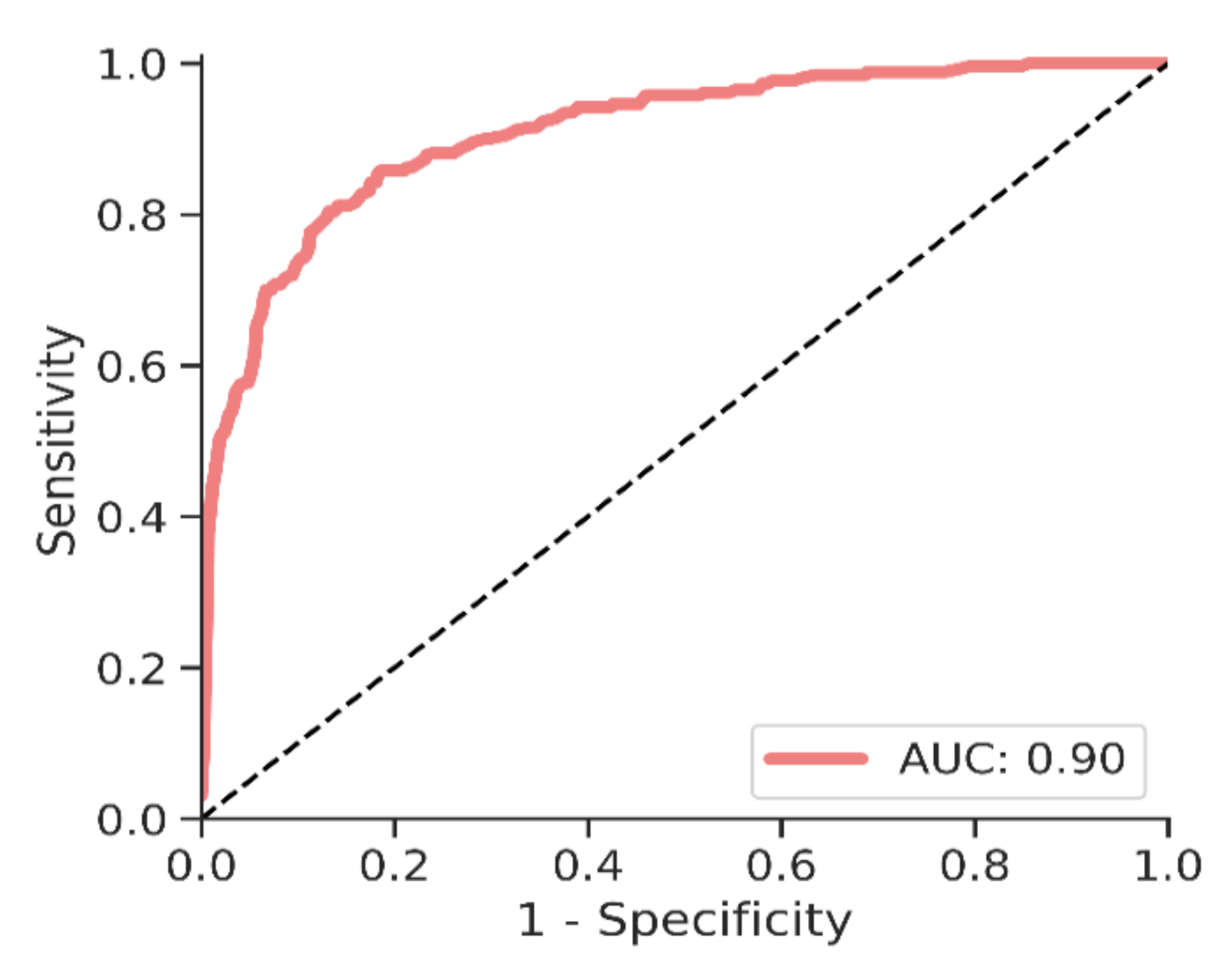
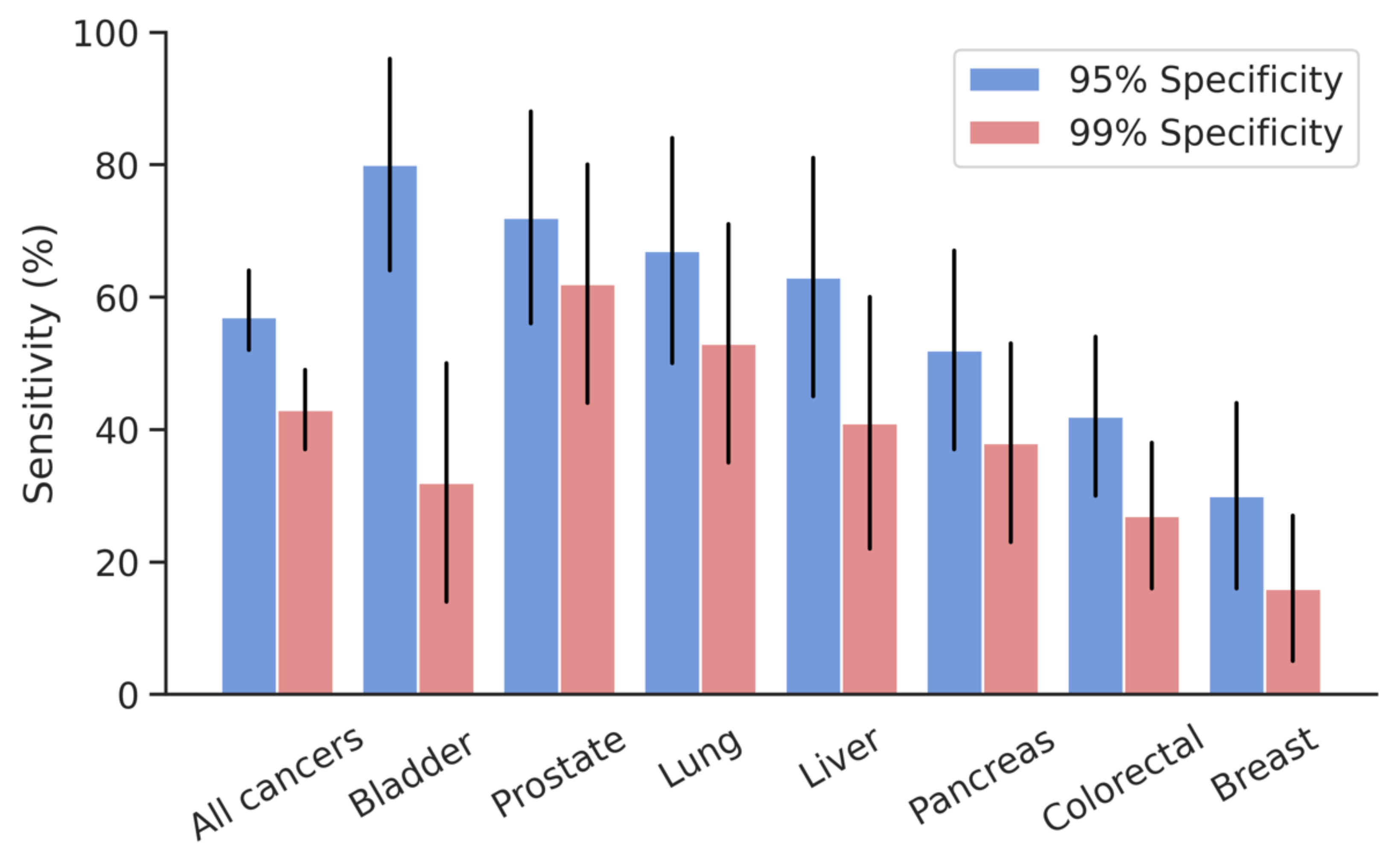
3.2. Cancer Identification

The overall detection sensitivity in the machine learning model for all cancers was 57% (CI: 52, 64) at 95% specificity, and 43% (CI: 37, 49) at 99% specificity. The area under the curve (AUC) for this model was 0.90. A receiver operating characteristics (ROC) curve for the “all cancers” model is shown in Figure 1.
At 95% specificity, the individual organ models ranged in sensitivity from 30% for breast cancer to 80% for bladder cancer, with sensitivities higher than 50% for prostate (72% (CI: 56, 88) sensitivity), lung (67% (CI: 50, 84) sensitivity), liver (63% (CI: 45, 81) sensitivity), and pancreas (52% (CI: 37, 67) sensitivity). At 99% specificity, the individual organ models ranged in sensitivity from 16% for breast cancer to 62% for prostate cancer (p = 0.001). The bladder differed from the breast model (p = 0.0018) and the colorectal model (p = 0.0102), the breast model from the lung (p = 0.0120) and prostate (p = 0.0052) models, and the prostate model from the colorectal model (p = 0.0268). Detection performance for the “all cancer” and individual organ models can be found in Table 2 and Figure 2.

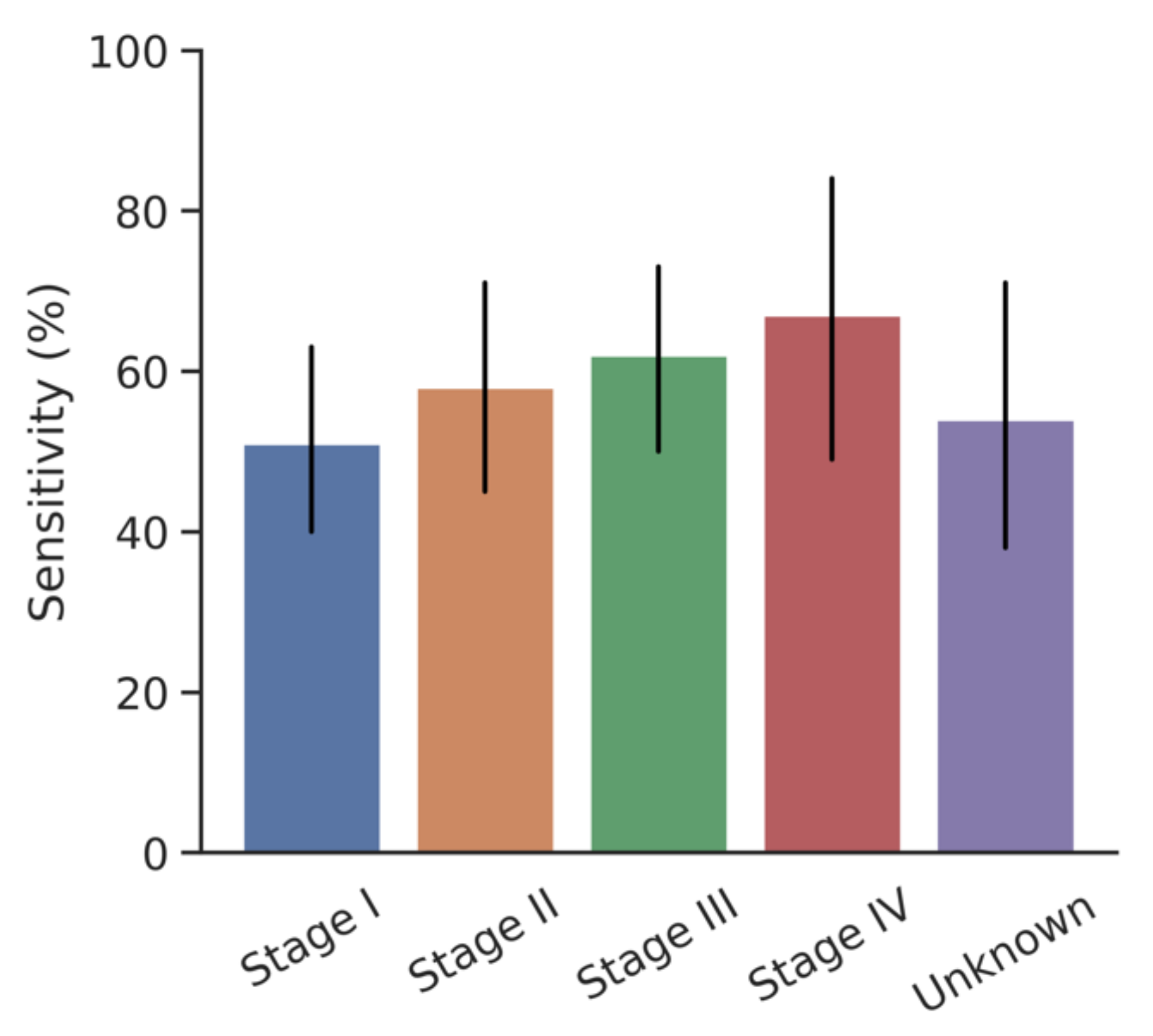
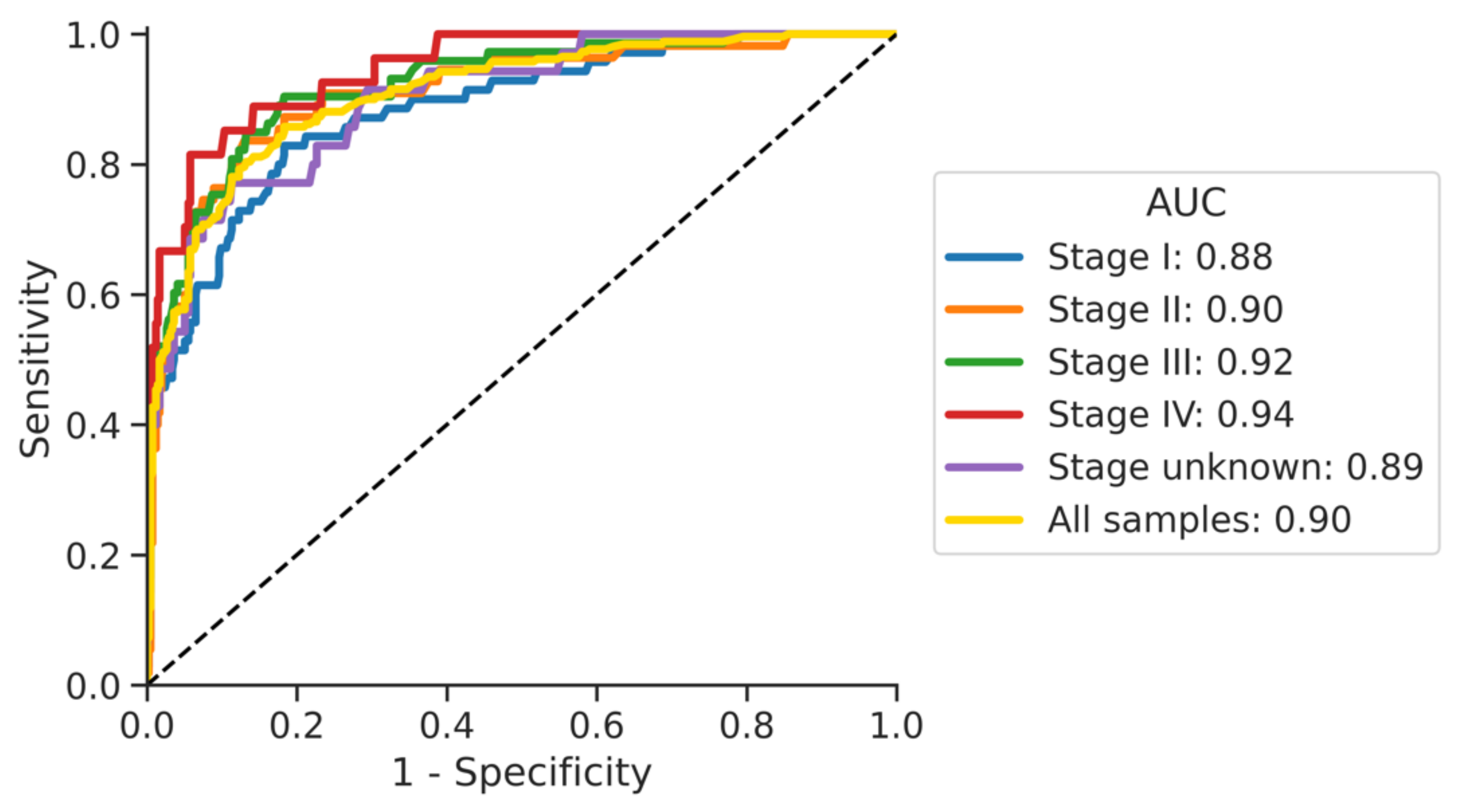
3.3. Impact of Cancer Stage

At 95% specificity, the overall cancer sensitivity was 51% (CI: 40, 63) for stage I, 58% (CI: 45, 71) for stage II, 62% (CI: 50, 73) for stage III, and 67% (CI: 49, 84) for stage IV. Figure 3 shows the stage-specific sensitivities at 95% specificity (p = 0.5375) and Figure 4 shows the stage-specific areas under the curve.
The area under the curve (AUC) for the cancer stages ranged from 0.88 for stage I to 0.94 for stage 4.

3.4. False Positive Controls

The “all cancers” model resulted in 21 positives of the 415 controls at 95% specificity. Twenty-one medical records were available with a mean follow-up of 30.9 (±5.2) months. Three study controls were morbidly obese and scheduled for bariatric surgery. Four patients were hospitalized for an acute inflammation and two of those required emergency cholecystectomies for acute cholecystitis and acute cholangitis, respectively. Two individuals were diagnosed with a benign tumor at the time of the liquid biopsy and three were found with a benign lesion at 8-, 20-, 24- and 36-months and post-study inclusion (one patient developed two independent benign lesions).

4. Discussion

These early results of a comprehensive prospective evaluation of the DEEPGENTM platform suggest a clinically viable capability to differentiate individuals with and without cancer for seven different organs of origin. This finding was observed for a “full model”—including all assessed cancers as well as organ-specific models with varying ranges of performance for the different cancers. As such, it seems plausible to assume that the described liquid biopsy and data analytics innovations not only transpire to an excellent technical performance [23], but also into a relevant clinical capability. This finding is in line with similar projects in the field of liquid biopsy using different technological approaches [7,21,22,26]. Important differences in performance have occurred across cancers and have been described previously. A variety of reasons might cause this observation, including differences in tumor biology (less production of cfDNA of certain cancers), panel variants that do not equally cover all cancers, study logistics, including the time from blood draw to DNA extraction, and others. Further research is required to understand the details around this variability in outcomes per organ of origin.
While this is still a preliminary analysis of the first few samples of a larger specimen collection, the obvious question is how this promising technology can be utilized for an impactful clinical application beyond research. As outlined above, current population-based screening is insufficient and flawed, and leaves room to significantly improve cancer outcomes through earlier detection [2,6,27,28]. At the same time, any new screening tool must provide robust performance with a healthy balance of sensitivity and specificity to be applied for a population-wide clinical application. As such, and considering the above-described results, the DEEPGENTM platform surely holds promise for becoming an instrument for early cancer detection in a variety of applications. Of particular value is the demonstrated ability to adjust sensitivity and specificity for the full model and each organ-specific model. To focus on clinical usability and to reduce the number of false-positive results, the specificities have been set at 95% and 99%. In both settings, the sensitivity of DEEPGENTM is lower than for most standard of care screening methods and also considering the low sample size; this tool should not be used as a replacement for any kind of recommended population-based screening at this point.
However, there may be significant value in diagnostic tests that affect the Bayesian probability of cancer, regardless of underlying genetics. This may include the evaluation of unexplained weight loss in the elderly or other conditions in which the pre-test probability of malignancy is high such as low gradient ascites and exudative pleural effusions. Other high-risk populations may also benefit from these types of screening technologies including patients undergoing evaluation for solid organ transplants and those people who are exposed to high-risk carcinogens or excessive radiation. There may also be value in complementing current screening regimes that can potentially be filled by this technology, such as patients with LungRads-3 screening CT scans for lung cancer, BIRADS-3 mammograms, and following colonoscopy for the removal of polyps with dysplasia. As such, a low sensitivity and high specificity setting is most suitable for individuals at clinical inflection points where there are indeterminate findings, but where additional radiographic or pathologic explorations confer morbidity. The DEEPGENTM approach could reduce exposure to potential morbidity—physically and/or psychologically—caused by further diagnostics and anxiety. On the other hand, a high sensitivity and low specificity setting is most appropriate for any individual subject to standard of care screening. In that instance, the higher rate of false-positive results turns into a benefit in which a wider spread of currently recommended pathways is fostered. This spread of currently recommended pathways will include all their proven medical and economic benefits since these diagnostics would be the next logical step in confirming cancer suspicion [29,30]. As such, even at this early stage, liquid biopsy platforms such as DEEPGENTM can help to identify cancers that would otherwise not be detected, and at the same time, can improve cancer diagnostics by enforcing existing screening programs.
Besides this potential concrete application for personalized cancer detection, this technology should also be viewed as a more global diagnostic platform. As a matter of fact, just in the space of conventional oncology, the ability to find signs of cancer at the earliest possible moment opens several opportunities to improve patient care. Beyond aiding in diagnosing cancer early, mutations and mutational profiles found in the patients’ blood have demonstrated value in determining treatment success by assessing the presence of minimal residual disease (MRD), recurrence detection, and tumor typing to support treatment decisions for advanced disease [31,32]. Additionally, research might demonstrate the role of cfDNA for a range of diseases beyond cancer, including neurodegenerative and cardiovascular diseases; as such, it is plausible to assume that such platforms might transform into global tools of the modern physician’s armory.
All of these technologies, including DEEPGENTM, are still at the beginning of their development and some important limitations exist to date. Besides the current overall performance, particularly the overlap of mutations across cancers, there is the problem of tissue-of-origin determination [33,34,35], and a liquid biopsy result that cannot firmly indicate the origin of cancer. As such, intelligent diagnostic algorithms must be developed and clinically studied before a widespread application in the screening sector can be imagined. In consideration of these potential challenges, the overall value of this technology needs to be studied in long-term clinical trials that analyze critical parameters, which include survival, quality of life, and costs. However, considering the promise of such technologies, significant investments should be made globally in the area of research and development. In the short- and mid-term, we foresee tremendous progress in all areas of this technology stack, ranging from DNA yielding, wet-lab technologies, and data analytics. In particular, the machine learning approach to cancer identification and tissue of origin capabilities will improve with growing datasets.
Despite a lot of promise in this data, several important shortcomings of this study need to be mentioned. First and foremost, this is a preliminary analysis of a larger collection; as such, the sample sizes are low. Additionally, the demographics are narrow and with limited demographic variety. Moreover, all the patients in the cancer cohort were already diagnosed with cancer; as such, this dataset can only demonstrate the general capability of the DEEPGENTM platform to identify cancer. On the other hand, this study addresses some shortcomings that have been discussed in previous projects [12,13,17,36]. Particularly worth mentioning is the diverse control cohort with individuals ranging from young and very healthy study participants to hospitalized patients. Nine of the positive controls were identified with possible explanations for the finding. Two individuals had a benign tumor at the time of the liquid biopsy, which can result in a signal similar to that of cancer. It is questionable if the three positives that were diagnosed with a benign lesion later were found because of an already existing pathology at time of the liquid biopsy. Previous literature has demonstrated that acute inflammations lead to an increase in somatic mutations that are otherwise associated with cancer [37,38]. All hospitalized patients with inflammation would not have been candidates for standard of care screening. Another interesting finding is the morbidly obese individuals before bariatric surgery having positive findings because this complex disease comes with chronic inflammation and an increased cancer risk. The poor health and chronic inflammatory conditions exhibited in these “controls”, identified as “cancers” by DEEPGENTM, reinforces the ultra-high sensitivity of the proprietary technology stack and opens up future research possibilities in areas of chronic inflammatory conditions that are increasingly considered to be precursors to cancer and other major diseases [39,40]. However, to find specific answers to these important questions, more global research in the field of liquid biopsy is needed.

5. Conclusions

Despite being at an early stage of development and there being room for improvements in areas such as blood-drawing to machine learning, the DEEPGENTM platform shows a clinically viable capability to differentiate patients with and without cancer. More systematic research is needed to define the exact clinical applications as well as its large-scale performance.

Author Contributions

Conceptualization of the work: F.R., M.H. and J.D.; acquisition of specimen: F.R., J.D., F.T., D.G., N.C.B., L.B., S.M., C.E.I., W.K., J.O., P.P., G.T.L., M.U.M., R.T., J.O., D.B. and C.T.; analysis or interpretation of data: F.R., M.H., J.J.N., Y.W., N.I. and J.D.; drafting the work: F.R., M.H. and J.D. All authors have read and agreed to the published version of the manuscript.

Funding

This study was funded by Quantgene Inc. (Santa Monica, CA, USA) in association with the identified study investigators under a cooperative clinical trial agreement. The authors had full control of the study execution, analysis, and development of the manuscript.

Institutional Review Board Statement

This study received ethics committee approval for all participating sites through either institutional review or Western IRB (Puyallup, WA, USA) and was registered on clinicaltrial.gov (NCT03517332). All procedures performed in this study were in accordance with the ethical standards of the institutional and national research committee and with the 1964 Helsinki declaration and its later amendments or comparable ethical standards.

Informed Consent Statement

Informed consent was obtained from all subjects involved in the study.

Data Availability Statement

The data presented in this study are available on request from the corresponding author. The data are not publicly available due to privacy and ethical restrictions.

Acknowledgments

We thank Petra Gambon-Stow (University Hospital Geneva) for study management and data collection.

Conflicts of Interest

Ris reports research funding from Quantgene, personal fees from Arthrex, Stryker, and Distal Motion, outside the submitted work. Douissard reports grants and non-financial support from Intuitive Surgical Inc., personal fees from Verb Surgical Inc., grants, personal fees, and non-financial support from Johnson&Johnson, outside the submitted work. Nieva reports holding stocks or other ownership from Epic Sciences, Cansera, Quantgene, and Indee, consulting or advisory roles for Astra Zeneca, Naveris, Fujirebio, research funding from Merck, patents, royalties, or other intellectual property from Cansera, outside this project. Triponez reports consulting fees and travel grants from Medtronic, Fluoptics, Stryker, outside this project. Hellan, Woo, Geller, NC Buchs, L. Bühler, Moenig, Iselin, Karenovics, Petignat, Lam, Undurraga Maliverno, Tuttle, Ouellete, Bose, Redan, Ismail, and Toso report no conflict of interest.

References

  1. Siegel, R.L.; Miller, K.D.; Fuchs, H.E.; Jemal, A. Cancer Statistics, 2021. CA Cancer J. Clin. 2021, 71, 7–33. [Google Scholar] [CrossRef]
  2. Smith, R.A.; Ba, K.S.A.; Brooks, D.; Fedewa, S.A.; Manassaram-Baptiste, D.; Saslow, D.; Wender, R.C. Cancer screening in the United States, 2019: A review of current American Cancer Society guidelines and current issues in cancer screening. CA Cancer J. Clin. 2019, 69, 184–210. [Google Scholar] [CrossRef]
  3. Singh, H.; Sethi, S.; Raber, M.; Petersen, L.A. Errors in cancer diagnosis: Current understanding and future directions. J. Clin. Oncol. 2007, 25, 5009–5018. [Google Scholar] [CrossRef]
  4. Dobson, C.M.; Russell, A.J.; Rubin, G.P. Patient delay in cancer diagnosis: What do we really mean and can we be more specific? BMC Health Serv. Res. 2014, 14, 387. [Google Scholar] [CrossRef] [PubMed] [Green Version]
  5. Hanna, T.P.; King, W.D.; Thibodeau, S.; Jalink, M.; Paulin, G.A.; Harvey-Jones, E.; O’Sullivan, D.E.; Booth, C.M.; Sullivan, R.; Aggarwal, A. Mortality due to cancer treatment delay: Systematic review and meta-analysis. BMJ 2020, 371, m4087. [Google Scholar] [CrossRef] [PubMed]
  6. Loomans-Kropp, H.A.; Umar, A. Cancer prevention and screening: The next step in the era of precision medicine. NPJ Precis. Oncol. 2019, 3, 3. [Google Scholar] [CrossRef] [PubMed] [Green Version]
  7. Chen, X.; Gole, J.; Gore, A.; He, Q.; Lu, M.; Min, J.; Yuan, Z.; Yang, X.; Jiang, Y.; Zhang, T.; et al. Non-invasive early detection of cancer four years before conventional diagnosis using a blood test. Nat. Commun. 2020, 11, 1–10. [Google Scholar] [CrossRef] [PubMed]
  8. Shyr, D.; Liu, Q. Next generation sequencing in cancer research and clinical application. Biol. Proced. Online 2013, 15, 4. [Google Scholar] [CrossRef] [PubMed] [Green Version]
  9. Metzker, M.L. Sequencing technologies—the next generation. Nat. Rev. Genet. 2010, 11, 31–36. [Google Scholar] [CrossRef] [Green Version]
  10. Shen, S.Y.; Singhania, R.; Fehringer, G.; Chakravarthy, A.; Roehrl, M.H.A.; Chadwick, D.; Zuzarte, P.C.; Borgida, A.; Wang, T.T.; Li, T.; et al. Sensitive tumour detection and classification using plasma cell-free DNA methylomes. Nat. Cell Biol. 2018, 563, 579–583. [Google Scholar] [CrossRef] [PubMed]
  11. Cohen, J.D.; Li, L.; Wang, Y.; Thoburn, C.; Afsari, B.; Danilova, L.; Douville, C.; Javed, A.A.; Wong, F.; Mattox, A.; et al. Detection and localization of surgically resectable cancers with a multi-analyte blood test. Science 2018, 359, 926–930. [Google Scholar] [CrossRef] [Green Version]
  12. Aarthy, R.; Mani, S.; Velusami, S.; Sundarsingh, S.; Rajkumar, T. Role of Circulating Cell-Free DNA in Cancers. Mol. Diagn. Ther. 2015, 19, 339–350. [Google Scholar] [CrossRef]
  13. Rachiglio, A.M.; Abate, R.E.; Sacco, A.; Pasquale, R.; Fenizia, F.; Lambiase, M.; Morabito, A.; Montanino, A.; Rocco, G.; Romano, C.; et al. Limits and potential of targeted sequencing analysis of liquid biopsy in patients with lung and colon carcinoma. Oncotarget 2016, 7, 66595–66605. [Google Scholar] [CrossRef] [PubMed] [Green Version]
  14. Heitzer, E.; Haque, I.S.; Roberts, C.E.S.; Speicher, M.R. Current and future perspectives of liquid biopsies in genomics-driven oncology. Nat. Rev. Genet. 2019, 20, 71–88. [Google Scholar] [CrossRef] [PubMed]
  15. Phallen, J.; Sausen, M.; Adleff, V.; Leal, A.; Hruban, C.; White, J.; Anagnostou, V.; Fiksel, J.; Cristiano, S.; Papp, E.; et al. Direct detection of early-stage cancers using circulating tumor DNA. Sci. Transl. Med. 2017, 9, eaan2415. [Google Scholar] [CrossRef] [PubMed] [Green Version]
  16. Diaz, L., Jr.; ABardelli, A. Liquid biopsies: Genotyping circulating tumor DNA. J. Clin. Oncol. 2014, 32, 579. [Google Scholar] [CrossRef] [PubMed]
  17. Trigg, R.; Martinson, L.; Parpart-Li, S.; Shaw, J. Factors that influence quality and yield of circulating-free DNA: A systematic review of the methodology literature. Heliyon 2018, 4, e00699. [Google Scholar] [CrossRef] [PubMed] [Green Version]
  18. Goldfeder, R.L.; Priest, J.R.; Zook, J.M.; Grove, M.E.; Waggott, D.; Wheeler, M.T.; Salit, M.; Ashley, E.A. Medical implications of technical accuracy in genome sequencing. Genome Med. 2016, 8, 1–12. [Google Scholar] [CrossRef] [PubMed] [Green Version]
  19. Gorgannezhad, L.; Umer, M.; Islam, M.N.; Nguyen, N.-T.; Shiddiky, M.J. Circulating tumor DNA and liquid biopsy: Opportunities, challenges, and recent advances in detection technologies. Lab Chip 2018, 18, 1174–1196. [Google Scholar] [CrossRef]
  20. Buscail, E.; Maulat, C.; Muscari, F.; Chiche, L.; Cordelier, P.; Dabernat, S.; Alix-Panabières, C.; Buscail, L. Liquid Biopsy Approach for Pancreatic Ductal Adenocarcinoma. Cancers 2019, 11, 852. [Google Scholar] [CrossRef] [PubMed] [Green Version]
  21. Kuderer, N.M.; Burton, K.A.; Blau, S.; Rose, A.L.; Parker, S.; Lyman, G.H.; Blau, C.A. Comparison of 2 Commercially Available Next-Generation Sequencing Platforms in Oncology. JAMA Oncol. 2017, 3, 996–998. [Google Scholar] [CrossRef] [PubMed]
  22. Stetson, D.; Ahmed, A.; Xu, X.; Nuttall, B.; Lubinski, T.J.; Johnson, J.H.; Barrett, J.C.; Dougherty, B.A. Orthogonal Comparison of Four Plasma NGS Tests with Tumor Suggests Technical Factors are a Major Source of Assay Discordance. JCO Precis. Oncol. 2019, 3, 1–9. [Google Scholar] [CrossRef]
  23. Hermann, B.T.; Pfeil, S.; Groenke, N.; Schaible, S.; Kunze, R.; Ris, F.; Bhakdi, J. DEEPGENTM—A Novel Variant Calling Assay for Low Frequency Variants. Genes 2021, 12, 507. [Google Scholar] [CrossRef]
  24. Pedregosa, F.; Varoquaux, G.; Gramfort, A.; Michel, V.; Thirion, B.; Grisel, O.; Duchesnay, E. Scikit-learn: Machine learning in Python. J. Mach. Learn. Res. 2011, 12, 2825–2830. [Google Scholar]
  25. Virtanen, P.; Gommers, R.; Oliphant, T.E.; Haberland, M.; Reddy, T.; Cournapeau, D.; Burovski, E.; Peterson, P.; Weckesser, W.; Bright, J.; et al. SciPy 1.0: Fundamental algorithms for scientific computing in Python. Nat. Methods 2020, 17, 261–272. [Google Scholar] [CrossRef] [Green Version]
  26. Thierry, A.R.; Mouliere, F.; El Messaoudi, S.; Mollevi, C.; Lopez-Crapez, E.; Rolet, F.; Gillet, B.; Gongora, C.; Dechelotte, P.; Robert, B.; et al. Clinical validation of the detection of KRAS and BRAF mutations from circulating tumor DNA. Nat. Med. 2014, 20, 430–435. [Google Scholar] [CrossRef]
  27. Guren, M.G. The global challenge of colorectal cancer. Lancet Gastroenterol. Hepatology 2019, 4, 894–895. [Google Scholar] [CrossRef] [Green Version]
  28. Pinsky, P.F.; Prorok, P.C.; Kramer, B.S. Prostate Cancer Screening-A Perspective on the Current State of the Evidence. N. Engl. J. Med. 2017, 376, 1285–1289. [Google Scholar] [CrossRef] [PubMed]
  29. Mandelker, D.; Zhang, L.; Kemel, Y.; Stadler, Z.K.; Joseph, V.; Zehir, A.; Pradhan, N.; Arnold, A.; Walsh, M.F.; Liying, Z.; et al. Mutation Detection in Patients with Advanced Cancer by Universal Sequencing of Cancer-Related Genes in Tumor and Normal DNA vs Guideline-Based Germline Testing. JAMA 2017, 318, 825–835. [Google Scholar] [CrossRef]
  30. Zubor, P.; Kubatka, P.; Kajo, K.; Dankova, Z.; Polacek, H.; Bielik, T.; Kudela, E.; Samec, M.; Liskova, A.; Vlcakova, D.; et al. Why the gold standard approach by mammography demands extension by multiomics? Application of liquid biopsy miRNA profiles to breast cancer disease management. Int. J. Mol. Sci. 2019, 20, 2878. [Google Scholar] [CrossRef] [PubMed] [Green Version]
  31. Pantel, K.A.; lix-Panabières, C. Liquid biopsy and minimal residual disease—latest advances and implications for cure. Nat. Rev. Clin. Oncol. 2019, 16, 409–424. [Google Scholar] [CrossRef]
  32. Ravandi, F.; Jorgensen, J.L.; Thomas, D.A.; O’Brien, S.; Garris, R.; Faderl, S.; Huang, X.; Wen, S.; Burger, J.A.; Ferrajoli, A.; et al. Detection of MRD may predict the outcome of patients with Philadelphia chromosome–positive ALL treated with tyrosine kinase inhibitors plus chemotherapy. Blood 2013, 122, 1214–1221. [Google Scholar] [CrossRef] [PubMed]
  33. Oien, K.A. Pathologic Evaluation of Unknown Primary Cancer. Semin. Oncol. 2009, 36, 8–37. [Google Scholar] [CrossRef]
  34. Kim, H.; Kim, Y.-M. Pan-cancer analysis of somatic mutations and transcriptomes reveals common functional gene clusters shared by multiple cancer types. Sci. Rep. 2018, 8, 6041. [Google Scholar] [CrossRef] [PubMed] [Green Version]
  35. Marquard, A.M.; Birkbak, N.J.; Thomas, C.E.; Favero, F.; Krzystanek, M.; Lefebvre, C.; Ferté, C.; Jamal-Hanjani, M.; Wilson, G.A.; Shafi, A.; et al. Tumor Tracer: A method to identify the tissue of origin from the somatic mutations of a tumor specimen. BMC Med. Genom. 2015, 8, 58. [Google Scholar] [CrossRef] [PubMed] [Green Version]
  36. Abbosh, C.; Birkbak, N.J.; Swanton, C. Early stage NSCLC—challenges to implementing ctDNA-based screening and MRD detection. Nat. Rev. Clin. Oncol. 2018, 15, 577–586. [Google Scholar] [CrossRef] [PubMed]
  37. Chechlinska, M.; Kowalewska, M.; Nowak, R. Systemic inflammation as a confounding factor in cancer biomarker discovery and validation. Nat. Rev. Cancer 2010, 10, 2–3. [Google Scholar] [CrossRef]
  38. Colotta, F.; Allavena, P.; Sica, A.; Garlanda, C.; Mantovani, A. Cancer-related inflammation, the seventh hallmark of cancer: Links to genetic instability. Carcinogenesis 2009, 30, 1073–1081. [Google Scholar] [CrossRef] [Green Version]
  39. Barbie, D.A.; Tamayo, P.; Boehm, J.; Kim, S.Y.; Moody, S.E.; Dunn, I.F.; Schinzel, A.C.; Sandy, P.; Meylan, E.; Scholl, C.; et al. Systematic RNA interference reveals that oncogenic KRAS-driven cancers require TBK1. Nat. Cell Biol. 2009, 462, 108–112. [Google Scholar] [CrossRef]
  40. Multhoff, G.; Molls, M.; Radons, J. Chronic inflammation in cancer development. Front. Immunol. 2012, 2, 98. [Google Scholar] [CrossRef] [Green Version]
Figure 1. ROC curve for diagnosis of all cancers (full model).
Cancers 13 04104 g001
Figure 2. Sensitivity of DEEPGENTM platform by model at 99% and 95% specificity.
Cancers 13 04104 g002
Figure 3. Sensitivity of DEEPGENTM platform by cancer stage at 95% specificity.
Cancers 13 04104 g003
Figure 4. ROC curves for cancer stages.
Cancers 13 04104 g004
Table 1. Demographic parameters.
Parameter Control
n = 415
All Cancer
n = 260
Bladder
n = 25
Prostate
n = 29
Lung
n = 30
Liver
n = 27
Pancreatic
n = 40
Colorectal
n = 66
Breast
n = 43
Age
Mean (standard deviation)54.9 (15.5)65.4 (10.9)68.7 (11.1)64.9 (7.3)64.2 (9.6)67.5 (9.7)66.9 (9.4)65.2 (12.3)61.9 (12.3)
Sex, n (%)
Male117 (28)136 (52)18 (72)29 (100)10 (33)20 (74)21 (52)38 (58)0 (0)
Female298 (72)124 (48)7 (28)0 (0)20 (67)7 (26)19 (48)28 (42)43 (100)
Comorbidities, n (%)
Alzheimer’s1 (0.2)1 (0.4)0 (0.0)0 (0.0)0 (0.0)0 (0.0)0 (0.0)0 (0.0)1 (2)
Cardiovascular disease27 (7)28 (11)8 (32)1 (3)3 (10)4 (15)4 (10)6 (9)2 (5)
Diabetes31 (7)36 (14)4 (16)2 (7)2 (7)8 (30)8 (20)8 (12)4 (9)
Hypertension75 (18)108 (42)18 (72)8 (28)12 (40)15 (56)18 (45)23 (35)14 (33)
Kidney disease5 (1)14 (5)4 (16)2 (7)1 (3)0 (0.0)0 (0.0)5 (8)2 (5)
Obesity40 (10)30 (12)8 (32)2 (7)5 (17)3 (11)0 (0.0)5 (8)7 (16)
Respiratory disease18 (4)35 (13)7 (28)3 (10)9 (30)4 (15)4 (10)2 (3)6 (14)
Other141 (34)113 (43)18 (72)17 (59)16 (53)18 (67)12 (30)14 (21)18 (42)
None185 (45)30 (12)1 (4)3 (10)3 (10)1 (4)3 (8)10 (15)9 (21)
Unknown32 (8)46 (18)0 (0.0)2 (7)1 (3)5 (19)13 (32)25 (38)0 (0.0)
Risk Factors, n (%)
Family History *182 (44)100 (38)4 (16)8 (28)14 (47)9 (33)14 (35)25 (38)26 (60)
Smoking History132 (32)110 (42)11 (44)11 (38)23 (77)13 (48)15 (38)25 (38)12 (28)
Medical Condition57 (14)65 (25)7 (28)11 (38)5 (17)8 (30)12 (30)18 (27)4 (9)
Other22 (5)47 (18)10 (40)8 (28)5 (17)14 (52)3 (8)5 (8)2 (5)
None104 (25)45 (17)8 (32)3 (10)2 (7)2 (7)3 (8)16 (24)11 (26)
Unknown51 (12)23 (9)4 (16)4 (14)0 (0)5 (19)6 (15)3 (5)1 (2)
* Across 4 generations (grandparents to children).
Table 2. Number of cancer samples and cancer finding/sensitivities at 95% and 99% specificity.
ModelNumber of Cancer SamplesCancer Findings at 95% SpecificityCancer Findings at 99% Specificity
nCorrectly Identified Cancers, nSensitivity, %95% Confidence Interval, Lower Bound, Upper BoundCorrectly Identified Cancers, nSensitivity, %95% Confidence Interval, Lower Bound, Upper Bound
All cancers2601505752, 641114337, 49
Bladder25208064, 9683214, 50
Breast43133016, 447165, 27
Colorectal66284230, 54182716, 38
Liver27176345, 81114122, 60
Lung30206750, 84165335, 71
Pancreas40215237, 67153823, 53
Prostate29217256, 88186244, 80
Publisher’s Note: MDPI stays neutral with regard to jurisdictional claims in published maps and institutional affiliations.

