An Analysis of Vertebral Body Growth after Proton Beam Therapy for Pediatric Cancer

 , , , ,
Abstract
:Simple Summary
Abstract
1. Introduction
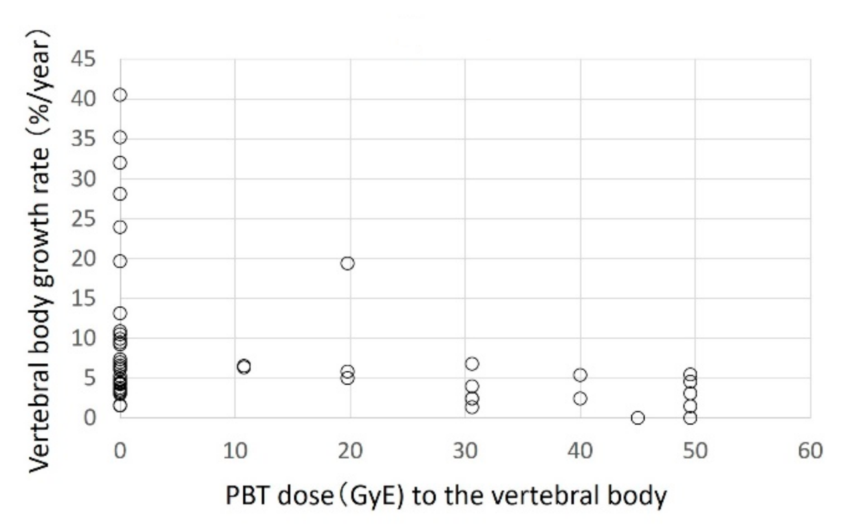
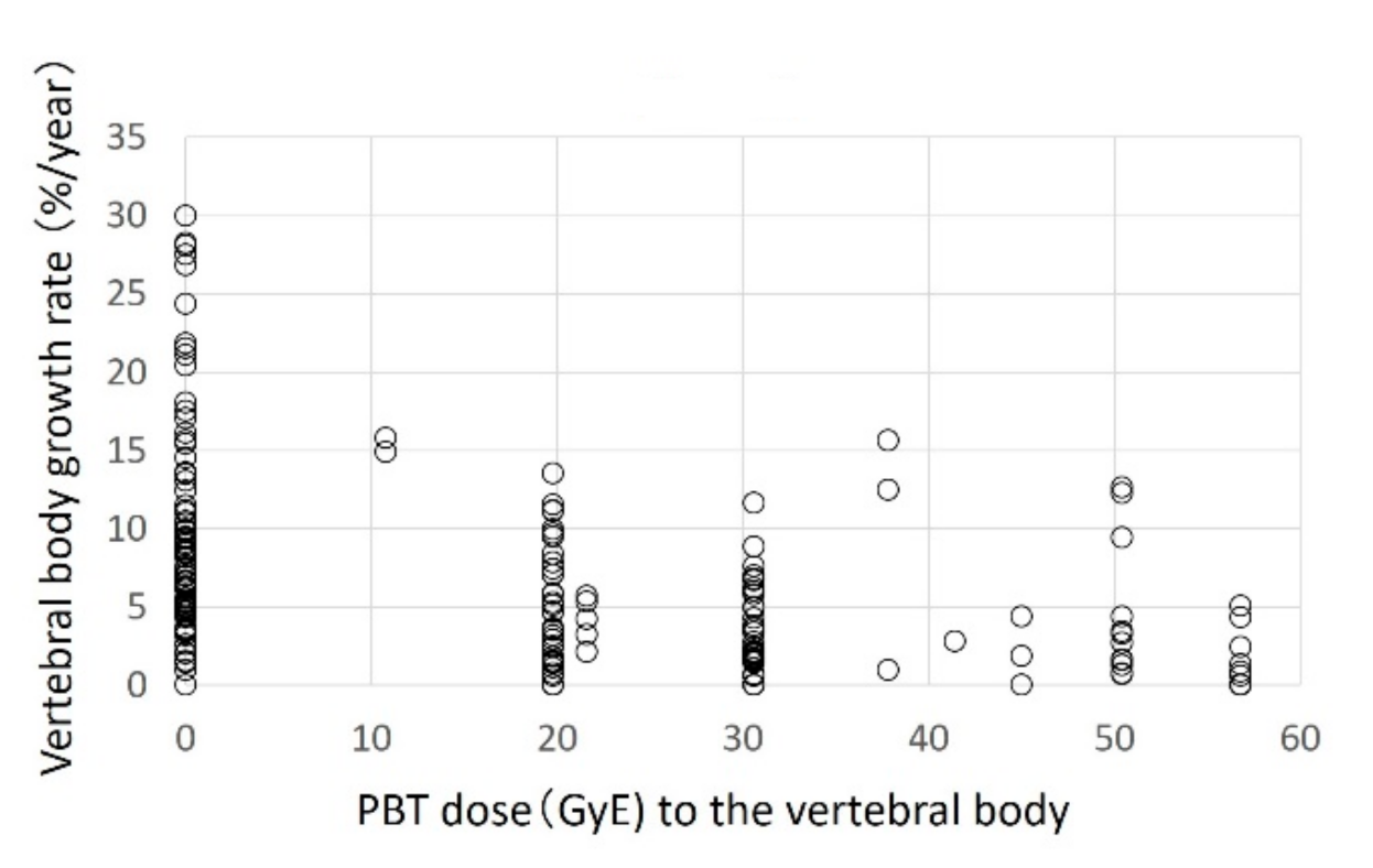
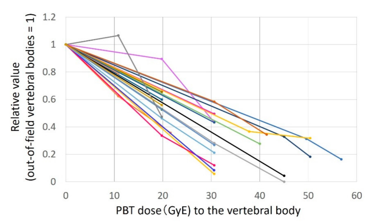
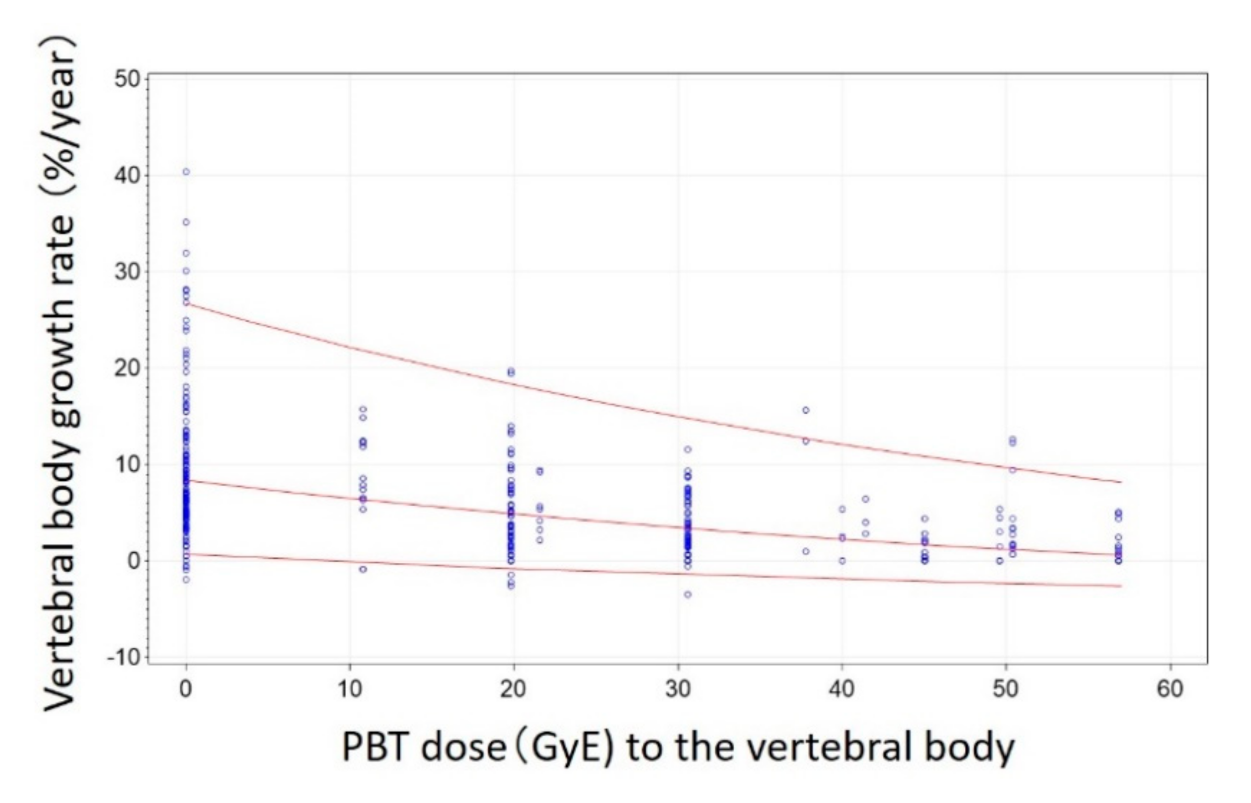
2. Results
3. Discussion
4. Patients and Methods
5. Conclusions
Author Contributions
Funding
Institutional Review Board Statement
Informed Consent Statement
Data Availability Statement
Conflicts of Interest
References
- Ward, E.; DeSantis, C.E.; Robbins, A.; Kohler, B.; Jemal, A. Childhood and adolescent cancer statistics, 2014. CA A Cancer J. Clin. 2014, 64, 83–103. [Google Scholar] [CrossRef]
- Armstrong, G.T.; Liu, Q.; Yasui, Y.; Neglia, J.P.; Leisenring, W.; Robison, L.L.; Mertens, A.C. Late mortality among 5-year survivors of childhood cancer: A summary from the Childhood Cancer Survivor Study. J. Clin. Oncol. 2009, 27, 2328–2338. [Google Scholar] [CrossRef] [Green Version]
- Mizumoto, M.; Oshiro, Y.; Yamamoto, T.; Kohzuki, H.; Sakurai, H. Proton Beam Therapy for Pediatric Brain Tumor. Neurol. Med. Chir. 2017, 57, 343–355. [Google Scholar] [CrossRef] [Green Version]
- Hartley, K.A.; Li, C.; Laningham, F.H.; Krasin, M.J.; Xiong, X.; Merchant, T.E. Vertebral Body Growth After Craniospinal Irradiation. Int. J. Radiat. Oncol. 2008, 70, 1343–1349. [Google Scholar] [CrossRef]
- Probert, J.C.; Parker, B.R.; Kaplan, H.S. Growth retardation in children after megavoltage irradiation of the spine. Cancer 1973, 32, 634–639. [Google Scholar] [CrossRef]
- Hoeben, B.A.; Carrie, C.; Timmermann, B.; Mandeville, H.C.; Gandola, L.; Dieckmann, K.; Albiac, M.R.; Magelssen, H.; Lassen-Ramshad, Y.; Ondrová, B.; et al. Management of vertebral radiotherapy dose in paediatric patients with cancer: Consensus recommendations from the SIOPE radiotherapy working group. Lancet Oncol. 2019, 20, e155–e166. [Google Scholar] [CrossRef]
- Michel, J.; Radulesco, T.; Mancini, J.; Paganelli, A.; Varoquaux, A.; Adalian, P.; Ranque, S.; Dessi, P. Maxillary sinus volume: New physiopathological data in fungal ball genesis? A retrospective study. Clin. Otolaryngol. 2017, 42, 831–836. [Google Scholar] [CrossRef]
- Oshiro, Y.; Mizumoto, M.; Okumura, T.; Sugahara, S.; Fukushima, T.; Ishikawa, H.; Nakao, T.; Hashimoto, T.; Tsuboi, K.; Ohkawa, H.; et al. Clinical results of proton beam therapy for advanced neuroblastoma. Radiat. Oncol. 2013, 8, 142. [Google Scholar] [CrossRef] [Green Version]
- Mizumoto, M.; Murayama, S.; Akimoto, T.; Demizu, Y.; Fukushima, T.; Ishida, Y.; Oshiro, Y.; Numajiri, H.; Fuji, H.; Okumura, T.; et al. Long-term follow-up after proton beam therapy for pediatric tumors: A Japanese national survey. Cancer Sci. 2017, 108, 444–447. [Google Scholar] [CrossRef] [Green Version]
- Bs, R.V.S.; Shih, H.A.; Yeap, B.Y.; Mouw, K.W.; Petersen, R.; Kim, D.Y.; Munzenrider, J.E.; Grabowski, E.; Rodriguez-Galindo, C.; Yock, T.I.; et al. Second nonocular tumors among survivors of retinoblastoma treated with contemporary photon and proton radiotherapy. Cancer 2014, 120, 126–133. [Google Scholar]
- Chung, C.S.; Yock, T.I.; Nelson, K.; Xu, Y.; Keating, N.L.; Tarbell, N.J. Incidence of Second Malignancies Among Patients Treated With Proton Versus Photon Radiation. Int. J. Radiat. Oncol. 2013, 87, 46–52. [Google Scholar] [CrossRef] [PubMed]
- Zhang, R.; Howell, R.M.; Taddei, P.J.; Giebeler, A.; Mahajan, A.; Newhauser, W.D. A comparative study on the risks of radiogenic second cancers and cardiac mortality in a set of pediatric medulloblastoma patients treated with photon or proton craniospinal irradiation. Radiother. Oncol. 2014, 113, 84–88. [Google Scholar] [CrossRef] [PubMed] [Green Version]
- Brodin, N.P.; Rosenschöld, P.M.A.; Aznar, M.C.; Kiil-Berthelsen, A.; Vogelius, I.R.; Nilsson, P.; Lannering, B.; Björk-Eriksson, T. Radiobiological risk estimates of adverse events and secondary cancer for proton and photon radiation therapy of pediatric medulloblastoma. Acta Oncol. 2011, 50, 806–816. [Google Scholar] [CrossRef] [PubMed]
- Yoon, M.; Shin, D.H.; Kim, J.; Kim, J.W.; Kim, D.W.; Park, S.Y.; Lee, S.B.; Kim, J.Y.; Park, H.-J.; Park, B.K.; et al. Craniospinal Irradiation Techniques: A Dosimetric Comparison of Proton Beams With Standard and Advanced Photon Radiotherapy. Int. J. Radiat. Oncol. 2011, 81, 637–646. [Google Scholar] [CrossRef] [PubMed]
- Mizumoto, M.; Okumura, T.; Hashimoto, T.; Fukuda, K.; Oshiro, Y.; Fukumitsu, N.; Abei, M.; Kawaguchi, A.; Hayashi, Y.; Ookawa, A.; et al. Proton Beam Therapy for Hepatocellular Carcinoma: A Comparison of Three Treatment Protocols. Int. J. Radiat. Oncol. 2011, 81, 1039–1045. [Google Scholar] [CrossRef] [PubMed]
- Oshiro, Y.; Mizumoto, M.; Okumura, T.; Hashimoto, T.; Fukumitsu, N.; Ohkawa, A.; Kanemoto, A.; Numajiri, H.; Ohno, T.; Sakae, T.; et al. Results of Proton Beam Therapy without Concurrent Chemotherapy for Patients with Unresectable Stage III Non-small Cell Lung Cancer. J. Thorac. Oncol. 2012, 7, 370–375. [Google Scholar] [CrossRef] [Green Version]
- Oshiro, Y.; Okumura, T.; Kurishima, K.; Homma, S.; Mizumoto, M.; Ishikawa, H.; Onizuka, M.; Sakai, M.; Goto, Y.; Hizawa, N.; et al. High-dose concurrent chemo-proton therapy for Stage III NSCLC: Preliminary results of a Phase II study. J. Radiat. Res. 2014, 55, 959–965. [Google Scholar] [CrossRef] [Green Version]
- Ishikawa, H.; Hashimoto, T.; Moriwaki, T.; Hyodo, I.; Hisakura, K.; Terashima, H.; Ohkohchi, N.; Ohno, T.; Makishima, H.; Mizumoto, M.; et al. Proton beam therapy combined with concurrent chemotherapy for esophageal cancer. Anticancer Res. 2015, 35, 1757–1762. [Google Scholar] [CrossRef]
- Grewal, A.S.; Schonewolf, C.; Min, E.J.; Chao, H.-H.; Both, S.; Lam, S.; Mazzoni, S.; Bekelman, J.; Christodouleas, J.; Vapiwala, N. Four-Year Outcomes From a Prospective Phase II Clinical Trial of Moderately Hypofractionated Proton Therapy for Localized Prostate Cancer. Int. J. Radiat. Oncol. 2019, 105, 713–722. [Google Scholar] [CrossRef]
- Mizumoto, M.; Yamamoto, T.; Ishikawa, E.; Matsuda, M.; Takano, S.; Ishikawa, H.; Okumura, T.; Sakurai, H.; Matsumura, A.; Tsuboi, K. Proton beam therapy with concurrent chemotherapy for glioblastoma multiforme: Comparison of nimustine hydrochloride and temozolomide. J. Neurooncol. 2016, 130, 165–170. [Google Scholar] [CrossRef]
- Mizumoto, M.; Sugahara, S.; Okumura, T.; Hashimoto, T.; Oshiro, Y.; Fukumitsu, N.; Nakahara, A.; Terashima, H.; Tsuboi, K.; Sakurai, H. Hyperfractionated concomitant boost proton beam therapy for esophageal carcinoma. Int. J. Radiat. Oncol. Biol. Phys. 2011, 81, e601–e606. [Google Scholar] [CrossRef] [PubMed]
- Hiroshima, Y.; Fukumitsu, N.; Saito, T.; Numajiri, H.; Murofushi, K.N.; Ohnishi, K.; Nonaka, T.; Ishikawa, H.; Okumura, T.; Sakurai, H. Concurrent chemoradiotherapy using proton beams for unresectable locally advanced pancreatic cancer. Radiother. Oncol. 2019, 136, 37–43. [Google Scholar] [CrossRef] [Green Version]
- Hirano, E.; Fuji, H.; Onoe, T.; Kumar, V.; Shirato, H.; Kawabuchi, K. Cost-effectiveness analysis of cochlear dose reduction by proton beam therapy for medulloblastoma in childhood. J. Radiat. Res. 2014, 55, 320–327. [Google Scholar] [CrossRef] [PubMed] [Green Version]
- Mailhot Vega, R.B.; Kim, J.; Bussière, M.; Hattangadi-Gluth, J.A.; Hollander, A.; Michalski, J.; Tarbell, N.; Yock, T.; Macdonald, S.M. Cost effectiveness of proton therapy compared with photon therapy in the management of pediatric medulloblastoma. Cancer 2013, 119, 4299–4307. [Google Scholar] [CrossRef]
- Lundkvist, J.; Ekman, M.; Ericsson, S.R.; Jönsson, B.; Glimelius, B. Cost-effectiveness of proton radiation in the treatment of childhood medulloblastoma. Cancer 2005, 103, 793–801. [Google Scholar] [CrossRef]
- Mailhot Vega, R.; Kim, J.; Hollander, A.; Hattangadi-Gluth, J.; Michalski, J.; Tarbell, N.J.; Yock, T.I.; Bussiere, M.; MacDonald, S.M. Cost effectiveness of proton versus photon radiation therapy with respect to the risk of growth hormone deficiency in children. Cancer 2015, 121, 1694–1702. [Google Scholar] [CrossRef]
- Guyuron, B.; Dagys, A.P.; Munro, I.R.; Ross, R.B. Effect of Irradiation on Facial Growth: A 7- to 25-Year Follow-Up. Ann. Plast. Surg. 1983, 11, 423–427. [Google Scholar] [CrossRef]
- Bloom, H.J.G.; Wallace, E.N.K.; Henk, J.M. The treatment and prognosis of medulloblastoma in children. Am. J. Roentgenol. 1969, 105, 43–62. [Google Scholar] [CrossRef]
- Silber, J.H.; Littman, P.S.; Meadows, A.T. Stature loss following skeletal irradiation for childhood cancer. J. Clin. Oncol. 1990, 8, 304–312. [Google Scholar] [CrossRef]
- Phillips, R.D.; Kimeldorf, D.J. Age and Dose Dependence of Bone Growth Retardation Induced by X-Irradiation. Radiat. Res. 1966, 27, 384–396. [Google Scholar] [CrossRef]
- Neuhauser, E.B.D.; Wittenborg, M.H.; Berman, C.Z.; Cohen, J. Irradiation Effects of Roentgen Therapy on the Growing Spine. Radiology 1952, 59, 637–650. [Google Scholar] [CrossRef] [PubMed]
- Johnson, S.B.; Hung, J.; Kapadia, N.; Oh, K.S.; Kim, M.; Hamstra, D.A. Spinal Growth Patterns after Craniospinal Irradiation in Children with Medulloblastoma. Pract. Radiat. Oncol. 2019, 9, e22–e28. [Google Scholar] [CrossRef]
- Paulino, A.C.; Fowler, B.Z. Risk factors for scoliosis in children with neuroblastoma. Int. J. Radiat. Oncol. 2005, 61, 865–869. [Google Scholar] [CrossRef]
- Paulino, A.; Suzawa, H.; Dreyer, Z. Scoliosis in children receiving craniospinal irradiation for medulloblastoma. In Proceedings of the Congress of the International Society of Paediatric Oncology (SIOP), Cape Town, South Africa, 8–11 October 2015; p. S209. [Google Scholar]
- Makipernaa, A.; Heikkilä, J.T.; Merikanto, J.; Marttinen, E.; Siimes, M.A. Spinal deformity induced by radiotherapy for solid tumours in childhood: A long-term follow up study. Eur. J. Pediatr. 1993, 152, 197–200. [Google Scholar] [CrossRef] [PubMed]
- Oshiro, Y.; Mizumoto, M.; Pan, H.; Kaste, S.C.; Gajjar, A.; Do, T.E.M. Spinal changes after craniospinal irradiation in pediatric patients. Pediatr. Blood Cancer 2020, 67, e28728. [Google Scholar] [CrossRef]
- Hogeboom, C.J.; Grosser, S.C.; Guthrie, K.A.; Thomas, P.R.; D’Angio, G.J.; Breslow, N.E. Stature loss following treatment for Wilms tumor. Med. Pediatr. Oncol. 2001, 36, 295–304. [Google Scholar] [CrossRef]
- Ng, L.W.; Wong, K.K.; Wu, C.-L.A.; Sposto, R.; Olch, A. Dose Sculpting Intensity Modulated Radiation Therapy for Vertebral Body Sparing in Children With Neuroblastoma. Int. J. Radiat. Oncol. 2018, 101, 550–557. [Google Scholar] [CrossRef] [PubMed]
- Mizumoto, M.; Oshiro, Y.; Pan, H.; Wang, F.; Kaste, S.C.; Gajjar, A.; Chemaitilly, W.; Do, T.E.M. Height after photon craniospinal irradiation in pediatric patients treated for central nervous system embryonal tumors. Pediatr. Blood Cancer 2020, 67, e28617. [Google Scholar] [CrossRef]


| Fixed Effect | Estimate | Standard Error | t-Value | p-Value | |
|---|---|---|---|---|---|
| Age | −0.04854 | 0.03281 | −1.48 | 0.1525 | |
| Sex | Female | −0.1799 | 0.1290 | −1.39 | 0.1781 |
| Male | 0 | ||||
| Site of vertebrae | Cervical | 0.1715 | 0.06332 | 2.71 | 0.0071 |
| Upper thoracic | 0.06079 | 0.05449 | 1.12 | 0.2654 | |
| Lower thoracic | 0.05647 | 0.04893 | 1.15 | 0.2493 | |
| Lumbar | 0 | ||||
| Dose | −0.01507 | 0.001246 | −12.09 | 0.0006 | |
| Square of dose | 0.000043 | 0.000099 | 0.44 | 0.6617 | |
| Interaction between the site and dose | Cervical | 0.000645 | 0.003581 | 0.18 | 0.8572 |
| Upper thoracic | −0.00048 | 0.003292 | −0.15 | 0.8833 | |
| Lower thoracic | −0.00138 | 0.003105 | −0.45 | 0.6565 | |
| Lumbar | 0 | ||||
| Item | Value | |
|---|---|---|
| Median age (range) | 4.4 (2.4–10.9) | |
| Sex (male/female) | 10/13 | |
| Disease | ||
| Neuroblastoma | 13 | |
| Wilms’ tumor | 3 | |
| Ewing sarcoma | 2 | |
| Ependymoma | 2 | |
| Pilocytic astrocytoma | 1 | |
| Nasopharyngeal carcinoma | 1 | |
| Clear cell sarcoma of kidney | 1 | |
| Median PBT Dose, Gy(RBE) (range) | 30.6 (10.8–56.8) | |
| Chemotherapy performed | 22 | |
| History of the irradiation for head | 2 | |
Publisher’s Note: MDPI stays neutral with regard to jurisdictional claims in published maps and institutional affiliations. |
© 2021 by the authors. Licensee MDPI, Basel, Switzerland. This article is an open access article distributed under the terms and conditions of the Creative Commons Attribution (CC BY) license (http://creativecommons.org/licenses/by/4.0/).
Share and Cite
Baba, K.; Mizumoto, M.; Oshiro, Y.; Shimizu, S.; Nakamura, M.; Hiroshima, Y.; Iizumi, T.; Saito, T.; Numajiri, H.; Nakai, K.; et al. An Analysis of Vertebral Body Growth after Proton Beam Therapy for Pediatric Cancer. Cancers 2021, 13, 349. https://0-doi-org.brum.beds.ac.uk/10.3390/cancers13020349
Baba K, Mizumoto M, Oshiro Y, Shimizu S, Nakamura M, Hiroshima Y, Iizumi T, Saito T, Numajiri H, Nakai K, et al. An Analysis of Vertebral Body Growth after Proton Beam Therapy for Pediatric Cancer. Cancers. 2021; 13(2):349. https://0-doi-org.brum.beds.ac.uk/10.3390/cancers13020349
Chicago/Turabian StyleBaba, Keiichiro, Masashi Mizumoto, Yoshiko Oshiro, Shosei Shimizu, Masatoshi Nakamura, Yuichi Hiroshima, Takashi Iizumi, Takashi Saito, Haruko Numajiri, Kei Nakai, and et al. 2021. "An Analysis of Vertebral Body Growth after Proton Beam Therapy for Pediatric Cancer" Cancers 13, no. 2: 349. https://0-doi-org.brum.beds.ac.uk/10.3390/cancers13020349