Next Issue
Volume 13, November-2
Previous Issue
Volume 13, October-2


Cancers, Volume 13, Issue 21 (November-1 2021) – 339 articles

Cover Story (view full-size image): 
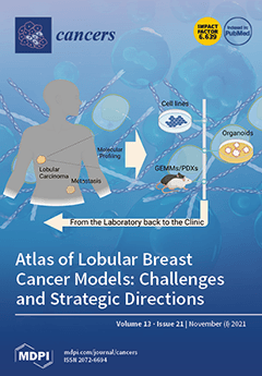
Invasive lobular carcinoma (ILC) is the most common ‘‘special type’’ breast cancer (BC) worldwide. ILC often has larger tumors on presentation and a unique pattern of metastatic spread compared to other BC types. Although molecular analyses of lobular carcinomas have recently revealed their unique mutational repertoire, preclinical models for studying ILC biology, progression, and predicting the efficacy of novel therapeutics are scarce. This study comprehensively presents the characteristics, peculiarities, and applications of the available in vitro, ex vivo, and in vivo genetically engineered mouse models (GEMMs) and patient-derived xenografts (PDX) ILC models. The data described in this paper will help to guide the experimental implementation of clinically relevant ILC models, ultimately improving the treatment of patients with ILC. Created with BioRender.com. View this paper
  • Issues are regarded as officially published after their release is announced to the table of contents alert mailing list.
  • You may sign up for e-mail alerts to receive table of contents of newly released issues.
  • PDF is the official format for papers published in both, html and pdf forms. To view the papers in pdf format, click on the "PDF Full-text" link, and use the free Adobe Reader to open them.
Order results
Result details
Section
Select all
Export citation of selected articles as:
14 pages, 2838 KiB  
Article
Response to FEC Chemotherapy and Oncolytic HSV-1 Is Associated with Macrophage Polarization and Increased Expression of S100A8/A9 in Triple Negative Breast Cancer
by Alyssa Vito, Nader El-Sayes, Omar Salem, Yonghong Wan and Karen L. Mossman
Cancers 2021, 13(21), 5590; https://doi.org/10.3390/cancers13215590 - 08 Nov 2021
Cited by 2 | Viewed by 2382
Abstract
The era of immunotherapy has seen an insurgence of novel therapies driving oncologic research and the clinical management of the disease. We have previously reported that a combination of chemotherapy (FEC) and oncolytic virotherapy (oHSV-1) can be used to sensitize otherwise non-responsive tumors [...] Read more.
The era of immunotherapy has seen an insurgence of novel therapies driving oncologic research and the clinical management of the disease. We have previously reported that a combination of chemotherapy (FEC) and oncolytic virotherapy (oHSV-1) can be used to sensitize otherwise non-responsive tumors to immune checkpoint blockade and that tumor-infiltrating B cells are required for the efficacy of our therapeutic regimen in a murine model of triple-negative breast cancer. In the studies herein, we have performed gene expression profiling using microarray analyses and have investigated the differential gene expression between tumors treated with FEC + oHSV-1 versus untreated tumors. In this work, we uncovered a therapeutically driven switch of the myeloid phenotype and a gene signature driving increased tumor cell killing. Full article
Show Figures

Figure 1

12 pages, 6662 KiB  
Article
Linearity of Age at Cancer Onset Worldwide: 25-Year Population-Based Cancer Registry Study
by Ettore Bidoli, Elda Lamaj, Tiziana Angelin, Ornella Forgiarini, Emilia De Santis and Diego Serraino
Cancers 2021, 13(21), 5589; https://0-doi-org.brum.beds.ac.uk/10.3390/cancers13215589 - 08 Nov 2021
Cited by 3 | Viewed by 1538
Abstract
Background The linear association between median age at cancer onset and median age of the underlying population has been described only for breast cancer. We quantified the shape and strength of such association for 20 cancer types using data from all population-based cancer [...] Read more.
Background The linear association between median age at cancer onset and median age of the underlying population has been described only for breast cancer. We quantified the shape and strength of such association for 20 cancer types using data from all population-based cancer registries (CRs) worldwide. Methods The patients’ median age at cancer onset and of the underlying population were extracted from all CRs listed in volumes VI (1983–1987 years) and XI (2008–2012 years) of Cancer Incidence in Five Continents. The association was assessed at cross-sectional level by linear regression models and longitudinally considering only the long-standing CRs active throughout the study period (i.e., 25-year span). Results During 2008–2012, each one-year increase in median population ageing was associated in men with a nearly half year increase of median age at onset of all cancers, but skin; and a 2/3 year increase in women. Variance explained by linear model was around 60%. In long-standing CRs a decrease in median age at cancer onset was observed for prostate and cervical cancers throughout the 25-year span. Conclusions Population ageing reflected 60% of the median age at cancer onset. Misinterpretation of peaks of cancer incidence in specific age groups may be avoided by examining population pyramids. Full article
(This article belongs to the Section Cancer Epidemiology and Prevention)
Show Figures

Figure 1

23 pages, 125196 KiB  
Article
ERAS, a Member of the Ras Superfamily, Acts as an Oncoprotein in the Mammary Gland
by Cristian Suarez-Cabrera, Isabel Ojeda-Perez, Raquel Sanchez-Baltasar, Angustias Page, Ana Bravo, Manuel Navarro and Angel Ramirez
Cancers 2021, 13(21), 5588; https://0-doi-org.brum.beds.ac.uk/10.3390/cancers13215588 - 08 Nov 2021
Viewed by 2359
Abstract
ERAS is a relatively uncharacterized gene of the Ras superfamily. It is expressed in ES cells and in the first stages of embryonic development; later on, it is silenced in the majority of cell types and tissues. Although there are several reports showing [...] Read more.
ERAS is a relatively uncharacterized gene of the Ras superfamily. It is expressed in ES cells and in the first stages of embryonic development; later on, it is silenced in the majority of cell types and tissues. Although there are several reports showing ERAS expression in tumoral cell lines and human tumor samples, it is unknown if ERAS deregulated expression is enough to drive tumor development. In this report, we have generated transgenic mice expressing ERAS in myoepithelial basal cells of the mammary gland and in basal cells of stratified epithelia. In spite of the low level of ERAS expression, these transgenic mice showed phenotypic alterations resembling overgrowth syndromes caused by the activation of the AKT-PI3K pathway. In addition, their mammary glands present developmental and functional disabilities accompanied by morphological and biochemical alterations in the myoepithelial cells. These mice suffer from tumoral transformation in the mammary glands with high incidence. These mammary tumors resemble, both histologically and by the expression of differentiation markers, malignant adenomyoepitheliomas. In sum, our results highlight the importance of ERAS silencing in adult tissues and define a truly oncogenic role for ERAS in mammary gland cells when inappropriately expressed. Full article
(This article belongs to the Special Issue Identification of Candidate Genes in Breast and Ovarian Cancer)
Show Figures

Figure 1

12 pages, 1016 KiB  
Article
Impact of Serum γ-Glutamyltransferase on Overall Survival in Men with Metastatic Castration-Resistant Prostate Cancer Treated with Docetaxel
by Minami Une, Kosuke Takemura, Kentaro Inamura, Hiroshi Fukushima, Masaya Ito, Shuichiro Kobayashi, Takeshi Yuasa, Junji Yonese, Philip G. Board and Fumitaka Koga
Cancers 2021, 13(21), 5587; https://0-doi-org.brum.beds.ac.uk/10.3390/cancers13215587 - 08 Nov 2021
Cited by 1 | Viewed by 1890
Abstract
Background: Reports on the prognostic significance of serum γ-glutamyltransferase (GGT) in men with metastatic castration-resistant prostate cancer (mCRPC) are limited. In addition, GGT expression status in cancer tissues has not been well characterized regardless of cancer types. Methods: This retrospective study included 107 [...] Read more.
Background: Reports on the prognostic significance of serum γ-glutamyltransferase (GGT) in men with metastatic castration-resistant prostate cancer (mCRPC) are limited. In addition, GGT expression status in cancer tissues has not been well characterized regardless of cancer types. Methods: This retrospective study included 107 consecutive men with mCRPC receiving docetaxel therapy. The primary endpoints were associations of serum GGT with overall survival (OS) and prostate-specific antigen (PSA) response. The secondary endpoint was an association of serum GGT with progression-free survival (PFS). Additionally, GGT expression status was immunohistochemically semi-quantified using tissue microarrays. Results: A total of 67 (63%) men died during follow-up periods (median 22.5 months for survivors). On multivariable analysis, high Log GGT was independently associated with adverse OS (HR 1.49, p = 0.006) as were low hemoglobin (HR 0.79, p = 0.002) and high PSA (HR 1.40, p < 0.001). In contrast, serum GGT was not significantly associated with PSA response or PFS. Moreover, incorporation of serum GGT into established prognostic models (i.e., Halabi and Smaletz models) increased their C-indices for predicting OS from 0.772 to 0.787 (p = 0.066) and from 0.777 to 0.785 (p = 0.118), respectively. Furthermore, there was a positive correlation between serum and tissue GGT levels (ρ = 0.53, p = 0.003). Conclusions: Serum GGT may be a prognostic biomarker in men with mCRPC receiving docetaxel therapy. GGT overexpression by prostate cancer cells appears to be responsible for the elevation of GGT in the serum. Full article
Show Figures

Figure 1

21 pages, 1211 KiB  
Review
Interplay between Cellular and Non-Cellular Components of the Tumour Microenvironment in Hepatocellular Carcinoma
by Tamás Sükei, Elena Palma and Luca Urbani
Cancers 2021, 13(21), 5586; https://0-doi-org.brum.beds.ac.uk/10.3390/cancers13215586 - 08 Nov 2021
Cited by 11 | Viewed by 3296
Abstract
Hepatocellular carcinoma (HCC) is one of the most common and lethal cancers worldwide. Currently, treatments available for advanced HCC provide dismal chances of survival, thus there is an urgent need to develop more effective therapeutic strategies. While much of the focus of recent [...] Read more.
Hepatocellular carcinoma (HCC) is one of the most common and lethal cancers worldwide. Currently, treatments available for advanced HCC provide dismal chances of survival, thus there is an urgent need to develop more effective therapeutic strategies. While much of the focus of recent decades has been on targeting malignant cells, promising results have emerged from targeting the tumour microenvironment (TME). The extracellular matrix (ECM) is the main non-cellular component of the TME and it profoundly changes during tumorigenesis to promote the growth and survival of malignant cells. Despite this, many in vitro models for drug testing fail to consider the TME leading to a high failure rate in clinical trials. Here, we present an overview of the function and properties of the ECM in the liver and how these change during malignant transformation. We also discuss the relationship between immune cells and ECM in the TME in HCC. Lastly, we present advanced, 3D culture techniques of cancer modelling and argue that the incorporation of TME components into these is essential to better recapitulate the complex interactions within the TME. Full article
Show Figures

Figure 1

15 pages, 2999 KiB  
Article
Identification of Risk Loci for Radiotoxicity in Prostate Cancer by Comprehensive Genotyping of TGFB1 and TGFBR1
by Manuel Guhlich, Laura Hubert, Caroline Patricia Nadine Mergler, Margret Rave-Fraenk, Leif Hendrik Dröge, Martin Leu, Heinz Schmidberger, Stefan Rieken, Andrea Hille and Markus Anton Schirmer
Cancers 2021, 13(21), 5585; https://0-doi-org.brum.beds.ac.uk/10.3390/cancers13215585 - 08 Nov 2021
Viewed by 1724
Abstract
Genetic variability in transforming growth factor beta pathway (TGFB) was suggested to affect adverse events of radiotherapy. We investigated comprehensive variability in TGFB1 (gene coding for TGFβ1 ligand) and TGFBR1 (TGFβ receptor-1) in relation to radiotoxicity. Prostate cancer patients treated with [...] Read more.
Genetic variability in transforming growth factor beta pathway (TGFB) was suggested to affect adverse events of radiotherapy. We investigated comprehensive variability in TGFB1 (gene coding for TGFβ1 ligand) and TGFBR1 (TGFβ receptor-1) in relation to radiotoxicity. Prostate cancer patients treated with primary radiotherapy (n = 240) were surveyed for acute and late toxicity. Germline polymorphisms (n = 40) selected to cover the common genetic variability in TGFB1 and TGFBR1 were analyzed in peripheral blood cells. Human lymphoblastoid cell lines (LCLs) were used to evaluate a possible impact of TGFB1 and TGFBR1 genetic polymorphisms to DNA repair capacity following single irradiation with 3 Gy. Upon adjustment for multiplicity testing, rs10512263 in TGFBR1 showed a statistically significant association with acute radiation toxicity. Carriers of the Cytosine (C)-variant allele (n = 35) featured a risk ratio of 2.17 (95%-CI 1.41–3.31) for acute toxicity ≥ °2 compared to Thymine/Thymine (TT)-wild type individuals (n = 205). Reduced DNA repair capacity in the presence of the C-allele of rs10512263 might be a mechanistic explanation as demonstrated in LCLs following irradiation. The risk for late radiotoxicity was increased by carrying at least two risk genotypes at three polymorphic sites, including Leu10Pro in TGFB1. Via comprehensive genotyping of TGFB1 and TGFBR1, promising biomarkers for radiotoxicity in prostate cancer were identified. Full article
(This article belongs to the Special Issue Emerging Biomarkers Used in Radiochemotherapy)
Show Figures

Figure 1

19 pages, 2881 KiB  
Article
Prediction of Radiation-Induced Hypothyroidism Using Radiomic Data Analysis Does Not Show Superiority over Standard Normal Tissue Complication Models
by Urszula Smyczynska, Szymon Grabia, Zuzanna Nowicka, Anna Papis-Ubych, Robert Bibik, Tomasz Latusek, Tomasz Rutkowski, Jacek Fijuth, Wojciech Fendler and Bartlomiej Tomasik
Cancers 2021, 13(21), 5584; https://0-doi-org.brum.beds.ac.uk/10.3390/cancers13215584 - 08 Nov 2021
Cited by 4 | Viewed by 2048
Abstract
State-of-art normal tissue complication probability (NTCP) models do not take into account more complex individual anatomical variations, which can be objectively quantitated and compared in radiomic analysis. The goal of this project was development of radiomic NTCP model for radiation-induced hypothyroidism (RIHT) using [...] Read more.
State-of-art normal tissue complication probability (NTCP) models do not take into account more complex individual anatomical variations, which can be objectively quantitated and compared in radiomic analysis. The goal of this project was development of radiomic NTCP model for radiation-induced hypothyroidism (RIHT) using imaging biomarkers (radiomics). We gathered CT images and clinical data from 98 patients, who underwent intensity-modulated radiation therapy (IMRT) for head and neck cancers with a planned total dose of 70.0 Gy (33–35 fractions). During the 28-month (median) follow-up 27 patients (28%) developed RIHT. For each patient, we extracted 1316 radiomic features from original and transformed images using manually contoured thyroid masks. Creating models based on clinical, radiomic features or a combination thereof, we considered 3 variants of data preprocessing. Based on their performance metrics (sensitivity, specificity), we picked best models for each variant ((0.8, 0.96), (0.9, 0.93), (0.9, 0.89) variant-wise) and compared them with external NTCP models ((0.82, 0.88), (0.82, 0.88), (0.76, 0.91)). We showed that radiomic-based models did not outperform state-of-art NTCP models (p > 0.05). The potential benefit of radiomic-based approach is that it is dose-independent, and models can be used prior to treatment planning allowing faster selection of susceptible population. Full article
(This article belongs to the Special Issue Radiomics/Radiogenomics in Cancer)
Show Figures

Figure 1

57 pages, 4650 KiB  
Review
In Vivo and In Vitro Models of Hepatocellular Carcinoma: Current Strategies for Translational Modeling
by Guilherme Ribeiro Romualdo, Kaat Leroy, Cícero Júlio Silva Costa, Gabriel Bacil Prata, Bart Vanderborght, Tereza Cristina da Silva, Luís Fernando Barbisan, Wellington Andraus, Lindsey Devisscher, Niels Olsen Saraiva Câmara, Mathieu Vinken and Bruno Cogliati
Cancers 2021, 13(21), 5583; https://0-doi-org.brum.beds.ac.uk/10.3390/cancers13215583 - 08 Nov 2021
Cited by 17 | Viewed by 6223
Abstract
Hepatocellular carcinoma (HCC) is the sixth most common cancer worldwide and the third leading cause of cancer-related death globally. HCC is a complex multistep disease and usually emerges in the setting of chronic liver diseases. The molecular pathogenesis of HCC varies according to [...] Read more.
Hepatocellular carcinoma (HCC) is the sixth most common cancer worldwide and the third leading cause of cancer-related death globally. HCC is a complex multistep disease and usually emerges in the setting of chronic liver diseases. The molecular pathogenesis of HCC varies according to the etiology, mainly caused by chronic hepatitis B and C virus infections, chronic alcohol consumption, aflatoxin-contaminated food, and non-alcoholic fatty liver disease associated with metabolic syndrome or diabetes mellitus. The establishment of HCC models has become essential for both basic and translational research to improve our understanding of the pathophysiology and unravel new molecular drivers of this disease. The ideal model should recapitulate key events observed during hepatocarcinogenesis and HCC progression in view of establishing effective diagnostic and therapeutic strategies to be translated into clinical practice. Despite considerable efforts currently devoted to liver cancer research, only a few anti-HCC drugs are available, and patient prognosis and survival are still poor. The present paper provides a state-of-the-art overview of in vivo and in vitro models used for translational modeling of HCC with a specific focus on their key molecular hallmarks. Full article
Show Figures

Figure 1

19 pages, 7655 KiB  
Systematic Review
Transcriptomics, Epigenetics, and Metabolomics of Primary Aldosteronism
by Ariadni Spyroglou, George P. Piaditis, Gregory Kaltsas and Krystallenia I. Alexandraki
Cancers 2021, 13(21), 5582; https://0-doi-org.brum.beds.ac.uk/10.3390/cancers13215582 - 08 Nov 2021
Cited by 6 | Viewed by 2679
Abstract
Introduction: Primary aldosteronism (PA) is the most common cause of endocrine hypertension, mainly caused by aldosterone-producing adenomas or hyperplasia; understanding its pathophysiological background is important in order to provide ameliorative treatment strategies. Over the past several years, significant progress has been documented in [...] Read more.
Introduction: Primary aldosteronism (PA) is the most common cause of endocrine hypertension, mainly caused by aldosterone-producing adenomas or hyperplasia; understanding its pathophysiological background is important in order to provide ameliorative treatment strategies. Over the past several years, significant progress has been documented in this field, in particular in the clarification of the genetic and molecular mechanisms responsible for the pathogenesis of aldosterone-producing adenomas (APAs). Methods: Systematic searches of the PubMed and Cochrane databases were performed for all human studies applying transcriptomic, epigenetic or metabolomic analyses to PA subjects. Studies involving serial analysis of gene expression and microarray, epigenetic studies with methylome analyses and micro-RNA expression profiles, and metabolomic studies focused on improving understanding of the regulation of autonomous aldosterone production in PA were all included. Results: In this review we summarize the main findings in this area and analyze the interplay between primary aldosteronism and several signaling pathways with differential regulation of the RNA and protein expression of several factors involved in, among others, steroidogenesis, calcium signaling, and nuclear, membrane and G-coupled protein receptors. Distinct transcriptomic and metabolomic patterns are also presented herein, depending on the mutational status of APAs. In particular, two partially opposite transcriptional and steroidogenic profiles appear to distinguish APAs carrying a KCNJ5 mutation from all other APAs, which carry different mutations. Conclusions: These findings can substantially contribute to the development of personalized treatment in patients with PA. Full article
Show Figures

Figure 1

15 pages, 2044 KiB  
Article
Impact of the Hypoechogenicity Criteria on Thyroid Nodule Malignancy Risk Stratification Performance by Different TIRADS Systems
by Nina Malika Popova, Maija Radzina, Peteris Prieditis, Mara Liepa, Madara Rauda and Kaspars Stepanovs
Cancers 2021, 13(21), 5581; https://0-doi-org.brum.beds.ac.uk/10.3390/cancers13215581 - 08 Nov 2021
Cited by 4 | Viewed by 3529
Abstract
Background: Various Thyroid Imaging and Reporting data systems (TIRADS) are used worldwide for risk stratification of thyroid nodules. Their sensitivity is high, while the specificity is suboptimal. The aim of the study was to compare several TIRADS systems and evaluate the effect of [...] Read more.
Background: Various Thyroid Imaging and Reporting data systems (TIRADS) are used worldwide for risk stratification of thyroid nodules. Their sensitivity is high, while the specificity is suboptimal. The aim of the study was to compare several TIRADS systems and evaluate the effect of hypoechogenicity as a sign of risk of malignancy on the overall assessment of diagnostic accuracy. Methods: The prospective study includes 274 patients with 289 thyroid nodules to whom US and risk of malignancy were assessed according to four TIRADS systems—European (EU-TIRADS), Korean (K-TIRADS), TIRADS by American College of Radiology (ACR TIRADS), and modified Kwak et al. TIRADS (L-TIRADS) systems, in which mild hypoechogenicity is not included in malignancy risk suggestive signs. For all thyroid nodules, a fine needle aspiration (FNA) biopsy was performed and evaluated according to the Bethesda system. For all systems, diagnostic accuracy was calculated. Results: Assessing the echogenicity of the thyroid nodules: from 81 of isoechogenic nodules, 2 were malignant (2.1%), from 151 mild hypoechogenic, 18 (12%) were malignant, and from 48 marked hypoechogenic nodules, 16 (33%) were malignant. In 80 thyroid nodules, mild hypoechogenicity was the only sign of malignancy and none appeared malignant. Assessing various TIRADS systems on the same cohort, sensitivity, specificity, PPV, NPV, and accuracy, firstly for EU-TIRADS, they were 97.2%; 39.9%; 18.7%; 99.0%, and 73.3%, respectively; for K-TIRADS they were 97.2%; 46.6%; 20.6%; 99.2%, and 53.9%; for ACR-TIRADS they were 97.2%; 41.1%, 19.0%; 99.0%, and 48.0%, respectively; finally, for L-TIRADS they were 80.6%; 72.7%; 29.6%; 96.3%, and 73.3%. Conclusions: This comparative research has highlighted that applying different TIRADS systems can alter the number of necessary biopsies by re-categorization of the thyroid nodules. The main pattern that affected differences was inconsistent hypoechogenicity interpretation, giving the accuracy superiority to the systems that raise the malignancy risk with marked hypoechogenicity, at the same time with minor compensation for sensitivity. Full article
Show Figures

Figure 1

20 pages, 3535 KiB  
Article
A Prostate Cancer Proteomics Database for SWATH-MS Based Protein Quantification
by Ammara Muazzam, Davide Chiasserini, Janet Kelsall, Nophar Geifman, Anthony D. Whetton and Paul A. Townsend
Cancers 2021, 13(21), 5580; https://0-doi-org.brum.beds.ac.uk/10.3390/cancers13215580 - 08 Nov 2021
Cited by 6 | Viewed by 2909
Abstract
Prostate cancer is the most frequent form of cancer in men, accounting for more than one-third of all cases. Current screening techniques, such as PSA testing used in conjunction with routine procedures, lead to unnecessary biopsies and the discovery of low-risk tumours, resulting [...] Read more.
Prostate cancer is the most frequent form of cancer in men, accounting for more than one-third of all cases. Current screening techniques, such as PSA testing used in conjunction with routine procedures, lead to unnecessary biopsies and the discovery of low-risk tumours, resulting in overdiagnosis. SWATH-MS is a well-established data-independent (DI) method requiring prior knowledge of targeted peptides to obtain valuable information from SWATH maps. In response to the growing need to identify and characterise protein biomarkers for prostate cancer, this study explored a spectrum source for targeted proteome analysis of blood samples. We created a comprehensive prostate cancer serum spectral library by combining data-dependent acquisition (DDA) MS raw files from 504 patients with low, intermediate, or high-grade prostate cancer and healthy controls, as well as 304 prostate cancer-related protein in silico assays. The spectral library contains 114,684 transitions, which equates to 18,479 peptides translated into 1227 proteins. The robustness and accuracy of the spectral library were assessed to boost confidence in the identification and quantification of prostate cancer-related proteins across an independent cohort, resulting in the identification of 404 proteins. This unique database can facilitate researchers to investigate prostate cancer protein biomarkers in blood samples. In the real-world use of the spectrum library for biomarker detection, using a signature of 17 proteins, a clear distinction between the validation cohort’s pre- and post-treatment groups was observed. Data are available via ProteomeXchange with identifier PXD028651. Full article
Show Figures

Figure 1

18 pages, 11001 KiB  
Article
Gossypol Treatment Restores Insufficient Apoptotic Function of DFF40/CAD in Human Glioblastoma Cells
by Laura Martínez-Escardó, Montse Alemany, María Sánchez-Osuna, Alejandro Sánchez-Chardi, Meritxell Roig-Martínez, Salvio Suárez-García, Daniel Ruiz-Molina, Noemi Vidal, Gerard Plans, Carles Majós, Judit Ribas, María Antonia Baltrons, Jose R. Bayascas, Carlos Barcia, Jordi Bruna and Victor J. Yuste
Cancers 2021, 13(21), 5579; https://0-doi-org.brum.beds.ac.uk/10.3390/cancers13215579 - 08 Nov 2021
Cited by 2 | Viewed by 3872
Abstract
Glioblastoma (GBM) is a highly aggressive brain tumor and almost all patients die because of relapses. GBM-derived cells undergo cell death without nuclear fragmentation upon treatment with different apoptotic agents. Nuclear dismantling determines the point-of-no-return in the apoptotic process. DFF40/CAD is the main [...] Read more.
Glioblastoma (GBM) is a highly aggressive brain tumor and almost all patients die because of relapses. GBM-derived cells undergo cell death without nuclear fragmentation upon treatment with different apoptotic agents. Nuclear dismantling determines the point-of-no-return in the apoptotic process. DFF40/CAD is the main endonuclease implicated in apoptotic nuclear disassembly. To be properly activated, DFF40/CAD should reside in the cytosol. However, the endonuclease is poorly expressed in the cytosol and remains cumulated in the nucleus of GBM cells. Here, by employing commercial and non-commercial patient-derived GBM cells, we demonstrate that the natural terpenoid aldehyde gossypol prompts DFF40/CAD-dependent nuclear fragmentation. A comparative analysis between gossypol- and staurosporine-treated cells evidenced that levels of neither caspase activation nor DNA damage were correlated with the ability of each compound to induce nuclear fragmentation. Deconvoluted confocal images revealed that DFF40/CAD was almost completely excluded from the nucleus early after the staurosporine challenge. However, gossypol-treated cells maintained DFF40/CAD in the nucleus for longer times, shaping a ribbon-like structure piercing the nuclear fragments and building a network of bridged masses of compacted chromatin. Therefore, GBM cells can fragment their nuclei if treated with the adequate insult, making the cell death process irreversible. Full article
(This article belongs to the Section Molecular Cancer Biology)
Show Figures

Figure 1

15 pages, 2367 KiB  
Article
Highly Expressed Progesterone Receptor B Isoform Increases Platinum Sensitivity and Survival of Ovarian High-Grade Serous Carcinoma
by Hao Lin, Kuo-Chung Lan, Yu-Che Ou, Chen-Hsuan Wu, Hong-Yo Kang, I-Chieh Chuang and Hung-Chun Fu
Cancers 2021, 13(21), 5578; https://0-doi-org.brum.beds.ac.uk/10.3390/cancers13215578 - 08 Nov 2021
Cited by 3 | Viewed by 2128
Abstract
Background: Expression of the progesterone receptor (PR) has been reported to influence survival outcomes in patients with ovarian high-grade serous carcinoma (HGSC). In the present study, we attempted to investigate the association among PR and its isoforms’ expression, platinum sensitivity, and survival in [...] Read more.
Background: Expression of the progesterone receptor (PR) has been reported to influence survival outcomes in patients with ovarian high-grade serous carcinoma (HGSC). In the present study, we attempted to investigate the association among PR and its isoforms’ expression, platinum sensitivity, and survival in ovarian HGSC. Material and methods: This retrospective study reviewed ovarian HGSC patients who received surgery followed by adjuvant chemotherapy. We analyzed total PR and PR isoform-B (PR-B) expression by immunohistochemical staining and quantified using the H-score. Then, we compared platinum sensitivity and survival outcomes between those patients with weak and strong PR-B expression. Cisplatin viability assays were carried out in ovarian HGSC cell lines (OC-3-VGH and OVCAR-3) with different PR-B expression. Results: Among 90 patients, 49 and 41 patients were considered to have platinum-sensitive and platinum-resistant disease, respectively. Pearson’s correlation model showed that the H-score of total PR correlated positively with PR-B (r = 0.813). The PR-B H-score of tumors was significantly higher in the platinum-sensitive group (p = 0.004). Multivariate analysis revealed that the PR-B H-score and optimal debulking status were independent factors predicting platinum sensitivity. When compared with strong PR-B expression, patients with weak PR-B had significantly poorer progression-free (p = 0.021) and cancer-specific survival (p = 0.046). In a cell model, cisplatin-resistant OC-3-VGH cells expressed a lower level of PR-B than wild-type cells. Overexpression of PR-B or progesterone could increase cisplatin sensitivity in both OC-3-VGH and OVCAR-3 cells via the mechanism of promoting cisplatin-related apoptosis. Conclusions: When compared to weak PR-B, ovarian HGSC patients with a strong PR-B expression had a better chance of platinum sensitivity and survival, and this finding was compatible with our experimental results. Progesterone seemed to be a platinum sensitizer, but the value of adding progesterone in the treatment of ovarian HGSC should be further investigated. Full article
Show Figures

Graphical abstract

16 pages, 1988 KiB  
Article
Variable Expression of the Disialoganglioside GD2 in Breast Cancer Molecular Subtypes
by Ramona Erber, Sareetha Kailayangiri, Hanna Huebner, Matthias Ruebner, Arndt Hartmann, Lothar Häberle, Julia Meyer, Simon Völkl, Andreas Mackensen, Laura Landgraf, Carol I. Geppert, Rüdiger Schulz-Wendtland, Matthias W. Beckmann, Peter A. Fasching, Nicole Farwick, Claudia Rossig and Paul Gass
Cancers 2021, 13(21), 5577; https://0-doi-org.brum.beds.ac.uk/10.3390/cancers13215577 - 08 Nov 2021
Cited by 5 | Viewed by 2640
Abstract
The disialoganglioside GD2 is a tumor-associated antigen that may allow for the application of targeted immunotherapies (anti-GD2 antibodies, GD2 CAR T cells) in patients with neuroblastoma and other solid tumors. We retrospectively investigated GD2 expression in a breast cancer cohort, using immunohistochemistry (IHC) [...] Read more.
The disialoganglioside GD2 is a tumor-associated antigen that may allow for the application of targeted immunotherapies (anti-GD2 antibodies, GD2 CAR T cells) in patients with neuroblastoma and other solid tumors. We retrospectively investigated GD2 expression in a breast cancer cohort, using immunohistochemistry (IHC) and immunofluorescence (IF) on tissue microarrays (TMAs), and its impact on survival. GD2 expression on IHC (n = 568) and IF (n = 503) was investigated in relation to subtypes and patient outcome. Overall, 50.2% of the 568 IHC-assessed samples and 69.8% of the 503 IF-assessed samples were GD2-positive. The highest proportion of GD2-positive tumors was observed in luminal tumors. Significantly fewer GD2-positive cases were detected in triple-negative breast cancer (TNBC) compared with other subtypes. The proportion of GD2-expressing tumors were significantly lower in HER2-positive breast cancer in comparison with luminal tumors on IF staining (but not IHC). GD2 expression of IHC or IF was not significantly associated with disease-free or overall survival, in either the overall cohort or in individual subtypes. However, GD2 expression can be seen in more than 50% of breast cancer cases, with the highest frequency in hormone receptor-positive tumors. With this high expression frequency, patients with GD2-positive advanced breast cancer of all subtypes may benefit from GD2-targeting immunotherapies, which are currently subject to clinical testing. Full article
Show Figures

Figure 1

1 pages, 146 KiB  
Editorial
Metastasis and Tumor Cell Migration of Solid Tumors
by Jens Hoeppner and Peter Bronsert
Cancers 2021, 13(21), 5576; https://0-doi-org.brum.beds.ac.uk/10.3390/cancers13215576 - 08 Nov 2021
Cited by 2 | Viewed by 1377
Abstract
The developmental process of local and distant metastases represents the major and defining trait of malignant tumors, whereby tumor cells sustain the capability to migrate from the initial tumor site, seed, and grow at a location other than that of the initial tumor [...] Read more.
The developmental process of local and distant metastases represents the major and defining trait of malignant tumors, whereby tumor cells sustain the capability to migrate from the initial tumor site, seed, and grow at a location other than that of the initial tumor [...] Full article
(This article belongs to the Special Issue Metastasis and Tumor Cell Migration of Solid Tumors)
21 pages, 711 KiB  
Review
Autophagy in Cancer Therapy—Molecular Mechanisms and Current Clinical Advances
by Ingo Ganzleben, Markus F. Neurath and Christoph Becker
Cancers 2021, 13(21), 5575; https://0-doi-org.brum.beds.ac.uk/10.3390/cancers13215575 - 08 Nov 2021
Cited by 11 | Viewed by 3233
Abstract
Autophagy is a crucial general survival tactic of mammalian cells. It describes the capability of cells to disassemble and partially recycle cellular components (e.g., mitochondria) in case they are damaged and pose a risk to cell survival or simply if their resources are [...] Read more.
Autophagy is a crucial general survival tactic of mammalian cells. It describes the capability of cells to disassemble and partially recycle cellular components (e.g., mitochondria) in case they are damaged and pose a risk to cell survival or simply if their resources are urgently needed elsewhere at the time. Autophagy-associated pathomechanisms have been increasingly recognized as important disease mechanisms in non-malignant (neurodegeneration, diffuse parenchymal lung disease) and malignant conditions alike. However, the overall consequences of autophagy for the organism depend particularly on the greater context in which autophagy occurs, such as the cell type or whether the cell is proliferating. In cancer, autophagy sustains cancer cell survival under challenging, i.e., resource-depleted, conditions. However, this leads to situations in which cancer cells are completely dependent on autophagy. Accordingly, autophagy represents a promising yet complex target in cancer treatment with therapeutically induced increase and decrease of autophagic flux as important therapeutic principles. Full article
(This article belongs to the Special Issue Targeting Autophagy for Cancer Treatment)
Show Figures

Figure 1

17 pages, 2295 KiB  
Article
Type I Interferon Promotes Antitumor T Cell Response in CRPC by Regulating MDSC
by Lilv Fan, Guiliang Xu, Jingjing Cao, Min Li, Huihui Zhang, Fanlin Li, Xinyue Qi, Xiaoqing Zhang, Zeyu Li, Ping Han and Xuanming Yang
Cancers 2021, 13(21), 5574; https://0-doi-org.brum.beds.ac.uk/10.3390/cancers13215574 - 08 Nov 2021
Cited by 5 | Viewed by 2495
Abstract
Background: Metastatic castration-resistant prostate cancer (CRPC) is the leading cause of death among prostate cancer patients. Here, our aim was to ascertain the immune regulatory mechanisms involved in CRPC development and identify potential immunotherapies against CRPC. Methods: A CRPC model was established using [...] Read more.
Background: Metastatic castration-resistant prostate cancer (CRPC) is the leading cause of death among prostate cancer patients. Here, our aim was to ascertain the immune regulatory mechanisms involved in CRPC development and identify potential immunotherapies against CRPC. Methods: A CRPC model was established using Myc-CaP cells in immune-competent FVB mice following castration. The immune cell profile of the tumor microenvironment (TME) was analyzed during CRPC development. Different immunotherapies were screened in the CRPC tumor model, and their efficacies and underlying mechanisms were investigated in vitro and in vivo. Results: During CRPC development, the proportion of granulocytic myeloid-derived suppressor cells (G-MDSCs) in the TME increased. Among the immunotherapies tested, IFNα was more effective than anti-PD-L1, anti-CTLA-4, anti-4-1BB, IL-2, and IL-9 in reducing Myc-CaP CRPC tumor growth. IFNα reduced the number of G-MDSCs both in vitro during differentiation and in vivo in CRPC mice. Furthermore, IFNα reduced the suppressive function of G-MDSCs on T cell proliferation and activation. Conclusion: G-MDSCs are crucial to effective immunotherapy against CRPC. Treatment with IFNα presents a promising therapeutic strategy against CRPC. Besides the direct inhibition of tumor growth and the promotion of T cell priming, IFNα reduces the number and the suppressive function of G-MDSCs and restores T cell activation. Full article
Show Figures

Figure 1

19 pages, 3670 KiB  
Article
Screening for the Key Proteins Associated with Rete Testis Invasion in Clinical Stage I Seminoma via Label-Free Quantitative Mass Spectrometry
by Lucia Borszéková Pulzová, Jan Roška, Michal Kalman, Ján Kliment, Pavol Slávik, Božena Smolková, Eduard Goffa, Dana Jurkovičová, Ľudovít Kulcsár, Katarína Lešková, Peter Bujdák, Michal Mego, Mangesh R. Bhide, Lukáš Plank and Miroslav Chovanec
Cancers 2021, 13(21), 5573; https://0-doi-org.brum.beds.ac.uk/10.3390/cancers13215573 - 08 Nov 2021
Cited by 4 | Viewed by 2165
Abstract
Rete testis invasion (RTI) is an unfavourable prognostic factor for the risk of relapse in clinical stage I (CS I) seminoma patients. Notably, no evidence of difference in the proteome of RTI-positive vs. -negative CS I seminomas has been reported yet. Here, a [...] Read more.
Rete testis invasion (RTI) is an unfavourable prognostic factor for the risk of relapse in clinical stage I (CS I) seminoma patients. Notably, no evidence of difference in the proteome of RTI-positive vs. -negative CS I seminomas has been reported yet. Here, a quantitative proteomic approach was used to investigate RTI-associated proteins. 64 proteins were differentially expressed in RTI-positive compared to -negative CS I seminomas. Of them, 14-3-3γ, ezrin, filamin A, Parkinsonism-associated deglycase 7 (PARK7), vimentin and vinculin, were validated in CS I seminoma patient cohort. As shown by multivariate analysis controlling for clinical confounders, PARK7 and filamin A expression lowered the risk of RTI, while 14-3-3γ expression increased it. Therefore, we suggest that in real clinical biopsy specimens, the expression level of these proteins may reflect prognosis in CS I seminoma patients. Full article
(This article belongs to the Special Issue Testicular Cancer)
Show Figures

Figure 1

9 pages, 814 KiB  
Article
Letermovir Prophylaxis and Cytomegalovirus Reactivation in Adult Hematopoietic Cell Transplant Recipients with and without Acute Graft Versus Host Disease
by Delaney Wolfe, Qiuhong Zhao, Emma Siegel, Marcin Puto, Danielle Murphy, Julianna Roddy, Yvonne Efebera and Justin Tossey
Cancers 2021, 13(21), 5572; https://0-doi-org.brum.beds.ac.uk/10.3390/cancers13215572 - 08 Nov 2021
Cited by 13 | Viewed by 3243
Abstract
Cytomegalovirus (CMV) is the most clinically significant infection after allogeneic hematopoietic-cell transplantation (allo-HCT) and is associated with increased mortality. The risk for CMV reactivation increases with graft versus host disease (GVHD). GVHD contributes to significant morbidity and mortality and is treated with immunosuppressive [...] Read more.
Cytomegalovirus (CMV) is the most clinically significant infection after allogeneic hematopoietic-cell transplantation (allo-HCT) and is associated with increased mortality. The risk for CMV reactivation increases with graft versus host disease (GVHD). GVHD contributes to significant morbidity and mortality and is treated with immunosuppressive therapies that can further increase CMV infection risk. Prophylaxis with letermovir, an oral antiviral approved to prevent CMV, has been shown to decrease the incidence of CMV infection post-allo-HCT in patients at high risk of CMV reactivation, but there is a lack of data confirming this benefit in patients with GVHD. In this single-center, retrospective study, we assessed the incidence of clinically significant CMV infection (CS-CMVi) in allo-HCT patients who received letermovir prophylaxis (n = 119) and who developed aGVHD compared to a control group (n = 143) who did not receive letermovir. Among aGVHD patients, letermovir prophylaxis decreased CS-CMVi in patients with aGVHD (HR 0.08 [95% CI 0.03–0.27], p < 0.001), reduced non-relapsed mortality (p = 0.04) and improved overall survival (p = 0.04). This data suggests that letermovir prophylaxis improves outcomes by preventing CS-CMVi in patients with aGVHD. Full article
(This article belongs to the Special Issue Autologous and Allogeneic Stem Cell Transplant in Cancer Therapy)
Show Figures

Graphical abstract

27 pages, 7879 KiB  
Article
Dual Hyaluronic Acid and Folic Acid Targeting pH-Sensitive Multifunctional 2DG@DCA@MgO-Nano-Core–Shell-Radiosensitizer for Breast Cancer Therapy
by Mostafa A. Askar, Noura M. Thabet, Gharieb S. El-Sayyad, Ahmed I. El-Batal, Mohamed Abd Elkodous, Omama E. El Shawi, Hamed Helal and Mohamed K. Abdel-Rafei
Cancers 2021, 13(21), 5571; https://0-doi-org.brum.beds.ac.uk/10.3390/cancers13215571 - 07 Nov 2021
Cited by 13 | Viewed by 4772
Abstract
Globally, breast cancer (BC) poses a serious public health risk. The disease exhibits a complex heterogeneous etiology and is associated with a glycolytic and oxidative phosphorylation (OXPHOS) metabolic reprogramming phenotype, which fuels proliferation and progression. Due to the late manifestation of symptoms, rigorous [...] Read more.
Globally, breast cancer (BC) poses a serious public health risk. The disease exhibits a complex heterogeneous etiology and is associated with a glycolytic and oxidative phosphorylation (OXPHOS) metabolic reprogramming phenotype, which fuels proliferation and progression. Due to the late manifestation of symptoms, rigorous treatment regimens are required following diagnosis. Existing treatments are limited by a lack of specificity, systemic toxicity, temporary remission, and radio-resistance in BC. In this study, we have developed CD44 and folate receptor-targeting multi-functional dual drug-loaded nanoparticles. This composed of hyaluronic acid (HA) and folic acid (FA) conjugated to a 2-deoxy glucose (2DG) shell linked to a layer of dichloroacetate (DCA) and a magnesium oxide (MgO) core (2DG@DCA@MgO; DDM) to enhance the localized chemo-radiotherapy for effective BC treatment. The physicochemical properties of nanoparticles including stability, selectivity, responsive release to pH, cellular uptake, and anticancer efficacy were thoroughly examined. Mechanistically, we identified multiple component signaling pathways as important regulators of BC metabolism and mediators for the inhibitory effects elicited by DDM. Nanoparticles exhibited sustained DDM release properties in a bio-relevant media, which was responsive to the acidic pH enabling eligibility to the control of drug release from nanoparticles. DDM-loaded and HA–FA-functionalized nanoparticles exhibited increased selectivity and uptake by BC cells. Cell-based assays revealed that the functionalized DDM significantly suppressed cancer cell growth and improved radiotherapy (RT) through inducing cell cycle arrest, enhancing apoptosis, and modulating glycolytic and OXPHOS pathways. By highlighting DDM mechanisms as an antitumor and radio-sensitizing reagent, our data suggest that glycolytic and OXPHOS pathway modulation occurs via the PI3K/AKT/mTOR/NF-κB/VEGFlow and P53high signaling pathway. In conclusion, the multi-functionalized DDM opposed tumor-associated metabolic reprogramming via multiple signaling pathways in BC cells as a promising targeted metabolic approach. Full article
(This article belongs to the Special Issue Development of Innovative Formulations for Breast Cancer Chemotherapy)
Show Figures

Graphical abstract

26 pages, 1952 KiB  
Review
Radioimmunotherapy in Oncology: Overview of the Last Decade Clinical Trials
by Aurélie Rondon, Jacques Rouanet and Françoise Degoul
Cancers 2021, 13(21), 5570; https://0-doi-org.brum.beds.ac.uk/10.3390/cancers13215570 - 07 Nov 2021
Cited by 18 | Viewed by 4254
Abstract
The specific irradiation of tumors with selective radiolabeled antibodies constitutes an attractive therapeutic approach. Consequent preclinical research has been conducted by both biologists to identify pertinent targets and to select corresponding antibodies (mAb) and by radiochemists to radiolabel mAbs. These numerous preclinical investigations [...] Read more.
The specific irradiation of tumors with selective radiolabeled antibodies constitutes an attractive therapeutic approach. Consequent preclinical research has been conducted by both biologists to identify pertinent targets and to select corresponding antibodies (mAb) and by radiochemists to radiolabel mAbs. These numerous preclinical investigations have ascertained the therapeutic interest of radioimmunotherapy (RIT) protocols in mice models. Here, we summarize the clinical studies that have been performed the last decade, including clinical trials (phases I, II, and III), prospective and retrospective studies, and cases series. We thereby reported 92 clinical studies. Among them, 62 concern the treatment of hematological malignancies, and 30 concern solid tumors. For hematologic diseases, the analysis was complex due to the high discrepancy of therapeutic strategies (first-line therapy, consolidation, stem cell transplantation conditioning) as well as the high variety of malignancies that were treated. The clinical studies from the last decade failed to expand anti-CD20 RIT indications but confirmed that RIT using radiolabeled anti-CD20 remains a pertinent choice for patients with relapse follicular lymphomas. For solid tumors, the positive benefit of RIT is more mitigated, apart for few malignancies that can be treated locally. Clinical trials also demonstrated the potential of some antibody formats, such as F(ab′)2, which has already been approved by the China State FDA under the trend name Licartin®. Despite disparate results, mAb fragments are an interesting prospect for the improvement of RIT efficiency as well as for pretargeted strategies that delay the injection of radioactive treatments from the mAb ones. Full article
(This article belongs to the Special Issue Molecular Imaging and Radio-Nuclide Therapy in Cancers)
Show Figures

Graphical abstract

26 pages, 4408 KiB  
Article
Targeting DDX3X Helicase Activity with BA103 Shows Promising Therapeutic Effects in Preclinical Glioblastoma Models
by Annalaura Brai, Valentina Riva, Letizia Clementi, Lucia Falsitta, Claudio Zamperini, Virginia Sinigiani, Claudio Festuccia, Samantha Sabetta, Davide Aiello, Camilla Roselli, Anna Garbelli, Claudia Immacolata Trivisani, Laura Maccari, Francesca Bugli, Maurizio Sanguinetti, Pierpaolo Calandro, Mario Chiariello, Paola Quaranta, Lorenzo Botta, Adriano Angelucci, Giovanni Maga and Maurizio Bottaadd Show full author list remove Hide full author list
Cancers 2021, 13(21), 5569; https://0-doi-org.brum.beds.ac.uk/10.3390/cancers13215569 - 07 Nov 2021
Cited by 5 | Viewed by 3372
Abstract
DDX3X is an ATP-dependent RNA helicase that has recently attracted interest for its involvement in viral replication and oncogenic progression. Starting from hit compounds previously identified by our group, we have designed and synthesized a new series of DDX3X inhibitors that effectively blocked [...] Read more.
DDX3X is an ATP-dependent RNA helicase that has recently attracted interest for its involvement in viral replication and oncogenic progression. Starting from hit compounds previously identified by our group, we have designed and synthesized a new series of DDX3X inhibitors that effectively blocked its helicase activity. These new compounds were able to inhibit the proliferation of cell lines from different cancer types, also in DDX3X low-expressing cancer cell lines. According to the absorption, distribution, metabolism, elimination properties, and antitumoral activity, compound BA103 was chosen to be further investigated in glioblastoma models. BA103 determined a significant reduction in the proliferation and migration of U87 and U251 cells, downregulating the oncogenic protein β-catenin. An in vivo evaluation demonstrated that BA103 was able to reach the brain and reduce the tumor growth in xenograft and orthotopic models without evident side effects. This study represents the first demonstration that DDX3X-targeted small molecules are feasible and promising drugs also in glioblastoma. Full article
(This article belongs to the Section Cancer Therapy)
Show Figures

Graphical abstract

10 pages, 847 KiB  
Article
Faecal Diagnostic Biomarkers for Colorectal Cancer
by Andrea Cruz, Carla M. Carvalho, Alexandra Cunha, Anais Crespo, Águeda Iglesias, Laura García-Nimo, Paulo P. Freitas and Joaquín Cubiella
Cancers 2021, 13(21), 5568; https://0-doi-org.brum.beds.ac.uk/10.3390/cancers13215568 - 07 Nov 2021
Cited by 7 | Viewed by 2385
Abstract
Background: Colorectal cancer (CRC) is a major cause of cancer-related death worldwide. Cancer progression, including invasion and metastasis, is a major cause of death among CRC patients. Current methods for CRC screening commonly consist of a combination of faecal immunochemical test (FIT) for [...] Read more.
Background: Colorectal cancer (CRC) is a major cause of cancer-related death worldwide. Cancer progression, including invasion and metastasis, is a major cause of death among CRC patients. Current methods for CRC screening commonly consist of a combination of faecal immunochemical test (FIT) for stool occult blood detection and invasive procedures such as colonoscopy. Considering the slow progression of CRC, and that symptoms usually emerge at advanced stages, its early diagnostic can limit cancer’s spread and provide a successful treatment. Biomarkers have a high potential for the diagnosis of CRC in either blood or stool samples. Methods: In this study, we analysed the diagnostic value of six different biomarkers in stool samples of patients with CRC, advanced adenomas, other lesions and healthy individuals. We have also assessed the overall performance of the combination of these biomarkers for CRC detection. Results: The results indicate that haemoglobin (Hb) and M2-pyruvate kinase (M2-PK) levels were increased in CRC patients in comparison to the controls. Conversely, the concentrations of matrix metalloproteinase (MMP)-2, MMP-9, and tumour necrosis factor-alpha (TNF-α) were not significantly different between the tested groups. Conclusion: The combination of FIT-Hb with the M2-PK levels increased the specificity or sensitivity for CRC detection and thus present potential as faecal diagnostic biomarkers for CRC. Full article
(This article belongs to the Special Issue Recent Advances in Colorectal Cancer Diagnostics and Treatments)
Show Figures

Figure 1

19 pages, 866 KiB  
Review
Papillary Thyroid Cancer Prognosis: An Evolving Field
by Salvatore Ulisse, Enke Baldini, Augusto Lauro, Daniele Pironi, Domenico Tripodi, Eleonora Lori, Iulia Catalina Ferent, Maria Ida Amabile, Antonio Catania, Filippo Maria Di Matteo, Flavio Forte, Alberto Santoro, Piergaspare Palumbo, Vito D’Andrea and Salvatore Sorrenti
Cancers 2021, 13(21), 5567; https://0-doi-org.brum.beds.ac.uk/10.3390/cancers13215567 - 07 Nov 2021
Cited by 57 | Viewed by 5289
Abstract
Over the last few years, a great advance has been made in the comprehension of the molecular pathogenesis underlying thyroid cancer progression, particularly for the papillary thyroid cancer (PTC), which represents the most common thyroid malignancy. Putative cancer driver mutations have been identified [...] Read more.
Over the last few years, a great advance has been made in the comprehension of the molecular pathogenesis underlying thyroid cancer progression, particularly for the papillary thyroid cancer (PTC), which represents the most common thyroid malignancy. Putative cancer driver mutations have been identified in more than 98% of PTC, and a new PTC classification into molecular subtypes has been proposed in order to resolve clinical uncertainties still present in the clinical management of patients. Additionally, the prognostic stratification systems have been profoundly modified over the last decade, with a view to refine patients’ staging and being able to choose a clinical approach tailored on single patient’s needs. Here, we will briefly discuss the recent changes in the clinical management of thyroid nodules, and review the current staging systems of thyroid cancer patients by analyzing promising clinicopathological features (i.e., gender, thyroid auto-immunity, multifocality, PTC histological variants, and vascular invasion) as well as new molecular markers (i.e., BRAF/TERT promoter mutations, miRNAs, and components of the plasminogen activating system) potentially capable of ameliorating the prognosis of PTC patients. Full article
(This article belongs to the Special Issue Updates in Thyroid Cancer Surgery)
Show Figures

Figure 1

15 pages, 2522 KiB  
Article
A Pilot Study to Evaluate Early Predictive Value of Thorax Perfusion-CT in Advanced NSCLC
by Francisco Aya, Mariana Benegas, Nuria Viñolas, Roxana Reyes, Ivan Vollmer, Ainara Arcocha, Marcelo Sánchez and Noemi Reguart
Cancers 2021, 13(21), 5566; https://0-doi-org.brum.beds.ac.uk/10.3390/cancers13215566 - 06 Nov 2021
Cited by 2 | Viewed by 2266
Abstract
Background: The role of perfusion computed tomography (pCT) in detecting changes in tumor vascularization as part of a response to antiangiogenic therapy in non-small cell lung cancer (NSCLC) remains unclear. Methods: In this prospective pilot study (IMPACT trial, NCT02316327), we aimed to determine [...] Read more.
Background: The role of perfusion computed tomography (pCT) in detecting changes in tumor vascularization as part of a response to antiangiogenic therapy in non-small cell lung cancer (NSCLC) remains unclear. Methods: In this prospective pilot study (IMPACT trial, NCT02316327), we aimed to determine the ability of pCT to detect early changes in blood flow (BF), blood volume (BV), and permeability (PMB), and to explore whether these changes could predict the response at day +42 in patients with advanced, treatment-naive, non-squamous NSCLC treated with cisplatin and gemcitabine plus bevacizumab. Results: All of the perfusion parameters showed a consistent decrease during the course of treatment. The BV difference between baseline and early assessment was significant (p = 0.013), whereas all perfusion parameters showed significant differences between baseline and day +42 (p = 0.003, p = 0.049, and p = 0.002, respectively). Among the 16 patients evaluable for efficacy, a significant decline in BV at day +7 from baseline was observed in tumors with no response (p = 0.0418). Conclusions: Our results confirm that pCT can capture early changes in tumor vasculature. A substantial early decline of BV from baseline might identify tumors less likely responsive to antiangiogenic-drugs. Full article
(This article belongs to the Special Issue Quantitative Imaging Dynamic Models in Cancer Research)
Show Figures

Figure 1

19 pages, 7200 KiB  
Article
MicroRNA-21 Plays Multiple Oncometabolic Roles in Colitis-Associated Carcinoma and Colorectal Cancer via the PI3K/AKT, STAT3, and PDCD4/TNF-α Signaling Pathways in Zebrafish
by Chi-Yu Lai, Kun-Yun Yeh, Bi-Feng Liu, Tzu-Ming Chang, Chuan-Hsun Chang, Yung-Feng Liao, Yi-Wen Liu and Guor Mour Her
Cancers 2021, 13(21), 5565; https://0-doi-org.brum.beds.ac.uk/10.3390/cancers13215565 - 06 Nov 2021
Cited by 18 | Viewed by 3322
Abstract
Colorectal cancer (CRC) is a leading cause of cancer-related mortality worldwide. Patients with inflammatory bowel disease (IBD) have a high risk of developing CRC. Inflammatory cytokines are regulated by complex gene networks and regulatory RNAs, especially microRNAs. MicroRNA-21 (miR-21) is amongst the most [...] Read more.
Colorectal cancer (CRC) is a leading cause of cancer-related mortality worldwide. Patients with inflammatory bowel disease (IBD) have a high risk of developing CRC. Inflammatory cytokines are regulated by complex gene networks and regulatory RNAs, especially microRNAs. MicroRNA-21 (miR-21) is amongst the most frequently upregulated microRNAs in inflammatory responses and cancer development. miR-21 has become a target for genetic and pharmacological regulation in various diseases. However, the association between inflammation and tumorigenesis in the gut is largely unknown. Hence, in this study, we generated a zebrafish model (ImiR-21) with inducible overexpression of miR-21 in the intestine. The results demonstrate that miR-21 can induce CRC or colitis-associated cancer (CAC) in ImiR-21 through the PI3K/AKT, PDCD4/TNF-α, and IL-6/STAT3 signaling network. miR-21 activated the PI3K/AKT and NF-κB signaling pathways, leading to initial inflammation; thereafter, miR-21 and TNF-α repressed PDCD4 and its tumor suppression activity. Eventually, active STAT3 stimulated a strong inflammatory response and activated the invasion/metastasis process of tumor cells. Hence, our findings indicate that miR-21 is critical for the development of CRC/CAC via the PI3K/AKT, STAT3, and PDCD4/TNF-α signaling networks. Full article
Show Figures

Graphical abstract

13 pages, 1758 KiB  
Article
A Prospective Study Assessing the Post-Prostatectomy Detection Rate of a Presumed Local Failure at mpMR with Either 64CuCl2 or 64CuPSMA PET/CT
by Adriana Faiella, Rosa Sciuto, Diana Giannarelli, Marta Bottero, Alessia Farneti, Luca Bertini, Sandra Rea, Valeria Landoni, Patrizia Vici, Maria Consiglia Ferriero and Giuseppe Sanguineti
Cancers 2021, 13(21), 5564; https://0-doi-org.brum.beds.ac.uk/10.3390/cancers13215564 - 06 Nov 2021
Cited by 6 | Viewed by 1705
Abstract
Background: We aimed assess the detection rate (DR) of positron emission tomography/computed tomography with two novel tracers in patients referred for salvage radiotherapy (sRT) with a presumed local recurrence at multiparametric magnetic resonance (mpMR) after radical prostatectomy (RP). Methods: The present prospective study [...] Read more.
Background: We aimed assess the detection rate (DR) of positron emission tomography/computed tomography with two novel tracers in patients referred for salvage radiotherapy (sRT) with a presumed local recurrence at multiparametric magnetic resonance (mpMR) after radical prostatectomy (RP). Methods: The present prospective study was conducted at a single institution between August 2017 and June 2020. Eligibility criteria were undetectable PSA after RP; subsequent biochemical recurrence (two consecutive PSA rises to 0.2 ng/mL or greater); a presumed local failure at mpMR; no distant metastases at 18F-fluorocholine PET/CT (CH/PET); no previous history of androgen deprivation therapy. Patients were offered both 64CuCl2 PET/CT (CU/PET) and 64Cu-PSMA PET/CT (PSMA/PET) before sRT. After image co-registration, PET findings were compared to mpMR ones in terms of DR and independent predictors of DR investigated at logistic regression. Results: A total of 62 patients with 72 nodules at mpMR were accrued. Compared to mpMR (DR = 100%, 95%CI: 94.9–100%), DRs were 47.2% (95%CI: 36.1–58.6%) and 54.4% (95%CI: 42.7–65.7%) for CU/PET and PSMA/PET, respectively (p < 0.001 for both). Both experimental PET/CT performed particularly poorly at PSA levels consistent with early sRT. Conclusions: The two novel radiotracers are inferior to mpMR in restaging the prostatic fossa for sRT planning purposes, particularly in the context of early salvage radiotherapy. Full article
(This article belongs to the Special Issue Clinical Studies on Imaging Biomarkers)
Show Figures

Figure 1

16 pages, 2391 KiB  
Article
ADAM17—A Potential Blood-Based Biomarker for Detection of Early-Stage Ovarian Cancer
by Christoph Rogmans, Jan Dominik Kuhlmann, Gerrit Hugendieck, Theresa Link, Norbert Arnold, Jörg Paul Weimer, Inken Flörkemeier, Anna-Christina Rambow, Wolfgang Lieb, Nicolai Maass, Dirk O. Bauerschlag and Nina Hedemann
Cancers 2021, 13(21), 5563; https://0-doi-org.brum.beds.ac.uk/10.3390/cancers13215563 - 06 Nov 2021
Cited by 11 | Viewed by 2740
Abstract
Ovarian cancer has the highest mortality rate among gynecological tumors. This is based on late diagnosis and the lack of early symptoms. To improve early detection, it is essential to find reliable biomarkers. The metalloprotease ADAM17 could be a potential marker, as it [...] Read more.
Ovarian cancer has the highest mortality rate among gynecological tumors. This is based on late diagnosis and the lack of early symptoms. To improve early detection, it is essential to find reliable biomarkers. The metalloprotease ADAM17 could be a potential marker, as it is highly expressed in many solid tumors, including ovarian and breast cancer. The aim of this work is to evaluate the relevance of ADAM17 as a potential diagnostic blood-based biomarker in ovarian cancer. Ovarian cancer cell lines IGROV-1 and A2780, as well as primary patient-derived tumor cells obtained from tumor tissue and ascitic fluid, were cultured to analyze ADAM17 abundance in the culture supernatant. In a translational approach, a cohort of 117 well-characterized ovarian cancer patients was assembled and ADAM17 levels in serum and corresponding ascitic fluid were determined at primary diagnosis. ADAM17 was quantified by enzyme-linked immunosorbent assay (ELISA). In the present study, ADAM17 was detected in the culture supernatant of ovarian cancer cell lines and primary cells. In addition, ADAM17 was found in serum and ascites of ovarian cancer patients. ADAM17 level was significantly increased in ovarian cancer patients compared to an age-matched control group (p < 0.0001). Importantly early FIGO I/II stages, which would not have been detected by CA-125, were associated with higher ADAM17 concentrations (p = 0.007). This is the first study proposing ADAM17 as a serum tumor marker in the setting of a gynecological tumor disease. Usage of ADAM17 in combination with CA-125 and other markers could help detect early stages of ovarian cancer. Full article
(This article belongs to the Special Issue Ovarian Cancer Biomarkers, Diagnostic and Therapeutic Technologies)
Show Figures

Figure 1

17 pages, 2794 KiB  
Article
Targeting HGF/c-Met Axis Decreases Circulating Regulatory T Cells Accumulation in Gastric Cancer Patients
by Juliette Palle, Laure Hirsch, Alexandra Lapeyre-Prost, David Malka, Morgane Bourhis, Simon Pernot, Elie Marcheteau, Thibault Voron, Florence Castan, Ariane Lacotte, Nadine Benhamouda, Corinne Tanchot, Eric François, François Ghiringhelli, Christelle de la Fouchardière, Aziz Zaanan, Eric Tartour, Julien Taieb and Magali Terme
Cancers 2021, 13(21), 5562; https://0-doi-org.brum.beds.ac.uk/10.3390/cancers13215562 - 05 Nov 2021
Cited by 7 | Viewed by 2829
Abstract
Elucidating mechanisms involved in tumor-induced immunosuppression is of great interest since it could help to improve cancer immunotherapy efficacy. Here we show that Hepatocyte Growth Factor (HGF), a pro-tumoral and proangiogenic factor, and its receptor c-Met are involved in regulatory T cells (Treg) [...] Read more.
Elucidating mechanisms involved in tumor-induced immunosuppression is of great interest since it could help to improve cancer immunotherapy efficacy. Here we show that Hepatocyte Growth Factor (HGF), a pro-tumoral and proangiogenic factor, and its receptor c-Met are involved in regulatory T cells (Treg) accumulation in the peripheral blood of gastric cancer (GC) patients. We observed that c-Met is expressed on circulating monocytes from GC patients. The elevated expression on monocytes is associated with clinical parameters linked to an aggressive disease phenotype and correlates with a worse prognosis. Monocyte-derived dendritic cells from GC patients differentiated in the presence of HGF adopt a regulatory phenotype with a lower expression of co-stimulatory molecules, impaired maturation capacities, and an increased ability to produce interleukin-10 and to induce Treg differentiation in vitro. In the MEGA-ACCORD20-PRODIGE17 trial, GC patients received an anti-HGF antibody treatment (rilotumumab), which had been described to have an anti-angiogenic activity by decreasing proliferation of endothelial cells and tube formation. Rilotumumab decreased circulating Treg in GC patients. Thus, we identified that HGF indirectly triggers Treg accumulation via c-Met-expressing monocytes in the peripheral blood of GC patients. Our study provides arguments for potential alternative use of HGF/c-Met targeted therapies based on their immunomodulatory properties which could lead to the development of new therapeutic associations in cancer patients, for example with immune checkpoint inhibitors. Full article
(This article belongs to the Special Issue Tumor Vasculature and Immunity in Cancer)
Show Figures

Graphical abstract

21 pages, 3655 KiB  
Article
Topological Analysis of γH2AX and MRE11 Clusters Detected by Localization Microscopy during X-ray-Induced DNA Double-Strand Break Repair
by Hannes Hahn, Charlotte Neitzel, Olga Kopečná, Dieter W. Heermann, Martin Falk and Michael Hausmann
Cancers 2021, 13(21), 5561; https://0-doi-org.brum.beds.ac.uk/10.3390/cancers13215561 - 05 Nov 2021
Cited by 8 | Viewed by 2502
Abstract
DNA double-strand breaks (DSBs), known as the most severe damage in chromatin, were induced in breast cancer cells and normal skin fibroblasts by 2 Gy ionizing photon radiation. In response to DSB induction, phosphorylation of the histone variant H2AX to γH2AX was observed [...] Read more.
DNA double-strand breaks (DSBs), known as the most severe damage in chromatin, were induced in breast cancer cells and normal skin fibroblasts by 2 Gy ionizing photon radiation. In response to DSB induction, phosphorylation of the histone variant H2AX to γH2AX was observed in the form of foci visualized by specific antibodies. By means of super-resolution single-molecule localization microscopy (SMLM), it has been recently shown in a first article about these data that these foci can be separated into clusters of about the same size (diameter ~400 nm). The number of clusters increased with the dose applied and decreased with the repair time. It has also been shown that during the repair period, antibody-labeled MRE11 clusters of about half of the γH2AX cluster diameter were formed inside several γH2AX clusters. MRE11 is part of the MRE11–RAD50–NBS1 (MRN) complex, which is known as a DNA strand resection and broken-end bridging component in homologous recombination repair (HRR) and alternative non-homologous end joining (a-NHEJ). This article is a follow-up of the former ones applying novel procedures of mathematics (topology) and similarity measurements on the data set: to obtain a measure for cluster shape and shape similarities, topological quantifications employing persistent homology were calculated and compared. In addition, based on our findings that γH2AX clusters associated with heterochromatin show a high degree of similarity independently of dose and repair time, these earlier published topological analyses and similarity calculations comparing repair foci within individual cells were extended by topological data averaging (2nd-generation heatmaps) over all cells analyzed at a given repair time point; thereby, the two dimensions (0 and 1) expressed by components and holes were studied separately. Finally, these mean value heatmaps were averaged, in addition. For γH2AX clusters, in both normal fibroblast and MCF-7 cancer cell lines, an increased similarity was found at early time points (up to 60 min) after irradiation for both components and holes of clusters. In contrast, for MRE11, the peak in similarity was found at later time points (2 h up to 48 h) after irradiation. In general, the normal fibroblasts showed quicker phosphorylation of H2AX and recruitment of MRE11 to γH2AX clusters compared to breast cancer cells and a shorter time interval of increased similarity for γH2AX clusters. γH2AX foci and randomly distributed MRE11 molecules naturally occurring in non-irradiated control cells did not show any significant topological similarity. Full article
Show Figures

Figure 1

Previous Issue
Back to TopTop