
Metastatic Progression of Osteosarcomas: A Review of Current Knowledge of Environmental versus Oncogenic Drivers

 , and
, and 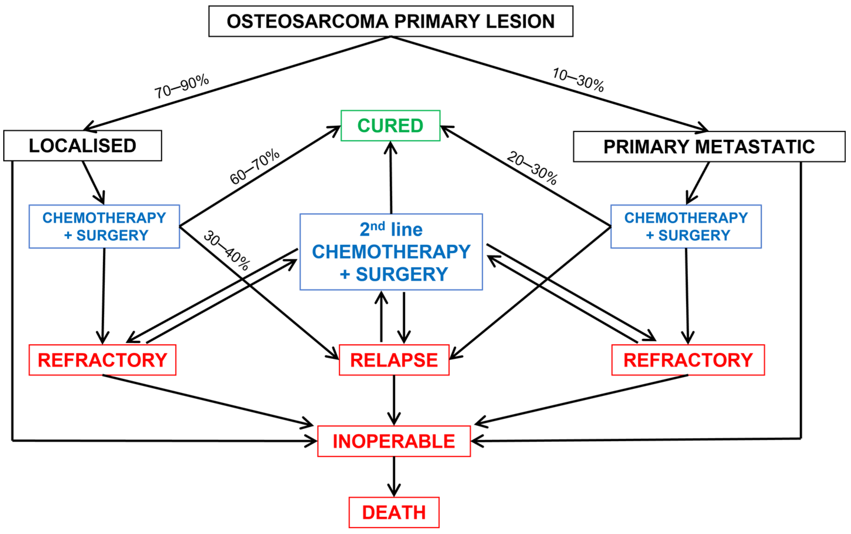
Abstract
:Simple Summary
Abstract
1. Introduction
2. Clinical Presentation of Metastases
2.1. Synchronous Metastases
2.2. Metachronous Metastases
2.3. Treatment of Metastatic Osteosarcoma
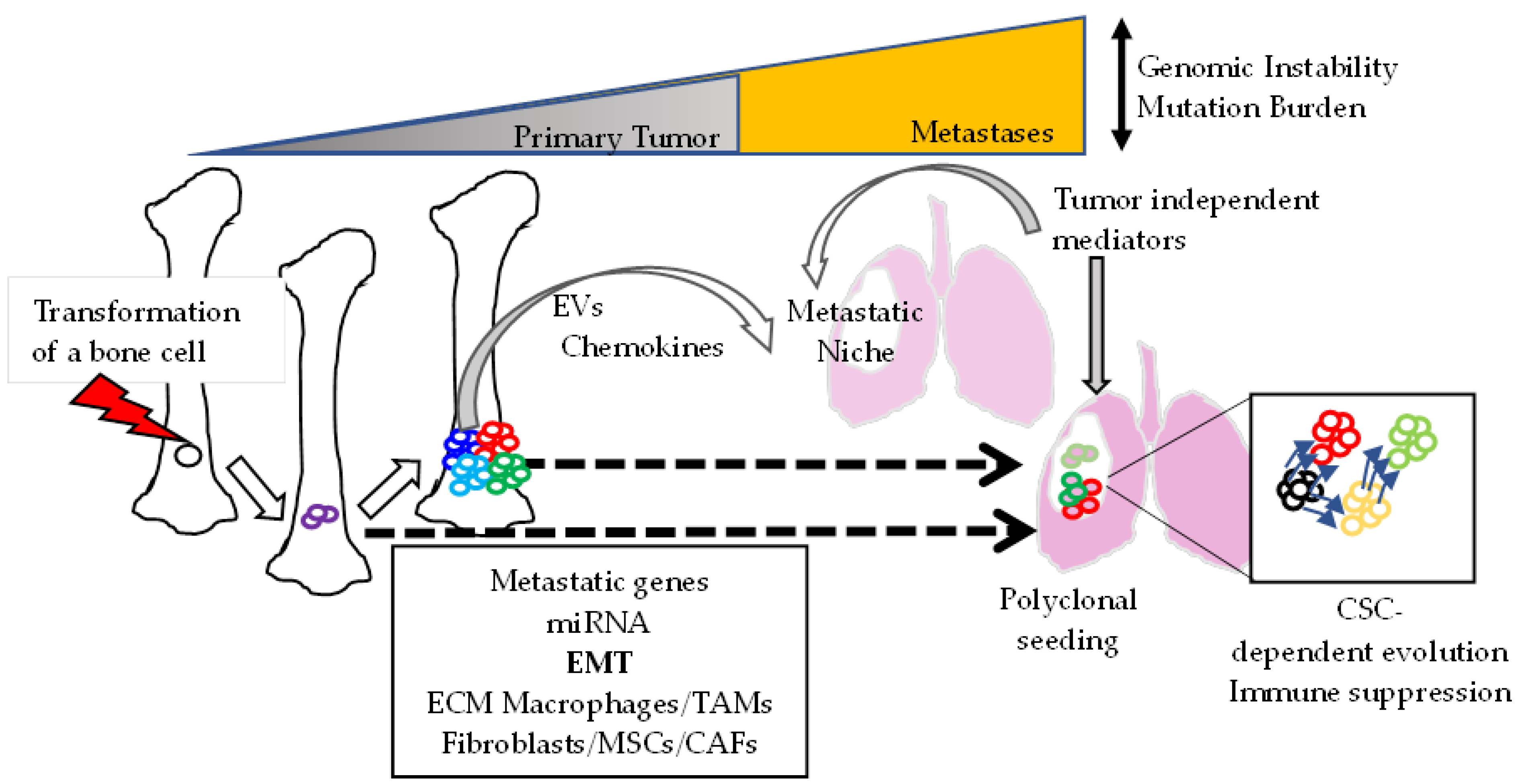
3. Mechanisms of Metastasis Formation
3.1. Metastasis Heterogeneity
3.2. Regulation of the Metastatic Capacity of Primary Tumor Cells
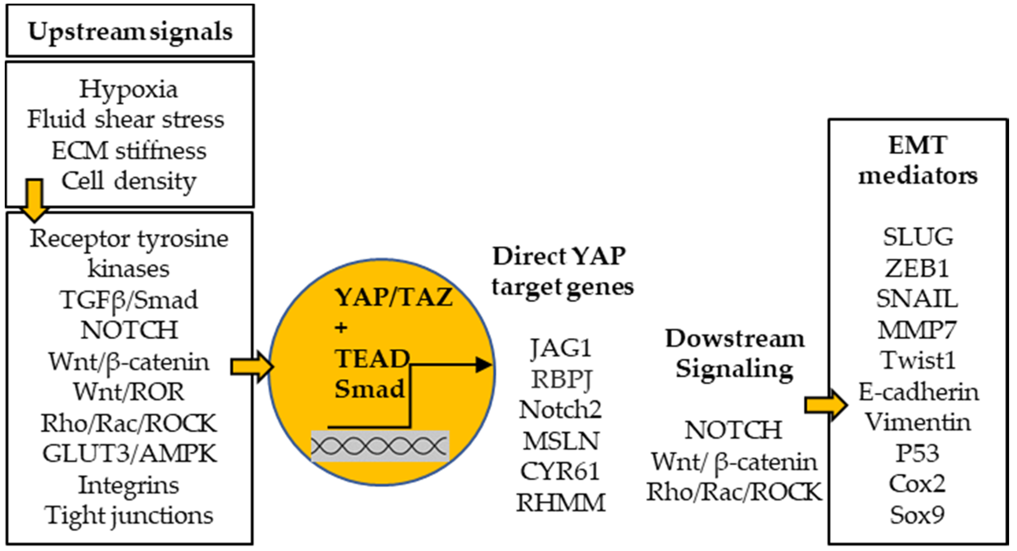
3.3. Epithelial–Mesenchymal Transition (EMT)
3.4. Role of the Microenvironment
3.4.1. Extracellular Matrix
3.4.2. Tumor Associated Macrophages
3.5. Regulation of the Metastatic Niche
3.6. Metastasis Cell Founders
4. Conclusions and Perspectives
Author Contributions
Funding
Conflicts of Interest
References
- Bielack, S.S.; Kempf-Bielack, B.; Delling, G.; Exner, G.U.; Flege, S.; Helmke, K.; Kotz, R.; Salzer-Kuntschik, M.; Werner, M.; Winkelmann, W.; et al. Prognostic factors in high-grade osteosarcoma of the extremities or trunk: An analysis of 1702 patients treated on Neoadjuvant Cooperative Osteosarcoma Study Group Protocols. J. Clin. Oncol. 2002, 20, 776–790. [Google Scholar] [CrossRef]
- Smeland, S.; Bielack, S.S.; Whelan, J.; Bernstein, M.; Hogendoorn, P.; Krailo, M.D.; Gorlick, R.; Janeway, K.A.; Ingleby, F.C.; Anninga, J.; et al. Survival and prognosis with osteosarcoma: Outcomes in more than 2000 patients in the EURAMOS-1 (European and American Osteosarcoma Study) cohort. Eur. J. Cancer 2019, 109, 36–50. [Google Scholar] [CrossRef] [Green Version]
- Kager, L.; Zoubek, A.; Pötschger, U.; Kastner, U.; Flege, S.; Kempf-Bielack, B.; Branscheid, D.; Kotz, R.; Salzer-Kuntschik, M.; Winkelmann, W.; et al. Primary metastatic osteosarcoma: Presentation and outcome of patients treated on neoadjuvant cooperative osteosarcoma study group protocols. J. Clin. Oncol. 2003, 21, 2011–2018. [Google Scholar] [CrossRef]
- Bacci, G.; Rocca, M.; Salone, M.; Balladelli, A.; Ferrari, S.; Palmerini, E.; Forni, C.; Briccoli, A. High grade osteosarcoma of the extremities with lung metastases at presentation: Treatment with neoadjuvant chemotherapy and simultaneous resection of primary and metastatic lesions. J. Surg. Oncol. 2008, 98, 415–420. [Google Scholar] [CrossRef] [PubMed]
- Kim, W.; Han, I.; Lee, J.S.; Cho, H.S.; Park, J.W.; Kim, H.S. Postmetastasis survival in high-grade extremity osteosarcoma: A retrospective analysis of prognostic factors in 126 patients. J. Surg. Oncol. 2018, 117, 1223–1231. [Google Scholar] [CrossRef] [PubMed]
- Meyers, P.A.; Heller, G.; Healey, J.H.; Huvos, A.; Applewhite, A.; Sun, M.; LaQuaglia, M. Osteogenic sarcoma with clinically detectable metastasis at initial presentation. J. Clin. Oncol. 1993, 11, 449–453. [Google Scholar] [CrossRef] [PubMed]
- Zhang, C.; Guo, X.; Xu, Y.; Han, X.; Cai, J.; Wang, X.; Wang, G. Lung metastases at the initial diagnosis of high-grade osteosarcoma: Prevalence, risk factors and prognostic factors. A large population-based cohort study. Sao Paulo Med. J. 2019, 137, 423–429. [Google Scholar] [CrossRef] [Green Version]
- Vasquez, L.; Silva, J.; Chavez, S.; Zapata, A.; Diaz, R.; Tarrillo, F.; Maza, I.; Sialer, L.; García, J. Prognostic Impact of Diagnostic and Treatment Delays in Children with Osteosarcoma. Pediatric Blood Cancer 2020, 67, e28180. [Google Scholar] [CrossRef]
- Yoshida, S.; Celaire, J.; Pace, C.; Taylor, C.; Kaneuchi, Y.; Evans, S.; Abudu, A. Delay in Diagnosis of Primary Osteosarcoma of Bone in Children: Have We Improved in the Last 15 Years and What Is the Impact of Delay on Diagnosis? J. Bone Oncol. 2021, 28, 100359. [Google Scholar] [CrossRef]
- Heaton, T.E.; Hammond, W.J.; Farber, B.A.; Pallos, V.; Meyers, P.A.; Chou, A.J.; Price, A.P.; LaQuaglia, M.P. A 20-Year Retrospective Analysis of CT-Based Pre-Operative Identification of Pulmonary Metastases in Patients with Osteosarcoma: A Single-Center Review. J. Pediatr. Surg. 2017, 52, 115–119. [Google Scholar] [CrossRef] [Green Version]
- Price, C.H.G.; Jeffree, G.M. Metastatic Spread of Osteosarcoma. Br. J. Cancer 1973, 28, 515–524. [Google Scholar] [CrossRef] [Green Version]
- Xie, L.; Huang, W.; Wang, H.; Zheng, C.; Jiang, J. Risk Factors for Lung Metastasis at Presentation with Malignant Primary Osseous Neoplasms: A Population-Based Study. J. Orthopaed. Surg. Res. 2020, 15, 32. [Google Scholar] [CrossRef] [Green Version]
- Jeffree, G.M.; Price, C.H.G.; Sissons, H.A. The Metastatic Patterns of Osteosarcoma. Br. J. Cancer 1975, 32, 87–107. [Google Scholar] [CrossRef] [PubMed] [Green Version]
- Giuliano, A.E.; Feig, S.; Eilber, F.R. Changing Metastatic Patterns of Osteosarcoma. Cancer 1984, 54, 2160–2164. [Google Scholar] [CrossRef]
- Le Aung, L.; Gorlick, R.; Healey, J.H.; Shi, W.; Thaler, H.T.; Shorter, N.A.; Huvos, A.G.; Meyers, P.A. Metachronous Skeletal Osteosarcoma in Patients Treated with Adjuvant and Neoadjuvant Chemotherapy for Nonmetastatic Osteosarcoma. J. Clin. Oncol. 2003, 21, 342–348. [Google Scholar] [CrossRef] [PubMed]
- Enneking, W.F.; Kagan, A. The Implications of “Skip” Metastases in Osteosarcoma. Clin. Orthopaed. Relat. Res. 1975, 111, 33–41. [Google Scholar] [CrossRef] [PubMed]
- Wuisman, P.; Enneking, W.F. Prognosis for Patients Who Have Osteosarcoma with Skip Metastasis. J. Bone Joint Surg. 1990, 72, 60–68. [Google Scholar] [CrossRef]
- Kager, L.; Zoubek, A.; Kastner, U.; Kempf-Bielack, B.; Potratz, J.; Kotz, R.; Exner, G.U.; Franzius, C.; Lang, S.; Maas, R.; et al. Skip Metastases in Osteosarcoma: Experience of the Cooperative Osteosarcoma Study Group. J. Clin. Oncol. 2006, 24, 1535–1541. [Google Scholar] [CrossRef]
- Li, N.; Wei, X.; Zhang, Z.; Zhang, Y. Use Of Microwave Thermal Ablation In Management Of Skip Metastases In Extremity Osteosarcomas. Cancer Manag. Res. 2019, 11, 9843–9848. [Google Scholar] [CrossRef] [Green Version]
- Barnett, J.R.; Gikas, P.; Gerrand, C.; Briggs, T.W.; Saifuddin, A. The Sensitivity, Specificity, and Diagnostic Accuracy of Whole-Bone MRI for Identifying Skip Metastases in Appendicular Osteosarcoma and Ewing Sarcoma. Skelet. Radiol. 2020, 49, 913–919. [Google Scholar] [CrossRef]
- Yang, P.; Gilg, M.; Evans, S.; Totti, F.; Stevenson, J.; Jeys, L.; Parry, M. Survival of Osteosarcoma Patients Following Diagnosis of Synchronous Skip Metastases. J. Orthopaed. 2019, 18, 121–125. [Google Scholar] [CrossRef] [PubMed]
- Saifuddin, A.; Michelagnoli, M.; Pressney, I. Skip Metastases in High-Grade Intramedullary Appendicular Osteosarcoma: An Indicator of More Aggressive Disease? Skelet. Radiol. 2021, 50, 2415–2422. [Google Scholar] [CrossRef] [PubMed]
- Corradi, D.; Wenger, D.E.; Bertoni, F.; Bacchini, P.; Bosio, S.; Goldoni, M.; Unni, K.K.; Sim, F.H.; Inwards, C.Y. Multicentric Osteosarcoma: Clinicopathologic and Radiographic Study of 56 Cases. Am. J. Clin. Pathol. 2011, 136, 799–807. [Google Scholar] [CrossRef]
- Gherlinzoni, F.; Antoci, B.; Canale, V. Case Report 250. Multicentric Osteosarcomata (Osteosarcomatosis). Skelet. Radiol. 1983, 10, 281–285. [Google Scholar] [CrossRef]
- Longhi, A.; Fabbri, N.; Donati, D.; Capanna, R.; Briccoli, A.; Biagini, R.; Bernini, G.; Ferrari, S.; Versari, M.; Bacci, G. Neoadjuvant Chemotherapy for Patients with Synchronous Multifocal Osteosarcoma: Results in Eleven Cases. J. Chemother. 2001, 13, 324–330. [Google Scholar] [CrossRef]
- Gok Durnali, A.; Paksoy Turkoz, F.; Ardic Yukruk, F.; Tokluoglu, S.; Yazici, O.K.; Demirci, A.; Bal, O.; Gundogdu Buyukbas, S.; Esbah, O.; Oksuzoglu, B.; et al. Outcomes of Adolescent and Adult Patients with Lung Metastatic Osteosarcoma and Comparison of Synchronous and Metachronous Lung Metastatic Groups. PLoS ONE 2016, 11, e0152621. [Google Scholar] [CrossRef] [PubMed] [Green Version]
- Wasilewski-Masker, K.; Liu, Q.; Yasui, Y.; Leisenring, W.; Meacham, L.R.; Hammond, S.; Meadows, A.T.; Robison, L.L.; Mertens, A.C. Late Recurrence in Pediatric Cancer: A Report from the Childhood Cancer Survivor Study. J. Natl. Cancer Instit. 2009, 101, 1709–1720. [Google Scholar] [CrossRef]
- Kaneuchi, Y.; Hakozaki, M.; Yamada, H.; Hasegawa, O.; Yamada, S.; Oka, Y.; Watanabe, K.; Konno, S. Very Late Relapse of High-Grade Osteosarcoma: A Case Report and Review of the Literature. Medicine 2020, 99, e21206. [Google Scholar] [CrossRef]
- Strauss, S.J.; McTiernan, A.; Whelan, J.S. Late Relapse of Osteosarcoma: Implications for Follow-up and Screening. Pediatr. Blood Cancer 2004, 43, 692–697. [Google Scholar] [CrossRef]
- Bacci, G.; Ruggieri, P.; Picci, P.; Briccoli, A.; Ferraro, A.; Ferrari, S.; Tienghi, A.; Iantorno, D.; Campanacci, M. Changing Pattern of Relapse in Osteosarcoma of the Extremities Treated with Adjuvant and Neoadjuvant Chemotherapy. J. Chemother. 1995, 7, 230–239. [Google Scholar] [CrossRef]
- Kempf-Bielack, B.; Bielack, S.S.; Jürgens, H.; Branscheid, D.; Berdel, W.E.; Exner, G.U.; Göbel, U.; Helmke, K.; Jundt, G.; Kabisch, H.; et al. Osteosarcoma Relapse after Combined Modality Therapy: An Analysis of Unselected Patients in the Cooperative Osteosarcoma Study Group (COSS). J. Clin. Oncol. 2005, 23, 559–568. [Google Scholar] [CrossRef]
- Bielack, S.S.; Kempf-Bielack, B.; Branscheid, D.; Carrle, D.; Friedel, G.; Helmke, K.; Kevric, M.; Jundt, G.; Kühne, T.; Maas, R.; et al. Second and Subsequent Recurrences of Osteosarcoma: Presentation, Treatment, and Outcomes of 249 Consecutive Cooperative Osteosarcoma Study Group Patients. J. Clin. Oncol. 2009, 27, 557–565. [Google Scholar] [CrossRef]
- Thebault, E.; Piperno-Neumann, S.; Tran, D.; Pacquement, H.; Marec-Berard, P.; Lervat, C.; Castex, M.P.; Cleirec, M.; Bompas, E.; Vannier, J.P.; et al. Successive Osteosarcoma Relapses after the First Line O2006/Sarcome-09 Trial: What Can We Learn for Further Phase-II Trials? Cancers 2021, 13, 1683. [Google Scholar] [CrossRef]
- Chi, S.N.; Conklin, L.S.; Qin, J.; Meyers, P.A.; Huvos, A.G.; Healey, J.H.; Gorlick, R. The Patterns of Relapse in Osteosarcoma: The Memorial Sloan-Kettering Experience. Pediatr. Blood Cancer 2004, 42, 46–51. [Google Scholar] [CrossRef] [PubMed]
- Ahmed, G.; Zamzam, M.; Kamel, A.; Ahmed, S.; Salama, A.; Zaki, I.; Kamal, N.; Elshafiey, M. Effect of Timing of Pulmonary Metastasis Occurrence on the Outcome of Metastasectomy in Osteosarcoma Patients. J. Pediatr. Surg. 2019, 54, 775–779. [Google Scholar] [CrossRef] [PubMed]
- Bao, J.; Zeng, J.; Song, C.; Yu, H.; Shi, Q.; Mai, W.; Qu, G. A Retrospective Clinicopathological Study of Osteosarcoma Patients with Metachronous Metastatic Relapse. J. Cancer 2019, 10, 2982–2990. [Google Scholar] [CrossRef] [PubMed] [Green Version]
- Slade, A.D.; Warneke, C.L.; Hughes, D.P.; Lally, P.A.; Lally, K.P.; Hayes-Jordan, A.A.; Austin, M.T. Effect of Concurrent Metastatic Disease on Survival in Children and Adolescents Undergoing Lung Resection for Metastatic Osteosarcoma. J. Pediatr. Surg. 2015, 50, 157–160. [Google Scholar] [CrossRef]
- Li, W.; Zhang, S. Survival of Patients with Primary Osteosarcoma and Lung Metastases. J. BUON 2018, 23, 1500–1504. [Google Scholar]
- Brandal, P.; Bjerkehagen, B.; Bruland, Ø.S.; Skjeldal, S.; Bogsrud, T.V.; Hall, K.S. Synchronous and Metachronous Skeletal Osteosarcomas: The Norwegian Radium Hospital Experience. Acta Oncol. 2009, 48, 1165–1172. [Google Scholar] [CrossRef] [PubMed] [Green Version]
- Simon, M.A.; Aschliman, M.A.; Thomas, N.; Mankin, H.J. Limb-Salvage Treatment versus Amputation for Osteosarcoma of the Distal End of the Femur. J. Bone Joint Surg. 1986, 68, 1331–1337. [Google Scholar] [CrossRef]
- Bertrand, T.E.; Cruz, A.; Binitie, O.; Cheong, D.; Letson, G.D. Do Surgical Margins Affect Local Recurrence and Survival in Extremity, Nonmetastatic, High-Grade Osteosarcoma? Clin. Orthopaed. Relat. Res. 2016, 474, 677–683. [Google Scholar] [CrossRef] [PubMed] [Green Version]
- Casali, P.G.; Bielack, S.; Abecassis, N.; Aro, H.T.; Bauer, S.; Biagini, R.; Bonvalot, S.; Boukovinas, I.; Bovee, J.V.M.G.; Brennan, B.; et al. Bone Sarcomas: ESMO-PaedCan-EURACAN Clinical Practice Guidelines for Diagnosis, Treatment and Follow-Up. Ann. Oncol. 2018, 29, iv79–iv95. [Google Scholar] [CrossRef]
- Meazza, C.; Scanagatta, P. Metastatic Osteosarcoma: A Challenging Multidisciplinary Treatment. Expert Rev. Anticancer Ther. 2016, 16, 543–556. [Google Scholar] [CrossRef]
- Gazouli, I.; Kyriazoglou, A.; Kotsantis, I.; Anastasiou, M.; Pantazopoulos, A.; Prevezanou, M.; Chatzidakis, I.; Kavourakis, G.; Economopoulou, P.; Kontogeorgakos, V.; et al. Systematic Review of Recurrent Osteosarcoma Systemic Therapy. Cancers 2021, 13, 1757. [Google Scholar] [CrossRef] [PubMed]
- Lavit, E.; Aldea, M.; Piperno-Neumann, S.; Firmin, N.; Italiano, A.; Isambert, N.; Kurtz, J.E.; Delcambre, C.; Lebrun, V.; Soibinet-Oudot, P.; et al. Treatment of 120 Adult Osteosarcoma Patients with Metachronous and Synchronous Metastases: A Retrospective Series of the French Sarcoma Group. Int. J. Cancer 2021, 150, 645–653. [Google Scholar] [CrossRef]
- Fagioli, F.; Aglietta, M.; Tienghi, A.; Ferrari, S.; Brach del Prever, A.; Vassallo, E.; Palmero, A.; Biasin, E.; Bacci, G.; Picci, P.; et al. High-Dose Chemotherapy in the Treatment of Relapsed Osteosarcoma: An Italian Sarcoma Group Study. J. Clin. Oncol. 2002, 20, 2150–2156. [Google Scholar] [CrossRef]
- Tawbi, H.A.; Burgess, M.; Bolejack, V.; van Tine, B.A.; Schuetze, S.M.; Hu, J.; D’Angelo, S.; Attia, S.; Riedel, R.F.; Priebat, D.A.; et al. Pembrolizumab in Advanced Soft-Tissue Sarcoma and Bone Sarcoma (SARC028): A Multicentre, Two-Cohort, Single-Arm, Open-Label, Phase 2 Trial. Lancet Oncol. 2017, 18, 1493–1501. [Google Scholar] [CrossRef]
- Burgess, M.; Tawbi, H. Immunotherapeutic Approaches to Sarcoma. Curr. Treat. Options Oncol. 2015, 16, 26. [Google Scholar] [CrossRef]
- Merchant, M.S.; Bernstein, D.; Amoako, M.; Baird, K.; Fleisher, T.A.; Morre, M.; Steinberg, S.M.; Sabatino, M.; Stroncek, D.F.; Venkatasan, A.M.; et al. Adjuvant Immunotherapy to Improve Outcome in High-Risk Pediatric Sarcomas. Clin. Cancer Res. 2016, 22, 3182–3191. [Google Scholar] [CrossRef] [PubMed] [Green Version]
- Ahmed, N.; Brawley, V.S.; Hegde, M.; Robertson, C.; Ghazi, A.; Gerken, C.; Liu, E.; Dakhova, O.; Ashoori, A.; Corder, A.; et al. Human Epidermal Growth Factor Receptor 2 (HER2)—Specific Chimeric Antigen Receptor—Modified T Cells for the Immunotherapy of HER2-Positive Sarcoma. J. Clin. Oncol. 2015, 33, 1688–1696. [Google Scholar] [CrossRef]
- Krishnamoorthy, N.; Desai, S.S.; Rekhi, B.; Jambhekar, N.A. A Clinico-Morphological Study of 95 Cases of Sarcomas with Metastases to the Lungs. Indian J. Cancer 2011, 48, 335–338. [Google Scholar] [CrossRef] [PubMed]
- Wang, D.; Niu, X.; Wang, Z.; Song, C.L.; Huang, Z.; Chen, K.N.; Duan, J.; Bai, H.; Xu, J.; Zhao, J.; et al. Multiregion Sequencing Reveals the Genetic Heterogeneity and Evolutionary History of Osteosarcoma and Matched Pulmonary Metastases. Cancer Res. 2019, 79, 7–20. [Google Scholar] [CrossRef] [PubMed] [Green Version]
- Xu, H.; Zhu, X.; Bao, H.; Shek, T.W.; Huang, Z.; Wang, Y.; Wu, X.; Wu, Y.; Chang, Z.; Wu, S.; et al. Genetic and Clonal Dissection of Osteosarcoma Progression and Lung Metastasis. Int. J. Cancer 2018, 143, 1134–1142. [Google Scholar] [CrossRef]
- Moriarity, B.S.; Otto, G.M.; Rahrmann, E.P.; Rathe, S.K.; Wolf, N.K.; Weg, M.T.; Manlove, L.A.; Larue, R.S.; Temiz, N.A.; Molyneux, S.D.; et al. A Sleeping Beauty Forward Genetic Screen Identifies New Genes and Pathways Driving Osteosarcoma Development and Metastasis. Nat. Genet. 2015, 47, 615–624. [Google Scholar] [CrossRef] [PubMed]
- Stoecklein, N.H.; Klein, C.A. Genetic Disparity between Primary Tumours, Disseminated Tumour Cells, and Manifest Metastasis. Int. J. Cancer 2010, 126, 589–598. [Google Scholar] [CrossRef] [PubMed]
- Klein, C.A. Parallel Progression of Primary Tumours and Metastases. Nat. Rev. Cancer 2009, 9, 302–312. [Google Scholar] [CrossRef]
- Li, M.; Lu, Y.; Long, Z.; Li, M.; Kong, J.; Chen, G.; Wang, Z. Prognostic and Clinicopathological Significance of Circulating Tumor Cells in Osteosarcoma. J. Bone Oncol. 2019, 16, 100236. [Google Scholar] [CrossRef]
- Wu, Z.J.; Tan, J.C.; Qin, X.; Liu, B.; Yuan, Z.C. Significance of Circulating Tumor Cells in Osteosarcoma Patients Treated by Neoadjuvant Chemotherapy and Surgery. Cancer Manag. Res. 2018, 10, 3333–3339. [Google Scholar] [CrossRef] [PubMed] [Green Version]
- Adamopoulos, C.; Gargalionis, A.N.; Basdra, E.K.; Papavassiliou, A.G. Deciphering Signaling Networks in Osteosarcoma Pathobiology. Exp. Biol. Med. 2016, 241, 1296–1305. [Google Scholar] [CrossRef] [Green Version]
- Li, Z.; Li, X.; Xu, D.; Chen, X.; Li, S.; Zhang, L.; Chan, M.T.V.; Wu, W.K.K. An Update on the Roles of Circular RNAs in Osteosarcoma. Cell Proliferat. 2021, 54, e12936. [Google Scholar] [CrossRef]
- Zhang, Y.; Li, J.; Wang, Y.; Jing, J.; Li, J. The Roles of Circular RNAs in Osteosarcoma. Med. Sci. Monit. 2019, 25, 6378–6382. [Google Scholar] [CrossRef] [PubMed]
- Kushlinskii, N.E.; Fridman, M.V.; Braga, E.A. Long Non-Coding RNAs as Competitive Endogenous RNAs in Osteosarcoma. Mol. Biol. 2020, 54, 776–801. [Google Scholar] [CrossRef]
- Botti, G.; Giordano, A.; Feroce, F.; Chiara, A.R. de Cantile, M. Noncoding RNAs as Circulating Biomarkers in Osteosarcoma Patients. J. Cell. Physiol. 2019, 234, 19249–19255. [Google Scholar] [CrossRef]
- Llobat, L.; Gourbault, O. Role of Micrornas in Human Osteosarcoma: Future Perspectives. Biomedicines 2021, 9, 463. [Google Scholar] [CrossRef] [PubMed]
- Zhang, J.; Yan, Y.G.; Wang, C.; Zhang, S.J.; Yu, X.H.; Wang, W.J. MicroRNAs in Osteosarcoma. Clin. Chim. Acta 2015, 444, 9–17. [Google Scholar] [CrossRef]
- Jamali, Z.; Taheri-Anganeh, M.; Shabaninejad, Z.; Keshavarzi, A.; Taghizadeh, H.; Razavi, Z.S.; Mottaghi, R.; Abolhassan, M.; Movahedpour, A.; Mirzaei, H. Autophagy Regulation by MicroRNAs: Novel Insights into Osteosarcoma Therapy. IUBMB Life 2020, 72, 1306–1321. [Google Scholar] [CrossRef]
- Zheng, D.; Xia, K.; Yu, L.; Gong, C.; Shi, Y.; Li, W.; Qiu, Y.; Yang, J.; Guo, W. A Novel Six Metastasis-Related Prognostic Gene Signature for Patients With Osteosarcoma. Front. Cell Dev. Biol. 2021, 9, 699212. [Google Scholar] [CrossRef]
- Ying, T.; Dong, J.L.; Yuan, C.; Li, P.; Guo, Q. The LncRNAs RP1-261G23.7, RP11-69E11.4 and SATB2-AS1 Are a Novel Clinical Signature for Predicting Recurrent Osteosarcoma. Biosci. Rep. 2020, 40, BSR20191251. [Google Scholar] [CrossRef] [Green Version]
- Shi, Y.; He, R.; Zhuang, Z.; Ren, J.; Wang, Z.; Liu, Y.; Wu, J.; Jiang, S.; Wang, K. A Risk Signature-Based on Metastasis-Associated Genes to Predict Survival of Patients with Osteosarcoma. J. Cell. Biochem. 2020, 121, 3479–3490. [Google Scholar] [CrossRef]
- Qi, W.; Yan, Q.; Lv, M.; Song, D.; Wang, X.; Tian, K. Prognostic Signature of Osteosarcoma Based on 14 Autophagy-Related Genes. Pathol. Oncol. Res. 2021, 27, 1609782. [Google Scholar] [CrossRef]
- Jiang, F.; Miao, X.-L.; Zhang, X.-T.; Yan, F.; Mao, Y.; Wu, C.-Y.; Zhou, G.-P. A Hypoxia Gene-Based Signature to Predict the Survival and Affect the Tumor Immune Microenvironment of Osteosarcoma in Children. J. Immunol. Res. 2021, 2021, 5523832. [Google Scholar] [CrossRef]
- Yang, M.; Ma, X.; Wang, Z.; Zhang, T.; Hua, Y. Identification of a Novel Glycolysis-Related Gene Signature for Predicting the Prognosis of Osteosarcoma Patients. Aging 2021, 13, 12896–12918. [Google Scholar] [CrossRef]
- Xiao, K.W.; Liu, Z.B.; Zeng, Z.H.; Yan, F.F.; Xiao, L.F.; Li, J.L.; Cai, L. Construction and Validation of a Macrophage-Associated Risk Model for Predicting the Prognosis of Osteosarcoma. J. Oncol. 2021, 2021, 9967954. [Google Scholar] [CrossRef]
- Chong, Z.X.; Yeap, S.K.; Ho, W.Y. Unraveling the Roles of MiRNAs in Regulating Epithelial-to-Mesenchymal Transition (EMT) in Osteosarcoma. Pharmacol. Res. 2021, 172, 105818. [Google Scholar] [CrossRef]
- Ehnman, M.; Chaabane, W.; Haglund, F.; Tsagkozis, P. The Tumor Microenvironment of Pediatric Sarcoma: Mesenchymal Mechanisms Regulating Cell Migration and Metastasis. Curr. Oncol. Rep. 2019, 21, 90. [Google Scholar] [CrossRef] [PubMed] [Green Version]
- Wang, J.; Zhang, H.; Sun, X.; Wang, X.; Ren, T.; Huang, Y.; Zhang, R.; Zheng, B.; Guo, W. Exosomal PD-L1 and N-Cadherin Predict Pulmonary Metastasis Progression for Osteosarcoma Patients. J. Nanobiotechnol. 2020, 18, 151. [Google Scholar] [CrossRef] [PubMed]
- Jiang, R.; Zhang, C.; Liu, G.; Gu, R.; Wu, H. MicroRNA-126 Inhibits Proliferation, Migration, Invasion, and EMT in Osteosarcoma by Targeting ZEB1. J. Cell. Biochem. 2017, 118, 3765–3774. [Google Scholar] [CrossRef] [PubMed]
- Yao, H.; Hou, G.; Wang, Q.Y.; Xu, W.B.; Zhao, H.Q.; Xu, Y.C. LncRNA SPRY4-IT1 Promotes Progression of Osteosarcoma by Regulating ZEB1 and ZEB2 Expression through Sponging of MiR-101 Activity. Int. J. Oncol. 2020, 56, 85–100. [Google Scholar] [CrossRef] [Green Version]
- Feng, T.; Zhu, Z.; Jin, Y.; Wang, H.; Mao, X.; Liu, D.; Li, Y.; Lu, L.; Zuo, G. The MicroRNA-708-5p/ZEB1/EMT Axis Mediates the Metastatic Potential of Osteosarcoma. Oncol. Rep. 2020, 43, 491–502. [Google Scholar] [CrossRef]
- Zhu, S.T.; Wang, X.; Wang, J.Y.; Xi, G.H.; Liu, Y. Downregulation of MiR-22 Contributes to Epithelial-Mesenchymal Transition in Osteosarcoma by Targeting Twist1. Front. Oncol. 2020, 10, 406. [Google Scholar] [CrossRef]
- Chen, Y.; Li, J.; Xiao, J.K.; Xiao, L.; Xu, B.W.; Li, C. The LncRNA NEAT1 Promotes the Epithelial-Mesenchymal Transition and Metastasis of Osteosarcoma Cells by Sponging MiR-483 to Upregulate STAT3 Expression. Cancer Cell Int. 2021, 21, 90. [Google Scholar] [CrossRef]
- Zhang, S.; Chen, H.; Liu, W.; Fang, L.; Qian, Z.; Kong, R.; Zhang, Q.; Li, J.; Cao, X. MiR-766-3p Targeting BCL9L Suppressed Tumorigenesis, Epithelial-Mesenchymal Transition, and Metastasis Through the β-Catenin Signaling Pathway in Osteosarcoma Cells. Front. Cell Dev. Biol. 2020, 8, 1086. [Google Scholar] [CrossRef] [PubMed]
- Wang, H.; Zhang, P. LncRNA-CASC15 Promotes Osteosarcoma Proliferation and Metastasis by Regulating Epithelial-Mesenchymal Transition via the Wnt/β-Catenin Signaling Pathway. Oncol. Rep. 2021, 45, 76. [Google Scholar] [CrossRef]
- Kovar, H.; Bierbaumer, L.; Radic-Sarikas, B. The YAP/TAZ Pathway in Osteogenesis and Bone Sarcoma Pathogenesis. Cells 2020, 9, 972. [Google Scholar] [CrossRef] [PubMed] [Green Version]
- Calvo, F.; Ege, N.; Grande-Garcia, A.; Hooper, S.; Jenkins, R.P.; Chaudhry, S.I.; Harrington, K.; Williamson, P.; Moeendarbary, E.; Charras, G.; et al. Mechanotransduction and YAP-Dependent Matrix Remodelling Is Required for the Generation and Maintenance of Cancer-Associated Fibroblasts. Nat. Cell Biol. 2013, 15, 637–646. [Google Scholar] [CrossRef]
- Pavel, M.; Renna, M.; Park, S.J.; Menzies, F.M.; Ricketts, T.; Füllgrabe, J.; Ashkenazi, A.; Frake, R.A.; Lombarte, A.C.; Bento, C.F.; et al. Contact Inhibition Controls Cell Survival and Proliferation via YAP/TAZ-Autophagy Axis. Nat. Commun. 2018, 9, 2961. [Google Scholar] [CrossRef] [Green Version]
- Xiang, L.; Gilkes, D.M.; Hu, H.; Luo, W.; Bullen, J.W.; Liang, H.; Semenza, G.L. HIF-1α and TAZ Serve as Reciprocal Co-Activators in Human Breast Cancer Cells. Oncotarget 2015, 6, 11768–11778. [Google Scholar] [CrossRef] [PubMed] [Green Version]
- Zhao, B.; Lv, Y. Suspension State and Shear Stress Enhance Breast Tumor Cells EMT through YAP by MicroRNA-29b. Cell Biol. Toxicol. 2021. [Google Scholar] [CrossRef]
- Di Benedetto, G.; Parisi, S.; Russo, T.; Passaro, F. YAP and TAZ Mediators at the Crossroad between Metabolic and Cellular Reprogramming. Metabolites 2021, 11, 154. [Google Scholar] [CrossRef]
- Varelas, X. The Hippo Pathway Effectors TAZ and YAP in Development, Homeostasis and Disease. Development 2014, 141, 1614–1626. [Google Scholar] [CrossRef] [Green Version]
- Jansen, S.; Gosens, R.; Wieland, T.; Schmidt, M. Paving the Rho in Cancer Metastasis: Rho GTPases and Beyond. Pharmacol. Ther. 2018, 183, 1–21. [Google Scholar] [CrossRef] [PubMed]
- Li, H.L.; Li, Q.Y.; Jin, M.J.; Lu, C.F.; Mu, Z.Y.; Xu, W.Y.; Song, J.; Zhang, Y.; Zhang, S.Y. A Review: Hippo Signaling Pathway Promotes Tumor Invasion and Metastasis by Regulating Target Gene Expression. J. Cancer Res. Clin. Oncol. 2021, 147, 1569–1585. [Google Scholar] [CrossRef] [PubMed]
- Tang, Y.; Weiss, S.J. Snail/Slug-YAP/TAZ Complexes Cooperatively Regulate Mesenchymal Stem Cell Function and Bone Formation. Cell Cycle 2017, 16, 399–405. [Google Scholar] [CrossRef] [PubMed] [Green Version]
- Sun, Z.; Schwenzer, A.; Rupp, T.; Murdamoothoo, D.; Vegliante, R.; Lefebvre, O.; Klein, A.; Hussenet, T.; Orend, G. Tenascin-C Promotes Tumor Cell Migration and Metastasis through Integrin A9b1–Mediated YAP Inhibition. Cancer Res. 2018, 78, 950–961. [Google Scholar] [CrossRef] [PubMed] [Green Version]
- Yudoh, K.; Matsui, H.; Kanamori, M.; Ohmori, K.; Yasuda, T.; Tsuji, H. Characteristics of High and Low Laminin-Adherent Dunn Osteosarcoma Cells Selected by Adhesiveness to Laminin. Tumor Biol. 1996, 17, 332–340. [Google Scholar] [CrossRef]
- Gvozdenovic, A.; Boro, A.; Meier, D.; Bode-Lesniewska, B.; Born, W.; Muff, R.; Fuchs, B. Targeting Avβ3 and Avβ5 Integrins Inhibits Pulmonary Metastasis in an Intratibial Xenograft Osteosarcoma Mouse Model. Oncotarget 2016, 7, 55141–55154. [Google Scholar] [CrossRef] [Green Version]
- Zandueta, C.; Ormazábal, C.; Perurena, N.; Martínez-Canarias, S.; Zalacaín, M.; Julián, M.S.; Grigoriadis, A.E.; Valencia, K.; Campos-Laborie, F.J.; De las Rivas, J.; et al. Matrix-Gla Protein Promotes Osteosarcoma Lung Metastasis and Associates with Poor Prognosis. J. Pathol. 2016, 239, 438–449. [Google Scholar] [CrossRef]
- Kitamura, T.; Qian, B.Z.; Pollard, J.W. Immune Cell Promotion of Metastasis. Nat. Rev. Immunol. 2015, 15, 73–86. [Google Scholar] [CrossRef]
- Han, Y.; Guo, W.; Ren, T.; Huang, Y.; Wang, S.; Liu, K.; Zheng, B.; Yang, K.; Zhang, H.; Liang, X. Tumor-Associated Macrophages Promote Lung Metastasis and Induce Epithelial-Mesenchymal Transition in Osteosarcoma by Activating the COX-2/STAT3 Axis. Cancer Lett. 2019, 440–441, 116–125. [Google Scholar] [CrossRef]
- Maloney, C.; Kallis, M.P.; Edelman, M.; Tzanavaris, C.; Lesser, M.; Soffer, S.Z.; Symons, M.; Steinberg, B.M. Gefitinib Inhibits Invasion and Metastasis of Osteosarcoma via Inhibition of Macrophage Receptor Interacting Serine-Threonine Kinase 2. Mol. Cancer Ther. 2020, 19, 1340–1350. [Google Scholar] [CrossRef]
- Nardin, A.; Lefebvre, M.; Labroquere, K.; Faure, O.; Abastado, J. Liposomal Muramyl Tripeptide Phosphatidylethanolamine: Targeting and Activating Macrophages for Adjuvant Treatment of Osteosarcoma. Curr. Cancer Drug Targets 2006, 6, 123–133. [Google Scholar] [CrossRef]
- Meyers, P.A. Muramyl Tripeptide-Phosphatidyl Ethanolamine Encapsulated in Liposomes (L-MTP-PE) in the Treatment of Osteosarcoma. Adv. Exp. Med. Biol. 2020, 1257, 133–139. [Google Scholar] [PubMed]
- Gomez-Brouchet, A.; Illac, C.; Gilhodes, J.; Bouvier, C.; Aubert, S.; Guinebretiere, J.M.; Marie, B.; Larousserie, F.; Entz-Werlé, N.; de Pinieux, G.; et al. CD163-Positive Tumor-Associated Macrophages and CD8-Positive Cytotoxic Lymphocytes Are Powerful Diagnostic Markers for the Therapeutic Stratification of Osteosarcoma Patients: An Immunohistochemical Analysis of the Biopsies Fromthe French OS2006 Phase 3 t. OncoImmunology 2017, 6, e1331193. [Google Scholar] [CrossRef] [PubMed]
- Buddingh, E.P.; Kuijjer, M.L.; Duim, R.A.J.; Bürger, H.; Agelopoulos, K.; Myklebost, O.; Serra, M.; Mertens, F.; Hogendoorn, P.C.W.; Lankester, A.C.; et al. Tumor-Infiltrating Macrophages Are Associated with Metastasis Suppression in High-Grade Osteosarcoma: A Rationale for Treatment with Macrophage Activating Agents. Clin. Cancer Res. 2011, 17, 2110–2119. [Google Scholar] [CrossRef] [PubMed] [Green Version]
- Dumars, C.; Ngyuen, J.M.; Gaultier, A.; Lanel, R.; Corradini, N.; Gouin, F.; Heymann, D.; Heymann, M.F. Dysregulation of Macrophage Polarization Is Associated with the Metastatic Process in Osteosarcoma. Oncotarget 2016, 7, 78343–78354. [Google Scholar] [CrossRef] [PubMed] [Green Version]
- Zhou, Y.; Yang, D.; Yang, Q.; Lv, X.; Huang, W.; Zhou, Z.; Wang, Y.; Zhang, Z.; Yuan, T.; Ding, X.; et al. Single-Cell RNA Landscape of Intratumoral Heterogeneity and Immunosuppressive Microenvironment in Advanced Osteosarcoma. Nat. Commun. 2020, 11, 6322. [Google Scholar] [CrossRef]
- Niu, J.; Yan, T.; Guo, W.; Wang, W.; Zhao, Z.; Ren, T.; Huang, Y.; Zhang, H.; Yu, Y.; Liang, X. Identification of Potential Therapeutic Targets and Immune Cell Infiltration Characteristics in Osteosarcoma Using Bioinformatics Strategy. Front. Oncol. 2020, 10, 1628. [Google Scholar] [CrossRef]
- Wong, C.W.; Lee, A.; Shientag, L.; Yu, J.; Dong, Y.; Kao, G.; Al-Mehdi, A.B.; Bernhard, E.J.; Muschel, R.J. Apoptosis: An Early Event in Metastatic Inefficiency. Cancer Res. 2001, 61, 333–338. [Google Scholar]
- Bonner, S.E.; Willms, E. Intercellular Communication through Extracellular Vesicles in Cancer and Evolutionary Biology. Prog. Biophys. Mol. Biol. 2021, 165, 80–87. [Google Scholar] [CrossRef]
- Macklin, R.; Wang, H.; Loo, D.; Martin, S.; Cumming, A.; Cai, N.; Lane, R.; Ponce, N.S.; Topkas, E.; Inder, K.; et al. Extracellular Vesicles Secreted by Highly Metastatic Clonal Variants of Osteosarcoma Preferentially Localize to the Lungs and Induce Metastatic Behaviour in Poorly Metastatic Clones. Oncotarget 2016, 7, 43570–43587. [Google Scholar] [CrossRef] [Green Version]
- Zhong, L.; Liao, D.; Li, J.; Liu, W.; Wang, J.; Zeng, C.; Wang, X.; Cao, Z.; Zhang, R.; Li, M.; et al. Rab22a-NeoF1 Fusion Protein Promotes Osteosarcoma Lung Metastasis through Its Secretion into Exosomes. Signal Transduct. Target. Ther. 2021, 6, 59. [Google Scholar] [CrossRef] [PubMed]
- Mazumdar, A.; Urdinez, J.; Boro, A.; Migliavacca, J.; Arlt, M.J.E.; Muff, R.; Fuchs, B.; Snedeker, J.G.; Gvozdenovic, A. Osteosarcoma-Derived Extracellular Vesicles Induce Lung Fibroblast Reprogramming. Int. J. Mol. Sci. 2020, 21, 5451. [Google Scholar] [CrossRef] [PubMed]
- Mazumdar, A.; Urdinez, J.; Boro, A.; Arlt, M.J.E.; Egli, F.E.; Niederöst, B.; Jaeger, P.K.; Moschini, G.; Muff, R.; Fuchs, B.; et al. Exploring the Role of Osteosarcoma-Derived Extracellular Vesicles in Pre-Metastatic Niche Formation and Metastasis in the 143-b Xenograft Mouse Osteosarcoma Model. Cancers 2020, 12, 3457. [Google Scholar] [CrossRef] [PubMed]
- Nineteenth Century Foundations of Cancer Research. Origins of Experimental Research—PubMed. Available online: https://pubmed-ncbi-nlm-nih-gov.proxy.insermbiblio.inist.fr/14106160/ (accessed on 23 September 2021).
- The Distribution of Secondary Growths in Cancer of the Breast. 1889—PubMed. Available online: https://pubmed-ncbi-nlm-nih-gov.proxy.insermbiblio.inist.fr/2673568/ (accessed on 23 September 2021).
- Gambera, S.; Abarrategi, A.; González-Camacho, F.; Morales-Molina, Á.; Roma, J.; Alfranca, A.; García-Castro, J. Clonal Dynamics in Osteosarcoma Defined by RGB Marking. Nat. Commun. 2018, 9, 3994. [Google Scholar] [CrossRef] [PubMed]
- Liu, Y.; Feng, W.; Dai, Y.; Bao, M.; Yuan, Z.; He, M.; Qin, Z.; Liao, S.; He, J.; Huang, Q.; et al. Single-Cell Transcriptomics Reveals the Complexity of the Tumor Microenvironment of Treatment-Naive Osteosarcoma. Front. Oncol. 2021, 11, 709210. [Google Scholar] [CrossRef]
- Couturier, C.P.; Ayyadhury, S.; Le, P.U.; Nadaf, J.; Monlong, J.; Riva, G.; Allache, R.; Baig, S.; Yan, X.; Bourgey, M.; et al. Single-Cell RNA-Seq Reveals That Glioblastoma Recapitulates a Normal Neurodevelopmental Hierarchy. Nat. Commun. 2020, 11, 3406. [Google Scholar] [CrossRef] [PubMed]
- Pan, X.W.; Zhang, H.; Xu, D.; Chen, J.X.; Chen, W.J.; Gan, S.S.; Qu, F.J.; Chu, C.M.; Cao, J.W.; Fan, Y.H.; et al. Identification of a Novel Cancer Stem Cell Subpopulation That Promotes Progression of Human Fatal Renal Cell Carcinoma by Single-Cell RNA-Seq Analysis. Int. J. Biol. Sci. 2020, 16, 3149–3162. [Google Scholar] [CrossRef]
- Tian, J.; Li, X.; Si, M.; Liu, T.; Li, J. CD271+ Osteosarcoma Cells Display Stem-Like Properties. PLoS ONE 2014, 9, e98549. [Google Scholar] [CrossRef]
- Gibbs, C.P.; Kukekov, V.G.; Reith, J.D.; Tchigrinova, O.; Suslov, O.N.; Scott, E.W.; Ghivizzani, S.C.; Ignatova, T.N.; Steindler, D.A. Stem-like Cells in Bone Sarcomas: Implications for Tumorigenesis. Neoplasia 2005, 7, 967–976. [Google Scholar] [CrossRef] [Green Version]
- Andrique, C.; Morardet, L.; Linares, L.K.; Cissé, M.Y.; Merle, C.; Chibon, F.; Provot, S.; Haÿ, E.; Ea, H.-K.; Cohen-Solal, M.; et al. Calpain-6 Controls the Fate of Sarcoma Stem Cells by Promoting Autophagy and Preventing Senescence. JCI Insight 2018, 3, 121225. [Google Scholar] [CrossRef]
- He, H.; Ni, J.; Huang, J. Molecular Mechanisms of Chemoresistance in Osteosarcoma (Review). Oncol. Lett. 2014, 7, 1352–1362. [Google Scholar] [CrossRef] [PubMed] [Green Version]
- Chandimali, N.; Koh, H.; Kim, J.; Lee, J.; Park, Y.H.; Sun, H.N.; Kwon, T. Brm270 Targets Cancer Stem Cells and Augments Chemo-Sensitivity in Cancer (Review). Oncol. Lett. 2020, 20, 103. [Google Scholar] [CrossRef]
- Marion, A.; Dieudonné, F.-X.; Patiño-Garcia, A.; Lecanda, F.; Marie, P.J.; Modrowski, D. Calpain-6 Is an Endothelin-1 Signaling Dependent Protective Factor in Chemoresistant Osteosarcoma. Int. J. Cancer 2012, 130, 2514–2525. [Google Scholar] [CrossRef] [PubMed]
- Li, Y.; Xian, M.; Yang, B.; Ying, M.; He, Q. Inhibition of KLF4 by Statins Reverses Adriamycin-Induced Metastasis and Cancer Stemness in Osteosarcoma Cells. Stem Cell Rep. 2017, 8, 1617–1629. [Google Scholar] [CrossRef] [Green Version]
- Lawson, D.A.; Bhakta, N.R.; Kessenbrock, K.; Prummel, K.D.; Yu, Y.; Takai, K.; Zhou, A.; Eyob, H.; Balakrishnan, S.; Wang, C.-Y.; et al. Single-Cell Analysis Reveals a Stem-Cell Program in Human Metastatic Breast Cancer Cells. Nature 2015, 526, 131–135. [Google Scholar] [CrossRef] [PubMed]
- Radisky, D.C.; LaBarge, M.A. Epithelial-Mesenchymal Transition and the Stem Cell Phenotype. Cell Stem Cell 2008, 2, 511–512. [Google Scholar] [CrossRef] [Green Version]
- Mani, S.A.; Guo, W.; Liao, M.J.; Eaton, E.N.; Ayyanan, A.; Zhou, A.Y.; Brooks, M.; Reinhard, F.; Zhang, C.C.; Shipitsin, M.; et al. The Epithelial-Mesenchymal Transition Generates Cells with Properties of Stem Cells. Cell 2008, 133, 704–715. [Google Scholar] [CrossRef] [Green Version]
- Pastushenko, I.; Mauri, F.; Song, Y.; de Cock, F.; Meeusen, B.; Swedlund, B.; Impens, F.; van Haver, D.; Opitz, M.; Thery, M.; et al. Fat1 Deletion Promotes Hybrid EMT State, Tumour Stemness and Metastasis. Nature 2021, 589, 448–455. [Google Scholar] [CrossRef]
- Castaño, Z.; San Juan, B.P.; Spiegel, A.; Pant, A.; DeCristo, M.J.; Laszewski, T.; Ubellacker, J.M.; Janssen, S.R.; Dongre, A.; Reinhardt, F.; et al. IL-1β Inflammatory Response Driven by Primary Breast Cancer Prevents Metastasis-Initiating Cell Colonization. Nat. Cell Biol. 2018, 20, 1084–1097. [Google Scholar] [CrossRef]
- Yao, J.; Lin, J.; He, L.; Huang, J.; Liu, Q. TNF-α/MiR-155 Axis Induces the Transformation of Osteosarcoma Cancer Stem Cells Independent of TP53INP1. Gene 2020, 726, 144224. [Google Scholar] [CrossRef]
- Xu, M.; Jin, H.; Xu, C.X.; Sun, B.; Song, Z.G.; Bi, W.Z.; Wang, Y. MiR-382 Inhibits Osteosarcoma Metastasis and Relapse by Targeting Y Box-Binding Protein 1. Mol. Ther. 2015, 23, 89–98. [Google Scholar] [CrossRef] [PubMed] [Green Version]
- Rodriguez-Torres, M.; Allan, A.L. Aldehyde Dehydrogenase as a Marker and Functional Mediator of Metastasis in Solid Tumors. Clin. Exp. Metastasis 2016, 33, 97–113. [Google Scholar] [CrossRef] [Green Version]
- Fuja, D.G.; Rainusso, N.C.; Shuck, R.L.; Kurenbekova, L.; Donehower, L.A.; Yustein, J.T. Transglutaminase-2 Promotes Metastatic and Stem-like Phenotypes in Osteosarcoma. Am. J. Cancer Res. 2018, 8, 1752–1763. [Google Scholar]
- Zhang, W.; Zhao, J.M.; Lin, J.; Hu, C.Z.; Zhang, W.B.; Yang, W.L.; Zhang, J.; Zhang, J.W.; Zhu, J. Adaptive Fibrogenic Reprogramming of Osteosarcoma Stem Cells Promotes Metastatic Growth. Cell Rep. 2018, 24, 1266–1277. [Google Scholar] [CrossRef] [PubMed] [Green Version]
- Silver, D.J.; Sinyuk, M.; Vogelbaum, M.A.; Ahluwalia, M.S.; Lathia, J.D. The Intersection of Cancer, Cancer Stem Cells, and the Immune System: Therapeutic Opportunities. Neuro-Oncol. 2016, 18, 153–159. [Google Scholar] [CrossRef]
- Pardoll, D. Cancer and Immune System: Basic Concepts and Targets for Intervention. Semin. Oncol. 2015, 42, 523–538. [Google Scholar] [CrossRef] [Green Version]
- Concha-Benavente, F.; Srivastava, R.; Ferrone, S.; Ferris, R.L. Immunological and Clinical Significance of HLA Class I Antigen Processing Machinery Component Defects in Malignant Cells. Oral Oncol. 2016, 58, 52–58. [Google Scholar] [CrossRef] [PubMed] [Green Version]
- Wu, Y.; Chen, M.; Wu, P.; Chen, C.; Xu, Z.P.; Gu, W. Increased PD-L1 Expression in Breast and Colon Cancer Stem Cells. Clin. Exp. Pharmacol. Physiol. 2017, 44, 602–604. [Google Scholar] [CrossRef] [PubMed]
- Wei, J.; Barr, J.; Kong, L.Y.; Wang, Y.; Wu, A.; Sharma, A.K.; Gumin, J.; Henry, V.; Colman, H.; Priebe, W.; et al. Glioblastoma Cancer-Initiating Cells Inhibit T-Cell Proliferation and Effector Responses by the Signal Transducers and Activators of Transcription 3 Pathway. Mol. Cancer Ther. 2010, 9, 67–78. [Google Scholar] [CrossRef] [Green Version]
- Jachetti, E.; Caputo, S.; Mazzoleni, S.; Brambillasca, C.S.; Parigi, S.M.; Grioni, M.; Piras, I.S.; Restuccia, U.; Calcinotto, A.; Freschi, M.; et al. Tenascin-C Protects Cancer Stem-like Cells from Immune Surveillance by Arresting T-Cell Activation. Cancer Res. 2015, 75, 2095–2108. [Google Scholar] [CrossRef] [Green Version]
- Marec-Berard, P.; Dalban, C.; Gaspar, N.; Brugieres, L.; Gentet, J.C.; Lervat, C.; Corradini, N.; Castex, M.P.; Schmitt, C.; Pacquement, H.; et al. A Multicentric Randomized Phase II Clinical Trial Evaluating High-Dose Thiotepa as Adjuvant Treatment to Standard Chemotherapy in Patients with Resectable Relapsed Osteosarcoma. Eur. J. Cancer 2020, 125, 58–68. [Google Scholar] [CrossRef] [PubMed]
- Italiano, A.; Mir, O.; Mathoulin-Pelissier, S.; Penel, N.; Piperno-Neumann, S.; Bompas, E.; Chevreau, C.; Duffaud, F.; Entz-Werlé, N.; Saada, E.; et al. Cabozantinib in Patients with Advanced Ewing Sarcoma or Osteosarcoma (CABONE): A Multicentre, Single-Arm, Phase 2 Trial. Lancet Oncol. 2020, 21, 446–455. [Google Scholar] [CrossRef]
- Boye, K.; Longhi, A.; Guren, T.; Lorenz, S.; Næss, S.; Pierini, M.; Taksdal, I.; Lobmaier, I.; Cesari, M.; Paioli, A.; et al. Pembrolizumab in Advanced Osteosarcoma: Results of a Single-Arm, Open-Label, Phase 2 Trial. Cancer Immunol. Immunother. 2021, 70, 2617–2624. [Google Scholar] [CrossRef] [PubMed]
- Qayed, M.; Cash, T.; Tighiouart, M.; MacDonald, T.J.; Goldsmith, K.C.; Tanos, R.; Kean, L.; Watkins, B.; Suessmuth, Y.; Wetmore, C.; et al. A Phase I Study of Sirolimus in Combination with Metronomic Therapy (CHOAnome) in Children with Recurrent or Refractory Solid and Brain Tumors. Pediatr. Blood Cancer 2020, 67, 28134. [Google Scholar] [CrossRef] [PubMed]


Publisher’s Note: MDPI stays neutral with regard to jurisdictional claims in published maps and institutional affiliations. |
© 2022 by the authors. Licensee MDPI, Basel, Switzerland. This article is an open access article distributed under the terms and conditions of the Creative Commons Attribution (CC BY) license (https://creativecommons.org/licenses/by/4.0/).
Share and Cite
Odri, G.A.; Tchicaya-Bouanga, J.; Yoon, D.J.Y.; Modrowski, D. Metastatic Progression of Osteosarcomas: A Review of Current Knowledge of Environmental versus Oncogenic Drivers. Cancers 2022, 14, 360. https://0-doi-org.brum.beds.ac.uk/10.3390/cancers14020360
Odri GA, Tchicaya-Bouanga J, Yoon DJY, Modrowski D. Metastatic Progression of Osteosarcomas: A Review of Current Knowledge of Environmental versus Oncogenic Drivers. Cancers. 2022; 14(2):360. https://0-doi-org.brum.beds.ac.uk/10.3390/cancers14020360
Chicago/Turabian StyleOdri, Guillaume Anthony, Joëlle Tchicaya-Bouanga, Diane Ji Yun Yoon, and Dominique Modrowski. 2022. "Metastatic Progression of Osteosarcomas: A Review of Current Knowledge of Environmental versus Oncogenic Drivers" Cancers 14, no. 2: 360. https://0-doi-org.brum.beds.ac.uk/10.3390/cancers14020360