
Multimodal Management of Grade 1 and 2 Pancreatic Neuroendocrine Tumors

 , , , ,
Abstract
:Simple Summary
Abstract
1. Introduction, Epidemiology and Staging
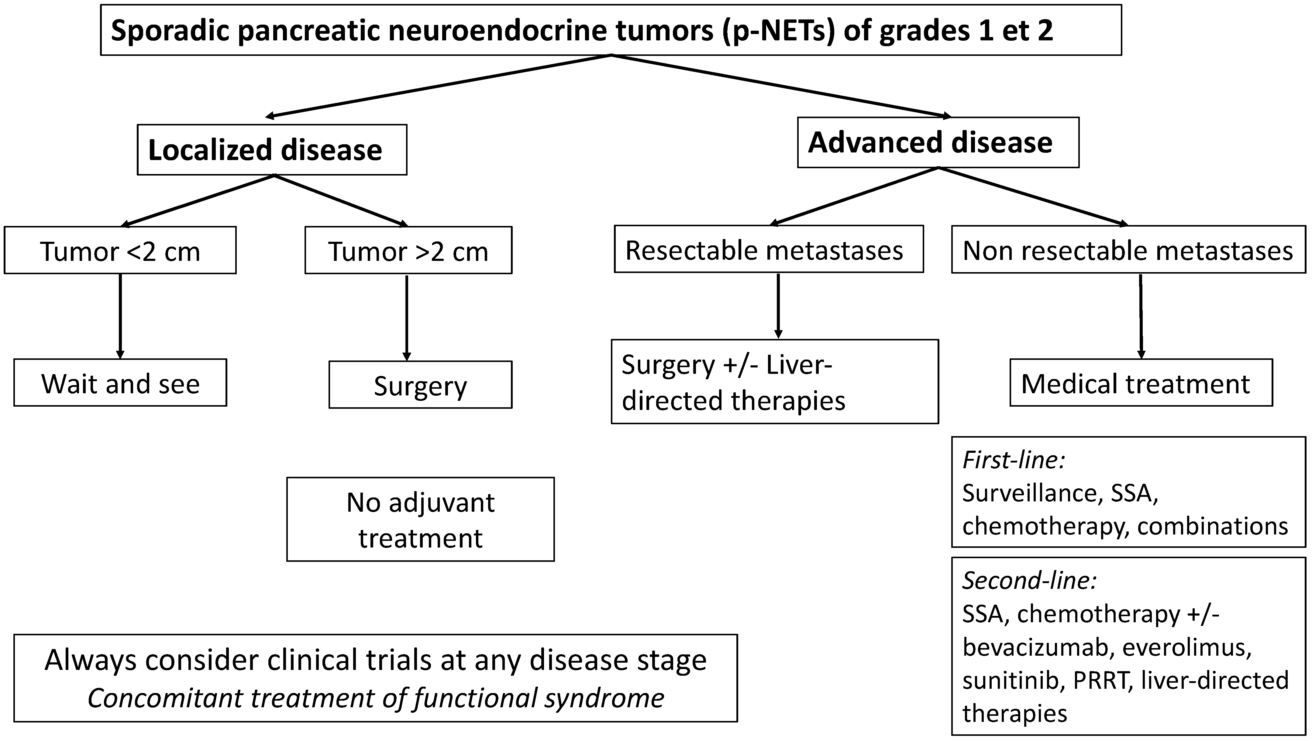
2. Multimodal Management of Localized Disease
2.1. Indications
2.2. Minimally Invasive Approach
2.3. Robotic Approach
2.4. Enucleation
2.5. Non-Sporadic p-NETs
3. Multimodal Strategy for Advanced p-NETs
3.1. Medical Treatment Options
3.2. Cytotoxic Chemotherapy
3.3. Somatostatin Analogs (SSA)
3.4. Targeted Therapy
3.4.1. Agents for Antiangiogenesis
3.4.2. mTOR Inhibitors
3.5. Peptide Receptor Radionuclide Therapy (PRRT)
3.6. External Radiotherapy
4. Surgical Management of Advanced p-NETs
4.1. Surgery of Liver Metastases
4.2. Management of Primary Tumor
4.3. Liver-Directed Therapies
4.4. Minimally Invasive Approach
4.5. Liver Transplantation
5. Conclusions
Author Contributions
Funding
Conflicts of Interest
References
- Ehehalt, F.; Saeger, H.D.; Schmidt, C.M.; Grützmann, R. Neuroendocrine Tumors of the Pancreas. Oncologist 2009, 14, 456–467. [Google Scholar] [CrossRef]
- Klöppel, G.; Hruban, R.H. WHO Classification of Tumours of Endocrine Organs; International Agency for Research on Cancer: Lyon, France, 2017. [Google Scholar]
- Dasari, A.; Shen, C.; Halperin, D.; Zhao, B.; Zhou, S.; Xu, Y.; Shih, T.; Yao, J.C. Trends in the Incidence, Prevalence, and Survival Outcomes in Patients With Neuroendocrine Tumors in the United States. JAMA Oncol. 2017, 3, 1335–1342. [Google Scholar] [CrossRef] [PubMed]
- Ito, T.; Lee, L.; Hijioka, M.; Kawabe, K.; Kato, M.; Nakamura, K.; Ueda, K.; Ohtsuka, T.; Igarashi, H. The Up-to-Date Review of Epidemiological Pancreatic Neuroendocrine Tumors in Japan. J. Hepatobiliary Pancreat. Sci. 2015, 22, 574–577. [Google Scholar] [CrossRef] [PubMed]
- Man, D.; Wu, J.; Shen, Z.; Zhu, X. Prognosis of patients with neuroendocrine tumor: A SEER database analysis. Cancer Manag. Res. 2018, 10, 5629–5638. [Google Scholar] [CrossRef] [Green Version]
- Díez, M.; Teulé, A.; Salazar, R. Gastroenteropancreatic Neuroendocrine Tumors: Diagnosis and Treatment. Ann. Gastroenterol. 2013, 26, 29–36. [Google Scholar]
- Ter-Minassian, M.; Chan, J.A.; Hooshmand, S.M.; Brais, L.K.; Daskalova, A.; Heafield, R.; Buchanan, L.; Qian, Z.R.; Fuchs, C.S.; Lin, X.; et al. Clinical Presentation, Recurrence, and Survival in Patients with Neuroendocrine Tumors: Results from a Prospective Institutional Database. Endocr. Relat. Cancer 2013, 20, 187–196. [Google Scholar] [CrossRef] [PubMed] [Green Version]
- Azoulay, A.; Cros, J.; Vullierme, M.P.; de Mestier, L.; Couvelard, A.; Hentic, O.; Ruszniewski, P.; Sauvanet, A.; Vilgrain, V.; Ronot, M. Morphological imaging and CT histogram analysis to differentiate pancreatic neuroendocrine tumor grade 3 from neuroendocrine carcinoma. Diagn. Interv. Imaging. 2020, 101, 821–830. [Google Scholar] [CrossRef]
- Pellat, A.; Cottereau, A.S.; Palmieri, L.-J.; Soyer, P.; Marchese, U.; Brezault, C.; Coriat, R. Digestive Well-Differentiated Grade 3 Neuroendocrine Tumors: Current Management and Future Directions. Cancers 2021, 13, 2448. [Google Scholar] [CrossRef]
- Yang, M.; Zeng, L.; Ke, N.; Tan, C.; Tian, B.; Liu, X.; Xiang, B.; Zhang, Y. World Health Organization Grading Classification for Pancreatic Neuroendocrine Neoplasms: A Comprehensive Analysis from a Large Chinese Institution. BMC Cancer 2020, 20, 906. [Google Scholar] [CrossRef] [PubMed]
- Hassan, M.M.; Phan, A.; Li, D.; Dagohoy, C.G.; Leary, C.; Yao, J.C. Family History of Cancer and Associated Risk of Developing Neuroendocrine Tumors: A Case-Control Study. Cancer Epidemiol. Biomarkers Prev. 2008, 17, 959–965. [Google Scholar] [CrossRef] [Green Version]
- Kartalis, N.; Mucelli, R.M.P.; Sundin, A. Recent Developments in Imaging of Pancreatic Neuroendocrine Tumors. Ann. Gastroenterol. 2015, 28, 193–202. [Google Scholar] [PubMed]
- Balogova, S.; Talbot, J.-N.; Nataf, V.; Michaud, L.; Huchet, V.; Kerrou, K.; Montravers, F. 18F-Fluorodihydroxyphenylalanine vs. Other Radiopharmaceuticals for Imaging Neuroendocrine Tumours According to Their Type. Eur. J. Nucl. Med. Mol. Imaging 2013, 40, 943–966. [Google Scholar] [CrossRef] [Green Version]
- Schreiter, N.F.; Nogami, M.; Steffen, I.; Pape, U.-F.; Hamm, B.; Brenner, W.; Röttgen, R. Evaluation of the Potential of PET-MRI Fusion for Detection of Liver Metastases in Patients with Neuroendocrine Tumours. Eur. Radiol. 2012, 22, 458–467. [Google Scholar] [CrossRef] [PubMed]
- Geurts, J.L. Inherited Syndromes Involving Pancreatic Neuroendocrine Tumors. J. Gastrointest. Oncol. 2020, 11, 559–566. [Google Scholar] [CrossRef] [PubMed]
- Chou, W.-C.; Lin, P.-H.; Yeh, Y.-C.; Shyr, Y.-M.; Fang, W.-L.; Wang, S.-E.; Liu, C.-Y.; Chang, P.M.-H.; Chen, M.-H.; Hung, Y.-P.; et al. Genes Involved in Angiogenesis and MTOR Pathways Are Frequently Mutated in Asian Patients with Pancreatic Neuroendocrine Tumors. Int. J. Biol. Sci. 2016, 12, 1523–1532. [Google Scholar] [CrossRef] [Green Version]
- Jiao, Y.; Shi, C.; Edil, B.H.; de Wilde, R.F.; Klimstra, D.S.; Maitra, A.; Schulick, R.D.; Tang, L.H.; Wolfgang, C.L.; Choti, M.A.; et al. DAXX/ATRX, MEN1 and MTOR Pathway Genes Are Frequently Altered in Pancreatic Neuroendocrine Tumors. Science 2011, 331, 1199–1203. [Google Scholar] [CrossRef] [Green Version]
- Cheema, A.; Weber, J.; Strosberg, J.R. Incidental Detection of Pancreatic Neuroendocrine Tumors: An Analysis of Incidence and Outcomes. Ann. Surg. Oncol. 2012, 19, 2932–2936. [Google Scholar] [CrossRef] [PubMed]
- Partelli, S.; Cirocchi, R.; Crippa, S.; Cardinali, L.; Fendrich, V.; Bartsch, D.K.; Falconi, M. Systematic review of active surveillance versus surgical management of asymptomatic small non-functioning pancreatic neuroendocrine neoplasms. Br. J. Surg. 2017, 104, 34–41. [Google Scholar] [CrossRef] [PubMed]
- Partelli, S.; Ramage, J.K.; Massironi, S.; Zerbi, A.; Kim, H.B.; Niccoli, P.; Panzuto, F.; Landoni, L.; Tomazic, A.; Ibrahim, T.; et al. Management of Asymptomatic Sporadic Nonfunctioning Pancreatic Neuroendocrine Neoplasms (ASPEN) ≤2 Cm: Study Protocol for a Prospective Observational Study. Front. Med. (Lausanne) 2020, 7, 598438. [Google Scholar] [CrossRef]
- Larghi, A.; Capurso, G.; Carnuccio, A.; Ricci, R.; Alfieri, S.; Galasso, D.; Lugli, F.; Bianchi, A.; Panzuto, F.; De Marinis, L.; et al. Ki-67 Grading of Nonfunctioning Pancreatic Neuroendocrine Tumors on Histologic Samples Obtained by EUS-Guided Fine-Needle Tissue Acquisition: A Prospective Study. Gastrointest. Endosc. 2012, 76, 570–577. [Google Scholar] [CrossRef]
- Lopez-Aguiar, A.G.; Zaidi, M.Y.; Beal, E.W.; Dillhoff, M.; Cannon, J.G.D.; Poultsides, G.A.; Kanji, Z.S.; Rocha, F.G.; Marincola Smith, P.; Idrees, K.; et al. Defining the Role of Lymphadenectomy for Pancreatic Neuroendocrine Tumors: An Eight-Institution Study of 695 Patients from the US Neuroendocrine Tumor Study Group. Ann. Surg. Oncol. 2019, 26, 2517–2524. [Google Scholar] [CrossRef] [PubMed]
- Bliss, L.A.; Yang, C.J.; Chau, Z.; Ng, S.C.; McFadden, D.W.; Kent, T.S.; Moser, A.J.; Callery, M.P.; Tseng, J.F. Patient Selection and the Volume Effect in Pancreatic Surgery: Unequal Benefits? HPB 2014, 16, 899–906. [Google Scholar] [CrossRef] [PubMed] [Green Version]
- El Amrani, M.; Clément, G.; Lenne, X.; Laueriere, C.; Turpin, A.; Theis, D.; Pruvot, F.-R.; Truant, S. Should All Pancreatic Surgery Be Centralized Regardless of Patients’ Comorbidity? HPB (Oxford) 2020, 22, 1057–1066. [Google Scholar] [CrossRef]
- Kahl, S.; Malfertheiner, P. Exocrine and Endocrine Pancreatic Insufficiency after Pancreatic Surgery. Best Pract. Res. Clin. Gastroenterol. 2004, 18, 947–955. [Google Scholar] [CrossRef]
- Kamilaris, C.D.C.; Stratakis, C.A. Multiple Endocrine Neoplasia Type 1 (MEN1): An Update and the Significance of Early Genetic and Clinical Diagnosis. Front. Endocrinol. (Lausanne) 2019, 10, 339. [Google Scholar] [CrossRef] [Green Version]
- Drymousis, P.; Raptis, D.A.; Spalding, D.; Fernandez-Cruz, L.; Menon, D.; Breitenstein, S.; Davidson, B.; Frilling, A. Laparoscopic versus Open Pancreas Resection for Pancreatic Neuroendocrine Tumours: A Systematic Review and Meta-Analysis. HPB (Oxford) 2014, 16, 397–406. [Google Scholar] [CrossRef] [PubMed] [Green Version]
- Van Hilst, J.; Korrel, M.; de Rooij, T.; Lof, S.; Busch, O.R.; Groot Koerkamp, B.; Kooby, D.A.; van Dieren, S.; Abu Hilal, M.; Besselink, M.G.; et al. Oncologic Outcomes of Minimally Invasive versus Open Distal Pancreatectomy for Pancreatic Ductal Adenocarcinoma: A Systematic Review and Meta-Analysis. Eur. J. Surg. Oncol. 2019, 45, 719–727. [Google Scholar] [CrossRef]
- De Rooij, T.; Jilesen, A.P.; Boerma, D.; Bonsing, B.A.; Bosscha, K.; van Dam, R.M.; van Dieren, S.; Dijkgraaf, M.G.; van Eijck, C.H.; Gerhards, M.F.; et al. A Nationwide Comparison of Laparoscopic and Open Distal Pancreatectomy for Benign and Malignant Disease. J. Am. Coll. Surg. 2015, 220, 263–270.e1. [Google Scholar] [CrossRef]
- De Rooij, T.; van Hilst, J.; van Santvoort, H.; Boerma, D.; van den Boezem, P.; Daams, F.; van Dam, R.; Dejong, C.; van Duyn, E.; Dijkgraaf, M.; et al. Minimally Invasive Versus Open Distal Pancreatectomy (LEOPARD): A Multicenter Patient-Blinded Randomized Controlled Trial. Ann. Surg. 2019, 269, 2–9. [Google Scholar] [CrossRef]
- Tyutyunnik, P.; Klompmaker, S.; Lombardo, C.; Lapshyn, H.; Menonna, F.; Napoli, N.; Wellner, U.; Izrailov, R.; Baychorov, M.; Besselink, M.G.; et al. Learning Curve of Three European Centers in Laparoscopic, Hybrid Laparoscopic, and Robotic Pancreatoduodenectomy. Surg. Endosc. 2022, 36, 1515–1526. [Google Scholar] [CrossRef]
- De Rooij, T.; Lu, M.Z.; Steen, M.W.; Gerhards, M.F.; Dijkgraaf, M.G.; Busch, O.R.; Lips, D.J.; Festen, S.; Besselink, M.G. Dutch Pancreatic Cancer Group Minimally Invasive Versus Open Pancreatoduodenectomy: Systematic Review and Meta-Analysis of Comparative Cohort and Registry Studies. Ann. Surg. 2016, 264, 257–267. [Google Scholar] [CrossRef]
- Klompmaker, S.; van Hilst, J.; Wellner, U.F.; Busch, O.R.; Coratti, A.; D’Hondt, M.; Dokmak, S.; Festen, S.; Kerem, M.; Khatkov, I.; et al. Outcomes After Minimally-Invasive Versus Open Pancreatoduodenectomy: A Pan-European Propensity Score Matched Study. Ann. Surg. 2020, 271, 356–363. [Google Scholar] [CrossRef]
- Yan, J.-F.; Pan, Y.; Chen, K.; Zhu, H.-P.; Chen, Q.-L. Minimally Invasive Pancreatoduodenectomy Is Associated with Lower Morbidity Compared to Open Pancreatoduodenectomy: An Updated Meta-Analysis of Randomized Controlled Trials and High-Quality Nonrandomized Studies. Medicine (Baltimore) 2019, 98, e16730. [Google Scholar] [CrossRef]
- Van Hilst, J.; de Rooij, T.; Bosscha, K.; Brinkman, D.J.; van Dieren, S.; Dijkgraaf, M.G.; Gerhards, M.F.; de Hingh, I.H.; Karsten, T.M.; Lips, D.J.; et al. Laparoscopic versus Open Pancreatoduodenectomy for Pancreatic or Periampullary Tumours (LEOPARD-2): A Multicentre, Patient-Blinded, Randomised Controlled Phase 2/3 Trial. Lancet Gastroenterol. Hepatol. 2019, 4, 199–207. [Google Scholar] [CrossRef]
- Zakaria, H.M.; Stauffer, J.A.; Raimondo, M.; Woodward, T.A.; Wallace, M.B.; Asbun, H.J. Total Pancreatectomy: Short- and Long-Term Outcomes at a High-Volume Pancreas Center. World J. Gastrointest. Surg. 2016, 8, 634–642. [Google Scholar] [CrossRef] [PubMed]
- Melvin, W.S.; Needleman, B.J.; Krause, K.R.; Schneider, C.; Wolf, R.K.; Michler, R.E.; Ellison, E.C. Computer-Enhanced Robotic Telesurgery. Initial Experience in Foregut Surgery. Surg. Endosc. 2002, 16, 1790–1792. [Google Scholar] [CrossRef] [PubMed]
- Yang, S.J.; Hwang, H.K.; Kang, C.M.; Lee, W.J. Revisiting the Potential Advantage of Robotic Surgical System in Spleen-Preserving Distal Pancreatectomy over Conventional Laparoscopic Approach. Ann. Transl. Med. 2020, 8, 188. [Google Scholar] [CrossRef]
- Chen, S.; Zhan, Q.; Chen, J.; Jin, J.; Deng, X.; Chen, H.; Shen, B.; Peng, C.; Li, H. Robotic Approach Improves Spleen-Preserving Rate and Shortens Postoperative Hospital Stay of Laparoscopic Distal Pancreatectomy: A Matched Cohort Study. Surg. Endosc. 2015, 29, 3507–3518. [Google Scholar] [CrossRef]
- Zhang, J.; Jin, J.; Chen, S.; Gu, J.; Zhu, Y.; Qin, K.; Zhan, Q.; Cheng, D.; Chen, H.; Deng, X.; et al. Minimally Invasive Distal Pancreatectomy for PNETs: Laparoscopic or Robotic Approach? Oncotarget 2017, 8, 33872–33883. [Google Scholar] [CrossRef] [Green Version]
- Daouadi, M.; Zureikat, A.H.; Zenati, M.S.; Choudry, H.; Tsung, A.; Bartlett, D.L.; Hughes, S.J.; Lee, K.K.; Moser, A.J.; Zeh, H.J. Robot-Assisted Minimally Invasive Distal Pancreatectomy Is Superior to the Laparoscopic Technique. Ann. Surg. 2013, 257, 128–132. [Google Scholar] [CrossRef] [Green Version]
- Ryan, C.E.; Ross, S.B.; Sukharamwala, P.B.; Sadowitz, B.D.; Wood, T.W.; Rosemurgy, A.S. Distal Pancreatectomy and Splenectomy: A Robotic or LESS Approach. JSLS 2015, 19, e2014.00246. [Google Scholar] [CrossRef] [Green Version]
- Lai, E.C.H.; Tang, C.N. Robotic Distal Pancreatectomy versus Conventional Laparoscopic Distal Pancreatectomy: A Comparative Study for Short-Term Outcomes. Front. Med. 2015, 9, 356–360. [Google Scholar] [CrossRef] [PubMed]
- Zhou, N.; Chen, J.; Liu, Q.; Zhang, X.; Wang, Z.; Ren, S.; Chen, X. Outcomes of Pancreatoduodenectomy with Robotic Surgery versus Open Surgery. Int. J. Med. Robot. 2011, 7, 131–137. [Google Scholar] [CrossRef]
- Rosemurgy, A.; Ross, S.; Bourdeau, T.; Craigg, D.; Spence, J.; Alvior, J.; Sucandy, I. Robotic Pancreaticoduodenectomy Is the Future: Here and Now. J. Am. Coll. Surg. 2019, 228, 613–624. [Google Scholar] [CrossRef]
- Boone, B.A.; Zenati, M.; Hogg, M.E.; Steve, J.; Moser, A.J.; Bartlett, D.L.; Zeh, H.J.; Zureikat, A.H. Assessment of Quality Outcomes for Robotic Pancreaticoduodenectomy: Identification of the Learning Curve. JAMA Surg. 2015, 150, 416–422. [Google Scholar] [CrossRef] [Green Version]
- Hackert, T.; Hinz, U.; Fritz, S.; Strobel, O.; Schneider, L.; Hartwig, W.; Büchler, M.W.; Werner, J. Enucleation in Pancreatic Surgery: Indications, Technique, and Outcome Compared to Standard Pancreatic Resections. Langenbecks Arch. Surg. 2011, 396, 1197. [Google Scholar] [CrossRef]
- Marchese, U.; Tzedakis, S.; Abou Ali, E.; Turrini, O.; Delpero, J.-R.; Coriat, R.; Fuks, D. Parenchymal Sparing Resection: Options in Duodenal and Pancreatic Surgery. J. Clin. Med. 2021, 10, 1479. [Google Scholar] [CrossRef] [PubMed]
- Santucci, N.; Gaujoux, S.; Binquet, C.; Reichling, C.; Lifante, J.-C.; Carnaille, B.; Pattou, F.; Mirallié, E.; Facy, O.; Mathonnet, M.; et al. Pancreatoduodenectomy for Neuroendocrine Tumors in Patients with Multiple Endocrine Neoplasia Type 1: An AFCE (Association Francophone de Chirurgie Endocrinienne) and GTE (Groupe d’étude Des Tumeurs Endocrines) Study. World J. Surg. 2021, 45, 1794–1802. [Google Scholar] [CrossRef]
- Herrera, M.F.; Åkerström, G.; Angelos, P.; Grant, C.S.; Hoff, A.O.; Pantoja, J.P.; Pérez-Johnston, R.; Sahani, D.V.; Wong, R.J.; Randolph, G. AACE/ACE Disease State Clinical Review: Pancreatic Neuroendocrine Incidentalomas. Endocr. Pract. 2015, 21, 546–553. [Google Scholar] [CrossRef] [Green Version]
- Yoo, Y.J.; Yang, S.J.; Hwang, H.K.; Kang, C.M.; Kim, H.; Lee, W.J. Overestimated Oncologic Significance of Lymph Node Metastasis in G1 Nonfunctioning Neuroendocrine Tumor in the Left Side of the Pancreas. Medicine (Baltimore) 2015, 94, e1404. [Google Scholar] [CrossRef] [PubMed]
- Zhang, R.-C.; Zhou, Y.-C.; Mou, Y.-P.; Huang, C.-J.; Jin, W.-W.; Yan, J.-F.; Wang, Y.-X.; Liao, Y. Laparoscopic versus Open Enucleation for Pancreatic Neoplasms: Clinical Outcomes and Pancreatic Function Analysis. Surg. Endosc. 2016, 30, 2657–2665. [Google Scholar] [CrossRef]
- Karaliotas, C.; Sgourakis, G. Laparoscopic versus Open Enucleation for Solitary Insulinoma in the Body and Tail of the Pancreas. J. Gastrointest. Surg. 2009, 13, 1869. [Google Scholar] [CrossRef] [PubMed]
- Sa Cunha, A.; Beau, C.; Rault, A.; Catargi, B.; Collet, D.; Masson, B. Laparoscopic versus Open Approach for Solitary Insulinoma. Surg. Endosc. 2007, 21, 103–108. [Google Scholar] [CrossRef] [PubMed]
- Triponez, F.; Sadowski, S.M.; Pattou, F.; Cardot-Bauters, C.; Mirallié, E.; Le Bras, M.; Sebag, F.; Niccoli, P.; Deguelte, S.; Cadiot, G.; et al. Long-term Follow-up of MEN1 Patients Who Do Not Have Initial Surgery for Small ≤2 cm Nonfunctioning Pancreatic Neuroendocrine Tumors, an AFCE and GTE Study: Association Francophone de Chirurgie Endocrinienne & Groupe d’Etude des Tumeurs Endocrines. Ann. Surg. 2018, 268, 158–164. [Google Scholar] [CrossRef] [Green Version]
- Niederle, B.; Selberherr, A.; Bartsch, D.K.; Brandi, M.L.; Doherty, G.M.; Falconi, M.; Goudet, P.; Halfdanarson, T.R.; Ito, T.; Jensen, R.T.; et al. Multiple Endocrine Neoplasia Type 1 and the Pancreas: Diagnosis and Treatment of Functioning and Non-Functioning Pancreatic and Duodenal Neuroendocrine Neoplasia within the MEN1 Syndrome—An International Consensus Statement. Neuroendocrinology 2021, 111, 609–630. [Google Scholar] [CrossRef] [PubMed]
- Van Beek, D.J.; Nell, S.; Verkooijen, H.M.; Borel Rinkes, I.H.M.; Valk, G.D.; (on behalf of the DutchMEN study group); Vriens, M.R.; International MEN1 Insulinoma Study Group. Surgery for Multiple Endocrine Neoplasia Type 1-Related Insulinoma: Long-Term Outcomes in a Large International Cohort. Br. J. Surg. 2020, 107, 1489–1499. [Google Scholar] [CrossRef] [PubMed]
- Bartsch, D.K.; Albers, M.; Knoop, R.; Kann, P.H.; Fendrich, V.; Waldmann, J. Enucleation and Limited Pancreatic Resection Provide Long-Term Cure for Insulinoma in Multiple Endocrine Neoplasia Type 1. Neuroendocrinology 2013, 98, 290–298. [Google Scholar] [CrossRef] [PubMed]
- Alexakis, N.; Connor, S.; Ghaneh, P.; Lombard, M.; Smart, H.; Evans, J.; Hughes, M.; Garvey, C.; Vora, J.; Vinjamuri, S.; et al. Hereditary pancreatic endocrine tumours. Pancreatology 2004, 4, 417–433. [Google Scholar] [CrossRef]
- Tirosh, A.; Sadowski, S.M.; Linehan, W.M.; Libutti, S.K.; Patel, D.; Nilubol, N.; Kebebew, E. Association of VHL Genotype With Pancreatic Neuroendocrine Tumor Phenotype in Patients With von Hippel-Lindau Disease. JAMA Oncol. 2018, 4, 124–126. [Google Scholar] [CrossRef]
- Tamura, K.; Nishimori, I.; Ito, T.; Yamasaki, I.; Igarashi, H.; Shuin, T. Diagnosis and Management of Pancreatic Neuroendocrine Tumor in von Hippel-Lindau Disease. World J. Gastroenterol. 2010, 16, 4515–4518. [Google Scholar] [CrossRef]
- Kouvaraki, M.A.; Ajani, J.A.; Hoff, P.; Wolff, R.; Evans, D.B.; Lozano, R.; Yao, J.C. Fluorouracil, Doxorubicin, and Streptozocin in the Treatment of Patients with Locally Advanced and Metastatic Pancreatic Endocrine Carcinomas. J. Clin. Oncol. 2004, 22, 4762–4771. [Google Scholar] [CrossRef]
- Moertel, C.G.; Lefkopoulo, M.; Lipsitz, S.; Hahn, R.G.; Klaassen, D. Streptozocin-Doxorubicin, Streptozocin-Fluorouracil or Chlorozotocin in the Treatment of Advanced Islet-Cell Carcinoma. N. Engl. J. Med. 1992, 326, 519–523. [Google Scholar] [CrossRef]
- Dilz, L.M.; Denecke, T.; Steffen, I.G.; Prasad, V.; von Weikersthal, L.F.; Pape, U.F.; Wiedenmann, B.; Pavel, M. Streptozocin/5-fluorouracil chemotherapy is associated with durable response in patients with advanced pancreatic neuroendocrine tumours. Eur. J. Cancer 2015, 51, 1253–1262. [Google Scholar] [CrossRef]
- Antonodimitrakis, P.C.; Sundin, A.; Wassberg, C.; Granberg, D.; Skogseid, B.; Eriksson, B. Streptozocin and 5-Fluorouracil for the Treatment of Pancreatic Neuroendocrine Tumors: Efficacy, Prognostic Factors and Toxicity. Neuroendocrinology 2016, 103, 345–353. [Google Scholar] [CrossRef] [PubMed]
- Strosberg, J.R.; Fine, R.L.; Choi, J.; Nasir, A.; Coppola, D.; Chen, D.-T.; Helm, J.; Kvols, L. First-Line Chemotherapy with Capecitabine and Temozolomide in Patients with Metastatic Pancreatic Endocrine Carcinomas. Cancer 2011, 117, 268–275. [Google Scholar] [CrossRef]
- Kunz, P.L.; Catalano, P.J.; Nimeiri, H.; Fisher, G.A.; Longacre, T.A.; Suarez, C.J.; Yao, J.C.; Kulke, M.H.; Hendifar, A.E.; Shanks, J.C.; et al. A Randomized Study of Temozolomide or Temozolomide and Capecitabine in Patients with Advanced Pancreatic Neuroendocrine Tumors: A Trial of the ECOG-ACRIN Cancer Research Group (E2211). JCO 2018, 36, 4004. [Google Scholar] [CrossRef]
- De Mestier, L.; Walter, T.; Brixi, H.; Evrard, C.; Legoux, J.L.; De Boissieu, P.; Hentic, O.; Cros, J.; Hammel, P.; Tougeron, D.; et al. Comparison of Temozolomide-Capecitabine to 5-Fluorouracile-Dacarbazine in 247 Patients with Advanced Digestive Neuroendocrine Tumors Using Propensity Score Analyses. Neuroendocrinology 2019, 108, 343–353. [Google Scholar] [CrossRef]
- Bajetta, E.; Rimassa, L.; Carnaghi, C.; Seregni, E.; Ferrari, L.; Di Bartolomeo, M.; Regalia, E.; Cassata, A.; Procopio, G.; Mariani, L. 5-Fluorouracil, Dacarbazine, and Epirubicin in the Treatment of Patients with Neuroendocrine Tumors. Cancer 1998, 83, 372–378. [Google Scholar] [CrossRef]
- Dussol, A.S.; Joly, M.O.; Vercherat, C.; Forestier, J.; Hervieu, V.; Scoazec, J.; Lombard-Bohas, C.; Walter, T. Gemcitabine and oxaliplatin or alkylating agents for neuroendocrine tumors: Comparison of efficacy and search for predictive factors guiding treatment choice. Cancer 2015, 121, 3428–3434. [Google Scholar] [CrossRef] [Green Version]
- Cassier, P.A.; Walter, T.; Eymard, B.; Ardisson, P.; Perol, M.; Paillet, C.; Chayvialle, J.-A.; Scoazec, J.-Y.; Hervieu, V.; Bohas, C.L. Gemcitabine and Oxaliplatin Combination Chemotherapy for Metastatic Well-Differentiated Neuroendocrine Carcinomas: A Single-Center Experience. Cancer 2009, 115, 3392–3399. [Google Scholar] [CrossRef] [PubMed]
- Caplin, M.E.; Pavel, M.; Ćwikła, J.B.; Phan, A.T.; Raderer, M.; Sedláčková, E.; Cadiot, G.; Wolin, E.M.; Capdevila, J.; Wall, L.; et al. Lanreotide in Metastatic Enteropancreatic Neuroendocrine Tumors. N. Engl. J. Med. 2014, 371, 224–233. [Google Scholar] [CrossRef]
- Yao, J.C. Neuroendocrine Tumors. Molecular Targeted Therapy for Carcinoid and Islet-Cell Carcinoma. Best Pract. Res. Clin. Endocrinol. Metab. 2007, 21, 163–172. [Google Scholar] [CrossRef]
- Plöckinger, U.; Wiedenmann, B. Treatment of gastroenteropancreatic neuroendocrine tumors. Virchows Arch. 2007, 451 (Suppl. 1), S71–S80. [Google Scholar] [CrossRef]
- Terris, B.; Scoazec, J.Y.; Rubbia, L.; Bregeaud, L.; Pepper, M.S.; Ruszniewski, P.; Belghiti, J.; Fléjou, J.; Degott, C. Expression of Vascular Endothelial Growth Factor in Digestive Neuroendocrine Tumours. Histopathology 1998, 32, 133–138. [Google Scholar] [CrossRef] [PubMed]
- Zhang, J.; Jia, Z.; Li, Q.; Wang, L.; Rashid, A.; Zhu, Z.; Evans, D.B.; Vauthey, J.-N.; Xie, K.; Yao, J.C. Elevated Expression of Vascular Endothelial Growth Factor Correlates with Increased Angiogenesis and Decreased Progression-Free Survival among Patients with Low-Grade Neuroendocrine Tumors. Cancer 2007, 109, 1478–1486. [Google Scholar] [CrossRef] [PubMed]
- Fjällskog, M.-L.H.; Lejonklou, M.H.; Oberg, K.E.; Eriksson, B.K.; Janson, E.T. Expression of Molecular Targets for Tyrosine Kinase Receptor Antagonists in Malignant Endocrine Pancreatic Tumors. Clin. Cancer Res. 2003, 9, 1469–1473. [Google Scholar]
- Mena, A.C.; Pulido, E.G.; Guillén-Ponce, C. Understanding the Molecular-Based Mechanism of Action of the Tyrosine Kinase Inhibitor: Sunitinib. Anticancer Drugs 2010, 21 (Suppl. 1), S3–S11. [Google Scholar] [CrossRef]
- Raymond, E.; Dahan, L.; Raoul, J.-L.; Bang, Y.-J.; Borbath, I.; Lombard-Bohas, C.; Valle, J.; Metrakos, P.; Smith, D.; Vinik, A.; et al. Sunitinib Malate for the Treatment of Pancreatic Neuroendocrine Tumors. N. Engl. J. Med. 2011, 364, 501–513. [Google Scholar] [CrossRef] [Green Version]
- Hobday, T.J.; Rubin, J.; Holen, K.; Picus, J.; Donehower, R.; Marschke, R.; Maples, W.; Lloyd, R.; Mahoney, M.; Erlichman, C. MC044h, a Phase II Trial of Sorafenib in Patients (Pts) with Metastatic Neuroendocrine Tumors (NET): A Phase II Consortium (P2C) Study. JCO 2007, 25, 4504. [Google Scholar] [CrossRef]
- Phan, A.T.; Halperin, D.M.; Chan, J.A.; Fogelman, D.R.; Hess, K.R.; Malinowski, P.; Regan, E.; Ng, C.S.; Yao, J.C.; Kulke, M.H. Pazopanib and Depot Octreotide in Advanced, Well-Differentiated Neuroendocrine Tumours: A Multicentre, Single-Group, Phase 2 Study. Lancet Oncol. 2015, 16, 695–703. [Google Scholar] [CrossRef] [Green Version]
- Chan, J.A.; Stuart, K.; Earle, C.C.; Clark, J.W.; Bhargava, P.; Miksad, R.; Blaszkowsky, L.; Enzinger, P.C.; Meyerhardt, J.A.; Zheng, H.; et al. Prospective study of bevacizumab plus temozolomide in patients with advanced neuroendocrine tumors. J. Clin. Oncol. 2012, 30, 2963–2968. [Google Scholar] [CrossRef] [PubMed] [Green Version]
- Ducreux, M.; Dahan, L.; Smith, D.; O’Toole, D.; Lepère, C.; Dromain, C.; Vilgrain, V.; Baudin, E.; Lombard-Bohas, C.; Scoazec, J.-Y.; et al. Bevacizumab Combined with 5-FU/Streptozocin in Patients with Progressive Metastatic Well-Differentiated Pancreatic Endocrine Tumours (BETTER Trial)--a Phase II Non-Randomised Trial. Eur. J. Cancer 2014, 50, 3098–3106. [Google Scholar] [CrossRef]
- Capdevila, J.; Fazio, N.; Lopez, C.; Teulé, A.; Valle, J.W.; Tafuto, S.; Custodio, A.; Reed, N.; Raderer, M.; Grande, E.; et al. Lenvatinib in Patients With Advanced Grade 1/2 Pancreatic and Gastrointestinal Neuroendocrine Tumors: Results of the Phase II TALENT Trial (GETNE1509). J. Clin. Oncol. 2021, 39, 2304–2312. [Google Scholar] [CrossRef] [PubMed]
- Yao, J.C.; Phan, A.T.; Chang, D.Z.; Wolff, R.A.; Hess, K.; Gupta, S.; Jacobs, C.; Mares, J.E.; Landgraf, A.N.; Rashid, A.; et al. Efficacy of RAD001 (Everolimus) and Octreotide LAR in Advanced Low- to Intermediate-Grade Neuroendocrine Tumors: Results of a Phase II Study. J. Clin. Oncol. 2008, 26, 4311–4318. [Google Scholar] [CrossRef]
- Yao, J.C.; Lombard-Bohas, C.; Baudin, E.; Kvols, L.K.; Rougier, P.; Ruszniewski, P.; Hoosen, S.; St Peter, J.; Haas, T.; Lebwohl, D.; et al. Daily Oral Everolimus Activity in Patients with Metastatic Pancreatic Neuroendocrine Tumors after Failure of Cytotoxic Chemotherapy: A Phase II Trial. J. Clin. Oncol. 2010, 28, 69–76. [Google Scholar] [CrossRef]
- Yao, J.C.; Shah, M.H.; Ito, T.; Bohas, C.L.; Wolin, E.M.; Van Cutsem, E.; Hobday, T.J.; Okusaka, T.; Capdevila, J.; de Vries, E.G.E.; et al. Everolimus for Advanced Pancreatic Neuroendocrine Tumors. N. Engl. J. Med. 2011, 364, 514–523. [Google Scholar] [CrossRef] [Green Version]
- Strosberg, J.; El-Haddad, G.; Wolin, E.; Hendifar, A.; Yao, J.; Chasen, B.; Mittra, E.; Kunz, P.L.; Kulke, M.H.; Jacene, H.; et al. Phase 3 Trial of 177Lu-Dotatate for Midgut Neuroendocrine Tumors. N. Engl. J. Med. 2017, 376, 125–135. [Google Scholar] [CrossRef]
- Brabander, T.; van der Zwan, W.A.; Teunissen, J.J.M.; Kam, B.L.R.; Feelders, R.A.; de Herder, W.W.; van Eijck, C.H.J.; Franssen, G.J.H.; Krenning, E.P.; Kwekkeboom, D.J. Long-Term Efficacy, Survival, and Safety of [177Lu-DOTA0,Tyr3]Octreotate in Patients with Gastroenteropancreatic and Bronchial Neuroendocrine Tumors. Clin. Cancer Res. 2017, 23, 4617–4624. [Google Scholar] [CrossRef] [Green Version]
- Chan, D.L.; Thompson, R.; Lam, M.; Pavlakis, N.; Hallet, J.; Law, C.; Singh, S.; Myrehaug, S. External Beam Radiotherapy in the Treatment of Gastroenteropancreatic Neuroendocrine Tumours: A Systematic Review. Clin. Oncol. (R Coll Radiol.) 2018, 30, 400–408. [Google Scholar] [CrossRef] [PubMed] [Green Version]
- Iwata, T.; Ueno, H.; Itami, J.; Ito, Y.; Inaba, K.; Morizane, C.; Kondo, S.; Sakamoto, Y.; Shiba, S.; Sasaki, M.; et al. Efficacy of radiotherapy for primary tumor in patients with unresectable pancreatic neuroendocrine tumors. JPN J. Clin. Oncol. 2017, 47, 826–831. [Google Scholar] [CrossRef] [PubMed]
- Sandhu, N.; Benson, K.R.K.; Kumar, K.A.; Eyben, R.V.; Chang, D.T.; Gibbs, I.C.; Hancock, S.L.; Meola, A.; Chang, S.D.; Li, G.; et al. Local control and toxicity outcomes of stereotactic radiosurgery for spinal metastases of gastrointestinal origin. J. Neurosurgery Spine 2020, 33, 87–94. [Google Scholar] [CrossRef] [PubMed]
- Kamieniarz, L.; Armeni, E.; O’Mahony, L.F.; Leigh, C.; Miah, L.; Narayan, A.; Bhatt, A.; Cox, N.; Mandair, D.; Navalkissoor, S.; et al. Orbital metastases from neuroendocrine neoplasms: Clinical implications and outcomes. Endocrine 2020, 67, 485–493. [Google Scholar] [CrossRef] [PubMed]
- Norton, J.A.; Warren, R.S.; Kelly, M.G.; Zuraek, M.B.; Jensen, R.T. Aggressive Surgery for Metastatic Liver Neuroendocrine Tumors. Surgery 2003, 134, 1057–1063; discussion 1063–1065. [Google Scholar] [CrossRef]
- Elias, D.; Lasser, P.; Ducreux, M.; Duvillard, P.; Ouellet, J.-F.; Dromain, C.; Schlumberger, M.; Pocard, M.; Boige, V.; Miquel, C.; et al. Liver Resection (and Associated Extrahepatic Resections) for Metastatic Well-Differentiated Endocrine Tumors: A 15-Year Single Center Prospective Study. Surgery 2003, 133, 375–382. [Google Scholar] [CrossRef]
- Watzka, F.M.; Fottner, C.; Miederer, M.; Schad, A.; Weber, M.M.; Otto, G.; Lang, H.; Musholt, T.J. Surgical Therapy of Neuroendocrine Neoplasm with Hepatic Metastasis: Patient Selection and Prognosis. Langenbecks Arch. Surg. 2015, 400, 349–358. [Google Scholar] [CrossRef]
- Mayo, S.C.; de Jong, M.C.; Pulitano, C.; Clary, B.M.; Reddy, S.K.; Gamblin, T.C.; Celinksi, S.A.; Kooby, D.A.; Staley, C.A.; Stokes, J.B.; et al. Surgical Management of Hepatic Neuroendocrine Tumor Metastasis: Results from an International Multi-Institutional Analysis. Ann. Surg. Oncol. 2010, 17, 3129–3136. [Google Scholar] [CrossRef]
- Maxwell, J.E.; Sherman, S.K.; O’Dorisio, T.M.; Bellizzi, A.M.; Howe, J.R. Liver-Directed Surgery of Neuroendocrine Metastases: What Is the Optimal Strategy? Surgery 2016, 159, 320–333. [Google Scholar] [CrossRef] [Green Version]
- Gaujoux, S.; Gonen, M.; Tang, L.; Klimstra, D.; Brennan, M.F.; D’Angelica, M.; Dematteo, R.; Allen, P.J.; Jarnagin, W.; Fong, Y. Synchronous Resection of Primary and Liver Metastases for Neuroendocrine Tumors. Ann. Surg. Oncol. 2012, 19, 4270–4277. [Google Scholar] [CrossRef] [PubMed]
- Zerbi, A.; Capitanio, V.; Boninsegna, L.; Delle Fave, G.; Pasquali, C.; Rindi, G.; Campana, D.; Falconi, M.; AISP-Network Study Group. Treatment of Malignant Pancreatic Neuroendocrine Neoplasms: Middle-Term (2-Year) Outcomes of a Prospective Observational Multicentre Study. HPB (Oxford) 2013, 15, 935–943. [Google Scholar] [CrossRef] [Green Version]
- Elias, D.; Lefevre, J.H.; Duvillard, P.; Goéré, D.; Dromain, C.; Dumont, F.; Baudin, E. Hepatic Metastases from Neuroendocrine Tumors with a “Thin Slice” Pathological Examination: They Are Many More than You Think. Ann. Surg. 2010, 251, 307–310. [Google Scholar] [CrossRef] [PubMed]
- Norlén, O.; Stålberg, P.; Zedenius, J.; Hellman, P. Outcome after Resection and Radiofrequency Ablation of Liver Metastases from Small Intestinal Neuroendocrine Tumours. Br. J. Surg. 2013, 100, 1505–1514. [Google Scholar] [CrossRef]
- Osborne, D.A.; Zervos, E.E.; Strosberg, J.; Strosberg, J.; Boe, B.A.; Malafa, M.; Rosemurgy, A.S.; Yeatman, T.J.; Carey, L.; Duhaine, L.; et al. Improved Outcome with Cytoreduction versus Embolization for Symptomatic Hepatic Metastases of Carcinoid and Neuroendocrine Tumors. Ann. Surg. Oncol. 2006, 13, 572–581. [Google Scholar] [CrossRef] [PubMed]
- Cusati, D.; Zhang, L.; Harmsen, W.S.; Hu, A.; Farnell, M.B.; Nagorney, D.M.; Donohue, J.H.; Que, F.G.; Reid-Lombardo, K.M.; Kendrick, M.L. Metastatic Nonfunctioning Pancreatic Neuroendocrine Carcinoma to Liver: Surgical Treatment and Outcomes. J. Am. Coll. Surg. 2012, 215, 117–124; discussion 124–125. [Google Scholar] [CrossRef] [PubMed]
- Steinmüller, T.; Kianmanesh, R.; Falconi, M.; Scarpa, A.; Taal, B.; Kwekkeboom, D.J.; Lopes, J.M.; Perren, A.; Nikou, G.; Yao, J.; et al. Consensus Guidelines for the Management of Patients with Liver Metastases from Digestive (Neuro)Endocrine Tumors: Foregut, Midgut, Hindgut, and Unknown Primary. Neuroendocrinology 2008, 87, 47–62. [Google Scholar] [CrossRef] [PubMed]
- Kandil, E.; Noureldine, S.I.; Koffron, A.; Yao, L.; Saggi, B.; Buell, J.F. Outcomes of Laparoscopic and Open Resection for Neuroendocrine Liver Metastases. Surgery 2012, 152, 1225–1231. [Google Scholar] [CrossRef]
- Kimura, W.; Tezuka, K.; Hirai, I. Surgical Management of Pancreatic Neuroendocrine Tumors. Surg. Today 2011, 41, 1332–1343. [Google Scholar] [CrossRef]
- Hanazaki, K.; Sakurai, A.; Munekage, M.; Ichikawa, K.; Namikawa, T.; Okabayashi, T.; Imamura, M. Surgery for a Gastroenteropancreatic Neuroendocrine Tumor (GEPNET) in Multiple Endocrine Neoplasia Type 1. Surg. Today 2013, 43, 229–236. [Google Scholar] [CrossRef]
- Pavel, M.; O’Toole, D.; Costa, F.; Capdevila, J.; Gross, D.; Kianmanesh, R.; Krenning, E.; Knigge, U.; Salazar, R.; Pape, U.-F.; et al. ENETS Consensus Guidelines Update for the Management of Distant Metastatic Disease of Intestinal, Pancreatic, Bronchial Neuroendocrine Neoplasms (NEN) and NEN of Unknown Primary Site. Neuroendocrinology 2016, 103, 172–185. [Google Scholar] [CrossRef]
- Mayo, S.C.; de Jong, M.C.; Bloomston, M.; Pulitano, C.; Clary, B.M.; Reddy, S.K.; Clark Gamblin, T.; Celinski, S.A.; Kooby, D.A.; Staley, C.A.; et al. Surgery versus Intra-Arterial Therapy for Neuroendocrine Liver Metastasis: A Multicenter International Analysis. Ann. Surg. Oncol. 2011, 18, 3657–3665. [Google Scholar] [CrossRef]
- Bettini, R.; Mantovani, W.; Boninsegna, L.; Crippa, S.; Capelli, P.; Bassi, C.; Scarpa, A.; Pederzoli, P.; Falconi, M. Primary Tumour Resection in Metastatic Nonfunctioning Pancreatic Endocrine Carcinomas. Dig. Liver Dis. 2009, 41, 49–55. [Google Scholar] [CrossRef]
- Scigliano, S.; Lebtahi, R.; Maire, F.; Stievenart, J.L.; Kianmanesh, R.; Sauvanet, A.; Vullierme, M.P.; Couvelard, A.; Belghiti, J.; Ruszniewski, P.; et al. Clinical and Imaging Follow-up after Exhaustive Liver Resection of Endocrine Metastases: A 15-Year Monocentric Experience. Endocr. Relat. Cancer 2009, 16, 977–990. [Google Scholar] [CrossRef] [PubMed] [Green Version]
- Eriksson, B.; Klöppel, G.; Krenning, E.; Ahlman, H.; Plöckinger, U.; Wiedenmann, B.; Arnold, R.; Auernhammer, C.; Körner, M.; Rindi, G.; et al. Consensus Guidelines for the Management of Patients with Digestive Neuroendocrine Tumors--Well-Differentiated Jejunal-Ileal Tumor/Carcinoma. Neuroendocrinology 2008, 87, 8–19. [Google Scholar] [CrossRef] [Green Version]
- Falconi, M.; Bartsch, D.K.; Eriksson, B.; Klöppel, G.; Lopes, J.M.; O’Connor, J.M.; Salazar, R.; Taal, B.G.; Vullierme, M.P.; O’Toole, D.; et al. ENETS Consensus Guidelines for the Management of Patients with Digestive Neuroendocrine Neoplasms of the Digestive System: Well-Differentiated Pancreatic Non-Functioning Tumors. Neuroendocrinology 2012, 95, 120–134. [Google Scholar] [CrossRef]
- Capurso, G.; Rinzivillo, M.; Bettini, R.; Boninsegna, L.; Delle Fave, G.; Falconi, M. Systematic Review of Resection of Primary Midgut Carcinoid Tumour in Patients with Unresectable Liver Metastases. Br. J. Surg. 2012, 99, 1480–1486. [Google Scholar] [CrossRef] [PubMed]
- Capurso, G.; Bettini, R.; Rinzivillo, M.; Boninsegna, L.; Delle Fave, G.; Falconi, M. Role of Resection of the Primary Pancreatic Neuroendocrine Tumour Only in Patients with Unresectable Metastatic Liver Disease: A Systematic Review. Neuroendocrinology 2011, 93, 223–229. [Google Scholar] [CrossRef]
- Nguyen, S.Q.; Angel, L.P.; Divino, C.M.; Schluender, S.; Warner, R.R.P. Surgery in Malignant Pancreatic Neuroendocrine Tumors. J. Surg. Oncol. 2007, 96, 397–403. [Google Scholar] [CrossRef] [PubMed]
- Solorzano, C.C.; Lee, J.E.; Pisters, P.W.; Vauthey, J.N.; Ayers, G.D.; Jean, M.E.; Gagel, R.F.; Ajani, J.A.; Wolff, R.A.; Evans, D.B. Nonfunctioning Islet Cell Carcinoma of the Pancreas: Survival Results in a Contemporary Series of 163 Patients. Surgery 2001, 130, 1078–1085. [Google Scholar] [CrossRef] [PubMed]
- Grazi, G.L.; Cescon, M.; Pierangeli, F.; Ercolani, G.; Gardini, A.; Cavallari, A.; Mazziotti, A. Highly Aggressive Policy of Hepatic Resections for Neuroendocrine Liver Metastases. Hepatogastroenterology 2000, 47, 481–486. [Google Scholar] [PubMed]
- Pascher, A.; Steinmüller, T.; Radke, C.; Hosten, N.; Wiedenmann, B.; Neuhaus, P.; Bechstein, W.O. Primary and Secondary Hepatic Manifestation of Neuroendocrine Tumors. Langenbecks Arch. Surg. 2000, 385, 265–270. [Google Scholar] [CrossRef]
- Nave, H.; Mössinger, E.; Feist, H.; Lang, H.; Raab, H. Surgery as Primary Treatment in Patients with Liver Metastases from Carcinoid Tumors: A Retrospective, Unicentric Study over 13 Years. Surgery 2001, 129, 170–175. [Google Scholar] [CrossRef]
- Jaeck, D.; Oussoultzoglou, E.; Bachellier, P.; Lemarque, P.; Weber, J.C.; Nakano, H.; Wolf, P. Hepatic Metastases of Gastroenteropancreatic Neuroendocrine Tumors: Safe Hepatic Surgery. World J. Surg. 2001, 25, 689–692. [Google Scholar] [CrossRef]
- Yao, K.A.; Talamonti, M.S.; Nemcek, A.; Angelos, P.; Chrisman, H.; Skarda, J.; Benson, A.B.; Rao, S.; Joehl, R.J. Indications and Results of Liver Resection and Hepatic Chemoembolization for Metastatic Gastrointestinal Neuroendocrine Tumors. Surgery 2001, 130, 677–682; discussion 682–685. [Google Scholar] [CrossRef] [PubMed]
- Norton, J.A.; Kivlen, M.; Li, M.; Schneider, D.; Chuter, T.; Jensen, R.T. Morbidity and Mortality of Aggressive Resection in Patients with Advanced Neuroendocrine Tumors. Arch. Surg. 2003, 138, 859–866. [Google Scholar] [CrossRef] [Green Version]
- Touzios, J.G.; Kiely, J.M.; Pitt, S.C.; Rilling, W.S.; Quebbeman, E.J.; Wilson, S.D.; Pitt, H.A. Neuroendocrine Hepatic Metastases: Does Aggressive Management Improve Survival? Ann. Surg. 2005, 241, 776–783; discussion 783–785. [Google Scholar] [CrossRef]
- Musunuru, S.; Chen, H.; Rajpal, S.; Stephani, N.; McDermott, J.C.; Holen, K.; Rikkers, L.F.; Weber, S.M. Metastatic Neuroendocrine Hepatic Tumors: Resection Improves Survival. Arch. Surg. 2006, 141, 1000–1004; discussion 1005. [Google Scholar] [CrossRef] [Green Version]
- Mazzaferro, V.; Pulvirenti, A.; Coppa, J. Neuroendocrine Tumors Metastatic to the Liver: How to Select Patients for Liver Transplantation? J. Hepatol. 2007, 47, 460–466. [Google Scholar] [CrossRef]
- Gomez, D.; Malik, H.Z.; Al-Mukthar, A.; Menon, K.V.; Toogood, G.J.; Lodge, J.P.A.; Prasad, K.R. Hepatic Resection for Metastatic Gastrointestinal and Pancreatic Neuroendocrine Tumours: Outcome and Prognostic Predictors. HPB (Oxford) 2007, 9, 345–351. [Google Scholar] [CrossRef] [Green Version]
- Zappa, M.; Abdel-Rehim, M.; Hentic, O.; Vullierme, M.-P.; Ruszniewski, P.; Vilgrain, V. Liver-Directed Therapies in Liver Metastases from Neuroendocrine Tumors of the Gastrointestinal Tract. Target. Oncol. 2012, 7, 107–116. [Google Scholar] [CrossRef] [PubMed]
- De Baere, T.; Deschamps, F.; Tselikas, L.; Ducreux, M.; Planchard, D.; Pearson, E.; Berdelou, A.; Leboulleux, S.; Elias, D.; Baudin, E. GEP-NETS Update: Interventional Radiology: Role in the Treatment of Liver Metastases from GEP-NETs. Eur. J. Endocrinol. 2015, 172, R151–R166. [Google Scholar] [CrossRef] [PubMed] [Green Version]
- Glazer, E.S.; Tseng, J.F.; Al-Refaie, W.; Solorzano, C.C.; Liu, P.; Willborn, K.A.; Abdalla, E.K.; Vauthey, J.-N.; Curley, S.A. Long-Term Survival after Surgical Management of Neuroendocrine Hepatic Metastases. HPB (Oxford) 2010, 12, 427–433. [Google Scholar] [CrossRef] [Green Version]
- Frilling, A.; Clift, A.K. Therapeutic Strategies for Neuroendocrine Liver Metastases. Cancer 2015, 121, 1172–1186. [Google Scholar] [CrossRef] [PubMed]
- Kandil, E.; Saeed, A.; Buell, J. Surgical Approaches for Liver Metastases in Carcinoid Tumors. Gland. Surg. 2015, 4, 442–446. [Google Scholar] [CrossRef]
- Fairweather, M.; Swanson, R.; Wang, J.; Brais, L.K.; Dutton, T.; Kulke, M.H.; Clancy, T.E. Management of Neuroendocrine Tumor Liver Metastases: Long-Term Outcomes and Prognostic Factors from a Large Prospective Database. Ann. Surg. Oncol. 2017, 24, 2319–2325. [Google Scholar] [CrossRef]
- Spolverato, G.; Bagante, F.; Aldrighetti, L.; Poultsides, G.A.; Bauer, T.W.; Fields, R.C.; Maithel, S.K.; Marques, H.P.; Weiss, M.; Pawlik, T.M. Management and Outcomes of Patients with Recurrent Neuroendocrine Liver Metastasis after Curative Surgery: An International Multi-Institutional Analysis. J. Surg. Oncol. 2017, 116, 298–306. [Google Scholar] [CrossRef]
- Abu Hilal, M.; Di Fabio, F.; Abu Salameh, M.; Pearce, N.W. Oncological Efficiency Analysis of Laparoscopic Liver Resection for Primary and Metastatic Cancer: A Single-Center UK Experience. Arch. Surg. 2012, 147, 42–48. [Google Scholar] [CrossRef] [Green Version]
- Cipriani, F.; Rawashdeh, M.; Stanton, L.; Armstrong, T.; Takhar, A.; Pearce, N.W.; Primrose, J.; Abu Hilal, M. Propensity Score-Based Analysis of Outcomes of Laparoscopic versus Open Liver Resection for Colorectal Metastases. Br. J. Surg. 2016, 103, 1504–1512. [Google Scholar] [CrossRef]
- Massironi, S.; Conte, D.; Sciola, V.; Pirola, L.; Paggi, S.; Fraquelli, M.; Ciafardini, C.; Spampatti, M.P.; Peracchi, M. Contrast-Enhanced Ultrasonography in Evaluating Hepatic Metastases from Neuroendocrine Tumours. Dig. Liver Dis. 2010, 42, 635–641. [Google Scholar] [CrossRef] [PubMed]
- Bonaccorsi-Riani, E.; Apestegui, C.; Jouret-Mourin, A.; Sempoux, C.; Goffette, P.; Ciccarelli, O.; Borbath, I.; Hubert, C.; Gigot, J.F.; Hassoun, Z.; et al. Liver Transplantation and Neuroendocrine Tumors: Lessons from a Single Centre Experience and from the Literature Review. Transpl. Int. 2010, 23, 668–678. [Google Scholar] [CrossRef]
- Soyer, P.; Roche, A.; Elias, D.; Levesque, M. Hepatic Metastases from Colorectal Cancer: Influence of Hepatic Volumetric Analysis on Surgical Decision Making. Radiology 1992, 184, 695–697. [Google Scholar] [CrossRef]
- Lehnert, T. Liver Transplantation for Metastatic Neuroendocrine Carcinoma: An Analysis of 103 Patients. Transplantation 1998, 66, 1307–1312. [Google Scholar] [CrossRef] [PubMed]
- Le Treut, Y.P.; Delpero, J.R.; Dousset, B.; Cherqui, D.; Segol, P.; Mantion, G.; Hannoun, L.; Benhamou, G.; Launois, B.; Boillot, O.; et al. Results of Liver Transplantation in the Treatment of Metastatic Neuroendocrine Tumors. A 31-Case French Multicentric Report. Ann. Surg. 1997, 225, 355–364. [Google Scholar] [CrossRef] [PubMed]
- Coppa, J.; Pulvirenti, A.; Schiavo, M.; Romito, R.; Collini, P.; Di Bartolomeo, M.; Fabbri, A.; Regalia, E.; Mazzaferro, V. Resection versus Transplantation for Liver Metastases from Neuroendocrine Tumors. Transpl. Proc. 2001, 33, 1537–1539. [Google Scholar] [CrossRef]
- Le Treut, Y.P.; Grégoire, E.; Klempnauer, J.; Belghiti, J.; Jouve, E.; Lerut, J.; Castaing, D.; Soubrane, O.; Boillot, O.; Mantion, G.; et al. Liver Transplantation for Neuroendocrine Tumors in Europe-Results and Trends in Patient Selection: A 213-Case European Liver Transplant Registry Study. Ann. Surg. 2013, 257, 807–815. [Google Scholar] [CrossRef] [PubMed]


| Well-Differentiated | Ki-67 Index (%) |
|---|---|
| NET 1 G1 (low-grade) | <3 |
| NET G2 (intermediate-grade) | 3–20 |
| NET G3 (high-grade) | >20 |
| Poorly differentiated NEN | |
| NEC 2 G3 Small-cell type or Large-cell type | >20 |
| Mixed neuroendocrine-nonneuroendocrine neoplasm (MiNEN) | |
Publisher’s Note: MDPI stays neutral with regard to jurisdictional claims in published maps and institutional affiliations. |
© 2022 by the authors. Licensee MDPI, Basel, Switzerland. This article is an open access article distributed under the terms and conditions of the Creative Commons Attribution (CC BY) license (https://creativecommons.org/licenses/by/4.0/).
Share and Cite
Marchese, U.; Gaillard, M.; Pellat, A.; Tzedakis, S.; Abou Ali, E.; Dohan, A.; Barat, M.; Soyer, P.; Fuks, D.; Coriat, R. Multimodal Management of Grade 1 and 2 Pancreatic Neuroendocrine Tumors. Cancers 2022, 14, 433. https://0-doi-org.brum.beds.ac.uk/10.3390/cancers14020433
Marchese U, Gaillard M, Pellat A, Tzedakis S, Abou Ali E, Dohan A, Barat M, Soyer P, Fuks D, Coriat R. Multimodal Management of Grade 1 and 2 Pancreatic Neuroendocrine Tumors. Cancers. 2022; 14(2):433. https://0-doi-org.brum.beds.ac.uk/10.3390/cancers14020433
Chicago/Turabian StyleMarchese, Ugo, Martin Gaillard, Anna Pellat, Stylianos Tzedakis, Einas Abou Ali, Anthony Dohan, Maxime Barat, Philippe Soyer, David Fuks, and Romain Coriat. 2022. "Multimodal Management of Grade 1 and 2 Pancreatic Neuroendocrine Tumors" Cancers 14, no. 2: 433. https://0-doi-org.brum.beds.ac.uk/10.3390/cancers14020433