Article

Exploring Metagenomic Enzymes: A Novel Esterase Useful for Short-Chain Ester Synthesis

by
Thaís Carvalho Maester
1,2,†,
Mariana Rangel Pereira
1,2,‡,
Aliandra M. Gibertoni Malaman
1,
Janaina Pires Borges
3,
Pâmela Aparecida Maldaner Pereira
1 and
Eliana G. M. Lemos
1,*
1
Department of Technology, São Paulo State University (UNESP), Jaboticabal, SP 14884-900, Brazil
2
Institute of Biomedical Sciences (ICB III), University of São Paulo (USP), São Paulo, SP 05508-900, Brazil
3
Institute of Biosciences, Languages and Exact Sciences, Department of Chemistry and Environmental Sciences, São Paulo State University (UNESP), São José do Rio Preto, SP 15054-000, Brazil
*
Author to whom correspondence should be addressed.
Current address: Supera Innovation and Technology Park, Ecobiotech Company, Ribeirão Preto, SP 14056-680, Brazil.
Current address: Department of Biochemistry, University of Cambridge, Cambridge CB2 1TN, UK.
Submission received: 13 August 2020 / Revised: 22 August 2020 / Accepted: 24 August 2020 / Published: 23 September 2020
(This article belongs to the Special Issue Biocatalytic Applications in Biotechnology)

Abstract

:
Enzyme-mediated esterification reactions can be a promising alternative to produce esters of commercial interest, replacing conventional chemical processes. The aim of this work was to verify the potential of an esterase for ester synthesis. For that, recombinant lipolytic enzyme EST5 was purified and presented higher activity at pH 7.5, 45 °C, with a Tm of 47 °C. Also, the enzyme remained at least 50% active at low temperatures and exhibited broad substrate specificity toward p-nitrophenol esters with highest activity for p-nitrophenyl valerate with a Kcat/Km of 1533 s−1 mM−1. This esterase exerted great properties that make it useful for industrial applications, since EST5 remained stable in the presence of up to 10% methanol and 20% dimethyl sulfoxide. Also, preliminary studies in esterification reactions for the synthesis of methyl butyrate led to a specific activity of 127.04 U·mg−1. The enzyme showed higher esterification activity compared to other literature results, including commercial enzymes such as LIP4 and CL of Candida rugosa assayed with butyric acid and propanol which showed esterification activity of 86.5 and 15.83 U·mg−1, respectively. In conclusion, EST5 has potential for synthesis of flavor esters, providing a concept for its application in biotechnological processes.

1. Introduction

With the increased environmental awareness, the utilization of enzymes may have special relevance. Industrial biocatalysts, in comparison to chemical catalysis, lead to cost savings in the application process, result in improved product quality, generate less waste, are more energy efficient, are able to reduce or eliminate the formation of by-products, and are safe and environmentally friendly [1,2,3]. The use of industrial enzymes has arisen as a significant solution for green and sustainable industrial products [4] and can be incorporated into the concept of the “circular economy” where nothing is wasted [5].
In this context, lipolytic enzymes are highly useful in many industrially significant processes due to their stability in the presence of solvents; exquisite chemo-, regio- and enantioselectivities; activity over a broad range of substrates; and no need for cofactors [6,7,8,9,10]. Such characteristics allow lipolytic enzymes to be used in the production of pharmaceuticals; in the food industry (with the global enzyme market being dominated by these two fields [11]); in the leather and paper industries; as additives in detergents; in the synthesis and degradation of plastics and biopolymers; for the production of biodiesel; and in bioremediation processes [12,13,14,15,16]. Thus, they are promising and have high-growth potential in the valuable world industrial enzymes market; the global lipase market is expected to reach $590.5 million in 2020 [17,18].
Lipolytic enzymes, comprising carboxylesterases (EC 3.1.1.1) and triacylglycerol lipases (EC 3.1.1.3), are members of the α/β hydrolase fold family and differ from each other in their biochemical properties. Carboxylesterases act on short-chain and usually water-soluble carboxylic esters, while lipases preferentially hydrolyze ester bonds of triglycerides with long-chain fatty acids which are often insoluble in water [19,20]. A classification of the bacterial lipases into eight families has been reported previously by Arpigny and Jaeger (1999), based on their conserved sequence motifs and some fundamental biological properties. Currently, many novel lipolytic enzymes have been discovered from different microorganisms, as yeasts [21,22], fungi [23,24,25] and bacteria [26,27,28,29].
Lipases and esterases catalyze hydrolytic reactions in aqueous media as well as the synthesis of esters from triglycerides/free fatty acids and alcohols in organic solvents or nonaqueous media [30]. Esterification reactions of carboxylic acids and alcohols generate important products as emulsifiers, biopolymers, and flavor esters. These products have been widely applied in cosmetics, plastics, medicine, food, etc. [12,16,31].
Conventionally, esterification reactions are performed using chemical catalysts, as acid solutions, which do not meet the concept of sustainable development. Therefore, enzyme-based biochemically produced carboxylic esters by lipolytic enzymes are interesting alternatives to their chemical counterparts [32,33], which lack specificity, resulting in the production of unwanted by-products; encounter difficulty in product recovery; consume more time and energy; have final products that may contain residues toxic to human health; and involve environmentally harmful production processes [33,34].
Therefore, characterization of novel lipases and esterases are potentially useful for industrial processes. Several proposals are feasible to search for new enzymes, such as metagenomics. Many lipolytic enzymes from metagenomic libraries have been discovered and characterized: the esterase Est906 of the family V from the metagenome of paper mill wastewater sediments that presented relevant characteristics for application in the detergent industry [35] and the lipase and its cognate foldase Lip-LifMF3 isolated from a metagenomic library from soil contaminated with fat that presented potential for application in biocatalysis [36]. In addition, we previously reported the characterization of the esterases ORF2, Est16 and EST3 from a metagenomic library isolated from a microbial consortium specialized in diesel oil degradation [37,38,39].
In a previous work, we sequenced the clone PL14.H10 and identified putative genes responsible for lipolytic activity. Cloning, expression and characterization of one of them was previous reported [39]. In this work, a novel enzyme from PL14.H10 was cloned and recombinantly expressed. Biochemical characterization of the enzyme was described, including substrate specificity, effect of additives on enzyme activity and effect of temperature and pH on enzyme conformation. In addition, the potential application of the enzyme as a catalyst for ester synthesis was highlighted.

2. Results and Discussion

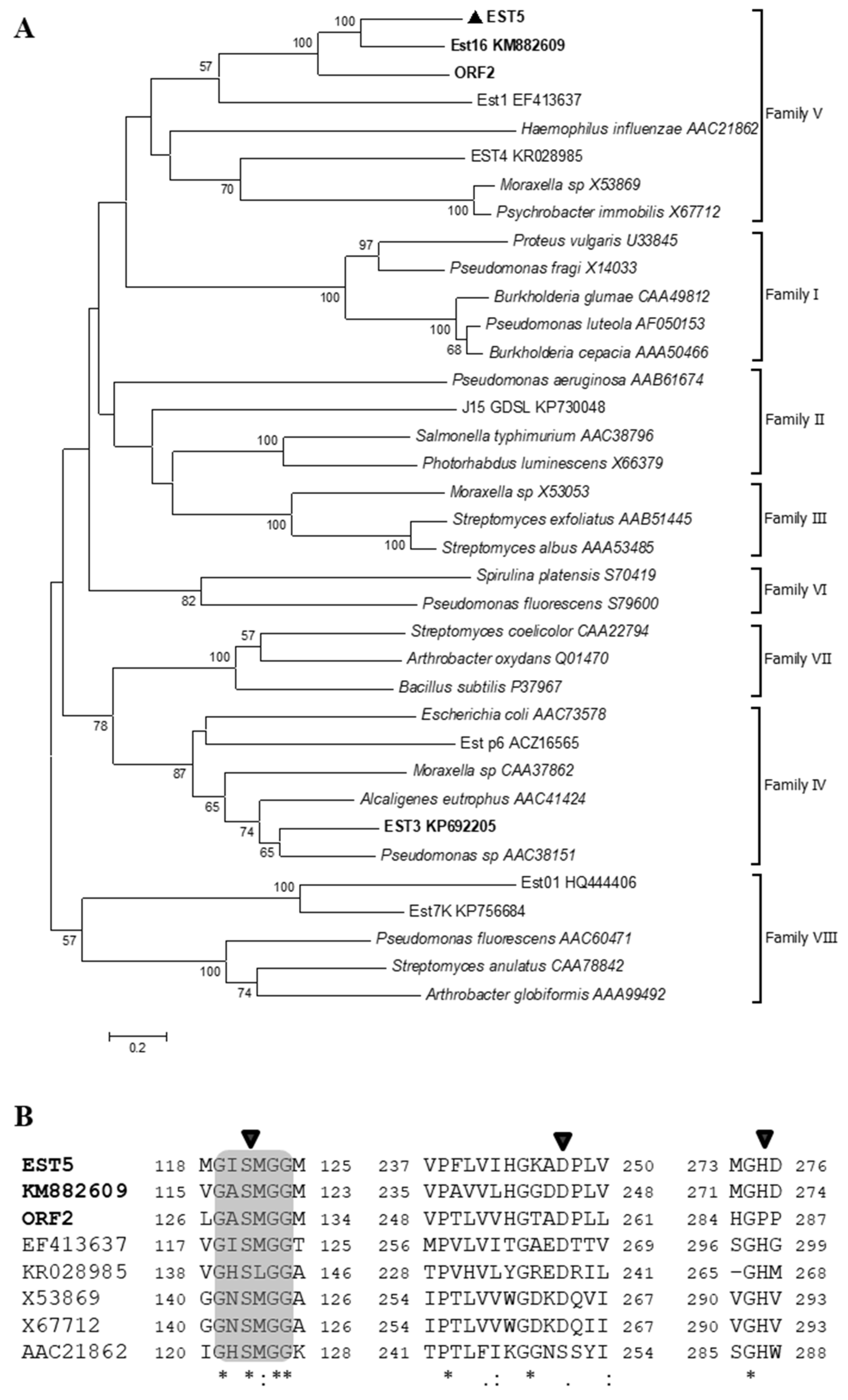
2.1. Analysis of EST5

The sequence analysis of the insert DNA from clone PL14.H10 showed the presence of one 912 bp open reading frame (ORF), encoding a protein of 340 amino acids with predicted molecular mass of 37 kDa and theoretical pI of 7.3, without putative signal peptide. The putative esterase gene was designated est5. BlastP analysis based on the information in the GenBank database revealed highest identity (78%) of the corresponding protein EST5 with a lipase/esterase from uncultured bacterium derived from sea sediment sample (accession number: ADM63077.1), followed by 74% identity with an alpha/beta hydrolase fold protein from Parvibaculum lavamentivorans (accession number: WP012110575.1), and 62% identity with an alpha/beta hydrolase from α-Proteobacterium Mf 1.05b.01 (accession number: WP029641682.1). The DNA sequence of est5 gene has been deposited in GenBank with the accession number KY563703.
To analyze the phylogenetic relationship of EST5 with other lipolytic enzymes, a neighbor-joining tree was constructed based on amino acid sequences of lipolytic enzymes representing the eight families proposed by Arpigny and Jaeger (1999). As shown in Figure 1A, EST5 is most closely related to esterases Est16 and ORF2, and all three are derived from the same metagenomic library. They formed a separated group and showed identities of 50% and 40% to EST5, respectively. EST5 contains a catalytic triad that is typical of proteins with α/β hydrolase fold: Ser120-Asp247-His27. Multiple sequence alignment with closely related homologues (Figure 1B) revealed the catalytic nucleophile Ser located in the typical family V conserved motif G-X-S-X-G-G; and the PTL motif varied among the enzyme sequences. Regarding the catalytic site of aspartate, the enzymes EST5, Est16 and ORF2 showed the conserved motif DPL.

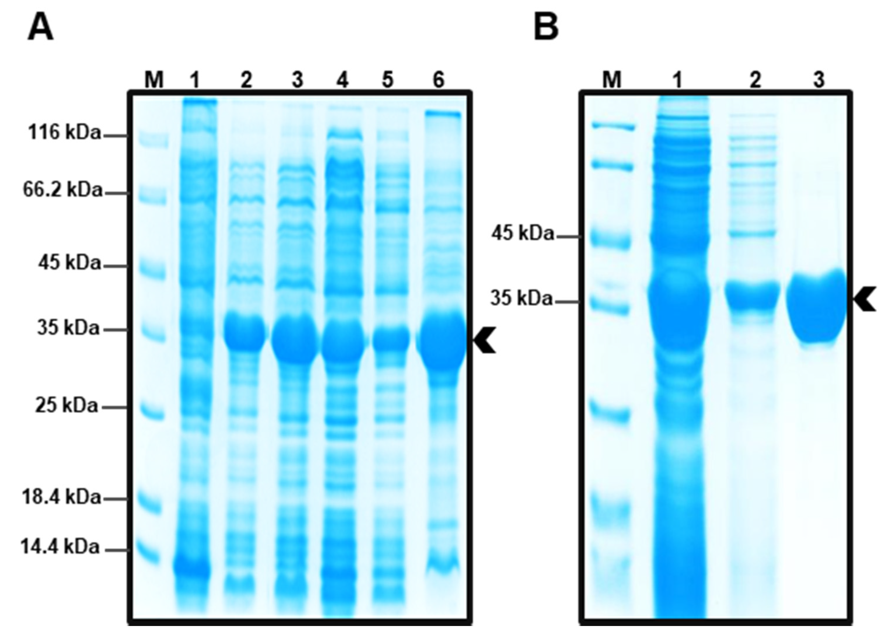
2.2. Obtaining Protein EST5

The full-length est5 sequence was amplified and cloned into the expression vector pET-28a(+) with an N-terminal 6× His tag. and expressed in E. coli BL21(DE3). The highest amount of protein was achieved at 30 °C for 4 h. Following induction, the encoded EST5 was expressed in active form in the soluble fraction of the host cells. Molecular mass of the recombinant enzyme was about 37 kDa, as analyzed by polyacrylamide gel electrophoresis (SDS-PAGE) (Figure 2A), in accordance with information predicted from amino acid sequence. Figure 2B shows the purification of the enzyme, in which two steps were combined: Ni-NTA affinity chromatography, where the protein was eluted mostly at 50 mM imidazole, and size exclusion chromatography, to proceed with the circular dichroism (CD) and fluorescence analysis.

2.3. Enzyme Activity and Its Affecting Factors

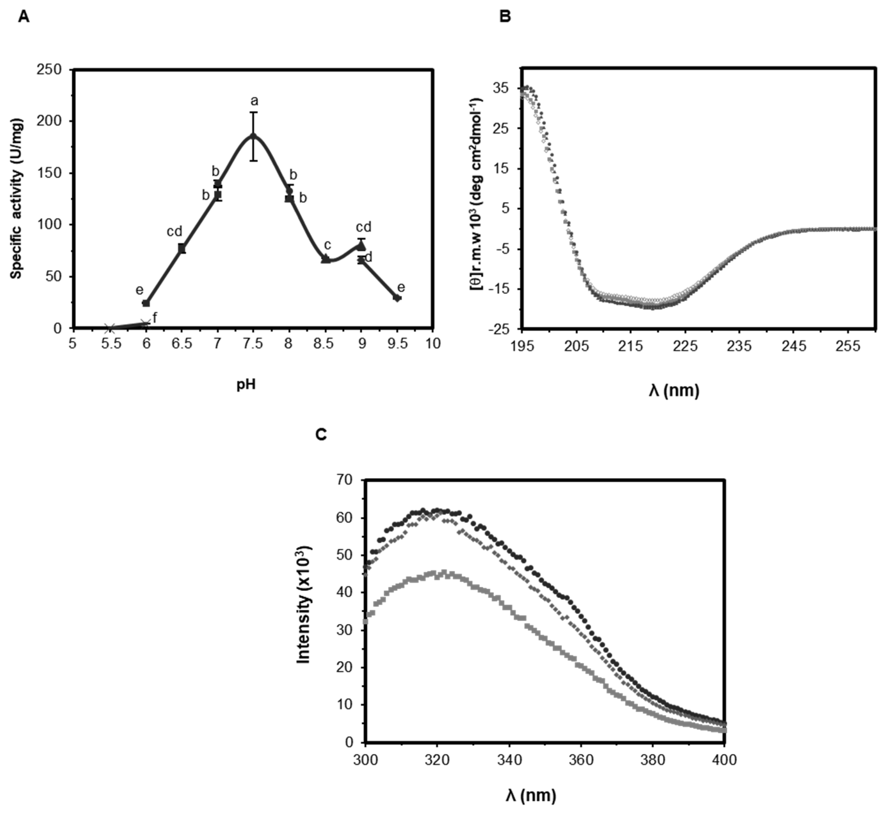
2.3.1. Effect of pH on Activity and Structure of EST5

The effect of pH on the enzymatic activity of EST5 was investigated using pNP-C4 as substrate in a pH range from 3 to 10. The enzyme exhibited activity from pH 6.0 to 9.5, with maximum activity at pH 7.5. EST5 retained more than 50% of its activity from pH 6.5 to 9, and the same activity among both bounds (Figure 3A). To assess if the secondary structure and protein folding was correlated to pH variation, CD spectra were measured at differing pH. The Far-UV CD spectra indicated that EST5 displays stronger negative bands at 208 and 222 nm, mostly at different pH values 7.0 and 8.0. On changing the pH to 5.6 and 9.0, there was a decrease of helical content, as shown by the slightly shallower signal in those wavelengths (Figure 3B) [40,41], in agreement with the pH that the enzyme exhibited lower activity. Still monitoring the structure of EST5 face the variation of pH, intrinsic tryptophan fluorescence was measured at pH 7.0, 8.0 and 9.0. EST5 has one tryptophan (W41); exposure of the protein to more basic pH caused a decrease in fluorescence intensity (Figure 3C), suggesting conformational changes of the protein. These data suggest that, at pH 8.0 and, mostly, at pH 9.0, some changes occur in the protein structure that may affect its activity.
To date, no studies regarding the influence of pH on esterase structure using both circular dichroism and intrinsic tryptophan fluorescence as a tool were reported. In our previous study the esterase Est16 exhibited maximal activity at pH 9.0, although the loss of activity in lower pH was not related to differences in the secondary structure, as evinced by CD analysis performed at pH 7.0, 8.0 and 9.0 [38].

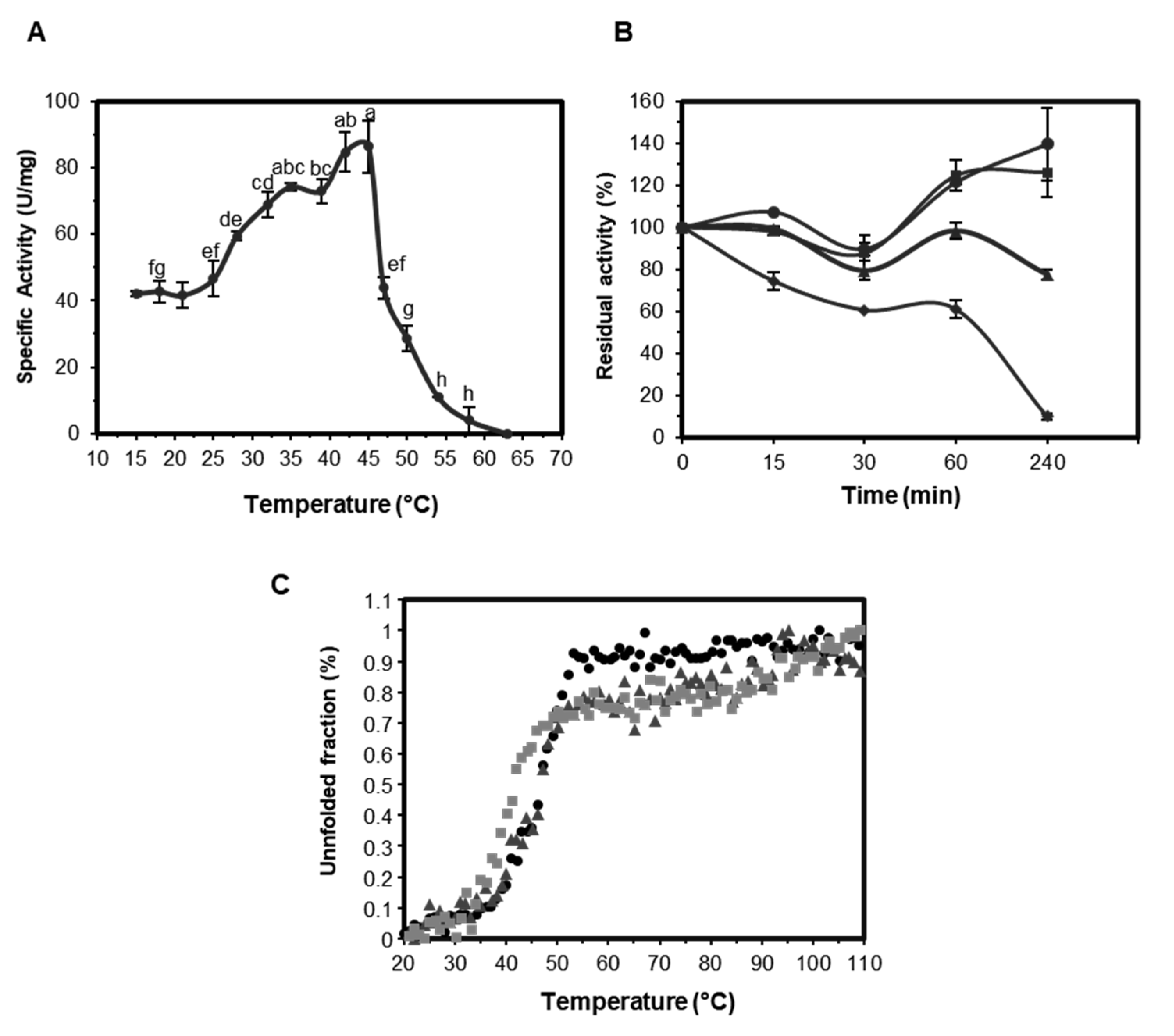
2.3.2. Effect of Temperature on the Activity and Stability of EST5

The effect of temperature variation on the enzymatic activity of EST5 was investigated using 50 mM sodium phosphate buffer, pH 7.5. EST5 displayed high activity in a temperature range of 30 °C to 45 °C with highest activity at 45 °C (Figure 4A). To test EST5 thermostability, the enzyme was preincubated at 10, 30, 37 and 45 °C and its residual activity was assayed. At 10 °C, 30 °C and 37 °C, the activity was not affected after 15 min of incubation, and was reduced to 74% at 45 °C. After 1 h, EST5 retained 60% of its activity at 45 °C; at 10 °C and 30 °C, the activity was higher than the control (no incubation) and remained higher up to the maximum incubation period (4 h) (Figure 4B).
The effect of temperature on stability of EST5 was also determined by CD spectra. The Tm value of the enzyme was 47 °C when evaluated at pH 7.0 and 8.0, and 42 °C using buffer at pH 9.0 (Figure 4C). These data confirm low activity of the enzyme above 47 °C inferred from enzymatic activity measurements and reinforce the lower activity of the protein in more basic pH, as conformational changes were observed through intrinsic tryptophan fluorescence tests. Some studies on lipolytic enzymes from metagenomic sources involving circular dichroism or fluorescence have already been reported: a lipase from a metagenomic library of a hot spring soil sample was evaluated based on variation of temperature. Circular dichroism assays revealed distortion in secondary structure at temperatures above 35 °C. However, the study of intrinsic fluorescence data revealed that even with the loss of secondary structure, its tertiary structure was retained [42]. The esterase rEst97 from a high Arctic intertidal zone sediment metagenomic library was considered a cold-adapted enzyme, by having optimum activity at mild temperature (35 °C) and loss of native protein structure at 35–40 °C, as indicated by CD and calorimetry analysis [43]. Est16 protein from our previous study showed two steps for complete unfolding when evaluated at pH 9.0 (40 °C and 95 °C) [38].
The Tm parameter of EST5 also indicated greater stability at the pH 7.0 and 8.0 in comparison to pH 9.0. By contrast, the esterase Est16 exhibited maximal activity at pH 9.0, although the loss of activity in lower pH was not related to differences in the secondary structure, as evinced by CD analysis performed at pH 7.0, 8.0 and 9.0.
Characterization of EST5 indicated highest hydrolytic activity at 45 °C. However, it is interesting that the enzyme remained at least 50% active at low temperatures. These results show that even though EST5 is a mesophilic enzyme, it has high activity at lower temperatures. Recently, many cold-active or cold-resistant esterases have been isolated, mostly from marine environments or psychrophilic organisms [44,45,46,47,48]. To our knowledge, EST5 is the first cold-active esterase that did not come from low-temperature environment. Moreover, thermal stability analysis showed that incubation of the enzyme up to 30 °C was responsible to increase its activity to 240%.

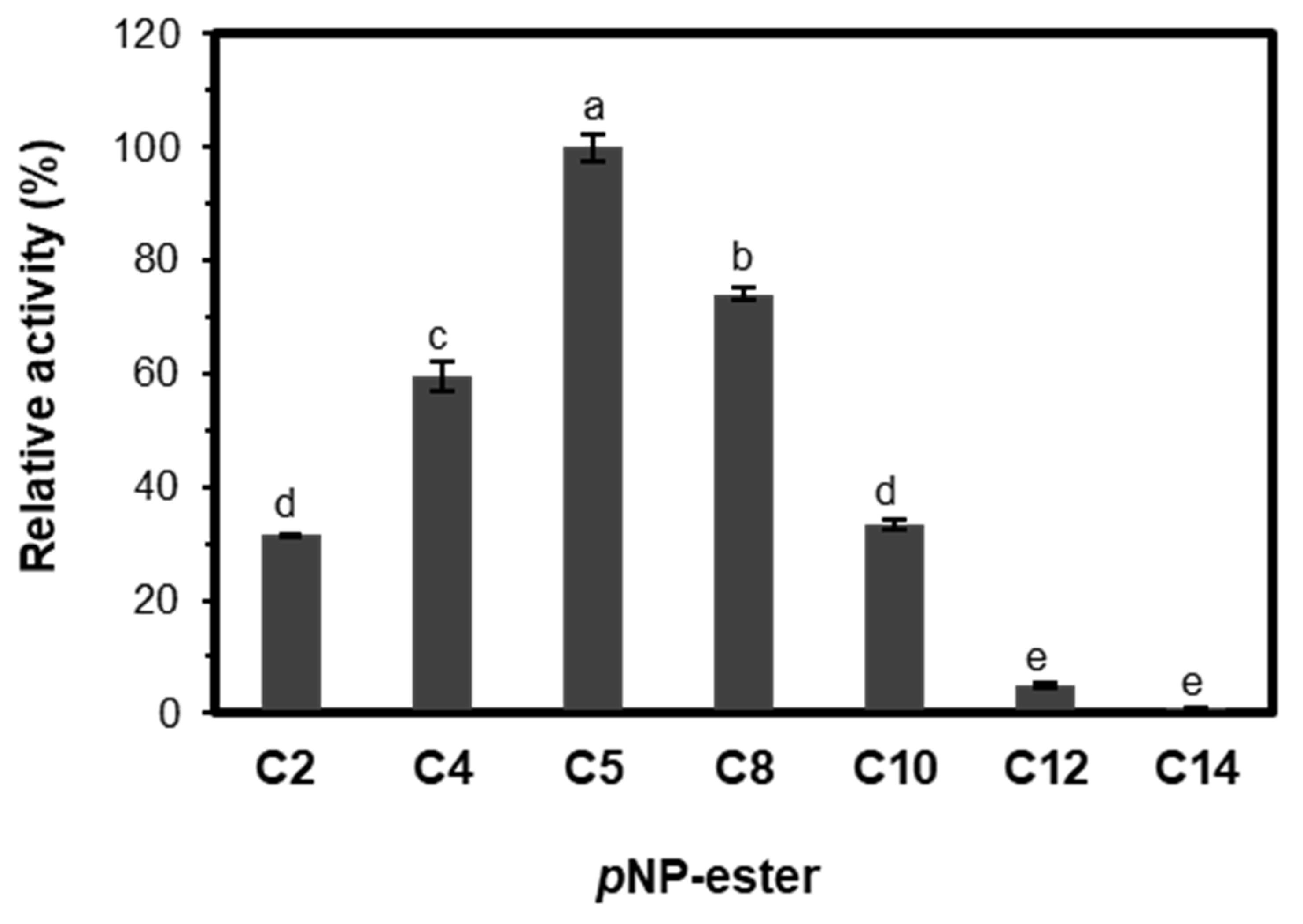
2.3.3. Substrate Specificity and Kinetic Parameters of EST5

The substrate preference of EST5 was determined under standard assay conditions at pH 7.5 and 45 °C, using pNP-esters with different chain lengths (C2–C14) as substrates. The enzyme could hydrolyze all evaluated substrates, as shown in Figure 5. EST5 exhibited higher activity toward pNP-C5 and 78% of the maximum activity against pNP-C8. The activities toward pNP-C2 and pNP-C10 were the same, 30% of the relative activity, whereas activities for longer-chain pNP-esters (C12 and C14) declined considerably.
Kinetic parameters of EST5 are summarized in Table 1. The enzyme showed higher ratio of Kcat/Km against pNP-C5 (1533.27 s−1·mM−1), suggesting that this was the most favored substrate among the pNP-esters tested. The Vmax and Kcat/Km values are, respectively, 1.8- and 2.3-fold higher for C8 in comparison to C4. The substrates C2 and C10 had similar values of catalytic efficiency (18.1 and 17.5 s−1 mM−1, respectively).
Despite EST5 ability to hydrolyze substrates with different acyl chains, the enzyme displayed relative activity markedly lower when the acyl chain length exceeded C10. In view of these findings, the lipolytic enzyme EST5 can be classified as esterase rather than a lipase, since esterases show preference for shorter triacylglycerols, while lipases act more efficiently on esters with longer carbon chain lengths [49].
To date, only a few esterases from family V have been biochemically characterized. Among them, esterase Est1 from metagenomic library derived from sediments of hot spring, which was capable of hydrolyzing pNP-esters from C2 to C16 [50]. As EST5, this enzyme showed higher activity toward C5, however Kcat/Km value about 1.6-fold lower. The esterase LC-Est1 from a leaf-branch compost metagenome library exhibited higher catalytic efficiency for C4, with Kcat/Km value of 19.33 s−1·mM−1 [51]. Mostly, esterases from metagenomic libraries from different sources exhibited higher activity on pNP-C4 [32,52,53]. Still comparing the catalytic efficiency of EST5 with esterases from family V, Est16 [38] exhibited higher kinetic parameters values for almost all evaluated substrates, although EST5 showed 2.3-fold higher catalytic efficiency for C5.

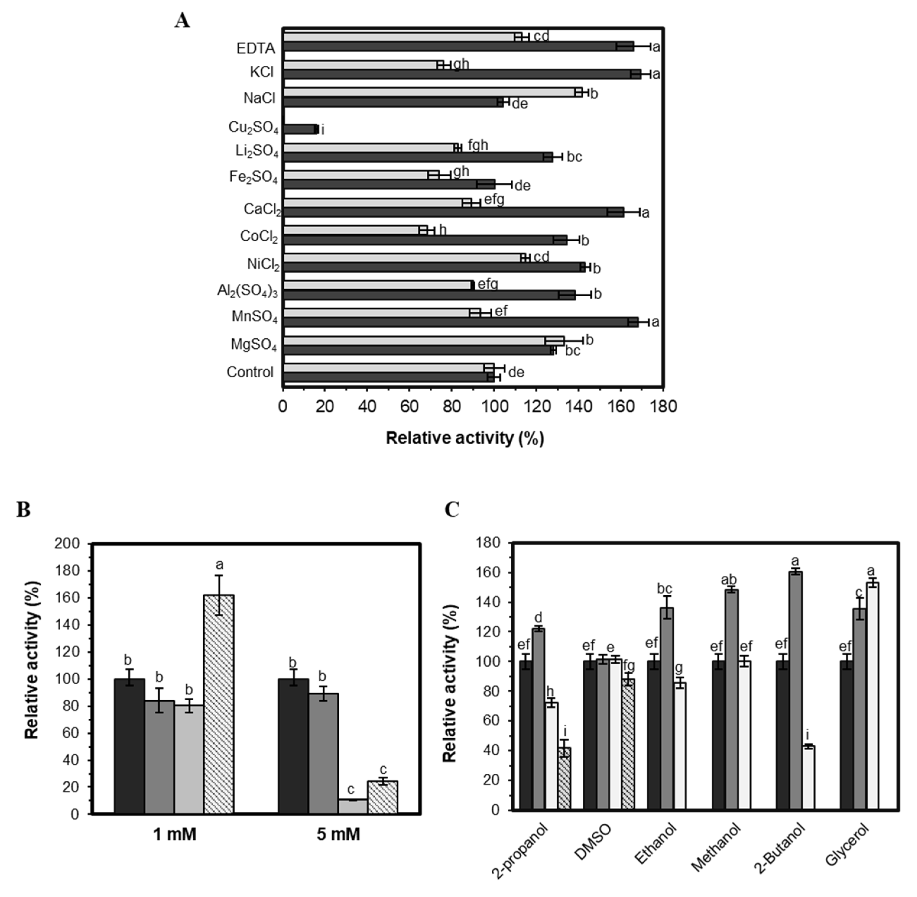
2.3.4. Effect of Additives on EST5 Activity

Many biotechnological processes occur in the presence of some ions in the reaction media that can modify enzyme activity [54]. Thus, the activity of EST5 was evaluated in the presence of ions and salts in the concentrations of 0.5 and 1 mM (Figure 6A). The activity increased in the presence of almost all ions and salts tested at 0.5 mM, mostly Mn2+, K+ and Ca2+, which were responsible for an increase of around 65%; Fe+2 and Na+ did not affect the enzyme activity, and neither Mn2+, Ni2+, Ca2+ and Al+3 at 1 mM. Mg+2 increased EST5 activity in both tested concentrations, with an increase of about 30%. By increasing the concentration of Na+ to 1 mM, the relative activity increased to 141%. Some metal ions can increase the solubility of substrates, increasing their availability for the enzyme [55]. In contrast, the enzyme was moderately inhibited by K+, Li+, Fe+2 and Co2+ (Figure 6A). Cu+2 inhibited the activity of EST5 at 0.5 mM to 15%, and the enzyme showed no activity in the presence of this ion at 1 mM. EST5 showed similar behavior with other esterases reported in the literature, responding differently to the presence of certain metal ions [56,57,58]. At 0.5 mM, EDTA increased the enzyme activity but showed slight influence when evaluated at 1 mM. The activity of the lipolytic enzyme Lp_3562 from Lactobacillus plantarum was enhanced in the presence of EDTA, showing relative activity of 121%, and also in the presence of Mg+2, Ni+2 and Mn+2 [59] similarly to the esterase EST5.
Detergents are typically added to the reaction media of lipolytic enzymes to improve the quality of the emulsion; however, high concentrations can denature the enzyme [60]. EST5 activity remained stable in the presence of Triton X-100 (1 mM) and Tween 20 (1 mM and 5 mM). 5 mM Triton X-100 inhibited the activity of the enzyme, remaining only 10% of relative activity and Tween 80 caused complete inhibition. Regarding ionic detergents, SDS completely inhibited enzyme activity; 1 mM CTAB increased by almost 70% the activity of EST5 and, at 5 mM, the remaining activity was 24% (Figure 6B).
The effect of organic solvents and glycerol at 1, 10 and 20% on the activity of EST5 is exposed in Figure 6C. The enzyme was almost unaffected in the presence of dimethyl sulfoxide (DMSO), in the three concentrations evaluated, or in the presence of 10% methanol, since no significant difference was detected in comparison to the control. Methanol, ethanol, 2-butanol, and 2-propanol increased enzyme activity at 1% concentration, however, except for methanol, 10% of these substances inhibit the activity of EST5, resulting in 70, 83 and 41% of relative activity, respectively. At 20% concentration of organic solvents, the activity was only detected in DMSO (86%) and 2-propanol (40%). In the presence of 1% and 10% glycerol, the activity of EST5 increased to 132% and 147%, respectively, being inhibited in the presence of 20% of this substance. It is known that, despite the cryoprotectant effect, high concentrations of glycerol have negative effects on the enzymatic activity due to reduced diffusion of the hydrophobic substrate in the active site of the enzyme [61]. The same pattern was observed for esterase EstOF4, which remained stable in 10% DMSO, was inhibited in the presence of 2-propanol and 2-butanol and was slightly stimulated by 10% glycerol [62].

2.4. EST5 Showed Esterification Activity

The esterification activity of EST5 was evaluated using butyric acid and methanol as substrates. The enzyme exhibited esterification activity of 127.04 U·mg−1. The esterase in the present study exhibited the same or even higher ability to be used on esterification reactions as the lipases described in Table 2. The commercial lipases Novozyme 435 (lipase B from Candida antarctica) and Lipozyme TLIM (lipase from Thermomyces lanuginosus) were analyzed using lauric acid and dodecanol, exhibiting activities of 9.89 and 3.54 U mg−1, respectively. The other commercial lipase CL from Candida rugosa showed an activity of 15.83 U·mg−1 when assayed with butyric acid and propanol. The esterification activity of EST5 was 1.47-fold higher than LIP4 from Candida rugosa and 14.32-fold higher than immobilized Pseudomonas cepacia lipase (PS). Higher activities values were achieved for LIP2 (from Candida rugosa), which shows preference for butyric acid as substrate, and for the lipase from yeast Sporidiobolus pararoseus. The free Candida rugosa lipase Lipomod 34MDP showed higher conversions in the esterification reactions of free fatty acids and polyols than two of the most popular commercially available immobilized lipases [63], showing that there is always a demand for new enzymes.
The fungal esterase RmEstA in free form produced butyl butyrate with an esterification efficiency of 56%, 35% less than the immobilized form of the enzyme [69].
Most currently commercially available esters are obtained by chemical synthesis using strong acids or bases as catalysts. Even though a considerable amount of current studies is focused on the production of aromas and flavors, only a few are obtained biotechnologically in industrial scale [70].
The ability to produce methyl butyrate makes EST5 attractive for possible application in the synthesis of flavor esters, especially fruity flavors used in food and pharmaceutical products [71,72]. Although complementary studies would be necessary, these preliminary studies of EST5 application shows promising results. Probably, after optimization of reaction parameters, higher conversion rates can be achieved.
Since EST5 showed preference for the pNP-esters C4, C5 and C8, valeric and caprylic acids could be possible substrates for its esterification reactions. Short- and medium-chain esters have known application as flavoring agents. Recently, environmental issues have driven research on sustainable sources for fuels. The valeric acid is a lignocellulosic material, considered a cheap and renewable feedstock. The esterification product of valeric acid and ethanol—ethyl valerate—was considered a possible second generation biofuel [73,74].

3. Materials and Methods

From the fosmid metagenomic library isolated from a microbial consortium specialized in diesel oil degradation, 30 positive clones were selected based on the formation of clear halos around individual colonies, which indicated hydrolysis of tributyrin in Luria–Bertani (LB) agar plates [38]. Recently, we sequenced the clone PL14.H10, which assembled four Open Reading Frames (ORFs) identified as putative genes encoding lipolytic enzymes [39]. One of the four potential lipolytic genes was selected to be characterized in this work.

3.1. Sequence and Phylogenetic Analysis

Homology searches were performed using BLAST analysis at the National Center for Biotechnology Information (NCBI, Bethesda, MA, USA). Signal peptides were predicted using SignalP 4.0 (http://www.cbs.dtu.dk/services/SignalP/) [75]. The molecular mass and pI of the encoded protein were analyzed via ProtParam tool in the ExPASy website (http://www.expasy.ch/tools/protparam.html).
Lipases/esterases from the eight families proposed by Arpigny and Jaeger (1999) as well as those novel enzymes recently described were used to construct a phylogenetic tree: EST3 [39], Est16 [38] and ORF2 [37] from the same metagenomic library as EST5 (this study); Est_p6 [76]; J15 GDSL [77]; Est7k [78]; Est1 [50] and EST4 [32]. The retrieved sequences were aligned in Clustal W [79] in the BioEdit Sequence Alignment Program (version 7.0.5.3, North Carolina State University, NC, USA, 1998). Phylogenetic tree was constructed by the neighbor-joining algorithm, as implemented in MEGA 6 [80] program package, with bootstrapping [81] based on 1,000 replicates.

3.2. Expression and Purification of the Recombinant Enzyme

For enzyme overexpression, the full length est5 gene was amplified using oligonucleotides forward 5′-GATGAATTCCCCATGACCGTCAACAT-3′ and reverse 5′-AGGTCTCGAGCCTTATTCCGCGG-3′ with the respective EcoRI and XhoI restriction sites (underlined). PCR was performed using Pfu DNA polymerase (Thermo Scientific, Waltham, MA, USA) according to the manufacturer recommendations. The purified PCR product was digested and cloned into pET-28a(+) vector to obtain the recombinant plasmid pET28a-est5. DNA sequencing confirmed the constructed plasmid integrity. Then, the recombinant plasmid was transformed into E. coli BL21(DE3) competent cells for heterologous expression of the protein. Recombinant E. coli cells were cultivated in LB liquid medium supplemented with 50 μg·mL−1 kanamycin and agitated at 200 rpm, 37 °C. For optimization tests, when the optical density OD600 of the culture reached an absorbance of 0.6, 0.1 mM IPTG (isopropyl-b-d-thiogalactopyranoside) was added to induce protein expression at different temperatures (22–37 °C) and time (2–8 h) in rotary shaking at 200 rpm. After induction, the culture was centrifuged at 11,953× g for 10 min at 4 °C and the pellet was then homogenized in Buffer A (50 mM Tris-HCl, pH 8.0; 100 mM NaCl and 10% glycerol). Lysozyme was added at a final concentration of 4 μg·mL−1 and the material was incubated at an ice bath for 1 h. Cells were disrupted by sonication in ice using the Branson sonifier equipment (Branson, CT, USA) with six cycles of 10 pulses at 30% amplitude with 20 s intervals and then centrifuged at 38,724× g for 20 min at 4 °C to pellet the cellular debris and collect the supernatant containing the targeted protein.
The enzyme was purified by immobilized metal ion chromatography and size-exclusion chromatography for characterization purposes. The recovered supernatant from the centrifugation step was mixed with Ni-NTA agarose resin (Qiagen, Venlo, The Netherlands) previously equilibrated with Buffer A containing 20 mM imidazole and loaded to a gravity flow column (Qiagen). The target protein was eluted and then concentrated with Vivaspin 20 30,000 molecular weight cut-off (MWCO) (Sartorius Stedim, Aubagne, France) under centrifugation at 200× g. Size-exclusion chromatography was used as an additional purification step in a HiLoad 16/60 Superdex 200 column (GE Healthcare, Little Chalfont, UK), at 4 °C using the elution buffer (20 mM Tris-HCl pH 8.0, 50 mM NaCl and 5% glycerol). Purity and concentration was analyzed by SDS-PAGE [82] and Nanodrop ND-1000 spectrophotometer (Thermo Scientific), respectively.

3.3. Biophysical Analysis

The effect of temperature and pH on enzyme conformation was monitored by the circular dichroism (CD) and fluorescence spectroscopic techniques. Circular dichroism measurements were performed with JASCO J-810 spectropolarimeter fitted with a Jasco Peltier-type temperature controller (PFD425S). Far-UV CD spectra were recorded at 10 °C in the wavelength region of 195–260 nm at 0.5 nm intervals, path length of 1 mm. 2.9 µmol of protein was used in buffers: 5 mM sodium citrate pH 5.6; 5 mM sodium phosphate, pH 7.0 and 8.0; and 5 mM N-Cyclohexyl-2-aminoethanesulfonic (CHES), pH 9.0. The thermal denaturation curve of the protein was obtained by monitoring the change in CD values at 222 nm as the temperature increased from 20 °C to 110 °C with spectra collected at 1 °C intervals. The midpoint temperature (Tm) of the unfolding transition was analyzed.
Intrinsic tryptophan fluorescence measurements were carried out on K2 multifrequency spectrofluorometer (ISS Inc., Champaign, IL, USA). Fluorescence spectra were measured at a protein concentration of 1 µM in 5 mM Tris-HCl at pH 7.0, 8.0 and 9.0. The excitation wavelength was set at 295 nm and the emission spectra were recorded between 300 to 400 nm.

3.4. Enzyme Characterization

Enzyme activity was measured at 405 nm with a SpectraMax M2e spectrophotometer (Molecular Devices, Sunnyvale, CA, USA) equipped with a temperature controller. Standard assays, unless otherwise indicated, were performed at 35 °C for 5 min in 100 µL mixture containing: 27.01 nM of the purified enzyme, 50 mM Tris-HCl buffer, pH 8.0, with 0.3% (v/v) Triton X-100 and 1 mM p-nitrophenyl butyrate dissolved in isopropanol/acetonitrile (4:1 v/v). The amount of p-nitrophenol produced by the reaction was determined from the absorbance with a molar extinction coefficient value of 17,000 M−1·cm−1. One enzyme unit is the amount of enzyme that releases 1 µmol of product from substrate per minute under standard assay conditions.
All measurements were carried out in triplicate and a blank reaction without enzyme was included for each experiment. Data was analyzed using the R software (R Foundation for Statistical Computing, Vienna, Austria, 2016). ANOVA and Tukey’s test at 5% probability were used to make comparisons among the different conditions evaluated.

3.4.1. Substrate Selectivity

Substrate specificity of the enzyme was carried out using pNP-esters: pNP-acetate (C2), pNP-butyrate (C4), pNP-valerate (C5), pNP-caprylate (C8), pNP-caprate (C10), pNP-laurate (C12), pNP-myristate (C14) and pNP-palmitate (C16) (Sigma, St. Louis, MO, USA). Assays were performed according to standard enzyme assay method. Initial reaction velocities were measured with pNP-esters at a concentration range from 0.04 mM to 2.5 mM. Kinetic parameters were obtained by nonlinear regression of the data on Michaelis–Menten equation using GraphPad Prism software version 5.0 (GraphPad Software, Inc, San Diego, CA, USA).

3.4.2. Effect of pH and Temperature on Enzyme Activity

To investigate the effect of pH on enzymatic activity, enzymatic assays were performed under standard assay conditions and the following buffers were used at 50 mM: citrate (pH 3.0 to 6.0), sodium phosphate (pH 6.0 to 8.0), 4-(2-Hydroxyethyl)piperazine-1-ethanesulfonic acid (HEPES) (6.5 to 8.0), Tris-HCl (pH 7.5 to 9.0), and 3-(Cyclohexylamino)-2-hydroxy-1-propanesulfonic acid (CAPSO) (pH 9.0 to 10). The temperature effects on enzyme activity were determined by assessing the enzyme activity from 15 to 65 °C, under standard conditions, with 50 mM HEPES buffer, pH 7.5. To study thermostability, the enzyme was incubated from 10 to 60 °C for up to 4 h. After the enzyme solution was heated to appointed temperature, the residual activity was measured under standard assay conditions.

3.4.3. Effect of Additives on Enzyme Activity

To evaluate the effect of detergents, metal ions, organic solvents and chelating agents, the enzyme was incubated in their presence for 5 min at 4 °C. The influence of metal ions (Na+, K+, Mg2+, Ca2+, Mn2+, Co2+, Cu+2, Fe+2, Ni+2, Al+3 or Li+) and the chelating agent EDTA (ethylenediaminetetraacetic acid) was investigated at final concentrations of 0.5 and 1 mM (w/v). To measure the effect of detergents on the enzyme activity Tween 20, Tween 80, Triton X-100, sodium dodecyl sulfate (SDS) or hexadecyltrimethylammonium bromide (CTAB) was added in the final concentrations of 1 and 5 mM (w/v). Stability of the enzyme in the presence of organic solvents was tested with 1%, 10%, and 20% (v/v) of methanol, ethanol, 2-propanol, 1-butanol and dimethyl sulfoxide (DMSO). The presence of glycerol was also investigated in the same concentrations. Enzyme activity without additives was defined as 100%.

3.5. Esterification Assays

Esterification activity was assayed according to method described previously [68]. A volume of 0.0395 µmol of protein in solution (10 mM Tris-HCl, pH 8.0, and 50 mM NaCl) was added to the reaction mixture containing 3.66 g of butyric acid and 0.7 g of methanol. The mixtures were incubated for 40 min at 40 °C and shaken at 150 rpm. After incubation time, reactions were stopped by adding 20 mL of an acetone/ethanol solution (1:1, v/v). Unreacted fatty acids in the mixture were titrated with 0.035 M NaOH up to pH 11.0. One unit of activity was defined as the amount of enzyme able to consume one µmol of fatty acid per minute under assay conditions.

4. Conclusions

Characterization of the esterase in this study provided useful information about its function and structure. EST5 showed activity toward a wide range of substrates and temperatures, was stable in the presence of some detergents and organic solvents, and incubation at 10 °C and 30 °C for up to 4 h increased its activity. In addition, EST5 had higher esterification activity than some previously reported lipases, including commercial enzymes. EST5-catalyzed esterification reactions can be conducted at lower temperatures in the presence of organic solvents and in solvents-free media. Thus, this enzyme displays a high potential for biotechnological applications.

Author Contributions

Conceptualization, T.C.M. and E.G.M.L.; Formal analysis, T.C.M., M.R.P., A.M.G.M. and J.P.B.; Funding acquisition, T.C.M. and E.G.M.L.; Investigation, T.C.M., M.R.P., A.M.G.M. and J.P.B.; Methodology, T.C.M. and E.G.M.L.; Writing—original draft, T.C.M.; Writing—review & editing, M.R.P., A.M.G.M., J.P.B., P.A.M.P. and E.G.M.L. All authors have read and agreed to the published version of the manuscript.

Funding

This research was funded by the São Paulo Research Foundation—FAPESP, grant numbers 2011/09064-6 and 2013/03568-8.

Acknowledgments

We are grateful for the Spectroscopy and Calorimetry Facility (Brazilian Biosciences National Laboratory-LNBio, Campinas, Brazil) for enabling the CD and fluorescence measurements. We thank the infrastructure offered by the São Paulo State University, Jaboticabal Campus, Brazil and the University of São Paulo, São Paulo, Brazil. We also thank Professor Eleni Gomes and the Institute of Biosciences, Languages and Exact Sciences, Department of Biology, which helped with the esther synthesis analysis.

Conflicts of Interest

The authors declare no conflict of interest.

References

  1. Sheldon, R.A.; van Pelt, S. Enzyme immobilisation in biocatalysis: Why, what and how. Chem. Soc. Rev. 2013, 42, 6223–6235. [Google Scholar] [CrossRef] [PubMed] [Green Version]
  2. Bezborodov, A.M.; Zagustina, N.A. Lipases in catalytic reactions of organic chemistry. Appl. Biochem. Microbiol. 2014, 50, 313–337. [Google Scholar] [CrossRef]
  3. De Miranda, A.S.; Miranda, L.S.M.; de Souza, R.O.M.A. Lipases: Valuable catalysts for dynamic kinetic resolutions. Biotechnol. Adv. 2015, 33, 372–393. [Google Scholar] [CrossRef] [PubMed]
  4. Chapman, J.; Ismail, A.E.; Dinu, C.Z. Industrial applications of enzymes: Recent advances, techniques, and outlooks. Catalysts 2018, 8, 238. [Google Scholar] [CrossRef] [Green Version]
  5. Littlechild, J.A. Improving the ‘tool box’ for robust industrial enzymes. J. Ind. Microbiol. Biotechnol. 2017, 44, 711–720. [Google Scholar] [CrossRef] [PubMed] [Green Version]
  6. Bornscheuer, U. Microbial carboxyl esterases: Classification, properties and application in biocatalysis. FEMS Microbiol. Rev. 2002, 26, 73–81. [Google Scholar] [CrossRef]
  7. Ferrer, M.; Bargiela, R.; Martínez-Martínez, M.; Mir, J.; Koch, R.; Golyshina, O.V.; Golyshin, P.N. Biodiversity for biocatalysis: A review of the α/β-hydrolase fold superfamily of esterases-lipases discovered in metagenomes. Biocatal. Biotransform. 2016, 2422, 1–15. [Google Scholar] [CrossRef]
  8. Hasan, F.; Shah, A.A.; Hameed, A. Industrial applications of microbial lipases. Enzyme Microb. Technol. 2006, 39, 235–251. [Google Scholar] [CrossRef]
  9. Kanmani, P.; Aravind, J.; Kumaresan, K. An insight into microbial lipases and their environmental facet. Int. J. Environ. Sci. Technol. 2015, 12, 1147–1162. [Google Scholar] [CrossRef] [Green Version]
  10. Carvalho, A.; Fonseca, T.; Mattos, M.; Oliveira, M.; Lemos, T.; Molinari, F.; Romano, D.; Serra, I. Recent Advances in Lipase-Mediated Preparation of Pharmaceuticals and Their Intermediates. Int. J. Mol. Sci. 2015, 16, 29682–29716. [Google Scholar] [CrossRef] [Green Version]
  11. Trono, D. Recombinant Enzymes in the Food and Pharmaceutical Industries. In Advances in Enzyme Technology; Elsevier: Amsterdam, The Netherlands, 2019; pp. 349–387. [Google Scholar]
  12. Samoylova, Y.V.; Sorokina, K.N.; Piligaev, A.V.; Parmon, V.N. Application of Bacterial Thermostable Lipolytic Enzymes in the Modern Biotechnological Processes: A Review. Catal. Ind. 2019, 11, 168–178. [Google Scholar] [CrossRef]
  13. Khambhaty, Y. Applications of enzymes in leather processing. Environ. Chem. Lett. 2020, 18, 747–769. [Google Scholar] [CrossRef]
  14. Zhang, Z.; Lan, D.; Zhou, P.; Li, J.; Yang, B.; Wang, Y. Control of sticky deposits in wastepaper recycling with thermophilic esterase. Cellulose 2017, 24, 311–321. [Google Scholar] [CrossRef]
  15. Sahay, S.; Chouhan, D. Study on the potential of cold-active lipases from psychrotrophic fungi for detergent formulation. J. Genet. Eng. Biotechnol. 2018, 16, 319–325. [Google Scholar] [CrossRef]
  16. Kumar, A.; Gudiukaite, R.; Gricajeva, A.; Sadauskas, M.; Malunavicius, V.; Kamyab, H.; Sharma, S.; Sharma, T.; Pant, D. Microbial lipolytic enzymes—Promising energy-efficient biocatalysts in bioremediation. Energy 2020, 192, 116674. [Google Scholar] [CrossRef]
  17. López-López, O.; Cerdán, M.E.; Gonzalez-Siso, M.I. New Extremophilic Lipases and Esterases from Metagenomics. Curr. Protein Pept. Sci. 2014, 15, 445–455. [Google Scholar] [CrossRef]
  18. Markets and Markets. Lipase Market by Source (Microbial Lipases, Animal Lipases), Application (Animal Feed, Dairy, Bakery, Confectionery, Others), & by Geography (North America, Europe, Asia-Pacific, Latin America, RoW)—Global Forecast to 2020; Markets and Markets: Pune, India, 2015. [Google Scholar]
  19. Fojan, P.; Jonson, P.H.; Petersen, M.T.N.; Petersen, S.B. What distinguishes an esterase from a lipase: A novel structural approach. Biochimie 2000, 82, 1033–1041. [Google Scholar] [CrossRef]
  20. Arpigny, J.L.; Jaeger, K.-E. Bacterial lipolytic enzymes: Classification and properties. Biochem. J. 1999, 343, 177–183. [Google Scholar] [CrossRef]
  21. Salgado, V.; Fonseca, C.; Lopes da Silva, T.; Roseiro, J.C.; Eusébio, A. Isolation and Identification of Magnusiomyces capitatus as a Lipase-Producing Yeast from Olive Mill Wastewater. Waste Biomass Valor. 2020, 11, 3207–3221. [Google Scholar] [CrossRef] [Green Version]
  22. Chen, C.C.; Chi, Z.; Liu, G.L.; Jiang, H.; Hu, Z.; Chi, Z.M. Production, purification, characterization and gene cloning of an esterase produced by Aureobasidium melanogenum HN6.2. Process Biochem. 2017, 53, 69–79. [Google Scholar] [CrossRef]
  23. Rade, L.L.; da Silva, M.N.P.; Vieira, P.S.; Milan, N.; de Souza, C.M.; de Melo, R.R.; Klein, B.C.; Bonomi, A.; de Castro, H.F.; Murakami, M.T.; et al. A Novel Fungal Lipase With Methanol Tolerance and Preference for Macaw Palm Oil. Front. Bioeng. Biotechnol. 2020. [Google Scholar] [CrossRef] [PubMed]
  24. Turati, D.F.M.; Almeida, A.F.; Terrone, C.C.; Nascimento, J.M.F.; Terrasan, C.R.F.; Fernandez-Lorente, G.; Pessela, B.C.; Guisan, J.M.; Carmona, E.C. Thermotolerant lipase from Penicillium sp. section Gracilenta CBMAI 1583: Effect of carbon sources on enzyme production, biochemical properties of crude and purified enzyme and substrate specificity. Biocatal. Agric. Biotechnol. 2019, 17, 15–24. [Google Scholar] [CrossRef]
  25. Lin, X.-S.; Zhao, K.-H.; Zhou, Q.-L.; Xie, K.-Q.; Halling, P.J.; Yang, Z. Aspergillus oryzae lipase-catalyzed synthesis of glucose laurate with excellent productivity. Bioresour. Bioprocess. 2016, 3, 2. [Google Scholar] [CrossRef] [Green Version]
  26. Bhardwaj, K.K.; Dogra, A.; Kapoor, S.; Mehta, A.; Gupta, R. Purification and Properties of an Esterase from Bacillus licheniformis and it’s Application in Synthesis of Octyl Acetate. Open Microbiol. J. 2020, 14, 113–121. [Google Scholar] [CrossRef]
  27. Noor, H.; Satti, S.M.; Din, S.U.; Farman, M.; Hasan, F.; Khan, S.; Badshah, M.; Shah, A.A. Insight on esterase from Pseudomonas aeruginosa strain S3 that depolymerize poly(lactic acid) (PLA) at ambient temperature. Polym. Degrad. Stab. 2020, 174, 109096. [Google Scholar] [CrossRef]
  28. Bharathi, D.; Rajalakshmi, G.; Komathi, S. Optimization and production of lipase enzyme from bacterial strains isolated from petrol spilled soil. J. King Saud Univ. Sci. 2019, 31, 898–901. [Google Scholar] [CrossRef]
  29. Boran, R.; Ugur, A.; Sarac, N.; Ceylan, O. Characterisation of Streptomyces violascens OC125-8 lipase for oily wastewater treatment. 3 Biotech 2019, 9, 5. [Google Scholar] [CrossRef]
  30. Kumar, A.; Dhar, K.; Kanwar, S.S.; Arora, P.K. Lipase catalysis in organic solvents: Advantages and applications. Biol. Proced. Online 2016, 18, 2. [Google Scholar] [CrossRef] [Green Version]
  31. Lai, O.-M.; Lee, Y.-Y.; Phuah, E.-T.; Akoh, C.C. Lipase/Esterase: Properties and Industrial Applications. In Encyclopedia of Food Chemistry; Elsevier: Amsterdam, The Netherlands, 2019; pp. 158–167. [Google Scholar]
  32. Gao, W.; Wu, K.; Chen, L.; Fan, H.; Zhao, Z.; Gao, B.; Wang, H.; Wei, D. A novel esterase from a marine mud metagenomic library for biocatalytic synthesis of short-chain flavor esters. Microb. Cell Fact. 2016, 15, 41. [Google Scholar] [CrossRef] [Green Version]
  33. Brault, G.; Shareck, F.; Hurtubise, Y.; Lépine, F.; Doucet, N. Short-chain flavor ester synthesis in organic media by an E. coli whole-cell biocatalyst expressing a newly characterized heterologous lipase. PLoS ONE 2014, 9, e91872. [Google Scholar] [CrossRef] [Green Version]
  34. SÁ, A.G.A.; Meneses, A.C.D.; Araújo, P.H.H.D.; Oliveira, D.D. A review on enzymatic synthesis of aromatic esters used as flavor ingredients for food, cosmetics and pharmaceuticals industries. Trends Food Sci. Technol. 2017, 69, 95–105. [Google Scholar] [CrossRef]
  35. Zhong, X.L.; Tian, Y.Z.; Jia, M.L.; Liu, Y.D.; Cheng, D.; Li, G. Characterization and purification via nucleic acid aptamers of a novel esterase from the metagenome of paper mill wastewater sediments. Int. J. Biol. Macromol. 2020, 153, 441–450. [Google Scholar] [CrossRef] [PubMed]
  36. Almeida, J.M.; Martini, V.P.; Iulek, J.; Alnoch, R.C.; Moure, V.R.; Müller-Santos, M.; Souza, E.M.; Mitchell, D.A.; Krieger, N. Biochemical characterization and application of a new lipase and its cognate foldase obtained from a metagenomic library derived from fat-contaminated soil. Int. J. Biol. Macromol. 2019, 137, 442–454. [Google Scholar] [CrossRef] [PubMed]
  37. Garcia, R.A.M.; Pereira, M.R.; Maester, T.C.; de Macedo Lemos, E.G. Investigation, Expression, and Molecular Modeling of ORF2, a Metagenomic Lipolytic Enzyme. Appl. Biochem. Biotechnol. 2015, 175, 3875–3887. [Google Scholar] [CrossRef]
  38. Pereira, M.R.; Mercaldi, G.F.; Maester, T.C.; Balan, A.; Lemos, E.G.M. Est16, a new esterase isolated from a metagenomic library of a microbial consortium specializing in diesel oil degradation. PLoS ONE 2015, 10, e0133723. [Google Scholar] [CrossRef]
  39. Maester, T.C.; Pereira, M.R.; Machado Sierra, E.G.; Balan, A.; de Macedo Lemos, E.G. Characterization of EST3: A metagenome-derived esterase with suitable properties for biotechnological applications. Appl. Microbiol. Biotechnol. 2016, 100, 5815–5827. [Google Scholar] [CrossRef] [Green Version]
  40. Greenfield, N.J. Using circular dichroism collected as a funcion of temperature to determine the thermodynamics of protein unfolding and binding interactions. Nat. Protoc. 2009, 1, 2527–2535. [Google Scholar] [CrossRef]
  41. Greenfield, N.J. Biomacromolecular Applications of Circular Dichroism and ORD BT—Encyclopedia of Spectroscopy and Spectrometry. In Encyclopedia of Spectroscopy and Spectrometry, 2nd ed.; Lindon, J.C., Tranter, G.E., Holmes, J.L., Eds.; Academic Press: San Diego, CA, USA, 1999; Volume 1, pp. 153–165. [Google Scholar]
  42. Sharma, P.K.; Singh, K.; Singh, R.; Capalash, N.; Ali, A.; Mohammad, O.; Kaur, J. Characterization of a thermostable lipase showing loss of secondary structure at ambient temperature. Mol. Biol. Rep. 2012, 39, 2795–2804. [Google Scholar] [CrossRef]
  43. Fu, J.; Leiros, H.K.S.; De Pascale, D.; Johnson, K.A.; Blencke, H.M.; Landfald, B. Functional and structural studies of a novel cold-adapted esterase from an Arctic intertidal metagenomic library. Appl. Microbiol. Biotechnol. 2013, 97, 3965–3978. [Google Scholar] [CrossRef]
  44. Dong, J.; Gasmalla, M.A.A.; Zhao, W.; Sun, J.; Liu, W.; Wang, M.; Han, L.; Yang, R. Characterisation of a cold adapted esterase and mutants from a psychotolerant Pseudomonas sp. strain. Biotechnol. Appl. Biochem. 2016, 64, 686–699. [Google Scholar] [CrossRef]
  45. Tchigvintsev, A.; Tran, H.; Popovic, A.; Kovacic, F.; Brown, G.; Flick, R.; Hajighasemi, M.; Egorova, O.; Somody, J.C.; Tchigvintsev, D.; et al. The environment shapes microbial enzymes: Five cold-active and salt-resistant carboxylesterases from marine metagenomes. Appl. Microbiol. Biotechnol. 2015, 99, 2165–2178. [Google Scholar] [CrossRef] [PubMed] [Green Version]
  46. Jiang, H.; Zhang, S.; Gao, H.; Hu, N. Characterization of a cold-active esterase from Serratia sp. and improvement of thermostability by directed evolution. BMC Biotechnol. 2016, 16, 7. [Google Scholar] [CrossRef] [PubMed] [Green Version]
  47. Borchert, E.; Selvin, J.; Kiran, S.G.; Jackson, S.A.; O’Gara, F.; Dobson, A.D.W. A novel cold active esterase from a deep sea sponge stelletta normani metagenomic library. Front. Mar. Sci. 2017, 4, 287. [Google Scholar] [CrossRef] [Green Version]
  48. De Santi, C.; Altermark, B.; Pierechod, M.M.; Ambrosino, L.; De Pascale, D.; Willassen, N.P. Characterization of a cold-active and salt tolerant esterase identified by functional screening of Arctic metagenomic libraries. BMC Biochem. 2016, 17, 1. [Google Scholar] [CrossRef] [PubMed] [Green Version]
  49. Jaeger, K.-E.; Dijkstra, B.W.; Reetz, M.T. Bacterial Biocatalysts: Molecular Biology, Three-Dimensional Structures, and Biotechnological Applications of Lipases. Annu. Rev. Microbiol. 1999, 53, 315–351. [Google Scholar] [CrossRef] [Green Version]
  50. Tirawongsaroj, P.; Sriprang, R.; Harnpicharnchai, P.; Thongaram, T.; Champreda, V.; Tanapongpipat, S.; Pootanakit, K.; Eurwilaichitr, L. Novel thermophilic and thermostable lipolytic enzymes from a Thailand hot spring metagenomic library. J. Biotechnol. 2008, 133, 42–49. [Google Scholar] [CrossRef]
  51. Okano, H.; Hong, X.; Kanaya, E.; Angkawidjaja, C.; Kanaya, S. Structural and biochemical characterization of a metagenome-derived esterase with a long N-terminal extension. Protein Sci. 2015, 24, 93–104. [Google Scholar] [CrossRef] [Green Version]
  52. Zarafeta, D.; Szabo, Z.; Moschidi, D.; Phan, H.; Chrysina, E.D.; Peng, X.; Ingham, C.J.; Kolisis, F.N.; Skretas, G. EstDZ3: A new esterolytic enzyme exhibiting remarkable thermostability. Front. Microbiol. 2016, 7, 1779. [Google Scholar] [CrossRef] [Green Version]
  53. Sukul, P.; Lupilov, N.; Leichert, L.I. Characterization of ML-005, a novel metaproteomics-derived esterase. Front. Microbiol. 2018, 9, 2716. [Google Scholar] [CrossRef]
  54. Bofill, C.; Prim, N.; Mormeneo, M.; Manresa, A.; Javier Pastor, F.I.; Diaz, P. Differential behaviour of Pseudomonas sp. 42A2 LipC, a lipase showing greater versatility than its counterpart LipA. Biochimie 2010, 92, 307–316. [Google Scholar] [CrossRef]
  55. Kumar, S.; Mathur, A.; Singh, V.; Nandy, S.; Khare, S.K.; Negi, S. Bioremediation of waste cooking oil using a novel lipase produced by Penicillium chrysogenum SNP5 grown in solid medium containing waste grease. Bioresour. Technol. 2012, 120, 300–304. [Google Scholar] [CrossRef] [PubMed]
  56. Maqbool, Q.U.A.; Johri, S.; Rasool, S.; Riyaz-ul-Hassan, S.; Verma, V.; Nargotra, A.; Koul, S.; Qazi, G.N. Molecular cloning of carboxylesterase gene and biochemical characterization of encoded protein from Bacillus subtilis (RRL BB1). J. Biotechnol. 2006, 125, 1–10. [Google Scholar] [CrossRef] [PubMed]
  57. Wu, C.; Sun, B. Identification of novel esterase from metagenomic library of Yangtze River. J. Microbiol. Biotechnol. 2009, 19, 187–193. [Google Scholar] [PubMed] [Green Version]
  58. Jeon, J.H.; Kim, J.T.; Lee, H.S.; Kim, S.J.; Kang, S.G.; Choi, S.H.; Lee, J.H. Novel lipolytic enzymes identified from metagenomic library of deep-sea sediment. Evid. Based Complement. Altern. Med. 2011, 2011, 271419. [Google Scholar] [CrossRef]
  59. Esteban-Torres, M.; Mancheño, J.M.; de las Rivas, B.; Muñoz, R. Characterization of a halotolerant lipase from the lactic acid bacteria Lactobacillus plantarum useful in food fermentations. LWT Food Sci. Technol. 2015, 60, 246–252. [Google Scholar] [CrossRef] [Green Version]
  60. Glogauer, A.; Martini, V.P.; Faoro, H.; Couto, G.H.; Müller-Santos, M.; Monteiro, R.A.; Mitchell, D.A.; de Souza, E.M.; Pedrosa, F.O.; Krieger, N. Identification and characterization of a new true lipase isolated through metagenomic approach. Microb. Cell Fact. 2011, 10, 1–15. [Google Scholar] [CrossRef] [Green Version]
  61. Xu, Y.; Nordblad, M.; Nielsen, P.M.; Brask, J.; Woodley, J.M. In situ visualization and effect of glycerol in lipase-catalyzed ethanolysis of rapeseed oil. J. Mol. Catal. B Enzym. 2011, 72, 213–219. [Google Scholar] [CrossRef]
  62. Rao, L.; Xue, Y.; Zheng, Y.; Lu, J.R.; Ma, Y. A Novel Alkaliphilic Bacillus Esterase Belongs to the 13th Bacterial Lipolytic Enzyme Family. PLoS ONE 2013, 8, e60645. [Google Scholar] [CrossRef]
  63. Cavalcanti, E.D.C.; Aguieiras, É.C.G.; da Silva, P.R.; Duarte, J.G.; Cipolatti, E.P.; Fernandez-Lafuente, R.; da Silva, J.A.C.; Freire, D.M.G. Improved production of biolubricants from soybean oil and different polyols via esterification reaction catalyzed by immobilized lipase from Candida rugosa. Fuel 2018, 215, 705–713. [Google Scholar] [CrossRef]
  64. Tang, S.J.; Sun, K.H.; Sun, G.H.; Chang, T.Y.; Lee, G.C. Recombinant expression of the Candida rugosa lip4 lipase in Escherichia coli. Protein Expr. Purif. 2000, 20, 308–313. [Google Scholar] [CrossRef]
  65. Lee, G.-C.; Lee, L.-C.; Sava, V.; Shaw, J.-F. Multiple mutagenesis of non-universal serine codons of the Candida rugosa LIP2 gene and biochemical characterization of purified recombinant LIP2 lipase overexpressed in Pichia pastoris. Biochem. J. 2002, 366, 603–611. [Google Scholar] [CrossRef]
  66. Liu, T.; Vora, H.; Khosla, C. Quantitative analysis and engineering of fatty acid biosynthesis in E. coli. Metab. Eng. 2010, 12, 378–386. [Google Scholar] [CrossRef]
  67. De Oliveira, D.; Feihrmann, A.C.; Dariva, C.; Cunha, A.G.; Bevilaqua, J.V.; Destain, J.; Oliveira, J.V.; Freire, D.M.G. Influence of compressed fluids treatment on the activity of Yarrowia lipolytica lipase. J. Mol. Catal. B Enzym. 2006, 39, 117–123. [Google Scholar] [CrossRef]
  68. Smaniotto, A.; Skovronski, A.; Rigo, E.; Tsai, S.M.; Durrer, A.; Foltran, L.L.; Paroul, N.; Di Luccio, M.; Vladimir Oliveira, J.; de Oliveira, D.; et al. Concentration, characterization and application of lipases from Sporidiobolus pararoseus strain. Braz. J. Microbiol. 2014, 45, 294–301. [Google Scholar] [CrossRef] [PubMed] [Green Version]
  69. Liu, Y.; Xu, H.; Yan, Q.; Yang, S.; Duan, X.; Jiang, Z. Biochemical characterization of a first fungal esterase from Rhizomucor miehei showing high efficiency of ester synthesis. PLoS ONE 2013, 8, e77856. [Google Scholar] [CrossRef] [PubMed] [Green Version]
  70. Radzi, S.M.; Mustafa, W.A.F.; Othman, S.S.; Noor, H.M. Green Synthesis of Butyl Acetate, A Pineapple Flavour via Lipase-Catalyzed Reaction. World Acad. Sci. Eng. Technol. 2011, 132, 7038–7042. [Google Scholar]
  71. Pires-Cabral, P.; da Fonseca, M.M.R.; Ferreira-Dias, S. Esterification activity and operational stability of Candida rugosa lipase immobilized in polyurethane foams in the production of ethyl butyrate. Biochem. Eng. J. 2010, 48, 246–252. [Google Scholar] [CrossRef]
  72. Kaur, M.; Mehta, A.; Gupta, R. Synthesis of methyl butyrate catalyzed by lipase from Aspergillus fumigatus. J. Oleo Sci. 2019, 68, 989–993. [Google Scholar] [CrossRef] [Green Version]
  73. Dong, L.-L.; He, L.; Tao, G.-H.; Hu, C. High yield of ethyl valerate from the esterification of renewable valeric acid catalyzed by amino acid ionic liquids. RSC Adv. 2013, 3, 4806. [Google Scholar] [CrossRef]
  74. Paes, F.C.; Kanda, L.R.S.; Voll, F.A.P.; Corazza, M.L. Liquid-liquid equilibrium of ternary systems comprising ethyl valerate(1), water(2), ethanol(3) and valeric acid(4). J. Chem. Thermodyn. 2017, 111, 185–190. [Google Scholar] [CrossRef]
  75. Petersen, T.N.; Brunak, S.; von Heijne, G.; Nielsen, H. SignalP 4.0: Discriminating signal peptides from transmembrane regions. Nat. Methods 2011, 8, 785–786. [Google Scholar] [CrossRef] [PubMed]
  76. Peng, Q.; Wang, X.; Shang, M.; Huang, J.; Guan, G.; Li, Y.; Shi, B. Isolation of a novel alkaline-stable lipase from a metagenomic library and its specific application for milkfat flavor production. Microb. Cell Fact. 2014, 13, 1–9. [Google Scholar] [CrossRef] [PubMed] [Green Version]
  77. Shakiba, M.H.; Ali, M.S.M.; Rahman, R.N.Z.R.A.; Salleh, A.B.; Leow, T.C. Cloning, expression and characterization of a novel cold-adapted GDSL family esterase from Photobacterium sp. strain J15. Extremophiles 2016, 20, 45–55. [Google Scholar] [CrossRef] [PubMed]
  78. Lee, H.W.; Jung, W.K.; Kim, Y.H.; Ryu, B.H.; Doohun Kim, T.; Kim, J.; Kim, H. Characterization of a novel alkaline family viii esterase with S-enantiomer preference from a compost metagenomic library. J. Microbiol. Biotechnol. 2016, 26, 315–325. [Google Scholar] [CrossRef] [PubMed]
  79. Thompson, J.D.; Higgins, D.G.; Gibson, T.J. CLUSTAL W: Improving the sensitivity of progressive multiple sequence alignment through sequence weighting, position-specific gap penalties and weight matrix choice. Nucleic Acids Res. 1994, 22, 4673–4680. [Google Scholar] [CrossRef] [PubMed] [Green Version]
  80. Tamura, K.; Stecher, G.; Peterson, D.; Filipski, A.; Kumar, S. MEGA6: Molecular evolutionary genetics analysis version 6.0. Mol. Biol. Evol. 2013, 30, 2725–2729. [Google Scholar] [CrossRef] [PubMed] [Green Version]
  81. Felsenstein, J. Confidence limits on phylogenies: An approach using the bootstrap. Evolution 1985, 39, 783–791. [Google Scholar] [CrossRef]
  82. Laemmli, U.K. Cleavage of structural proteins during the assembly of the head of bacteriophage T4. Nature 1970, 227, 680–685. [Google Scholar] [CrossRef]
Figure 1. Sequence analysis of EST5. (A) Phylogenetic relationship of EST5 (filled triangle) and other lipolytic enzymes. Lipolytic enzymes from the same metagenomic source as EST5 are in black. Only bootstrap values higher than 50% are shown. The scale bar represents 0.2 changes per amino acid. (B) Multiple sequence alignment of conserved regions of lipolytic enzymes belonging to family V. Conserved motif is indicated with a gray box. The catalytic triad is indicated with black triangles. Shaded areas (gray) represent the conserved pentapeptide of the lipolytic enzymes and the residues of the catalytic triad are marked with an inverted triangle (▼). Identical (*) or similar residues (. and :) are symbolized below the alignment. The numbers next to the sequence indicate the positions of the amino acids.
Catalysts 10 01100 g001
Figure 2. SDS-PAGE analysis of EST5 purification. (A) Ni-NTA affinity chromatography purification fractions of EST5 from E. coli BL21(DE3) cells carrying the pET28a-est5 vector. M: molecular weight standard (Thermo Scientific). Lane 1: crude extract of non-induced cells. Lane 2: crude extract of induced cells. Lane 3: soluble extract. Lane 4: flow-through fraction from affinity chromatography. Lanes 5 and 6: eluted fractions with 20 and 50 mM imidazole, respectively. (B) EST5 crude extract (Lane 1), after affinity chromatography (Lane 2) and size exclusion chromatography (Lane 3). M represents the molecular weight standard (Thermo Scientific).
Catalysts 10 01100 g002
Figure 3. Effect of pH on EST5 activity and structure. (A) The effect of pH on enzyme activity. The pH profile was determined in 50 mM Citrate (x), Phosphate (■), HEPES (●), Tris-HCl (▲) and CAPSO (♦) buffers. The amount of released p-nitrophenol from pNP-butyrate was monitored at 348 nm. Measurements were performed in triplicate assays and error bars represent standard deviation. Small letters on top indicate the significant difference between each condition performed in the experiment, according to ANOVA and Tukey’s test at 5% probability. (B) Circular dichroism spectra at pH 5.6 (■), 7.0 (●), 8.0 (▲) and 9.0 (◊). The spectrum was obtained in the range from 195 to 260 nm with 2.9 µmol of protein. (C) Intrinsic tryptophan fluorescence recorded at 340 nm at pH 7.0 (●), 8.0 (♦) and 9.0 (■).
Catalysts 10 01100 g003
Figure 4. Effect of temperature on EST5 activity and stability. (A) Enzyme activity determined from 15 to 65 °C. (B) Thermostability. The activity of EST5 was evaluated at different periods of incubation at 10 °C (■), 30 °C (●), 37 °C (▲) and 45 °C (♦). The assays in A and B were performed in 50 mM sodium phosphate pH 7.5 using pNP-butyrate as substrate. (C) Thermal denaturation profile of EST5 monitored by following the ellipticity at 222 nm from 20 to 110 °C at pH 7,0 (●), 8,0 (▲) and 9,0 (■). Measurements were performed in triplicate assays and error bars represent standard deviation. Small letters on top indicate the significant difference between each condition performed in the experiment, according to ANOVA and Tukey’s test at 5% probability.
Catalysts 10 01100 g004
Figure 5. Substrate specificities EST5 on pNP-esters. pNP-esters of different chain lengths (C2-C14) were assayed at 35 °C in 50 mM Tris-HCl buffer, pH 8.0. Measurements were performed in triplicate assays and error bars represent standard deviation. Small letters on top indicate the significant difference between each condition performed in the experiment, according to ANOVA and Tukey’s test at 5% probability.
Catalysts 10 01100 g005
Figure 6. Effect of additives on EST5 activity. (A) Effects of metal ions and salts. The enzyme activity was determined in the presence of 0.5 mM (light gray) and 1 mM (dark gray) metal ions, salts and EDTA. (B) Effect of detergents at 1 mM and 5 mM: Control (black), Tween 20 (dark gray), Triton X-100 (light gray) and CTAB (striped). (C) Effect of organic solvents and glycerol at 1% (dark gray), 10% (light gray), and 20% (striped). Controls are in black bars. Reactions were performed at 35 °C using pNP-valerate in 50 mM sodium phosphate buffer, pH 7.5. All measurements were performed in triplicate assays and error bars represent standard deviation. Small letters on top of the bars indicate the significant difference between each condition performed in the experiment, according to ANOVA and Tukey’s test at 5% probability.
Catalysts 10 01100 g006
Table 1. Kinetic parameters of esterase EST5 on pNP-esters.
SubstrateVmax (µM·s−1)Km (mM)Kcat (s−1)Kcat/Km (s−1 mM−1)
pNP-acetate (C2)0.5 ± 0.031.1 ± 0.1318.97 ± 1.1818.1
pNP-butyrate (C4)0.78 ± 0.040.4 ± 0.0529.0 ± 1.4667.7
pNP-valerate (C5)2.4 ± 0.040.1 ± 0.0188.9 ± 1.021533.27
pNP-octanoate (C8)1.4 ± 0.030.3 ± 0.0452.2 ± 1.28158.24
pNP-decanoate (C10)0.3 ± 0.020.73 ± 0.112.79 ± 0.7417.5
Table 2. Specific esterification activity of lipolytic enzymes.
EnzymeSubstrateEsterification (U·mg−1)Reference
EST5Butyric acid/Methanol127.04This study
LIP4Butyric acid/Propanol86.5[64,65]
CLButyric acid/Propanol15.83[65]
LIP2Butyric acid/Propanol166[65]
Immobilized Lipase PSLauric acid/Dodecanol8.87[66]
Novozyme 435Lauric acid/Dodecanol9.89[66]
Lipozyme TLIMLauric acid/Dodecanol3.54[66]
YLLLauric acid/Propanol0.35[67]
Sporidiobolus pararoseus lipaseOleic Acid/Ethanol489.65[68]

