Mimicking the Catalytic Center for the Water-Splitting Reaction in Photosystem II
by
 , and
, and
Yanxi Li
1,2,† ,
,
Ruoqing Yao
1,2,†,
Yang Chen
1,2,
Boran Xu
1,2,
Changhui Chen
1,* and
Chunxi Zhang
1,* 1
Laboratory of Photochemistry, Institute of Chemistry, Chinese Academy of Sciences, Beijing 100190, China
2
University of Chinese Academy of Sciences, Beijing 100049, China
*
Authors to whom correspondence should be addressed.
†
These authors contributed equally to this work.
Catalysts 2020, 10(2), 185; https://0-doi-org.brum.beds.ac.uk/10.3390/catal10020185
Submission received: 30 December 2019
/
Revised: 29 January 2020
/
Accepted: 31 January 2020
/
Published: 3 February 2020
(This article belongs to the Special Issue Electrocatalysis/Photocatalysis for CO2 Conversion, H2 Production, and Pollutant Removal)
Abstract
:The oxygen-evolving center (OEC) in photosystem II (PSII) of plants, algae and cyanobacteria is a unique natural catalyst that splits water into electrons, protons and dioxygen. The crystallographic studies of PSII have revealed that the OEC is an asymmetric Mn4CaO5-cluster. The understanding of the structure-function relationship of this natural Mn4CaO5-cluster is impeded mainly due to the complexity of the protein environment and lack of a rational chemical model as a reference. Although it has been a great challenge for chemists to synthesize the OEC in the laboratory, significant advances have been achieved recently. Different artificial complexes have been reported, especially a series of artificial Mn4CaO4-clusters that closely mimic both the geometric and electronic structures of the OEC in PSII, which provides a structurally well-defined chemical model to investigate the structure-function relationship of the natural Mn4CaO5-cluster. The deep investigations on this artificial Mn4CaO4-cluster could provide new insights into the mechanism of the water-splitting reaction in natural photosynthesis and may help the development of efficient catalysts for the water-splitting reaction in artificial photosynthesis.
1. Introduction
The oxygen-evolving center (OEC) in photosystem II (PSII) of plants, algae and cyanobacteria is a unique natural catalyst that provides electrons and protons to produce the biomass or biofuel, and a dioxygen molecule to maintain the oxygenic atmosphere on our planet [1,2,3,4,5,6,7,8]. Due to its significantly fundamental interests and potential applications, the investigation of the OEC has attracted extensive attention during the last several decades. A long-standing goal in science seeks to reveal the structure-function relationship and the catalytic mechanism of the OEC, which would provide a blueprint to develop efficient artificial catalysts for the water-splitting reaction in artificial photosynthesis [2,7,9,10].
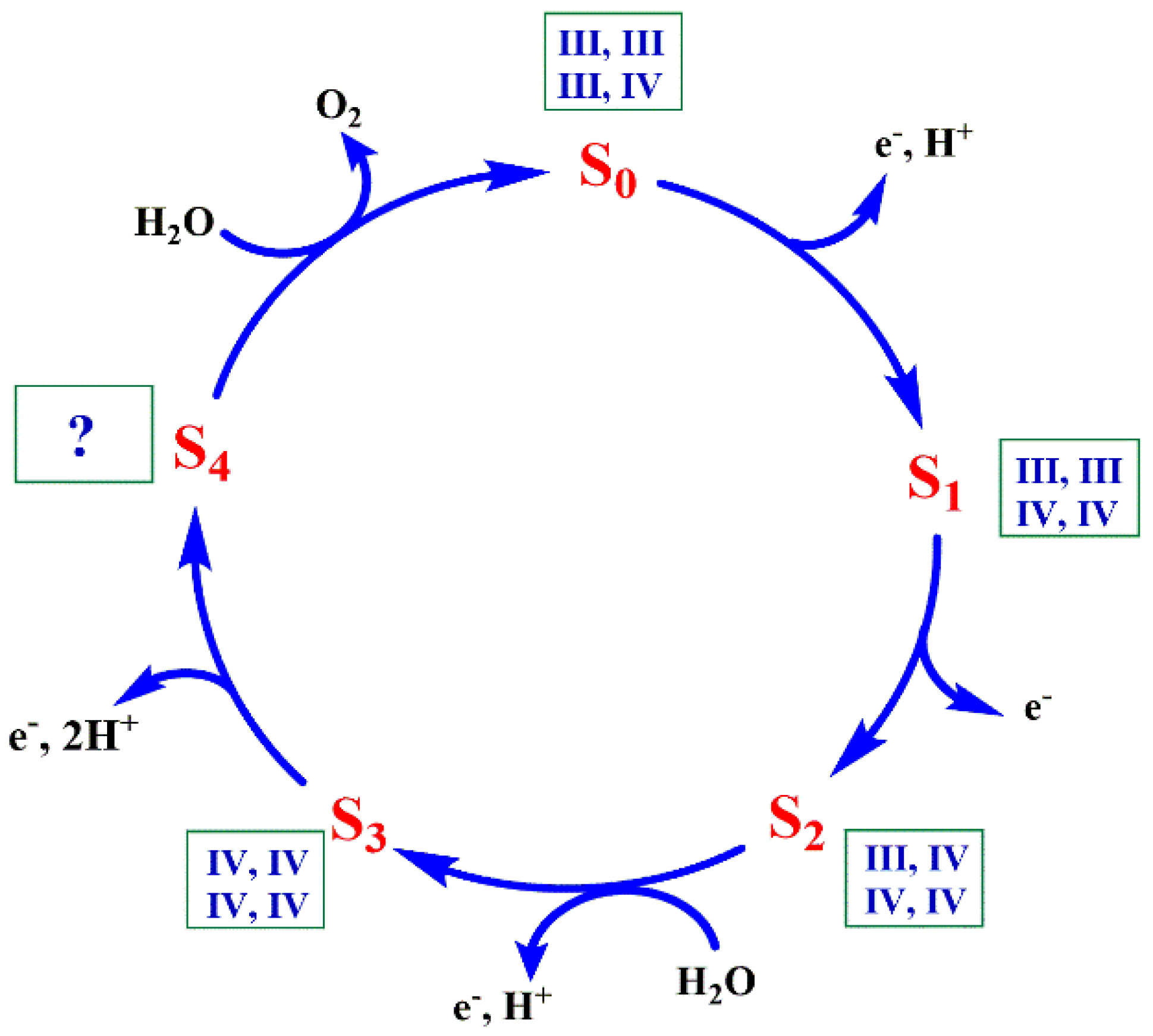
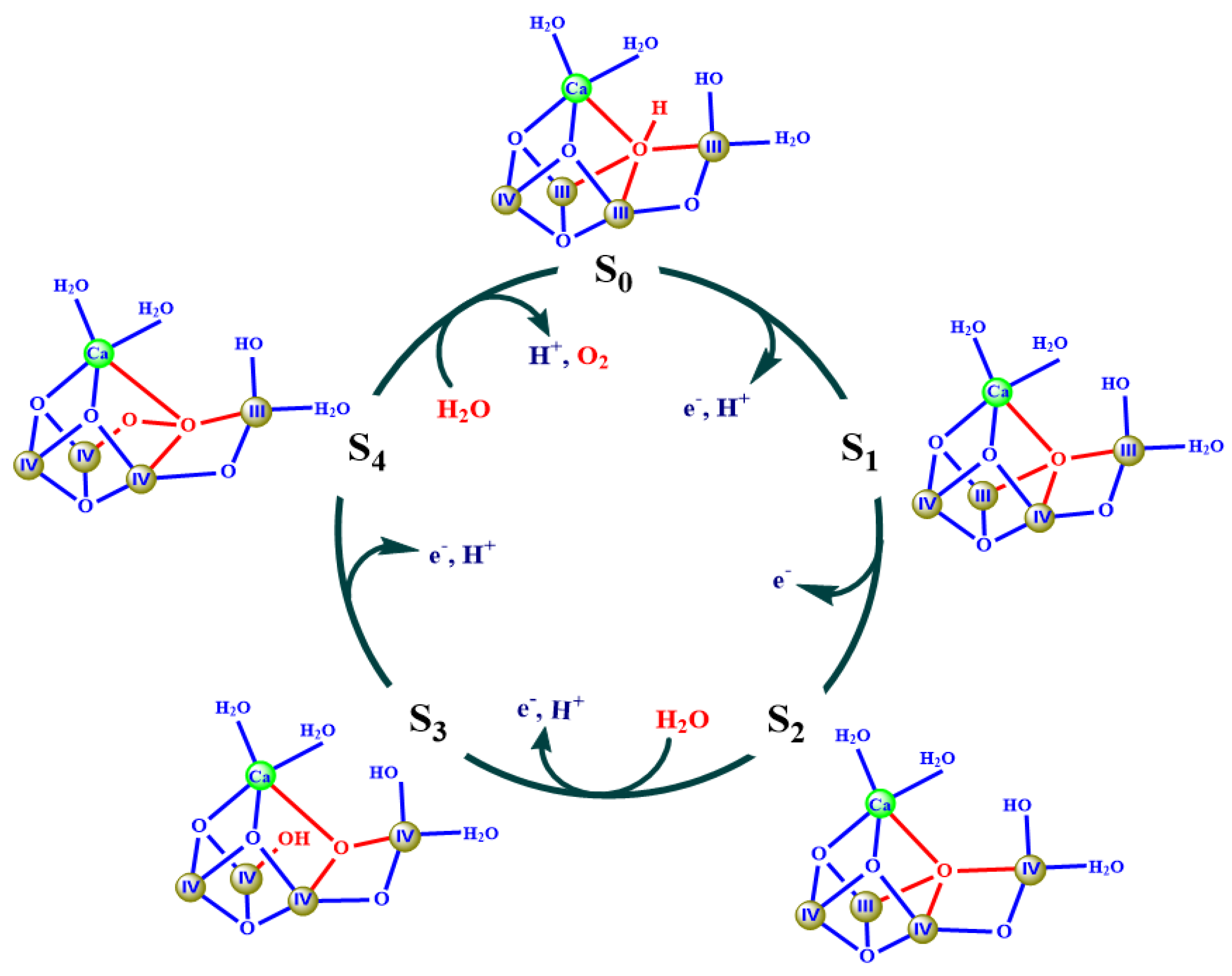
It is well known that the water-splitting reaction involves five different redox states (Sn, n = 0–4) of the OEC (Figure 1) [11,12], wherein the S0 state is the initial and most reduced state and the S1 state is the dark-stable state. The S2 and S3 states are metastable and decay eventually to the dark-stable S1 state, whereas the S4 state is a transient state that releases dioxygen and decays to the S0 state. In 1980s, it was revealed that the OEC is composed by one calcium and four manganese ions, embedded into the large protein environment of PSII through some carboxylate and imidazole groups [13,14,15]. Based on the X-ray absorption spectroscopy (XAS) and electron paramagnetic resonance (EPR) investigations of the OEC in different S-states, it has been found that changes of the valences of the four manganese ions take place during the catalytic turnover [6,16,17,18,19]. The valences for the four manganese ions have been suggested to be S0 (III, III, III, IV) or (II, III, IV, IV), S1 (III, III, IV, IV), S2 (III, IV, IV, IV) and S3 (IV, IV, IV, IV) [17,18,20,21]. This is the “high-oxidation paradigm” that has been widely adopted in the field of photosynthetic research [17,18,20,21]. However, some groups proposed a low-oxidation paradigm, corresponding to S0 (II, III, III, III), S1 (III, III, III, III), or (II, III, III, IV), S2 (III, III, III, IV), and S3 (III, III, IV, IV), respectively [22,23,24,25,26,27]. The calcium is an indispensable cofactor for the function of the OEC, and its depletion results in the complete loss of the water oxidation capability of PSII and it can only be functionally replaced by strontium [28,29,30]. The structure and catalytic mechanism of the OEC have attracted extensive studies during the last three decades [2,6,7].
2. Structure of the OEC
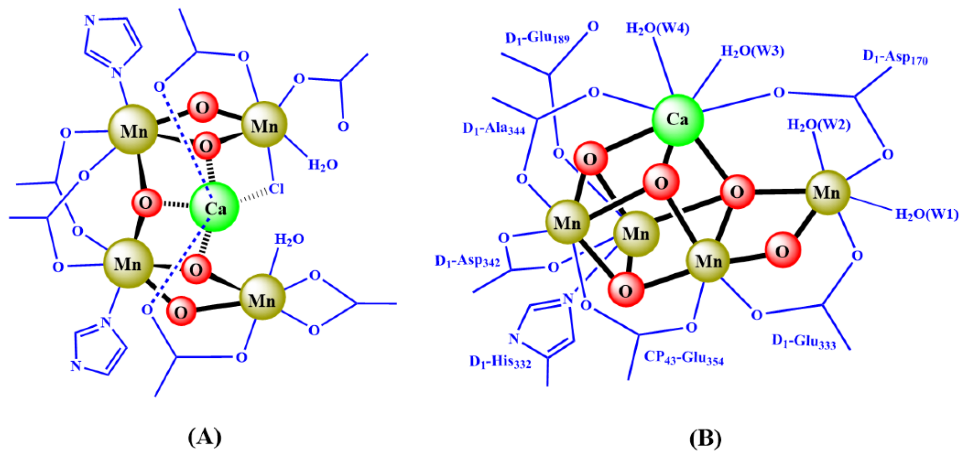
It is a long-standing issue to reveal the detailed structure of the OEC in the field of photosynthetic research. Before the appearance of the crystal structure of PSII, most structural information of the OEC came from X-ray absorption spectroscopy (XAS) [18,20], electron paramagnetic resonance (EPR) [6,16,31] investigations, and theoretical calculations [32]. Different structural models were suggested to explain different experimental observations of the OEC in PSII [33,34,35,36,37]. Figure 2A shows the structural model proposed by Zhang et al. in 1999 [37,38] in which, apart from all other models [33,34,35,36], the key component of calcium was suggested to be located in the middle of the OEC and connected with four manganese ions through three oxide bridges and two carboxylate groups [37].
The crystal structure of the OEC has emerged since the beginning of this century [39,40,41,42,43,44,45,46]. In 2001, Zouni et al. [40] reported the first crystal structure of PSII from thermophilic cyanobacterium at a resolution of 3.8 Å. In 2004, Ferreira et al. [42] reported the structure of PSII at 3.5 Å resolution, and proposed that the OEC could be comprised of a Mn3CaO4 cubane attached with a “dangler” Mn ion via one bridging oxide, which forms a Mn4CaO4-cluster. However, the detailed core and peripheral ligands of the OEC were still elusive due to the low resolution and the radiation reduction induced by the X-ray beam during the crystallographic structural determination [45,47,48,49].
The more detailed structure of the OEC was revealed by the crystal structure of PSII at a resolution of 1.9 Å reported by Umena et al. in 2011 [39]. In this structure, all possible binding ligands of the OEC have been clearly resolved, including four water molecules with one imidazole group from D1-His332 and six carboxylate groups from D1-Asp170, D1-Glu189, D1-Glu333, D1-Asp342, D1-Ala344, and CP43-Glu354, respectively. Importantly, one additional µ2-oxo (O4) bridge linking the dangler Mn and Mn3CaO4 cubane in the OEC was observed (Figure 2B). The whole structure of the OEC is an asymmetric Mn4CaO5-cluster. In this structure, the key component, Ca2+, is located in the middle of the OEC and connected to the four manganese ions through three oxide bridges and two carboxylate groups, which is consistent with our previous proposal (Figure 2A) published in 1999 [37,38].
The structure of the OEC (Figure 2B) was further confirmed by the 1.95 Å resolution data obtained by an X-ray free electron laser (XFEL) reported by Shen’s group [50,51,52] and other groups [46,53]. It was also supported by the 2.44 Å resolution reported by Hellmich et al. [44] and the 1.87 Å resolution reported by Tanaka et al. [54] when using a conventional synchrotron radiation source at an extremely low X-ray dose (0.03 MGy). Recently, the structure of the OEC in higher plants (e.g., spinach and pea) has also been revealed by single-particle cryo-electron microscopy (Cryo-EM) at the resolution of 3.2~2.7 Å [55,56].
The structures of the S2 and S3 states of the OEC have also been reported recently by using XFEL, and it was found that a new oxygen (termed O6 or OX) occupies the sixth coordination site of Mn1 during the S2 → S3 state transition [51,52,53]. The structures revealed by XFEL have been suggested to correspond to the native structure of the OEC in PSII [50]. However, consensus of the atomic positions of the S1 state OEC revealed by XFEL is still not fully reached for all structures with the results of extended X-ray absorption fine structure (EXAFS) spectroscopy studies on the active sample [49,57]. To evaluate the oxidation valences of the four manganese ions in the structure of the OEC revealed by XFEL, we have carried out bond-valence sum (BVS) calculations [58,59]. The BVS method is a popular method in coordination chemistry to estimate the valences of atoms [60,61]. It is derived from the bond-valence model [60], which is a simple yet robust model for validating chemical structures with localized bonds or used to predict some of their properties. This method has been used extensively to estimate the oxidation state of the active site in various metalloenzymes as well [62,63]. Table 1 lists the results of the BVS calculations on the XFEL structures of the OEC in the ‘native’ S1, S2, and S3 states, respectively. Surprisingly, the oxidation valences of the four manganese ions of all these states are remarkably lower than that of widely adopted S1(III, III, IV, IV) in the field of photosynthetic research (Figure 1) [16,17,18,19]. It is likely that the reduction of the high valences of manganese ions in the OEC could take place during the structural determination. Alternatively, some significant changes of the coordination spheres of the manganese ions in the OEC could take place during the X-ray diffraction measurement [57]. If it was the case, one would expect that the XFEL structures of the OEC would be different from the native structure of these intermediate states during the catalytic cycle. Recently, it has been found that the structural modifications of the OEC would take place due to the radiation damage induced by XFEL such as the position of the μ4-oxide bridge (O5, Figure 2), which can be significantly disturbed by XFEL [57,64,65].
3. Mechanism for the Water-Splitting Reaction in the OEC
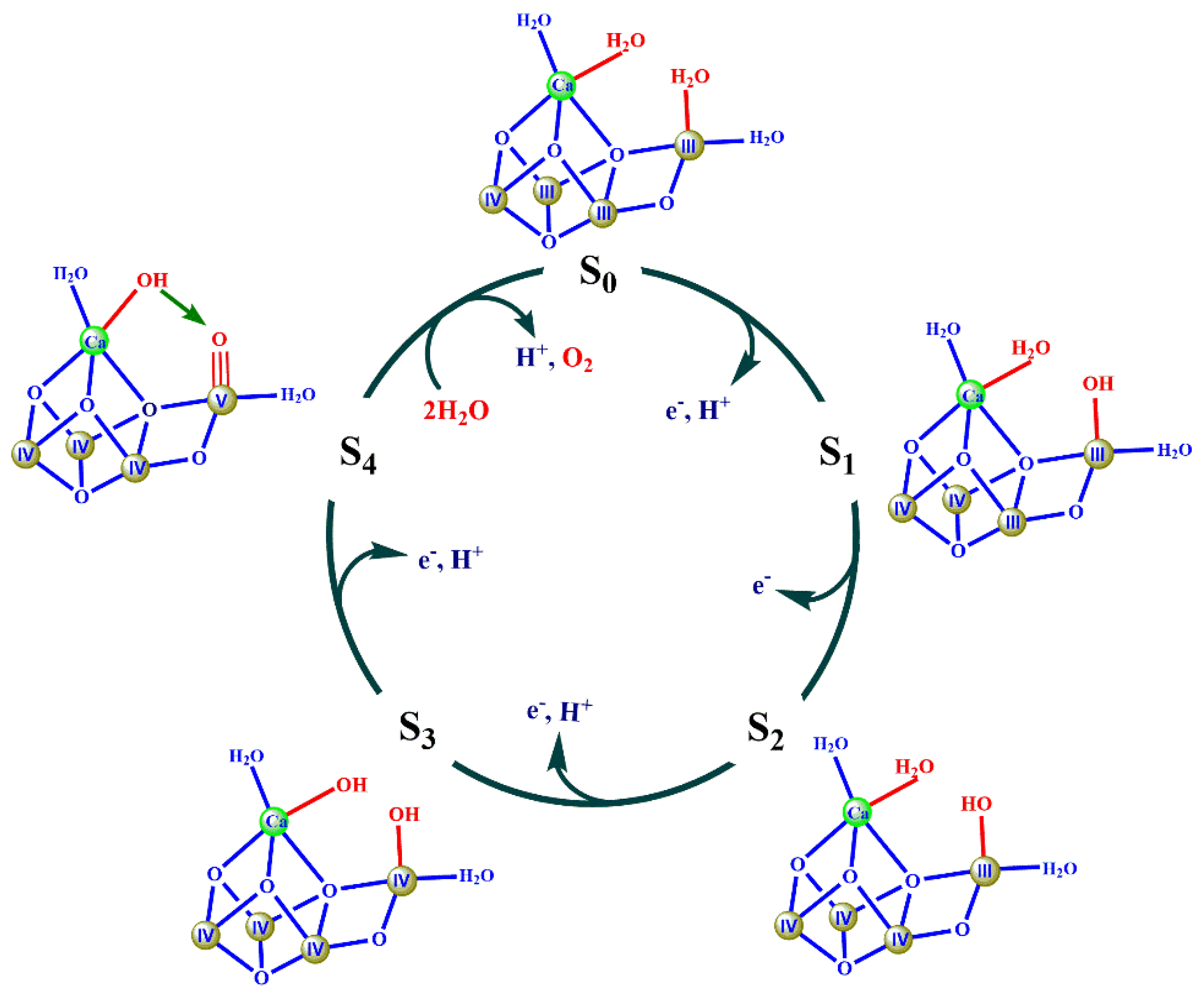
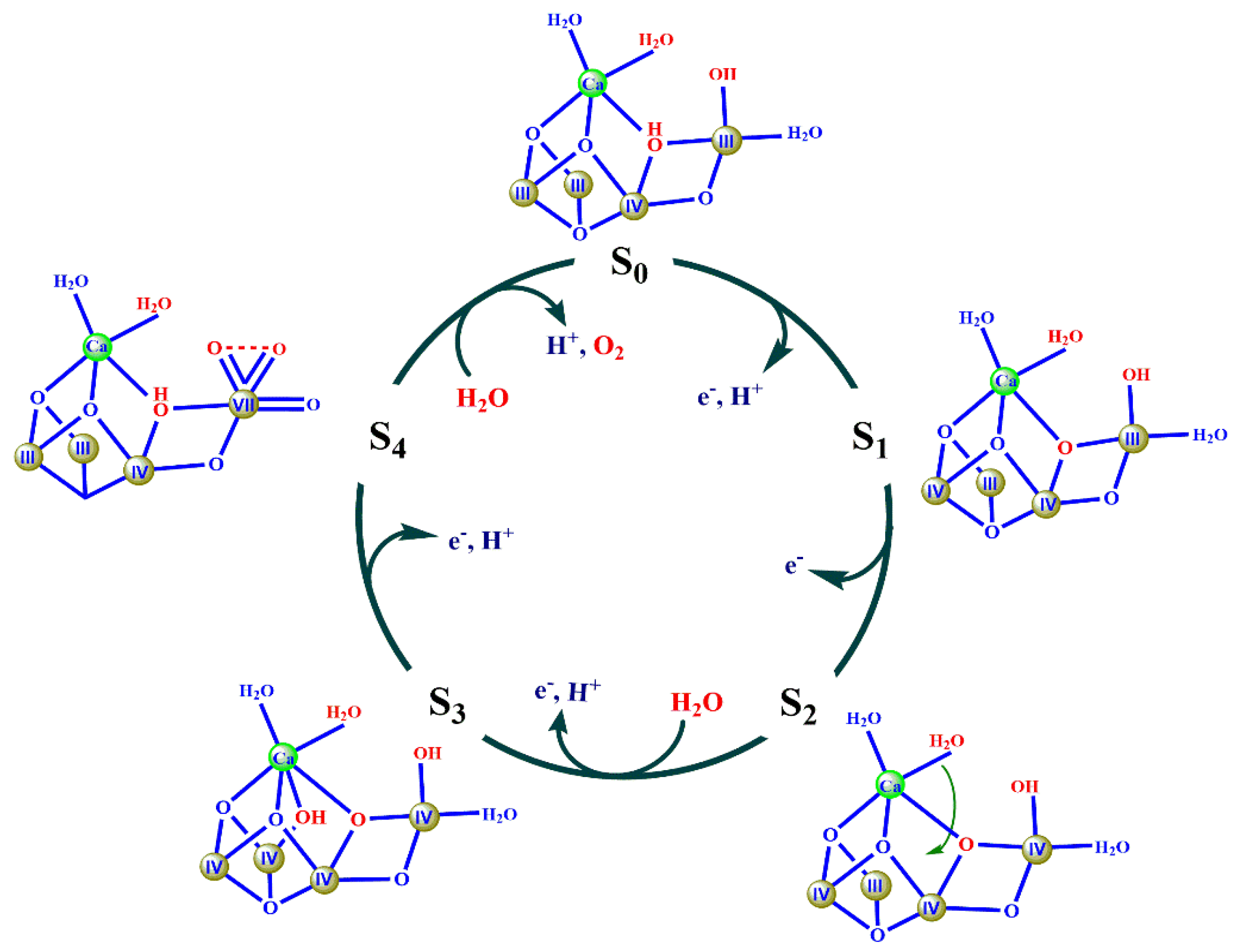
Based on recent crystallographic studies [39,42,46,50,51,52,53], spectroscopic investigations, and theoretical calculations [17,18,66,67,68,69,70,71,72,73,74,75], different proposals for the O-O bond formation have been suggested [6,49,66,75,76,77,78,79,80,81]. Figure 3, Figure 4, Figure 5 and Figure 6 show four typical proposals from different groups.
The mechanism in Figure 3 was suggested by Barber’s group [77] in which the two oxygens of two water molecules (W2 and W3, Figure 2B) were proposed to serve as the oxygen sources for the formation of the O-O bond. The key feature of this mechanism is that the O-O bond is formed by a nucleophilic attack of a calcium ligated hydroxyl group onto an electrophilic oxo of MnV≡O or MnIV-O•, derived from the deprotonation of the second substrate water molecule. Similar proposals have been suggested by other groups [80,82,83]. However, this proposal was not supported by a recent theoretical calculation reported by Siegbahn [84].
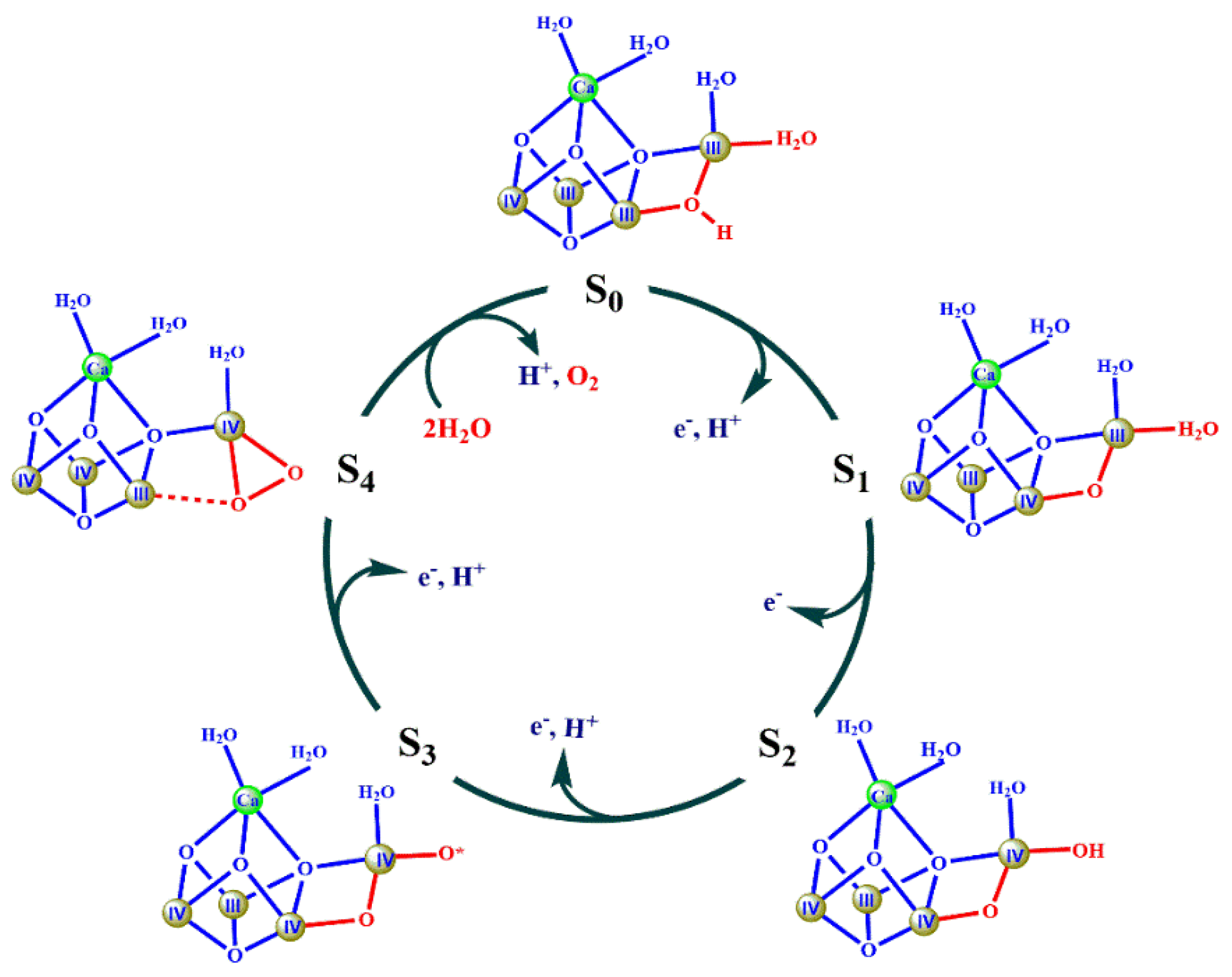
The second proposal for the mechanism of the water-splitting reaction was suggested by Ishikita’s group, as shown in Figure 4 [79]. In this mechanism, the μ2-oxide bridge (O4) and one water molecule (W1) were suggested to provide the oxygen atoms to form the O-O bond. The key feature of this proposal is that the O-O bond is formed through the coupling of a bridged oxo and an Mn(IV)-O• oxyl radical. However, the valences (III, IV, IV, IV) of the four manganese ions in the S3 state were not consistent with the widely accepted valences of (IV, IV, IV, IV) [17,18,20].
The third mechanism was first suggested by Siegbahn based on theoretical calculations [67] (Figure 5). The main feature of this proposal is that the μ4-oxide bridge (O5) serves as the active site for the O-O bond formation. According to this mechanism, the release of O2 from the S4 state would result in the formation of four unsaturated metal ions, which includes three 5-coordinated manganese (i.e., Mn1, Mn3, Mn4) and one 6-coordinated calcium. This could certainly require very high activation energy [85]. Thus, one would expect that the dioxygen release could be the rate-limited step during the catalytic cycle. However, this is inconsistent with the fast release of the O2 observed in the natural system [2,86]. Although some spectroscopic studies [6,74] show experimental evidence to support this proposal to some degree [78], it is still an open question whether the Mn1 is the active site for the binding site of the second substrate water molecule [2,7,58,59]. In addition, it is also noted that the suggestion of the protonated μ4-oxide bridge (i.e., OH) for O5 in the S0 state has not been supported by the theoretical studies from the other group [65,87].
Figure 6 shows a hypothesis proposed by Zhang and Sun [88]. In all previous proposals, the highest oxidation valance of the manganese ion was MnV (e.g., Figure 3). Remarkably, in the proposal shown in Figure 6, the authors suggested that the highest oxidation valance of the manganese ion, MnVII, could be present in the S4 state induced by charge and structural rearrangements of the first coordination spheres around the MnVII-oxo site on the dangling Mn4 with de-coordination and re-coordination of carboxylates (D1-Glu333 and D1-Asp170) [88]. Generally, MnVII ion displays a special UV-visible absorption at a range of 400–600 nm. Therefore, if this is the case, one could observe the typical MnVII absorption feature during the turnover of the catalytic cycle.
As mentioned above, although various mechanisms for the water-splitting reaction have been proposed [67,77,79,80,82,89,90], the detailed mechanism for the O-O bond formation is still elusive [2,7,59] mainly due to the complexity of the huge protein environment and the dynamic structural changes of the OEC during the water-splitting reaction. In this regard, precisely structural data for different S states of the OEC are still highly required in the future [46,51,52,53,77,85].
4. Challenge for the Synthesis of the OEC in the Laboratory
In order to better understand the structure and properties of the OEC as well as to develop highly efficient and cheap man-made catalysts for the water-splitting reaction to overcome the bottleneck of the artificial photosynthesis, many groups have tried to synthesize the OEC in the laboratory since the 1990s. However, it was a great challenge for chemists to synthesize the whole structure of the OEC due to several reasons [38,91] including: i) It is very difficult to incorporate Ca2+ into the Mn4-cluster through µ-oxo bridges because the affinity of Ca2+ to µ-oxo is significantly weaker than that of the Mn ion. In general, only a homometallic cluster, instead of the heterometallic manganese-calcium cluster, can be isolated. ii) The core structure of the OEC is an asymmetric Mn4Ca-cluster [1]. It was fully unknown whether such an asymmetric structure could be synthesized in a chemical system. iii) The ligands of the natural OEC are mainly composed of carboxylate groups and water molecules, which are drastically different from the multi-pyridine ligands used in most previous chemical model systems [92,93,94,95]. iv) The redox potential of the OEC is very high (+0.8 ~ +1.0 V vs. normal hydrogen electrode(NHE)) [4,96] due to the presence of the high valence Mn(IV)/Mn(III) ions.
A large number of artificial Mn complexes have been reported in the literature [82,92,95,97,98,99,100,101,102,103,104]. Among them, tetra-manganese complexes containing Mn4O4-cubane [101,105,106,107,108] are attractive. However, both the structure and properties of most model complexes are remarkably different from that of the OEC in a natural system.
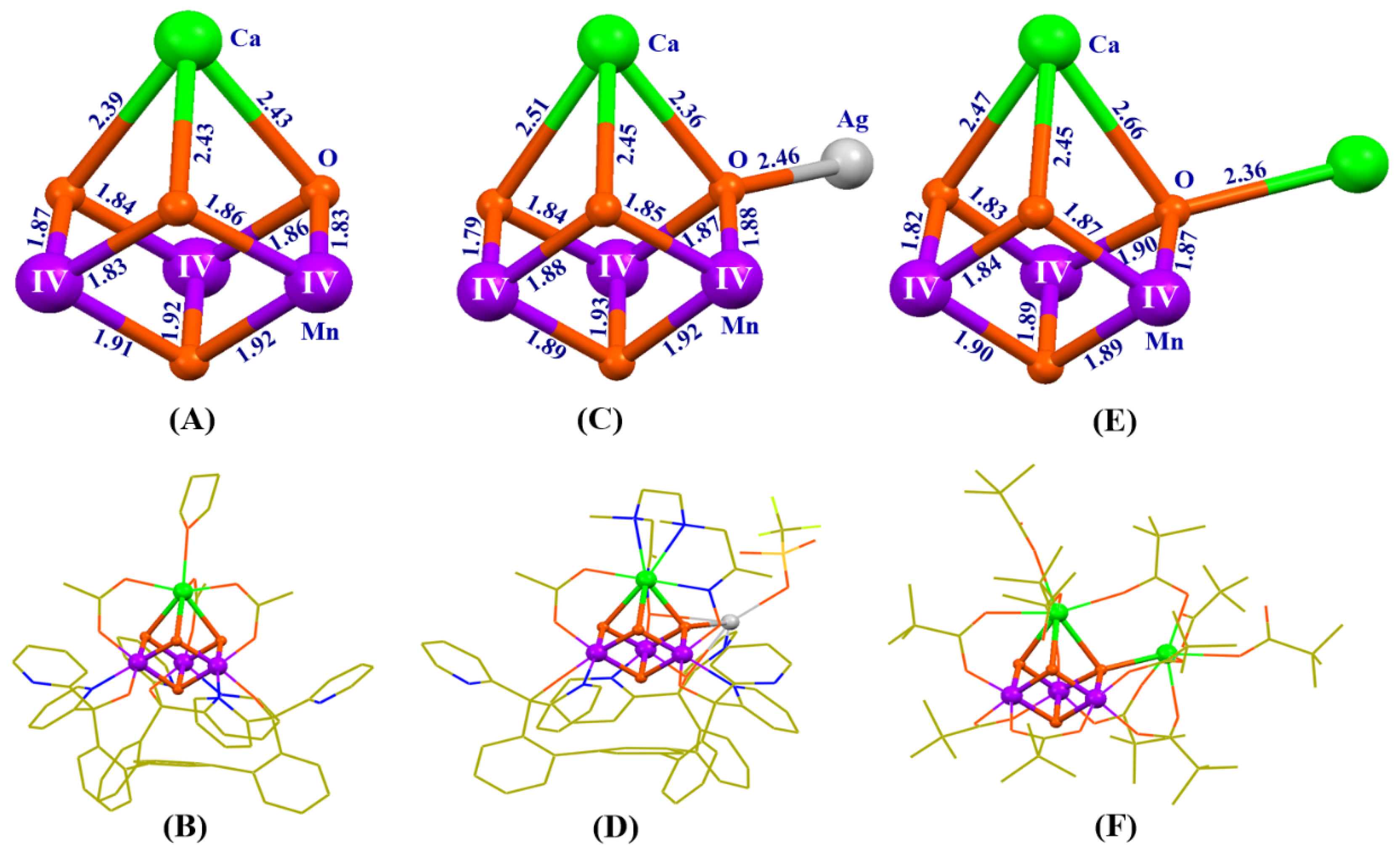
Significant advances for the synthesis of the OEC have emerged since 2011. Agapie’s group reported the first artificial Mn3CaO4-cluster using a multi-pyridylalkoxide ligand (i.e., 1,3,5–triarylbenzene motif appended with alkoxide and pyridine donors) [107] (Figure 7A,B). By treating the Mn3CaO4-cluster with Ln(CF3SO3)3 (Ln = La3+, Ce3+, Gd3+, etc), they isolated different Mn3LnO4-clusters, and observed a linear correlation between the redox potential of the cluster and the pKa of the lanthanide metal ions [109]. The same group reported a series of analogues or derivatives [110] for the artificial Mn3CaAgO4-complex [111] (Figure 7C,D).
In 2012, Christou’s group reported the Mn3Ca2O4-complex with one Ca2+ attached to the Mn3CaO4 cubane [112] (Figure 7E,F). Distinct from previous Mn3CaO4-complexes and its derivatives, the peripheral ligands of the Mn3Ca2O4-complex are pivalic anions or neutral pivalic acid, which closely mimics the peripheral carboxylate ligands of the OEC in PSII. In these artificial complexes, all the manganese ions are in an IV oxidation state, and the typical bond lengths for Mn-O and Ca-O range from 1.8–1.9 Å to 2.4–2.7 Å, respectively. The distances of the Mn…Mn and Mn…Ca range from 2.7–2.8 Å to 3.2–3.5 Å, respectively.
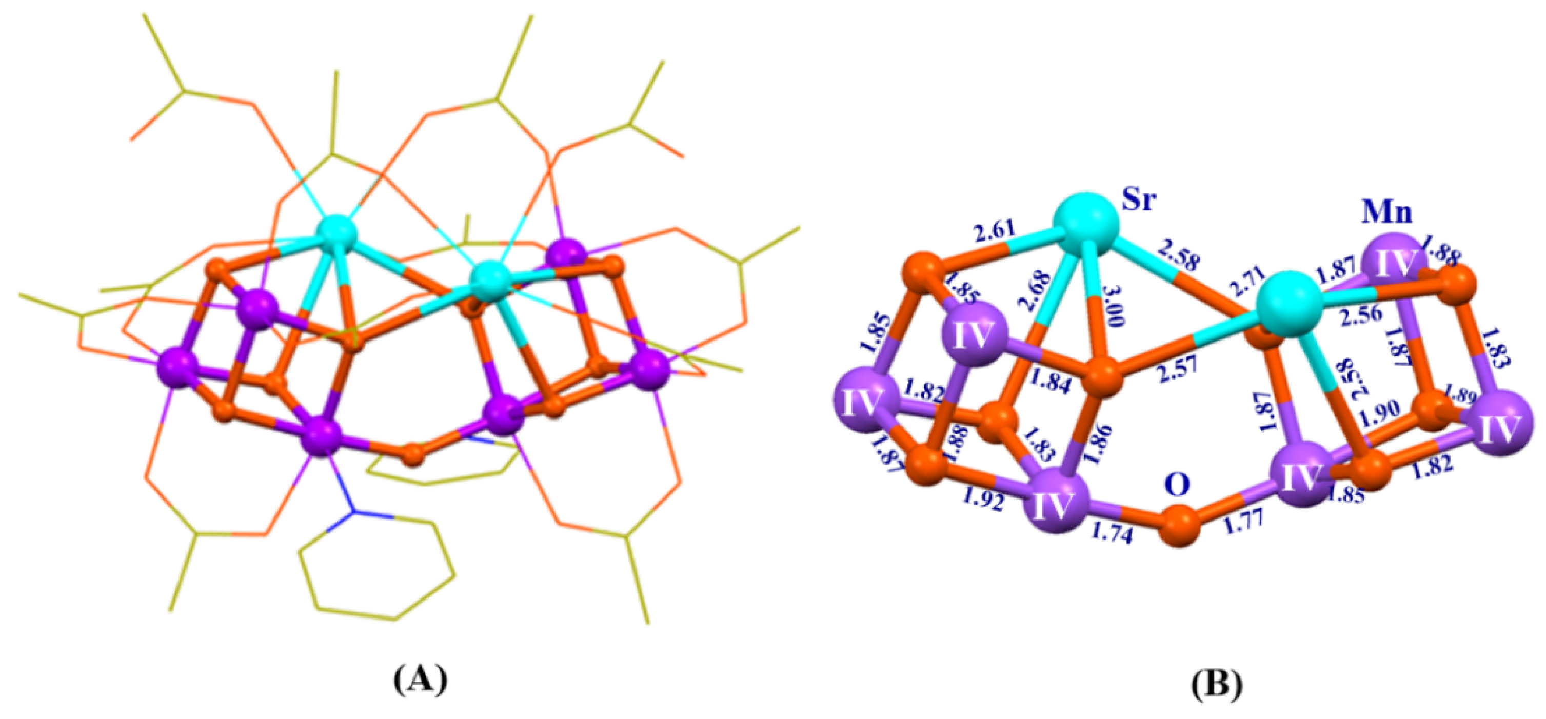
In 2014, we reported a heterometallic cluster containing two MnIV3SrO4-clusters linked by one μ2-oxide bridge [113], which mimics the three types of oxide bridges (μ2-oxide, μ3-oxide, and μ4-oxide) and the Mn3SrO4 cubane of the Sr2+-containing OEC [30] at the same time (Figure 8).
It should be pointed out that, even though all complexes shown in Figure 7 and Figure 8 have mimicked some key structural motifs of the Mn3CaO4 or Mn3SrO4 cubane in the OEC, until recently, it remains a great challenge to synthesize the entire Mn4Ca-cluster with similar ligands, as seen in the OEC of PSII.
5. Closer Mimicking of the OEC
Inspired by the evolution of the OEC [114] and assembly processes of the OEC in PSII [115,116,117], in 2015, we successfully prepared the first artificial Mn4CaO4-cluster [118] (Figure 9 C,D), which was synthesized through a two-step procedure using inexpensive commercial chemicals. The first step was to synthesize a precursor through a reaction of Bun4NMnO4 (Bun = n-butyl), Mn(CH3CO2)2· (H2O)4, and Ca(CH3CO2)2·H2O (molar ratio of 4:1:1) in boiling acetonitrile in the presence of an excess of pivalic acid. The second step was to treat the precursor with organic base (pyridine) in ethyl acetate, which leads to the formation of a final product. We have found that both the acetonitrile solvent and pivalic acid are crucial for the formation of the final product, and the replacements of them by other organic solvents (e.g., THF, CH3OH) and/or organic acids (e.g., acetic acid and propionic acid) lead to failing to prepare the Mn4CaO4-complex.
The artificial Mn4CaO4-complex contains a Mn3CaO4 cubane attached with a dangler Mn ion, which forms an asymmetric Mn4CaO4-core structure. This is exactly the same as the OEC structure proposed by Ferreira et al. in 2004 [42]. The surrounding ligands of the Mn4CaO4-cluster are provided by eight (CH3)3CCO2- anions and three exchangeable neutral ligands (two pivalic acid and one pyridine molecules), which are similar to the peripheral ligands of the OEC (Figure 9B).
It should be pointed out that the structure of the artificial Mn4CaO4-cluster is well-defined and the effect from the X-ray radiation reduction is limited mainly due to the absence of water as solvent in the crystal of the artificial Mn4CaO4-complex. BVS calculations have clearly shown that the oxidation states of the four manganese ions of the artificial Mn4CaO4-complex are (III, III, IV, IV), which are essentially the same as that proposed for the OEC in the S1 state in the high-oxidation paradigm (Figure 1).
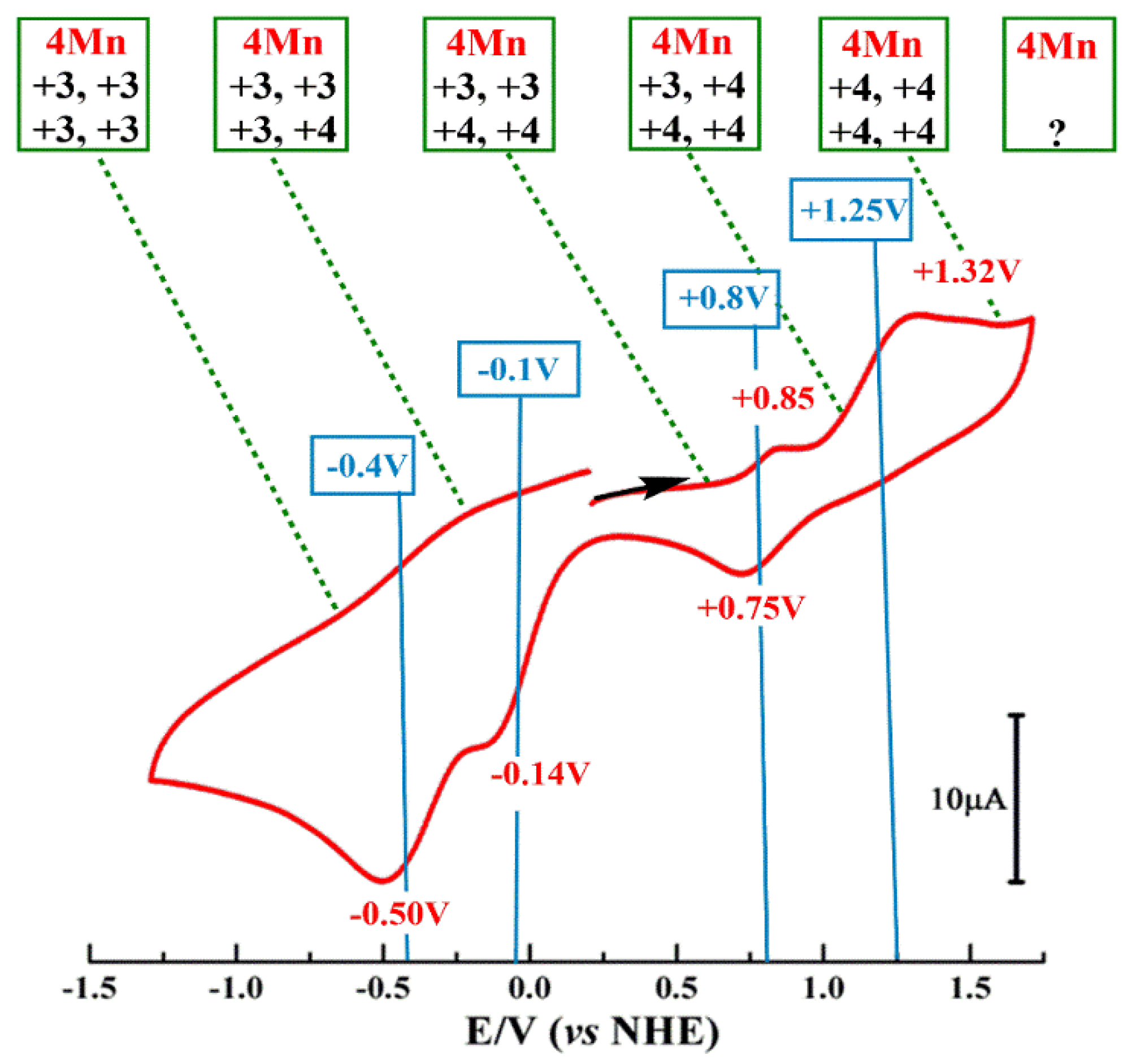
The similarity between the artificial Mn4CaO4-cluster and the natural OEC was further supported by the observation of four redox transitions revealed by cyclic voltammogram (CV) measurements (Figure 10). The redox potential of ~ 0.8 eV (vs. NHE) for the S1→S2 transition of artificial Mn4CaO4-complex is close to the estimated potential of the corresponding OEC redox transition (≥0.9 V) [4,96], but it is remarkably different from that of the previously Mn3CaO4-complex without a dangling Mn ion [107]. This result indicates that the dangler manganese ion could play a crucial role in tuning the redox potential of the Mn4CaO4-cluster.
Moreover, the one-electron oxidation of the artificial Mn4CaO4-complex gave rise to two distinct electron paramagnetic resonance (EPR) signals (g = 4.9 and g = 2.0) [118] (Figure 11), which is similar to the g ≈ 4 and g = 2.0 EPR signals observed in PSII for the OEC in the S2 state [16,119,120,121,122,123]. In the field of photosynthetic research, the latter two EPR signals have been considered as fingerprint spectroscopic characteristics to evaluate the structure and function of the OEC. Therefore, the experimental observation of two EPR signals suggested that the artificial Mn4CaO4-cluster would have a similar electronic structure as that of the OEC in the biological system.
The origin of the g = 2 and g ≈ 4 EPR signals in both natural and artificial Mn4Ca-clusters have been theoretically investigated recently [71,72,100,120,124,125,126,127,128]. It was suggested that the two EPR signals could be raised from two different conformations of the OEC in the S2 state [72,120,125], and the g = 2 EPR signal could correspond to the structure with an open Mn3CaO4 cubane, while the g ≈ 4 EPR signal may be raised with a closed Mn3CaO4 cubane. Narzi et al. suggested that the conversion between these two conformations could be crucial for the function of the OEC [71]. On the contrary, Corry and O’Malley suggested that the high-spin (g ≈ 4) and low spin (g = 2) EPR signals could be raised from the same conformation of the OEC, but have different protonation states of O4 (i.e., μ2-O2- or μ2-OH-) in the Mn4CaO5-cluster [125]. Pushkar et al. recently proposed that the high spin (g = 4) EPR signal could be raised from the early binding of the substrate oxygen to the five-coordinate Mn1 ion in the S2 state [128]. Some theoretical studies on the two EPR signals of the artificial Mn4CaO4-cluster have been reported [126,127], while the real origin of these signals is still elusive. So far, the origin of these two EPR signals observed in both a natural and artificial Mn4Ca-cluster is still an open question [123,124,128,129]. Considering the lack of μ2-oxo (O4) in the reported artificial Mn4CaO4-cluster resulting in a large and significant change in the orientation of dangling Mn4, as compared to the Mn4CaO5-cluster in OEC, we suggest that the precise mimic of the OEC with the presence of the μ2-oxo (O4) is pressingly needed in the future. This would provide crucial information for the entities and process of the biological Mn4CaO5-cluster in PSII.
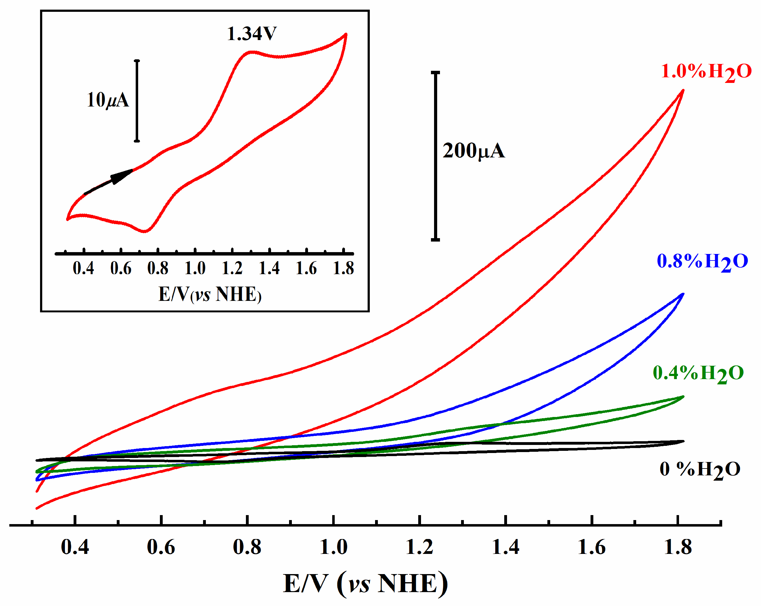
Considering the close mimicking of both the geometric and electronic structures as well as the redox properties of the OEC, we expect that the artificial Mn4CaO4-cluster should be able to serve as a catalyst for the water-splitting reaction. A remarkable catalytic current was observed during the CV measurement in the presence of an artificial Mn4CaO4-complex and 1% water in acetonitrile [118] (Figure 12). The artificial Mn4CaO4-complexes can also catalyze an oxygen-evolving reaction efficiently by using (CH3)3COOH (tert-butyl hydroperoxide) as an oxidant in acetonitrile [130]. However, it should be pointed out that the artificial complex is very sensitive to the experimental conditions. Quantification of the catalytic reaction is difficult due to the rapid degradation of the catalyst in the presence of water in solution. Particularly, the calcium ion in the artificial cluster has been found to be easily dissociated in the presence of water, which leads to the formation of multi-manganese complexes.
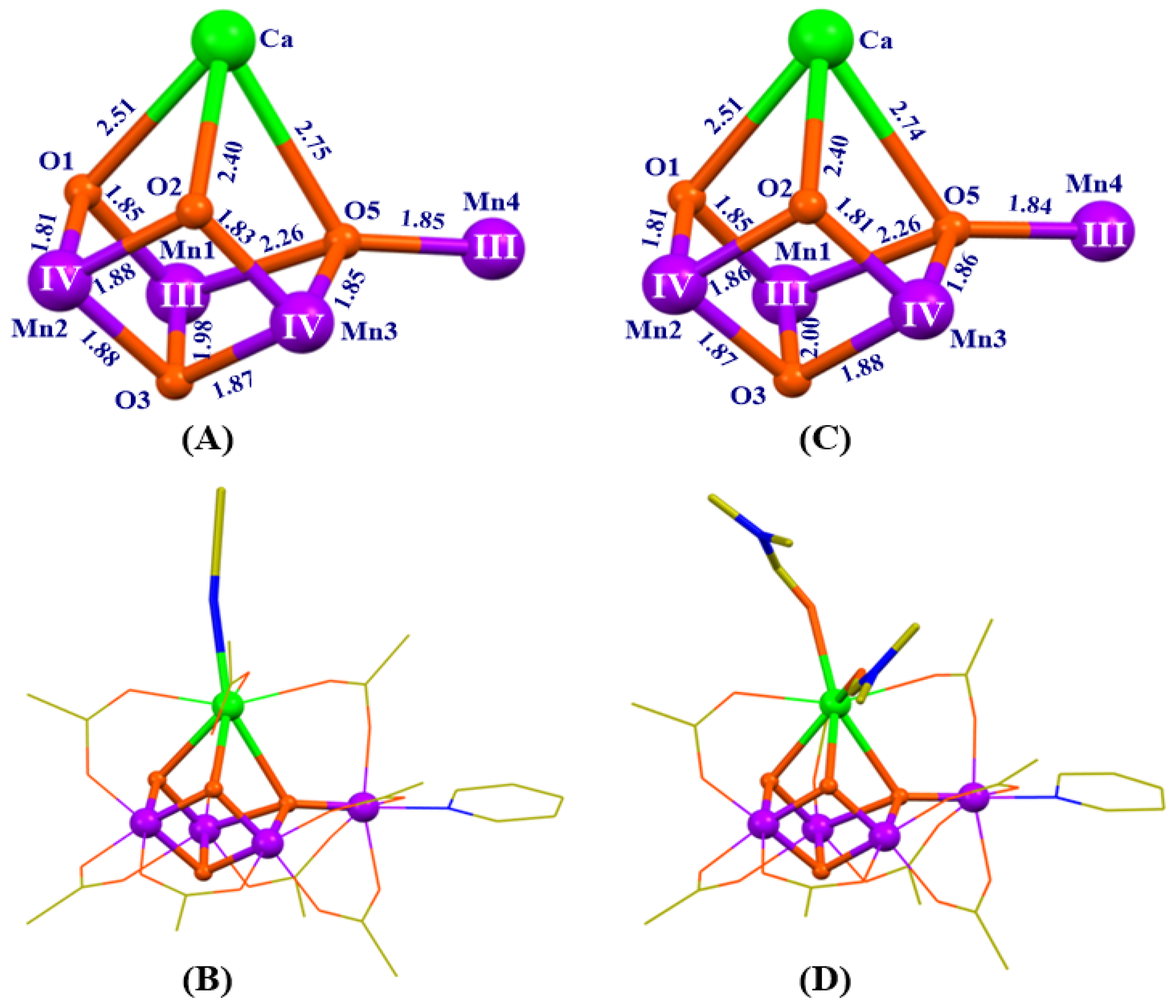
To improve the stability of the artificial Mn4CaO4-cluster, we have recently prepared two new Mn4CaO4-clusters with an exchangeable solvent known as acetonitrile or N, N-dimethylformamide (DMF) [58] on the calcium, as shown in Figure 13. Remarkably, the replacement of one or two ligands on the calcium by organic solvent molecules does not modify the core structure and valences of the four manganese ions as well as the main peripheral environmental ligands. More importantly, these new Mn4CaO4-clusters become more stable in the polar solvent, which may provide a great opportunity to investigate the catalytic activity of the artificial Mn4CaO4-cluster in the future.
6. Implications for the Mechanism of the Water-Splitting Reaction in OEC
Artificial Mn4CaO4-clusters shown in Figure 9 and Figure 13 have mimicked the main structural motifs of the OEC, which could provide distinct chemical insight into understanding the principle of the OEC [127,131,132]. First, the successful isolation of different artificial Mn4CaO4-complexes demonstrates that the Mn4CaO4-cluster is thermodynamically stable, which supports the proposal that the Mn4CaO4-cluster could be an evolutionary origin of the natural OEC by Barber [133]. Second, from the structure of an artificial Mn4CaO4-cluster, we can clearly see that the µ4-O2- bridge (O5) is tightly bound to four metal ions (one calcium and three manganese ions). This is a position less prone to be removed or replaced, which indicates that the similar µ4-O2- bridge (O5) in OEC could not be directly involved in serving as an oxygen source to form the O-O bond, as proposed recently [6,52,66,67,78]. Kawashima et al. have recently proposed that the μ2-O2- bridge (O4) in OEC (Figure 4) may play a role as the substrate binding site to form the O-O bond [79]. In this proposal and some previous suggestions [77,80,82], the Mn4CaO4 fragment does not undertake significant changes during the water-splitting reaction, which is consistent with the isolation of the stable artificial Mn4CaO4-clusters described in this case. It should be pointed out that the μ2-O2- (O4) in OEC is absent in the artificial Mn4CaO4-cluster [100,134], which is replaced by a bridging carboxyl group in the latter. Clearly, the future investigation of this missing μ2-O2- bridge in an artificial Mn4Ca-cluster may provide new insights into the mechanism for the O-O bond formation during the OEC turnover.
7. Conclusions
In summary, the crystallographic studies of PSII have revealed that the OEC is composed by an asymmetric Mn4CaO5-cluster. Although extensive investigations have been carried out on both the natural and artificial OEC in the last three decades, the detailed mechanism for the water-splitting reaction in a natural system is still elusive due to the complexity of the large protein environment and structural uncertainty of the OEC in different S-states during the catalytic turnover. A long-standing goal in science seeks to understand the structure-function relationship of the OEC and develop efficient man-made water-splitting catalysts in artificial photosynthesis. Recently, different model complexes have been synthesized to mimic the OEC in the laboratory where the artificial Mn4CaO4-cluster closely mimics both the geometric and electronic structures of the OEC. This artificial Mn4CaO4-cluster provides a rational chemical model to investigate the structure-function relationship of the OEC, and also sheds new insights into the mechanism of the water-splitting reaction in natural photosynthesis. These recent advances in the mimicking of the OEC may help develop efficient man-made catalysts for the water-splitting reaction by using earth-abundant and non-toxic chemical elements in artificial photosynthesis in the future.
Funding
The National Key Research & Development Program of China (No. 2017YFA0503704), the Strategic Priority Research Program of Chinese Academy of Sciences (No. XDA21010212, XDB17030600), and the National Natural Science Foundation of China (No. 31770258; 91961203) supported this work.
Conflicts of Interest
The authors declare no conflict of interest.
References
- Shen, J.R. The structure of photosystem II and the mechanism of water oxidation in photosynthesis. Annu. Rev. Plant Biol. 2015, 66, 23–48. [Google Scholar] [CrossRef] [PubMed] [Green Version]
- Junge, W. Oxygenic photosynthesis: History, status and perspective. Q. Rev. Biophys. 2019, 52, e1. [Google Scholar] [CrossRef] [PubMed]
- Barber, J. Photosynthetic energy conversion: Natural and artificial. Chem. Soc. Rev. 2009, 38, 185–196. [Google Scholar] [CrossRef] [PubMed]
- Dau, H.; Zaharieva, I. Principles, efficiency and blueprint character of solar-energy conversion in photosynthetic water oxidation. Acc. Chem. Res. 2009, 42, 1861–1870. [Google Scholar] [CrossRef] [PubMed]
- Nelson, N.; Yocum, C.F. Structure and function of photosystem I and II. Annu. Rev. Plant Biol. 2006, 57, 521–565. [Google Scholar] [CrossRef] [Green Version]
- Lubitz, W.; Chrysina, M.; Cox, N. Water oxidation in photosystem II. Photosyn. Res. 2019, 142, 105–125. [Google Scholar] [CrossRef] [Green Version]
- Pantazis, D.A. Missing pieces in the puzzle of biological water oxidation. ACS Catal. 2018, 8, 9477–9507. [Google Scholar] [CrossRef]
- Cardona, T.; Sedoud, A.; Cox, N.; Rutherford, A.W. Charge separation in Photosystem II: A comparative and evolutionary overview. Biochim. Biophys. Acta 2012, 1817, 26–43. [Google Scholar] [CrossRef] [Green Version]
- Zhang, B.; Sun, L. Artificial photosynthesis: Opportunities and challenges of molecular catalysts. Chem. Soc. Rev. 2019, 48, 2216–2264. [Google Scholar] [CrossRef] [Green Version]
- Matheu, R.; Garrido-Barros, P.; Gil-Sepulcre, M.; Ertem, M.Z.; Sala, X.; Gimbert-Suriñach, C.; Llobet, A. The development of molecular water oxidation catalysts. Nat. Rev. Chem. 2019, 3, 331–341. [Google Scholar] [CrossRef]
- Kok, B.; Forbush, B.; McGloin, M. Cooperation of charges in photosynthetic O2 evolution. I. A linear four step mechanism. Photochem. Photobiol. 1970, 11, 457–475. [Google Scholar] [CrossRef] [PubMed]
- Dau, H.; Haumann, M. Eight steps preceding O–O bond formation in oxygenic photosynthesis—A basic reaction cycle of the Photosystem II manganese complex. Biochim. Biophys. Acta 2007, 1767, 472–483. [Google Scholar] [CrossRef] [PubMed] [Green Version]
- Debus, R.J. The manganese and calcium ions of photosynthetic oxygen evolution. Biochim. Biophys. Acta 1992, 1102, 269–352. [Google Scholar] [CrossRef]
- Debus, R.J. Amino acid residues that modulate the properties of tyrosine YZ and the manganese cluster in the water oxidizing complex of photosystem II. Biochim. Biophys. Acta 2001, 1503, 164–186. [Google Scholar] [CrossRef] [Green Version]
- Diner, B.A. Amino acid residues involved in the coordination and assembly of the manganese cluster of photosystem II. Proton-coupled electron transport of the redox-active tyrosines and its relationship to water oxidation. Biochim. Biophys. Acta 2001, 1503, 147–163. [Google Scholar] [CrossRef] [Green Version]
- Peloquin, J.M.; Britt, R.D. EPR/ENDOR characterization of the physical and electronic structure of the OEC Mn-cluster. Biochim. Biophys. Acta 2001, 1503, 96–111. [Google Scholar] [CrossRef] [Green Version]
- Krewald, V.; Retegan, M.; Cox, N.; Messinger, J.; Lubitz, W.; DeBeer, S.; Neese, F.; Pantazis, D.A. Metal oxidation states in biological water splitting. Chem. Sci. 2015, 6, 1676–1695. [Google Scholar] [CrossRef] [Green Version]
- Yano, J.; Yachandra, V.K. Mn4Ca-cluster in photosynthesis: Where and how water is oxidized to dioxygen. Chem. Rev. 2014, 114, 4175–4205. [Google Scholar] [CrossRef]
- Dau, H.; Haumann, M. The manganese complex of photosystem II in its reaction cycle—Basic framework and possible realization at the atomic level. Coord. Chem. Rev. 2008, 252, 273–295. [Google Scholar] [CrossRef]
- Dau, H.; Grundmeier, A.; Loja, P.; Haumann, M. On the structure of the manganese complex of photosystem II: Extended-range EXAFS data and specific atomic-resolution models for four S-states. Philos. Trans. R. Soc. Lond. B 2008, 363, 1237–1244. [Google Scholar] [CrossRef] [Green Version]
- Sauer, K.; Yano, J.; Yachandra, V.K. X-ray spectroscopy of the photosynthetic oxygen-evolving complex. Coord. Chem. Rev. 2008, 252, 318–335. [Google Scholar] [CrossRef] [PubMed] [Green Version]
- Zheng, M.; Dismukes, G.C. Orbital configuration of the valence electrons, ligand field symmetry, and manganese oxidation states of the photosynthetic water oxidizing complex: Analysis of the S2 state multiline EPR signals. Inorg. Chem. 1996, 35, 3307–3319. [Google Scholar] [CrossRef] [PubMed]
- Pace, R.J.; Jin, L.; Stranger, R. What spectroscopy reveals concerning the Mn oxidation levels in the oxygen evolving complex of photosystem II: X-ray to near infra-red. Dalton Trans. 2012, 41, 11145–11160. [Google Scholar] [CrossRef] [PubMed]
- Jin, L.; Smith, P.; Noble, C.J.; Stranger, R.; Hanson, G.R.; Pace, R.J. Electronic structure of the oxygen evolving complex in photosystem II, as revealed by 55Mn Davies ENDOR studies at 2.5 K. Phys. Chem. Chem. Phys. 2014, 16, 7799–7812. [Google Scholar] [CrossRef]
- Petrie, S.; Stranger, R.; Pace, R.J. Explaining the different geometries of the water oxidising complex in the nominal S3 state crystal structures of photosystem II at 2.25 Å and 2.35 Å. ChemPhysChem 2018, 19, 3296–3309. [Google Scholar] [CrossRef]
- Petrie, S.; Pace, R.J.; Stranger, R. Resolving the differences between the 1.9 Å and 1.95 Å crystal structures of photosystem II: A single proton relocation defines two tautomeric forms of the water-oxidizing complex. Angew. Chem. Int. Ed. 2015, 54, 7120–7124. [Google Scholar] [CrossRef]
- Petrie, S.; Stranger, R.; Pace, R.J. Rationalizing the 2.25 Å resolution crystal structure of the water oxidising complex of photosystem II in the S3 state. ChemPhysChem 2017, 18, 2924–2931. [Google Scholar] [CrossRef]
- van Gorkom, H.J.; Yocum, C.F. The calcium and chloride cofactor. In Photosystem II: The Light-Driven Water: Plastoquinone Oxidoreductase; Wydrzynski, T.J., Satoh, K., Eds.; Springer: Dordrecht, The Netherlands, 2005; pp. 307–328. [Google Scholar]
- Yocum, C.F. The calcium and chloride requirements of the O2 evolving complex. Coord. Chem. Rev. 2008, 252, 296–305. [Google Scholar] [CrossRef]
- Koua, F.H.M.; Umena, Y.; Kawakami, K.; Shen, J.R. Structure of Sr-substituted photosystem II at 2.1 Å resolution and its implications in the mechanism of water oxidation. Proc. Natl. Acad. Sci. USA 2013, 110, 3889–3894. [Google Scholar] [CrossRef] [Green Version]
- Cox, N.; Pantazis, D.A.; Neese, F.; Lubitz, W. Biological water oxidation. Acc. Chem. Res. 2013, 46, 1588–1596. [Google Scholar] [CrossRef]
- Siegbahn, P.E.M. Structures and energetics for O2 formation in photosystem II. Acc. Chem. Res. 2009, 42, 1871–1880. [Google Scholar] [CrossRef] [PubMed]
- Tommos, C.; Babcock, G.T. Oxygen production in nature: A light-driven metalloradical enzyme process. Acc. Chem. Res. 1998, 31, 18–25. [Google Scholar] [CrossRef]
- Cinco, R.M.; Robblee, J.H.; Rompel, A.; Fernandez, C.; Yachandra, V.K.; Sauer, K.; Klein, M.P. Strontium EXAFS reveals the proximity of calcium to the manganese cluster of oxygen-evolving photosystem II. J. Phys. Chem. B 1998, 102, 8248–8256. [Google Scholar] [CrossRef] [PubMed] [Green Version]
- Peloquin, J.M.; Campbell, K.A.; Randall, D.W.; Evanchik, M.A.; Pecoraro, V.L.; Armstrong, W.H.; Britt, R.D. 55Mn ENDOR of the S2-state multiline EPR signal of photosystem II: Implications on the structure of the tetranuclear Mn cluster. J. Am. Chem. Soc. 2000, 122, 10926–10942. [Google Scholar] [CrossRef]
- Vrettos, J.S.; Limburg, J.; Brudvig, G.W. Mechanism of photosynthetic water oxidation: Combining biophysical studies of photosystem II with inorganic model chemistry. Biochim. Biophys. Acta 2001, 1503, 229–245. [Google Scholar] [CrossRef] [Green Version]
- Zhang, C.; Pan, J.; Li, L.; Kuang, T. New structure model of oxygen-evolving center and mechanism for oxygen evolution in photosynthesis. Chin. Sci. Bull. 1999, 44, 2209–2215. [Google Scholar] [CrossRef]
- Zhang, C. From natural photosynthesis to artificial photosynthesis. Sci. Sin. Chim. 2016, 46, 1101–1109. [Google Scholar] [CrossRef]
- Umena, Y.; Kawakami, K.; Shen, J.R.; Kamiya, N. Crystal structure of oxygen-evolving photosystem II at a resolution of 1.9Å. Nature 2011, 473, 55–60. [Google Scholar] [CrossRef]
- Zouni, A.; Witt, H.T.; Kern, J.; Fromme, P.; Kraub, N.; Saenger, W.; Orth, P. Crystal structure of photosystem II from Synechococcus elongatus at 3.8Å resolution. Nature 2001, 409, 739–743. [Google Scholar] [CrossRef]
- Kamiya, N.; Shen, J.R. Crystal structure of oxygen-evolving photosystem II from Thermosynechococcus vulcanus at 3.7 Å resolution. Proc. Natl. Acad. Sci. USA 2003, 100, 98–103. [Google Scholar] [CrossRef] [Green Version]
- Ferreira, K.N.; Iverson, T.M.; Maghlaoui, K.; Barber, J.; Iwata, S. Architecture of the photosynthetic oxygen-evolving center. Science 2004, 303, 1831–1838. [Google Scholar] [CrossRef] [PubMed] [Green Version]
- Guskov, A.; Kern, J.; Gabdulkhakov, A.; Broser, M.; Zouni, A.; Saenger, W. Cyanobacterial photosystem II at 2.9-Å resolution and the role of quinones, lipids, channels and chloride. Nat. Struct. Mol. Biol. 2009, 16, 334–342. [Google Scholar] [CrossRef] [PubMed]
- Hellmich, J.; Bommer, M.; Burkhardt, A.; Ibrahim, M.; Kern, J.; Meents, A.; Müh, F.; Dobbek, H.; Zouni, A. Native-like photosystem II superstructure at 2.44 Å resolution through detergent extraction from the protein crystal. Structure 2014, 22, 1607–1615. [Google Scholar] [CrossRef] [PubMed] [Green Version]
- Yano, J.; Kern, J.; Sauer, K.; Latimer, M.J.; Pushkar, Y.; Biesiadka, J.; Loll, B.; Saenger, W.; Messinger, J.; Zouni, A.; et al. Where water is oxidized to dioxygen: Structure of the photosynthetic Mn4Ca cluster. Science 2006, 314, 821–825. [Google Scholar] [CrossRef] [Green Version]
- Young, I.D.; Ibrahim, M.; Chatterjee, R.; Gul, S.; Fuller, F.D.; Koroidov, S.; Brewster, A.S.; Tran, R.; Alonso-Mori, R.; Kroll, T.; et al. Structure of photosystem II and substrate binding at room temperature. Nature 2016, 540, 453–457. [Google Scholar] [CrossRef] [Green Version]
- Yano, J.; Kern, J.; Irrgang, K.D.; Latimer, M.J.; Bergmann, U.; Glatzel, P.; Pushkar, Y.; Biesiadka, J.; Loll, B.; Sauer, K.; et al. X-ray damage to the Mn4Ca complex in single crystals of photosystem II: A case study for metalloprotein crystallography. Proc. Natl. Acad. Sci. USA 2005, 102, 12047–12052. [Google Scholar] [CrossRef] [Green Version]
- Grabolle, M.; Haumann, M.; Müller, C.; Liebisch, P.; Dau, H. Rapid loss of structural motifs in the manganese complex of oxygenic photosynthesis by X-ray irradiation at 10-300K. J. Biol. Chem. 2006, 281, 4580–4588. [Google Scholar] [CrossRef] [Green Version]
- Askerka, M.; Brudvig, G.W.; Batista, V.S. The O2-evolving complex of photosystem II: Recent insights from quantum mechanics/molecular mechanics (QM/MM), extended X-ray absorption fine structure (EXAFS), and femtosecond X-ray crystallography data. Acc. Chem. Res 2017, 50, 41–48. [Google Scholar] [CrossRef]
- Suga, M.; Akita, F.; Hirata, K.; Ueno, G.; Murakami, H.; Nakajima, Y.; Shimizu, T.; Yamashita, K.; Yamamoto, M.; Ago, H.; et al. Native structure of photosystem II at 1.95Å resolution revealed by a femtosecond X-ray laser. Nature 2015, 517, 99–103. [Google Scholar] [CrossRef]
- Suga, M.; Akita, F.; Sugahara, M.; Kubo, M.; Nakajima, Y.; Nakane, T.; Yamashita, K.; Umena, Y.; Nakabayashi, M.; Yamane, T.; et al. Light-induced structural changes and the site of O=O bond formation in PSII caught by XFEL. Nature 2017, 543, 131–135. [Google Scholar] [CrossRef]
- Suga, M.; Akita, F.; Yamashita, K.; Nakajima, Y.; Ueno, G.; Li, H.; Yamane, T.; Hirata, K.; Umena, Y.; Yonekura, S.; et al. An oxyl/oxo mechanism for oxygen-oxygen coupling in PSII revealed by an x-ray free-electron laser. Science 2019, 366, 334–338. [Google Scholar] [CrossRef] [PubMed] [Green Version]
- Kern, J.; Chatterjee, R.; Young, I.D.; Fuller, F.D.; Lassalle, L.; Ibrahim, M.; Gul, S.; Fransson, T.; Brewster, A.S.; Alonso-Mori, R.; et al. Structures of the intermediates of Kok’s photosynthetic water oxidation clock. Nature 2018, 563, 421–425. [Google Scholar] [CrossRef] [PubMed]
- Tanaka, A.; Fukushima, Y.; Kamiya, N. Two different structures of the oxygen-evolving complex in the same polypeptide frameworks of photosystem II. J. Am. Chem. Soc. 2017, 139, 1718–1721. [Google Scholar] [CrossRef] [PubMed]
- Wei, X.; Su, X.; Cao, P.; Liu, X.; Chang, W.; Li, M.; Zhang, X.; Liu, Z. Structure of spinach photosystem II–LHCII supercomplex at 3.2Å resolution. Nature 2016, 534, 69–74. [Google Scholar] [CrossRef]
- Su, X.; Ma, J.; Wei, X.; Cao, P.; Zhu, D.; Chang, W.; Liu, Z.; Zhang, X.; Li, M. Structure and assembly mechanism of plant C2S2M2-type PSII-LHCII supercomplex. Science 2017, 357, 815–820. [Google Scholar] [CrossRef] [Green Version]
- Askerka, M.; Vinyard, D.J.; Wang, J.; Brudvig, G.W.; Batista, V.S. Analysis of the radiation-damage-free X-ray structure of photosystem II in light of EXAFS and QM/MM data. Biochemistry 2015, 54, 1713–1716. [Google Scholar] [CrossRef]
- Chen, C.; Chen, Y.; Yao, R.; Li, Y.; Zhang, C. Artificial Mn4Ca clusters with exchangeable solvent molecuels mimicking the oxygen-evolving center in photosynthesis. Angew. Chem. Int. Ed. 2019, 58, 3939–3942. [Google Scholar] [CrossRef]
- Chen, C.; Chen, Y.; Zhang, C. Mimicking the oxygen-evolving center in photosystem II. In Oxygen Production and Reduction in Artificial and Natural Systems; Barber, J., Ruban, A.V., Nixon, P.J., Eds.; World Scientific Publishing Co. Pte. Ltd.: Singapore, 2019; pp. 167–189. [Google Scholar]
- Pauling, L. The principles determining the structure of complex ionic crystals. J. Am. Chem. Soc. 1929, 51, 1010–1026. [Google Scholar] [CrossRef]
- Brown, I.D. Recent developments in the methods and applications of the bond valence model. Chem. Rev. 2009, 109, 6858–6919. [Google Scholar] [CrossRef] [Green Version]
- Liu, W.; Thorp, H.H. Bond valence sum analysis of metal-ligand bond lengths in metalloenzymes and model complexes. 2. Refined distances and other enzymes. Inorg. Chem. 1993, 32, 4102–4105. [Google Scholar] [CrossRef]
- Gatt, P.; Petrie, S.; Stranger, R.; Pace, R.J. Rationalizing the 1.9 Å crystal structure of photosystem II—A remarkable Jahn-Teller balancing act induced by a single proton transfer. Angew. Chem. Int. Ed. 2012, 51, 12025–12028. [Google Scholar] [CrossRef]
- Amin, M.; Badawi, A.; Obayya, S.S. Radiation damage in XFEL: Case study from the oxygen-evolving complex of photosystem II. Sci. Rep. 2016, 6, 36492. [Google Scholar] [CrossRef] [PubMed] [Green Version]
- Saito, K.; Ishikita, H. Mechanism of protonation of the over-reduced Mn4CaO5 cluster in photosystem II. Biochim. Biophys. Acta 2019, 1860, 148059. [Google Scholar] [CrossRef] [PubMed]
- Yamaguchi, K.; Shoji, M.; Isobe, H.; Yamanaka, S.; Kawakami, T.; Yamada, S.; Katouda, M.; Nakajima, T. Theory of chemical bonds in metalloenzymes XXI. Possible mechanisms of water oxidation in oxygen evolving complex of photosystem II. Mol. Phys. 2018, 116, 717–745. [Google Scholar] [CrossRef]
- Siegbahn, P.E.M. Water oxidation mechanism in photosystem II, including oxidations, proton release pathways, O-O bond formation and O2 release. Biochim. Biophys. Acta 2013, 1827, 1003–1019. [Google Scholar] [CrossRef] [PubMed] [Green Version]
- Corry, T.A.; O’Malley, P.J. Evidence of O−O bond formation in the final metastable S3 state of nature’s water oxidizing complex implying a novel mechanism of water oxidation. J. Phys. Chem. Lett. 2018, 9, 6269–6274. [Google Scholar] [CrossRef] [Green Version]
- Beal, N.J.; Corry, T.A.; O’Malley, P.J. A comparison of experimental and broken symmetry density functional theory (BS-DFT) calculated electron paramagnetic resonance (EPR) parameters for intermediates involved in the S2 to S3 state transition of nature’s oxygen evolving complex. J. Phys. Chem. B 2018, 122, 1394–1407. [Google Scholar] [CrossRef]
- Pushkar, Y.; Davis, K.M.; Palenik, M.C. Model of the oxygen evolving complex which is highly predisposed to O−O bond formation. J. Phys. Chem. Lett. 2018, 9, 3525–3531. [Google Scholar] [CrossRef]
- Narzi, D.; Bovi, D.; Guidoni, L. Pathway for Mn-cluster oxidation by tyrosine-Z in the S2 state of photosystem II. Proc. Natl. Acad. Sci. USA 2014, 111, 8723–8728. [Google Scholar] [CrossRef] [Green Version]
- Krewald, V.; Retegan, M.; Neese, F.; Lubitz, W.; Pantazis, D.A.; Cox, N. Spin state as a marker for the structural evolution of nature’s water-splitting catalyst. Inorg. Chem. 2016, 55, 488–501. [Google Scholar] [CrossRef]
- Pushkar, Y.; Yano, J.; Sauer, K.; Boussac, A.; Yachandra, V.K. Structural changes in the Mn4Ca cluster and the mechanism of photosynthetic water splitting. Proc. Natl. Acad. Sci. USA 2008, 105, 1879–1884. [Google Scholar] [CrossRef] [Green Version]
- Cox, N.; Retegan, M.; Neese, F.; Pantazis, D.A.; Boussac, A.; Lubitz, W. Electronic structure of the oxygen-evolving complex in photosystem II prior to O-O bond formation. Science 2014, 345, 804–808. [Google Scholar] [CrossRef]
- Isobe, H.; Shoji, M.; Suzuki, T.; Shen, J.R.; Yamaguchi, K. Spin, valence, and structural isomerism in the S3 state of the oxygen-evolving complex of photosystem II as a manifestation of multimetallic cooperativity. J. Chem. Theory Comput. 2019, 15, 2375–2391. [Google Scholar] [CrossRef] [PubMed]
- Zhang, B.; Sun, L. Why nature chose the Mn4CaO5 cluster as water-splitting catalyst in photosystem II: A new hypothesis for the mechanism of O-O bond formation. Dalton Trans. 2018, 47, 14381–14387. [Google Scholar] [CrossRef] [Green Version]
- Barber, J. A mechanism for water splitting and oxygen production in photosynthesis. Nat. Plants 2017, 3, 17041. [Google Scholar] [CrossRef] [PubMed]
- Britt, R.D.; Marchiori, D.A. Photosystem II, poised for O2 formation. Science 2019, 366, 305–306. [Google Scholar] [CrossRef]
- Kawashima, K.; Takaoka, T.; Kimura, H.; Saito, K.; Ishikita, H. O2 evolution and recovery of the water-oxidizing enzyme. Nat. Commun. 2018, 9, 1247. [Google Scholar] [CrossRef] [PubMed] [Green Version]
- Vinyard, D.J.; Khan, S.; Brudvig, G.W. Photosynthetic water oxidation: Binding and activation of substrate water for O-O bond formation. Faraday Discuss. 2015, 185, 37–50. [Google Scholar] [CrossRef] [Green Version]
- Hoganson, C.W.; Babcock, G.T. A metalloradical mechanism for the generation of oxygen from water in photosynthesis. Science 1997, 277, 1953–1956. [Google Scholar] [CrossRef] [PubMed] [Green Version]
- Chen, C.; Zhang, C.; Dong, H.; Zhao, J. Artificial synthetic MnIVCa-oxido complexes mimic the oxygen-evolving complex in photosystem II. Dalton Trans. 2015, 44, 4431–4435. [Google Scholar] [CrossRef]
- Pecoraro, V.L.; Baldwin, M.J.; Caudle, M.T.; Hsieh, W.Y.; Law, N.A. A proposal for water oxidation in photosystem II. Pure Appl. Chem. 1998, 70, 925–929. [Google Scholar] [CrossRef] [Green Version]
- Siegbahn, P.E.M. Nucleophilic water attack is not a possible mechanism for O-O bond formation in photosystem II. Proc. Natl. Acad. Sci. USA 2017, 114, 4966–4968. [Google Scholar] [CrossRef] [PubMed] [Green Version]
- Zhang, C.; Kuang, T. A new milestone for photosyntehsis. Nat. Sci. Rev. 2018, 5, 444–445. [Google Scholar] [CrossRef] [Green Version]
- Haumann, M.; Liebisch, P.; Muller, C.; Barra, M.; Grabolle, M.; Dau, H. Photosynthetic O2 formation tracked by time-resolved x-ray experiments. Science 2005, 310, 1019–1021. [Google Scholar] [CrossRef] [PubMed]
- Ishikita, H. Protein environment that facilitates proton transfer and electron transfer in photosystem II. In Oxygen Production and Reduction in Artificial and Natural Systems; Barber, J., Ruban, A.V., Nixon, P.J., Eds.; World Scientific Publishing Co. Pte. Ltd.: Singapore, 2019; pp. 191–208. [Google Scholar]
- Zhang, B.; Sun, L. Across the board: Licheng Sun on the mechanism of O-O bond formation in photosystem II. ChemSusChem 2019, 12, 3401–3404. [Google Scholar] [CrossRef] [PubMed]
- Isobe, H.; Shoji, M.; Yamanaka, S.; Umena, Y.; Kawakami, K.; Kamiya, N.; Shen, J.R.; Yamaguchi, K. Theoretical illumination of water-inserted structures of the CaMn4O5-cluster in the S2 and S3 states of oxygen-evolving complex of photosystem II: Full geometry optimizations by B3LYP hybrid density functional. Dalton Trans. 2012, 41, 13727–13740. [Google Scholar] [CrossRef]
- Perez-Navarro, M.; Neese, F.; Lubitz, W.; Pantazis, D.A.; Cox, N. Recent developments in biological water oxidation. Curr. Opin. Chem. Biol. 2016, 31, 113–119. [Google Scholar] [CrossRef]
- Zhang, C. The first artificial Mn4Ca-cluster mimicking the oxygen-evolving center in photosystem II. Sci. Chin. Life Sci. 2015, 58, 816–817. [Google Scholar] [CrossRef] [Green Version]
- Mullins, C.S.; Pecoraro, V.L. Reflections on small molecule manganese models that seek to mimic photosynthetic water oxidation chemistry. Coord. Chem. Rev. 2008, 252, 416–443. [Google Scholar] [CrossRef] [Green Version]
- Tsui, E.Y.; Kanady, J.S.; Agapie, T. Synthetic cluster models of biological and heterogeneous manganese catalysts for O2 evolution. Inorg. Chem. 2013, 52, 13833–13848. [Google Scholar] [CrossRef] [Green Version]
- Kärkäs, M.D.; Verho, O.; Johnston, E.V.; Åkermark, B. Artificial photosynthesis: Molecular systems for catalytic water oxidation. Chem. Rev. 2014, 114, 11863–12001. [Google Scholar] [CrossRef] [PubMed]
- Limburg, J.; Vrettos, J.S.; Liable-Sands, L.M.; Rheingold, A.L.; Crabtree, R.H.; Brudvig, G.W. A functional model for O-O bond formation by the O2-evolving complex in photosystem II. Science 1999, 283, 1524–1527. [Google Scholar] [CrossRef] [PubMed]
- Vass, I.; Styring, S. pH-dependent charge equilibria between tyrosine-D and the S states in photosystem II. Estimation of relative midpoint redox potentials. Biochemistry 1991, 30, 830–839. [Google Scholar] [CrossRef] [PubMed]
- Mukhopadhyay, S.; Mandal, S.K.; Bhaduri, S.; Armstrong, W.H. Manganese clusters with relevance to photosystem II. Chem. Rev. 2004, 104, 3981–4026. [Google Scholar] [CrossRef]
- Najafpour, M.M.; Renger, G.; Hołyńska, M.; Moghaddam, A.N.; Aro, E.M.; Carpentier, R.; Nishihara, H.; Eaton-Rye, J.J.; Shen, J.R.; Allakhverdiev, S.I. Manganese compounds as water-oxidizing catalysts: From the natural water-oxidizing complex to nanosized manganese oxide structures. Chem. Rev. 2016, 116, 2886–2936. [Google Scholar] [CrossRef]
- Gerey, B.; Goure, E.; Fortage, J.; Pecaut, J.; Collomb, M.N. Manganese-calcium/strontium heterometallic compounds and their relevance for the oxygen-evolving center of photosystem II. Coord. Chem. Rev. 2016, 319, 1–24. [Google Scholar] [CrossRef]
- Paul, S.; Neese, F.; Pantazis, D.A. Structural models of the biological oxygen-evolving complex: Achievements, insights, and challenges for biomimicry. Green Chem. 2017, 19, 2309–2325. [Google Scholar] [CrossRef] [Green Version]
- Dismukes, G.C.; Brimblecombe, R.; Felton, G.A.N.; Pryadun, R.S.; Sheats, J.E.; Spiccia, L.; Swiegers, G.F. Development of bioinspired Mn4O4-cubane water oxidation catalysts: Lessons from photosynthesis. Acc. Chem. Res. 2009, 42, 1935–1943. [Google Scholar] [CrossRef]
- Chang, W.; Chen, C.; Dong, H.; Zhang, C. Artificial Mn4-oxido complexes mimic the oxygen-evolving center in photosynthesis. Sci. Bull. 2017, 62, 665–668. [Google Scholar] [CrossRef] [Green Version]
- Maayan, G.; Gluz, N.; Christou, G. A bioinspired soluble manganese cluster as a water oxidation electrocatalyst with low overpotential. Nat. Cat. 2018, 1, 48–54. [Google Scholar] [CrossRef]
- Karlsson, E.A.; Lee, B.L.; Åkermark, T.; Johnston, E.V.; Kärkäs, M.D.; Sun, J.; Hansson, Ő.; Bäckvall, J.E.; Åkermark, B. Photosensitized water oxidation by use of a bioinspired manganese catalyst. Angew. Chem. Int. Ed. 2011, 50, 11715–11718. [Google Scholar] [CrossRef] [PubMed]
- Ruettinger, W.F.; Campana, C.; Dismukes, G.C. Synthesis and characterization of Mn4O4L6 complexes with cubane-like core structure: A new class of models of the active site of the photosynthetic water oxidase. J. Am. Chem. Soc. 1997, 119, 6670–6671. [Google Scholar] [CrossRef]
- Chakov, N.E.; Abboud, K.A.; Zakharov, L.N.; Rheingold, A.L.; Hendrickson, D.N.; Christou, G. Reaction of [Mn12O12(O2CR)16(H2O)4] single-molecule magnets with non-carboxylate ligands. Polyhedron 2003, 22, 1759–1763. [Google Scholar] [CrossRef]
- Kanady, J.S.; Tsui, E.Y.; Day, M.W.; Agapie, T. A synthetic model of the Mn3Ca subsite of the oxygen-evolving complex in photosystem II. Science 2011, 333, 733–736. [Google Scholar] [CrossRef] [PubMed] [Green Version]
- Chakov, N.E.; Thuijs, A.E.; Wernsdorfer, W.; Rheingold, A.L.; Abboud, K.A.; Christou, G. Unusual Mn(III/IV)4 cubane and Mn(III)16M4 (M = Ca, Sr) looplike clusters from the use of dimethylarsinic acid. Inorg. Chem. 2016, 55, 8468–8477. [Google Scholar] [CrossRef]
- Lin, P.H.; Takase, M.K.; Agapie, T. Investigations of the effect of the non-manganese metal in heterometallic-oxido cluster models of the oxygen evolving complex of photosystem II: Lanthanides as substitutes for calcium. Inorg. Chem. 2015, 54, 59–64. [Google Scholar] [CrossRef] [Green Version]
- Tsui, E.Y.; Agapie, T. Reduction potentials of heterometallic manganese–oxido cubane complexes modulated by redox-inactive metals. Proc. Natl. Acad. Sci. USA 2013, 110, 10084–10088. [Google Scholar] [CrossRef] [Green Version]
- Kanady, J.S.; Lin, P.H.; Carsch, K.M.; Nielsen, R.J.; Takase, M.K.; Goddard, W.A.; Agapie, T. Toward models for the full oxygen-evolving complex of photosystem II by ligand coordination to lower the symmetry of the Mn3CaO4 cubane: Demonstration that electronic effects facilitate binding of a fifth metal. J. Am. Chem. Soc. 2014, 136, 14373–14376. [Google Scholar] [CrossRef] [Green Version]
- Mukherjee, S.; Stull, J.A.; Yano, J.; Stamatatos, T.C.; Pringouri, K.; Stich, T.A.; Abboud, K.A.; Britt, R.D.; Yachandra, V.K.; Christou, G. Synthetic model of the asymmetric [Mn3CaO4] cubane core of the oxygen-evolving complex of photosystem II. Proc. Natl. Acad. Sci. USA 2012, 109, 2257–2262. [Google Scholar] [CrossRef] [Green Version]
- Chen, C.; Zhang, C.; Dong, H.; Zhao, J. A synthetic model for the oxygen-evolving complex in Sr2+-containing photosystem II. Chem. Commun. 2014, 50, 9263–9265. [Google Scholar] [CrossRef]
- Cardona, T.; Rutherford, A.W. Evolution of photochemical reaction centres: More twists? Trends Plant Sci. 2019, 24, 1008. [Google Scholar] [CrossRef]
- Dasgupta, J.; Ananyev, G.M.; Dismukes, G.C. Photoassembly of the water-oxidizing complex in photosystem II. Coord. Chem. Rev. 2008, 252, 347–360. [Google Scholar] [CrossRef] [Green Version]
- Zhang, M.; Bommer, M.; Chatterjee, R.; Hussein, R.; Yano, J.; Dau, H.; Kern, J.; Dobbek, H.; Zouni, A. Structural insights into the light-driven auto-assembly process of the water-oxidizing Mn4CaO5-cluster in photosystem II. eLife 2017, 6, e26933. [Google Scholar] [CrossRef]
- Vinyard, D.J.; Badshah, S.L.; Riggio, M.R.; Kaur, D.; Fanguy, A.R.; Gunner, M.R. Photosystem II oxygen-evolving complex photoassembly displays an inverse H/D solvent isotope effect under chloride-limiting conditions. Proc. Natl. Acad. Sci. USA 2019, 116, 18917–18922. [Google Scholar] [CrossRef] [Green Version]
- Zhang, C.; Chen, C.; Dong, H.; Shen, J.R.; Dau, H.; Zhao, J. A synthetic Mn4Ca-cluster mimicking the oxygen-evolving center of photosynthesis. Science 2015, 348, 690–693. [Google Scholar] [CrossRef]
- Boussac, A.; Rutherford, A.W. Comparative study of the g=4.1 EPR signals in the S2 state of photosystem II. Biochim. Biophys. Acta 2000, 1457, 145–156. [Google Scholar] [CrossRef] [Green Version]
- Pantazis, D.A.; Ames, W.; Cox, N.; Lubitz, W.; Neese, F. Two interconvertible structures that explain the spectroscopic properties of the oxygen-evolving complex of photosystem II in the S2 state. Angew. Chem. Int. Ed. 2012, 51, 9935–9940. [Google Scholar] [CrossRef]
- Dismukes, G.C.; Siderer, Y. Intermediates of a polynuclear manganese center involved in photosynthetic oxidation of water. Proc. Natl. Acad. Sci. USA 1981, 78, 274–278. [Google Scholar] [CrossRef] [Green Version]
- Boussac, A.; Ugur, I.; Marion, A.; Sugiura, M.; Kaila, V.R.I.; Rutherford, A.W. The low spin-high spin equilibrium in the S2-state of the water oxidizing enzyme. Biochim. Biophys. Acta 2018, 1859, 342–356. [Google Scholar] [CrossRef]
- Chatterjee, R.; Lassalle, L.; Gul, S.; Fuller, F.D.; Young, I.D.; Ibrahim, M.; Lichtenberg, C.D.; Cheah, M.H.; Zouni, A.; Messinger, J.; et al. Structural isomers of the S2 state in photosystem II: Do they exist at room temperature and are they important for function? Physiol. Plant. 2019, 166, 60–72. [Google Scholar] [CrossRef]
- Bovi, D.; Narzi, D.; Guidoni, L. The S2 state of the oxygen-evolving complex of photosystem II explored by QM/MM dynamics: Spin surfaces and metastable states suggest a reaction path towards the S3 state. Angew. Chem. Int. Ed. 2013, 52, 11744–11749. [Google Scholar] [CrossRef] [Green Version]
- Corry, T.A.; O’Malley, P.J. Proton isomers rationalize the high- and low-spin forms of the S2 state intermediate in the water-oxidizing reaction of photosystem II. J. Phys. Chem. Lett. 2019, 10, 5226–5230. [Google Scholar] [CrossRef]
- Shoji, M.; Isobe, H.; Shen, J.R.; Yamaguchi, K. Geometric and electronic structures of the synthetic Mn4CaO4 model compound mimicking the photosynthetic oxygen-evolving complex. Phys. Chem. Chem. Phys. 2016, 18, 11330–11340. [Google Scholar] [CrossRef]
- Paul, S.; Cox, N.; Pantazis, D.A. What can we learn from a biomimetic model of nature’s oxygen evolving complex? Inorg. Chem. 2017, 56, 3875–3888. [Google Scholar] [CrossRef]
- Pushkar, Y.; Ravari, A.K.; Jensen, S.C.; Palenik, M. Early binding of substrate oxygen is responsible for a spectroscopically distinct S2 state in photosystem II. J. Phys. Chem. Lett. 2019, 10, 5284–5291. [Google Scholar] [CrossRef]
- Mino, H.; Nagashima, H. Orientation of ligand field for dangling manganese in photosynthetic oxygen-evolving complex of photosystem II. J. Phys. Chem. B 2020, 124, 128–133. [Google Scholar] [CrossRef]
- Chen, C.; Li, Y.; Zhao, G.; Yao, R.; Zhang, C. Natural and artificial Mn4Ca cluster for the water splitting reaction. ChemSusChem 2017, 10, 4403–4408. [Google Scholar] [CrossRef] [Green Version]
- Kuang, T. A breakthrough of artificial photosynthesis. Nat. Sci. Rev. 2016, 3, 2–3. [Google Scholar] [CrossRef] [Green Version]
- Yu, Y.; Hu, C.; Liu, X.; Wang, J. Synthetic model of the oxygen-evolving center: Photosystem II under the spotlight. ChemBioChem 2015, 16, 1981–1983. [Google Scholar] [CrossRef]
- Barber, J. Mn4Ca cluster of photosynthetic oxygen-evolving center: Structure, function and evolution. Biochemistry 2016, 55, 5901–5906. [Google Scholar] [CrossRef]
- Sun, L. A closer mimic of the oxygen evolution complex of photosystem II. Science 2015, 348, 635–636. [Google Scholar] [CrossRef]
Figure 1.
The turnover of the OEC in PSII. The valences of the four manganese ions in different S states are according to the high-oxidation paradigm (see main text for details).
Figure 1.
The turnover of the OEC in PSII. The valences of the four manganese ions in different S states are according to the high-oxidation paradigm (see main text for details).


Figure 3.
One possible mechanism for the water-splitting reaction by OEC suggested by Barber’s group [77]. Significant changes during the catalytic cycle are given in a red color. Mn and Ca are shown in brown and green, respectively. Roman numerals indicate the oxidation states of manganese ions. For clarity, all protein ligands of the OEC are omitted.

Figure 4.
The second proposal for the O-O bond formation involving the μ2-oxide bridge (O4) and one water molecule (W1) on the dangler Mn [79]. All other depictions are the same as that in Figure 3. For clarity, all protein ligands of the OEC are omitted. The proton released during the S3→S4 state transition is from D1-Asp61 instead of the binding water molecule.

Figure 5.
The third proposal for the O-O bond formation involving the μ4-oxide bridge (O5) [52,67,78]. All other depictions are the same as that in Figure 3. For clarity, all protein ligands of the OEC are omitted.

Figure 6.
The hypothesis for the O-O bond formation involving a MnVII species [88]. All other depictions are the same as that in Figure 3. For clarity, all protein ligands of OEC are omitted. During the S3→S4 state transition, two protons of two hydroxide groups bound to the dangling manganese ion are delivered to D1-Asp170 and D1-Glu333, respectively [88].

Figure 7.
Structures of artificial complexes containing the Mn3CaO4 cubane. (A) Core structure of the Mn3CaO4-complex [107]. (B) Whole structure of the Mn3CaO4-complex [107]. (C) Core structure of the Mn3CaAgO4-complex [111]. (D) Whole structure of the Mn3CaAgO4-complex [111]. (E) Core structure of the Mn3Ca2O4-complex [112]. (F) Whole structure of the Mn3Ca2O4-complex [112]. Distances are given in Å units. Mn, Ca, Ag, O, N, F, S, and C are shown in purple, green, gray, orange, blue, green yellow, bright yellow, and yellow, respectively. For clarity, all hydrogen atoms are not shown.

Figure 8.
Structure of the Mn6Sr2O9-complex [113]. (A) Whole structure of the Mn6Sr2O9-complex. (B) Core structure of the Mn6Sr2O9-complex. Mn, Sr, O, N, and C are shown in purple, cyan, orange, blue, and yellow, respectively. For clarity, all the methyl groups and hydrogen atoms are not shown.
Figure 8.
Structure of the Mn6Sr2O9-complex [113]. (A) Whole structure of the Mn6Sr2O9-complex. (B) Core structure of the Mn6Sr2O9-complex. Mn, Sr, O, N, and C are shown in purple, cyan, orange, blue, and yellow, respectively. For clarity, all the methyl groups and hydrogen atoms are not shown.

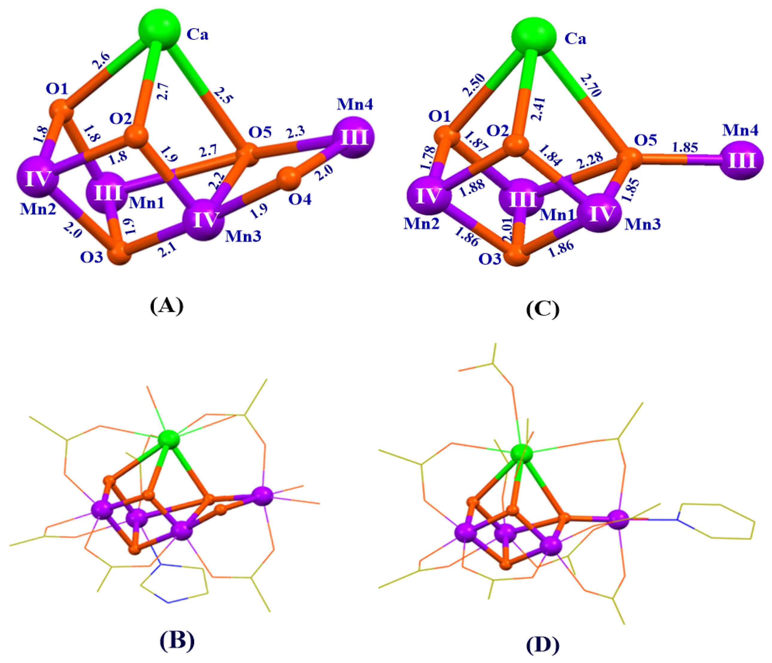
Figure 9.
Structures of the natural OEC [50] (A,B) and the artificial Mn4Ca-cluster [118] (C,D). Distances are given in Å units. Mn, Ca, O, N and C are shown in purple, green, orange, blue, and yellow, respectively. The oxidation states of the four manganese ions in A are directly taken from the previous suggestion [50], which are different from the BVS calculations listed in Table 1. For clarity, all the methyl groups and hydrogen atoms are not shown.

Figure 10.
Cyclic voltammogram (CV) of the artificial Mn4CaO4-complex [118]. Peak positions of CV waves are in red and estimated midpoint potentials are in blue. The likely oxidation states of the four Mn ions in various oxidation states of artificial complex are indicated using black numbers.
Figure 10.
Cyclic voltammogram (CV) of the artificial Mn4CaO4-complex [118]. Peak positions of CV waves are in red and estimated midpoint potentials are in blue. The likely oxidation states of the four Mn ions in various oxidation states of artificial complex are indicated using black numbers.

Figure 11.
Electron paramagnetic resonance (EPR) spectra for the artificial Mn4CaO4-cluster [118] (top) and natural Mn4CaO5-cluster [16] (below) in the S2 state.

Figure 12.
Catalytic properties of the artificial Mn4CaO4-cluster [118]. Cyclic voltammogram (CV) measurement of the artificial Mn4CaO4-complex in acetonitrile with different amounts of H2O. The inset shows the CV without H2O (0% H2O).
Figure 12.
Catalytic properties of the artificial Mn4CaO4-cluster [118]. Cyclic voltammogram (CV) measurement of the artificial Mn4CaO4-complex in acetonitrile with different amounts of H2O. The inset shows the CV without H2O (0% H2O).

Figure 13.
Crystal structures of two new artificial Mn4CaO4-complexes with an exchangeable solvent, acetonitrile (A, B) and DMF (C, D) on the calcium [58]. Mn, Ca, O, N, and C are shown in purple, green, orange, blue, and yellow, respectively. Distances are given in Å. The oxidation states of four manganese ions are obtained by the BVS calculation. For clarity, all the methyl groups in pivalic groups and hydrogen atoms are omitted.


Table 1.
Bond-valence sum (BVS) calculations on the structures of the OEC revealed at different resolutions in different S states. Roman numerals in parentheses indicate the assignment of the possible oxidation valences of four manganese ions in the OEC based on BVS calculations. All atomic coordinates were taken from the first monomer of PSII in the crystal structure data with the Protein Data Bank (PDB) codes: 4UB6 [50], 5B5E [54], 6DHF [53], 6JLK [52], 6DHO [53], and 6JLL [52], respectively.
| S1 (1.95 Å) (4UB6) | S1 (1.87 Å) (5B5E) | S2 (2.08 Å) (6DHF) | S2 (2.15 Å) (6JLK) | S3 (2.07 Å) (6DHO) | S3 (2.15 Å) (6JLL) | |
|---|---|---|---|---|---|---|
| Mn1 | 3.075 (III) | 3.244 (III) | 3.232 (III) | 3.204 (III) | 3.901 (IV) | 4.300 (IV) |
| Mn2 | 3.237 (III) | 3.057 (III) | 4.316 (IV) | 3.775 (IV) | 4.193 (IV) | 3.852 (IV) |
| Mn3 | 2.980 (III) | 2.951 (III) | 3.784 (IV) | 3.347 (III) | 3.232 (III) | 3.243 (III) |
| Mn4 | 2.318 (II) | 2.603 (III) | 3.139 (III) | 2.597 (III) | 2.932 (III) | 2.531 (III) |
© 2020 by the authors. Licensee MDPI, Basel, Switzerland. This article is an open access article distributed under the terms and conditions of the Creative Commons Attribution (CC BY) license (http://creativecommons.org/licenses/by/4.0/).
Share and Cite
MDPI and ACS Style
Li, Y.; Yao, R.; Chen, Y.; Xu, B.; Chen, C.; Zhang, C. Mimicking the Catalytic Center for the Water-Splitting Reaction in Photosystem II. Catalysts 2020, 10, 185. https://0-doi-org.brum.beds.ac.uk/10.3390/catal10020185
AMA Style
Li Y, Yao R, Chen Y, Xu B, Chen C, Zhang C. Mimicking the Catalytic Center for the Water-Splitting Reaction in Photosystem II. Catalysts. 2020; 10(2):185. https://0-doi-org.brum.beds.ac.uk/10.3390/catal10020185
Chicago/Turabian StyleLi, Yanxi, Ruoqing Yao, Yang Chen, Boran Xu, Changhui Chen, and Chunxi Zhang. 2020. "Mimicking the Catalytic Center for the Water-Splitting Reaction in Photosystem II" Catalysts 10, no. 2: 185. https://0-doi-org.brum.beds.ac.uk/10.3390/catal10020185
Note that from the first issue of 2016, this journal uses article numbers instead of page numbers. See further details here.