Communication

Dual-Modified Cu2S with MoS2 and Reduced Graphene Oxides as Efficient Photocatalysts for H2 Evolution Reaction

1
College of Health Science and Environmental Engineering, Shenzhen Technology University, Shenzhen 518118, China
2
Department of Applied Biology and Chemical Technology, State Key Laboratory of Chirosciences, The Hong Kong Polytechnic University, Kowloon 999077, China
3
College of New Materials and New Energies, Shenzhen Technology University, Shenzhen 518118, China
*
Author to whom correspondence should be addressed.
These authors contributed equally to this work.
Submission received: 27 September 2021 / Revised: 14 October 2021 / Accepted: 20 October 2021 / Published: 22 October 2021

Abstract

:
Noble metal-free cocatalysts have drawn great interest in accelerating the catalytic reactions of metal chalcogenide semiconductor photocatalyst. In particular, great efforts have been made on modifying a semiconductor with dual cocatalysts, which show synergistic effect of a fast transfer of exciton and energy simultaneously. Herein, we report the dual-modified Cu2S with MoS2 and reduced graphene oxides (Cu2S-MoS2/rGO). The in situ growth of Cu2S nanoparticles in the presence of MoS2/rGO resulted in high density of nanoscale interfacial contacts among Cu2S nanoparticles, MoS2, and rGO, which is beneficial for reducing the photogenerated electrons’ and holes’ recombination. The Cu2S-MoS2/rGO system also demonstrated stable photocatalytic activity for H2 evolution reaction for the long term.

Graphical Abstract

1. Introduction

Chalcocite copper(I) sulfide (Cu2S) is a typical p-type semiconductor with a bulk band gap of 1.2 eV [1], which has been extensively studied and used in many applications, such as cancer therapy [2,3], batteries [4], solar cell [5], catalyst [6], etc. Many efforts have been focused on the synthesis of Cu2S nanostructures with different morphologies, sphere [7,8,9], nanowires [10,11], nanorods [12,13], nanosheets [14], and nanoplates [15,16]. In addition to its low cost, high earth abundancy, and low-toxicity, Cu2S is nearly ideal to capture and convert sunlight into useful chemical fuel. The conduction band (CB) of Cu2S is more negative than the proton reduction potential, and the narrow band gap enables the absorption of solar energy in the visible regime [17]. However, the issue remains its poor photocatalytic activity due to a rapid exciton recombination. Although much effort has been made to synthesize the Cu2S of various nanostructures, a systematic study on the morphology–performance relationship and the suppression of photoelectron recombination remain the key challenges to improve their photocatalytic performances.
Formation of heterogeneous structures is a key approach for improving the activity of photocatalysts by suppressing the recombination probability of photoexcited electron-hole pairs [18,19]. One popular way is to decorate or incorporate photocatalysts with noble metal nanoparticles, such as platinum (Pt) or gold (Au), in order to shuttle the electrons from photocatalysts to the metal, as well as to provide more active sites for the redox reaction. However, the high cost and low availability of noble metals limit their industrial application. Molybdenum disulfide (MoS2), graphene, and their hybrids were reported to be promising two-dimensional nanomaterials that can replace Pt as a co-catalyst in the photocatalytic H2 evolution reaction (HER) [20,21]. For instance, the formation of 2H phase MoS2 nanosheets on novel Cu2S snowflakes greatly enhanced the photocatalytic degradation of methyl orange [22]. A CuS@defect-rich MoS2 core-shell structure exhibits good electrocatalytic hydrogen generation performance [23]. Our previous studies on the Cu2ZnSnS4 (CZTS)-MoS2/rGO heterostructure also confirmed its better photocatalytic performance in hydrogen generation compared to noble metal-decorated CZTS [24].
On the other hand, 1T phase MoS2, although not an intrinsically active semiconductor to capture light in photocatalytic reaction, showed good metallic properties and dense active sites compared to the traditional 2H phase [25]. By coupling 1T MoS2 and Cu2S through the rGO network, we expect pronounced enhancements in photocatalytic performance of Cu2S, which has previously been inferior to many other systems. Herein, we synthesized the Cu2S-MoS2/rGO composites through a simple wet chemistry approach. We also systematically investigated the morphology effects on photocatalytic HER performance of Cu2S nanostructures. The spherical Cu2S nanoparticles anchored on MoS2/rGO showed the highest photocatalytic hydrogen production rate with excellent long-term photostability, which surpasses those of many noble metal-modified semiconductors reported in the literature.

2. Results and Discussion

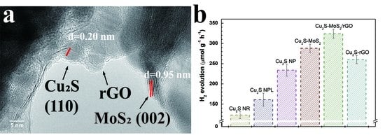
Figure 1a–e displays the typical transmission electron microscopic (TEM) images of as-synthesized Cu2S, graphene oxide, MoS2, MoS2/rGO hybrids and Cu2S-MoS2/rGO composites. Cu2S nanoparticles were grown on the MoS2/rGO hybrid nanosheet to form a heterojunction structure. The Cu2S nanoparticles are uniformly grown in a spherical shape with an average particle size of 8.7 ± 2.5 nm. The MoS2 displays a multilayered structure with good contact with graphene oxide. High-resolution TEM images of Cu2S-MoS2/rGO heterostructure, shown in Figure 1f, clearly reveal the lattice fringes of a hexagonal Cu2S (110) plane with d-spacing of 0.20 nm, which indicates their high degree of crystallinity and nanoscale interfacial contact. A 1T-MoS2 (002) plane with d-spacing of 0.95 nm was also observed. In the preparation progress, ammonium diethyldithiocarbamate and dodecanthiol bind to the defective sites and exposed metal cations of MoS2-rGO through polar covalent bond, where the Cu2S can be in situ nucleated at a high temperature. Elemental analysis taken by EDS (Figure S1) shows a close stoichiometric number of Cu:S = 2:1 and Mo:S = 1:2.
The X-ray powder diffraction (XRD) patterns of the as-prepared Cu2S-MoS2/rGO composites, Cu2S nanoparticles alone, and MoS2/rGO hybrid are compared in Figure 2. For Cu2S spherical nanoparticles, the characteristic peaks were observed at 37.4°, 45.8°, 48.5°, and 53.7°, which were well matched with the hexagonal chalcocite Cu2S (JCPDS No. 026-1116). Peak broadening was observed since the crystallites are nano-sized. The as-prepared MoS2/rGO showed the characteristic (002) peak of 1T MoS2 at around 9° with d-spacing of 0.95 nm. For MoS2/rGO, a second-order diffraction peak appeared at 18°, which could be assigned to a new (004) crystal plane. The corresponding d spacing difference between the two (002) peaks was around 0.33 nm, which is consistent with the hydrogen-bonding diameter (≈0.35 nm) of the ammonium ions in metal disulfides [18]. Cu2S-MoS2/rGO composites showed the pure phase of hexagonal Cu2S (JCPDS No. 026-1116) without any side products. The peaks for 1T MoS2 were not obvious in the composites, possibly due to the low loading amount of 1T MoS2 (8.7 wt%) in the composites. This phenomenon has also been reported in other systems (ZnS-MoS2/rGO and CZTS-MoS2/rGO) [24,26].
X-ray photoelectron spectroscopy (XPS) analyses were performed to investigate the chemical states and surface status of Cu2S-MoS2/rGO composites. The wide scan XPS spectrum, shown in Figure 3a, confirmed the coexistence of Cu, S, Mo, and C elements in the Cu2S-MoS2/rGO heterostructure. In the high-resolution XPS spectrum, Cu2S-MoS2/rGO showed a major C 1s signal from C-C at 284.9 eV and a tiny shoulder for C-O peak at 286.1 eV (Figure 3b,c), which identified the reduction of the O=C–O and C O bonds in graphene oxide and the formation of a good electron transfer network during the hydrothermal synthesis of MoS2/rGO hybrid followed by further annealing for the growth of Cu2S [25,27,28]. Two peaks observed at 932.2 and 952 eV in the high-resolution XPS spectra were determined to be 2p3/2 and 2p1/2 states of Cu(I). The sulfur 2p3/2 and 2p1/2 peaks were identified at 161.8 and 162.6 eV, which agreed with the sulfides phase in the range of 160 and 164 eV. The tiny peaks at 232 and 229 eV were attributed to the Mo 3d3/2 and 3d5/2 orbitals, respectively, and were due to the small loading amount in the composite. The UV-Vis absorption spectra of Cu2S nanoparticles, MoS2/rGO hybrid, and Cu2S-MoS2/rGO composites are shown in Figure S2a. Compared with the spectrum of Cu2S alone, the composite showed a much broader peak at the range of 300 to 400 nm, which originated from the strong absorption and interaction of MoS2-rGO in this regime [24].
Note that Cu2S could be synthesized into several morphologies, but the simplest form of nanoparticles should work the best due to its copious number of active sites. To verify this, we first compared the photocatalytic hydrogen production rates of three types of Cu2S nanocrystals, including nanoparticles, nanorods, and nanoplates. The preparation and characterization details of Cu2S nanorods and nanoplates can be found in Figures S3 and S4, including TEM and XRD data. The photocatalytic hydrogen production rates of three types of Cu2S nanocrystals are summarized in Figure 4a. The spherical Cu2S nanoparticles showed the highest H2 production rate of 234 μmol g−1 h−1, compared with nanorods (123 μmol g−1 h−1) and nanoplates (161 μmol g−1 h−1). The small size (average 8.7 nm in diameter) of the nanoparticles resulted in a large surface area, which may provide more potential active sites for hydrogen evolution. The estimated band gap of 1.6 eV was significantly larger than bulk Cu2S due to the quantum confinement effect, which may also decrease the chance of electron hole recombination, as shown in Figure S2b.
Cu2S nanoparticles of the highest activity were selected for combining with co-catalyst MoS2 and mediator rGO. The addition of 10 wt% rGO to Cu2S nanoparticles showed a higher hydrogen generation rate of 260 μmol g−1 h−1. An even higher photocatalytic activity can be achieved by loading 10 wt% MoS2, resulting in a rate of 288 μmol g−1 h−1. Compared to using rGO or MoS2 as a single component, further enhancement was observed from the Cu2S nanoparticles incorporated with MoS2-rGO hybrid. The highest H2 production rate (324 μmol g−1 h−1) was obtained by the composites with the weight ratio of Cu2S:MoS2:rGO = 90:8.7:1.3. As a control experiment, Cu2S nanoparticles and MoS2-rGO hybrid were physically mixed with the ratio of Cu2S:MoS2:rGO = 90:8.7:1.3 in ethanol, sonicated for 2 h, and stirred overnight. The as-prepared physical mixture exhibited even lower photocatalytic activity than Cu2S alone, as shown in Figure S5. Since the bare MoS2/rGO hybrid showed a lower photocatalytic activity than Cu2S, a simple physical mixing without forming interfacial contacts between Cu2S and MoS2/rGO hybrid did not enhance the photocatalytic activity. In summary, Cu2S nanoparticles-decorated MoS2-rGO hybrids exhibited the highest hydrogen generation rate of 324 µmol g−1 h−1. The stability of Cu2S-MoS2/rGO composite was investigated by a continuous 9-h reaction (Figure 4b). The produced amount of H2 remained steady over the entire reaction time, indicating its excellent photocatalytic stability. The photocatalytic hydrogen production of Cu2S-MoS2/rGO composites was compared to those of previously reported relative photocatalysts, and are summarized in Table 1. The Cu2S-MoS2/rGO composites showed better hydrogen evolution performance than the Cu2S/Pt and TiO2/MoS2/rGO system, even with lower light irradiation power.
A proposed mechanism for enhanced photocatalytic hydrogen evolution is illustrated in Scheme 1. Under the light irradiation, the electrons from the VB of Cu2S were excited to the CB, leaving positively charged holes. The narrow bandgap enabled Cu2S to efficiently absorb visible light, but it also make the excited electrons easily fall back to VB, resulting in electron-hole recombination, which lowered the photocatalytic activity. By the introduction of MoS2 and rGO, the band structure of Cu2S and MoS2, as well as the Fermi level of rGO, allowed the photoexcited electrons to get transferred either directly to the CB of the nearby MoS2 or through rGO backbone to a remote MoS2. The presence of rGO nanosheet with distinguished electron mobility served as an efficient electron collector [31]. Meanwhile, MoS2 provided more active sites on the nanocrystal edges and fast electrochemical kinetics for hydrogen evolution reaction [32,33]. The dual-composite cocatalyst synergistically enhanced the photocatalytic hydrogen production of Cu2S-MoS2/rGO heterostructure. The photoexcited electrons on both Cu2S and MoS2 sulfur edges reacted with the adsorbed protons to produce H2. Meanwhile, the holes were consumed by sacrificial agents, S2− and SO32−. Such electron transfer pathway suppressed the recombination of the electron-hole pairs, prolonged the lifetime of the excitons, provided more active sites for proton adsorption and hydrogen evolution, and consequently improved the photocatalytic hydrogen evolution rate.

3. Experimental

3.1. Synthesis of Graphene Oxide (GO)

GO was prepared by an improved Hummer’s method [34], which used strong oxidizing agents to chemically exfoliate the graphite sheet. Typically, 1 g graphite flakes were added to 150 mL (9:1 v/v) mixture of H2SO4/H3PO4, followed by slowly adding 6 g KMnO4. After 36 h at room temperature, the mixture was poured into 200 mL ice with 3 mL H2O2 (30%). Then the mixture was centrifuged and washed in succession with DI water until the pH of the supernatant was between 4~5. The solid was further purified by dialysis using Wizard® SVmini columns (A129B) for 1 week. After the purification steps, the mixture was then under ultrasonication for 1 h, centrifugation, and air drying for the supernatant. This procedure was repeated for the remaining precipitates.

3.2. Synthesis of 1T-MoS2/rGO Hybrid

The 1T-MoS2 and 1T-MoS2/rGO hybrid were prepared through a hydrothermal method [25]. Sodium molybdate dehydrates (Na2MoO4·2H2O) and thiourea (CS(NH2)2) were chosen as Mo precursor and S precursor, respectively. For the synthesis of 1T-MoS2/rGO hybrid, 0.242 g Na2MoO4·2H2O, 2.28 g CS(NH2)2, and a certain amount of GO was dispersed into 30 mL DI water by the assistance of ultrasonication for 20 min. The pH value was adjusted to 6.5 by NaOH solution. Then the mixture was transferred to a 45-mL Teflon-lined stainless steel autoclave, maintained at 200 °C for 24 h, and allowed naturally to cool down to room temperature. The product was washed with ethanol several times and vacuum dried at 60 °C.

3.3. Synthesis of Cu2S Nanostructures and Cu2S-MoS2/rGO Composites

The nanoparticles, nanorods, and nanoplates of Cu2S were synthesized through a hot injection method, reported previously by Yue Wu et al. [16], Marta Kruszynska et al. [12], and Yan Wang et al. [16], respectively. In a typical synthesis of Cu2S nanoparticles, 281.6 mg ammonium diethyldithiocarbamate and 10 mL 1-dodecanethiol (1-DDT) were mixed with 17 mL oleic acid. After the purging process, the reaction solution was heated up to 110 °C under N2 protection, and a suspension of 261.8 mg copper (II) acetylacetonate [Cu(acac)2] and 3 mL oleic acid was injected into the system. Then the temperature was raised to 180 °C and kept for 10 min. The resulting product was cooled to room temperature, washed with chloroform and ethanol at least three times, and dried in a vacuum oven. The following Cu2S nanostructures were also purified in this way.
In the preparation of Cu2S nanoplates, 181.6 mg copper (II) acetate and 2.5 g trioctylphosphine oxide (TOPO) were dissolved in 30 mL 1-octadecene (ODE) in a three-neck flask. The mixture was stirred under vacuum for 1 h followed by bubbling with nitrogen gas (N2) to remove any low-boiling-point solvents. Thereafter, the reaction solution was heated up to 160 °C under N2 flow followed by a quick injection of 2.5 mL 1-DDT. Then the temperature was raised to 200 °C and kept for 1.5 h. The resulting product was cooled to room temperature, washed with chloroform and ethanol at least three times, and dried in a vacuum oven. After the reaction, the resulting product was separated, cleaned, and dried.
To obtain Cu2S nanorods, the replacement of 1-DDT by tert-dodecanethiol (t-DDT) was required. Then, 181.6 mg copper (II) acetate and 1.9 g TOPO were dissolved in 10 mL ODE, and the mixture was purged in the same manner. Under N2 flow, 3.75 mL of t-DDT was injected into the system when the reaction solution was heated to 120 °C. Then, the temperature was raised to 180 °C. The reaction was allowed to proceed for 3–5 min, and the product was then cooled, purified, and dried.
For the synthesis of Cu2S-MoS2/rGO composites, a certain amount of MoS2/rGO was dispersed into sulfur precursor solution by 30-min sonication and reacted with a metal precursor.

3.4. Characterization and Photocatalytic Test

The crystalline structure of the nanostructures was characterized by powder X-ray diffraction (PXRD), which was performed on a SmartLab® X-ray diffractometer (Rigaku, Tokyo, Japan) at room temperature with Cu Kα radiation (λ = 1.5418 Å) and a diffraction angle 2θ ranging from 5° to 90°. The structural and morphological information was obtained by using a JEM-2100F (JEOL Ltd., Tokyo, Japan) scanning transmission electron microscope (STEM) and selected area electron diffraction (SAED) operated at 200-kV accelerating voltage. The analysis of elemental compositions was conducted on an energy dispersive X-ray spectrometer (EDS) attached to the STEM. For TEM and EDS measurements, samples were dispersed in chloroform by sonication and drop-dried on holey carbon-coated 400-mesh Ni grids. UV-Visible absorption spectra were recorded on an Agilent 8453 Diode-Array UV-Visible spectrophotometer (Santa Clara, CA, USA) within the wavelength range of 200~1000 nm. Before the measurement, samples were dispersed in chloroform in a quartz cuvette of 1-cm path length. The XPS results were obtained by the instrument of Thermo Fisher Scientific K-Alpha (Waltham, MA, USA).
For the measurement of photocatalytic hydrogen evolution, light irradiation was performed at ambient temperature and atmospheric pressure with a Newport solar simulator (Xenon lamp, ozone free, 150 W) (Irvine, CA, USA) of light intensity of 100 mW cm−2 for 3 h. In a typical photocatalytic experiment, the catalyst of ca. 5 mg was dispersed by 10-min sonication in 25 mL aqueous sulfide/sulfite solution (0.35M Na2S/0.25M Na2SO3). Prior to irradiation, Argon was bubbled through the reactor for 30 min to achieve anaerobic conditions. The solution was stirred throughout the irradiation period in order to keep the photocatalyst in suspension status. After irradiation, 250 μL gas was injected and analyzed by Agilent 7890B gas chromatograph system (Palo Alto, CA, USA) (TCD, nitrogen as carrier gas).

4. Conclusions

In this work, a novel Cu2S-based composite photocatalyst containing MoS2/rGO hybrid as a cocatalyst was synthesized and characterized. The study of the morphology effect of Cu2S nanocrystals showed that the Cu2S nanoparticles with chalcocite structure had a higher photocatalytic hydrogen production rate (234 μmol g−1 h−1) compared with Cu2S nanoplates (chalcocite, 161 μmol g−1 h−1) and Cu2S nano-rods (djurleite, 123 μmol g−1 h−1). The Cu2S-MoS2/rGO composites with a ratio of Cu2S:MoS2:rGO = 90:8.7:1.3 showed the highest photocatalytic hydrogen production rate of 324 μmol g−1 h−1. It is believed that the synergistic effect between the photocatalyst Cu2S and cocatalyst/mediator MoS2/rGO hybrid can effectively isolate photoexcited electrons and reduce the recombination of the electron-hole pairs, thus enhancing the photocatalytic hydrogen evolution performance.

Supplementary Materials

The following are available online at https://0-www-mdpi-com.brum.beds.ac.uk/article/10.3390/catal11111278/s1. Figure S1: EDS spectrum of Cu2S-MoS2/rGO composites. Figure S2: (a) UV-Vis spectra of Cu2S-MoS2/rGO, MoS2/rGO, and Cu2S; (b) the band gap calculation of Cu2S by using Tauc plot. Figure S3: Typical TEM images of (a) Cu2S nanoparticles, (c) nanorods, (e) nanoplates and their corresponding size distribution histograms (b, d, f). Figure S4: Powder XRD patterns of as-synthesized Cu2S nanoparticles, nanoplates, and nanorods. Figure S5: H2 generation from various Cu2S-MoS2/rGO composites with different ratios compared to their physical mixture.

Author Contributions

Conceptualization, E.H. and J.H.; methodology, Z.X.; formal analysis, Z.X.; data curation, D.L. and J.Z.; writing—original draft preparation, E.H. and Z.X.; writing—review and editing, T.J. and X.H.; supervision, L.W. and J.H.; funding acquisition, E.H. and J.H. All authors have read and agreed to the published version of the manuscript.

Funding

This research was funded by the Natural Science Foundation of Top Talent of SZTU (Grant No. 2019205), National Natural Science Foundation of China (Grant Nos. 21701135, 21902104, and 21561031), Shenzhen Science and Technology Research Project (Grant No. JCYJ20170818093553012), the Open Project Program of Key Laboratory for Analytical Science of Food Safety and Biology, Ministry of Education (Grant No. FS2004), Foundation for Young Innovative Talents in Higher Education of Guangdong (Grant No. 2018KQNCX401), and the Guangdong Basic and Applied Basic Research Foundation (Grant No. 2020A1515010258).

Acknowledgments

The authors would like to thank the helpful discussion and support from Lawrence Yoon Suk Lee and Kwok-Yin Wong at the Hong Kong Polytechnic University, Hong Kong SAR.

Conflicts of Interest

The authors declare no conflict of interest.

References

  1. Liu, M.; Liu, Y.; Gu, B.; Wei, X.; Xu, G.; Wang, X.; Swihart, M.T.; Yong, K.-T. Recent advances in copper sulphide-based nanoheterostructures. Chem. Soc. Rev. 2019, 48, 4950–4965. [Google Scholar] [CrossRef]
  2. Nikam, A.N.; Pandey, A.; Fernandes, G.; Kulkarni, S.; Mutalik, S.P.; Padya, B.S.; George, S.D.; Mutalik, S. Copper sulphide based heterogeneous nanoplatforms for multimodal therapy and imaging of cancer: Recent advances and toxicological perspectives. Coord. Chem. Rev. 2020, 419, 213356. [Google Scholar] [CrossRef]
  3. Cui, J.; Jiang, R.; Guo, C.; Bai, X.; Xu, S.; Wang, L. Fluorine Grafted Cu7S4–Au Heterodimers for Multimodal Imaging Guided Photothermal Therapy with High Penetration Depth. J. Am. Chem. Soc. 2018, 140, 5890–5894. [Google Scholar] [CrossRef]
  4. Peng, Q.K.; Zhang, S.P.; Yang, H.; Sheng, B.B.; Xu, R.; Wang, Q.S.; Yu, Y. Boosting Potassium Storage Performance of the Cu2S Anode via Morphology Engineering and Electrolyte Chemistry. ACS Nano 2020, 14, 6024–6033. [Google Scholar] [CrossRef] [PubMed]
  5. Chen, X.; Xu, W.; Ding, N.; Ji, Y.; Pan, G.; Zhu, J.; Zhou, D.; Wu, Y.; Chen, C.; Song, H. Dual Interfacial Modification Engineering with 2D MXene Quantum Dots and Copper Sulphide Nanocrystals Enabled High-Performance Perovskite Solar Cells. Adv. Funct. Mater. 2020, 30, 2003295. [Google Scholar] [CrossRef]
  6. Srinivas, B.; Kumar, B.G.; Muralidharan, K. Stabilizer free copper sulphide nanostructures for rapid photocatalytic decomposition of rhodamine B. J. Mol. Catal. A Chem. 2015, 410, 8–18. [Google Scholar] [CrossRef]
  7. Zhuang, Z.; Peng, Q.; Zhang, B.; Li, Y. Controllable Synthesis of Cu2S Nanocrystals and Their Assembly into a Superlattice. J. Am. Chem. Soc. 2008, 130, 10482–10483. [Google Scholar] [CrossRef]
  8. Xie, Y.; Riedinger, A.; Prato, M.; Casu, A.; Genovese, A.; Guardia, P.; Sottini, S.; Sangregorio, C.; Miszta, K.; Ghosh, S.; et al. Copper Sulfide Nanocrystals with Tunable Composition by Reduction of Covellite Nanocrystals with Cu+ Ions. J. Am. Chem. Soc. 2013, 135, 17630–17637. [Google Scholar] [CrossRef]
  9. Wu, Y.; Wadia, C.; Ma, W.; Sadtler, B.; Alivisatos, A.P. Synthesis and Photovoltaic Application of Copper(I) Sulfide Nanocrystals. Nano Lett. 2008, 8, 2551–2555. [Google Scholar] [CrossRef]
  10. Liu, Z.; Xu, D.; Liang, J.; Shen, J.; Zhang, S.; Qian, Y. Growth of Cu2S Ultrathin Nanowires in a Binary Surfactant Solvent. J. Phys. Chem. B 2005, 109, 10699–10704. [Google Scholar] [CrossRef]
  11. Law, M.; Greene, L.E.; Johnson, J.C.; Saykally, R.; Yang, P. Nanowire dye-sensitized solar cells. Nat. Mater. 2005, 4, 455–459. [Google Scholar] [CrossRef]
  12. Kruszynska, M.; Borchert, H.; Bachmatiuk, A.; Rümmeli, M.H.; Büchner, B.; Parisi, J.; Kolny-Olesiak, J. Size and Shape Control of Colloidal Copper(I) Sulfide Nanorods. ACS Nano 2012, 6, 5889–5896. [Google Scholar] [CrossRef] [PubMed]
  13. Sigman, M.B.; Ghezelbash, A.; Hanrath, T.; Saunders, A.E.; Lee, F.; Korgel, B.A. Solventless Synthesis of Monodisperse Cu2S Nanorods, Nanodisks, and Nanoplatelets. J. Am. Chem. Soc. 2003, 125, 16050–16057. [Google Scholar] [CrossRef] [PubMed]
  14. Hui, Z.; Xu, W.; Li, X.; Guo, P.; Zhang, Y.; Liu, J. Cu2S nanosheets for ultrashort pulse generation in the near-infrared region. Nanoscale 2019, 11, 6045–6051. [Google Scholar] [CrossRef] [PubMed]
  15. Li, X.; Shen, H.; Niu, J.; Li, S.; Zhang, Y.; Wang, H.; Li, L.S. Columnar Self-Assembly of Cu2S Hexagonal Nanoplates Induced by Tin(IV)−X Complex as Inorganic Surface Ligand. J. Am. Chem. Soc. 2010, 132, 12778–12779. [Google Scholar] [CrossRef] [PubMed]
  16. Wang, Y.; Hu, Y.; Zhang, Q.; Ge, J.; Lu, Z.; Hou, Y.; Yin, Y. One-Pot Synthesis and Optical Property of Copper(I) Sulfide Nanodisks. Inorg. Chem. 2010, 49, 6601–6608. [Google Scholar] [CrossRef] [PubMed]
  17. Liu, G.; Schulmeyer, T.; Brötz, J.; Klein, A.; Jaegermann, W. Interface properties and band alignment of Cu2S/CdS thin film solar cells. Thin Solid Films 2003, 431-432, 477–482. [Google Scholar] [CrossRef]
  18. Marschall, R. Semiconductor Composites: Strategies for Enhancing Charge Carrier Separation to Improve Photocatalytic Activity. Adv. Funct. Mater. 2014, 24, 2421–2440. [Google Scholar] [CrossRef]
  19. Zhu, J.; Hu, L.; Zhao, P.; Lee, L.Y.S.; Wong, K.-Y. Recent Advances in Electrocatalytic Hydrogen Evolution Using Nanoparticles. Chem. Rev. 2020, 120, 851–918. [Google Scholar] [CrossRef]
  20. Lin, H.; Zhang, K.; Yang, G.; Li, Y.; Liu, X.; Chang, K.; Xuan, Y.; Ye, J. Ultrafine nano 1T-MoS2 monolayers with NiOx as dual co-catalysts over TiO2 photoharvester for efficient photocatalytic hydrogen evolution. Appl. Catal. B Environ. 2020, 279, 119387. [Google Scholar] [CrossRef]
  21. Wang, Y.; Lu, F.; Su, K.; Zhang, N.; Zhang, Y.; Wang, M.; Wang, X. Engineering Mo-O-C interface in MoS2@rGO via charge transfer boosts hydrogen evolution. Chem. Eng. J. 2020, 399, 126018. [Google Scholar] [CrossRef]
  22. Zhang, X.; Guo, Y.; Tian, J.; Sun, B.; Liang, Z.; Xu, X.; Cui, H. Controllable growth of MoS2 nanosheets on novel Cu2S snowflakes with high photocatalytic activity. Appl. Catal. B Environ. 2018, 232, 355–364. [Google Scholar] [CrossRef]
  23. Liu, L.; Liu, X.; Jiao, S. CuS@defect-rich MoS2 core-shell structure for enhanced hydrogen evolution. J. Colloid Interface Sci. 2020, 564, 77–87. [Google Scholar] [CrossRef]
  24. Ha, E.; Liu, W.; Wang, L.; Man, H.-W.; Hu, L.; Tsang, S.C.E.; Chan, C.T.-L.; Kwok, W.-M.; Lee, L.Y.S.; Wong, K.-Y. Cu2ZnSnS4/MoS2-Reduced Graphene Oxide Heterostructure: Nanoscale Interfacial Contact and Enhanced Photocatalytic Hydrogen Generation. Sci. Rep. 2017, 7, 39411. [Google Scholar] [CrossRef] [Green Version]
  25. Liu, Q.; Li, X.; He, Q.; Khalil, A.; Liu, D.; Xiang, T.; Wu, X.; Song, L. Gram-Scale Aqueous Synthesis of Stable Few-Layered 1T-MoS2: Applications for Visible-Light-Driven Photocatalytic Hydrogen Evolution. Small 2015, 11, 5556–5564. [Google Scholar] [CrossRef]
  26. Zhu, B.; Lin, B.; Zhou, Y.; Sun, P.; Yao, Q.; Chen, Y.; Gao, B. Enhanced photocatalytic H2 evolution on ZnS loaded with graphene and MoS2 nanosheets as cocatalysts. J. Mater. Chem. A 2014, 2, 3819–3827. [Google Scholar] [CrossRef]
  27. Xiang, Q.; Yu, J.; Jaroniec, M. Synergetic Effect of MoS2 and Graphene as Cocatalysts for Enhanced Photocatalytic H2 Production Activity of TiO2 Nanoparticles. J. Am. Chem. Soc. 2012, 134, 6575–6578. [Google Scholar] [CrossRef] [PubMed]
  28. Wang, L.; Lian, J.; Cui, P.; Xu, Y.; Seo, S.; Lee, J.; Chan, Y.; Lee, H. Dual n-type doped reduced graphene oxide field effect transistors controlled by semiconductor nanocrystals. Chem. Commun. 2012, 48, 4052–4054. [Google Scholar] [CrossRef] [PubMed]
  29. Yu, X.; Shi, J.; Wang, L.; Wang, W.; Bian, J.; Feng, L.; Li, C. A novel Au NPs-loaded MoS2/RGO composite for efficient hydrogen evolution under visible light. Mater. Lett. 2016, 182, 125–128. [Google Scholar] [CrossRef]
  30. Wang, B.; An, W.; Liu, L.; Chen, W.; Liang, Y.; Cui, W. Novel Cu2S quantum dots coupled flower-like BiOBr for efficient photocatalytic hydrogen production under visible light. RSC Adv. 2015, 5, 3224–3231. [Google Scholar] [CrossRef]
  31. Geim, A.K.; Novoselov, K.S. The rise of graphene. Nat. Mater. 2007, 6, 183–191. [Google Scholar] [CrossRef] [PubMed]
  32. Jaramillo, T.F.; Jørgensen, K.P.; Bonde, J.; Nielsen, J.H.; Horch, S.; Chorkendorff, I. Identification of Active Edge Sites for Electrochemical H2 Evolution from MoS2 Nanocatalysts. Science 2007, 317, 100–102. [Google Scholar] [CrossRef] [PubMed] [Green Version]
  33. Wang, X.; Long, R. Rapid Charge Separation Boosts Solar Hydrogen Generation at the Graphene–MoS2 Junction: Time-Domain Ab Initio Analysis. J. Phys. Chem. Lett. 2021, 12, 2763–2769. [Google Scholar] [CrossRef] [PubMed]
  34. Marcano, D.C.; Kosynkin, D.V.; Berlin, J.M.; Sinitskii, A.; Sun, Z.; Slesarev, A.; Alemany, L.B.; Lu, W.; Tour, J.M. Improved Synthesis of Graphene Oxide. ACS Nano 2010, 4, 4806–4814. [Google Scholar] [CrossRef]
Figure 1. TEM images of (a) Cu2S, (b) graphene oxide, (c) MoS2, (d) MoS2/rGO, (e) Cu2S-MoS2/rGO, and (f) high-resolution TEM images of Cu2S-MoS2/rGO composites.
Catalysts 11 01278 g001
Figure 2. Powder XRD patterns of as-synthesized MoS2/rGO, Cu2S, and Cu2S-MoS2/rGO composites.
Catalysts 11 01278 g002
Figure 3. (a) Wide scan XPS spectrum and (b) high-resolution XPS spectrum in C 1s region of as-synthesized Cu2S-MoS2/rGO composite. (c) High-resolution XPS spectra for individual elements in Cu2S-MoS2/rGO composite, including C 1s, Cu 2p, Mo 3d, and S 2p.
Catalysts 11 01278 g003
Figure 4. (a) Comparison of photocatalytic H2 evolution from Cu2S (Cu2S nanorods: Cu2S NR; Cu2S nanoplates: Cu2S NPL; Cu2S nanoparticles: Cu2S NP), Cu2S-rGO, Cu2S-MoS2, and Cu2S-MoS2/rGO composites. Experimental conditions: 1-h irradiation by solar simulator (150 W Xe lamp, 100 mW cm−2); (b) photocatalytic H2 evolution by Cu2S-MoS2/rGO composites in a 9-h reaction. The composition of photocatalyst: 85 wt% Cu2S, 13 wt% MoS2, and 2 wt% rGO.
Catalysts 11 01278 g004
Scheme 1. Proposed mechanism of interfacial charge transfer and photocatalytic redox reaction in Cu2S-MoS2/rGO composites.
Catalysts 11 01278 sch001
Table 1. Comparative photocatalytic hydrogen evolution with reference photocatalysts.
PhotocatalystLight SourceSacrificial ReagentH2 Production Rate
(μmol g−1 h−1)
Reference
Cu2S-MoS2/rGO150 W Xenon0.35M Na2S/0.25M Na2SO3324This work
Au/MoS2/rGO300 W Xenon0.25M methanol aq.115.4[29]
Cu2S/Pt300 W Xenon0.1M Na2S/0.5M Na2SO3241.2[30]
TiO2/MoS2/rGO350 W Xenon25% (v/v) ethanol/water165.3[27]
Cu2ZnSnS4/MoS2/rGO150 W Xenon0.35M Na2S/0.25M Na2SO3104[24]
Publisher’s Note: MDPI stays neutral with regard to jurisdictional claims in published maps and institutional affiliations.