Article

Fagonia cretica-Mediated Synthesis of Manganese Oxide (MnO2) Nanomaterials Their Characterization and Evaluation of Their Bio-Catalytic and Enzyme Inhibition Potential for Maintaining Flavor and Texture in Apples

1
Department of Health and Biological Sciences, Abasyn University, Peshawar 25000, Pakistan
2
Institute of Biotechnology and Microbiology, Bacha Khan University, Charsadda 24460, Pakistan
3
Department of Microbiology, Abdul Wali Khan University, Mardan 23200, Pakistan
4
Institute of Molecular Biology and Biotechnology, Bahauddin Zakariya University, Multan 60800, Pakistan
5
Center for Biotechnology and Microbiology, University of Swat, Odigram, Mingora 19130, Pakistan
6
Department of Pharmacognosy (Medicinal Aromatic and Poisonous Plants Research Center), College of Pharmacy, King Saud University, Riyadh 11495, Saudi Arabia
7
Department of Biology, College of Science, Princess Nourah bint Abdulrahman University, Riyadh 11671, Saudi Arabia
8
Zoology Department, Faculty of Science, Cairo University, Giza 12613, Egypt
*
Author to whom correspondence should be addressed.
Submission received: 15 April 2022 / Revised: 5 May 2022 / Accepted: 11 May 2022 / Published: 19 May 2022
(This article belongs to the Special Issue Catalysis for Flavours and Fragrances)

Abstract

:
The apple is the most widely used fruit globally. Apples are more prone to fungal spoilage, which leads to browning and subsequent changes in their flavor and texture. Browning is also caused by the tyrosinase enzyme. By inhibiting tyrosinase initiation and fungal spoilage in fruits, the natural flavor and texture of fruits can be maintained. Biogenic NPs can act as antioxidants to inhibit tyrosinase and due to oxidative stress, it also catalyzes the deformation of fungal hyphae and spores. Nanotechnology is a research hotspot that has gained considerable interest due to its potential inferences in biosciences and food preservation technology. The present study aims to use biomass from the Fagonia cretica to create bio-inspired manganese oxide MnO2 NPs and to evaluate its bio-catalytic potential for antifungal anti-browning through the inhibition of tyrosinase and its antioxidant potential for preserving apple flavor and texture. The green synthesized nanoparticles were extensively analyzed using UV spectroscopy, XRD, SEM, EDX, and FTIR techniques. Moreover, the synthesized manganese oxide nanoparticles (MnO2 NPs) were evaluated for their bio-catalytic potential as anti-fungal and anti-spoiling agents. The values of antifungal activity among all the samples were 14.2 ± 86 mm, 8.9 ± 6.0 mm, 17.7 ± 1.26, and 20.7 ± 4.38 mm for Penicillium expansum, Monilinia fructigena, Penicillium chrysogenum, and Aspergillus oryzae at 200 µg/well, respectively. Moreover, the biogenic NPs were evaluated for their anti-browning potential through the inhibition of tyrosinase. MnO2 NPs have been shown to have considerable inhibitory effects on tyrosinase up to 64.8 ± 0.16 at 200 µg/mL and (27.2 ± 0.58) at 25 µg/mL. Biogenic MnO2 NPs can also act as antioxidants to inhibit tyrosinase and fungal growth by the formation of free radicals that damage the fungal hyphae and, as a result, slow down browning. The maximum DPPH free radical scavenging activity was 74.5 ± 0.39% at 200 µg/mL, and the minimum was 12.4 ± 0.27 at 25 µg/mL. The biogenic MnO2 NPs are biocompatible and play a potent role in maintaining the flavor and texture of apples.

1. Introduction

Fruits are the outcome of fertilized ovaries, and, as such, they have long been a valuable source of sustenance for humans. Despite the multiple benefits they bring, fruits survival and longevity are threatened by a range of factors, the most serious of which are fungal attacks [1]. Fungi may induce fruit spoilage, which refers to a number of changes that make the fruits less pleasant or desirable in terms of flavor and scent, as well as appearance and texture. As a result of the discovery of a huge number of toxigenic fungi in damaged fruits, rotting microorganisms are often viewed as poisonous or pathogenic [1,2]. Even when food is refrigerated, microbes, such as molds and other fungi, may produce mycotoxins in different forms, posing a health concern to consumers. Fruits with preservatives have a longer shelf life because they prevent them from rotting. Antimicrobials, such as nitrogen oxides, nitrogen trioxides (NOx), benzoates, and sulfur dioxide, destroy or inhibit bacteria, yeast, and mold development in fruits [2]. However, since certain synthetic food and cosmetic ingredients have been shown to be carcinogenic and very dangerous, it is best to avoid using them. Synthetic chemical additives and preservatives should be avoided due to a lack of sufficient testing [1].
The plant-based synthesis of NPs has revolutionized drug discovery. Fagonia cretica contains many important bioactive elements that play a promising role in the synthesis of NPs. The chemical constituents are saponins, sapogenins, alkaloids, terpenoids, sterols, flavonoids, proteins, amino acids, coumarins, vitamins, and trace elements [3]. Biosynthesized nanoparticles, on the other hand, are safer and more effective in slowing the degradation of fruits and preserving their texture and flavor. Biogenic MnO2 NPs have the ability to act as a biocatalyst while simultaneously being poisonous to cells and damaging membranes [4]. According to one study, membrane rupture and free radical production are two of the most prevalent mechanisms of NP-induced cellular damage [5]. The interaction of nanoparticles with fungal hyphae and spores has been proven to inhibit fungal growth.
Fungi may also cause fruit to become brown, altering the flavor and texture of the fruit as a consequence of the browning. Browning is the gradual change in the color of food from white to brown or dark brown over time, which may have a positive or negative influence on the nutritional content of the meal. Browning may be classed as enzymatic or non-enzymatic browning in food, depending on the method involved. During the harvesting, transportation, storage, and processing phases of FV goods, enzymatic browning is the most common. Enzymatic browning produces a significant quantity of waste. Tyrosinase is also responsible for enzymatic browning in apples, which affects the fruits’ flavor and texture [6]. The oxidative process is supposed to diminish the flavor and nutritional value of fruits and vegetables due to the browning caused by tyrosinase. As a consequence, a variety of tyrosinase inhibitors have been produced, including various glycyl dipeptides, L-ascorbic acid, ellagic acid, and others [7]. These commercially available chemical inhibitors, on the other hand, have a variety of deleterious effects on apple quality. As a consequence, novel biocompatible tyrosinase inhibitors, capable of lowering tyrosinase activity in apples while maintaining their flavor and texture, have been developed.
Metal oxide nanoparticles, on the other hand, could not be used by fruits owing to the use of harmful substances that act as reducing and capping agents in the chemical synthesis of these nanoparticles [8]. These novel materials are nontoxic and safer thanks to the use of plant metabolites that act as reducing and capping agents in the biogenesis of metal oxide nanoparticles. There have been many reports of tyrosinase enzyme inhibitory nanoparticles [5,6]. Nanoparticles have greater anti-tyrosinase action than equivalent plant metabolites, according to research [5]. Biogenic NPs may also act as antioxidants by inhibiting the initiation of tyrosinase, which prevents browning in fruits [8]. The capacity of biogenic nanoparticles to inhibit tyrosinase, as well as their bio-catalytic activity and mechanisms of action, is seldom recorded [9].
In the present study, we attempted to synthesize MnO2 using Fagonia cretica as a reducing agent. The synthesized MnO2 were characterized by UV spectroscopy, XRD, SEM, EDX, and FTIR techniques to visualize their morphology, involvement of bioactive compounds, stability, crystallinity of the particles, etc. Furthermore, the tyrosinase inhibitory and antioxidant potential of MnO2 nanoparticles for slowing down browning and its bio-catalytic potential as an antifungal agent against spoilage causing fungi was also evaluated.

2. Results

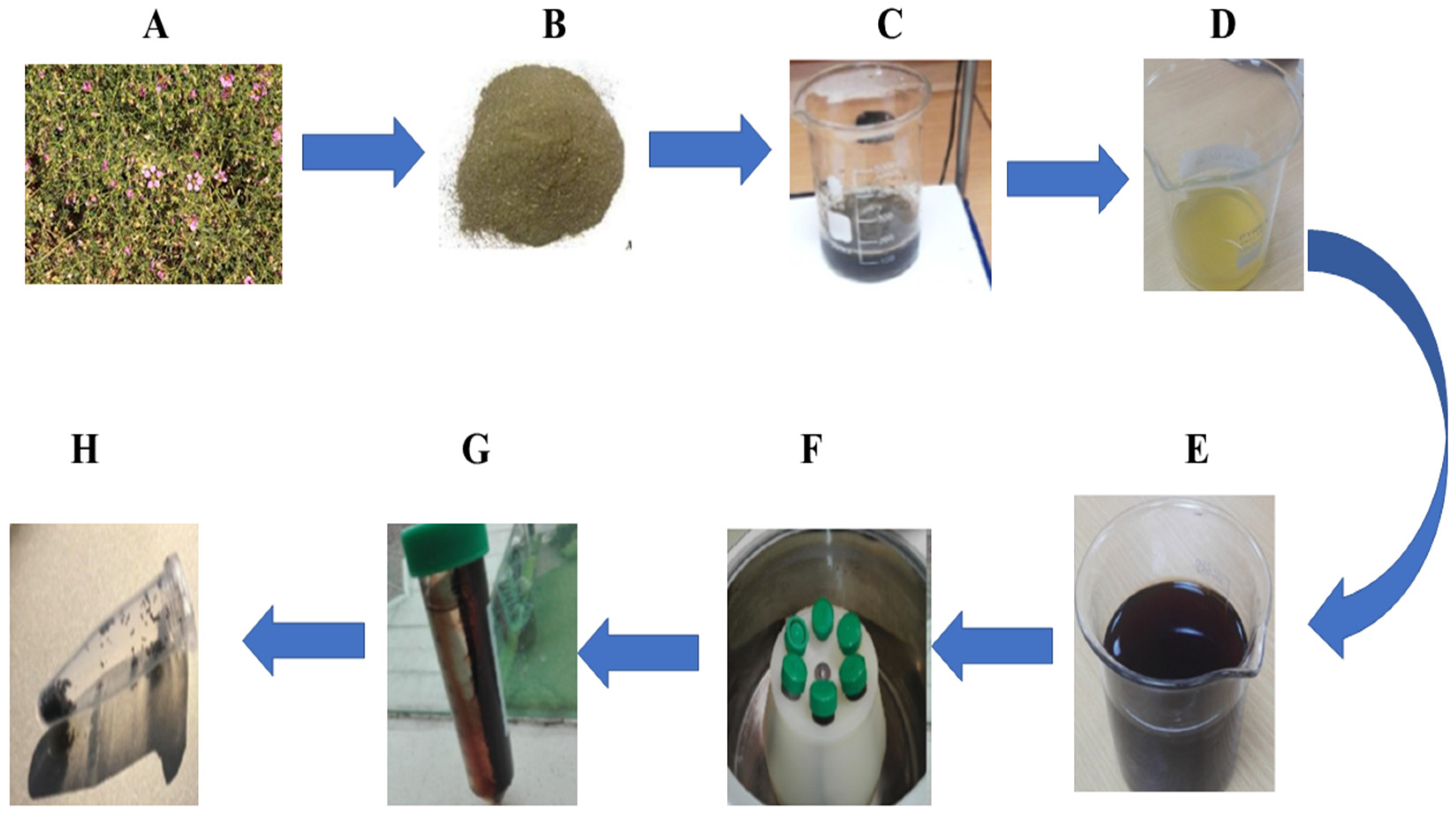
2.1. Synthesis Mechanism

The leaf extract of Fagonia cretica was used as a reducing and capping agent in the synthesis of MnO2 NPs. The synthesis of MnO2 NPs was visually monitored by detecting the color change generated by the addition of a precursor to the leaf extraction. The color of the reaction mixture changed from yellowish green to brownish, indicating the production of manganese dioxide NPs. The color change was caused by the surface plasmon resonance activity of the nanoparticle. Several studies have shown that the Fagonia cretica leaf’s extract is a rich source of biogenic phytomolecules such as alkaloids, flavonoids, tannins, phenolic compounds, saponins, and triterpenoids. These phytomolecules may work during the biosynthesis process by converting manganese ions to zero-valent species through a reduction and oxidation mechanism, resulting in keto form products. Other secondary metabolites (surfactants, proteins, alkaloids, and so on) found in the Fagonia cretica leaf extract also stabilized and capped the zero-valent Mn0 species. The zero-valent species of Mn0 would quickly oxidize and transform into MnO2 nanoparticles capped with phytomolecules of plant leaf extract during air drying and calcination at 200 °C. (See Figure 1).

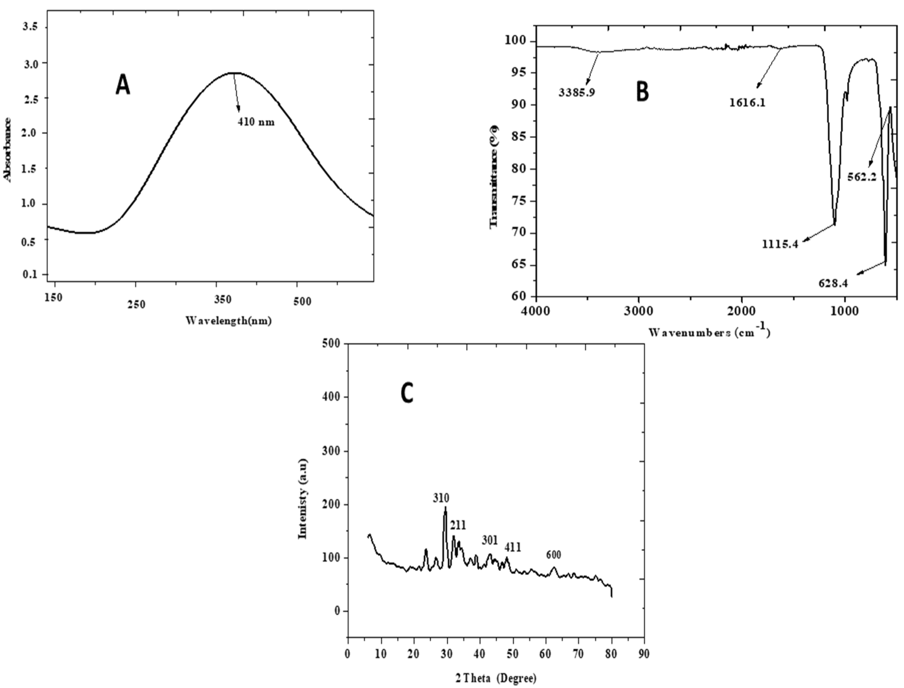
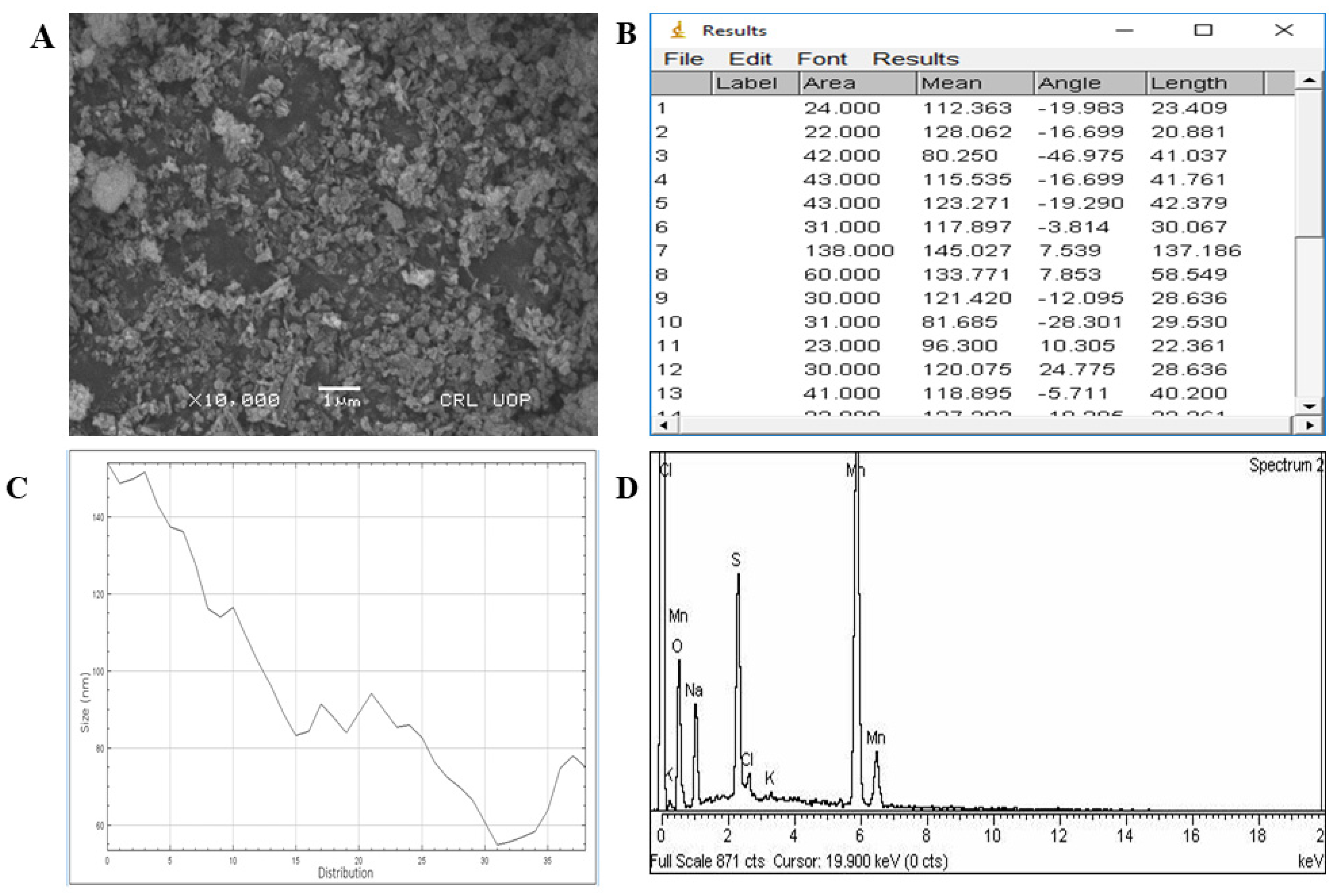
2.2. Characterization

The dark brownish hue is a marker of nanoparticle production, according to UV–visible spectroscopy in the 300 nm to 800 nm region. An examination of zinc nanoparticle absorption at 2.25 au shows a distinct peak at 410 nm, indicating that they were manufactured. This is seen in Figure 2a. In metallic nanoparticles, there is only one SPR peak, which is detected in MnO2 NPs. Infrared spectroscopy confirms the creation of the produced nanoparticles. Figure 2b depicts the essential infrared stretching vibrations. The absorption band at 562.2 cm−1 was ascribed to the normal stretching collision of O–Mn–O, indicating that MnO2 NPs were present in the produced sample. Aromatic unsaturation (C=C) of stabilized Fagonia cretica is represented by absorption bands at 1616.1 and 1115.4 cm−1, respectively, whereas C–O stretching in the Fagonia cretica molecule is represented by an absorption band at 628.4 cm−1. The wide band at 3385.9 cm−1 is the typical absorption for O–H–O of water present in the solution, which may play a role in manganese nanoparticle production and stability in the aqueous medium. The crystallinity of MnO2 NPs produced using the Fagonia cretica leaf extract was investigated using XRD analysis. The XRD pattern of MnO2 NPs is shown in Figure 2c. The XRD pattern shows five distinct peaks at 2θ 28.78°, 37.66°, 42.14°, 49.90°, and 56.44°, corresponding to the crystal planes of MnO2 NPs (310), (211), (301), (411), and (600) (JSPDF 44-0141). Furthermore, the strength of the peaks in the XRD pattern suggests that the MnO2 NPs are very crystalline. The EDX analysis was then used to identify the chemical composition of the MnO2 NPs. The EDX pattern is shown in Figure 3D. Four distinct peaks in the EDX spectra correspond to sodium, potassium, sulphur, chlorine, and manganese. In EDX spectra, a tiny peak of Mn can also be seen. The SEM pictures of MnO2 NPs are shown in Figure 3A. The produced MnO2 NPs are spherical with homogenous dispersity, as seen in the SEM picture. The particle size of the MnO2 NPs produced was determined to be 15.5 ± 0.85 nm.

2.3. Anti-Browning Activity of NPs

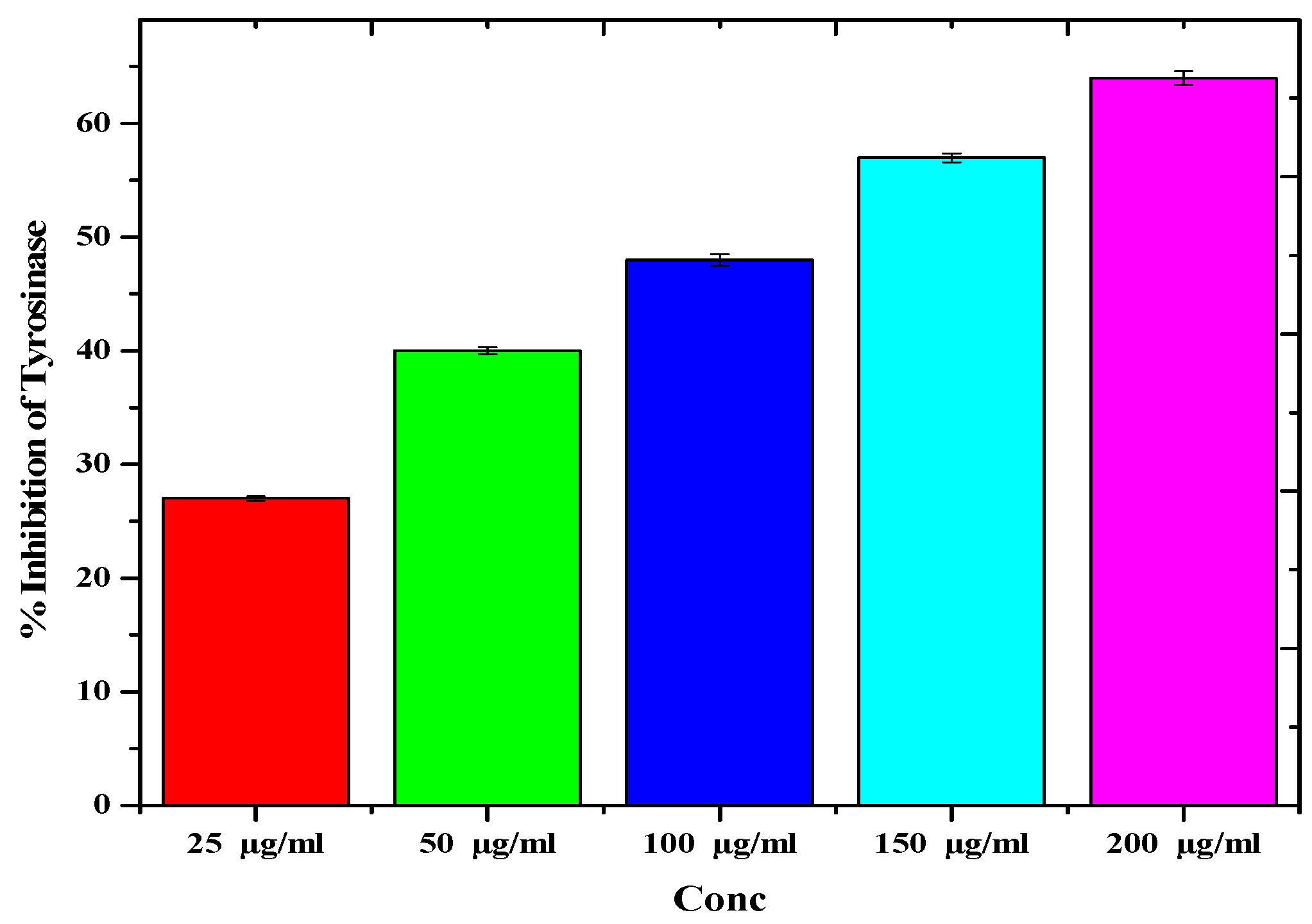
2.3.1. Tyrosinase Assay

The actual mechanism that causes browning in apples involves an enzyme called tyrosinase. The inhibitory ability of NPs against the production of tyrosinase was determined. MnO2 NPs block the active site and cause enzyme inhibition; thus they bear anti-browning characteristics and may be employed as anti-browning agents. MnO2 NPs were used as anti-tyrosinase agents in our work. MnO2 NPs have been shown to have considerable inhibitory effects on tyrosinase up to 64.8 ± 0.16 at 200 µg/mL and (27.2 ± 0.58) at 25 µg/mL, as shown in Figure 4.

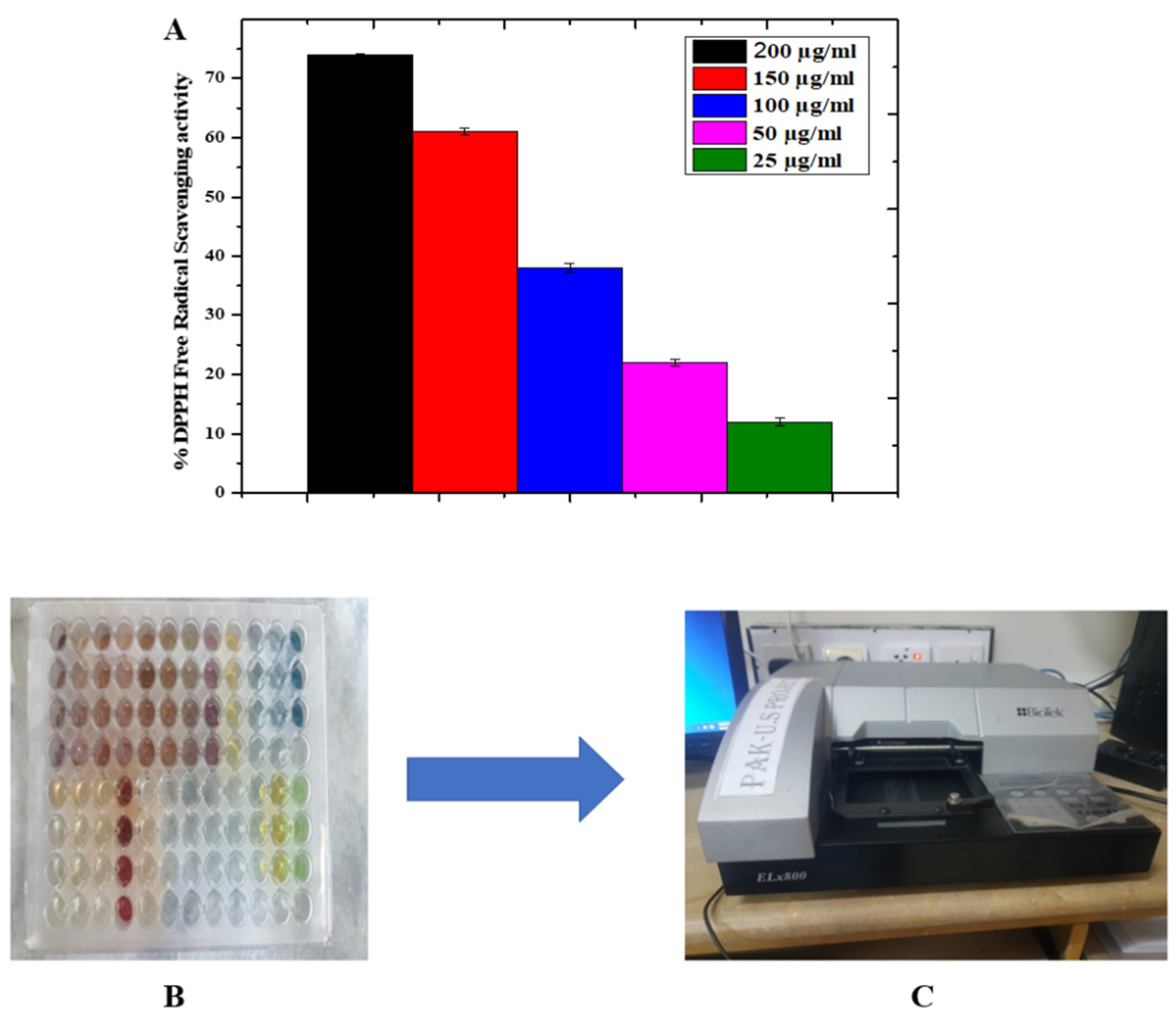
2.3.2. Antioxidant Assay

Antioxidants can react with oxygen to suppress the initiation of browning. They are also able to react with the intermediate products, thereby breaking the chain reaction and inhibiting tyrosinase formation. 2,2-diphenyl-1-picrylhydrazyl (DPPH) free radicals were exposed to test samples of varying concentrations in order to evaluate the antioxidant potential of MnO2 NPs, as shown in Figure 5. The maximum DPPH free radical scavenging activity was 74.5 ± 0.39% at (200 µg/mL), and the minimum was 12.4 ± 0.27 at (25 µg/mL).

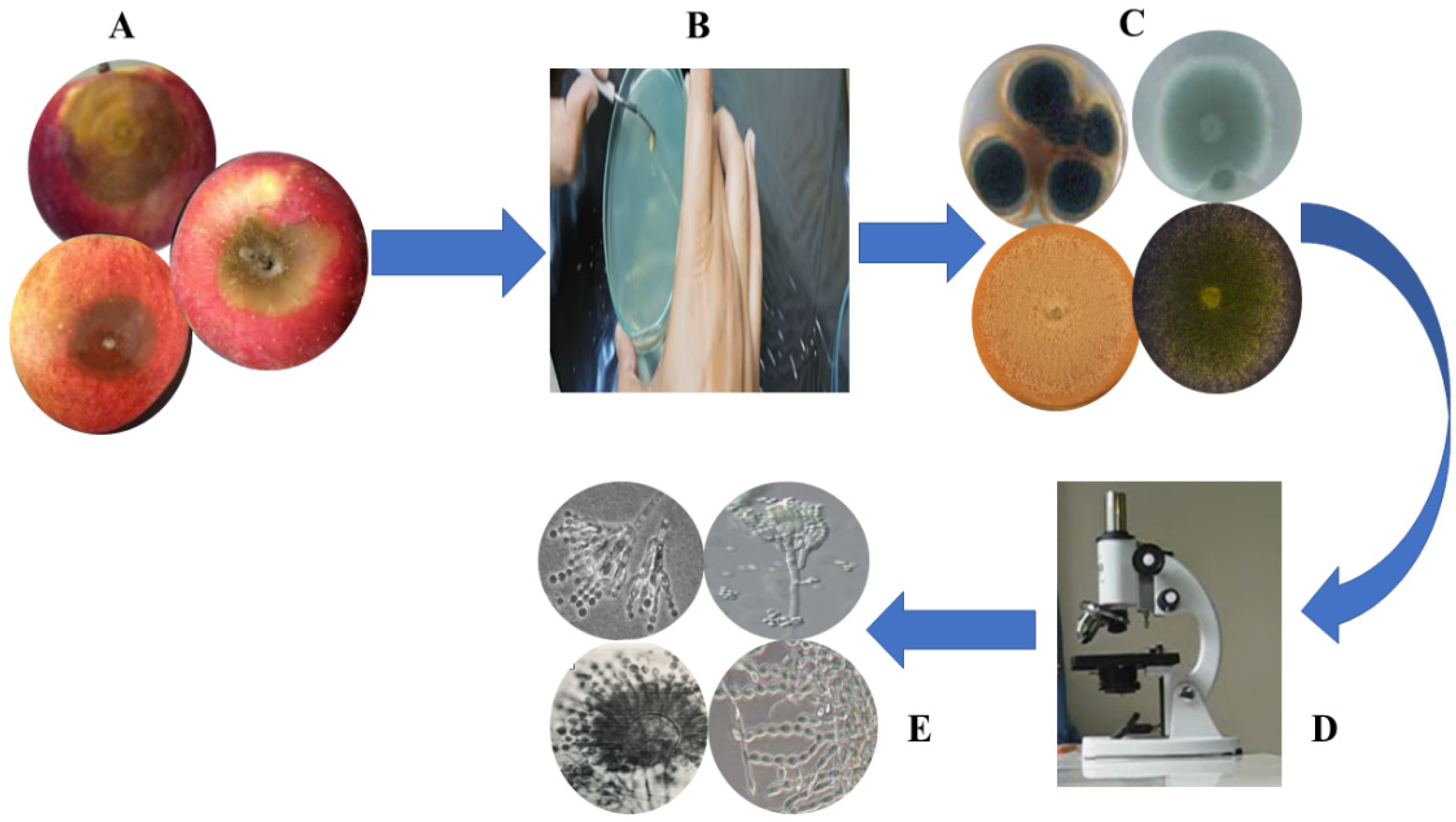
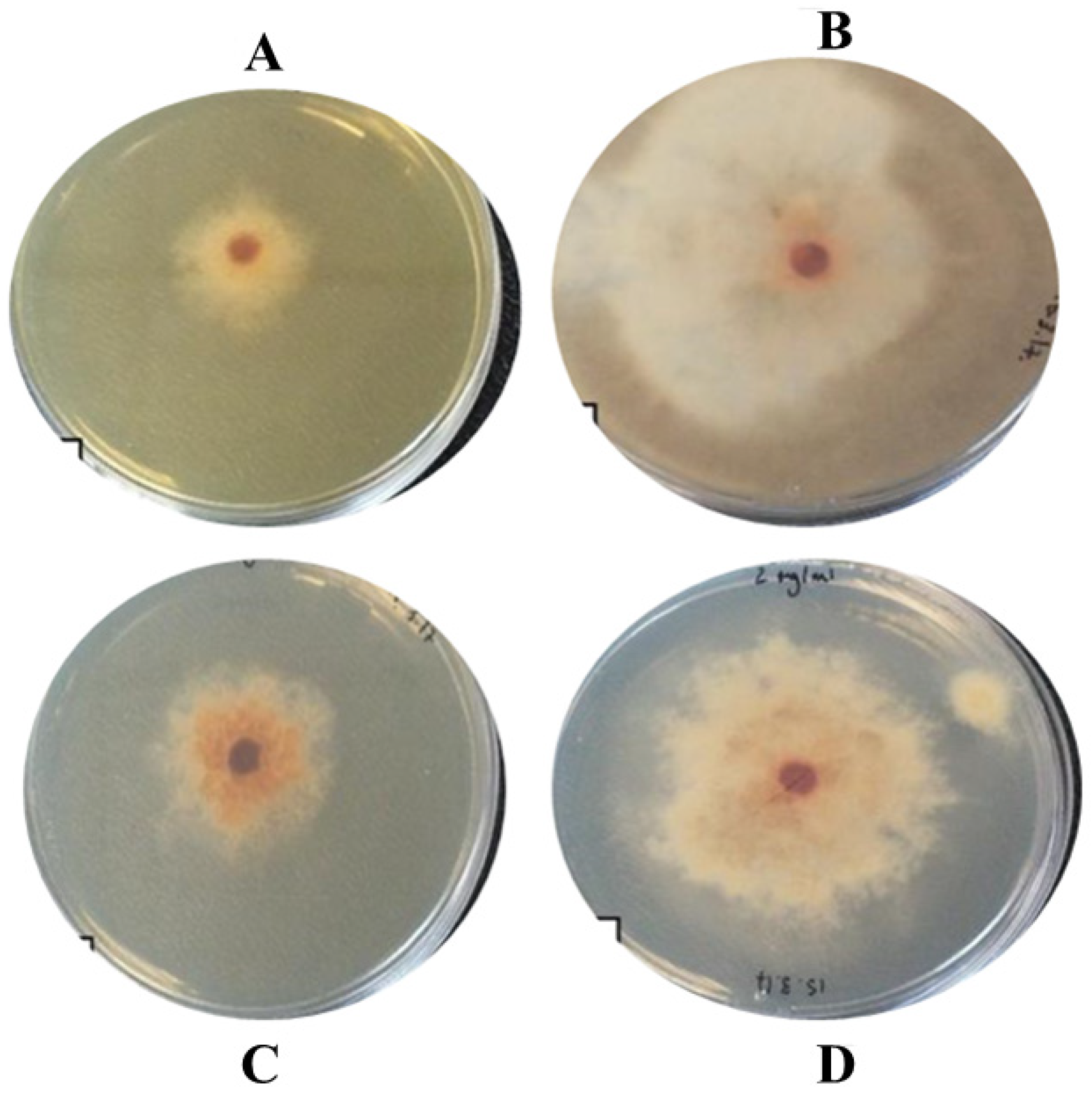
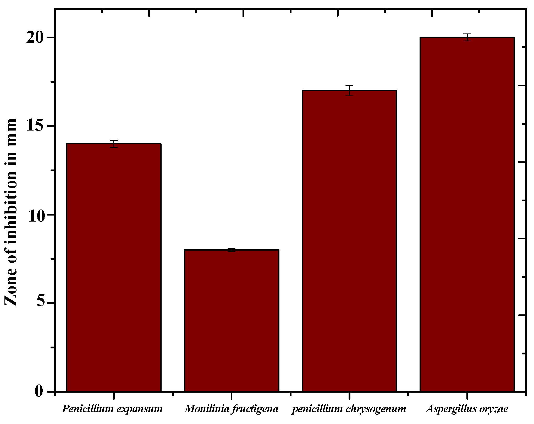
2.3.3. Anti-Spoil Activity of NPs against Spoil-Causing Fungal Isolate Species

Characterization of fungal flora: The characterization of fungal flora from fruits obtained in the Peshawar market was shown in Figure 6A. The results show the occurrence of four different fungal species distributed among the spoilt fruits: Penicillium expansum, Monilinia fructigena, penicillium chrysogenum, and Aspergillus oryzaeFigure 6 shows the identification of fungus. Figure 7 show the inhibition zone of MnO2 NPs. The outcomes of MnO2 NPs antifungal potential are depicted in Figure 8. In this study, MnO2 was added to each well. The values of antifungal activity of all the samples were 14.2 ± 86 mm, 8.9 ± 6.0 mm, 17.7 ± 1.26 mm, and 20.7 ± 4.38 mm for Penicillium expansum, Monilinia fructigena, Penicillium chrysogenum, and Aspergillus oryzae at 200 µg/mL in each well, respectively. According to several studies, the most prevalent routes of NP-induced cellular toxicity include membrane disruption and free radical production. Fungal growth is inhibited by the interaction of nanoparticles with fungal hyphae and spores.

3. Discussion

Because of its simplicity of use and cost efficiency, as well as its potential for large-scale manufacturing, the NP synthesis approach has piqued the scientific community’s attention in recent years. We used a biosynthesis approach to make MnO2 NPs from Fagonia cretica, and we tested NPs as bio-catalysts for antifungal agents and enzyme inhibitors in food preservation and flavor maintenance. During the first steps, the MnO2 NPs were explored for their morphological features by employing diverse analytical tools, including UV spectroscopy, FTIR, XRD, SEM, and EDX. According to UV spectroscopy, the sample absorbed energy at 410 nm, which is a sign of typical peak value for MnO2 NPs. Aside from that, an absorption peak at 410 nm with no other peak demonstrated the NPs’ exceptional purity. Many investigations revealed a significant absorption peak of MnO2 NPs 410 nm wavelengths, related to the samples’ red shift at 500 °C and 700 °C [10]. FTIR analysis was used to observe the stretching vibration of the different functional groups, which show different absorption bands. The absorption band at 562.2 cm−1 shows a collision of O–Mn–O; 628.4 cm−1 shows C–O stretching; 1115.4 cm−1, 1616.1 cm−1 indicates aromatic unsaturation (C=C) of the stabilized Fagonia cretica system; and 3385.9 cm−1 is the typical absorption for O–H–O of water present in the solution, respectively; our FTIR results are in correspondence with [11]. The produced MnO2 NPs had crystalline in the range of 15.5 ± 0.85 nm, as estimated by Nano-measurer and ImageJ analysis, as confirmed by SEM micrographs. The size of NPs was larger in this work than in [12], which might be attributed to changes in synthesis settings such as temperature, incubation period, bacterial extract type, and handling applications. Furthermore, EDX analysis showed pure MnO2 NPs phases and a strong peak in the EDX spectrum, showing that the test sample contained pure manganese. The EDX spectra of MnO2 NPs were obtained using a simple precipitation process using manganese as the starting material. Pure MnO2 NPs with substantial peaks were successfully synthesized, according to the EDX spectrum. However, additional peaks in the spectrum were detected, suggesting that bacterial biomolecules were involved in the nanoparticle synthesis. In comparison to other reports, we found the same EDX pattern of MnO2 NPs with great purity [12]. The size and crystallinity of the bio-fabricated MnO2 NPs were measured using an XRD profile. The XRD pattern shows five distinct peaks at 2θ = 28.78°, 37.66°, 42.14°, 49.90°, and 56.44°, corresponding to the crystal planes of MnO2 NPs (310), (211), (301), (411), and (600) (JSPDF 44-0141). Furthermore, the strength of the peaks in the XRD pattern suggests that the MnO2 NPs are very crystalline [12]. After thorough morphological and chemical analysis, the produced NPs were tested for important applications, such as bio-catalysts as antifungal agents, and antioxidant and enzyme inhibitors in food preservation and flavor maintenance. We also checked the anti-browning mechanism that causes browning in apples involving an enzyme called tyrosinase. The inhibitory ability of NPs against the production of tyrosinase was determined. MnO2 NPs block the active site and cause enzyme inhibition, which produce anti-browning characteristics and may be employed as anti-browning agents, which were used in our recent work. APE has been shown to have considerable inhibitory effects on tyrosinase up to 64.8 ± 0.16 at 200 µg/mL and (27.2 ± 0.58) at 25 µg/mL. Similar results were reported by [13]. A variety of concentrations of synthesized NPs (25, 50, 100, 150, and 200 ppm) were evaluated as antioxidant agents. 2, 2-diphenyl-1-picrylhydrazyl (DPPH) free radicals were exposed to test samples of varying concentrations in order to evaluate the antioxidant potential of MnO2 NPs. The maximum DPPH free radical scavenging activity was 74.5 ± 0.39% (200 µg/mL) and the minimum was 12.4 ± 0.27 at (25 µg/mL), respectively, in accordance with previous results [14]. We tested the NPs against spoil-causing fungal isolate in apples using well diffusion methods, and the antifungal potential of MnO2 NPs was studied. The values of antifungal activity among all the samples were 14.2 ± 86 mm, 8.9 ± 6.0 mm, 17.7 ± 1.26, and 20.7 ± 4.38 mm for Penicillium expansum, Monilinia fructigena, Penicillium chrysogenum, and Aspergillus oryzae at 200 µg/mL in each well, respectively. Our results are congruent with earlier reports [15].

4. Materials and Methods

4.1. Chemicals

All of the chemicals used in this study were analytical grade and bought from Sigma Chemicals Co. (St. Louis, MO, USA) and Merck (Darmstadt, Germany). For NP synthesis, commercially available manganese acetate salt was purchased from US Research Nanomaterials, Inc. (3302 Twig Leaf Lane, Houston, TX 77084, USA).

4.2. Collection of the Plant Material

Fresh Fagonia cretica leaves were obtained in Peshawar, Pakistan’s surrounding regions.

4.3. Preparation of Leaves Extract of Fagonia cretica

Using 20 g of fresh Fagonia cretica leaves, a Fagonia cretica leaf extract was made. To eliminate any pollutants or dust, the leaves were carefully washed with deionized (DI) water and air-dried at 30 °C. The dried leaves were chopped into tiny bits and crushed in a professional blender before being transferred to a 500 mL beaker. After that, 150 mL of DI water was added and agitated for 60 min at 60 °C. After that, the Fagonia cretica leaves extract was cooled to room temperature and then filtered. For later usage, the filtrate was collected and kept at 4 °C in an airtight glass container.

4.4. Biogenic Synthesis of Manganese Dioxide NPs (MnO2 NPs)

One millimeter of manganese acetate was added to twenty-five milliliters of Fagonia cretica leaves extract for the biogenic production of manganese dioxide NPs. At pH 7.15, the resultant liquid was heated for 70 min at 40 °C with continuous stirring. After that, the produced manganese dioxide NPs were isolated from the reaction mixture by centrifugation at 3000 rpm for 30 min. After centrifugation, the resulting NPs were washed three times with ethanol, dried at 40 °C, and then calcined for three hours in a muffle furnace at 200 °C. Finally, the green produced manganese dioxide NPs were designated MnO2 NPs and kept in a glass container for further analysis.

4.5. Characterization of Biosynthesized MnO2 NPs

Advanced tools were used to assess the physicochemical and morphological features of biosynthesized MnO2 NPs [16]. The UV–vis-NIR spectrophotometer UV-3600 Plus Shimadzu was used for performing UV spectroscopy in the typical range of 200 to 700 nm to monitor the interaction between biomass and metallic salt. The crystal nature of biologically synthesized MnO2 NPs was determined using the X-ray diffraction (XRD) profile. The Panalytical’s X’Pert X-ray diffractometer was utilized to produce the XRD peaks at CuKα (=1.54056 Ǻ). MnO2 NPs were studied using the IRTracer-100 Fourier transform infrared (FTIR) spectrophotometer in the 400–4000 cm−1 spectrum region to reveal and assess related functional groups involved in their biosynthesis approach [17]. Scanning electron microscopy (SEM) was applied to measure the physical dimensions and morphological characteristics of biosynthesized MnO2 NPs (JSM-5910, Tokyo, Japan). The biosynthesized MnO2 NPs were subjected to energy-dispersive X-ray (EDX) spectroscopy to ascertain their elemental composition.

4.6. Anti-Browning Activity of NPs

4.6.1. Tyrosinase Assay

The actual mechanism that causes browning in apples involves an enzyme called tyrosinase. The inhibitory ability of MnO2 NPs against the production of tyrosinase was determined. A tyrosinase test was performed using the previously reported technique of [18], which employed L-DOPA (5 mM; Sigma Aldrich, St. Louis, MO, USA). The test sample was combined with 10 mL of an L-DOPA diphenolase substrate and a sodium phosphate buffer (50 mM, pH 6.8). By adding 0.2 mg/mL of mushroom tyrosinase solution to the reaction mixture, the final volume increased to 200 mL (Sigma Aldrich). As a control, the DMSO solvent was utilized to replace the tested sample. At 475 nm, the reaction activities were monitored using a microplate reader (BioTek ELX800; BioTek Instruments, Bad Friedrichshall, Germany). The percent inhibition was calculated in comparison to the matching control tyrosinase effect.

4.6.2. Antioxidant Assay

Antioxidants can react with oxygen to suppress the initiation of browning. They are also able to react with intermediate products, thereby breaking the chain reaction and inhibiting tyrosinase formation. 2,2-diphenyl-1-picrylhydrazyl (DPPH) free radicals were exposed to test samples of varying concentrations in order to evaluate the antioxidant potential of MnO2 NPs nanoparticles [19]. A DPPH solution was prepared and 180 μL was poured into specified wells of a 96-well titer plate. Afterwards, 20 μL of different concentrations (25–400 μg/mL) of nanoparticles was added to each well and incubated for 1 h at 37 °C. DMSO was used as a negative while ascorbic acid was used as a positive control. After incubation, the samples were exposed to absorbance at 517 nm under the microplate reader and absorbance was recorded for each well. The antioxidant potential was calculated using the following formula:
DPPH % = 1 A b s o r b a n c e   o f   S a m p l e A b s o r b a n c e   o f   C o n t r o l e × 100

4.7. Anti-Spoil Activity of NPs against Spoil-Causing Fungal Isolate Species

4.7.1. Sample Collection of Spoilt Apple

Apple fruit samples were selected at random from both wholesalers and merchants in Peshawar. The ruined fruits were detected using the Bukar et al. [20] approach of morphological evaluation. Before mycological examination, the materials were maintained in the refrigerator at 4 °C.

4.7.2. Culture Media Preparation

Chloramphenicol (30 mg mL−1) was added to potato dextrose agar (PDA). The culture media were produced according to the instructions provided by the manufacturer. The suitable medium or foundation medium was weighed in its various amounts. After that, the weighed quantity of media was suspended in 400 mL of distilled water. Over a Bunsen flame, the media were brought to a boil until the agar melted. The pH of the molten agar medium was adjusted according to the manufacturer’s recommendations after cooling to 45 °C. The media were cotton-plugged and covered in aluminum foil before being autoclaved for 15 min at 121 °C. The media were aseptically distributed in 20 mL aliquots sterile Petri plates after sterilization, and then allowed to set on the flat before being aseptically dispensed into sterile Petri dishes. The Petri dishes were labelled and placed in the refrigerator to be used later.

4.7.3. Isolation of Fungi from Spoilt Fruits

Dashwood et al. [21] and Balali et al. [22] used the same procedure to isolate the mycological flora. The diseased fruits were surface-sterilized for 2 min with cotton wool soaked in 0.1 percent mercury chloride (HgCl), then washed three times in distilled water. To avoid bacterial development, a sterile blade and forceps were used to cut a tiny segment of tissue (3 mm diameter), including both the healthy and rotten portions, and then plated on solidified potato dextrose agar (PDA) with chloramphenicol (30 mg/mL). For 7 days, the infected plates were kept at room temperature (25 °C). As stated by Fawole and Oso [23], the various colonies detected on the plates were differentiated based on their cultural traits, such as colony size, shape, color, consistency, and hemolytic properties. To achieve pure isolates, the fungal isolates were subculture on PDA slants.

4.7.4. Identification of Fungal Isolates

Slide culture methods were used to identify fungal isolates collected from the slant based on their gross morphology, which included colony development pattern, conidial morphology, and pigmentation [24,25]. A small portion of aerial mycelia from the representative culture was picked with a sterile inoculating needle and inoculated on a slide containing a fraction of a prepared solidified potato dextrose agar and incubated for 48 h, after which it was viewed under a light microscope, first with a low resolution objective of ×10 and then with a high resolution objective of ×40 to detect spore, hyphae, and other special structures according to the mycological atlas of Domsch et al. [26]. We validated and verified the morphological traits and appearance of the fungal isolates from decaying fruits utilized in this investigation.

4.8. Antifungal Activity

It was determined that MnO2 NPs have antifungal activity using the agar well diffusion assay [27] after they were generated in the laboratory. In this experiment, fungal strains were disseminated on potato dextrose agar (PDA) plates, and uniform lawns were created using the spread plate method. In each plate, 5 mm wells were bored using a sterile well borer, and varied quantities of nanoparticles were put into each well. The plates were incubated at 28 °C for 72 h. Amphotericin B was utilized as a positive control, while DMSO was employed as a negative control in this study. The exercise was performed three times, and the mean inhibition zones were measured with the use of a Vernier caliper for each repetition.

5. Conclusions

In this study, the Fagonia cretica plant biomass-based MnO2 NPs was prepared and characterized by various instrumental techniques and was then investigated as bio-catalytic for antifungal and anti-browning potential in order to preserve apple flavor and texture. The biomass of Fagonia cretica contains proteins, carbohydrates, and lipids—chemicals which helped in the capping and reduction of Mn0 into MnO2 NPs. The prepared nanoparticles showed potent antifungal potential against spoil-causing fungal isolate species. DPPH and tyrosinase were potently inhibited by these nanoparticles, indicating that they could be effectively used as anti-browning agents for preserving apple flavor and texture.

Author Contributions

Conceptualization, S.F.; methodology, S.F. and S.Z.; software, A., validation, M.R., M.A. and F.A.; formal analysis, R.U.; investigation, G.M.A.; resources, S.F.; data curation, H.R.H.M.; writing—original draft preparation, S.F. and S.Z.; writing—review and editing, S.F. and S.K.; visualization, R.U.; supervision, S.F.; project administration, S.F.; funding acquisition, G.M.A. All authors have read and agreed to the published version of the manuscript.

Funding

This research was supported by Princess Nourah bint Abdulrahman University Researchers Supporting Project number (PNURSP2022R30), Princess Nourah bint Abdulrahman University, Riyadh, Saudi Arabia.

Data Availability Statement

All required data are present in this file.

Conflicts of Interest

The authors declare no conflict of interest.

References

  1. Muk, M.F.; Wu, N.R.; Danl, U.I.; Salisu, N. Isolation and characterization of fungal species from spoilt fruits in Utako market, Abuja, Nigeria. J. Appl. Sci. 2018, 19, 15–19. [Google Scholar] [CrossRef]
  2. Anand, S.P.; Sati, N. Artificial preservatives and their harmful effects: Looking toward nature for safer alternatives. Int. J. Pharm. Sci. Res. 2013, 4, 2496. [Google Scholar]
  3. Kumbi, Y. Phytochemical Characterization of Fagonia Indica and its Effects on MCF-7 Breast Cancer Cell Line. 2019. Available online: https://ir.library.oregonstate.edu/downloads/cj82kd531 (accessed on 14 April 2022).
  4. Pillaiyar, T.; Manickam, M.; Namasivayam, V. Skin whitening agents: Medicinal chemistry perspective of tyrosinase inhibitors. J. Enzym. Inhib. Med. Chem. 2017, 32, 403–425. [Google Scholar] [CrossRef] [PubMed] [Green Version]
  5. Basavegowda, N.; Idhayadhulla, A.; Lee, R.Y. Tyrosinase inhibitory activity of silver nanoparticles treated with Hoveniadulcisfruit extract: An in-vitro study. Mater. Lett. 2014, 129, 28–30. [Google Scholar] [CrossRef]
  6. Abbas, Q.; Saleem, M.; Phull, A.R.; Rafiq, M.; Hassan, M.; Seo, K.L.S. Green synthesis of silver nanoparticles using bidensfrondosaextract and their tyrosinase activity. Iran. J. Pharm. Res. 2017, 16, 763–770. [Google Scholar]
  7. Faisal, S.; Jan, H.; Abdullah; Alam, I.; Rizwan, M.; Hussain, Z.; Sultana, K.; Ali, Z.; Uddin, M.N.U.M.N. In Vivo Analgesic, Anti-Inflammatory, and Anti-Diabetic Screening of Bacopa monnieri-Synthesized Copper Oxide Nanoparticles. ACS Omega 2022, 7, 4071–4082. [Google Scholar] [CrossRef]
  8. Faisal, S.; Abdullah; Rizwan, M.; Ullah, R.; Alotaibi, A.; Khattak, A.; Bibi, N.; Idrees, M. Paraclostridium benzoelyticum Bacterium-Mediated Zinc Oxide Nanoparticles and Their In Vivo Multiple Biological Applications. Oxid. Med. Cell. Longev. 2022, 2022, 5994033. [Google Scholar] [CrossRef]
  9. Sabir, M.S.; Rajendra, C.D.; Amol, S.; Poournima, S.S. A review on: Preservatives used in pharmaceuticals and impacts on health. PharmaTutor 2016, 4, 25–32. [Google Scholar]
  10. Moon, S.A.; Salunke, B.K.; Alkotaini, B.; Sathiyamoorthi, E.; Kim, B.S. Biological synthesis of manganese dioxide nanoparticles by Kalopanax pictus plant extract. IET Nanobiotechnol. 2015, 9, 220–225. [Google Scholar] [CrossRef]
  11. Prasad, K.S.; Patra, A. Green synthesis of MnO2 nanorods using Phyllanthus amarus plant extract and their fluorescence studies. Green Process. Synth. 2017, 6, 549–554. [Google Scholar] [CrossRef]
  12. Lu, H.; Zhang, X.; Khan, S.A.; Li, W.; Wan, L. Biogenic synthesis of MnO2 Nanoparticles with leaf extract of Viola betonicifolia for enhanced antioxidant, antimicrobial, cytotoxic, and biocompatible applications. Front. Microbiol. 2021, 12, 3329. [Google Scholar] [CrossRef] [PubMed]
  13. Selvaraj, K.; Daoud, A.; Alarifi, S.; Idhayadhulla, A. Tel-Cu-NPs catalyst: Synthesis of naphtho [2, 3-g] phthalazine derivatives as potential inhibiters of tyrosinase enzymes and their investigation in kinetic, molecular docking, and cytotoxicity studies. Catalysts 2020, 10, 1442. [Google Scholar] [CrossRef]
  14. Mahlangeni, N.T.; Moodley, R. Biosynthesis of manganese oxide nanoparticles using Urginea sanguinea and their effects on cytotoxicity and antioxidant activity. Adv. Nat. Sci. Nanosci. Nanotechnol. 2021, 12, 015015. [Google Scholar] [CrossRef]
  15. Thirumurugan, A.; Ramachandran, S.; Shiamala Gowri, A. Combined effect of bacteriocin with gold nanoparticles against food spoiling bacteria-an approach for food packaging material preparation. Int. Food Res. J. 2013, 20, 1909–1912. [Google Scholar]
  16. Rizwan, M.; Hussain, M.; Muhammad; Rauf, A.; Zafar, M.N.; Mabkhot, Y.N.; Maalik, A. Green synthesis and antimicrobial evaluation of silver nanoparticles mediated by leaf extract of Syzygium cumini against poultry pathogens. Micro Nano Lett. 2020, 15, 600–605. [Google Scholar] [CrossRef]
  17. Rizwan, M.; Amin, S.; Malikovna, B.K.; Rauf, A.; Siddique, M.; Ullah, K.; Bawazeer, S.; Farooq, U.; Mabkhot, Y.N.; Ramadan, M.F. Green synthesis and antimicrobial potential of silver nanoparticles with Boerhavia procumbens extract. J. Pure Appl. Microbiol. 2020, 14, 1437–1451. [Google Scholar] [CrossRef]
  18. Ekennia, A.; Uduagwu, D.; Olowu, O.; Nwanji, O.; Oje, O.; Daniel, B.; Mgbii, S.; Emma-Uba, C. Biosynthesis of zinc oxide nanoparticles using leaf extracts of Alchornea laxiflora and its tyrosinase inhibition and catalytic studies. Micron 2020, 141, 102964. [Google Scholar] [CrossRef]
  19. Tian, H.; Ghorbanpour, M.; Kariman, K. Manganese oxide nanoparticle-induced changes in growth, redox reactions and elicitation of antioxidant metabolites in deadly nightshade (Atropa belladonna L.). Ind. Crop. Prod. 2018, 126, 403–414. [Google Scholar] [CrossRef]
  20. Bukar, A.; Mukhtar, M.D.; Adamu, S. Isolation and identification of postharvest spoilage fungi associated with sweet oranges (Citrus sinensis) traded in kano metropolis. Bayero J. Pure Appl. Sci. 2010, 2, 122–124. [Google Scholar] [CrossRef] [Green Version]
  21. Dashwood, E.P.; Fox, R.A.; Perry, D.A. Effect of inoculum source on root and tuber infection by potato blemish disease fungi. Plant Pathol. 1992, 41, 215–223. [Google Scholar] [CrossRef]
  22. Balali, G.R.; Neate, S.M.; Scott, E.S.; Whisson, D.L.; Wicks, T.J. Anastomosis group and pathogenicity of isolates of Rhizoctonia solani from potato crops in South Australia. Plant Pathol. 1995, 44, 1050–1057. [Google Scholar] [CrossRef]
  23. Fawole, M.O.; Oso, B.A. Laboratory Manual of Microbiology; Spectrum Book Ltd.: Ibadan, Nigeria, 1988; pp. 22–45. [Google Scholar]
  24. Mailafia, S.; Okoh, H.O.G.R.; Olabode, K.; Osanupin, R. Isolation and identification of fungi associated with spoilt fruits vended in Gwagwalada market, Abuja, Nigeria. Vet. World 2017, 10, 393. [Google Scholar] [CrossRef] [PubMed] [Green Version]
  25. Rizwan, H.M.; Zhimin, L.; Harsonowati, W.; Waheed, A.; Qiang, Y.; Yousef, A.F.; Munir, N.; Wei, X.; Scholz, S.S.; Reichelt, M.; et al. Identification of fungal pathogens to control postharvest passion fruit (Passiflora edulis) decays and multi-omics comparative pathway analysis reveals purple is more resistant to pathogens than a yellow cultivar. J. Fungi 2021, 7, 879. [Google Scholar] [CrossRef] [PubMed]
  26. Domsch, K.H.; Gams, W.; Anderson, T.-H. Compendium of Soil Fungi; Academic Press (London) Ltd.: London, UK, 1982. [Google Scholar] [CrossRef]
  27. Manogar, P. Studies on the Efficient Dual Performance of Lyngbya Majuscula Extract with Manganese Dioxide Nanoparticles in Photodegradation and Antimicrobial Activity. 2021. Available online: https://assets.researchsquare.com/files/rs-1102629/v1/88518079-8f06-4541-b77e-b7ca039a2884.pdf?c=1638287458 (accessed on 14 April 2022).
Figure 1. The entire process of making MnO2 NPs is depicted in this schematic picture. (A) Fagonia cretica plant, (B) Fagonia cretica powder, (C) Fagonia cretica extract, (D) filtrate, (E) reduction and capping of MnO ions by Fagonia cretica extract, (F) centrifugations, (G) pellets of NPs, and (H) purified MnO2 NPs.
Catalysts 12 00558 g001
Figure 2. (A) UV–visible spectroscopy, (B) Fourier transform infrared (FTIR) spectroscopy, and (C) XRD pattern for the green synthesized MnO2 NPs.
Catalysts 12 00558 g002
Figure 3. (A) Scanning electron microscopy (SEM), (B) image J analysis of SEM image, (C) size distribution, and (D) energy-dispersive X-ray (EDX) for the green synthesized MnO2 NPs.
Catalysts 12 00558 g003
Figure 4. Anti-browning potential of biosynthesized MnO2 NPs by inhibition of tyrosinase.
Catalysts 12 00558 g004
Figure 5. (A) Antioxidant potential of biosynthesized MnO2 NPs, (B) Test solution in the 96-well plates and (C) microplate reader.
Catalysts 12 00558 g005
Figure 6. (A) Spoilt apple fruits, (B) culture on PDA media, (C) growth of fungus on PDA media, (D) microscope, and (E) microscopic identification of fungus.
Catalysts 12 00558 g006
Figure 7. Shows the zone of inhibitions of NPs against (A) Penicillium expansum, (B) Monilinia fructigena, (C) Penicillium chrysogenum, and (D) Aspergillus oryzae.
Catalysts 12 00558 g007
Figure 8. Anti-fungal potential of MnO2 NPs against spoil-causing fungal isolates.
Catalysts 12 00558 g008
Publisher’s Note: MDPI stays neutral with regard to jurisdictional claims in published maps and institutional affiliations.

