Review

Microbial Phosphotriesterase: Structure, Function, and Biotechnological Applications

by
Wahhida Latip
1,*,
Victor Feizal Knight
1,
Norhana Abdul Halim
2,
Keat Khim Ong
2,
Noor Azilah Mohd Kassim
2,
Wan Md Zin Wan Yunus
3,
Siti Aminah Mohd Noor
2,* and
Mohd Shukuri Mohamad Ali
4
1
Center for Chemical Defence, National Defence University of Malaysia, Kem Perdana Sungai Besi, Kuala Lumpur 57000, Malaysia
2
Center for Defence Foundation Studies, National Defence University of Malaysia, Kem Perdana Sungai Besi, Kuala Lumpur 57000, Malaysia
3
Center for Tropicalisation, National Defence University of Malaysia, Kem Perdana Sungai Besi, Kuala Lumpur 57000, Malaysia
4
Enzyme and Microbial Technology Research Center, Department of Biochemistry, Faculty of Biotechnology and Biomolecular sciences, Universiti Putra Malaysia, Serdang 43400, Malaysia
*
Authors to whom correspondence should be addressed.
Submission received: 24 June 2019 / Revised: 17 July 2019 / Accepted: 18 July 2019 / Published: 7 August 2019
(This article belongs to the Special Issue Biocatalytic Applications in Biotechnology)

Abstract

:
The role of phosphotriesterase as an enzyme which is able to hydrolyze organophosphate compounds cannot be disputed. Contamination by organophosphate (OP) compounds in the environment is alarming, and even more worrying is the toxicity of this compound, which affects the nervous system. Thus, it is important to find a safer way to detoxify, detect and recuperate from the toxicity effects of this compound. Phosphotriesterases (PTEs) are mostly isolated from soil bacteria and are classified as metalloenzymes or metal-dependent enzymes that contain bimetals at the active site. There are three separate pockets to accommodate the substrate into the active site of each PTE. This enzyme generally shows a high catalytic activity towards phosphotriesters. These microbial enzymes are robust and easy to manipulate. Currently, PTEs are widely studied for the detection, detoxification, and enzyme therapies for OP compound poisoning incidents. The discovery and understanding of PTEs would pave ways for greener approaches in biotechnological applications and to solve environmental issues relating to OP contamination.

1. Introduction

Organophosphate (OP) compounds—otherwise known as phosphotriesters—are a class of highly neurotoxic synthetic compounds that are widely used as agricultural insecticides, petroleum additives, plasticizers, refrigerants, dyes, and even as chemical warfare agents [1]. Contamination by these compounds may occur because of terrorist attacks, unsafe disposal methods, and spillage from poor handling by agricultural workers [2]. OP compounds are known to inhibit mammalian acetylcholinesterases (AChE) [3,4,5]. Acetylcholinesterase (AChE) is an enzyme that hydrolyzes the neurotransmitter acetylcholine (ACh) and serves as a regulator of neurotransmission [3]. The most terrifying fact of its action is that the inhibition process is naturally irreversible, whereby the enzyme can no longer hydrolyze acetylcholine resulting in paralysis of muscles, seizures, difficulty in breathing and death by suffocation. In 2009, the World Health Organization reported that over 3 million people were poisoned by OP pesticides, and 200,000 had died and furthermore these numbers are increasing yearly. Moreover, OP compounds have been reported to be involved in gene mutations, DNA damage, cancerogenesis, and endocrine disorders.
OP compounds can be briefly divided into three major groups, which are the phosphotriesters, thiophosphotriesters, and phosphorothiolesters [6]. Phosphotriesters such as paraoxon contain a phosphate in the center with an o-linked group Figure 1A. Similarly, in thiophosphotriesters (methyl parathion) and phosphorothiolesters (malathion), the phosphate group is still in the center but the phosphoryl oxygen is replaced by sulfur and one or more of the ester oxygens are replaced by sulfur Figure 1B,C. To date these compounds are still widely used especially in agricultural industries [7]. Moreover, OPs compounds are easily drained into ground water that in turn will pollute water supplies and surrounding land areas [8]. Due to its high toxicity, numerous efforts to detect the presence of OP compounds and to detoxify their effects on humans and the environment have been made.
Many alternative technologies have been studied and developed to resolve this problem, using various chemical, physical, and biological methods. Unfortunately, most of these methods are expensive and may not suitable or even safe for the decontamination of human beings [7]. They may be very harsh and may not be very specific [3]. Currently, using enzymes is one of the more promising and safer ways to solve this problem. Organophosphate hydrolysis enzymes (OPHs) are enzymes that can hydrolyze the OP compounds to become nontoxic agents [9]. These enzymes have been reported to be found in bacteria as well as in cephalopods (squids) and mammals [10,11]. The OP compounds have been reported as being used as food by several bacteria species where they would cleave the phosphorester bond, and thereby reduce their toxicity [12,13]. A few OPHs have been isolated recently and are classified according to their protein homology. Phosphotriesterases (PTEs) are a group of these OPHs and are classified under the aryldialkylphosphatase super family [14]. This enzyme has been extensively studied in molecular biology and with much emphasis on their applications.
In this review, the 3D structure, mechanism of hydrolysis, and substrate specificity of PTEs are described. The biotechnological application of this enzyme in bioremediation, biosensor development and human therapy is also discussed.

2. Structure and Function

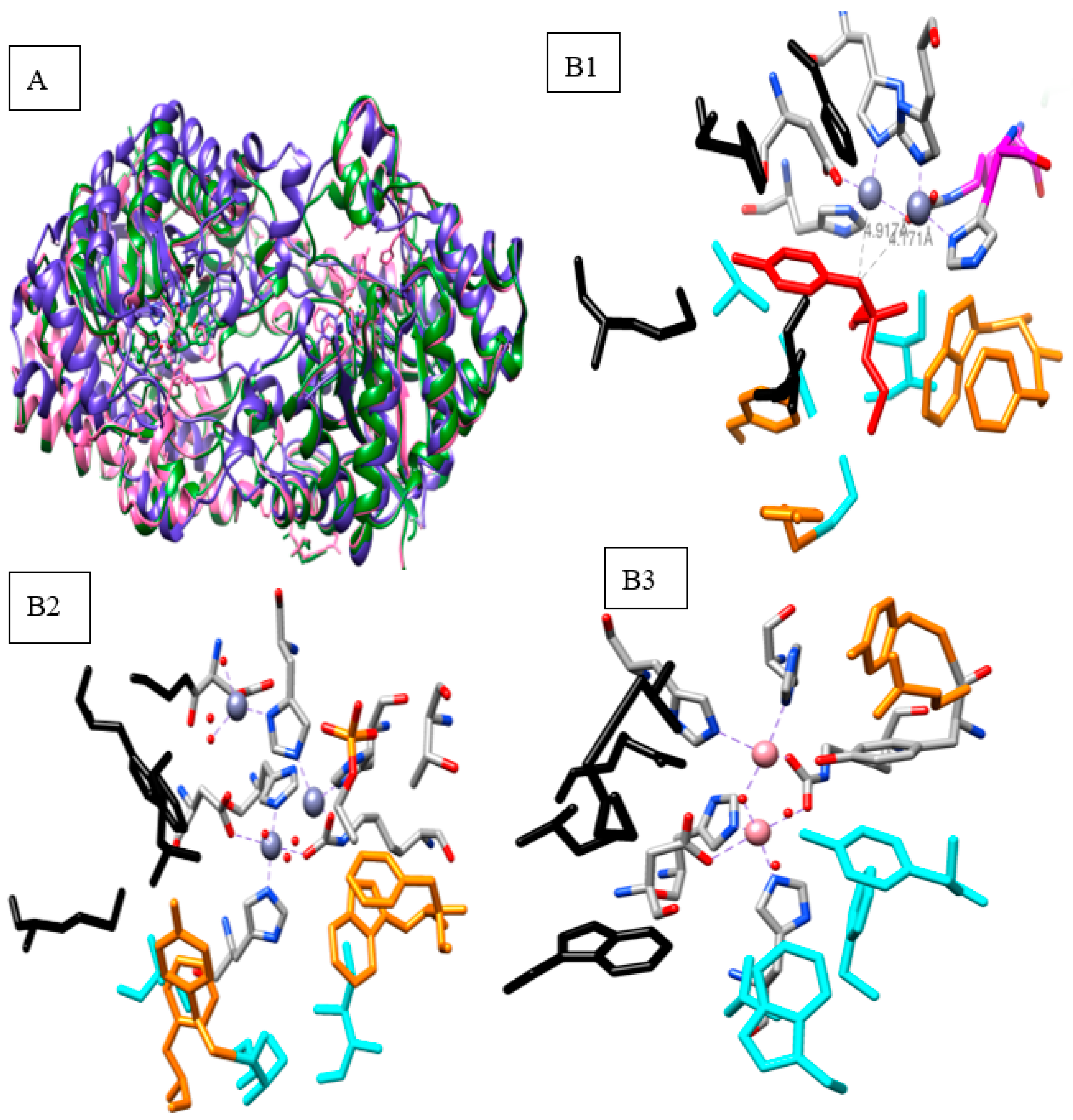
2.1. Bacterial Phosphotriesterase 3D Structures

A number of bacteria that are capable of degrading OP pesticides have been identified from soil samples from different parts of the world [15]. These include Pseudomonas pseudoalcaligenes, Pseudomonas diminuta, Agrobacterium radiobacter, Alteromonas haloplanktis and Saccharomyces cerevisiae, Pseudomonas monteilii, and Geobacillus stearothermophilus [13,16,17]. Flavobacterium sp. ATCC27551 is the first OP degrading bacteria and was isolated in the Philippines in 1973 [1]. The gene encoding the phosphotriesterase (PTE) enzyme was isolated and expressed in several expression hosts including E. coli, yeasts, insect cells and in in vitro compartmentalization [13,14,18,19]. Some of the genes showed a conserved sequence homology between two species such as with Pseudomonas diminuta and Flavobacterium sp. ATCC27551 even though the plasmid containing the PTE genes were discovered from among different plasmids [20]. Pseudomonas diminuta, Flavobacterium sp., and Alteromonas sp. possess the best characterized PTE enzymes isolated from bacteria [19,21].
Phosphotriesterase (PTE) is a membrane-associated protein that is translated as a 29 amino acid-long target peptide [22]. PTE is classified as a metalloenzyme or metal-dependent enzyme which means that without the metal, the enzyme will not function [23]. The natural substrate for PTE is still as yet unknown; however, the ability of PTE to hydrolyze the insecticide paraoxon at a rate that approaches the diffusion-controlled limit, is remarkable [9,19,24]. Although PTE can hydrolyze paraoxon efficiently, the PTE hydrolysis rates for malathion, phosalone, and acephate are approximately 1000-fold slower compared to that of paraoxon [13]. Thus, many efforts have been done to improve the hydrolysis rate using molecular approaches such as mutation at the active site pocket, varying the ribosomal operon and expression host. The enzyme has been reported to have three binding pockets to accommodate the substrate [6,25,26]. The active site pocket of the PTE in a hydrophobic condition contains two metal ions (Zn2+ Mn2+, Co2+, and Cd2+) bridged with side-chains of histidine residues that are clustered around the metal atoms [22,27]. A number of the crystal structures of PTE isolated from bacteria such as Pseudomonas pseudoalcaligenes (2.1 Å), Pseudomonas diminuta (2.0 Å), Mycobacterium tuberculosis (2.27 Å), Agrobacterium radiobacter (1.99 Å), Geobacillus stearothermophilus (2.09 Å), and Geobacillus kaustophilus HTA426 (2.05 Å) have been determined [4,7,16,28,29]. In this review, three crystal structures of PTEs from Pseudomonas diminuta, Sphingobium fuliginis strain ATCC 27551, and Geobacillus stearothermophilus (PLL) are used as examples in this section to shown the similarities and differences in their 3D structure Figure 2. All crystal structures reveal that there is some degree of similarity in their structures, such as each containing an α/β-barrel motif Figure 2. All of these PTEs are metal-dependent hydrolases and each naturally exists as a homodimer [5]. Although some researchers reported that PTEs had varying protein folds ((β/α) 8 or TIM-barrel, β-lactamase folds, and pita bread folds), all of these crystal structures are actually TIM-barrel folds [2,6,19]. In addition, all PTEs require divalent metals to directly ligate the substrate to simplify the hydrolysis process Figure 2(B1–B3) [5,6]. In this case, the metal ion in Pseudomonas diminuta and Sphingobium fuliginis strain ATCC 27551 PTE have Zn2+, as the ion, whereas for the Geobacillus stearothermophilus PTE it is Co2+ [7,30]. In the absence of a substrate, the α zinc metal was reported as buried near to His55, His 57, and Asp 301, and the β zinc metal was more exposed to the solvent [6,7,30]. The divalent metal ions in the active site center are reported to contribute to the activity and stability of the enzyme. The zinc is reported to have the greatest effect on stability while, Co in the center of the enzyme, increases the enzyme activity up to 300% [21]. Moreover, the carboxylated lysine and four histidine residues, which coordinate the two divalent cations, are required in the active center for activity [28,31]. The metals ions in turn coordinate an activated hydroxyl group that functions as the nucleophile in the OP hydrolysis reaction [32].
The 3D structure shows that the PTE enzymes have three detached binding pockets (small, large and leaving groups) that allow the substrate (paraoxon) to attach to the phosphorus center Figure 2B [2,6,32]. Due to their high similarity (of up to 90%) in structure and protein homology [10,12]. PTE from Pseudomonas sp. and Sphingobium fuliginis have similar active site pockets. The small binding pockets for Pseudomonas diminuta and Sphingobium fuliginis are Gly-60, Leu-303/Thr-303, Ser-308, and Ile-106 [11,12,28]. Similarly, with the leaving group pocket, all hydrophobic residues were conserved (Phe-306, Phe-132, Trp131, and Tyr-309) Figure 2(B1,B2) [16,23]. Unlike the large pocket, His-254 and 257 are substituted by Gly-254 and Trp-257. Other residues namely Leu-271 and Met-317 are also well conserved [33]. The active site pocket for Geobacillus stearothermophilus was found to be different from that of Pseudomonas sp. and Sphingobium fuliginis where the small binding pocket contained F28, Y30, T70, C74, V268, W271, the large pocket (R230, I233, M236, V237, and W289), and the leaving group pocket (Y100 and E103), see Figure 2(B3) (17) [30]. Moreover, the metal ion at the center of Geobacillus stearothermophilus PTE is different from that of Pseudomonas sp. and Sphingobium fuliginis, which is Co2+ [30]. The two metal ions are thus designated as α and β. The α metal is more buried and is in direct coordination with His55, His57, and Asp301 (Pseudomonas sp. and Sphingobium fuliginis) [17]. The β-metal is more solvent-exposed and coordinated towards His201 and His230 [7,16].

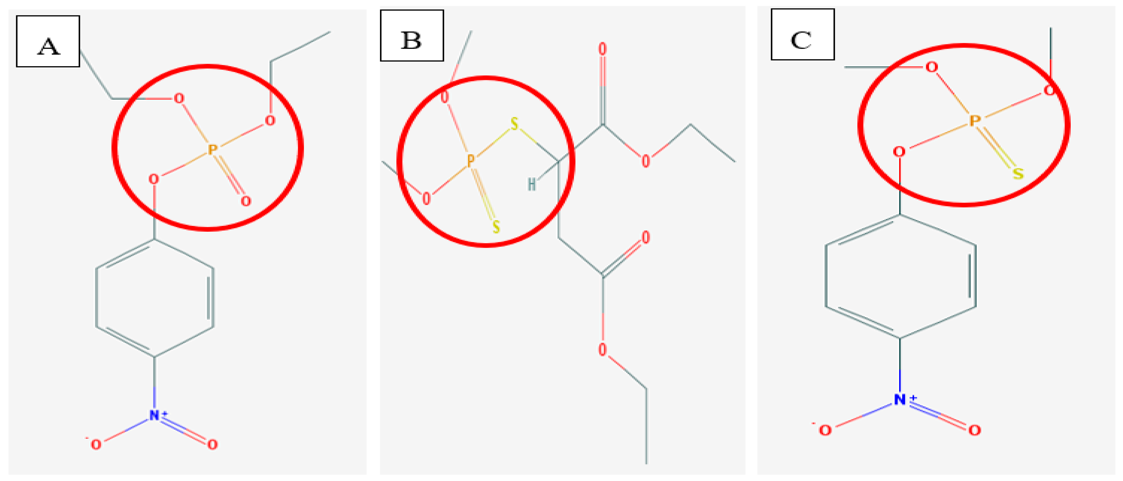
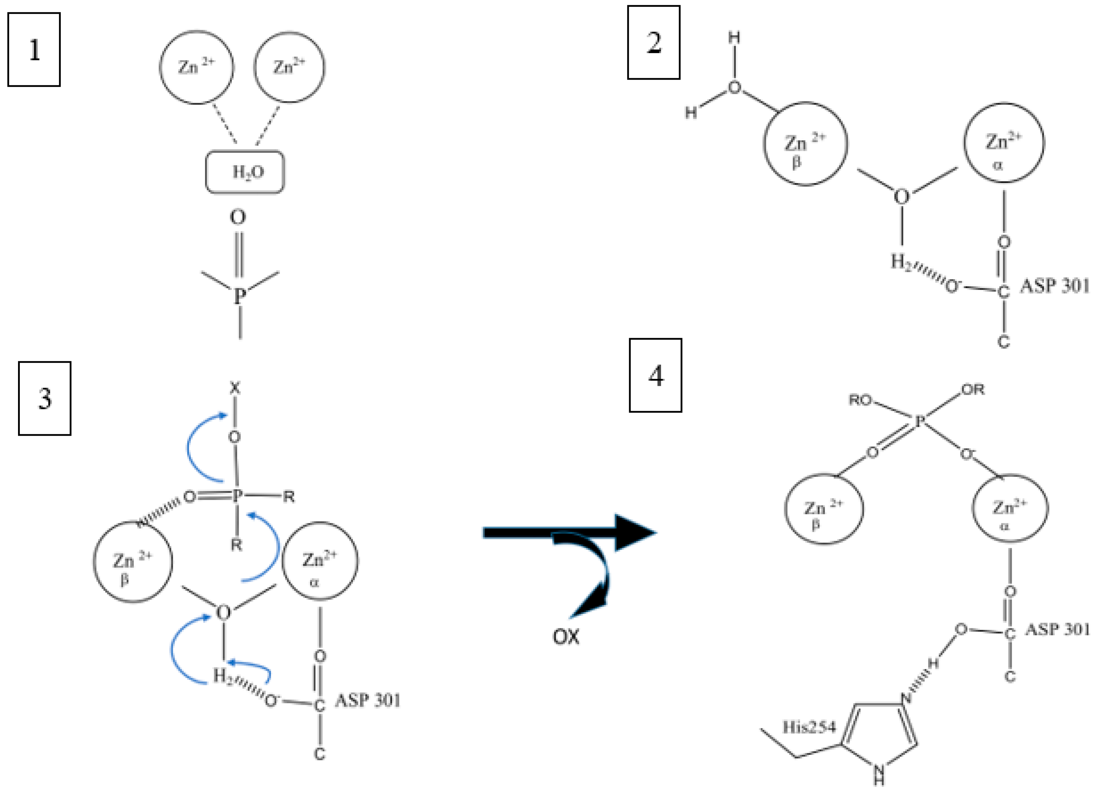
2.2. Mechanism of Hydrolysis and Stereoselectivity of PTE

Hydrolysis of OP by PTE depends on three factors: the metal ion in the enzyme, the pH of the environment, and the substrate itself [22,34]. In detail, the structure of the PTE shows that the two metal ions coordinated in the enzyme interact with a carboxylated lysine (Lys169) and a hydroxide ion or water molecule [17,24,35]. The nucleophilic attack starts with these three components which are water bridged between two divalent cations, the bimetal ion, and substrate in the active PTE site [2,6]. The 3D crystal structure shows the presence of this hydroxide or water bridge between the metal [6,19]. The nucleophilic attack occurs on the phosphorus center (P–O bond) followed by a proton transfer [6,11,17]. Histidine 254 and Aspartic acid 233 extend the hydrogen bond network in the binuclear metal center and Asp301 helps to shuttle protons from the active site to the solvent [6,17]. The polarization of the P–O bond of the substrate (pH and substrate factors) also contributes to the nucleophilic attack [17,21,24]. The OH− then attacks the phosphorus center of the substrate, followed by a proton transfer event to Asp301 Figure 3 [17,19]. The P–O bond is thus broken, and the products are released from the active site. The proton is shuttled away from the active site with the assistance of His254 and Asp233 [6,17]. This mechanism is called the SN2 mechanism where one bond is broken and one bond is formed synchronously [5,36]. Moreover, the metal ion was found to depend on the pH where protonation occurred from the bridging hydroxyl group [11,17]. The catalytic activity is lost at a low pH because of the protonation [17]. Furthermore, polarizing the P–O bond of the substrate, makes it more susceptible for a nucleophilic attack to occur [6,21,24].
The three distinct active site pockets (small, large, and leaving group) play an important role in the stereoselectivity of PTE. Wild type PTE prefers paraoxon as a substrate and can hydrolyze a variety of insecticides including phosphotriesters, thiophosphotriesters, and phosphorothioesters [2,37]. Each pocket plays its own role in stereoselectivity [19,25,37]. The leaving group pocket has been reported to play an important role in dictating the enzyme specificity [6,25]. The binding site pockets which are the small pockets and large pockets play a role in the determination of the specificity for the side ester groups on the substrate [6,27]. The PTE of the wild type is known to prefer SP-enantiomers for most phosphate substrates [9,25]. The small pocket of the PTE has been reported as too small for malathion to bind and a mutation has been done to enlarge the pocket [21,33]. Reducing the size of the small pocket has been done by mutation at G60A and was found to increase the selectivity for the RP-enantiomer compound from 9.4 × 10⁻¹ M−1 s−1 to 2.1 × 10⁴ M−1 s−1 by factor of 2 × 10⁴ [2,6]. Similarly with the large pocket of the PTE, mutation has been done by switching the residue H254Q/H257F, the catalytic constant for Sp-enantiomer enhanced 2 times from the wild type [9,11]. Moreover, mutation at the substrate binding pocket (H257Y/L303T) also enhances the kcat/Km of YT mutant towards G-type nerve gases as compared to the wild type from 2.4 × 10⁵ to 2 × 10⁶; 1.5 × 10⁴ to 5 × 10⁵; and 2.3 × 10⁵ to 8 × 10⁵ M−1 s−1 for sarin (GB), soman (GD), and cyclosarin (GF), respectively [9,38]. The manipulation of large and small pockets of PTE allows the researcher to enhance, diminish, or reverse the hydrolysis of the substrate [6,7,15]. These approaches have enhanced the hydrolysis of the most toxic enantiomers of GB and GD by over two orders of magnitude and approximately 25-fold for the nerve agent VX [11,39]. In addition, mutation near the small pocket of PTE or Loop 7 using error prone PCR has also given reliable results where the mutant L7ep-3a enhanced a 600-fold (2.6 × 10³ M−1 s−1) faster action in the detoxification of VR and VX nerve agents [40]. The substitution of the metal ion has also been done to identify the influence of the metal ion towards selectivity and stability of the PTE [4,22] Zinc is the native metal ion for PTE but can be substituted with other metals such as Co2+, Ni2+, Cd2+, or Mn2+ to retain the catalytic activity [17]. The value of kcat/Km has been shown to decrease when the Zn2+/Zn2+ (6.8 × 10⁶ M−1 s−1) was substituted with Cd2+/Cd2+ (1.32 × 10⁶ M−1 s−1) and Zn2+/Cd2+ (2.8 × 10⁶ M−1 s−1) with paraoxon as the substrate, but not for parathion. The PTE with Zn2+/Cd2+ showed an increment value of up to 5.0 × 10⁵ M−1 s−1 for this substrate [17] Therefore, by modifying the active sites and their surrounding areas, the applicability of these enzyme can be expanded for industrial purpose.

2.3. Industrial Application of Bacterial Phosphotriesterase

2.3.1. Biosensors

The specificity of PTE and its mutant to hydrolyze a broad range of OP compounds even at low-level concentrations of these OPs is well documented [19,33]. This enzyme characteristic has been exploited to develop detectors to identify the presence of OP compounds in the environment. Even though many methods have been proposed for this purpose, the techniques are time-consuming and may not be easily portable for use. Moreover, the techniques require trained technicians to perform the analysis competently [41]. Thus, portable and specific enzymatic biosensors (especially for use in field OP monitoring), with high selectivity and sensitivity, would be very beneficial [42,43].
In enzymatic biosensing of OP compounds, the detection is based on either their inhibition of AChE or the hydrolysis process by the OP hydrolysis enzyme [42,43]. Detection using an inhibition-based sensor has been reported to have a high sensitivity and is capable of ultralow OP detection [42,44]. However, enzymes can easily be inhibited by many other factors which include heavy metals, temperature, pH, and detergents that normally found in the environment and may be subject to a slow detection rate (owing to prolonged incubation and regeneration periods) [41,45,46]. Therefore, direct measurement of the hydrolysis reaction is preferable compared to just measurement of inhibition. Furthermore, the detection limit of sensors based on the PTE reaction is better when compared with that of AChE activity detection [31,43].
PTE isolated from Brevudimonas dimunita and Flavobacterium sp. have been studied and used as potentiometric, amperometric and optical biosensors [45,47,48]. The PTE biosensor is reported to have high reusability (0.3% per time sensed), achieving up to 90% stability in 1000 s and 70% longevity and selectivity (with the detection of six similar nerve agents) [41]. The development of this biosensor is based on the mechanism of hydrolysis of PTE in the OP compound [19,49,50]. Products from the hydrolysis process will change the pH and electrical charge, proton transfer to Asp 301 will occur [11,17,42]. Immobilized PTE has also been used as a bioreceptor to detect OP compounds. Immobilization supports such as graphene, chitosan, sulphate chitosan, bovine serum, and glutaraldehyde have been used to stabilize and enhance the enzyme properties [41,50,51]. Currently, a novel microelectronic device based on a CMOS-compatible ISFET chip and PTE enzyme, combined with a microfluidic system has been developed by Pozio and his coworkers to detect OP compounds [43,49].

2.3.2. Bioremediation and Detoxification

Currently, PTE and other OPH enzymes have emerged as one of the biocatalyst candidates for bioremediation purposes and the detoxification of agricultural pesticides as well as chemical warfare agents [3,34,39]. Since the OPs are not toxic to bacteria and some species of bacteria are also able to utilize OPs as a nutrient and carbon source, OPH bacteria like Pseudomonas, Aspergillus, Arthrobacter, and Chlorella have been used as organophosphate-degrading microorganisms [15,37,52,53]. The use of microbes offers effectiveness, low-cost, and an environmentally friendly method of removing these toxic compounds from the environment [2]. Similarly, PTE isolated from Pseudomonas, Altromonas, Geobacillus, and yeast have been also used as bioremediation to hydrolyze OP compounds [13,53,54].
PTE has also been considered as an important enzyme in the development of an enzyme-based cocktail for the decontamination of chemical warfare agents and pesticides in a certain country [55,56]. In 2004, an OPH enzyme was patented by Genencor International (a company manufacturing industrial enzymes and the Edgewood Chemical and Biological Center) for scale up and commercial purposes [8]. Detoxification of environments exposed to OP compounds is also a matter of crucial concern. There is a need to develop an enzyme that meets the requirements for a decontamination system that is easy to store and to transport. On top of that, the Defense Threat Reduction Agency (DTRA) has also their own guidelines for an enzyme candidate that they want to use as a decontamination agent [11]. The enzyme must be environmentally friendly, have activity/stability over broad pH and temperature ranges, and be stable in the presence of harsh solvents, metals, detergents, and/or denaturants (DTRA 2008) [8,56]. Contamination can occur in the air, water and soil [3,13,15]. As OP compounds can easily seep or dissolve into water, Istamboulie and his coworkers have tried to detoxify water using a PTE enzyme as the detoxification agent. The pilot batch was tested in a column where water mixes with the OP compound. The water was successfully detoxified by the PTE using an enzyme concentration of 500 IU [57]. These findings show that the role of this enzyme is very important in developing an ability to clean up the environment and has a bright future in industry.

2.3.3. Enzyme Therapy

Recently, OP compound intoxication cases have been seen to gradually increase in occurrence and represents a major public health problem [3]. An urgent need for the development of efficient and safe detoxification treatments for OP poisoning is a must in the management of cases involving the accidental use or misuse of these compounds [44]. A lot of effort has been made towards this goal. OPs are inhibitors of acetylcholinesterase (AChE) and this enzyme is a neurotransmitter in the peripheral nervous system [3,31]. Late treatment will result in the AChE aging and will ultimately result in death [10,44]. Symptoms such as the acute effects of respiratory failure, central and peripheral nervous damage, loss of muscle control and cardiac arrest are consequences of exposure to OP compounds [31,35,44]. Current treatment of over exposure to OP compound is by the use of oximes, atropine and benzodiazepines which are not necessarily successful [54].
PTE is also used in medical applications as an antidote or a therapeutic in preventing OP poisoning [2,56]. PTE has emerged as one of leading candidates for the development of prophylactic and postexposure treatments against OP compounds [2,8]. These enzymes break down the OP compounds to become nontoxic products before they enter the blood circulation and produce their toxic effects at the neuromuscular junctions [2,35]. Moreover, some tests have been done to look at the efficacy of PTE as a prophylactic in various animal models [2,10,40]. PTE was tested in mice which were pretreated with PTE. The treated mice were able to resist even higher doses of nerve agents [3,10,19].
PTE can also be considered to be a bioscavenger in OP poisoning treatments. This has been demonstrated from PTEs which were isolated from Pseudomonas sp., Plesiomonas sp., as well as the dimethoate-degrading enzyme from Aspergillus niger, and chlorpyrifos-degrading enzyme from an Enterobacter strain which showed an improvement in animal survivability in vitro [10,53,56,58]. The enzymes from Pseudomonas diminuta, Flavobacterium sp., and Alteromonas sp. can be considered the best characterized bacterial PTEs [10]. The current issue at hand is how to deliver the enzyme into our body and at the same time protect the enzyme from our own immune system. A number of experiments have been done using erythrocytes and liposomes as carriers for PTE inside mice [3,49]. However, the enzyme remained active for only 45 h at the most [8,14]. Similar results using encapsulation techniques showed the enzyme activity could last up to 45 h, whereafter the enzyme began to decrease in activity once beyond its half-life [1,35]. To prolong the enzyme half-life and to retain enzyme in an active state inside the immune system, a safe and efficient nanoparticle delivery system is needed and will be the focal point of research in the years to come.

3. Conclusions

In this review, it is clear that bacterium PTE possess an amazing property in which they are able to hydrolyze OP compounds efficiently. Microbial PTEs are easy to manipulate and can be overexpressed compared to the PTE of other organisms. These enzymes have wide hydrolysis activity levels with varying OP compounds which occurs at the hydrophobic active site. The 3D structures of PTEs show variations in protein folding and contain bimetal at the active site. Biotechnological applications using microbial PTEs possess huge potential impact on detoxification, detection and therapy activities against OP compound contamination. Large-scale commercialization of the production of these enzymes will make them easier to obtain, and therefore their application in the detection, treatment and removal of OP contamination which therefore can be carried out effectively. The use of PTEs as biosensors, or in detoxification and biomedical applications, shows that they have a wide spectrum of application. However, further work is crucial to engineer robust enzymes that meet the various requirements of the industry and address the characteristics of OP contamination events. A molecular strategy can be one of the tools used to enhance the stability and stereo selectivity of these enzymes, in addition to increasing their catalytic activity. It is expected that through these approaches, the development of PTE enzymes as one of the leading biocatalysts for OP compound contamination management will be achieved in the near future.

Author Contributions

W.L. wrote and improved the draft. M.S.M.A., V.F.K., S.A.M.N., K.K.O., N.A.M.K., N.A.H., and W.M.Z.W.Y. reviewed the draft of the article.

Funding

This research was funded by Ministry of Education Malaysia, Development Fund F0020.

Acknowledgments

Ministry of Education Malaysia.

Conflicts of Interest

The authors declare no conflict of interest.

References

  1. Singh, B.K. Organophosphorus-degrading bacteria: Ecology and industrial applications. Nat. Rev. Microbiol. 2009, 7, 156–164. [Google Scholar] [CrossRef] [PubMed]
  2. Iyengar, A.R.S.; Pande, A.H. Organophosphate-Hydrolyzing Enzymes as First-Line of Defence Against Nerve Agent-Poisoning: Perspectives and the Road Ahead. Protein J. 2016, 35, 424–439. [Google Scholar] [CrossRef] [PubMed] [Green Version]
  3. Alejo-González, K.; Hanson-Viana, E.; Vazquez-Duhalt, R. Enzymatic detoxification of organophosphorus pesticides and related toxicants. J. Pestic. Sci. 2018, 43, 1–9. [Google Scholar] [CrossRef] [PubMed] [Green Version]
  4. De Castro, A.A.; Caetano, M.S.; Silva, T.C.; Mancini, D.T.; Rocha, E.P.; da Cunha, E.F.F.; Ramalho, T.C. Molecular Docking, Metal Substitution and Hydrolysis Reaction of Chiral Substrates of Phosphotriesterase. Comb. Chem. High Throughput Screen. 2016, 19, 334–344. [Google Scholar] [CrossRef] [PubMed]
  5. Benning, M.M.; Kuo, J.M.; Raushel, F.M.; Holden, H.M. Three-Dimensional Structure of Phosphotriesterase: An Enzyme Capable of Detoxifying Organophosphate Nerve Agents. Biochemistry 1994, 33, 15001–15007. [Google Scholar] [CrossRef] [PubMed]
  6. Andrew, N.B.; Raushel, F.M. Catalytic Mechanism for phosphotriesterases. Biochim. Biophys. Acta 2014, 1834, 443–453. [Google Scholar]
  7. Manco, G.; Porzio, E.; Suzumoto, Y. Enzymatic detoxification: A sustainable means of degrading toxic organophosphate pesticides and chemical warfare nerve agents. J. Chem. Technol. Biotechnol. 2018, 93, 2064–2082. [Google Scholar] [CrossRef]
  8. Theriot, C.M.; Grunden, A.M. Hydrolysis of organophosphorus compounds by microbial enzymes. Appl. Microbiol. Biotechnol. 2011, 89, 35–43. [Google Scholar] [CrossRef]
  9. Tsai, P.C.; Fox, N.; Bigley, A.N.; Harvey, S.P.; Barondeau, D.P.; Raushel, F.M. Enzymes for the homeland defense: Optimizing phosphotriesterase for the hydrolysis of organophosphate nerve agents. Biochemistry 2012, 51, 6463–6475. [Google Scholar] [CrossRef]
  10. Bird, S.B.; Dawson, A.; Ollis, D. University of Massachusetts Medical Center, Worcester, Massachusetts, USA, 2 South Asian Clinical Toxicology Research Collaboration (SACTRC), Faculty of Medicine, University of Peradeniya, Peradeniya, Sri Lanka and School of Population and Health, Univers. Front. Biosci. 2010, S2, 209–220. [Google Scholar] [CrossRef]
  11. Bigley, A.N.; Mabanglo, M.F.; Harvey, S.P.; Raushel, F.M. Variants of Phosphotriesterase for the Enhanced Detoxification of the Chemical Warfare Agent VR. Biochemistry 2015, 54, 5502–5512. [Google Scholar] [CrossRef] [PubMed]
  12. Chu, X.Y.; Wu, N.F.; Deng, M.J.; Tian, J.; Yao, B.; Fan, Y.L. Expression of organophosphorus hydrolase OPHC2 in Pichia pastoris: Purification and characterization. Protein Expr. Purif. 2006, 49, 9–14. [Google Scholar] [CrossRef] [PubMed]
  13. Makkar, R.S. Enzyme-Mediated Bioremediation of Organophosphates Using Stable Yeast Biocatalysts. J. Bioremed. Biodeg. 2013, 4, 2. [Google Scholar] [CrossRef]
  14. Merone, L.; Mandrich, L.; Rossi, M.; Manco, G. Enzymes with Phosphotriesterase and Lactonase Activities in Archaea Enzymes with Phosphotriesterase and Lactonase Activities in Archaea. Curr. Chem. Biol. 2008, 2, 237–248. [Google Scholar]
  15. Kumar, S.; Kaushik, G.; Dar, M.A.; Nimesh, S.; LÓpez-chuken, U.J.; Villarreal-chiu, J.F. Microbial Degradation of Organophosphate Pesticides: A Review. Pedosphere 2018, 28, 190–208. [Google Scholar] [CrossRef]
  16. Gotthard, G.; Hiblot, J.; Gonzalez, D.; Elias, M.; Chabriere, E. Structural and enzymatic characterization of the phosphotriesterase OPHC2 from Pseudomonas pseudoalcaligenes. PLoS ONE 2013, 8, e77995. [Google Scholar] [CrossRef]
  17. Aubert, S.D.; Li, Y.; Raushel, F.M. Mechanism for the Hydrolysis of Organophosphates by the Bacterial Phosphotriesterase. Biochemistry 2004, 43, 5707–5715. [Google Scholar] [CrossRef]
  18. Wang, J.B.; Chen, D.C.; Hu, D.X.; Su, X.F.; Tang, X.M. Cloning, Expression, Purification, and Characterization of an Organophosphate-Degrading Enzyme in Escherichia Coli. Adv. Mater. Res. 2012, 610, 203–209. [Google Scholar] [CrossRef]
  19. Ghanem, E.; Raushel, F.M. Detoxification of organophosphate nerve agents by bacterial phosphotriesterase. Toxicol. Appl. Pharmacol. 2005, 207, 459–470. [Google Scholar] [CrossRef]
  20. Dave, K.I.; Miller, C.E.; Wild, J.R. Characterization of organophosphorus hydrolases and the genetic manipulation of the phosphotriesterase from Pseudomonas diminuta. Chem. Biol. Interact. 1993, 87, 55–68. [Google Scholar] [CrossRef]
  21. Del Giudice, I.; Coppolecchia, R.; Merone, L.; Porzio, E.; Carusone, T.M.; Mandrich, L.; Worek, F.; Manco, G. An efficient thermostable organophosphate hydrolase and its application in pesticide decontamination. Biotechnol. Bioeng. 2016, 113, 724–734. [Google Scholar] [CrossRef]
  22. Carletti, E.; Jacquamet, L.; Loiodice, M.; Rochu, D.; Masson, P.; Nachon, F. Update on biochemical properties of recombinant Pseudomonas diminuta phosphotriesterase. J. Enzyme Inhib. Med. Chem. 2009, 24, 1045–1055. [Google Scholar] [CrossRef]
  23. Roodveldt, C.; Tawfik, D.S. Directed evolution of phosphotriesterase from Pseudomonas diminuta for heterologous expression in Escherichia coli results in stabilization of the metal-free state. Protein Eng. Des. Sel. 2005, 18, 51–58. [Google Scholar] [CrossRef] [Green Version]
  24. Buchbinder, J.; Stephenson, R.; Dresser, M.; Pitera, J.; Scanlan, T.; Fletterick, R. Biochemical characterization and crystallographic structure of an Escherichia coli protein from the phosphotriesterase gene family. Biochemistry 1998, 37, 5096–5106. [Google Scholar] [CrossRef]
  25. Chen-Goodspeed, M.; Sogorb, M.A.; Wu, F.; Raushel, F.M. Enhancement, Relaxation, and Reversal of the Stereoselectivity for Phosphotriesterase by Rational Evolution of Active Site Residues† Misty. Biochemistry 2004, 40, 1332–1339. [Google Scholar] [CrossRef] [PubMed]
  26. Manuscript, A.; Niswender, C.M.; Conn, P.J.; Sommer, M.A.; Wurtz, R.H. NIH Public Access. Changes 2010, 38, 319–335. [Google Scholar]
  27. Shim, H.; Hong, S.B.; Raushel, F.M. Hydrolysis of phosphodiesters through transformation of the bacterial phosphotriesterase. J. Biol. Chem. 1998, 273, 17445–17450. [Google Scholar] [CrossRef] [PubMed]
  28. Pedroso, M.M.; Ely, F.; Mitić, N.; Carpenter, M.C.; Gahan, L.R.; Wilcox, D.E.; Larrabee, J.L.; Ollis, D.L.; Schenk, G. Comparative investigation of the reaction mechanisms of the organophosphate-degrading phosphotriesterases from Agrobacterium radiobacter (OpdA) and Pseudomonas diminuta (OPH). J. Biol. Inorg. Chem. 2014, 19, 1263–1275. [Google Scholar] [CrossRef]
  29. Jacquet, P.; Hiblot, J.; Daudé, D.; Bergonzi, C.; Gotthard, G.; Armstrong, N.; Chabrière, E.; Elias, M. Rational engineering of a native hyperthermostable lactonase into a broad spectrum phosphotriesterase. Sci. Rep. 2017, 7, 16745. [Google Scholar] [CrossRef] [Green Version]
  30. Hawwa, R.; Aikens, J.; Turner, R.J.; Santarsiero, B.D.; Andrew, D.; Brook, O. Structural basis for thermostability revealed through the identification and characterization of a highly thermostable phosphotriesterase-like lactonase from Geobacillus stearothermophilus. Arch. Biochem. Biophys. 2017, 488, 109–120. [Google Scholar] [CrossRef]
  31. Kern, R.J. Enzyme-Based Detoxification of Organophosphorus Neurotoxic Pesticides and Chemical Warfare Agents. Ph. D. Thesis, Texas A&M University, College Station, TX, USA, 2007. [Google Scholar]
  32. Bigley, A.N.; Xiang, D.F.; Ren, Z.; Xue, H.; Hull, K.G.; Romo, D.; Raushel, F.M. Chemical Mechanism of the Phosphotriesterase from Sphingobium sp. Strain TCM1, an Enzyme Capable of Hydrolyzing Organophosphate Flame Retardants. J. Am. Chem. Soc. 2016, 138, 2921–2924. [Google Scholar] [CrossRef]
  33. Jeong, Y.S.; Choi, J.M.; Kyeong, H.H.; Choi, J.Y.; Kim, E.J.; Kim, H.S. Rational design of organophosphorus hydrolase with high catalytic efficiency for detoxifying a V-type nerve agent. Biochem. Biophys. Res. Commun. 2014, 449, 263–267. [Google Scholar] [CrossRef]
  34. Munnecke, D.M. Enzymatic hydrolysis of organophosphate insecticides, a possible pesticide disposal method. Appl. Environ. Microbiol. 1976, 32, 7–13. [Google Scholar] [Green Version]
  35. Nachon, F.; Brazzolotto, X.; Trovaslet, M.; Masson, P. Progress in the development of enzyme-based nerve agent bioscavengers. Chem. Biol. Interact. 2013, 206, 536–544. [Google Scholar] [CrossRef]
  36. Wong, K.Y.; Gao, J. The reaction mechanism of paraoxon hydrolysis by phosphotriesterase from combined QM/MM simulations. Biochemistry 2007, 46, 13352–13369. [Google Scholar] [CrossRef]
  37. Horne, I.; Sutherland, T.D.; Oakeshott, J.G.; Russell, R.J. Cloning and expression of the phosphotriesterase gene hocA from Pseudomonas monteilii C11. Microbiology 2002, 148, 2687–2695. [Google Scholar] [CrossRef]
  38. Tsai, P.-C.; Fan, Y.; Kim, J.; Yang, L.; Almo, S.C.; Gao, Y.Q.; Raushel, F.M. Structural Determinants for the Stereoselective Hydrolysis of Chiral Substrates by Phosphotriesterase. Biochemistry 2010, 49, 7988–7997. [Google Scholar] [CrossRef] [Green Version]
  39. Reeves, T.E.; Wales, M.E.; Grimsley, J.K.; Li, P.; Cerasoli, D.M.; Wild, J.R. Balancing the stability and the catalytic specificities of OP hydrolases with enhanced V-agent activities. Protein Eng. Des. Sel. 2008, 21, 405–412. [Google Scholar] [CrossRef] [Green Version]
  40. Bigley, A.N.; Xu, C.; Henderson, T.J.; Harvey, S.P.; Raushel, F.M. Enzymatic neutralization of the chemical warfare agent VX: Evolution of phosphotriesterase for phosphorothiolate hydrolysis. J. Am. Chem. Soc. 2013, 135, 10426–10432. [Google Scholar] [CrossRef]
  41. Hondred, J.A.; Breger, J.C.; Alves, N.J.; Trammell, S.A.; Walper, S.A.; Medintz, I.L.; Claussen, J.C. Printed Graphene Electrochemical Biosensors Fabricated by Inkjet Maskless Lithography for Rapid and Sensitive Detection of Organophosphates. ACS Appl. Mater. Interfaces 2018, 10, 11125–11134. [Google Scholar] [CrossRef]
  42. Anh, D.H.; Cheunrungsikul, K.; Wichitwechkarn, J.; Surareungchai, W. A colorimetric assay for determination of methyl parathion using recombinant methyl parathion hydrolase. Biotechnol. J. 2011, 6, 565–571. [Google Scholar] [CrossRef]
  43. Andrianova, M.S.; Gubanova, O.V.; Komarova, N.V.; Kuznetsov, E.V.; Kuznetsov, A.E. Development of a Biosensor Based on Phosphotriesterase and n-Channel ISFET for Detection of Pesticides. Electroanalysis 2016, 28, 1311–1321. [Google Scholar] [CrossRef]
  44. Nurulain, S.M. Different approaches to acute organophosphorus poison treatment. J. Pak. Med. Assoc. 2012, 62, 712–717. [Google Scholar]
  45. Wang, J.; Krause, R.; Block, K.; Musameh, M.; Mulchandani, A.; Mulchandani, P.; Chen, W.; Schöning, M.J. Dual amperometric-potentiometric biosensor detection system for monitoring organophosphorus neurotoxins. Anal. Chim. Acta 2002, 469, 197–203. [Google Scholar] [CrossRef]
  46. Latip, W.; Raja Abd Rahman, R.N.Z.; Leow, A.T.C.; Mohd Shariff, F.; Kamarudin, N.H.A.; Mohamad Ali, M.S. The effect of N-terminal domain removal towards the biochemical and structural features of a thermotolerant lipase from an antarctic Pseudomonas sp. Strain AMS3. Int. J. Mol. Sci. 2018, 19, 560. [Google Scholar] [CrossRef]
  47. Wang, J.; Krause, R.; Block, K.; Musameh, M.; Mulchandani, A.; Schöning, M.J. Flow injection amperometric detection of OP nerve agents based on an organophosphorus-hydrolase biosensor detector. Biosens. Bioelectron. 2002, 18, 255–260. [Google Scholar] [CrossRef]
  48. Lei, Y.; Mulchandani, P.; Wang, J.; Chen, W.; Mulchandani, A. Highly sensitive and selective amperometric microbial biosensor for direct determination of p-nitrophenyl-substituted organophosphate nerve agents. Environ. Sci. Technol. 2005, 39, 8853–8857. [Google Scholar] [CrossRef]
  49. Porzio, E.; Bettazzi, F.; Mandrich, L.; Del Giudice, I.; Restaino, O.F.; Laschi, S.; Febbraio, F.; De Luca, V.; Borzacchiello, M.G.; Carusone, T.M.; et al. Innovative Biocatalysts as Tools to Detect and Inactivate Nerve Agents. Sci. Rep. 2018, 8, 13773. [Google Scholar] [CrossRef]
  50. Efremenko, E.; Peregudov, A.; Kildeeva, N.; Perminov, P.; Varfolomeyev, S. New enzymatic immobilized biocatalysts for detoxification of organophosphorus compounds. Biocatal. Biotransformation 2005, 23, 103–108. [Google Scholar] [CrossRef]
  51. Mulchandani, A.; Mulchandani, P.; Chen, W. Enzyme Biosensor for Determination of Organophosphates. Field Anal. Chem. Technol. Chem. Technol. 1998, 2, 363–369. [Google Scholar] [CrossRef]
  52. Rosenberg, A.; Alexander, M. Microbial cleavage of various organophosphorus insecticides. Appl. Environ. Microbiol. 1979, 37, 886–891. [Google Scholar] [Green Version]
  53. Singh, B.K.; Walker, A.; Morgan, J.A.W.; Wright, D.J. Biodegradation of chlorpyrifos by Enterobacter strain B-14 and its use in bioremediation of contaminated soils. Appl. Environ. Microbiol. 2004, 70, 4855–4863. [Google Scholar] [CrossRef]
  54. De Castro, A.A.; Prandi, I.G.; Kuca, K.; Ramalho, T.C. Organophosphorus degrading enzymes: Molecular basis and perspectives for enzymatic bioremediation of agrochemicals. Cienc. Agrotecnologia 2017, 41, 471–482. [Google Scholar]
  55. Vyas, N.K.; Nickitenko, A.; Rastogi, V.K.; Shah, S.S.; Quiocho, F.A. Structural insights into the dual activities of the nerve agent degrading organophosphate anhydrolase/prolidase. Biochemistry 2010, 49, 547–559. [Google Scholar] [CrossRef]
  56. Wille, T.; Neumaier, K.; Koller, M.; Ehinger, C.; Aggarwal, N.; Ashani, Y.; Goldsmith, M.; Sussman, J.L.; Tawfik, D.S.; Thiermann, H.; et al. Single treatment of VX poisoned guinea pigs with the phosphotriesterase mutant C23AL: Intraosseous versus intravenous injection. Toxicol. Lett. 2016, 258, 198–206. [Google Scholar] [CrossRef]
  57. Istamboulie, G.; Durbiano, R.; Fournier, D.; Marty, J.L.; Noguer, T. Biosensor-controlled degradation of chlorpyrifos and chlorfenvinfos using a phosphotriesterase-based detoxification column. Chemosphere 2010, 78, 1–6. [Google Scholar] [CrossRef]
  58. Liu, Y.H.; Chung, Y.C.; Xiong, Y. Purification and Characterization of a Dimethoate-Degrading Enzyme of Aspergillus niger ZHY256, Isolated from Sewage. Appl. Environ. Microbiol. 2001, 67, 3746–3749. [Google Scholar] [CrossRef]
Figure 1. Three major groups of organophosphate compound. (A) Phosphotriester (paraoxon), (B) thiophosphotriester (methyl parathion), and (C) phosphorothiolester (Malathion). The red circles embody the differences between the OP-S compounds. The structures were adopted from Pubchem.
Catalysts 09 00671 g001
Figure 2. Superposition of 3D structure and substrate binding sites of phosphotriesterase. (A) Colored pink: Pseudomonas dimunita PTE; green: Sphingobium fuliginis PTE; blue: Geobacillus stearothermophilus PTE. (B1) Pseudomonas dimunita PTE; (B2) Sphingobium fuliginis PTE; (B3) Geobacillus stearothermophilus PTE. Residue colors: Gray: hexahedron residue for Zn2+; Cyan: Small binding pocket; Orange: Large pocket; Black: Leaving group pocket. All coordinates were taken from PDB 1DPM, 1P6B, and 3F4D. The cartoon diagram was generated using Chimera software.
Catalysts 09 00671 g002
Figure 3. Proposed mechanism of PTE hydrolysis. 1: Three components for nucleophilic attack; 2: Active site of PTE without the substrate; 3: Mechanism hydrolysis of OP compound; 4: Proton transfer by Asp301.
Catalysts 09 00671 g003

