Article

Structural, Non-Covalent Interaction, and Natural Bond Orbital Studies on Bromido-Tricarbonyl Rhenium(I) Complexes Bearing Alkyl-Substituted 1,4-Diazabutadiene (DAB) Ligands

Chemistry Department, Sharif University of Technology, Tehran P.O. Box 11155-3516, Iran
*
Author to whom correspondence should be addressed.
Submission received: 28 February 2020 / Revised: 30 March 2020 / Accepted: 31 March 2020 / Published: 1 April 2020

Abstract

:
The synthesis, characterization, structural and computational studies of Re(I) tricarbonyl bromo complexes bearing alkyl-substituted 1,4-diazabutadiene ligands, [Re(CO)3(1,4-DAB)Br], where 1,4-DAB = N,N-bis(2,4-dimethylbenzene)-1,4-diazabutadiene, 2,4-Me2DAB (1); N,N-bis(2,4-dimethylbenzene)-2,3-dimethyl-1,4-diazabutadiene, 2,4-Me2DABMe (2); N,N-bis(2,4,6-trimethylbenzene)-1,4-diazabutadiene, 2,4,6-Me3DAB (3); and N,N-bis(2,6-diisopropylbenzene)-1,4-diazabutadiene, 2,6-ipr2DAB (4) are reported. The complexes were characterized by different spectroscopic methods such as FT-IR, 1H-NMR, 13C-NMR, and elemental analyses and their solid-state structures were confirmed by X-ray diffraction. In each complex, the Re(I) centre shows a distorted octahedral shape with a facial geometry of carbonyl groups. The gas phase geometry of the complexes was identified by density functional theory. Interesting intermolecular nπ* interactions of complexes 1 and 3 were investigated by non-covalent interaction index (NCI), and natural bond orbital (NBO) analyses. The intramolecular nσ*, σπ*, πσ* interactions were also studied in complexes 3 and 4.

Graphical Abstract

1. Introduction

Transition metal carbonyl complexes have become one of the most important classes of coordination compounds in inorganic chemistry. These complexes are not only a subject of interest for basic synthesis and study in academic research but are also very important as homo- and heterogeneous catalysts in industry. The chemical bonding in transition metal carbonyl complexes themselves, or in metal carbonyl bearing diimine ligands, is based on the classical concept of synergistic σ-donation and π-back donation between the ligand (carbonyl or diimine) and the metal, introduced by Dewar-Chatt-Duncanson in 1951. An understanding of such properties of transition metal carbonyl complexes helps produce required knowledge of the properties of the molecular orbitals, spectra, and appropriate excited states [1]. Among different metal carbonyl complexes, Re(I)-tricarbonyl complexes with diimine ligands of the type [Re(CO)3(α-diimine)(X)]0/+, in which X is a halide, bridging ligand, organic donor/acceptor, nitrogen donor or some other monodentate or ambidentate ligands, have been the subject of much attention, mainly because of their photophysical and photochemical properties [2,3,4] and their use in the photoreduction and electroreduction of CO2 to CO [5,6], a key process in the conversion and storage of solar energy as a model in natural photosynthesis, and in supramolecular chemistry and catalysis [7,8,9,10]. The photo-behaviour of these complexes may be interpreted in terms of three types of excited states: metal-to-ligand charge transfer (MLCT) states, ligand-to-ligand charge transfer (LLCT) states, and intra-ligand (IL) states [11,12,13,14,15,16]. On the other hand, the spectroscopic properties of the Re(I)-tricarbonyl complexes are ligand-dependent and can be tuned by changing the chelated diimine and/or axial ligands. As such, the quantitative description of the electronic properties of diimine ligands based on their σ-donation and π-back donation nature can clarify such properties. By changing the electronic and steric effects in the DAB ligands in their Ni(II) and Pd(II) complexes, they can be utilized as efficient catalysts in alkene polymerization [17,18]. The tuning of such electronic and steric effects has been examined before in some iminophenol complexes [19,20]. Besides all the aforementioned properties, one of the interesting features of metal carbonyl complexes is the presence of intra- and/or intermolecular nπ* interactions, which were not the subject of much attention experimentally and theoretically until their importance was noticed by Echeverría [21]. Since its introduction by Burgi-Dunitz, the so-called Burgi-Dunitz trajectory in the geometrical reaction coordinates in the nucleophilic addition to carbonyl group, most of the studies were focused on organic and biological systems [22,23,24,25,26,27,28,29,30,31,32].
It has been demonstrated that M–CO(lone pair)⋯π interactions are relevant in the structures of a number of transition metal carbonyl complexes, and they have important effects on their internal geometry in the related complexes and supramolecular interactions of metal carbonyl complexes [33,34]. In spite of their inherently weak nature, M–CO(lone pair)⋯π* interactions stabilize precise molecular conformations that maximize the overlap between the involved donor and acceptor orbitals in the interaction and can also provide a measure of stability to their crystal structures and lead to supramolecular architectures [35].
Therefore, the structural and computational studies of such interactions are of great interest and a new topic in the structural and computational chemistry of metal carbonyl complexes [36,37,38]. In continuation of our work on synthesis, characterization, structural chemistry, and computational studies of transition metal-carbonyl complexes [39,40], we here report the synthesis, spectroscopic, structural and computational studies of new Re(I)-tricarbonyl bromo complexes bearing 1,4-diazabutadiene as non-heterocyclic diimine ligands, namely: N,N-bis(2,4-dimethylbenzene)-1,4-diazabutadiene, 2,4-Me2DAB (1); N,N-bis(2,4-dimethylbenzene)-2,3-dimethyl-1,4-diazabutadiene, 2,4-Me2DABMe (2); N,N-bis(2,4,6-trimethylbenzene)-1,4-diazabutadiene, 2,4,6-Me3DAB (3); and N,N-bis(2,6-diisopropylbenzene)-1,4-diazabutadiene, 2,6-ipr2DAB (4). The solid-state structures of complexes 1–4 were confirmed by single-crystal X-ray diffraction, and the intra- and intermolecular interaction results from X-ray diffraction were elucidated by NCI and NBO calculations.

2. Experimental

2.1. General Methods

All chemicals used were analytical reagent grade. All solvents purchased from Merck were reagent grade and purified by standard techniques where required. CH3CN was distilled over P2O5 for synthesis. Commercially available Re(CO)5Br from Aldrich was used as received. The 1H-NMR and 13C-NMR (125 MHz) spectra were recorded using a BRUKER AVANCE 500 MHz spectrometer in CDCl3. IR spectra in the region of 4000–400 cm−1 were recorded in KBr pellets with a Shimadzu IRPrestige-21 FTIR spectrophotometer. Electronic absorption spectra were measured with a Rayleh 5E spectrophotometer in dichloromethane solutions. The elemental analyses were done using a LECO CHNS instrument. The preparation of all Re(I) complexes (Scheme 1) was achieved using the previously reported procedure based on phenanthroline-type ligands, except that DAB ligands were used [41]. The FTIR, 1H-NMR, and 13C{1H}-NMR spectra of the complexes are listed in the supplementary materials. The DAB ligands, L1L4, were prepared by condensation of glyoxal or diacetyl with the appropriate primary amine, according to literature procedures (see Supporting Information) [42]. The analytical data on L1L4 are shown in Figures S1–S8.
[(2,4-Me2DAB)Re(CO)3Br] (1). A mixture of Re(CO)5Br (203 mg, 0.5 mmol) and 2,4-Me2DAB (132 mg, 0.5 mmol) in a mixture of CH2Cl2 (10 mL) and toluene (30 mL) was heated at reflux for 4 h to give a dark-brown solution. The volume of the solution was reduced to 10 mL and by addition of cold n-hexane the complex was precipitated. The crude material recrystallized from CH2Cl2/hexane to give the complex as a pure dark-brown microcrystalline powder. Anal. Calc. for C13H8BrN2O3Re: C, 41.05; H, 3.28; N, 4.56. Found: C, 41.04; H, 3.25, N, 4.58. 1HNMR (δppm, CDCl3): 2.40 (s, 6H, 2-CH3), 2.41 (s, 6H, 4-CH3), 7.10–7.46 (m, 6H, aromatic protons), 8.61 (s, 2H, iminic protons). 13C{1H}-NMR (125 MHz, CDCl3): 18.06 (2-CH3), 21.05 (4-CH3), 123.14 (C6), 127.12 (C2), 127.55 (C5), 132.14 (C3), 138.64 (C4), 148.85 (C1), 165.02 (iminic carbon), 182.94 (COax), 194.79 (COeq). IR (KBr, cm−1): υmax 2020 (COax), 1938 and 1890 (COeq). UV–Vis in DCM: λmax (ɛ): 230 (16750), 330 (3207), 394 (4387), 505 (3918).
[(2,4-Me2DABMe)Re(CO)3Br] (2). The complex was prepared by a procedure similar to 1 using 146 mg (0.5 mmol) of 2,4-Me2DABMe. The crude material recrystallized from CH2Cl2/hexane to give the complex as a pure dark-brown microcrystalline powder. Anal. Calc. for C17H12BrN2O3Re: C, 42.99; H, 3.76; N, 4.36. Found; C, 42.97; H, 3.75; N, 4.39. 1HNMR (δppm, CDCl3): 2.10 (s, 6H, 2-CH3), 2.20 (m, 6H, 4-(CH3)), 2.35 (s, 6H, 7-CH3), 7.0–7.5 (m, 6H, aromatic protons). 13C{1H}-NMR (125 MHz, CDCl3): 17.02 (2-CH3), 20.41 (4-CH3), 20.97 (7-CH3), 121.37 (C6), 126.09 (C2), 128.36 (C5), 132.16 (C3), 137.31 (C4), 146.39 (C1), 174.97 (iminic carbon), 185.01 (COax), 195.28 (COeq). IR (KBr, cm−1): υmax 2019 (COax), 1825 and 1896 (COeq). UV–Vis in DCM: λmax, (ε): 231 (36301), 460 (6571).
[(2,4,6-Me3DAB)Re(CO)3Br] (3). The complex was prepared by a procedure similar to 1 using 146 mg (0.5 mmol) of 2,4,6-Me3DAB. The crude material recrystallized from CH2Cl2/hexane to give the complex as a pure dark-brown microcrystalline powder. Anal. Calc. for C17H12BrN2O3Re: C, 42.99; H, 3.76; N, 4.36. Found; C, 42.95; H, 3.77; N, 4.38. 1HNMR (δppm, CDCl3): 2.28 (s, 6H, 6-CH3), 2.37 (s, 6H, 2-CH3), 2.60 (s, 6H, 4-CH3), 7.0–7.28 (m, 4H, aromatic protons), 8.69 (s, 2H, iminic protons). 13C{1H}-NMR (125 MHz, CDCl3): 19.03 (6-CH3), 20.77 (2-CH3), 20.89 (4-CH3), 128.29 (C6), 129.64 (C5), 129.94 (C2), 130.25 (C3), 137.97 (C4), 148.44 (C1), 165.88 (iminic carbon), 183.89 (COax), 194.17 (COeq). IR (KBr, cm−1): υmax 2026 (COax), 1936 and 1895 (COeq). UV–Vis in DCM: λmax (ε): 233 (18406), 328 (2726), 400 (3847), 512 (5325).
[(2,6-ipr2DAB)Re(CO)3Br] (4). The complex was prepared by a procedure similar to 1 using 188 mg (0.5 mmol) of 2,6-ipr2DAB. The crude material recrystallized from CH2Cl2/hexane to give the complex as a pure dark-brown microcrystalline powder. Anal. Calc. for C17H12N3O5Re: C, 47.93; H, 4.99; N, 3.85. Found; C, 47.80; H, 4.97; N, 3.87. 1HNMR (δppm, CDCl3): 1.1 (m, 12H, 8-(CH3)2), 1.35 (m, 12H, 9-(CH3)2), 2.75 (m, 2H, isopropyl protons (8-CH)), 4 (m, 2H, isopropyl protons (9-CH)), 7.1–7.4 (m, 6H, aromatic protons), 8.7 (s, 2H, iminic protons). 13C{1H}-NMR (125 MHz, CDCl3): 23.18 and 26.44 and 27.08 and 28.38 (methyl groups), 28.55 (isopropyl carbon), 124.19 (C5), 124.91 (C3), 129.09 (C4), 139.54 (C6), 141.01 (C2), 148.14 (C1), 166.22 (iminic carbon), 182.58 (COax), 194.19 (COeq). IR (KBr, cm−1): υmax 2027 (COax), 1930 (COeq). UV–Vis in DCM: λmax (ε): 229 (9909), 332 (1295), 516 (3213).

2.2. X-ray Crystallography

Single crystals of 14, suitable for X-ray diffraction analysis, were grown by slow vapor diffusion of n-hexane into a dichloromethane solution of the complexes. X-ray intensity data were collected using the full-sphere routine by φ and ω scans strategy on the Agilent SuperNova dual wavelength EoS S2 diffractometer with mirror monochromated Mo radiation (λ = 0.71073 Ǻ) for 1, 2, and 4 and with Cu radiation for 3. For all data collections, the crystals were cooled to 150(2) K using an Oxford diffraction Cryojet low-temperature attachment. The data reduction, including an empirical absorption correction using spherical harmonics, implemented in the SCALE3 ABSPACK scaling algorithm, was performed using the CrysAlisPro software package [43]. The crystal structures of 14 were solved by direct methods using the online version of AutoChem 2.0 in conjunction with the OLEX2 suite of programs [44] implemented in the CrysAlis software, and then refined by full-matrix least-squares (SHELXL-2018) on F2 [45]. The non-hydrogen atoms were refined anisotropically. All of the hydrogen atoms were positioned geometrically in idealized positions and refined with the riding model approximation, with Uiso(H) = 1.2 or 1.5 Ueq(C). For the molecular graphics, the program SHELXTL was used [45]. All geometric calculations were carried out using the PLATON software [46]. The full crystal data, bond lengths and angles are listed in the supplementary materials.

2.3. Computational Details

Density functional theory (DFT) calculations have been performed using the Gaussian09 package [47] to perform geometry optimizations for vibrational frequencies and electronic structures of complexes 14. The structures of all complexes were optimized using the computational model (PBE1PBE) by combining the Perdew–Burke–Erzenrhof with the quasi-relativistic Stuttgart–Dresden (SDD) effective core pseudopotential (ECP) and corresponding set of basic functions for the Re atom and 6-31G* (five pure d functions) for C, H, N, O, and 6-311+G* for Br [48,49].

3. Results and Discussion

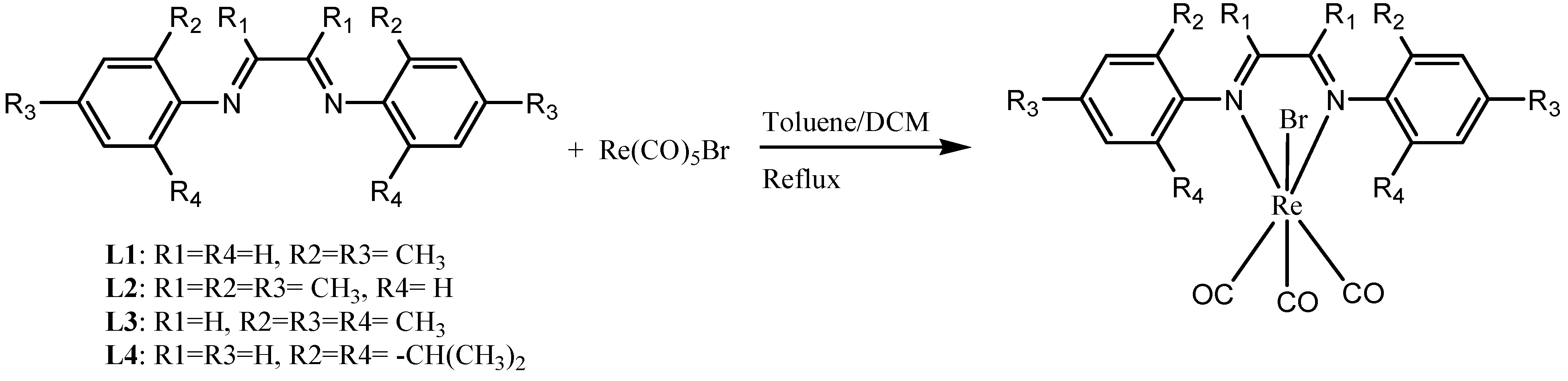
3.1. Synthesis and Characterization

The new [(NN)Re(CO)3X] (NN = diazabutadiene) complexes were synthesized via substitution reaction from the DAB ligands and Re(CO)5Br in a mixture of CH2Cl2 and toluene under reflux condition. Complexes 14 were recrystallized from a mixture of CH2Cl2/n-hexane, and the purity of all complexes was confirmed by elemental analyses. The characteristic feature of the complexes incorporating fac-[Re(CO)3]+ by losing two carbonyl groups in cis positions is the appearance of three intense carbonyl bands at about 2050–1880 cm−1, including a sharp intense band at about 2030–2050 cm−1 and two closely spaced lower energy bands consistent with the A(cis), A′′(cis), and A(trans) modes expected in Cs symmetry, with energy ordering A(cis) >A′′(cis) > A(trans) as reported by Cotton-Karihanzel based on force field calculation [50,51,52]. As a normal trend, the position of the absorption bands is influenced by the electronic nature of the axial ligand. Normally, with the weakly donating ligands in the axial position, the ν(CO) is further increased. The electronic nature of the axial ligand (X) in fac-Re(CO)3(NN)(X) complexes influences merging or splitting of the lower energy bands of the carbonyl groups. A stronger π–acceptor ligand in the basal plane shifts the CO stretching bands to higher frequencies. For complexes 2 and 4, by merging two lower energy bands, there seems to be an approximate C3v spectroscopic symmetry, leading to a virtual “E” band. The FT-IR spectra of complexes 14 are shown in Figures S9–S12. The stretching frequencies of the complexes are listed in the experimental section. The 1H-NMR and 13C{1H}-NMR spectra of complexes 14 are shown in Figures S13–S20.

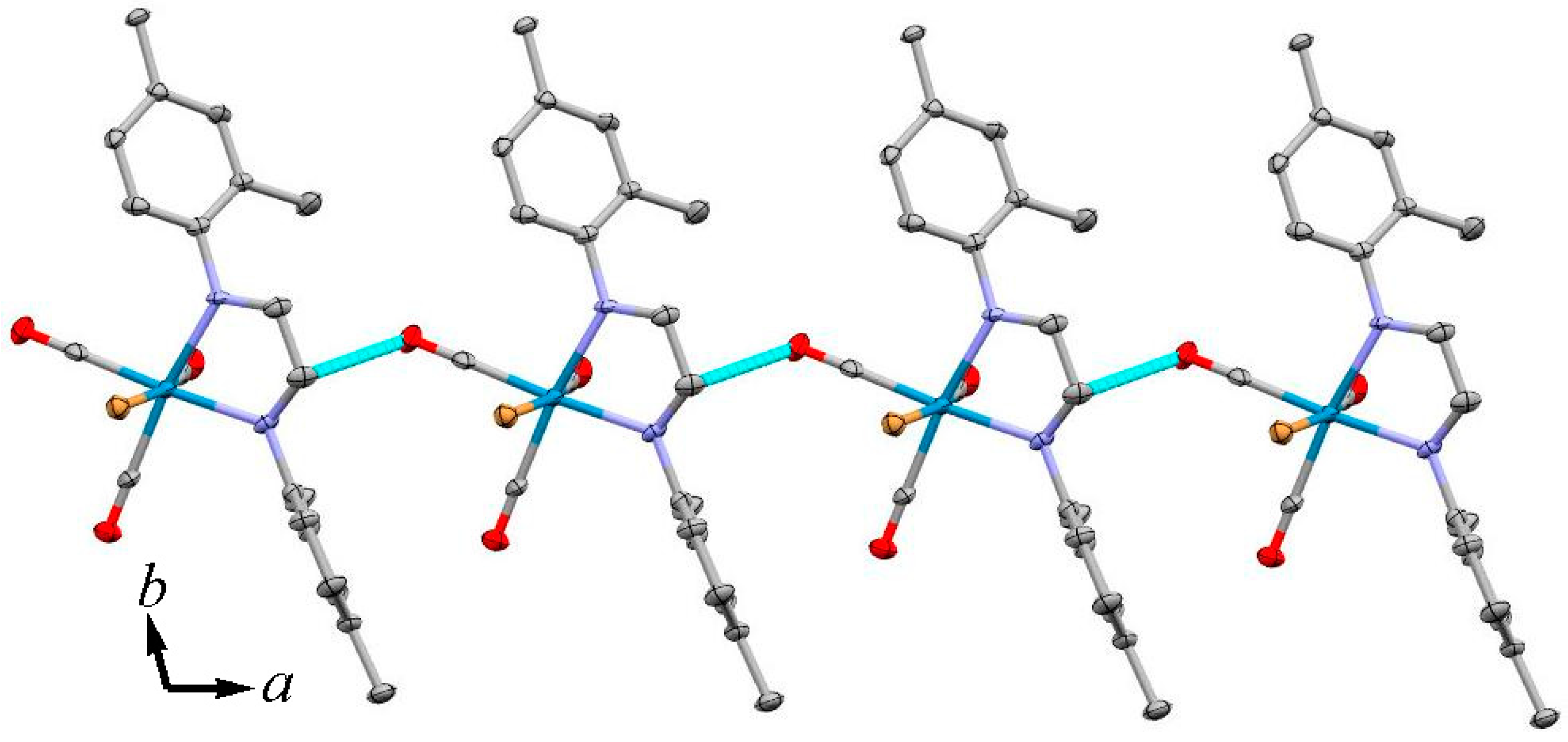
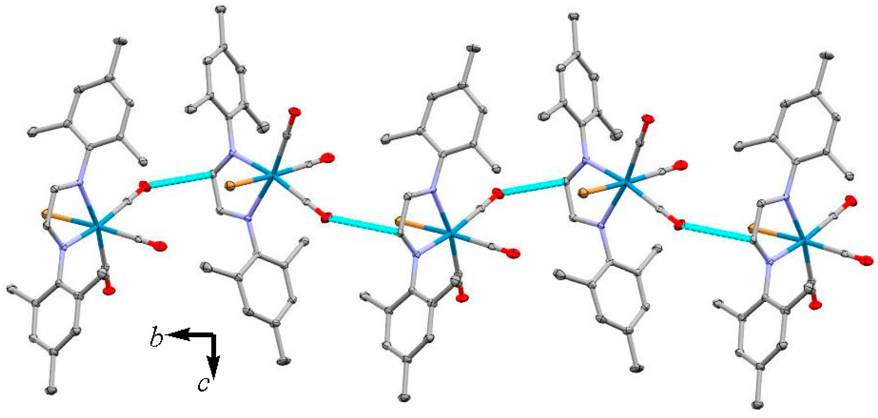
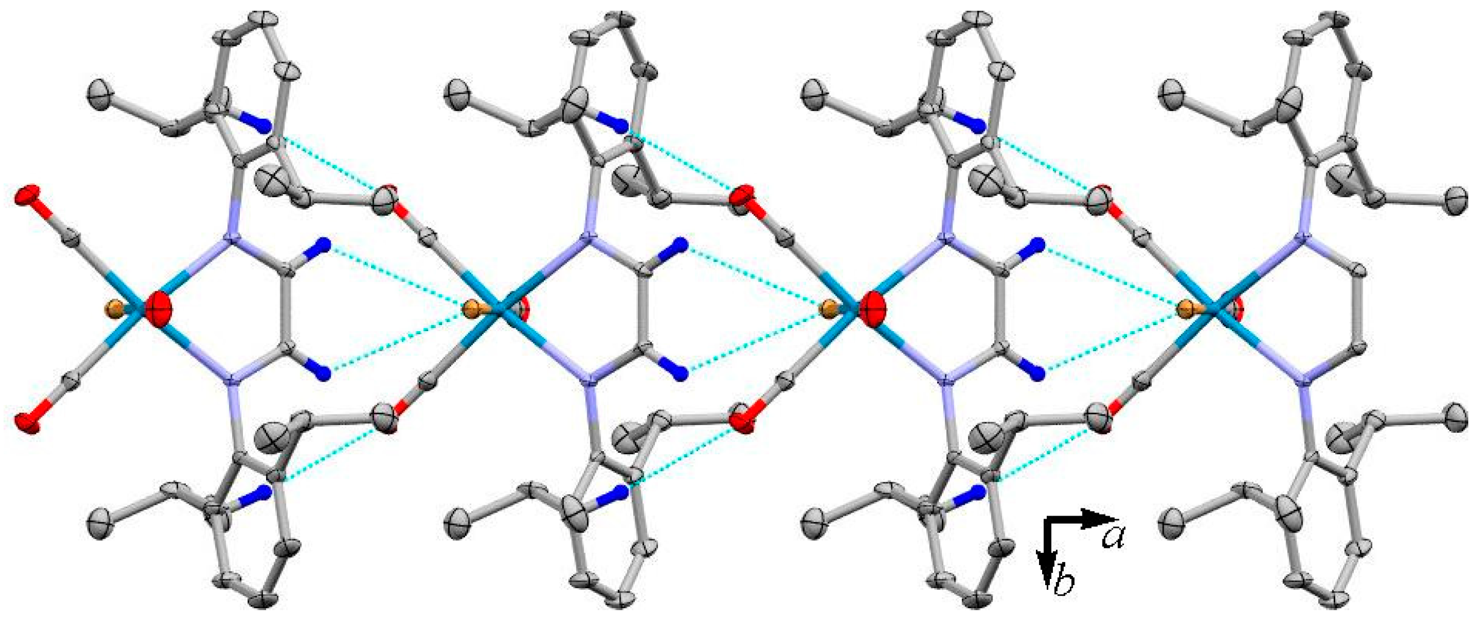
3.2. X-ray Crystal Structures

The solid-state structures of complexes 14 were determined by X-ray crystallography and are shown with their atom labelling scheme in Figure 1. Details of data collection and refinement parameters are given in Table 1. Selected bond lengths and angles for complexes 14 are listed in Table 2. The details of the hydrogen bonding interactions are listed in Table 3.
The geometry around the Re(I) is a distorted octahedron involving the carbonyl groups in facial arrangement, a DAB ligand and axial bromo group. In complex 2, the methyl group in the ortho position was disordered over two positions with a refined site-occupancy ratio of 0.81(2)/0.19(2). The most significant angular distortion is associated with the bite angles of the DAB ligands N(1)–Re(1)–N(2) in the range of 73.3(4)–75.62(12)° which is due to the formation of the strained five-membered chelate ring. The trans angle C(1)–Re(1)–Br(1) in 14 falls in the range of 173.55(12)–177.7(4)°, respectively. The bond lengths, angles and coordination geometry of the crystal structures in 1–4 are similar to those structures reported previously [53,54]. There is only one report related to the crystal structure of 1,4-alkyldiazabutadiene, namely 1,4-di(tert-butyl) diazabutadiene, which shows significantly different Re—N [2.170(15), 2.226(19) Å], C≡O [1.13(3), 1.16(3), and 1.01(3) Å] bond lengths compared to 1,4-aryldiazabutadiene, but the Re—Br bond length is similar to complexes 1–4 [13]. The interesting feature of complexes 1 and 3 is the intermolecular nπ* interactions which connects neighbouring molecules into a 1-D extended chain along the a- and b-axis, respectively (Figure 2 and Figure 3). As it is summarized in Table 3, the crystal packing of the complexes is consolidated mainly by the intermolecular non-classic C–HO and C–HBr hydrogen bonding. In complexes 1 and 4, the intermolecular C–HBr and C–HO interactions form individual dimers in the crystal packing, respectively. In case of complexes 2 and 3, intermolecular C–HBr and C–HO interactions connect neighbouring molecules into an extended infinite chain along the b-axis, respectively. Figure 4 shows one-dimensional extended chain formation along the a-axis through intermolecular C–HO and C–HBr interactions in complex 4.

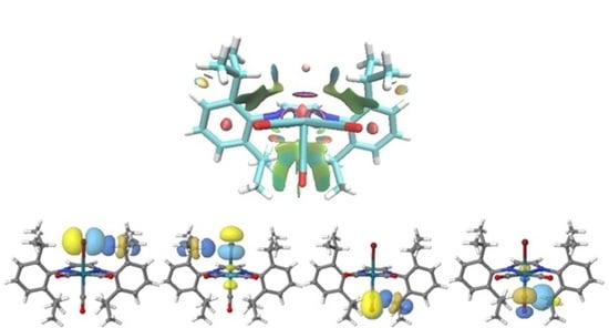
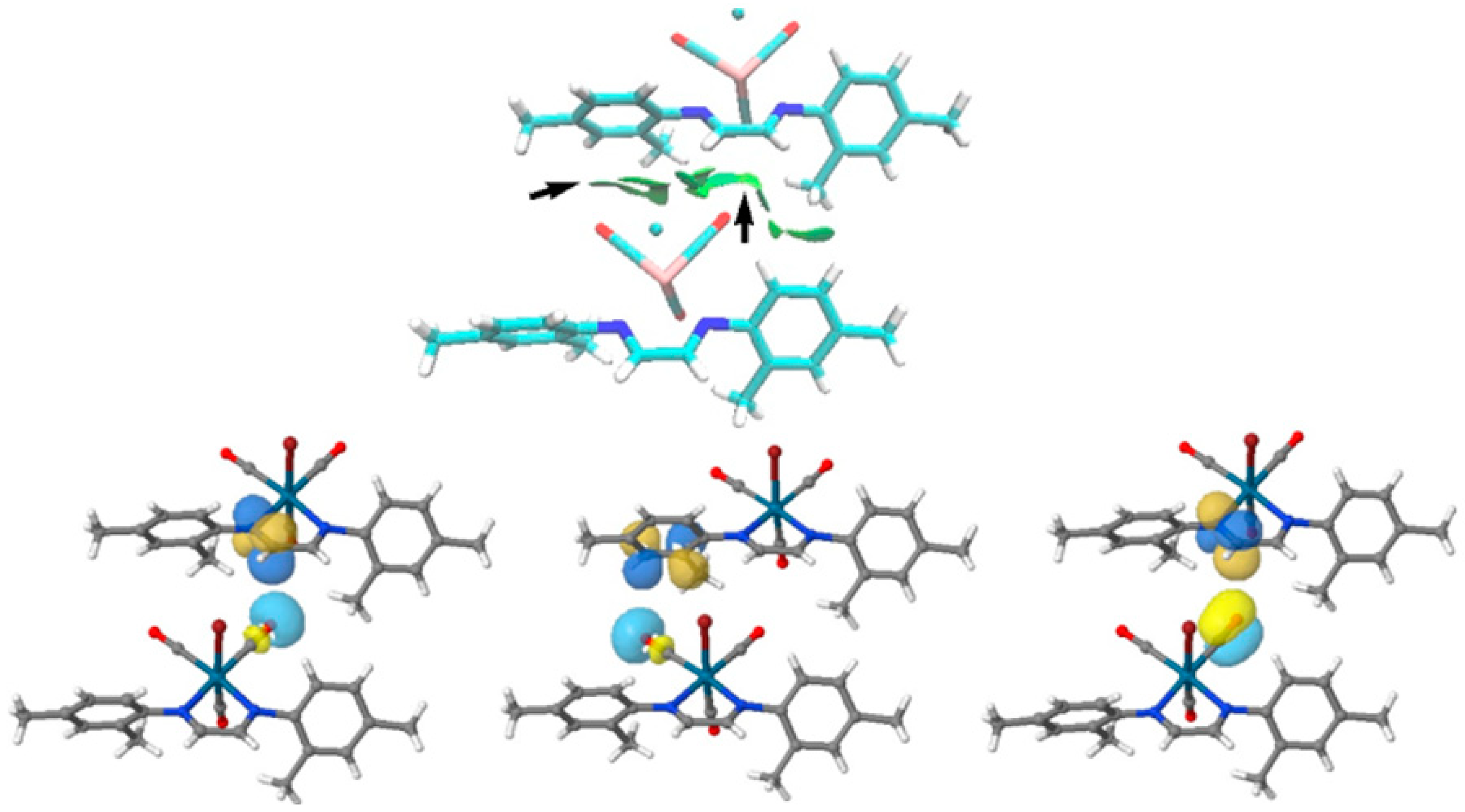
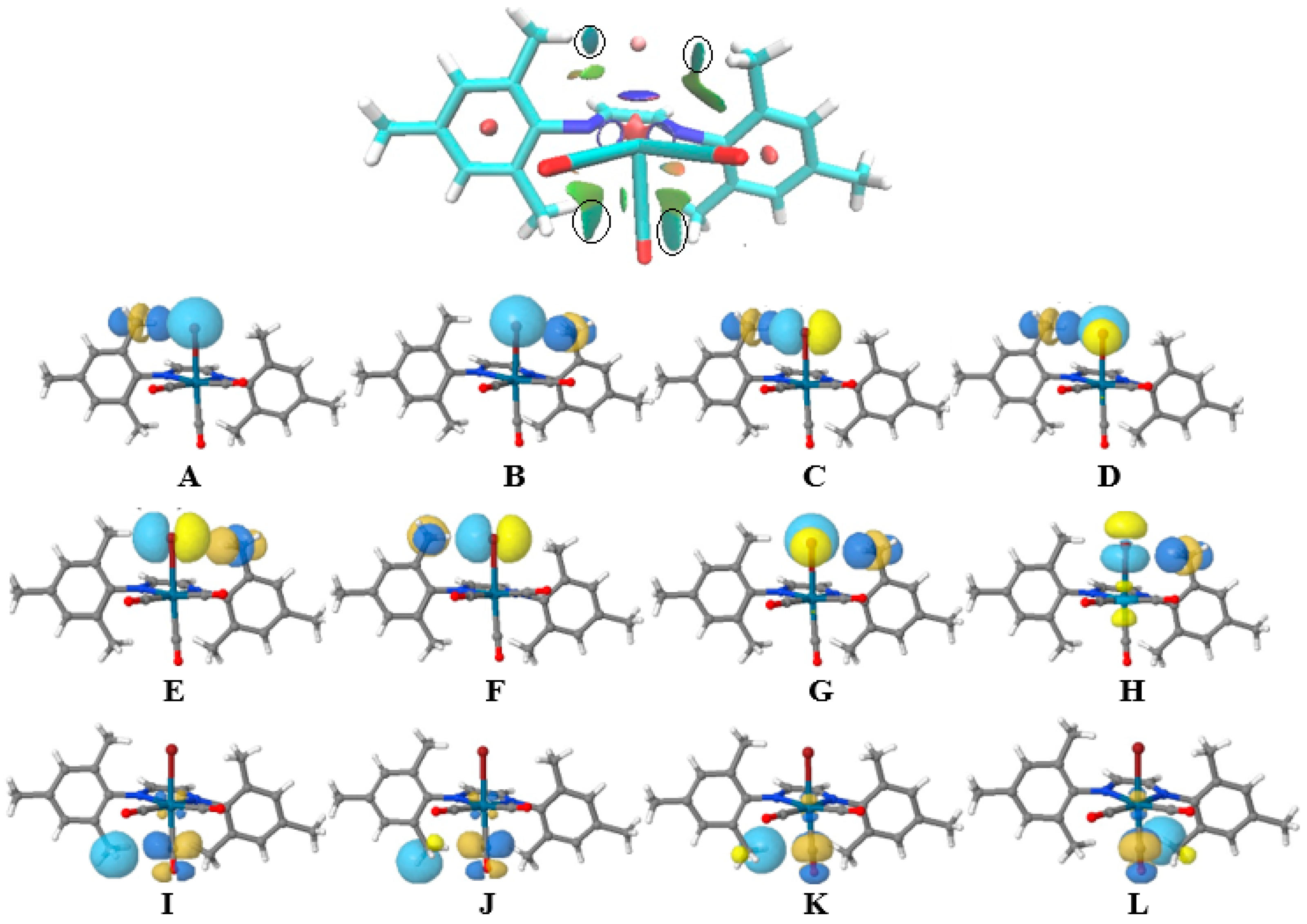
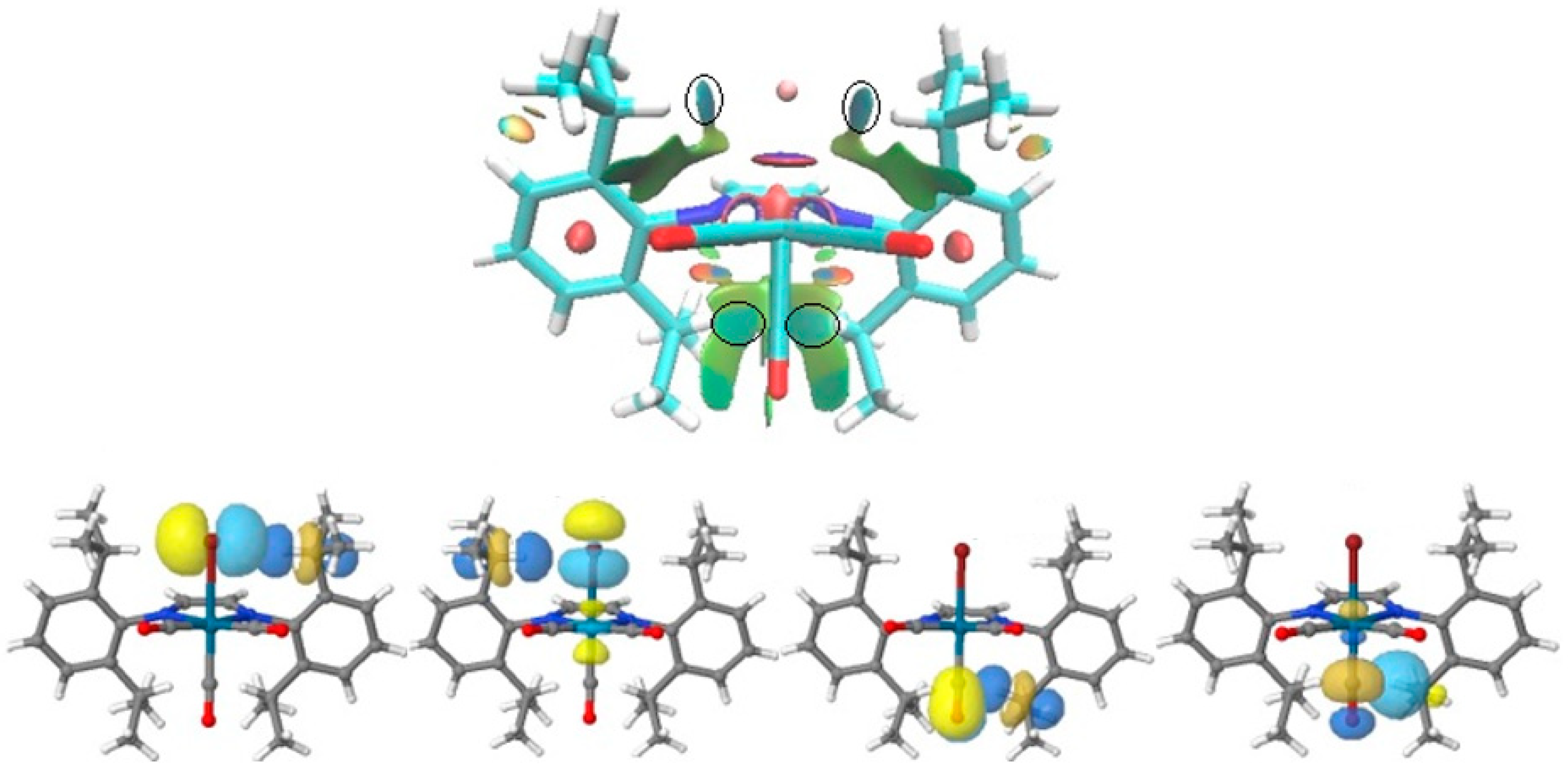
3.3. Non-Covalent Interaction Index and Natural Bond Orbitals (NBOs)

Non-covalent interactions were evaluated using the non-covalent index (NCI) approach, which relies on the topological analysis of the electron density and its derivatives at low density regions based on the reduced density gradient (RDG) [55,56]. The NCI isosurface regions show both stabilizing and destabilizing weak interactions. These are distinguishable according to the total sign of the second eigenvalue of the Hessian (λ2) matrix, where the sign of the λ2 quantity can vary accordingly and are thus suggested as a useful descriptor to characterize such situations. Negative values of the product given by ρ*sign (λ2), denote stabilizing interactions. Values close to zero account for weak interactions (van der Waals forces), while positive values account for weak repulsive cases. One of the main reasons to include the Br ligand in the axial position of the complexes was to increase the possibility of intra- and intermolecular interactions through halogenhalogen and halogenoxygen bonding, since Br has a greater tendency to act as halogen bond acceptor compared to Cl, because of the greater σ-hole [57]. Complexes 1, 3, and 4 showed some interesting intra- and intermolecular interactions which were investigated by non-covalent interaction index (NCI) and NBO calculations. The NCI analyses for single molecules were done based on the optimized structures, but in case of the dimer pairs they were calculated directly from the crystallographically generated dimers without optimization. The surface NCI analyses are depicted in Figure 5, Figure 6, Figure 7 and Figure 8, revealing stabilizing weak non-covalent intramolecular interactions in single-molecule and intermolecular interactions between interacting pairs, respectively. On the other hand, to shed light on the nature and strength of the intra- and intermolecular interactions depicted by NCI calculations, NBO analysis of other suitable descriptors for bond analysis was used with more details. Natural bond orbital calculations were performed on 1, 3, and 4 with the same level of theory for molecular orbital calculations [58]. The NBO analysis shows that the attractive nature is associated with donor–acceptor orbital interactions in single molecules and between the pairs. Figure 5 shows the intermolecular interactions in 1 with contributions from the lone pair of oxygen in the carbonyl group and the π* of the imine functional group, ns(O3)π*(C10=N1) and π(C3≡O3)π*(C10=N1), with 0.47 kcal mol−1 according to the NBO analysis from second-order perturbation energy. The energy contribution of ns(O2)π*(C8=C9) was negligible.
The NCI plot and NBO analyses of 3 are shown in Figure 6. The NCI plot shows interactions between the C–H bond of the methyl group and Br [AH] and the C—H bond and the carbonyl group, IL, in axial positions with light-blue color, confirming attractive interactions. These interactions are due to the overlap of the perpendicular occupied s and p orbitals of Br to the empty σ* orbital of the C‒H bond with overall energy release of 8.07 kcal mol−1, based on the NBO analysis from second-order perturbation energy. The contribution from np(Br)σ*(C—H), C-D, was 2.48, 2.27, and 2.09 kcal mol−1, respectively.
On the other hand, the NCI plot and NBO calculations of the interaction pairs of 3 were also analyzed and depicted in Figure 7. The total energy release of the n(O)π* and n(O)σ* was 0.68 kcal mol−1, while the contribution from n(Br)π* was 0.31 kcal mol−1 based on second-order perturbation energy calculations.
The NCI plot and NBO analyses of 4 are shown in Figure 8. The NCI plot shows some intramolecular interactions among the Br, isopropyl and CO groups with green-blue color, confirming attractive interaction. The NBO study confirms the np(Br)σ*, π(CO)σ*, and σπ*(CO) interactions in 4 with energy releases of 3.46, 0.28 and 0.44 kcal mol−1 based on the NBO analysis from second-order perturbation energy, respectively.

4. Conclusions

In this paper, we have described the synthesis, characterization, structural and full computational studies of four bromide- tricarbonyl Re(I) complexes 14, bearing substituted diazabutadiene ligands. The molecular structures of the complexes were established by single-crystal X-ray diffraction, feature the metal in a distorted octahedral environment with facial arrangement of the carbonyl groups in the complexes, which was also confirmed by FT-IR spectroscopy. The nature and energy of the intermolecular n(O)π* interactions between carbonyl-bound metal and π* of benzene ring and imine segments in 1 and n(O)π* and n(Br)π* in 3 were investigated in detail. The presence of such interactions was also confirmed by NCI index based on colour codes. The complexes 3 and 4 showed interesting intramolecular np(Br)σ* interactions, which stabilized the internal geometry of the coordinated ligand around the metal center.

Supplementary Materials

The following are available online at https://0-www-mdpi-com.brum.beds.ac.uk/2073-4352/10/4/267/s1. CCDC 1962581(1), 1962582(2), 1962583(3), and 1962580(4) contain the supplementary crystallographic data for this paper. These data can be obtained free of charge from The Cambridge Crystallographic Data Centre via www.ccdc.cam.ac.uk/data_request/cif. FT-IR of complexes 14, the crystal packing of complexes 14, some of the important frontier molecular orbitals of 5, and crystallographic data of complexes 14 in CIF format. Figure S1. The 11H-NMR spectrum of complex L1; Figure S2. The 13C{1H}-NMR spectrum of complex L1; Figure S3. The 1H-NMR spectrum of complex L2; Figure S4. The 13C{1H}-NMR spectrum of complex L2; Figure S5. The 1H-NMR spectrum of complex L3; Figure S6. The 13C{1H}-NMR spectrum of complex L3; Figure S7. The 1H-NMR spectrum of complex L4; Figure S8. The 13C{1H}-NMR spectrum of complex L4; Figure S9. The FTIR spectrum of 1 in KBr pellet; Figure S10. The FTIR spectrum of 2 in KBr pellet; Figure S11. The FTIR spectrum of 3 in KBr pellet; Figure S12. The FTIR spectrum of 4 in KBr pellet; Figure S13. The 1H-NMR spectrum of complex 1; Figure S14. The 13C{1H}-NMR spectrum of complex 1; Figure S15. The 1H-NMR spectrum of complex 2; Figure S16. The 13C{1H}-NMR spectrum of complex 2; Figure S17. The 1H-NMR spectrum of complex 3; Figure S18. The 13C{1H}-NMR spectrum of complex 3; Figure S19. The 1H-NMR spectrum of complex 4; Figure S20. The 13C{1H}-NMR spectrum of complex 4.

Author Contributions

Manuscript preparation, X-ray structure determination, final structure analysis, and final editing of manuscript: R.K.; Synthesis and spectroscopic characterization of the complexes, manuscript review A.K. All authors have read and agreed to the published version of the manuscript.

Funding

This research was funded by Sharif University of Technology Research Council for financial support.

Conflicts of Interest

The funder had no role in the design of the study and in the decision to publish the results.

References

  1. Ehlers, A.W.; Dapprich, S.; Vyboishchikov, S.F.; Frenking, G. Structure and Bonding of the Transition-Metal Carbonyl Complexes M(CO)5L (M = Cr, Mo, W) and M(CO)3L (M = Ni, Pd, Pt; L = CO, SiO, CS, N2, NO+, CN-, NC-, HCCH, CCH2, CH2, CF2, H2). Organometallics 1996, 15, 105–117. [Google Scholar] [CrossRef]
  2. Lee, A.J. Luminescence properties of organometallic complexes. Chem. Rev. 1987, 87, 711–743. [Google Scholar]
  3. Farrell, I.R.; Vlcek, A., Jr. Mechanisms of ultrafast metal–ligand bond splitting upon MLCT excitation of carbonyl-diimine complexes. Coord. Chem. Rev. 2000, 208, 87–101. [Google Scholar] [CrossRef]
  4. Striplin, D.R.; Crosby, G.A. Photophysical investigations of rhenium(I)Cl(CO)3(phenanthroline) complexes. Coord. Chem. Rev. 2001, 211, 163–175. [Google Scholar] [CrossRef]
  5. Collin, J.P.; Sauvage, J.P. Electrochemical reduction of carbon dioxide mediated by molecular catalysts. Coord. Chem. Rev. 1989, 93, 245–268. [Google Scholar] [CrossRef]
  6. Rossenaar, B.R.; Hartl, F.; Stufkens, D.J. Reduction of [Re(X)(CO)3(R′-DAB)] (X = Otf-, Br-; DAB = Diazabutadiene; R′ = iPr, pTol, pAn) and [Re(R)(CO)3(iPr-DAB)] (R = Me, Et, Bz) Complexes:  A Comparative (Spectro)electrochemical Study at Variable Temperatures. Inorg. Chem. 1996, 35, 6194–6203. [Google Scholar] [CrossRef] [Green Version]
  7. Balzani, V.; Juris, A.; Venturi, M.; Campagna, S.; Serroni, S. Luminescent and Redox-Active Polynuclear Transition Metal Complexes. Chem. Rev. 1996, 96, 759–834. [Google Scholar] [CrossRef]
  8. Slone, R.V.; Hupp, J.T. Synthesis, Characterization, and Preliminary Host−Guest Binding Studies of Porphyrinic Molecular Squares Featuring fac-Tricarbonylrhenium(I) Chloro Corners. Inorg. Chem. 1997, 36, 5422–5423. [Google Scholar] [CrossRef]
  9. Nieto, S.; Perez, J.; Riera, L.; Riera, V.; Miguel, D. Non-covalent interactions between anions and a cationic rhenium diamine complex: Structural characterization of the supramolecular adducts. New J. Chem. 2006, 30, 838–843. [Google Scholar] [CrossRef]
  10. Hevia, E.; Perez, J.; Riera, V.; Miguel, D.; Kassel, S.; Rheingold, A.L. New Synthetic Routes to Cationic Rhenium Tricarbonyl Bipyridine Complexes with Labile Ligands. Inorg. Chem. 2002, 41, 4673–4679. [Google Scholar] [CrossRef]
  11. Machura, B.; Wolff, M.; Jaworska, M.; Lodowski, P.; Benoist, E.; Carrayon, C.; Saffon, N.; Kruszynski, R.; Mazurak, Z. Rhenium(I) carbonyl complex of 4,7-diphenyl-1,10-phenanthroline–Spectroscopic properties, X-Ray structure, theoretical studies of ground and excited electronic states. J. Organomet. Chem. 2011, 696, 3068–3075. [Google Scholar] [CrossRef]
  12. Drozdz, A.; Bubrin, M.; Fiedler, J.; Zalis, S.; Kaim, W. (α-Diimine)tricarbonylhalorhenium complexes: The oxidation side. Dalton Trans. 2012, 41, 1013–1019. [Google Scholar] [CrossRef] [PubMed]
  13. Grupp, A.; Bubrin, M.; Ehret, F.; Kvapilova, H.; Zalis, S.; Kaim, W. Oxidation and reduction response of α-diimine complexes with tricarbonylrhenium halides and pseudohalides. J. Organomet. Chem. 2014, 751, 678–685. [Google Scholar] [CrossRef]
  14. Kumar, A.; Sun, S.-S.; Lees, A.J. Photophysics and Photochemistry of Organometallic Rhenium Diimine Complexes. Top. Organomet. Chem. 2010, 29, 1–35. [Google Scholar]
  15. Kirgan, R.A.; Sullivan, B.P.; Rillema, D.P. Photochemistry and Photophysics of Coordination Compounds: Rhenium. Top. Curr. Chem. 2007, 281, 45–100. [Google Scholar]
  16. Kinghat, R.; Khatyr, A.; Knorr, M.; Kubicki, M.M.; Vigier, E.; Villafañe, F. Mono- and di-nuclear 2,3-diazabutadiene and 2-azabutadiene complexes of Rhenium(I): Syntheses, luminescence spectra and X-ray structures. Inorg. Chem. Commun. 2008, 11, 1060. [Google Scholar] [CrossRef]
  17. Zou, W.; Chen, C. Influence of Backbone Substituents on the Ethylene (Co)polymerization Properties of a-diimine Pd(II) and Ni(II) Catalysts. Organometallics 2016, 35, 1794–1801. [Google Scholar] [CrossRef]
  18. Kong, S.; Song, K.; Liang, T.; Guo, C.-Y.; Sun, W.-H.; Redshaw, C. 18 Methylene-bridged bimetallic α-diimino nickel(II) complexes: Synthesis and high efficiency in ethylene polymerization. Dalton Trans. 2013, 42, 9176–9178. [Google Scholar] [CrossRef]
  19. Abakumov, G.A.; Druzhkov, N.O.; Egorova, E.N.; Kocherova, T.N.; Shavyrin, A.S.; Cherkasov, A.V. Intramolecular cyclization–decyclization of new sterically hindered diiminophenol. Synthesis and coordination abilities. RSC Adv. 2014, 4, 14495–14500. [Google Scholar] [CrossRef]
  20. Egorova, E.N.; Druzhkov, N.O.; Shavyrin, A.S.; Cherkasov, A.V.; Abakumova, G.A.; Fedorov, A.Y. The group 13 metal complexes of stericallyhindered substituted iminophenol: Synthesis and structure. RSC Adv. 2015, 5, 19362–19367. [Google Scholar] [CrossRef]
  21. Echeverría, J. The n → π* interaction in metal complexes. Chem. Commun. 2018, 54, 3061–3064. [Google Scholar] [CrossRef] [PubMed] [Green Version]
  22. Burgi, H.B.; Dunitz, J.D.; Lehn, J.M.; Wipff, G. Stereochemistry of reaction paths at carbonyl centres. Tetrahedron 1974, 30, 1563–1572. [Google Scholar] [CrossRef]
  23. Burgi, H.B.; Dunitz, J.D.; Shefter, E. Geometrical reaction coordinates. II. Nucleophilic addition to a carbonyl group. J. Am. Chem. Soc. 1973, 95, 5065–5067. [Google Scholar] [CrossRef]
  24. Mooibroek, T.J.; Gamez, P.; Reedijk, J. Lone pair–π interactions: A new supramolecular bond? CrystEngComm 2008, 10, 1501–1515. [Google Scholar] [CrossRef]
  25. Jakobsche, C.E.; Choudhary, A.; Miller, S.J.; Raines, R.T. n → π* Interaction and n)(π Pauli Repulsion Are Antagonistic for Protein Stability. J. Am. Chem. Soc. 2010, 132, 6651–6653. [Google Scholar] [CrossRef] [Green Version]
  26. Kramer, K.J.; Choudhary, A.; Raines, R.T. Intimate Interactions with Carbonyl Groups: Dipole–Dipole or n→π*? J. Org. Chem. 2013, 78, 2099–2103. [Google Scholar] [CrossRef] [Green Version]
  27. Bartlett, G.J.; Newberry, R.W.; Van Veller, B.; Raines, R.T.; Woolfson, D.N. Interplay of Hydrogen Bonds and n → π* Interactions in Proteins. J. Am. Chem. Soc. 2013, 135, 18682–18688. [Google Scholar] [CrossRef] [Green Version]
  28. Singh, S.K.; Das, A. The n → π* interaction: A rapidly emerging non-covalent interaction. Phys. Chem. Chem. Phys. 2015, 17, 9596–9612. [Google Scholar] [CrossRef] [Green Version]
  29. Velásquez, J.D.; Echeverría, J.; Alvarez, S. Effect of the Substituents on the Nature and Strength of Lone-Pair–Carbonyl Interactions in Acyl Halides. Cryst. Growth Des. 2019, 11, 6511–6518. [Google Scholar] [CrossRef]
  30. Newberry, R.W.; Raines, R.T. The n→π* Interaction. Acc. Chem. Res. 2017, 50, 1838–1846. [Google Scholar] [CrossRef] [Green Version]
  31. Doppert, M.T.; van Overeem, H.; Mooibroek, T.J. Intermolecular π-hole/n→π* interactions with carbon monoxide ligands in crystal structures. Chem. Commun. 2018, 54, 12049–12052. [Google Scholar] [CrossRef] [PubMed]
  32. van der Werve Ad, R.; van Dijk, Y.R.; Mooibroek, T.J. π-Hole/n→π* interactions with acetonitrile in crystal structures. Chem. Commun. 2018, 54, 10742–10745. [Google Scholar] [CrossRef] [PubMed]
  33. Zukerman-Schpector, J.; Haiduc, I.; Tiekink, E.R.T. The metal–carbonyl⋯π(aryl) interaction as a supramolecular synthon for the stabilisation of transition metal carbonyl crystal structures. Chem. Commun. 2011, 47, 12682–12684. [Google Scholar] [CrossRef] [PubMed]
  34. Zukerman-Schpector, J.; Haidu, I.; Tiekink, E.R.T. Supramolecular Self-assembly of Transition Metal Carbonyl Molecules Through M–CO(Lone Pair)…π(Arene) Interactions. Adv. Organomet. Chem. 2012, 60, 49–92. [Google Scholar]
  35. Wan, C.-Q.; Chen, X.-D.; Mak, T.C.W. Supramolecular frameworks assembled via intermolecular lone pair-aromatic interaction between carbonyl and pyridyl groups. CrystEngComm 2008, 10, 475–478. [Google Scholar] [CrossRef]
  36. Echeverría, J. Intermolecular Carbonyl Carbonyl Interactions in Transition-Metal Complexes. Inorg. Chem. 2018, 57, 5429–5437. [Google Scholar] [CrossRef]
  37. Murcia-García, C.; Bauzá, A.; Schnakenburg, G.; Frontera, A.; Streubel, R. Surprising behaviour of M–CO(lone pair)⋯π(arene) interactions in the solid state of fluorinated oxaphosphirane complexes. CrystEngComm 2015, 17, 1769–1772. [Google Scholar] [CrossRef]
  38. Mark-Lee, W.F.; Chong, Y.Y.; Kassim, M.B. Supramolecular structures of rhenium (I) complexes mediated by ligand planarity via the interplay of substituents. Acta Crystallogr. Sect. C Struct. Chem. 2018, 74, 997–1006. [Google Scholar]
  39. Kia, R.; Hosseini, M.; Abdolrahimi, A.; Mahmoudi, M. Intermolecular C–H⋯O and n→π* and short intramolecular σ → π* interactions in the molybdenum(0) tetracarbonyl complex of a very twisted 14-membered tetraazaannulene macrocyclic ligand: Structural and computational studies. CrystEngComm 2019, 21, 5222–5226. [Google Scholar] [CrossRef]
  40. Kia, R.; Mahmoudi, S.; Raithby, P.R. New rhenium-tricarbonyl complexes bearing halogen-substituted bidentate ligands: Structural, computational and Hirshfeld surfaces studies. CrystEngComm 2019, 21, 77–93. [Google Scholar] [CrossRef] [Green Version]
  41. Villegas, J.M.; Stoyanov, S.R.; Huang, W.; Rillema, D.P. A spectroscopic and computational study on the effects of methyl and phenyl substituted phenanthroline ligands on the electronic structure of Re(I) tricarbonyl complexes containing 2,6-dimethylphenylisocyanide. Dalton Trans. 2005, 34, 1042–1051. [Google Scholar] [CrossRef] [PubMed]
  42. Kliegman, J.M.; Barnes, R.K. Glyoxal derivatives-I: Conjugated aliphatic diimines from glyoxal and aliphatic primary amines. Tetrahedron 1970, 26, 2555–2560. [Google Scholar] [CrossRef]
  43. Clark, R.C.; Reid, J.S. The analytical calculation of absorption in multifaceted crystals. Acta Cryst. 1995, A51, 887–897. [Google Scholar] [CrossRef]
  44. Dolomanov, O.V.; Bourhis, L.J.; Gildea, R.J.; Howard, J.A.K.; Puschmann, H. OLEX2: A complete structure solution, refinement and analysis program. J. Appl. Cryst. 2009, 42, 339–341. [Google Scholar] [CrossRef]
  45. Sheldrick, G.M. A short history of SHELX. Acta Cryst. 2008, A64, 112–122. [Google Scholar] [CrossRef] [Green Version]
  46. Spek, A.L. Structure validation in chemical crystallography. Acta Cryst. 2009, D65, 148–155. [Google Scholar] [CrossRef]
  47. Frisch, M.J.; Trucks, G.W.; Schlegel, H.B.; Scuseria, G.E.; Robb, M.A.; Cheeseman, J.R.; Scalmani, G.; Barone, V.; Mennucci, B. Gaussian 09, Revision A.02; ScienceOpen: Berlin, Germany, 2014. [Google Scholar]
  48. Perdew, J.P.; Burke, K.; Ernzerhof, M. Generalized Gradient Approximation Made Simple. Phys. Rev. Lett. 1996, 77, 3865. [Google Scholar] [CrossRef] [Green Version]
  49. Andrae, D.; Häußermann, U.; Dolg, M.; Stoll, H.; Preuß, H. Energy-adjustedab initio pseudopotentials for the second and third row transition elements. Theor. Chim. Acta 1990, 77, 123–141. [Google Scholar] [CrossRef]
  50. Cotton, F.A.; Karihanzel, C.S. Vibrational Spectra and Bonding in Metal Carbonyls. I. Infrared Spectra of Phosphine-substituted Group VI Carbonyls in the CO Stretching Region. J. Am. Chem. Soc. 1962, 84, 4432–4438. [Google Scholar] [CrossRef]
  51. Klein, A.; Vogler, C.; Kaim, W. The δ in 18 + δ Electron Complexes:  Importance of the Metal/Ligand Interface for the Substitutional Reactivity of “Re(0)” Complexes (α-diimine)ReI(CO)3(X). Organometallics 1996, 15, 236–244. [Google Scholar] [CrossRef]
  52. Staal, L.H.; Oskam, A.; Vrieze, K. The syntheses and coordination properties of M(CO)3X(DAB) (M = Mn, Re; X = Cl, Br, I.; DAB = 1,4-diazabutadiene). J. Organomet. Chem. 1979, 170, 235–245. [Google Scholar] [CrossRef]
  53. Vollmer, M.V.; Machan, C.W.; Clark, M.L.; Antholine, W.E.; Agarwal, J.; Schaefer III, H.F.; Kubiak, C.P.; Walensky, J.R. Synthesis, Spectroscopy, and Electrochemistry of (α-Diimine)M(CO)3Br, M = Mn, Re, Complexes: Ligands Isoelectronic to Bipyridyl Show Differences in CO2 Reduction. Organometallics 2015, 34, 3–12. [Google Scholar] [CrossRef] [PubMed] [Green Version]
  54. Abramov, P.A.; Dmitriev, A.A.; Kholin, K.V.; Gritsan, N.P.; Kadirov, M.K.; Gushchin, A.L.; Sokolov, M.N. Mechanistic study of the [(dpp-bian)Re(CO)3Br] electrochemical reduction using in situ EPR spectroscopy and computational chemistry. Electrochim. Acta 2018, 270, 526–534. [Google Scholar] [CrossRef]
  55. Johnson, E.R.; Keinan, S.; Mori-Sánchez, P.; Contreras-Garcia, J.; Cohen, A.J.; Yang, W. Revealing Noncovalent Interactions. J. Am. Chem. Soc. 2010, 132, 6498–6506. [Google Scholar] [CrossRef] [Green Version]
  56. Contreras-García, J.; Johnson, E.R.; Keinan, S.; Chaudret, R.; Piquemal, J.-P.; Beratan, D.N.; Yang, W. NCIPLOT: A program for plotting non-covalent interaction regions. J. Chem. Theory Comput. 2010, 7, 625–632. [Google Scholar] [CrossRef]
  57. Usoltsev, A.N.; Adonin, S.A.; Novikov, A.S.; Samsonenko, D.G.; Sokolova, M.N.; Fedin, V.P. One-dimensional polymeric polybromotellurates (IV): Structural and theoretical insights into halogen···halogen contacts. CrystEngComm 2017, 19, 5934–5939. [Google Scholar] [CrossRef]
  58. Glendening, E.D.; Landis, C.R.; Weinhold, F. Natural bond orbital methods. WIREs Comput. Mol. Sci. 2012, 2, 1–42. [Google Scholar] [CrossRef]
Scheme 1. Synthesis pathway of complexes 14.
Crystals 10 00267 sch001
Figure 1. The molecular structure of complexes 14, showing 40% probability displacement ellipsoids and the atomic numbering (H atoms are shown as blue wire). The symmetry code for unlabeled atoms in 4 is x, -y+1/2, z.
Crystals 10 00267 g001
Figure 2. Part of the crystal packing of 1, showing one-dimensional extended chain formation along the a-axis through intermolecular n→π* interaction (H atoms omitted for clarity).
Crystals 10 00267 g002
Figure 3. Part of the crystal packing of 3, showing one-dimensional extended chain formation along the b-axis through intermolecular n→π* interaction (H atoms omitted for clarity).
Crystals 10 00267 g003
Figure 4. Part the crystal packing of 4, showing one-dimensional extended chain formation along the a-axis through intermolecular C–HO and C–HBr interaction (H atoms omitted for clarity).
Crystals 10 00267 g004
Figure 5. NCI plot of interacting dimer in 1 and the donor–acceptor interacting orbitals from NBO calculations. The area of nπ* and ππ* are shown in the NCI plot with arrows.
Crystals 10 00267 g005
Figure 6. NCI plot of 3 and the representation of interacting donor–acceptor ns(Br)σ*(CH), AB; np(Br)σ*(CH), CH; σ(CH)π*(CO), IL; orbitals from NBO calculations.
Crystals 10 00267 g006
Figure 7. NCI plot of the interacting dimer in 3 and the representation of donor–acceptor interacting n(O)π*, n(O)σ*, and n(Br)π* orbitals from NBO calculations. The area is shown in the NCI plot with arrows.
Crystals 10 00267 g007
Figure 8. NCI plot of interacting dimer in 4 and the donor–acceptor interacting orbitals from NBO calculations through np(Br)σ*, np(Br)σ*, π(CO)σ*, σπ*(CO) from left to right, respectively. The interaction area is shown with black circles.
Crystals 10 00267 g008
Table 1. Crystal data and refinement parameters of complexes 1–4.
Complex1234
Empirical Formula
Formula Mass
Crystal Size (mm)
Colour
Crystal System
Space Group
θmax (°)
a (Å)
b (Å)
c (Å)
α (°)
β (°)
γ (°)
V3)
Z
Dcalc (Mg/m3)
μ (mm−1)
F (000)
Index Ranges
 
 
No. of Measured Reflns.
No. of independent
reflns./Rint
No. of observed
reflns. I > 2σ(I)
No. of parameters
Goodness-of-fit (GOF)
R1 (observed data)
wR2 (all data)
C21H20BrN2O3Re
614.50
0.10 × 0.15 × 0.25
Dark-brown
Monoclinic
P21/n
26
7.3182(2)
21.8786(9)
13.0930(5)
90
95.658(3)
90
2086.13(13)
4
1.957
7.764
1176
−9 ≤ h ≤8
−26 ≤ k ≤26
−17 ≤ l ≤16
16734
 
4084/0.046
 
3477
257
1.07
0.0261
0.0574
C23H24BrN2O3Re
642.55
0.04 × 0.08 × 0.15
Dark-brown
Triclinic
P-1
30.4
8.1105(3)
8.3373(4)
18.3651(11)
93.857(4)
97.380(4)
104.588(4)
1185.41(10)
2
1.800
6.836
620
−11 ≤ h ≤ 11
−11 ≤ k ≤ 11
−25 ≤ l ≤ 25
23113
 
6361/0.067
 
5247
282
1.18
0.0783
0.1694
C23H24N2O3Re
642.55
0.10 × 0.18 × 0.35
Dark-brown
Orthorhombic
Pbca
72.9
14.0730(2)
13.8361(2)
23.2653(3)
90
90
90
4530.11(11)
8
1.884
12.775
2480
−17 ≤ h ≤11
−14 ≤ k ≤17
−28 ≤ l ≤28
21739
 
4448/0.03
 
4228
277
1.13
0.0288
0.0726
C29H36BrN2O3Re
726.71
0.10 × 0.18 × 0.35
Dark-red
Orthorhombic
Pnma
29.4
13.3103(5)
21.7931(9)
10.3165(5)
90
90
90
2992.5(2)
4
1.613
5.462
1432
−18 ≤ h ≤15
−27 ≤ k ≤18
−13 ≤ l ≤8
8882
 
3587/0.047
 
2868
173
1.02
0.0377
0.0722
Table 2. Selected bond lengths (Ǻ) and angles (°) of 14.
Bond Lengths (Ǻ)1234
Re(1)–N(1)
Re(1)–N(2)
Re(1)–C(1)
Re(1)–C(2)
Re(1)–C(3)
C(1)–O(1)
C(2)–O(2)
C(3)–O(3)
Re(1)–Br(1)
Bond Angles (°)
N(1)–Re(1)–N(2)
C(1)–Re(1)–N(1)
C(2)–Re(1)–N(2)
N(1)–Re(1)–C(3)
N(2)–Re(1)–C(3)
C(1)–Re(1)–C(3)
C(2)–Re(1)–C(3)
C(1)–Re(1)–Br(1)
2.180(3) [2.175] a
2.150(4) [2.188]
2.6114(6) [1.916]
1.933(4) [1.935]
1.923(4) [1.936]
1.141(5) [1.161]
1.142(5) [1.156]
1.141(5) [1.157]
2.6114(6) [2.655]
 
74.59(13) [74.03]
99.20(14) [92.51]
169.99(15) [170.0]
171.97(16)
[170.22]
99.45(15) [96.54]
86.35(17) [91.24]
90.09(16) [90.51]
2.169(9) 2.173]
2.159(9) [2.173]
1.870(14) [1.911]
1.922(16) [1.936]
1.921(13) [1.936]
1.167(19) [1.163]
1.15(2) [1.157]
1.135(17) [1.157]
2.6166(18) [2.668]
 
73.3(4) [72.98]
95.5(5) [95.11]
171.2(6) [170.04]
168.9(5) [170.05]
95.4(8) [98.08]
89.6(6) [89.98]
89.1(6) [90.44]
177.7(4) [179.19]
2.182(3) [2.209]
2.179(3) [2.182]
1.994(4) [1.914]
1.934(4) [1.938]
1.924(4) [1.932]
1.002(6) [1.162]
1.142(5) [1.156]
1.145(5) [1.157]
2.6161(5) [2.664]
 
74.08(11) [74.11]
99.86(14) [98.94]
171.19(15) [171.16]
169.73(14) [168.91]
99.32(15) [97.47]
88.05(17) [88.38]
89.49(18) [91.17]
173.55(12) [174.76]
2.176(3) [2.223]
-
1.893(7) [1.927]
1.918(4) [1.951]
-
1.166(9) [1.165]
1.148(5) [1.159]
-
2.6122(7) [2.712]
 
75.62(12) [73.92]
96.53(18) [97.10]
-
-
-
-
-
176.1(2) [170.9]
a The values in bracket are from theoretical calculations.
Table 3. Hydrogen bonding interaction parameters in complexes 1–4.
D−H···AH···A (Å)D···A (Å)D−H···A (°)
Complex 1C(11)–H(11)Br(1)i2.753.629(4)158
Complex 2C(13)–H(16)Br(1)ii2.923.841(12)168
Complex 3C(11)–H(11)O(1)iii2.453.375(6)162
C(20)–H(20B)Br(1)2.653.572(5)162
C(21)–H(21A)O(1)2.513.303(6)140
C(23)–H(23C)Br(1)2.783.681(5)156
Complex 4C(6)–H(6)O(2)iv2.523.451(6)177
C(10)–H(10)Br(1)2.623.564(4)163
Symmetry codes: (i) 1 - x, 1- y, 1 - z (ii) 1 + x, 1 + y, z (iii) ½ + x, ½ - y, -1/2 + z (iv) 1 - x, ½ + y, -1 – z.

