Article

Characterization, X-ray Absorption Spectroscopic Analysis and Photocatalytic Activity of Co/Zn Co-Doped TiO2 Nanoparticles Synthesized by One-Step Sonochemical Process

by
Wanichaya Mekprasart
1,
Sorapong Pavasupree
2,
C. K. Jayasankar
3,
Balaji Rao Ravuri
4,
Chakkaphan Wattanawikkam
5,* and
Wisanu Pecharapa
1
1
College of Materials Innovation and Technology, King Mongkut’s Institute of Technology, Ladkrabang, Bangkok 10520, Thailand
2
Department of Materials and Metallurgical Engineering, Faculty of Engineering, Rajamangala University of Technology, Thanyaburi, Pathum Thani 12110, Thailand
3
Department of Physics, Sri Venkateswara University, Tirupati 517502, India
4
Department of Physics, School of Science, GITAM Deemed to Be University, Hyderabad 502329, India
5
Division of Physics, Faculty of Science and Technology, Rajamangala University of Technology, Thanyaburi, Pathum Thani 12110, Thailand
*
Author to whom correspondence should be addressed.
Submission received: 14 July 2021 / Revised: 6 October 2021 / Accepted: 8 October 2021 / Published: 15 October 2021
(This article belongs to the Section Hybrid and Composite Crystalline Materials)

Abstract

:
A novel one-step preparation of sonochemical method was applied to synthesize Co/Zn co-doped TiO2 nanoparticles using a sonicator of 750 W, 20 kHz for 30 min at room temperature. The formation of the anatase TiO2 phase for all as-prepared samples was observed from XRD results with a crystalline size in nanoscale. The use of ultrasound allowed for the successful doping of both Co and Zn into the TiO2 lattice, which was confirmed by Synchrotron light including X-ray near edge structure (XANES) and Extended X-ray absorption fine structure (EXAFS) spectroscopy. Ti K-edge, Co K-edge, and Zn K-edge XANES spectra exhibited the dominating +4, +2, and +2 valence state of Ti, Co, and Zn in as-prepared samples, respectively. A detailed XANES and EXAFS data analysis give strong evidence that the Co/Zn dopants partially replace the Ti atom of the TiO2 host. The Co/Zn co-doping extends the light absorption of the host to the visible region and restricts the e+/h+ recombination. The photocatalytic activity of samples was tested for degradation of Rhodamine B dye solution under visible light irradiation. The as-synthesized of the co-doped catalyst was presented as highly efficient, with 2.5 and 5 times dye degradation compared with single-doped and bare TiO2.

1. Introduction

The development of photo-catalyst materials to inactivate pathogenic in aqueous media, degradation of hazardous materials, environmental cleanup, and the purification of pollution in water and air is a matter of growing interest [1]. Particularly, the use of visible light for driving the photocatalytic activity employing solar light is a great challenge. Of the semiconductor photocatalytic materials, titanium dioxide (TiO2) has been the most extensively premeditated and utilized in various applications due to high chemical stability, great oxidizing strength, cheapness, and environmental friendliness [2,3]. However, because of the large energy band gap (3.0–3.2 eV for anatase and rutile phase), TiO2 can only absorb UV light and is limited as a solar light-driven photocatalyst [4]. In addition, TiO2 has a high recombination rate between the electron (e) and hole (h+) pairs which results in low quantum yield. Hence, in order to enhance the visible light absorption and to slow down the e/h+ recombination, one of the approaches is to dope with metal/nonmetal, such as Zn, Mn, Co, Fe, Ni, Cu, V, Cr, Mo, Ce, Zr, Au, Y, F, N, I [5,6,7,8,9,10,11,12,13,14,15,16,17,18,19] and so on. Moreover, recent reports are focused on co-doping of TiO2 photocatalysts with different ions (metal/metal and metal/nonmetal), allowing better separation of the photogenerated electron and holes, assuring an effective enhancement of visible light adsorption together with higher photocatalytic activity [20,21,22]. Among various metal ions, 3d-transition metals ions such as Zn2+ and Co2+ are considered to be the candidate dopants, which the radii ions are similar, and the electronegativity ions are considerably approximate. So, the Co and Zn can enter the lattice of TiO2 resulting in the improvement of the visible light harvesting and act as the trap for e/h+ pairs resulting in the enhance of charge carrier separation. In the report by J. Cai et al., highly efficient degradation of organic pollutants was presented by the stable boron and cobalt co-doped TiO2 nanotubes [23]. Photocatalytic degradation of opaque dye acid fuchsin by Pr-Co co-doped TiO2 was reported by J. Yu [24]. Additionally, detailed atomistic knowledge and local structure are the most important for the understanding of materials functions. X-ray absorption spectroscopy (XAS) is a more specialized technique to examine the local atomic, electronic structure, atomic geometry, and site/symmetry selected density of unoccupied electronic states [25,26]. Many researchers have worked on the development of TiO2-based nanoparticles, but most of the work involves the fabrication of nanostructures using conventional methods, these methods providing time-consuming and costly procedures. The facile and clean synthesis using ultrasounds has been confirmed to be useful for fabricating nanostructured materials, with fine dispersion and size reduction of particles. In addition, it prevents agglomeration and uniformly distributes particles [27]. The cavities are created in the solution due to the ultrasonic irradiation, then the cavities collapse after reaching a certain size releasing a large magnitude of energy. Under harsh conditions, various physical changes and reactions occur. So, various nanostructure materials can be successfully formed with desired nanosized. In previous work, we have reported the use of the sonochemical method to prepare nanomaterials at room temperature, such as Mn-Zn co-doped TiO2 nanoparticles [28], perovskite ZnTiO3 nanoparticles [29], and Co and Mn-doped ZnTiO3 nanoparticles [30].
Hence, the present study aimed to investigate the fabrication of Co/Zn co-doped TiO2 nanoparticles via facile one-step of the sonochemical method. The effect of dopant ions on structural phase, morphology, chemical composition, and optical properties were examined. The local structure of prepared samples was characterized by X-ray absorption near-edge structure and extended X-ray absorption fine structure technique. Rhodamine B dye was considered as a model pollutant to study the photocatalytic degradation capability of the synthesized nanoparticles under visible light irradiation.

2. Materials and Methods

Titanium isopropoxide (C12H28O4Ti), zinc nitrate hexahydrate (Zn(NO3)2·6H2O), and cobalt nitrate hexahydrate (Co(NO3)2·6H2O) purchased from Aldrich were used as Ti, Zn and Co precursors, respectively. The molar ratio of Zn dopant was fixed to 1 mol% and varying the Co concentration at 1, 2, 3, and 5 mol%. First, 0.05 M titanium isopropoxide was dissolved in absolute ethanol and zinc nitrate was then added to the solution under a magnetic stirrer for 20 min. After that, the Co at different concentrations was added to the solution followed by DI water. The mixing solution was kept under stirring for 15 min. Finally, the mixed precursor was irradiated under a high ultrasonic sonicator (750 W, 20 kHz) for 30 min at room temperature together with the slow addition of NaOH solution (6g of NaOH pallet in 20 mL of DI) until the precipitation solution was obtained. The obtained solution was washed thoroughly with deionized water until the pH became 7 and then dried at 120 °C for 12 h. Finally, as-synthesized TiO2 nanopowders were obtained.
Phase structure of as-synthesized samples was characterized by the X-ray diffraction technique (XRD). Surface morphology and particles size analysis were obtained by the Schottky field emission scanning electron microscope (FESEM) model: SU8230. The UV-visible diffused reflection spectroscopy (UV-vis DRS) model: Perkin Elmer model Lamda 750s was used to study the optical properties of all samples. The chemical composition of samples was obtained by the X-ray absorption spectroscopy (XAS) technique at Ti, Zn, and Co K-edge using Beamline-8, Synchrotron Light Research Institute (SLRI) Thailand, with the storage ring running at 1.2 GeV and beam current of beam current 80–120 mA. The data collection was carried out in transmission mode for Ti K-edge (at 4966 eV) and fluorescent mode for Zn K-edge (9659 eV) and Co K-edge (7709 eV) using double crystal Ge(220) monochromator. ATHENA and ATHEMIS programs were used for all XAS analyses. The specific area of prepared samples was measured by the surface area and pore size analyzer, Quantachrome, model: Autosorb iQ-C-XR. The photocatalytic behavior of prepared samples was evaluated using RhB degradation as a test reaction. In a typical, the photocatalytic performance was evaluated by the degradation of Rhodamine B (0.01 mmol) dye in an aqueous solution under visible light irradiation. The 0.08 g of catalyst was added to 100 mL of dye solution, the suspension was stirred for 20 min in the dark to ensure the establishment of adsorption/desorption between dye and catalyst, and then the visible light was irradiated. During the photocatalytic reaction, 4 mL of dye solution was taken at selected time intervals. The concentration of RhB in the supernatants was measured by UV-vis spectroscopy at a wavelength of 554 nm.

3. Results

3.1. Phase Structure

The crystallographic structures of as-synthesized pure TiO2, single-doping, and co-doping nanopowders prepared by the single-step sonochemical method were confirmed by XRD investigation as seen in Figure 1. The diffraction position of 2θ at 25.3°, 37.6°, 48.1°, and 54.1° corresponded to the (101), (112), (200), and (105) crystallographic planes of the anatase-TiO2 phase, respectively. All XRD diffractograms of co-doping samples clearly exhibit the formation of pure anatase TiO2 phase without any secondary phase of metal oxide. By the increase in the cobalt content in TiO2 samples, the intensities of the main anatase phase slightly declined, and the width of diffraction peaks of 101 became broader, clearly representing the reduction of the crystalline size. These may have indicated the shrinkage of some of the TiO2 lattices caused by the incorporation of the doped Co/Zn ions. From the results, it can be proposed the sonochemical reaction mechanism that the hydrolytic species of TTIP in water condensed to form a large number of tiny gel nuclei, which aggregated to form larger clusters. Ultrasonic irradiation from the sonicator generates a lot of local hot spots within the solution, outside which the polycondensation of Ti–OH or Ti–OR is promoted [31]. The local hot spot is an important role in the rapid loss of organic residue and water. This further causes the formation of a large number of seed nuclei, which leads to smaller grain size. In this case, the preferred region for TiO2 crystallization is the interfacial zone of the ultrasound cavity, which is due to the low vapor pressure of the reactants. Compared with the traditional stirring technology, sonochemistry has been demonstrated to stimulate the speed of the reaction in liquid. Hence, high intensity of ultrasounds can accelerate the crystallization of TiO2, and nanocrystalline products were obtained without the heat-treatment process.

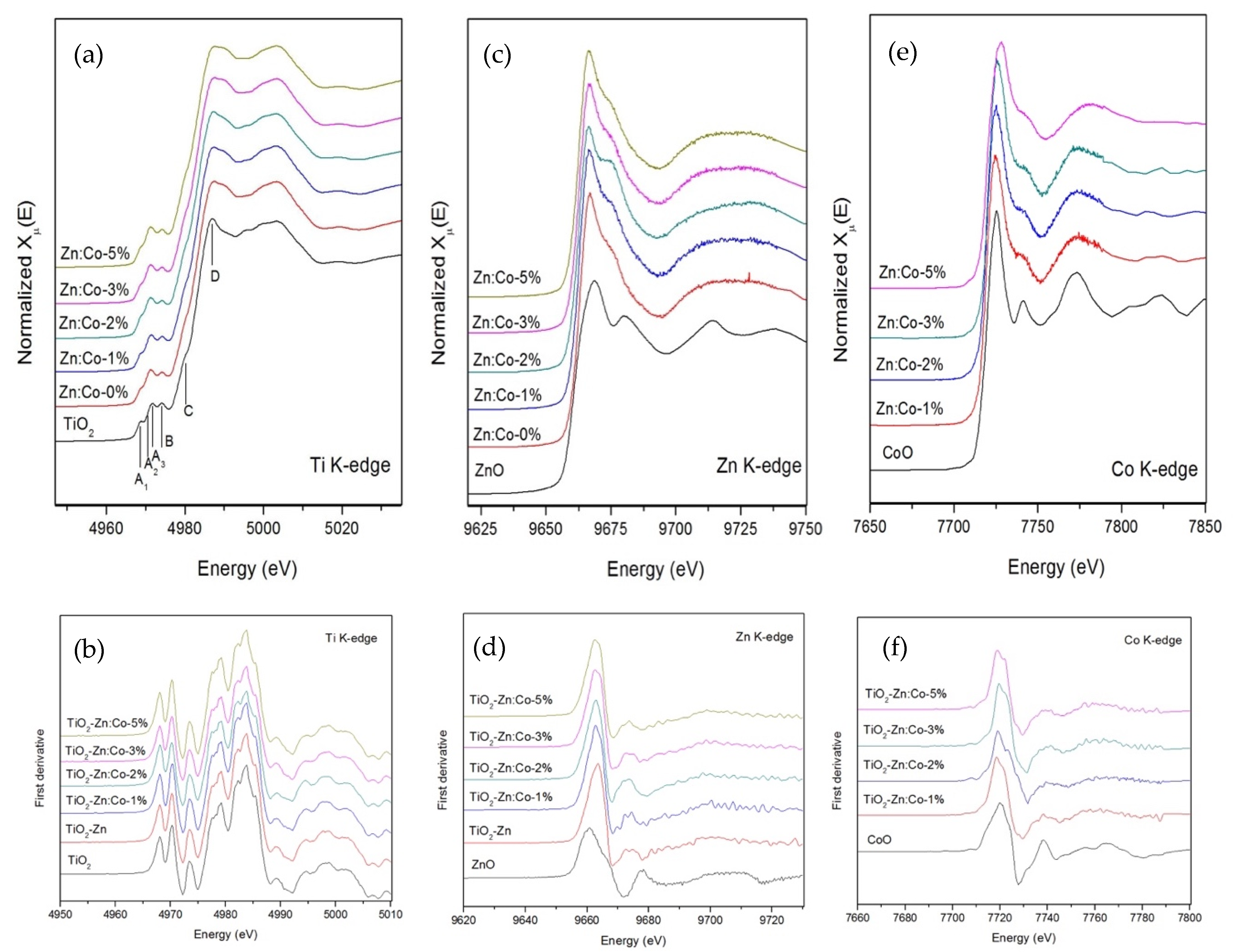
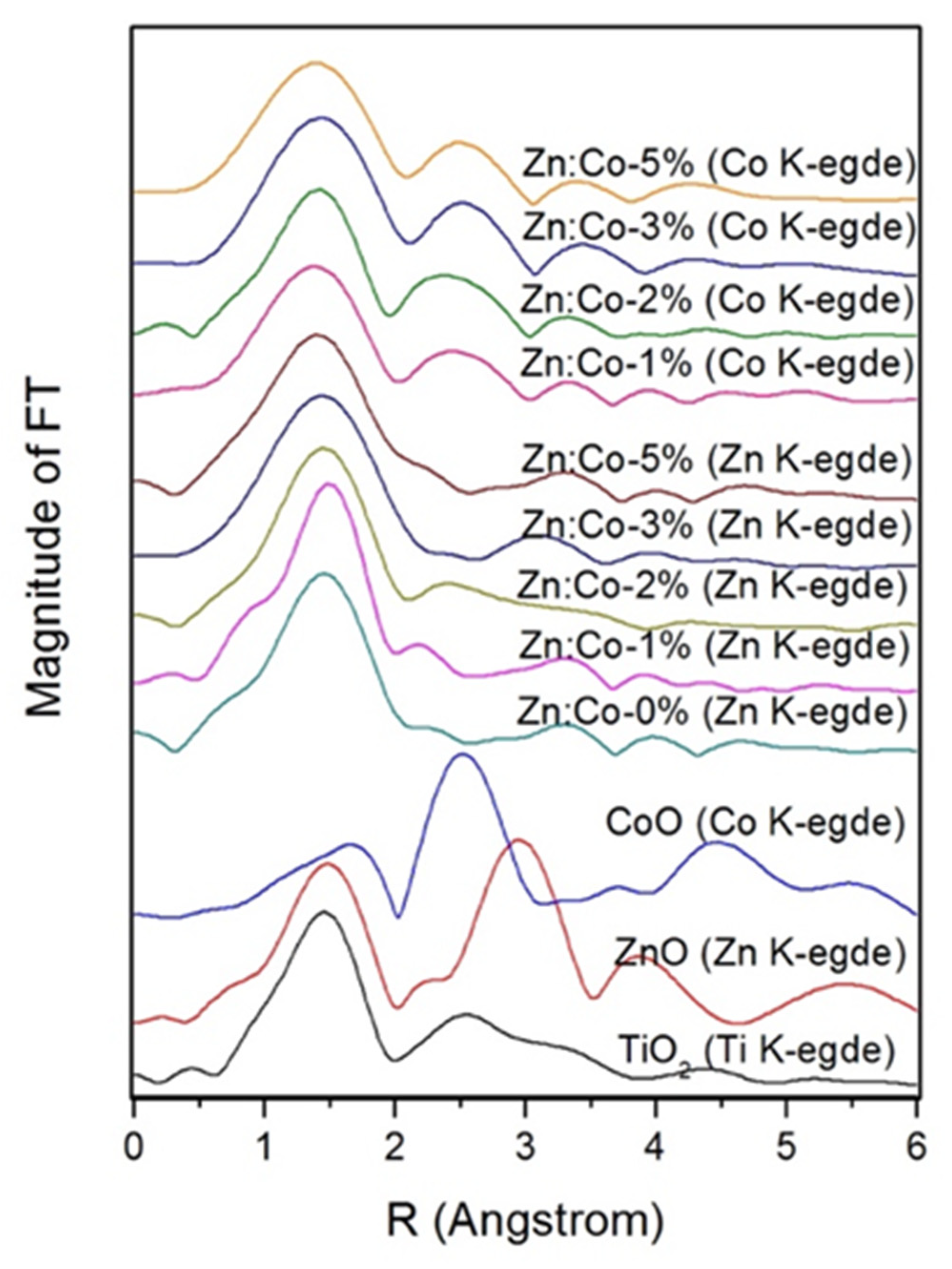
3.2. Structural Study

The XAS technique with synchrotron light including X-ray absorption near edge structure (XANES) and extended X-ray absorption fine structure (EXAFS) was used to investigate the electronic nature of Co/Zn co-doped TiO2 samples. The normalized XANES spectra of single-doped, co-doped TiO2 and the reference standards are shown in Figure 2. Generally, the interpretation of Ti K-edge is intricate, of which the main features of post-edge edge (>4980 eV) can be determined to dipole transition from Ti 1s 4p states, whereas the pre-edge (<4980 eV) can be assigned to the electronic transition to the crystal field split hybridized states between Ti 4p and 3d orbital based on a mixture of quadrupole and dipole excitations [6]. By examining these data and comparing with the literature, as displayed in Figure 2a, the Ti K-edge for TiO2, single-doping, and co-doping samples describes the pre-edge spectra of anatase TiO2 based specimens including four characteristic spectra denoted as A1, A2, A3, and B in the energy region of 4988–4996 eV. The pre-edge Ti K-edge XANES structure arises from 1s to 4d dipole electric transition, with the intensities of the peaks affected by the local geometry and particular medium-range structure of the sample around central Ti atoms [6]. Moreover, the features of pre-edge such as height and position indicate that these samples contain hexa-coordinated Ti4+ atoms. The partial split of pre-edge spectra of Ti K-edge could be observed in the first derivative XANES as written in Figure 2b, representing the edge raising of structures, which has been discussed above.
To confirm the existence of Co and Zn dopants ions in TiO2 lattice, Co- and Zn K-edge XANES measurements were conducted. For the Zn K-edge, the normalized XANES spectra and the first derivative of prepared samples and ZnO reference standard are shown in Figure 2c,d, respectively. Following the inspection of the spectra, it was found that the spectra exhibit a strong absorption edge in a range of 9660–9670 eV with no pre-edge region because the 3d orbital in Zn2+ ion is fully occupied by electrons. The energy absorption spectra of all samples correspond to the Zn2+ of the ZnO reference standard. Thus, the Zn species being introduced into the TiO2 host is evidently present in a 2+ oxidation state. In the case of Co K-edge, as seen in Figure 2e,f, normalized XANES spectra and their first derivative of all as-prepared samples reveals the main absorption energy at ~7710 eV corresponding to the transition to higher energy levels. The strong absorption edge of the co-doped sample corresponds to the CoO reference standard. It can be concluded that prepared samples comprise Co 2+ of oxidation state. Moreover, both post-edge spectra of Zn K-edge (after 9670 eV) and Co K-edge (after 7730 eV) are not likely to occur ZnO and CoO standard spectrum, respectively, which is presumed the Zn2+ and Co2+ cations partially replaced Ti4+ cations to octahedrally coordinated with six oxygen anions. However, the Co-dopants do not exhibit the changing in pre- and post-edge of XANES feature, this implies that the dopant ion might not change the host structure but be incorporated in the TiO2 host.
For an in-depth elucidation of the local structure in as-prepared samples, the EXAFS inquisition was employed at Ti K-edge, Zn K-edge, and Co-kedge. EXAFS spectra are Fourier-transformed (FT) with k2-weighting to produce radial distribution functions, as shown in Figure 3. By analysis of Ti as an absorber atom for TiO2, there are two peaks appearing at around 1.9 Å and 2.7 Å (Black line). The first peak is the Ti-O coordination, associating with the single scattering path of absorbing Ti atom with the nearest neighboring O atoms by six-coordinated around the Ti atom. While the second peak is the Co/Zn-Ti coordination assigned to the single and multiple scattering paths of the absorbing Ti atom with the nearest neighboring Ti atoms and with the nearest neighboring Co/Zn atoms. For describing the environment around Zn- and Co-dopants ions, FT spectra of co-coped samples at Zn and Co were analyzed. It can be seen that the radial distribution features for the Zn/Co dopants in all TiO2 samples studied are similar to the Ti in TiO2. This indicates that the environment around dopant ions is identical with the host Ti, which can be implied the Zn/Co dopants replaced in the Ti site of samples.

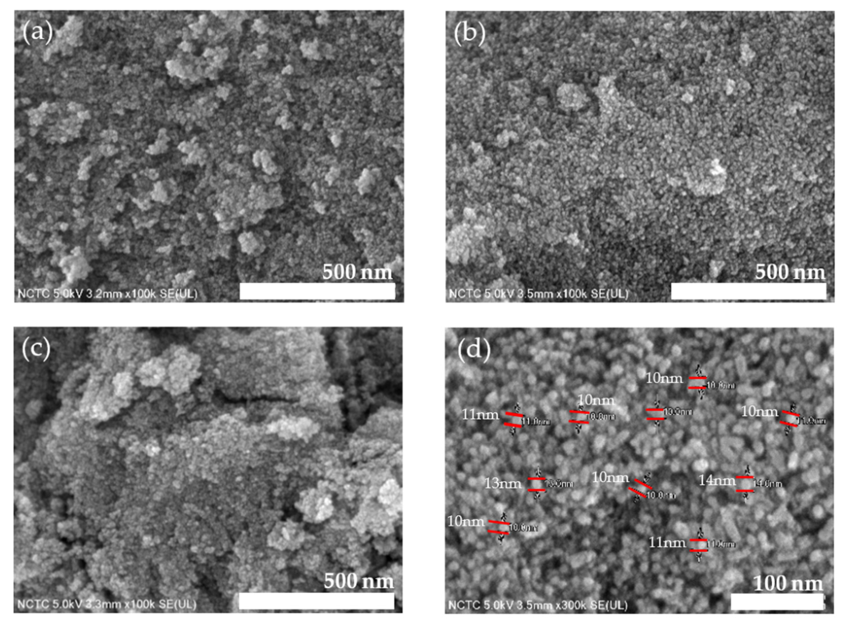
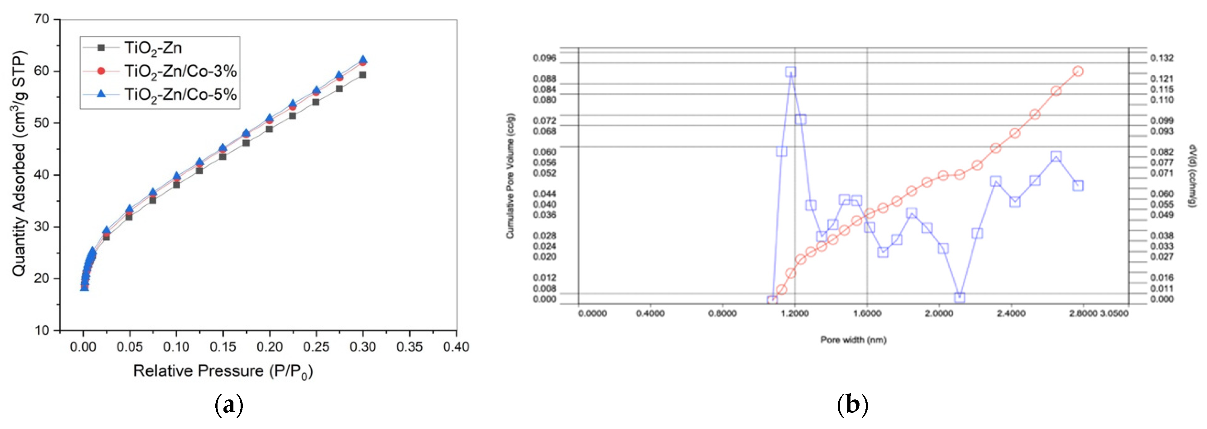
3.3. Morphology and Surface Area Analysis

The FESEM images of single-doped and co-doped samples are displayed in Figure 4. As shown in Figure 4a, almost spherical-shaped particles with particle agglomeration were observed in the single Zn doped TiO2 samples. For 3% and 5% co-doping samples (Figure 4b,c), FESEM images exhibit the uniform distribution of nanoparticles, especially the Co-3%, as seen in Figure 4b. In addition, the micrograph in Figure 4d demonstrated that the particle size analysis by FESEM image, the average particle sizes are around 10–20 nm.
For the specific area of all as-prepared samples, N2 adsorption-desorption and corresponding pore size distribution calculated by BJH (TiO2-Zn/Co-3%) are shown in Figure 5a,b, respectively. It can be seen that the single- and co-doping shows isotherm of type II, which is a typical medium microporous structure. The surface area of Zn-TiO2, 1%-Co/Zn-TiO2, 3%-Co/Zn-TiO2, and 5%-Co/Zn-TiO2 are 190.86, 201.77, 205.27, 206.40, 208.83 m2/g, respectively. The large surface of co-doped samples compared to pure-, single-doped, and previous studies may increase the interaction in photocatalytic activity, which is beneficial for photo photocatalytic efficiency.

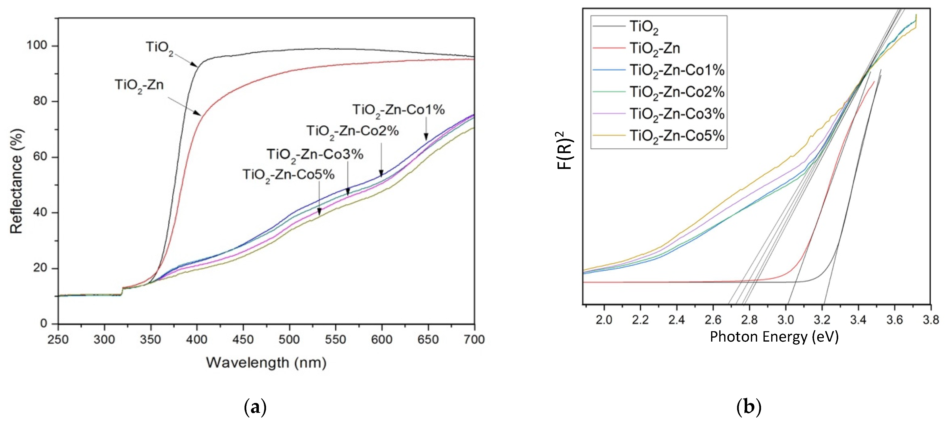
3.4. Optical Properties

The UV-vis DRS measurement of pure TiO2, single-doped, and co-doped samples is shown in Figure 6. For the single-doped samples, extended visible light absorption is observed compared to pure TiO2. Furthermore, the larger absorption in the visible region is noticed in the co-doped samples. It is suggested that increasing co-dopant concentration could result in the reduction of bandgap energy and the enhancement in visible light-harvesting of TiO2. This feature would be correlated to the incorporation of dopant ions in the host lattice that could generate new electronic states in the TiO2 bandgap. Hence the distance of charge transfers between the d electron of the dopant ion and the conduction or valence band is narrowed, leading to the increase in visible light response of the material. Moreover, the different valence states of dopants including Co2+ and Zn2+ from the host matrix Ti4+ may induce the substantial generation of oxygen deficiencies. The bandgap energies of all samples were calculated from the DRS spectra employing a modified Kubelka-Munk equation as plotted in Figure 6b. It is obviously noticed that the deposition of co-dopants significantly affects the decrease of the bandgap energy of the host TiO2 which has an estimated value of 3.21, 3.01, 2.78, 2.75, 2.72, and 2.68 eV for pure TiO2, Zn-TiO2, 1%Co/Zn-TiO2, 3%Co/Zn-TiO2, and 5%Co/Zn-TiO2, respectively. From these results, it was inferred that the Co/Zn co-doped TiO2 could be activated effectively under visible light illumination.

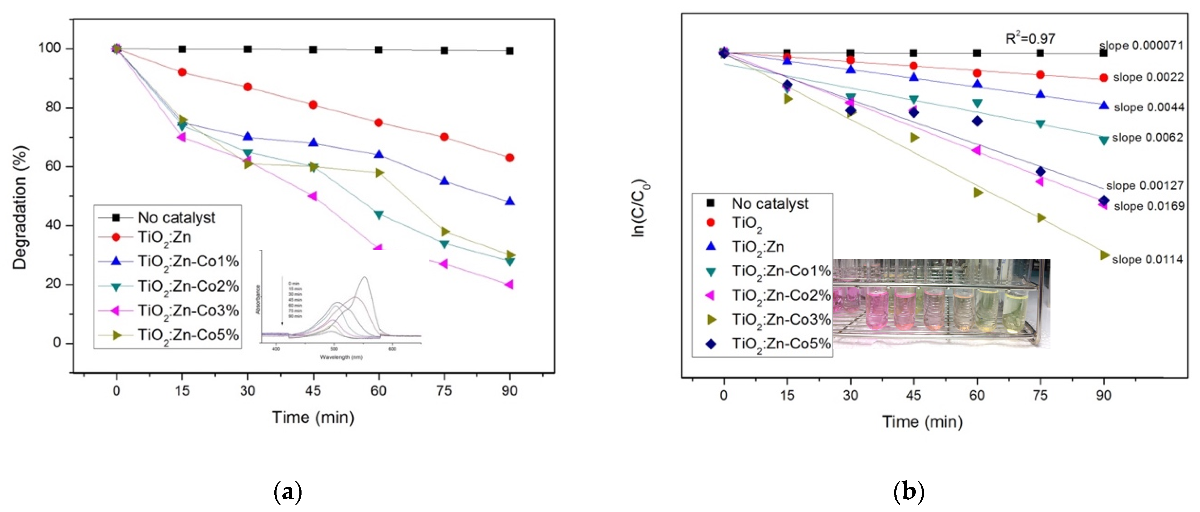
3.5. Photocatalytic Activity

The photocatalytic performance of the prepared samples was employed for rhodamine b dye decolorization under visible light irradiation. Pure RhB exhibit the absorption band at 544 nm, which is assigned to the chromophoric group. The percentage of degradation (D) for RhB dye was calculated using the following equation;
D = [ A 0 A t ] A 0 × 100 %
where A0 is the absorbance value of the initial RhB solution, At is the absorbance value of degraded RhB solution at the reaction time t. Under the visible light irradiation without the catalyst, no decrease in the RhB concentration is observed, implying that the self-photolysis of RhB is negligible when irradiated with visible light. When the reaction was carried out by a single doped sample, around 40% of RhB degradation after 90 min was obtained a seen in Figure 6. For the Co/Zn co-doped TiO2 (Figure 7a), the superior degradation of around 80% was found in the samples containing 3% of Co-content after 90 min of irradiation time while the sample with 1, 2, and 5% Co doping content exhibited about 51, 73 and 71% of photo-decolorization, respectively. According to the results, all as-synthesized co-doped catalysts show higher degradation rates than Co- and Zn- single-doped and bare TiO2. Obviously, a comparison in the plots of dye degradation shows that the concentration of as-prepared catalyst can indeed influence the degradation efficiency. The increase of dopant content to 3 mol% resulted in the increase in dye degradation, which might be attributed to the Co increased, the surface barrier becomes higher and narrower space charge obtained, leading to easy of e/h pairs. However, a slight decrease of dye decomposition was observed when the concentration of Co-doped was further increased to 5mol%, this may be due to when the content of cobalt dopant is increased beyond the limit, the space charge recombination is lower and penetration depth of light into the catalyst greatly exceeds the layer of space charge, indicating in high recombination of e/h pairs [32].
Reaction kinetics gives information about the reaction rates and the mechanisms by which the reactants are converted to the products. The equation for pseudo-first-order kinetic is given below;
l n ( C C 0 ) = k t
where C and C0 are the final and initial concentrations (ppm) of RhB, k is the apparent reaction rate constant (min−1) and t is the irradiation time (min). Figure 7b demonstrates the photocatalytic reaction kinetic of RhB degradation in the solution based on the data plotted in Figure 6. As it can be seen, a rather good correlation to pseudo-first-order reaction kinetics (R2 = 0.97) was found, which suggests that the RhB degradation over these samples follows the first-order kinetics. Among all as-prepared catalysts, 3% of Co/Zn doped TiO2 shows the highest reaction rate constant of 0.0114 min−1, which is about 5.2 and 2.6 times that over pure and single-doping catalysts, respectively.
It is observed that the superior performance of organic compounds photodegradation was found in Co/Zn co-doping TiO2. It may be due to many factors, such as the increase of visible light harvesting due to the narrowing energy band gap, slower time for e/h+ pairs recombination, and greater hydroxyl radical generation. Additionally, the possible mechanism of photodegradation for organics compounds using metal-doped TiO2 was studied by comparing with similarly reported [33,34,35,36]. They reported that when the light irradiation on the metal-doped TiO2 catalyst, photoexcited electrons were transferred from the valence band to newly created electronic states in the band structure via metals incorporated into the TiO2 matrix. Then, the transferred electron reacted with an oxygen molecule ( O 2 ) to generate a superoxide anion ( · O 2 ) ; e + O 2 · O 2 , and a positive hole in the valence band of TiO2 reacted with (i) a water molecule (H2O) or (ii) hydroxyl ion (OH) to form a hydroxyl radical ( · OH ) ; (i) h + + H 2 O · O H + H + and (ii) h + + O H · O H . Finally, the reactive species · O 2 and · OH could oxidize the target organic compounds to generate CO2, CO, and other byproducts.

4. Conclusions

In summary, Co/Zn co-doped TiO2 was successfully synthesized by a facile one-step sonochemical method at room temperature. The use of ultrasounds allowed for the successful doping of both Co and Zn into the TiO2 lattice, which caused a shift in the light absorption toward the higher wavelength. XANES measurement confirmed the oxidation state of 4+, 2+, and 2+ for Ti, Zn, and Co, respectively. EXAFS analysis reveals that the cobalt and zinc are possibly occupied into TiO2 lattice, by replacing some Ti4+. The Co/Zn co-doped TiO2 photocatalyst exhibited good photodegradation of RhB dye under visible light irradiation. The best decolorization performance was achieved in the content of 3% for cobalt doping concentration. It is presumed that (i) the sonochemical method provides the small nanoparticles resulting in higher surface area and (ii) the incorporation of Co and Zn lead to enhance visible light harvesting and to diminish the electron-hole recombination, which improved the photocatalytic behavior.

Author Contributions

Conceptualization, C.W., W.M., S.P. and W.P.; methodology, W.M., S.P. and C.W.; software, C.W.; validation, W.P., S.P. and C.K.J.; formal analysis, C.W., W.P., S.P. and B.R.R.; investigation, C.W. and S.P.; resources, W.P. and S.P.; data curation, W.M.; writing—original draft preparation, C.W., W.M. and S.P.; writing—review and editing, W.P., C.K.J. and S.P.; visualization, C.W.; supervision, W.P.; project administration, W.P.; funding acquisition, S.P. and W.P.. All authors have read and agreed to the published version of the manuscript.

Funding

This research was funded by Fundamental Research Budget Allocation (RFB630004/0168) grant funded by Thailand Science Research and Innovation (TSRI) (Granted No. IRF63A0704A.4).

Data Availability Statement

Not applicable.

Acknowledgments

Authors would like to thank the Division of Physics, Faculty of Science and Technology, Rajamangala University of Technology Thanyaburi (RMUTT), College of Materials Innovation and Technology, King Mongkut’s Institute of Technology Ladkrabang (KMITL), and Smart Materials Research Unit (SMRU) for supporting the facilities. Authors gratefully acknowledge BL-8 at Synchrotron Light Research Institute (SLRI), Nakhon Ratchasima, Thailand, for XAS measurement.

Conflicts of Interest

The authors declare no conflict of interest.

References

  1. Bhatkhande, D.S.; Pangarkar, V.G.; Beenackers, A.A.C.M. Photocatalytic degradation for environmental applications—A review. J. Chem. Technol. Biotechnol. 2001, 77, 102–116. [Google Scholar] [CrossRef]
  2. Rajeshwar, K.; Chenthamarakshan, C.R.; Goeringer, S.; Djukic, M. Titania-based heterogeneous photocatalysis. Materials, mechanistic issues, and implications for environmental remediation. Pure Appl. Chem. 2001, 73, 1849–1860. [Google Scholar] [CrossRef]
  3. Nakata, K.; Fujishima, A. TiO2 photocatalysis: Design and applications. J. Photochem. Photobiol. C Photochem. Rev. 2012, 13, 169–189. [Google Scholar] [CrossRef]
  4. Anpo, M.; Takeuchi, M. The design and development of highly reactive titanium oxide photocatalysts operating under visible light irradiation. J. Catal. 2003, 216, 505–516. [Google Scholar] [CrossRef]
  5. Hernández, J.V.; Coste, S.; Murillo, A.G.; Romo, F.C.; Kassiba, A. Effects of metal doping (Cu, Ag, Eu) on the electronic and optical behavior of nanostructured TiO2. J. Alloy Compd. 2017, 710, 355–363. [Google Scholar] [CrossRef]
  6. Jaihindh, D.P.; Verma, A.; Chen, C.-C.; Huang, Y.-C.; Dong, C.-L.; Fu, Y.-P. Study of oxidation states of Fe- and Co-doped TiO2 photocatalytic energy materials and their visible-light-driven photocatalytic behavior. Int. J. Hydrogen Energy 2019, 44, 15892–15906. [Google Scholar] [CrossRef]
  7. Wattanawikkam, C.; Pecharapa, W.; Ishihara, K.N. X-ray absorption spectroscopy analysis and magnetic properties of M-doped TiO2 nanoparticles (M=Co, Mn, Ni and Zn) prepared by co-precipitation method. Ceram. Int. 2017, 43, S397–S402. [Google Scholar] [CrossRef]
  8. Chen, W.-T.; Dong, Y.; Yadav, P.; Aughterson, R.D.; Sun-Waterhouse, D.; Waterhouse, G.I. Effect of alcohol sacrificial agent on the performance of Cu/TiO2 photocatalysts for UV-driven hydrogen production. Appl. Catal. A Gen. 2020, 602, 117703. [Google Scholar] [CrossRef]
  9. Noonuruk, R.; Wattanawikkam, C. Visible-light-driven Photodegradation of Commercial Dyes by the Cooperation of Co-doped TiO2 Material. Curr. Appl. Sci. Technol. 2020, 20, 43–51. [Google Scholar] [CrossRef]
  10. Inturi, S.N.R.; Boningari, T.; Suidan, M.; Smirniotis, P.G. Visible-light-induced photodegradation of gas phase acetonitrile using aerosol-made transition metal (V, Cr, Fe, Co, Mn, Mo, Ni, Cu, Y, Ce, and Zr) doped TiO2. Appl. Catal. B Environ. 2014, 144, 333–342. [Google Scholar] [CrossRef]
  11. Kerkez-Kuyumcu, Ö.; Kibar, M.E.; Dayıoğlu, K.; Gedik, F.; Akın, A.N.; Ozkara-Aydinoglu, S. A comparative study for removal of different dyes over M/TiO2 (M=Cu, Ni, Co, Fe, Mn and Cr) photocatalysts under visible light irradiation. J. Photochem. Photobiol. A Chem. 2015, 311, 176–185. [Google Scholar] [CrossRef]
  12. Wattanawikkam, C.; Pecharapa, W. Synthesis and Characterization of Zn-Doped TiO2 Nanoparticles via Sonochemical Method. Integr. Ferroelectr. 2015, 165, 167–175. [Google Scholar] [CrossRef]
  13. Makdee, A.; Unwiset, P.; Chanapattharapol, K.C.; Kidkhunthod, P. Effects of Ce addition on the properties and photocatalytic activity of TiO2, investigated by X-ray absorption spectroscopy. Mater. Chem. Phys. 2018, 213, 431–443. [Google Scholar] [CrossRef]
  14. Katoueizadeh, E.; Zebarjad, S.M.; Janghorban, K. Synthesis and enhanced visible-light activity of N-doped TiO2 nano-additives applied over cotton textiles. J. Mater. Res. Technol. 2018, 7, 204–211. [Google Scholar] [CrossRef]
  15. Kahattha, C.; Wongpisutpaisan, N.; Vittayakorn, N.; Pecharapa, W. Physical properties of V-doped TiO2 nanoparticles synthesized by sonochemical-assisted process. Ceram. Int. 2013, 39, S389–S393. [Google Scholar] [CrossRef]
  16. Wang, Q.; Rhimi, B.; Wang, H.; Wang, C. Efficient photocatalytic degradation of gaseous toluene over F-doped TiO2/exfoliated bentonite. Appl. Surf. Sci. 2020, 530, 147286. [Google Scholar] [CrossRef]
  17. Suwannaruang, T.; Kidkhunthod, P.; Chanlek, N.; Soontaranon, S.; Wantala, K. High anatase purity of nitrogen-doped TiO2 nanorice particles for the photocatalytic treatment activity of pharmaceutical wastewater. Appl. Surf. Sci. 2019, 478, 1–14. [Google Scholar] [CrossRef]
  18. Saroj, S.; Singh, L.; Singh, S.V. Solution-combustion synthesis of anion (iodine) doped TiO2 nanoparticles for photocatalytic degradation of Direct Blue 199 dye and regeneration of used photocatalyst. J. Photochem. Photobiol. A Chem. 2020, 396, 112532. [Google Scholar] [CrossRef]
  19. Gordon, W.; Balboa, A.; Giles, S.; Epshteyn, A.; Ávalos-Ovando, O.; Govorov, A.; McEntee, M.; Baturina, O. Visible Light-Induced Reactivity of Plasmonic Gold Nanoparticles Incorporated into TiO2 Matrix towards 2-Chloroethyl Ethyl Sulfide. Crystals 2021, 11, 659. [Google Scholar] [CrossRef]
  20. Siddiqa, A.; Masih, D.; Anjum, D.; Siddiq, M. Cobalt and sulfur co-doped nano-size TiO2 for photodegradation of various dyes and phenol. J. Environ. Sci. 2015, 37, 100–109. [Google Scholar] [CrossRef] [Green Version]
  21. Isari, A.A.; Hayati, F.; Kakavandi, B.; Rostami, M.; Motevassel, M.; Dehghanifard, E. N, Cu co-doped TiO2@functionalized SWCNT photocatalyst coupled with ultrasound and visible-light: An effective sono-photocatalysis process for pharmaceutical wastewaters treatment. Chem. Eng. J. 2020, 392, 123685. [Google Scholar] [CrossRef]
  22. Bramhankar, T.; Pawar, S.; Shaikh, J.; Gunge, V.; Beedri, N.; Baviskar, P.; Pathan, H.; Patil, P.; Kambale, R. Effect of Nickel–Zinc Co-doped TiO2 blocking layer on performance of DSSCs. J. Alloy Compd. 2020, 817, 152810. [Google Scholar] [CrossRef]
  23. Cai, J.; Zhou, M.; Xu, X.; Du, X. Stable boron and cobalt co-doped TiO2 nanotubes anode for efficient degradation of organic pollutants. J. Hazard. Mater. 2020, 396, 122723. [Google Scholar] [CrossRef]
  24. Yu, J.; Zou, J.; Xu, P.; He, Q. Three-dimensional photoelectrocatalytic degradation of the opaque dye acid fuchsin by Pr and Co co-doped TiO2 particle electrodes. J. Clean. Prod. 2020, 251, 119744. [Google Scholar] [CrossRef]
  25. Sharma, A.; Varshney, M.; Shin, H.J.; Lee, B.-H.; Chae, K.H.; Won, S.O. Effect of Cu insertion on structural, local electronic/atomic structure and photocatalyst properties of TiO2, ZnO and Ni(OH)2 nanostructures: XANES-EXAFS study. Mater. Chem. Phys. 2017, 191, 129–144. [Google Scholar] [CrossRef]
  26. Bootchanont, A.; Rujirawat, S.; Yimnirun, R.; Guo, R.; Bhalla, A. Local structure study of phase transition behavior in Ba(Ti,Sn)O3 perovskite by X-ray absorption fine structure. Ceram. Int. 2016, 42, 8151–8154. [Google Scholar] [CrossRef] [Green Version]
  27. Yadav, R.S.; Mishra, P.; Pandey, A.C. Growth mechanism and optical property of ZnO nanoparticles synthesized by sonochemical method. Ultrason. Sonochemistry 2008, 15, 863–868. [Google Scholar] [CrossRef]
  28. Wattanawikkam, C.; Pecharapa, W. Structural studies and photocatalytic properties of Mn and Zn co-doping on TiO2 prepared by single step sonochemical method. Radiat. Phys. Chem. 2020, 171, 108714. [Google Scholar] [CrossRef]
  29. Wattanawikkam, C.; Pecharapa, W. Sonochemical Synthesis, Characterization, and Photocatalytic Activity of Perovskite ZnTiO3 Nanopowders. IEEE Trans. Ultrason. Ferroelectr. Freq. Control. 2016, 63, 1663–1667. [Google Scholar] [CrossRef] [PubMed]
  30. Wattanawikkam, C.; Kansa-Ard, T.; Pecharapa, W. X-ray absorption spectroscopy analysis and photocatalytic behavior of ZnTiO3 nanoparticles doped with Co and Mn synthesized by sonochemical method. Appl. Surf. Sci. 2019, 474, 169–176. [Google Scholar] [CrossRef]
  31. Jiang, W.; Zhang, X.; Gong, X.; Yan, F.; Zhang, Z. Sonochemical synthesis and characterization of magnetic separable Fe3O4–TiO2 nanocomposites and their catalytic properties. Int. J. Smart Nano Mater. 2010, 1, 278–287. [Google Scholar] [CrossRef]
  32. Miao, Y.; Zhai, Z.; Jiang, L.; Shi, Y.; Yan, Z.; Duan, D.; Zhen, K.; Wang, J. Facile and new synthesis of cobalt doped mesoporous TiO2 with high visible-light performance. Powder Technol. 2014, 266, 365–371. [Google Scholar] [CrossRef]
  33. Lee, J.Y.; Choi, J.-H. Sonochemical Synthesis of Ce-doped TiO2 Nanostructure: A Visible-Light-Driven Photocatalyst for Degradation of Toluene and O-Xylene. Materials 2019, 12, 1265. [Google Scholar] [CrossRef] [Green Version]
  34. Abdelraheem, W.; Patil, M.K.; Nadagouda, M.N.; Dionysiou, D.D. Hydrothermal synthesis of photoactive nitrogen- and boron-codoped TiO2 nanoparticles for the treatment of bisphenol A in wastewater: Synthesis, photocatalytic activity, degradation byproducts and reaction pathways. Appl. Catal. B Environ. 2019, 241, 598–611. [Google Scholar] [CrossRef]
  35. Abdelraheem, W.H.; Nadagouda, M.N.; Dionysiou, D.D. Solar light-assisted remediation of domestic wastewater by NB-TiO2 nanoparticles for potable reuse. Appl. Catal. B Environ. 2020, 269, 118807. [Google Scholar] [CrossRef]
  36. Peñas-Garzón, M.; Abdelraheem, W.H.; Belver, C.; Rodriguez, J.J.; Bedia, J.; Dionysiou, D.D. TiO2-carbon microspheres as photocatalysts for effective remediation of pharmaceuticals under simulated solar light. Sep. Purif. Technol. 2021, 275, 119169. [Google Scholar] [CrossRef]
Figure 1. Powder XRD pattern of Zn-TiO2, Co/Zn co-doped TiO2 nanoparticles.
Crystals 11 01254 g001
Figure 2. Normalized XANES spectra and first derivative XANES of Co/Zn co-doped TiO2: (a,b) Ti K-edge, (c,d) Zn K-edge, and (e,f) Co K-edge.
Crystals 11 01254 g002
Figure 3. Fourier transform magnitudes of EXAFS spectra of k2-weighted data for Co/Zn co-doped TiO2 with their reference standard at Ti, Zn, and Co K-edge.
Crystals 11 01254 g003
Figure 4. FESEM images of (a) TiO2-Zn-1%, (b) TiO2-Zn/Co-3%, (c) TiO2-Zn/Co-5% and (d) particle size analysis of TiO2-Zn/Co-3%.
Crystals 11 01254 g004
Figure 5. (a) Nitrogen sorption isotherms of the as-synthesized singles-dope TiO2 and Zn/Co co-doped TiO2 and (b) pore diameter distribution curve TiO2-Zn/Co-3%.
Crystals 11 01254 g005
Figure 6. (a) UV-vis diffuse reflectance spectra and (b) Energy band gap of Co/Zn co-doped TiO2.
Crystals 11 01254 g006
Figure 7. (a) Photocatalytic degradation curves with time independence UV-vis and (b) kinetic linear fitting curves for Co/Zn co-doped TiO2 catalyst under visible light irradiation in the presence of RhB dye.
Crystals 11 01254 g007
Publisher’s Note: MDPI stays neutral with regard to jurisdictional claims in published maps and institutional affiliations.

