
Weak Interactions in Cocrystals of Isoniazid with Glycolic and Mandelic Acids

 ,
,  , , and
Abstract
:1. Introduction
2. Experimental and Theoretical Methods
2.1. Materials and Methods
2.2. Crystallography
2.3. Cocrystal Screening
2.4. Cocrystal Synthesis
2.5. Hirshfeld Surface Analysis
2.6. Theoretical Methods
3. Results and Discussion
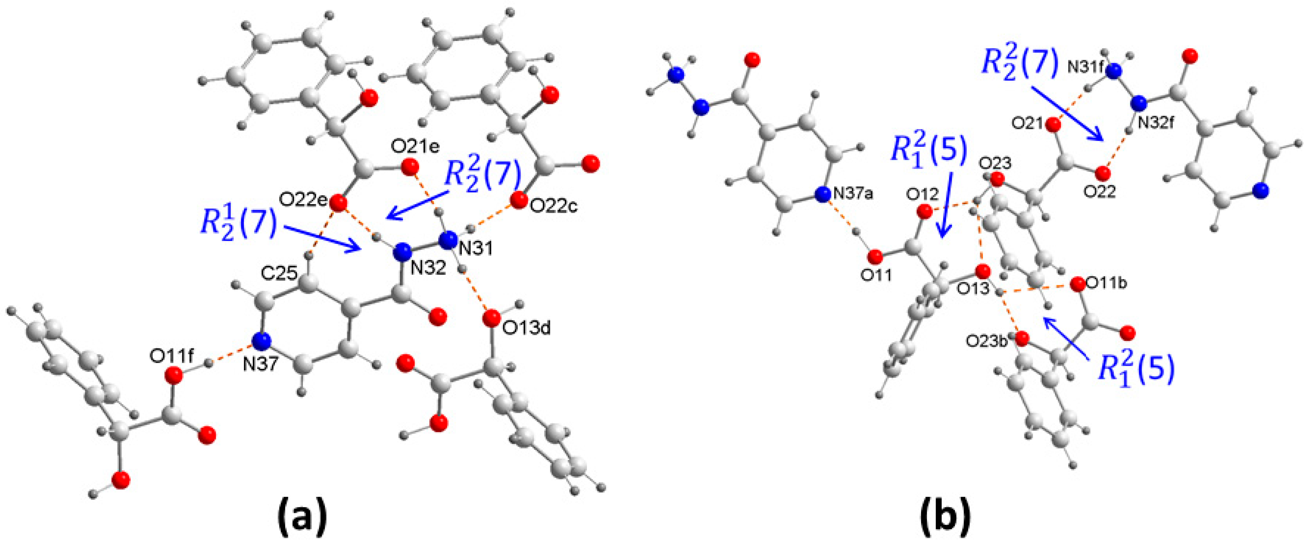
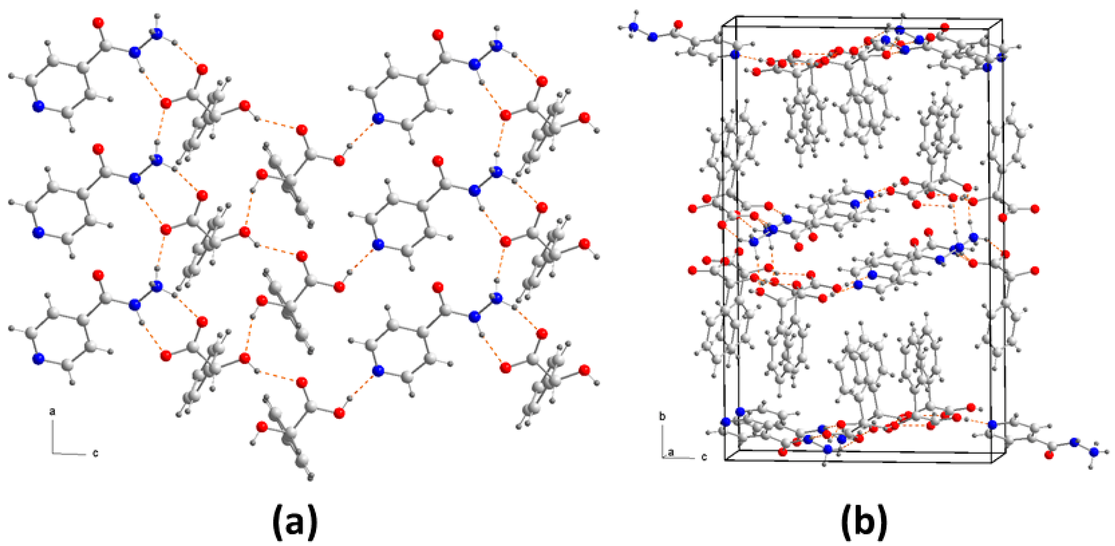
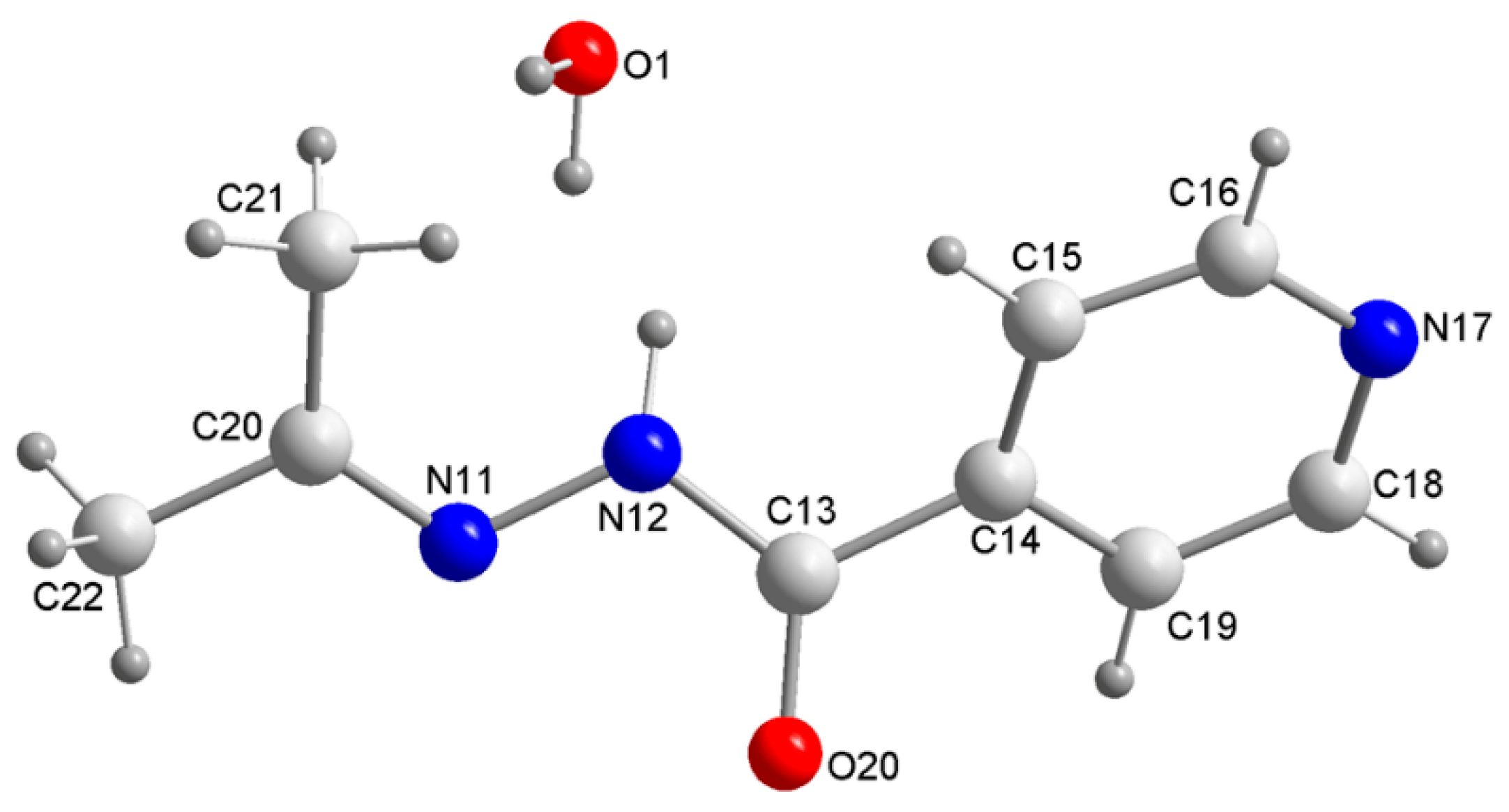
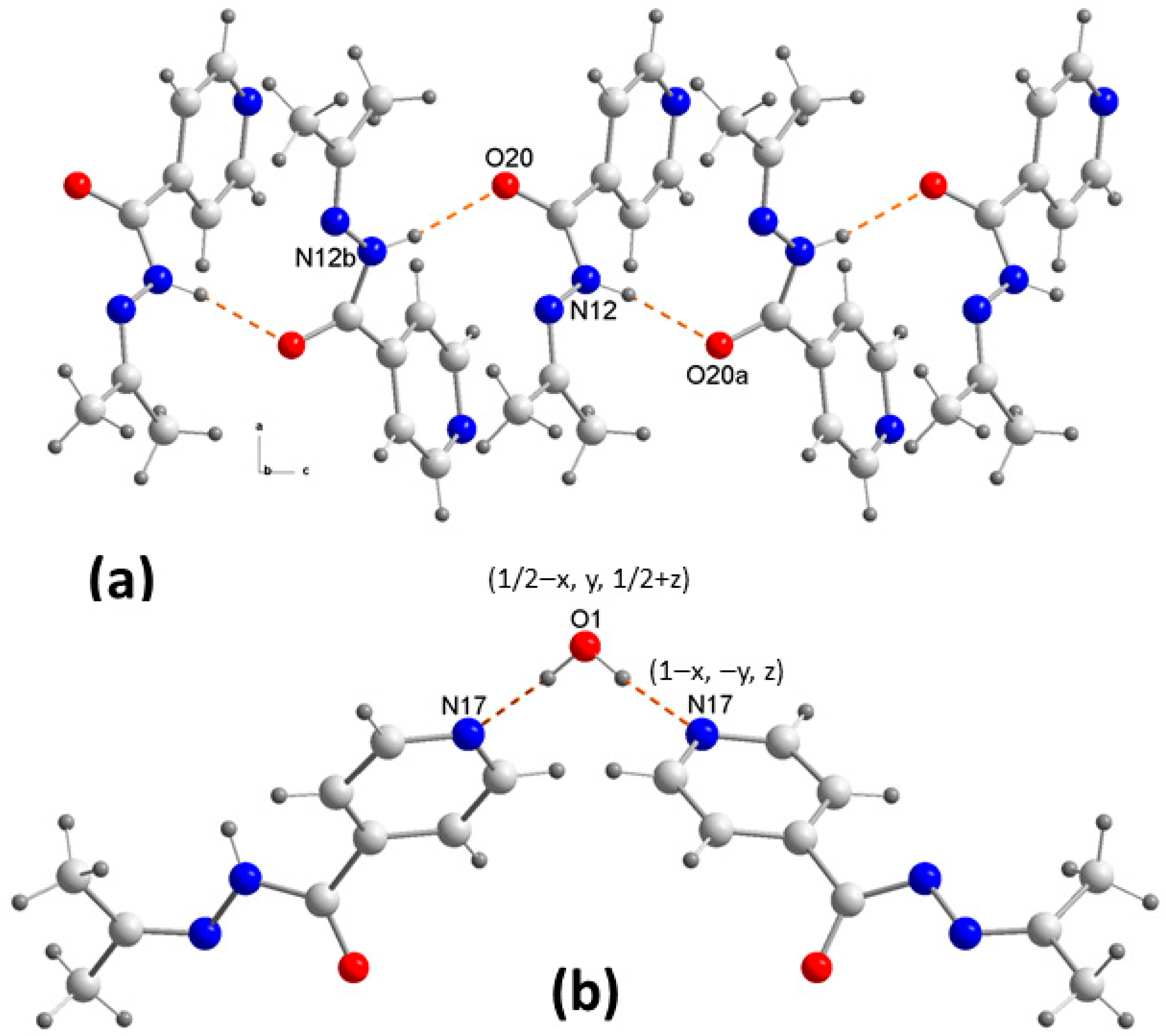
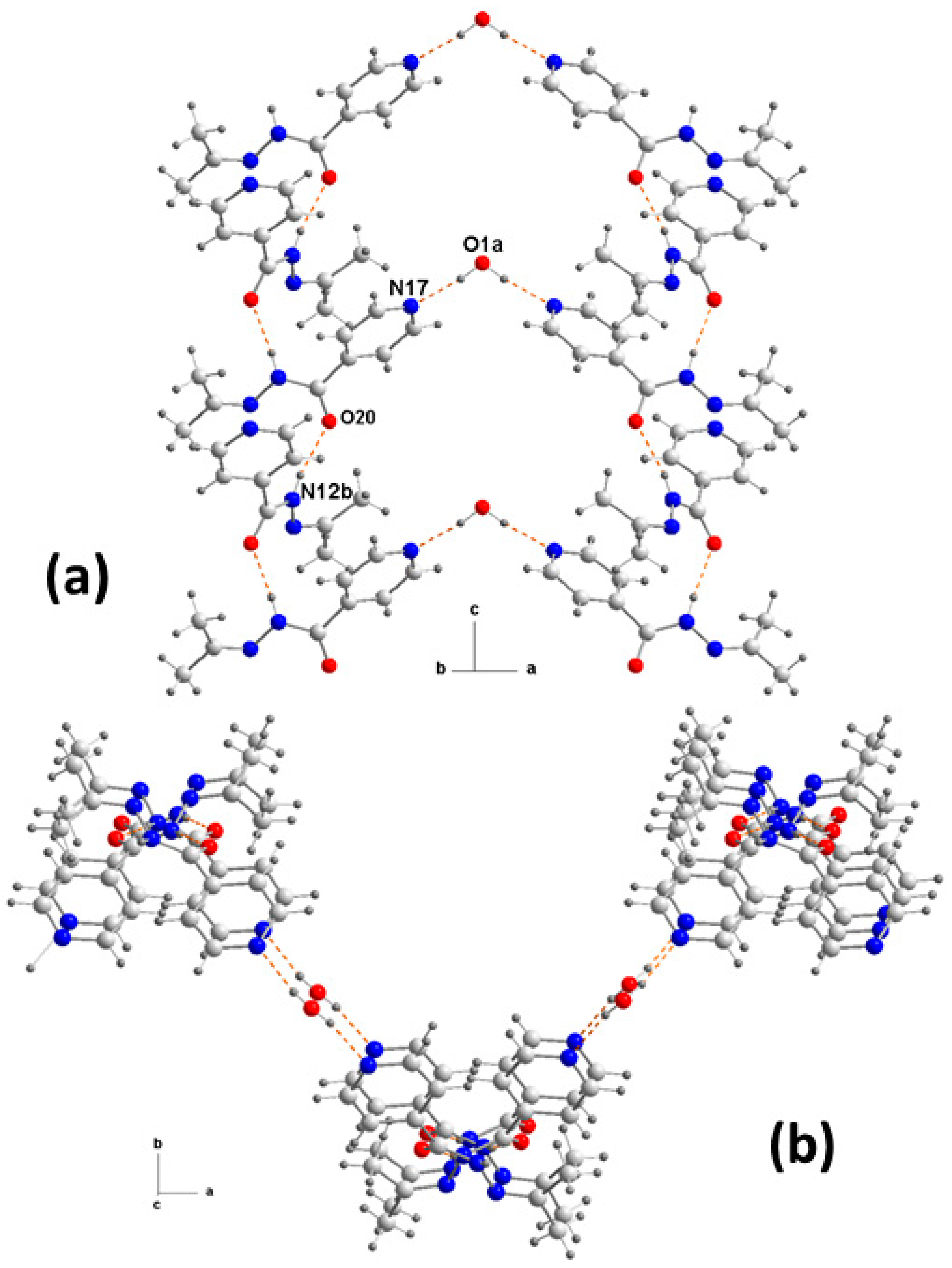
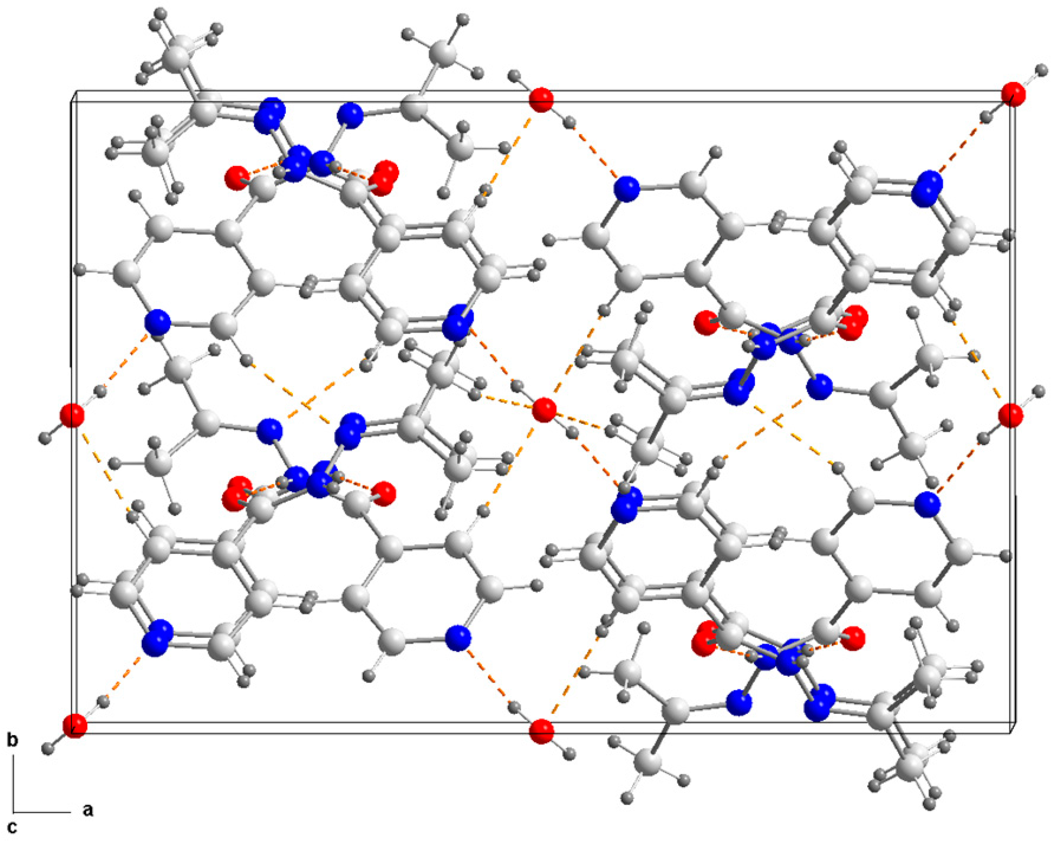
3.1. Structural Description and Supramolecular Analysis
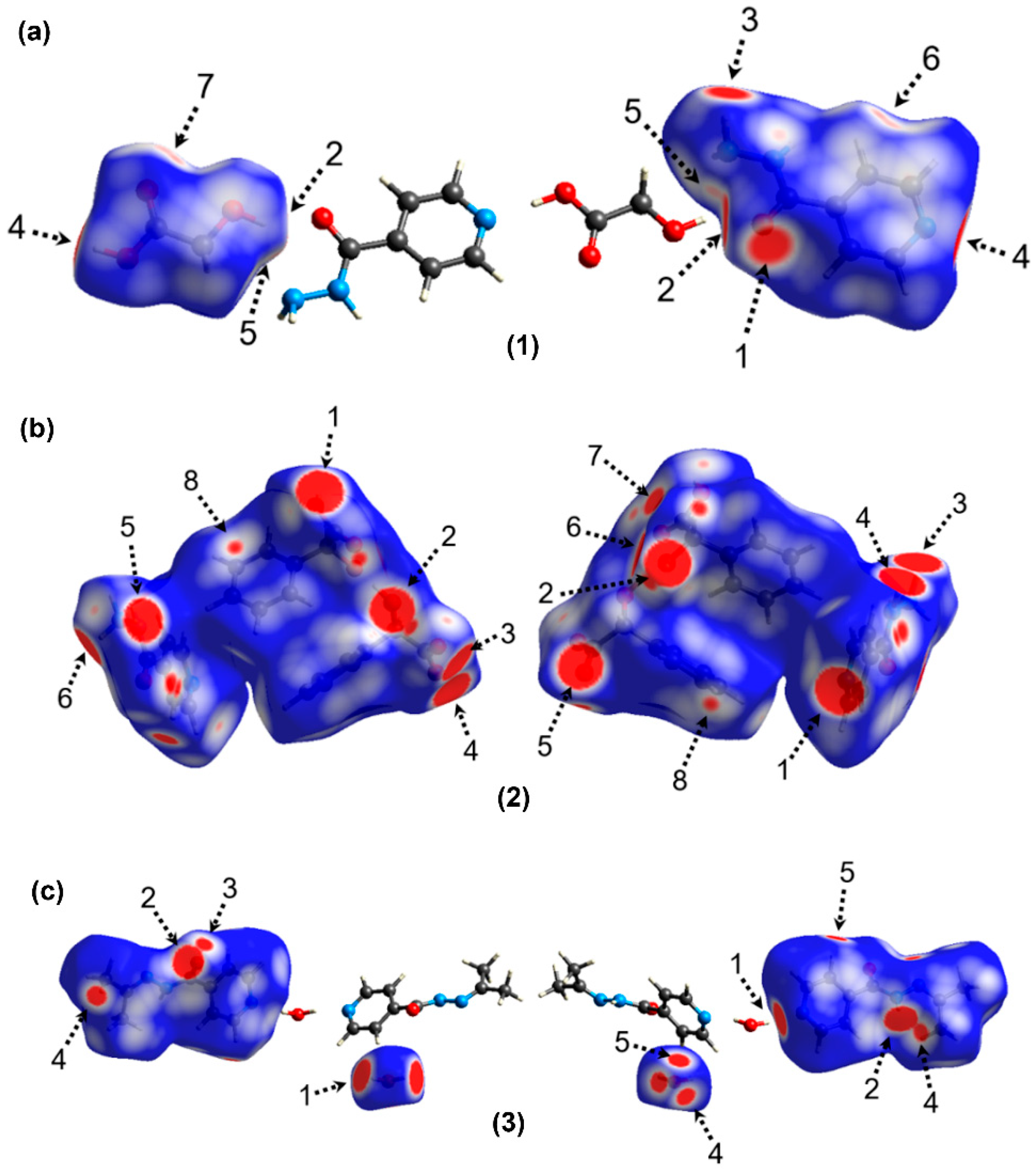
3.2. Hirshfeld Surface Analysis
3.3. DFT Calculations
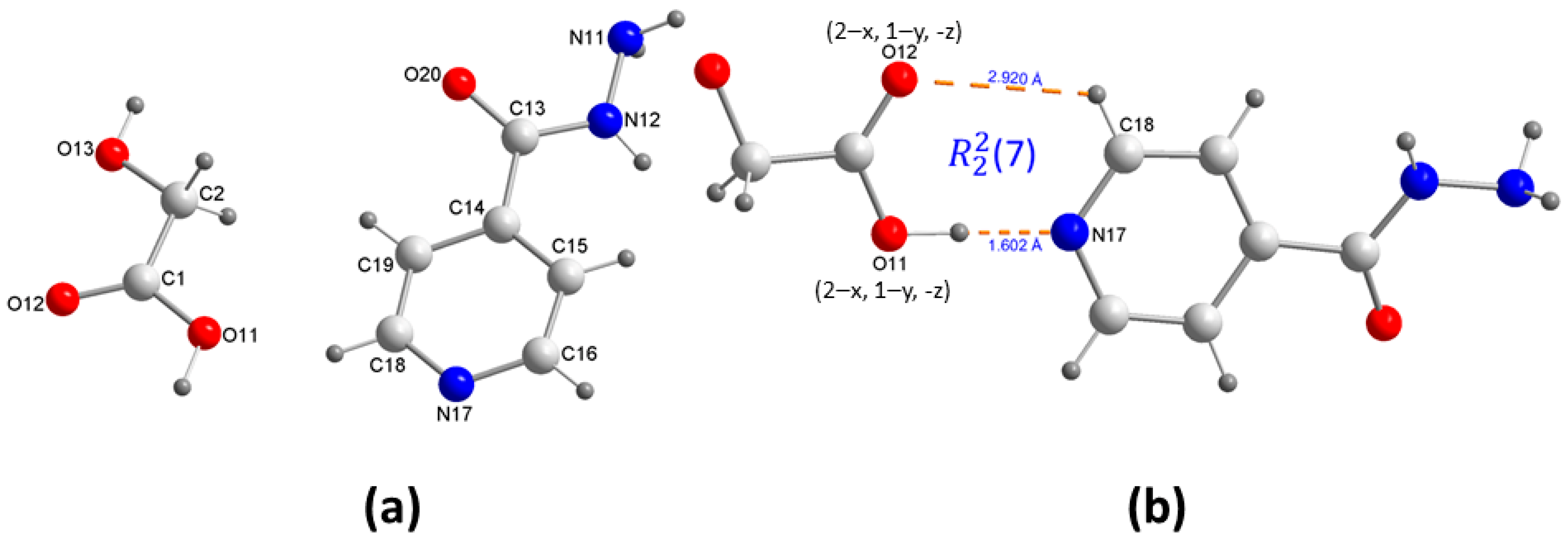
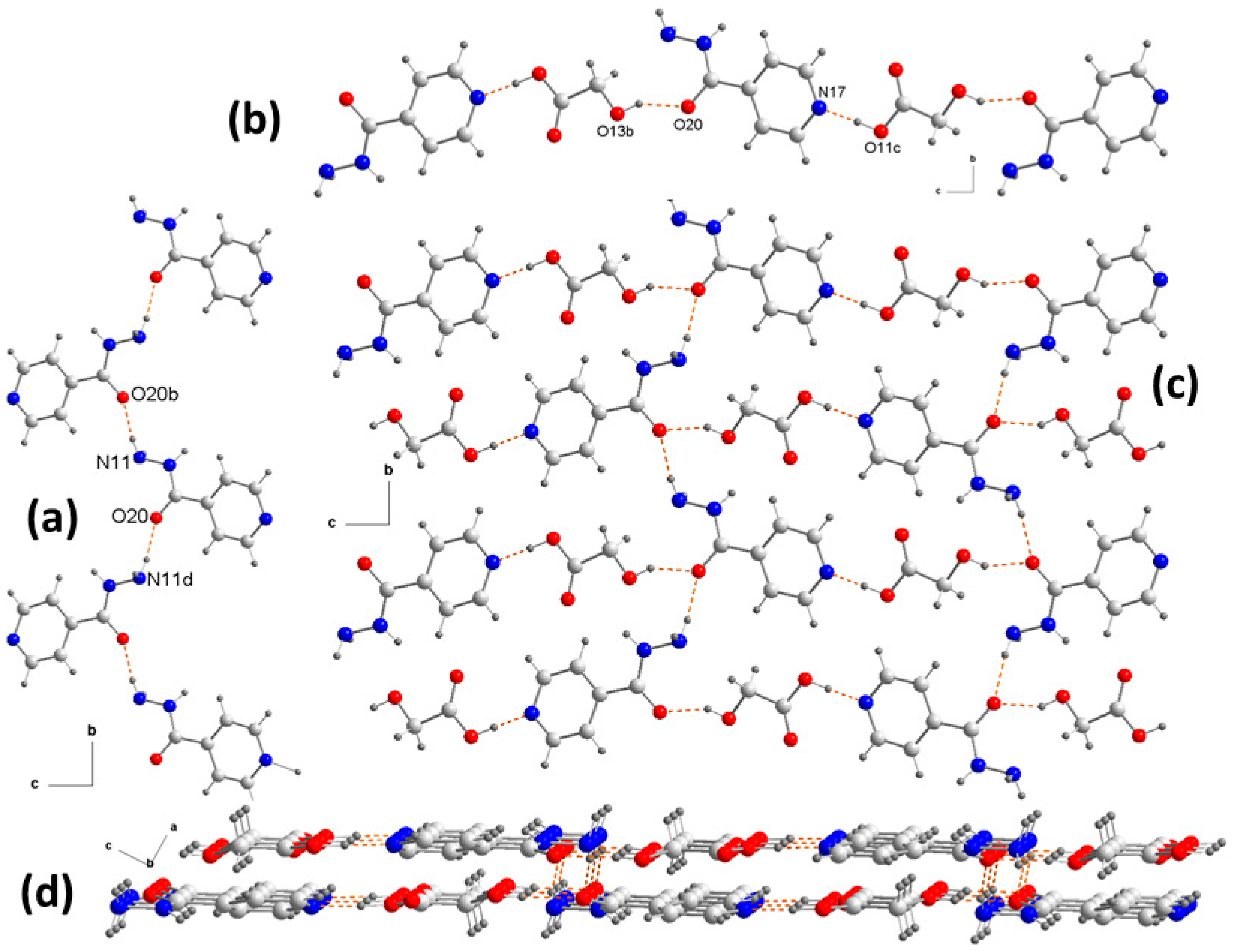
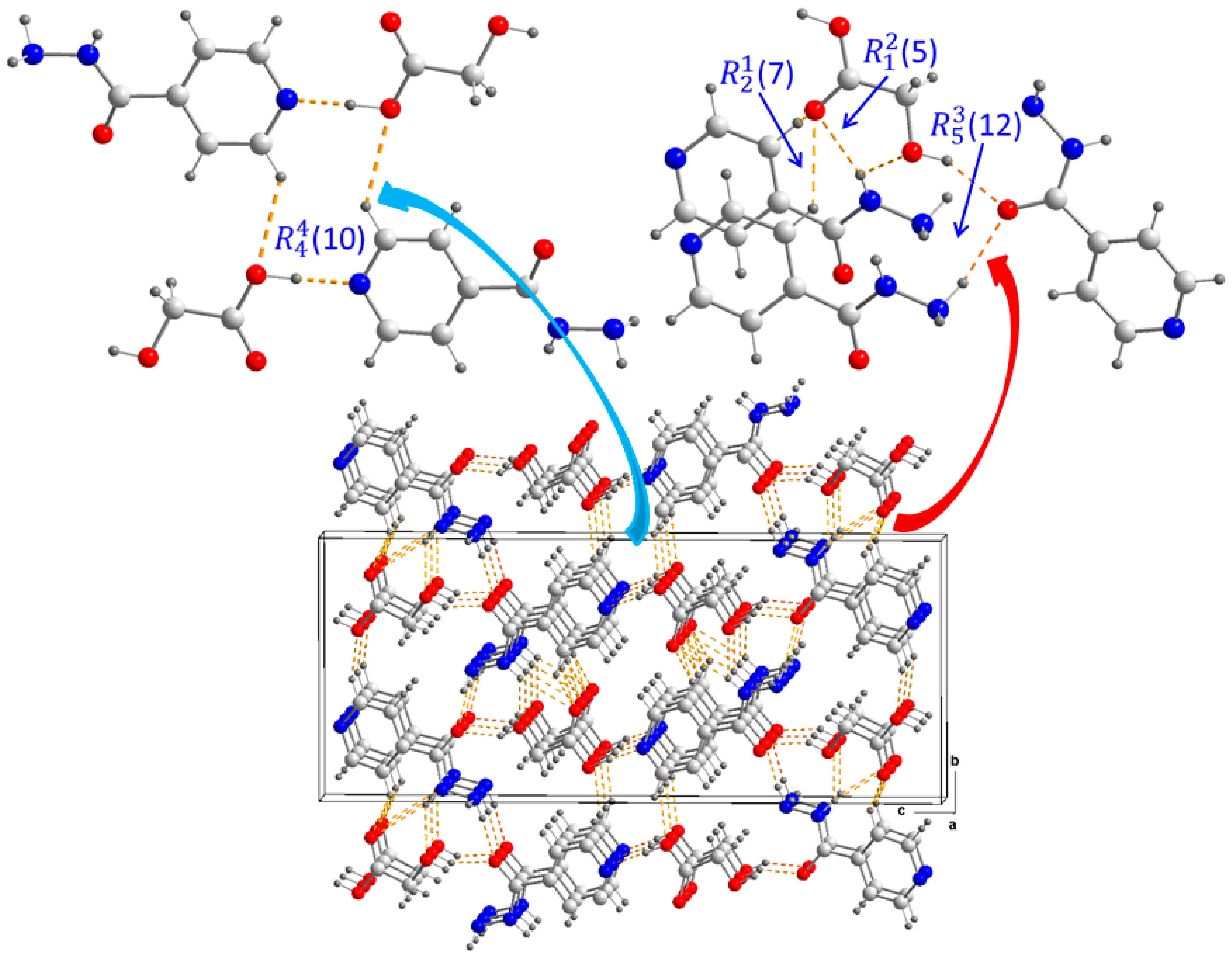
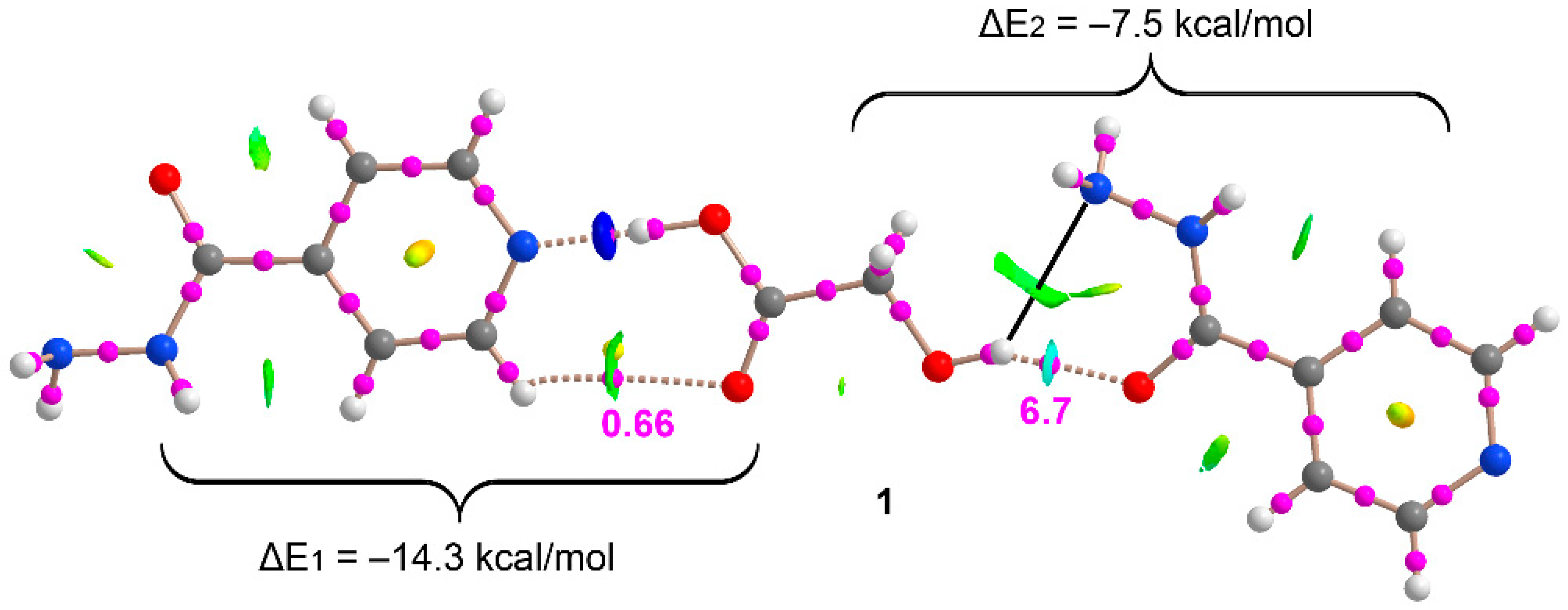
3.3.1. Compound 1
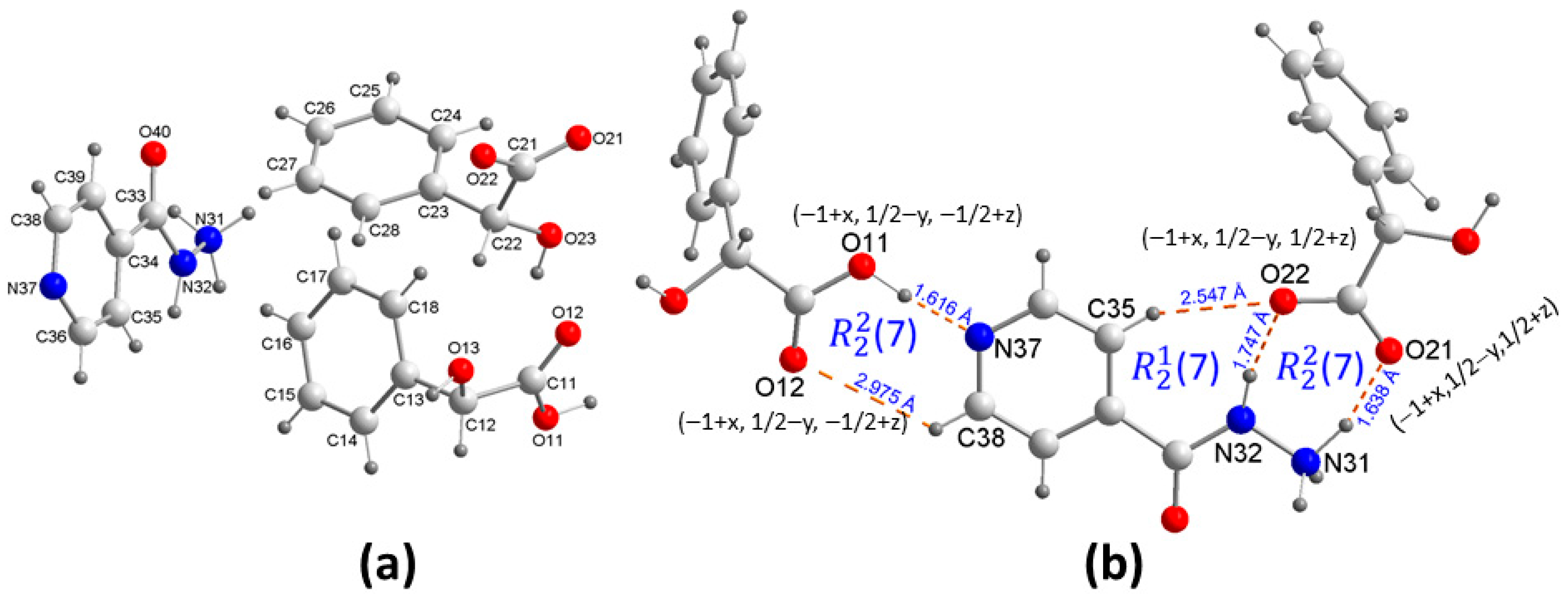
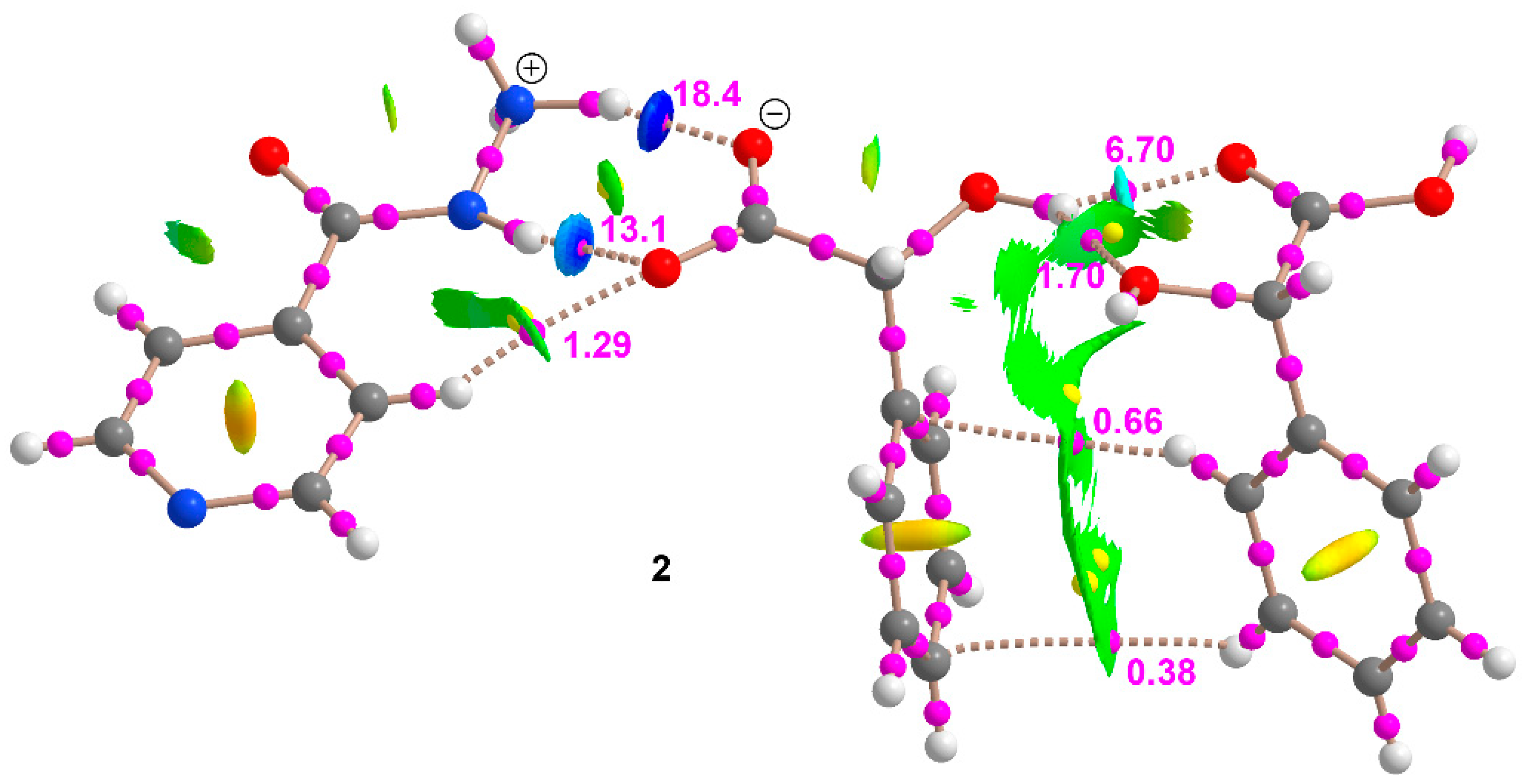
3.3.2. Compound 2
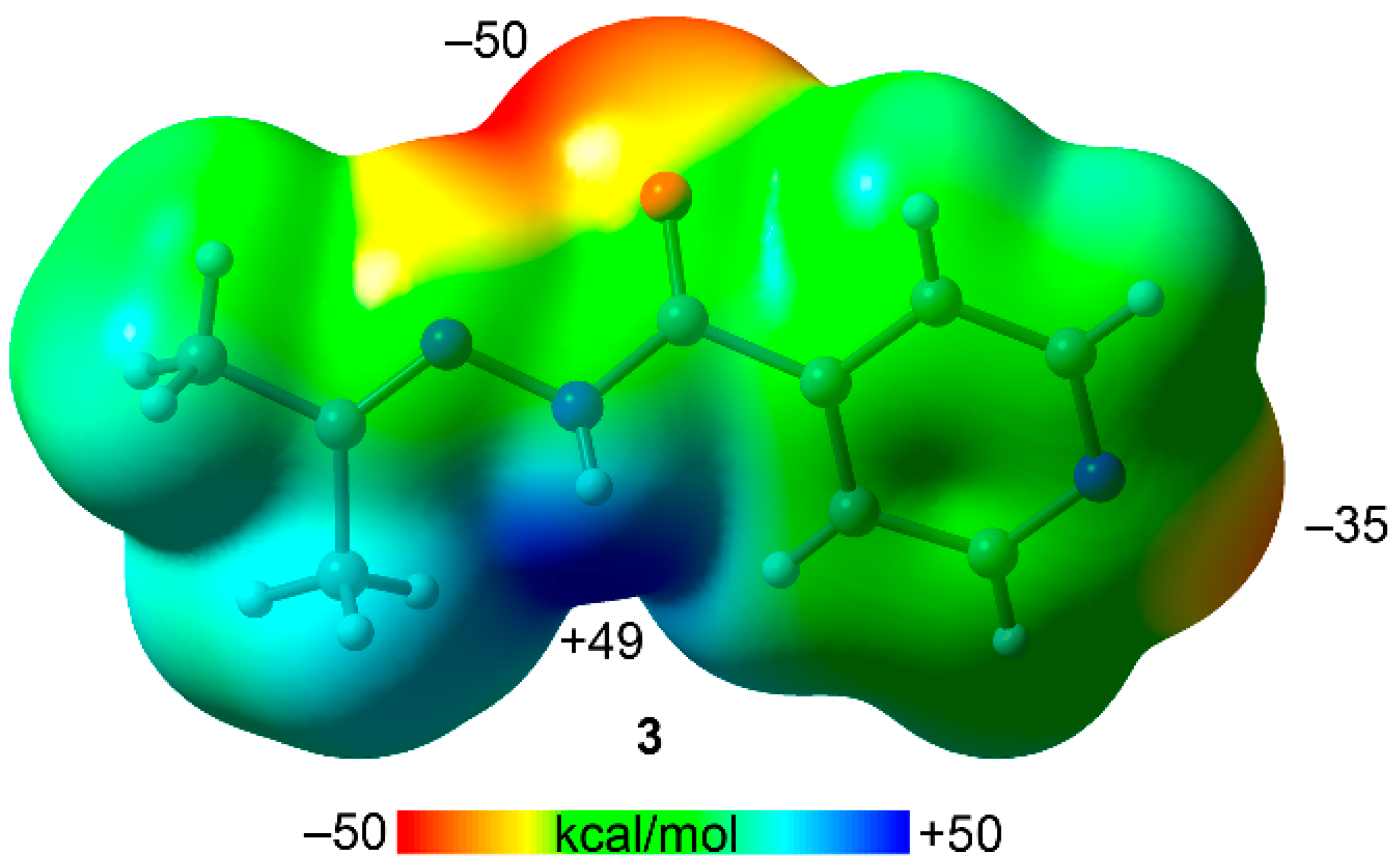
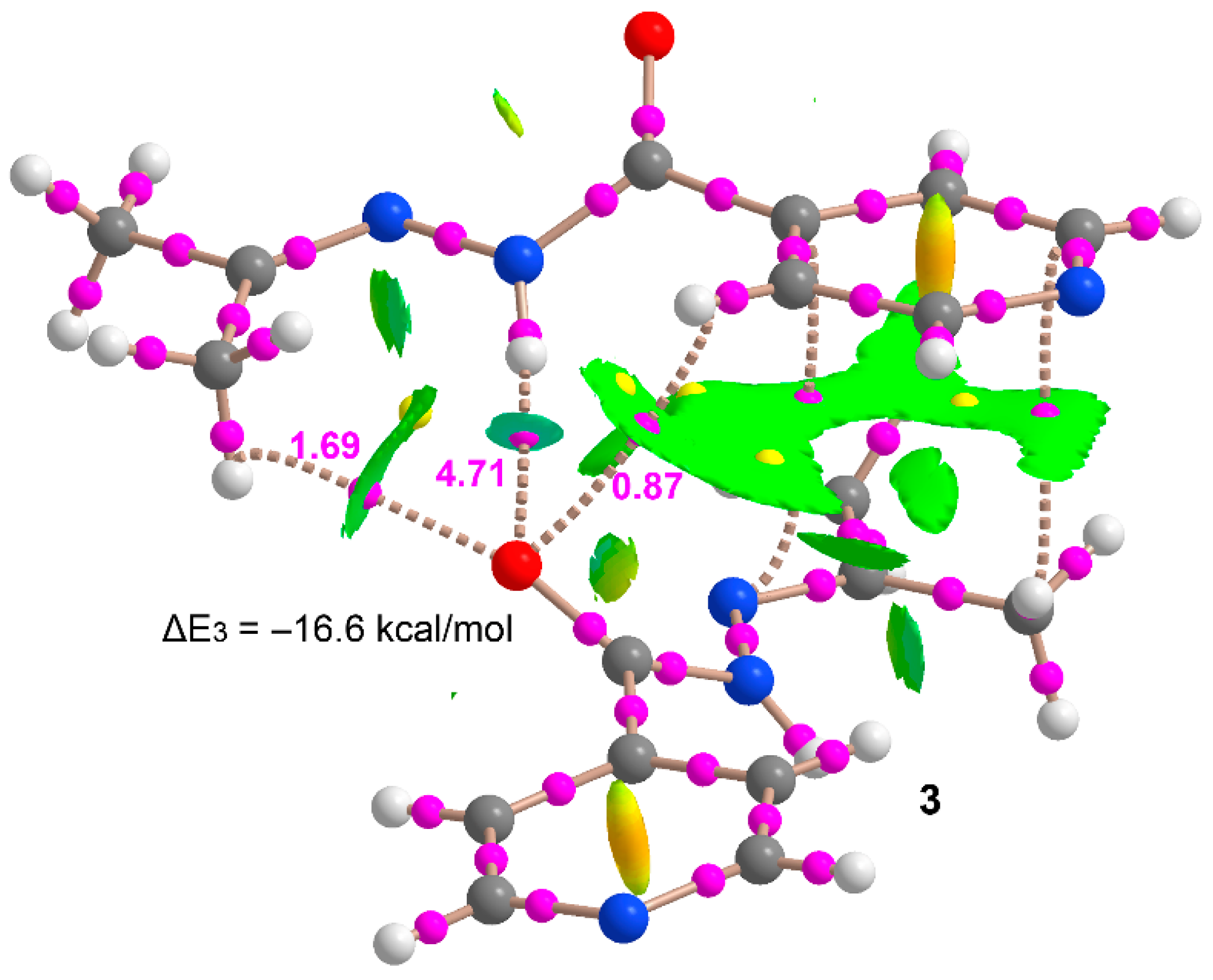
3.3.3. Compound 3
3.3.4. Comparison of H-Bond Energies in Compounds 1–3 with Experimental Values from the Literature
4. Concluding Remarks
Supplementary Materials
Author Contributions
Funding
Institutional Review Board Statement
Informed Consent Statement
Data Availability Statement
Acknowledgments
Conflicts of Interest
References
- Stahly, G.P. A Survey of Cocrystals Reported Prior to 2000. Cryst. Growth Des. 2009, 9, 4212–4229. [Google Scholar] [CrossRef]
- Du, M.; Zhang, Z.H.; Wang, X.G.; Wu, H.F.; Wang, Q. Flexible Building Blocks of N,N’-Bis(picolinoyl)hydrazine for Hydrogen-Bonding Directed Cocrystallization: Structural Diversity, Concomitant Polymorphs, and Synthon Prediction. Cryst. Growth Des. 2006, 6, 1867–1875. [Google Scholar] [CrossRef]
- Wouters, J.; Quere, L. (Eds.) Pharmaceutical Salts and Co-Crystals; RSC Drug Discovery Series No. 16; RSC Publisher: Cambridge, UK, 2011. [Google Scholar]
- Diniz, L.F.; Souza, M.S.; Carvalho, P.S., Jr.; da Silva, C.C.P.; D’Vries, R.F.; Ellena, J. Novel Isoniazid cocrystals with aromatic carboxylic acids: Crystal engineering, spectroscopy and thermochemical investigations. J. Mol. Struct. 2018, 1153, 58–68. [Google Scholar] [CrossRef]
- Foces-Foces, C.; Llamas-Saiz, A.L.; Lorente, P.; Golubev, N.S.; Limbach, H.-H. Three 2,4,6-trimethylpyridine-benzoic acid complexes at 150 K. Acta Cryst. 1999, C55, 377–381. [Google Scholar]
- Seaton, C.C. Proton location in acid⋯pyridine hydrogen bonds of multi-component crystals. CrystEngComm 2014, 16, 5878–5886. [Google Scholar] [CrossRef] [Green Version]
- Oruganti, M.; Khade, P.; Dasc, U.K.; Trivedi, D.R. The hierarchies of hydrogen bonds in salts/cocrystals of isoniazid and its Schiff base—A case study. RSC Adv. 2016, 6, 15868–15876. [Google Scholar] [CrossRef]
- Mironov, A.V.; Tafeenko, V.A.; Grebenkin, D.Y.; Oblezov, A.E. Hydrogen bonding in hydroxypyridium salts. Z. Kristallogr. 2018, 233, 501–506. [Google Scholar] [CrossRef]
- Goswami, P.K.; Kumar, V.; Ramanan, A. Multicomponent solids of diclofenac with pyridine based coformers. J. Mol. Struct. 2020, 1210, 128066. [Google Scholar] [CrossRef]
- Shattock, T.R.; Arora, K.K.; Vishweshwar, P.; Zaworotko, M.J. Hierarchy of Supramolecular Synthons: Persistent Carboxylic Acid⋯Pyridine Hydrogen Bonds in Cocrystals That also Contain a Hydroxyl Moiety. Cryst. Growth Des. 2008, 8, 4533–4545. [Google Scholar] [CrossRef]
- Iseman, M.D. Tuberculosis therapy: Past, present and future. Eur. Respir. J. 2002, 20 (Suppl. 36), 87s–94s. [Google Scholar] [CrossRef] [Green Version]
- Hearn, M.J.; Cynamon, M.H.; Chen, M.F.; Coppins, R.; Davis, J.; Kang, H.J.-O.; Noble, A.; Tu-Sekine, B.; Terrot, M.S.; Trombino, D.; et al. Preparation and antitubercular activities in vitro and in vivo of novel Schiff bases of isoniazid. Eur. J. Med. Chem. 2009, 44, 4169–4178. [Google Scholar] [CrossRef] [Green Version]
- Lemmerer, A. Covalent assistance to supramolecular synthesis: Modifying the drug functionality of the antituberculosis API isoniazid in situ during co-crystallization with GRAS and API compounds. CrystEngComm 2012, 14, 2465–2478. [Google Scholar] [CrossRef]
- Sarcevica, I.; Orola, L.; Veidis, M.V.; Podjava, A.; Belyakov, S. Crystal and Molecular Structure and Stability of Isoniazid Cocrystals with Selected Carboxylic Acids. Cryst. Growth Des. 2013, 13, 1082–1090. [Google Scholar] [CrossRef]
- Aitipamula, S.; Wong, A.B.H.; Shan Chowa, P.; Tan, R.B.H. Novel solid forms of the anti-tuberculosis drug, Isoniazid: Ternary and polymorphic cocrystals. CrystEngComm 2013, 15, 5877–5887. [Google Scholar] [CrossRef]
- Castiñeiras, A.; García-Santos, I.; González-Perez, J.M.; Bauzá, A.; Zaręba, J.K.; Niclós-Gutiérrez, J.; Torres, R.; Vilchez, E.; Frontera, A. Multicomponent Supramolecular Assemblies of Melamine and α-hydroxycarboxylic Acids: Understanding the Hydrogen Bonding Patterns and Their Physicochemical Consequences. Cryst. Growth Des. 2018, 18, 6786–6800. [Google Scholar] [CrossRef]
- Alvarez-Lorenzo, C.; Castiñeiras, A.; Frontera, A.; García-Santos, I.; González-Pérez, J.M.; Niclós-Gutiérrez, J.; Rodríguez-González, I.; Vílchez-Rodríguez, E.; Zaręba, J.K. Recurrent motifs in pharmaceutical cocrystals involving Glycolic acid: X-ray characterization, Hirshfeld surface analysis and DFT calculations. CrystEngComm 2020, 22, 6674–6689. [Google Scholar] [CrossRef]
- Zhou, Z.-H.; Hou, S.-Y.; Cao, Z.-X.; Wan, H.-L.; Ng, S.-W. Syntheses, crystal structures and biological relevance of glicolato and S-lactato molybdates. J. Inorg. Biochem. 2004, 98, 1037–1044. [Google Scholar] [CrossRef] [PubMed]
- Tumanova, N.; Payen, R.; Springuel, G.; Norberg, B.; Robeyns, K.; Le Duff, C.; Wouters, J.; Leyssens, T. Cocrystallization out of the blue: Dl-mandelic acid/ethyl-DL-mandelate cocrystal. J. Mol. Struct. 2017, 1127, 397–402. [Google Scholar] [CrossRef]
- Brunner, H.; Maiterth, F.; Treittinger, B. Synthesis and antitumor activity of water-soluble 2-benzyl-1,2-diaminobutane-α-oxycarboxylatoplatinum(II) complexes. Inorg. Chim. Acta 1992, 200, 79–84. [Google Scholar] [CrossRef]
- Bruker, APEX3 Software; v2018.7-2; Bruker AXS Inc.: Madison, WI, USA, 2018.
- Sheldrick, G.M. SADABS. Program for Empirical Absorption Correction of Area Detector Data; University of Goettingen: Göttingen, Germany, 1997. [Google Scholar]
- Sheldrick, G.M. A short history of SHELX. Acta Cryst. 2008, A64, 112–122. [Google Scholar] [CrossRef] [PubMed] [Green Version]
- Flack, H.D.; Bernardinelli, G. Absolute structure and absolute configuration. Acta Cryst. 1999, A55, 908–915. [Google Scholar] [CrossRef] [Green Version]
- Putz, H.; Brandenburg, K. DIAMOND—Crystal and Molecular Structure Visualization Version 4.6.2; Crystal Impact GbR: Bonn, Germany, 2020. [Google Scholar]
- Spackman, M.A.; Jayatilaka, D. Hirshfeld surface analysis. CrystEngComm 2009, 11, 19–32. [Google Scholar] [CrossRef]
- Spackman, M.A.; McKinnon, J.J. Fingerprinting intermolecular interactions in molecular crystals. CrystEngComm 2002, 66, 378–392. [Google Scholar] [CrossRef]
- McKinnon, J.J.; Jayatilaka, D.; Spackman, M.A. Towards quantitative analysis of intermolecular interactions with Hirshfeld surfaces. Chem. Commun. 2007, 37, 3814–3816. [Google Scholar] [CrossRef] [PubMed]
- Turner, M.J.; McKinnon, J.J.; Wolf, S.K.; Grimwood, D.J.; Spackman, P.R.; Jayatilaka, D.; Spackman, M.A. CrystalExplorer17; University of Western Australia: Perth, Australia, 2017. [Google Scholar]
- Young, D.; Ding, F.; Lipparini, F.; Egidi, F.; Goings, J.; Peng, B.; Petrone, A.; Henderson, T.; Ranasinghe, D.; Zakrzewski, V.G.; et al. Gaussian 16, Revision A.01; Gaussian, Inc.: Wallingford, CT, USA, 2016. [Google Scholar]
- Perdew, J.P.; Burke, K.; Ernzerhof, M. Generalized Gradient Approximation Made Simple. Phys. Rev. Lett. 1996, 77, 3865–3868. [Google Scholar] [CrossRef] [PubMed] [Green Version]
- Grimme, S.; Antony, J.; Ehrlich, S.; Krieg, H. A consistent and accurate ab initio parametrization of density functional dispersion correction (DFT-D) for the 94 elements H-Pu. J. Chem. Phys. 2010, 132, 154104. [Google Scholar] [CrossRef] [PubMed] [Green Version]
- Weigend, F.; Ahlrichs, R. Balanced basis sets of split valence, triple zeta valence and quadruple zeta valence quality for H to Rn: Design and assessment of accuracy. Phys. Chem. Chem. Phys. 2005, 7, 3297–3305. [Google Scholar] [CrossRef]
- Bader, R.F.W. A quantum theory of molecular structure and its applications. Chem. Rev. 1991, 91, 893–928. [Google Scholar] [CrossRef]
- AIMAll, Version 19.10.12 ed; Todd A. Keith, TK Gristmill Software: Overland Park, KS, USA, 2019; Available online: aim.tkgristmill.com (accessed on 26 February 2021).
- Boys, S.F.; Bernardi, F. The calculation of small molecular interactions by the differences of separate total energies. Some procedures with reduced errors. Mol. Phys. 1970, 19, 553–566. [Google Scholar] [CrossRef]
- Espinosa, E.; Molins, E.; Lecomte, C. Hydrogen bond strengths revealed by topological analyses of experimentally observed electron densities. Chem. Phys. Lett. 1998, 285, 170–173. [Google Scholar] [CrossRef]
- Efimenko, Z.M.; Eliseeva, A.A.; Ivanov, D.M.; Galmes, B.; Frontera, A.; Bokach, N.A.; Kukushkin, V.Y. Bifurcated μ2-I···(N,O) Halogen Bonding: The Case of (Nitrosoguanidinate)NiII Cocrystals with Iodine(I)-Based σ-Hole Donors. Cryst. Growth Des. 2021, 21, 588–596. [Google Scholar] [CrossRef]
- Zelenkov, L.E.; Ivanov, D.M.; Sadykov, E.K.; Bokach, N.A.; Galmes, B.; Frontera, A.; Kukushkin, V.Y. Semicoordination Bond Breaking and Halogen Bond Making Change the Supramolecular Architecture of Metal-Containing Aggregates. Cryst. Growth Des. 2020, 20, 6956–6965. [Google Scholar] [CrossRef]
- Soldatova, N.S.; Postnikov, P.S.; Suslonov, V.V.; Kissler, T.Y.; Ivanov, D.M.; Yusubov, M.S.; Galmes, B.; Frontera, A.; Kukushkin, V.Y. Diaryliodonium as a double σ-hole donor: The dichotomy of thiocyanate halogen bonding provides divergent solid state arylation by diaryliodonium cations. Org. Chem. Front. 2020, 7, 2230–2242. [Google Scholar] [CrossRef]
- Katlenok, E.A.; Haukka, M.; Levin, O.V.; Frontera, A.; Kukushkin, V.Y. Supramolecular Assembly of Metal Complexes by (Aryl)I···dz2 [PtII] Halogen Bonds. Chem. Eur. J. 2020, 26, 7692–7701. [Google Scholar] [CrossRef]
- Rozhkov, A.V.; Eliseeva, A.A.; Baykov, S.V.; Galmes, B.; Frontera, A.; Kukushkin, V.Y. One-Pot Route to X-perfluoroarenes (X = Br, I) Based on FeIII-Assisted C-F Functionalization and Utilization of These Arenes as Building Blocks for Crystal Engineering Involving Halogen Bonding. Cryst. Growth Des. 2020, 20, 5908–5921. [Google Scholar] [CrossRef]
- Rozhkov, A.V.; Ananyev, I.V.; Gomila, R.M.; Frontera, A.; Kukushkin, V.Y. π-Hole···dz2[PtII] Interactions with Electron-Deficient Arenes Enhance the Phosphorescence of PtII-Based Luminophores. Inorg. Chem. 2020, 59, 9308–9314. [Google Scholar] [CrossRef]
- Verdugo-Escamilla, C.; Alarcón-Payer, C.; Frontera, A.; Acebedo-Martínez, F.J.; Domínguez-Martín, A.; Gómez-Morales, J.; Choquesillo-Lazarte, D. Interconvertible Hydrochlorothiazide–Caffeine Multicomponent Pharmaceutical Materials: A Solvent Issue. Crystals 2020, 10, 1088. [Google Scholar] [CrossRef]
- Barbas, R.; Kumar, V.; Vallcorba, O.; Prohens, R.; Frontera, A. Sildenafil–Resorcinol Cocrystal: XRPD Structure and DFT Calculations. Crystals 2020, 10, 1126. [Google Scholar] [CrossRef]
- Johnson, E.R.; Keinan, S.; Mori-Sánchez, P.; Contreras-García, J.; Cohen, A.J.; Yang, W. Revealing Noncovalent Interactions. J. Am. Chem. Soc. 2010, 132, 6498–6506. [Google Scholar] [CrossRef] [Green Version]
- Contreras-García, J.; Johnson, E.R.; Keinan, S.; Chaudret, R.; Piquemal, J.-P.; Beratan, D.N.; Yang, W. NCIPLOT: A Program for Plotting Noncovalent Interaction Regions. J. Chem. Theor. Comput. 2011, 7, 625–632. [Google Scholar] [CrossRef]
- Atta, N.F.; Galal, A.; Ahmed, R.A. Voltammetric Behavior and Determination of Isoniazid Using PEDOT Electrode in Presence of Surface Active Agents. Int. J. Electrochem. Sci. 2011, 6, 5097–5113. [Google Scholar]
- Banerjee, S.S.; Bhanja, K.; Chattopadhyay, P.K. Quantum chemical predictions of aqueous pKa values for OH groups of some α-hydroxycarboxylic acids based on ab initio and DFT calculations. Comput. Theor. Chem. 2018, 1125, 29–38. [Google Scholar] [CrossRef]
- Childs, S.L.; Stahly, G.P.; Pak, A. The Salt−Cocrystal Continuum: The Influence of Crystal Structure on Ionization State. Mol. Pharm. 2007, 4, 323–338. [Google Scholar] [CrossRef] [Green Version]
- Cruz-Cabeza, A.J. Acid–base crystalline complexes and the pKa rule. CrystEngComm 2012, 14, 6362–6365. [Google Scholar] [CrossRef]
- Lemmerer, A.; Bernstein, J.; Kahlenberg, V. One-pot covalent and supramolecular synthesis of pharmaceutical co-crystals using the API isoniazid: A potential supramolecular reagent. CrystEngComm 2010, 12, 2856–2864. [Google Scholar] [CrossRef]
- Madeley, L.G.; Levendis, D.C.; Lemmerer, A. Covalent-assisted supramolecular synthesis: The effect of hydrogen bonding in cocrystals of 4-tertbutylbenzoic acid with isoniazid and its derivatized forms. Acta Cryst. 2019, C75, 200–207. [Google Scholar] [CrossRef]
- Kataeva, O.; Nohr, M.; Ivshin, K.; Hampel, S.; Büchner, B.; Knupfer, M. Understanding Intermolecular Interactions in a Tetracene–F4TCNQ Cocrystal via Its Electron Density Distribution and Topology. Cryst. Growth Des. 2021, 21, 471–481. [Google Scholar] [CrossRef]
- Koritsanszky, T.S.; Coppens, P. Chemical Applications of X-ray Charge-Density Analysis. Chem. Rev. 2001, 101, 1583–1627. [Google Scholar] [CrossRef]
- Münch, A.; Knauer, L.; Ott, H.; Sindlinger, C.; Herbst-Irmer, R.; Strohmann, C.; Stalke, D. Insight into the Bonding and Aggregation of Alkyllithiums by Experimental Charge Density Studies and Energy Decomposition Analyses. J. Am. Chem. Soc. 2020, 142, 15897–15906. [Google Scholar] [CrossRef]
- Borissova, A.O.; Lyssenko, K.A.; Gurinov, A.A.; Shenderovich, I.G. Energy Analysis of Competing Non-Covalent Interaction in 1: 1 and 1: 2 Adducts of Collidine with Benzoic Acids by Means of X-Ray Diffraction. Z. Phys. Chem. 2013, 227, 775–790. [Google Scholar] [CrossRef]


| Compound | 1 | 2 | 3 |
|---|---|---|---|
| Empirical formula | C8H11N3O4 | C22H23N3O7 | C9H12N3O1.5 |
| Formula weight | 213.20 | 441.43 | 186.22 |
| Crystal system | Monoclinic | Monoclinic | Orthorhombic |
| Space group | P21/n | P21/c | Aba2 |
| Unit cell dimensions | |||
| a/Å | 3.8930(3) | 5.6049(5) | 18.8351(5) |
| b/Å | 9.9754(5) | 24.388(3) | 12.6568(4) |
| c/Å | 23.6410(12) | 15.1471(16) | 8.0435(3) |
| α/° | 90 | 90 | 90 |
| β/° | 92.480(3) | 92.025(7) | 90 |
| γ/° | 90 | 90 | 90 |
| Volume/Å−3 | 917.22(10) | 2069.2(4) | 1917.51(11) |
| Z | 4 | 4 | 8 |
| Calc. density/Mg/m3 | 1.544 | 1.417 | 1.290 |
| Absorp. coefc./mm−1 | 0.125 | 0.107 | 0.091 |
| F(000) | 448 | 928 | 792 |
| Crystal size | 0.19 × 0.14 × 0.05 | 0.46 × 0.40 × 0.03 | 0.66 × 0.24 × 0.13 |
| θ range/° | 1.724–26.399 | 2.145–28.280 | 2.163–36.313 |
| Limiting indices/h,k,l | −4/4, −12/12, −29/29 | −7/7, −32/32, −20/19 | −30/31, −20/21, −13/13 |
| Refl. collect/unique (Rint) | 13443 / 1879 [0.0501] | 32295 / 5150 [0.0703] | 76482 / 4651 [0.0447] |
| Completeness θ/° | 25.242–99.9 | 25.242–99.9 | 25.242–99.9 |
| Absorp. correct. | Multi-scans | Multi-scans | Multi-scans |
| Max./min. transm. | 1.0000/0.8327 | 1.000/0.868 | 1.000/0.883 |
| Data/parameters | 1879/156 | 5150/317 | 4651/132 |
| Goodness-of-fit on F2 | 1.047 | 1.003 | 1.030 |
| Final R indices | R1 = 0.0417, wR2 = 0.0901 | R1 = 0.0501, wR2 = 0.0985 | R1 = 0.0341, wR2 = 0.0899 |
| R indices (all data) | R1 = 0.0645, wR2 = 0.0980 | R1 = 0.0888, wR2 = 0.1114 | R1 = 0.0402, wR2 = 0.0938 |
| Largest dif. peak/hole | 0.224/−0.280 | 0.300/−0.266 | 0.373 /−0.179 |
| Cmpnd | D–H···A | D–H | H···A | D···A | ∠DHA | Symmetry Code |
|---|---|---|---|---|---|---|
| 1 | N12–H12A···O12a | 0.84(2) | 2.43(2) | 3.2617(19) | 168.1(19) | x − 1, y + 1, z |
| N12–H12A···O13a | 0.84(2) | 2.59(2) | 3.1168(19) | 121.5(17) | ||
| N11–H11A···O20b | 0.89(2) | 2.14(2) | 3.022(2) | 167.4(18) | −x + 1/2, y + 1/2, −z + 1/2 | |
| N11–H11B···O13a | 0.89(2) | 2.62(2) | 3.049(2) | 110.2(16) | ||
| O11–H11···N17c | 1.00(3) | 1.60(3) | 2.6064(19) | 178(2) | −x + 2, −y + 1, −z | |
| O13–H13A···O20d | 0.89(3) | 1.91(3) | 2.7399(18) | 154(2) | −x+1/2, y−1/2, −z + 1/2 | |
| O13–H13A···N11d | 0.89(3) | 2.60(3) | 3.294(2) | 135(2) | ||
| C15–H15···O12e | 0.95 | 2.53 | 3.118(2) | 119.8 | x, y + 1, z | |
| C18–H18···O11f | 0.95 | 2.49 | 3.235(2) | 134.8 | X − 1, y, z | |
| 2 | O11–H11···N37a | 1.01(2) | 1.62(2) | 2.6252(16) | 171(2) | X + 1, −y + 1/2, z + 1/2 |
| O13–H13···O21b | 0.87(2) | 2.51(2) | 3.0671(16) | 122.1(17) | X − 1, y, z | |
| O13–H13···O23b | 0.87(2) | 1.86(2) | 2.7106(15) | 162(2) | ||
| O23–H23···O12 | 0.97(2) | 1.91(2) | 2.7716(18) | 146.3(19) | ||
| O23–H23···O13 | 0.97(2) | 2.42(2) | 3.1756(16) | 135.1(18) | ||
| N31–H31A···O22c | 0.94(2) | 1.87(2) | 2.7971(17) | 170.1(18) | x, −y + 1/2, z + 1/2 | |
| N31–H31B···O13d | 0.96(2) | 1.85(2) | 2.807(2) | 172.7(19) | −x + 1, y− 1/2, −z + 1/2 | |
| N31–H31C···O21e | 1.07(2) | 1.64(2) | 2.677(2) | 162.5(19) | x − 1, −y + 1/2, z + 1/2 | |
| N32–H32···O22e | 0.96(2) | 1.75(2) | 2.7009(18) | 168.5(18) | ||
| C12–H12···O12b | 0.98 | 2.57 | 3.4869(19) | 155.7 | ||
| C35–H35···O22e | 0.93 | 2.55 | 3.411(2) | 154.8 | ||
| 3 | N12–H12A···O20a | 0.86(2) | 2.02(2) | 2.8683(13) | 169.7(19) | −x + 1/2, y, z + 1/2 |
| O1–H1A···N17b | 0.89(2) | 2.00(2) | 2.8714(12) | 168(2) | −x + 1/2, y, z − 1/2 | |
| C16–H16···N11c | 0.95 | 2.58 | 3.4475(15) | 152.1 | x, y − 1/2, z + 1/2 | |
| C19–H19···O1d | 0.95 | 2.42 | 3.3328(11) | 161.2 | −x + 1/2, y + 1/2, z | |
| C21–H21B···O20a | 0.98 | 2.57 | 3.0523(14) | 110.1 | −x + 1/2, y, z + 1/2 | |
| C22–H22C···O1e | 0.98 | 2.43 | 3.3599(15) | 159.0 | x, y + 1/2, z − 1/2 |
Publisher’s Note: MDPI stays neutral with regard to jurisdictional claims in published maps and institutional affiliations. |
© 2021 by the authors. Licensee MDPI, Basel, Switzerland. This article is an open access article distributed under the terms and conditions of the Creative Commons Attribution (CC BY) license (http://creativecommons.org/licenses/by/4.0/).
Share and Cite
Álvarez-Vidaurre, R.; Castiñeiras, A.; Frontera, A.; García-Santos, I.; Gil, D.M.; González-Pérez, J.M.; Niclós-Gutiérrez, J.; Torres-Iglesias, R. Weak Interactions in Cocrystals of Isoniazid with Glycolic and Mandelic Acids. Crystals 2021, 11, 328. https://0-doi-org.brum.beds.ac.uk/10.3390/cryst11040328
Álvarez-Vidaurre R, Castiñeiras A, Frontera A, García-Santos I, Gil DM, González-Pérez JM, Niclós-Gutiérrez J, Torres-Iglesias R. Weak Interactions in Cocrystals of Isoniazid with Glycolic and Mandelic Acids. Crystals. 2021; 11(4):328. https://0-doi-org.brum.beds.ac.uk/10.3390/cryst11040328
Chicago/Turabian StyleÁlvarez-Vidaurre, Raquel, Alfonso Castiñeiras, Antonio Frontera, Isabel García-Santos, Diego M. Gil, Josefa M. González-Pérez, Juan Niclós-Gutiérrez, and Rocío Torres-Iglesias. 2021. "Weak Interactions in Cocrystals of Isoniazid with Glycolic and Mandelic Acids" Crystals 11, no. 4: 328. https://0-doi-org.brum.beds.ac.uk/10.3390/cryst11040328